Embed Size (px)
Citation preview

Published 02/10/2011
© Copyright 2011Vidul et al. This is an open accessarticle distributed under the terms ofthe Creative Commons AttributionLicense CC-BY 3.0., which permitsunrestricted use, distribution, andreproduction in any medium,provided the original author andsource are credited.
Difficult Diagnosis and DifferentiatedTreatment in Synchronous ThoracicHemangiomasViorica Vidul , Vasile Popa , Genevieve Wu
1. Center for General Surgery and Liver Transplantation, Clinical Institute of Digestive Diseases andLiver Transplant, Fundeni, Bucharest 2. Thoracic Surgery Department, Emergency Clinical Hospital,"Sfântul Ioan", Bucharest, Romania
Corresponding author: Valentin M. Munteanu, [email protected] Disclosures can be found in Additional Information at the end of the article
AbstractThoracic hemangiomas are rare vascular neoplasia among adults. Even with advanced imaging,diagnosis is sometimes impossible, thereby mandating histopathological examination. Thedifferential diagnosis of thoracic hemangiomas can include arteriovenous malformations,thoracic sarcomas (angiosarcoma, paravertebral mesothelioma), neurinoma, andneurofibroma. We herein present a case which was intitially suggestive of a malignancy thatultimately proved to be a capillary cavernous paravertebral and vertebral hemangioma. Giventhe unique topography of this lesion, a correct preoperative diagnosis was not possible and ainterdisciplinary surgical approach was required.
Categories: Cardiac/Thoracic/Vascular Surgery, General Surgery, NeurosurgeryKeywords: tumor, capillary-cavernous hemangioma, vertebral and paravertebral, surgical treatment
IntroductionConsidered a relatively rare disease in adult pathology, hemangiomas represent a controversialentity in terms of their pathogenic, clinical and therapeutic characteristics. The nosologicframework of hemangioma continues to be debated, having been defined in the past aseither congenital vascular malformations (hamartomas) or benign vascular malignancies with alocally destructive nature. In an adult population, hemangiomas, especially the cavernous type,are located in bones, especially the vertebrae, thorax, brain, liver, and skeletal muscle. Sincethe clinical and imaging apperance of hemangioma can mimic a malignanttumor, histopathological examination is frequently required.
Because there is no universally accepted standard treatment of hemangiomas, severaltherapeutic alternatives are generally considered: surgery, embolization, direct injection ofethanol (alcohol), chemotherapy, radiotherapy, corticosteroids, immunotherapy depending onage, topography, histopathological type, complications, and local destructive nature. In about10% of cases, hemangiomas are synchronous. In these situations, the differential diagnosis iseven more difficult; it is imperative to rule out malignancy. Ultimately, treatmentvaries depending on topography and the evolutionary stage of the lesion.
Since hemangiomas are relatively rare entities, with especially misleading clinical and imagingcharacteristics, their management can be challenging. The case we present herein bothillustrates these difficulties as well as demonstrates the value of interdisciplinary collaboration.
1 2
Open Access CaseReport DOI: 10.7759/cureus.22
How to cite this articleVidul V, Popa V, Wu G (February 10, 2011) Difficult Diagnosis and Differentiated Treatment inSynchronous Thoracic Hemangiomas. Cureus 3(2): e22. DOI 10.7759/cureus.22

Case PresentationThis 45-year-old male patient, without a significant antecedent history of disease, presentedwith a several year history of progressively severe chest pain that responded poorly to a rangeof pain medications. Despite his severe chest pain, physical examination was grosslynormal. However, chest computed tomography (CT) revealed a solid tumor located in the leftparavertebral muscles, adhering to the parietal pleura. Imaging was suggestive for aparavertebral mesothelioma or muscle sarcoma.
FIGURE 1: Aspect of chest computer tomography: leftparavertebral tumor
Within the posterior mediastinum, a contrast magnetic resonance imaging (MRI) revealed a leftparavertebral tumor of 5.5 cm, located in the V-VI intercostal space. The lesion was extra-pleural in location, polilobar in shape and both hyperintense on T2-weighted andhypointense on T1- weighted MRI. Furthermore, imaging demonstrated the mass to beinhomogeneous, well-vascularized, and without invasion or destruction of the adjacent ribsor neural foramen (Figure 2A, 2B).
2011 Vidul et al. Cureus 3(2): e22. DOI 10.7759/cureus.22 2 of 12

FIGURE 2: Aspect of magnetic resonance with contrast study:paravertebral and intercostals T5-6 tumorA. Sagital image B. Transversal image a level T5-T6;
The appearance was most consistent with an intercostal nerve sheath tumor. Meanwhile, asecond lesion was seen on MRI adjacent to and contacting the T6 vertebral body, but withoutviolating the integrity of cortical bone (Figure 3A, 3B).
FIGURE 3: Aspect of magnetic resonance imaging withangiographic study: vertebral hemangioma T6 with corticalbone limits (A,B)A. Sagital image B. Transversal image a level T6
2011 Vidul et al. Cureus 3(2): e22. DOI 10.7759/cureus.22 3 of 12

FIGURE 4: Aspect of magnetic resonance imaging withangiographic study: paravertebral tumor with vertebral T6contact.
Given a presumptive diagnosis of an intercostal neurinoma, but also considering the possibilityof a paravertebral mesothelioma, the patient underwent surgery by the Thoracic SurgeryDepartment of the St. John's Hospital. A left axillary thoracotomy with sectioning of thelatissimus dorsi and serratus muscles was performed. A purple, partially encapsulatedpolilobar lesion was found intraoperatively located in the intercostal space, which was attachedto a thoracic spine vascular/nervous pedicle. The neural foramen was closed and the tumor wasfound to have destroyed the adjacent intercostal muscle (Figure 5).
2011 Vidul et al. Cureus 3(2): e22. DOI 10.7759/cureus.22 4 of 12

FIGURE 5: Intraoperative appearance of the vertebral capillarycavernous hemangioma with intra-thoracic extension.
We performed a en-block resection of the lesion, including the parietal pleura of the thoracicspine and the remaining intercostal muscles. Because of serious intraoperative hemorrhage, itproved necessary to dissect, ligate and section the posterior intercostal artery (Figure 6).Parietal plural and paravertebral hemostasis was achieved with TachoSil (Figure 7).
2011 Vidul et al. Cureus 3(2): e22. DOI 10.7759/cureus.22 5 of 12
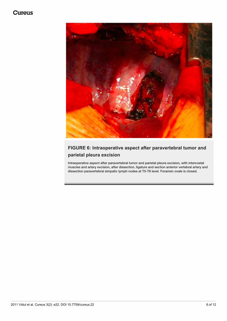
FIGURE 6: Intraoperative aspect after paravertebral tumor andparietal pleura excisionIntraoperative aspect after paravertebral tumor and parietal pleura excision, with intercostalmuscles and artery excision, after dissection, ligature and section anterior vertebral artery anddissection paravertebral simpatic lymph nodes at T5-T6 level. Foramen ovale is closed.
2011 Vidul et al. Cureus 3(2): e22. DOI 10.7759/cureus.22 6 of 12

FIGURE 7: Intraoperative aspect. Hemostasis with TachoSil(paravertebral application)
Histopathological intraoperative examination revealed a cavernous capillary hemangioma, thefindings of which were consistent with final paraffin-embedded microscopy (Figure 8).
2011 Vidul et al. Cureus 3(2): e22. DOI 10.7759/cureus.22 7 of 12

FIGURE 8: Capillary cavernous paravertebral hemangioma.Microscopic study (20xHE).
The lesion was positive for CD34 immunohistochemical markers (Figure 9) and actine(Figure 10) within vessel walls, confirming its benign nature and vascular origin, with limitedproliferative tendency.
2011 Vidul et al. Cureus 3(2): e22. DOI 10.7759/cureus.22 8 of 12

FIGURE 9: Immunohistochemical study. CD 34 marker pozitivon vessels wall. (20xCD34)
FIGURE 10: Immunohistochemical study. Actine positive on
2011 Vidul et al. Cureus 3(2): e22. DOI 10.7759/cureus.22 9 of 12
vessels wall (20x OB)
Postoperatively, there was a good outcome with healing per primam and no evidence of tumorrecurrence after two years. Postoperative MRI confirmed a significant resection of the lesionwhich over the ensuing two years remained stable in size and continues to respect the osseousboundaries of the adjacent veterbra (Figure 11A, 11B).
FIGURE 11: IRM aspect two years ago at postoperative care.A. Paravertebral region after surgery without recidives B. Vertebral body T6 hemangioma withcortical bone limits.
DiscussionHemangiomas are a relatively rare disease in adult pathology, estimated to account for about0.5% of all mediastinal tumors [1]. The most frequently thoracic localizations are in thevertebrae, in the intercostal muscles, in subcutaneous tissue, skin, lung parenchyma, in theribs, and in the posterior mediastinum, and in 25-30% of cases, they may be multiple[2]. Mediastinal localizations are very rare. One hundred cases have been communicated in theliterature with the favored location being in the posterior and anterior-superior mediastinum[3].
2011 Vidul et al. Cureus 3(2): e22. DOI 10.7759/cureus.22 10 of 12

From a nosological point of view, it has been asserted that hemangiomas are in a class withcongenital arteriovenous malformation despite a tumor-like appearance (hamartoma) [4]. However, the modern perspective classifies hemangioma as benign neoplasia of a vascularorigin that include neoformation vessels and have the potential to be locally destructive [3, 5].Furthermore, recently reported chromosomal alterations, "mass" effect with compression ofneighboring structures, and a macroscopic appearance of solid or mixed tumors also supportthe status of a "true neoplasia" [4].
Histopathologically, two main types of hemangiomas are found among thoracic lesions: 1) acavernous type, frequently with significant expansion of ducts that store considerablequantities of blood and which contains vascular lakes, and a pseudo-capsule without vascularelements as well as fibrosis, post-thrombotic drainage and smooth muscle cells. Such lesionscan have either a nodular or diffuse appearance and harbor a tendency for spontaneousregression [4], and 2) a capillary type with smaller neoformation vessels, fibroblasts, and fewmitoses within endothelial cells, and which, in the pediatric age group, have a proclivityfor spontaneous regression [4].
The pathogenic defect among hemangioblastoma is believed to center on a dysfunction ofangiogenesis occurring during periods of blood vessel formation within primitive vascularnetworks: in the plexiform stage, when there is a network of capillaries that communicate, mayoccur capillary hemangiomas or in the retiform stage when exist big tubular vessels, with atendency to coalescence, it forms cavernous hemangiomas and arteriovenous malformations[4].
Although it appears that most hemangiomas in adults are asymptomatic, there are rare cases inwhich chest pain is the dominant symptom. Meanwhile, there are situations in which theclinical onset consists of severe complications that can even become surgical emergenciesassociated with massive haemopneumothorax, mediastinum perforation, and chest wallhematoma. Other serious symptomotology includes recurrent pleuresis and severethrombocytopenia identified with Kasabach Merritt syndrome or with Maffuci syndrome. Paraplegia, spinal cord compression from hemorrhage, and progressive muscle weakness incases of vertebral cavernous hemangiomas have also been reported in 15% of cases. Given thedeceptive preoperative clinical picture that often mimics a malignancy, diagnosis of cavernoushemangiomas is difficult, and is often only resolved by histopathologic examination.
However, it is appreciated that diagnostic value of angio-MRI with three-dimensional study iscompelling, especially for assessing invasion of the neighboring tissue, the degree of bone lysis,the spinal compression, and intra-thoracic development of the vertebral hemangiomas and issuggestive for differential diagnosis with other thoracic tumors, although, in the spinalcapillary hemangioma, the selective arteriography has a superior value. Some authorsrecommend transthoracic ultrasound for differential diagnosis of chest wall tumors and fortherapeutic purposes, for thoracentesis and percutaneous biopsy, and computed tomographyscan, but MRI is the method of choice.
This case was, however, particularly difficult, because of the topography and structure withvery rare capillary and cavernous combination of V-VI intercostal hemangioma, but alsobecause of the synchronization with a T6 vertebral body hemangioma, with suggestivecavernous type appearance on MRI. A paravertebral location, sarcoma-like appearance on MRI,and the development along the intercostal space allowed differential diagnosis withparavertebral mesothelioma and neurinoma, although it was also possibly a chest wallhemangiosarcoma.
However, the striking peculiarity of the presented case is the synchronism of those two
2011 Vidul et al. Cureus 3(2): e22. DOI 10.7759/cureus.22 11 of 12

hemangiomas situated in the T6 vertebral body and V-VI paravertebral space, a situation whichallowed a differential diagnosis with "dumbbell shaped" tumor type, very rarely reported in theliterature. But in this context, the two synchronous hemangiomas actually represent a"dumbbell shaped" lesion type?
Tumor invasion of the intervertebral foramen is often accompanied by a gradual painfulradiculopathy which, in rare cases, can progress to paraplegia. Clearly, the imaging procedureof choice is MRI which best highlights the widening of the intervertebral space. Nevertheless,there are reported cases in which foraminal invasion could be definitely demonstratedintraoperatively, but which was invisible on preoperative MRI.
In the patient we present, there was no epidural or foraminal invasion, which is consistent withthe absence of radiculopathy. Meanwhile, the MRI appearance of a cavernous hemangiomaconfined to the vertebral body, without breeching the cortical surface (Figure 3A, 3B), wasconfirmed intraoperatively (Figure 6). Based on their topography, we presume that theparavertebral hemangioma represents an extra-thoracic extension of the vertebral lesion.
ConclusionsThoracic hemangiomas are rare malignancies with vascular origin, which have misleadingclinical-evolutionary aspects, requiring a difficult differential diagnosis. In terms of imaging,MRI with three-dimensional angiography, and sometimes arteriography, represent methods ofchoice for diagnosis. The differential diagnosis should include the arteriovenousmalformations, malignant cancers, and, in the particular case of synchronous lesions withparavertebral topography, "dumbbell shaped" tumor type, because the treatment is different.The main treatment is surgical, based on the complete excision of the lesion, but there arecomplementary or alternative therapies, such as embolization, alcohol injection with spinalreconstruction, chemotherapy, corticosteroids, and anti-tumor immunotherapy. There is nostandard treatment, but treatment options tailored to topography and evolutionary stage,especially in synchronous forms. The presented case is particular due to topography,histopathological structure, locally destructive nature, and "special type" synchronization,which requires a "particular" differential diagnosis and may represent a model for aninterdisciplinary treatment approaches.
Additional InformationDisclosuresConflicts of interest: The authors have declared that no conflicts of interest exist.
References1. Klecker Rosemary, Sinclair D, King M, Christoforidis AJ, Mueller CF: Chest: Case of the day.
AJR. 2000, 175:866-871.2. Hwang PM: Vertebral abnormality in a patient with suspected malignancy . Proc (Bayl Univ
Med Cent). 2002, 15:325-326.3. DeVita V, Hellman S, Rosenberg S: Cancer-Principles and Practice of Oncology . JB Lippincott
Company, Philadelphia; 1993.4. Rosai J, Ackerman S: Surgical Pathology. Elsevier Inc, Amsterdam; 1993.5. Hernigou P, Djindjian M, Ricolfi F, Dahhan P: Neuro-aggressive dorsal vertebral hemangioma
and vertebrectomy. Apropos of 2 cases. Review of the literature. Rev Chir Orthop ReparatriceAppar Mot. 1994, 80:542-50 [Article in French].
2011 Vidul et al. Cureus 3(2): e22. DOI 10.7759/cureus.22 12 of 12