Embed Size (px)
Citation preview

Case ReportHematochezia: An Uncommon Presentationof Colonic Tuberculosis
Fares Ayoub,1 Vikas Khullar,2 Harry Powers,1 Angela Pham,2
Shehla Islam,3 and Amitabh Suman2
1Department of Medicine, University of Florida, Gainesville, FL 32608, USA2Department of Medicine, Division of Gastroenterology, University of Florida, Gainesville, FL 32608, USA3Department of Medicine, Division of Infectious Disease, University of Florida, Gainesville, FL 32608, USA
Correspondence should be addressed to Fares Ayoub; [email protected]
Received 16 February 2017; Accepted 28 March 2017; Published 3 April 2017
Academic Editor: Tetsuo Hirata
Copyright © 2017 Fares Ayoub et al. This is an open access article distributed under the Creative Commons Attribution License,which permits unrestricted use, distribution, and reproduction in any medium, provided the original work is properly cited.
Abdominal tuberculosis (TB) is an uncommon entity in the United States. Colonic TB is reported in 2-3% of patients withabdominal TB. It is frequently misdiagnosed as Crohn’s disease or carcinoma of the colon due to their shared clinical, radiographic,and endoscopic presentations. We present a case of a 72-year-old male with colonic tuberculosis presenting as hematochezia. Ourpatient presented with shortness of breath and weight loss. Chest X-ray demonstrated ill-defined bilateral parenchymal opacitiesin the perihilar, mid, and lower lung zones. The patient was diagnosed and treated for community acquired pneumonia, with noimprovement. Hematochezia complicated by symptomatic hypotension developed later in the course of admission. Colonoscopyrevealed multiple ulcers at the anus and transverse and ascending colon as well as the cecum with stigmata of bleeding. Biopsy of asigmoid ulcer was consistent with colonic tuberculosis. Antitubercular therapy was initiated, but the patient passed away secondaryto multiorgan failure 29 days into admission.
1. Introduction
After a resurgence in the incidence of tuberculosis (TB)infections in the United States between 1985 and 1992 dueto the human immunodeficiency virus (HIV) epidemic,the incidence of TB had annually declined. However, asof 2015, TB incidence has leveled in the US and TBelimination (defined as <1 TB case per 1 million personsannually) remains elusive [1]. Gastrointestinal tuberculosisis a manifestation of extrapulmonary tuberculosis. In 2014,extrapulmonary TB constituted 20.57% of TB cases reportedto the CDC and continues to be a missed diagnosis forproviders not considering TB in their differential diagnoses[2].
Tuberculosis affecting the gastrointestinal tract was rec-ognized as early as the fourth century BC in texts byHippocrates [3]. While TB of the gastrointestinal tract is notas common as pulmonary TB, it is an important cause forTB related morbidity and mortality. The pathophysiology of
this form of tuberculosis involves spread of mycobacteria tothe gastrointestinal tract by number of means: hematogenousspread, swallowing of sputum contaminated with live M.tuberculosis bacilli, ingestion of contaminated food, or directspread from adjacent organs [4]. The terminal ileum andthe ileocecal valve are the most commonly affected parts,followed by the ascending colon which is usually affectedthrough continuous involvement extending from the cecum.This predilection for the terminal ileum has been attributedto the presence of large amounts of lymphoid tissue and thelonger contact duration of gastrointestinal contents with thelumen [5].
Classically, gastrointestinal TB may present with fever,weight loss, anorexia, abdominal pain, nausea, vomiting,or diarrhea. Hematochezia is a less common presentation[5]. Physical exam findings are nonspecific but may includeabdominal tenderness, ascites, and hepatomegaly [4, 5].Diagnosis is often delayed, as this form of tuberculosis iscommonly misdiagnosed as Crohn’s disease or carcinoma
HindawiCase Reports in Gastrointestinal MedicineVolume 2017, Article ID 7831907, 5 pageshttps://doi.org/10.1155/2017/7831907
2 Case Reports in Gastrointestinal Medicine
of the colon due to their similar clinical, radiographic, andendoscopic presentations [5, 6]. Laboratory testing is alsononspecific but may reveal anemia, leukocytosis, increasedalkaline phosphatase, and hypoalbuminemia. A chest X-raymay demonstrate evidence of pulmonary TB; however, anormal reading does not exclude disease, as only 15–20%of intestinal TB is associated with active pulmonary TB [7].CT scanning of the abdomen can exhibit mural thickening,extramural inflammation, and strictures [8]. Colonoscopycan demonstrate ulceration, nodularity, polyps, and luminalnarrowing. Mukewar et al. found ulceration to be the mostcommon lesion, found in 88% of patients. Ulcers werelargely linear, transverse, or circumferential, covered withyellowish or whitish exudates and surrounding mucosa wasinflamed with edema and nodularity. Other lesions found oncolonoscopy include polyps which mimicked carcinoma ofthe colon, as well as unnegotiable luminal narrowing in asmaller subset of patients [5]. Biopsy of colonic ulcers typi-cally demonstrates either caseating or noncaseating granulo-mas with predominantly lymphocytic chronic inflammation.Acid-fast bacilli (AFB) staining can demonstrate the presenceof mycobacteria; however, in one series, this was reportedto be positive in only 36% of cases. Other diagnostic testsinclude tissue culture, PCR, and immunostaining [9].
Treatment of gastrointestinal tuberculosis is analogousto the treatment of pulmonary TB, with 2 months ofconventional antituberculous therapy (rifampicin, isoniazid,pyrazinamide, and ethambutol), followed by rifampicin andisoniazid for an additional 4 months [10]. Surgical resectionmay be required in cases of severe stricture causing high-grade intestinal obstruction.
2. Case
A 72-year-old African American male with no past medicalhistory was hospitalized for shortness of breath and unin-tentional weight loss for a month prior to presentation. Onfurther questioning, he reported that his shortness of breathhad developed over the past year and was associated withan intermittent dry cough. A few days prior to presentation,he had developed fatigue, subjective fever, and chills. He wasotherwise asymptomatic. At the time of presentation, he hada blood pressure of 134/85mmHg, a heart rate of 128 bpm, arespiratory rate of 18 bpm, and an oral temperature of 37.1∘C.On physical examination, he appeared to be cachectic. Hischest and abdominal exams were within normal limits. Theremainder of the examination was unremarkable.
Laboratory testing revealed a myriad of abnormalities.A complete blood count was significant for normocyticanemia with a hemoglobin of 12 g/dL and an MCV of 81.0 fLas well as thrombocytosis with a platelet count of 552 ×103/microliter. A metabolic panel revealed hyponatremiawith a sodium level of 123mmol/L, hyperkalemia with apotassium of 6.2mmol/L, acute renal failure with a creatinineof 8.0mg/dL and a BUN of 98mg/dL, and an elevatedanion gap metabolic acidosis with a pH of 7.3, a serumCO2 of 11mmol/L, and an anion gap of 28mmol/L. Testingfor human immunodeficiency virus (HIV) was negative.Urinalysis was remarkable for pyuria with >180WBC/high
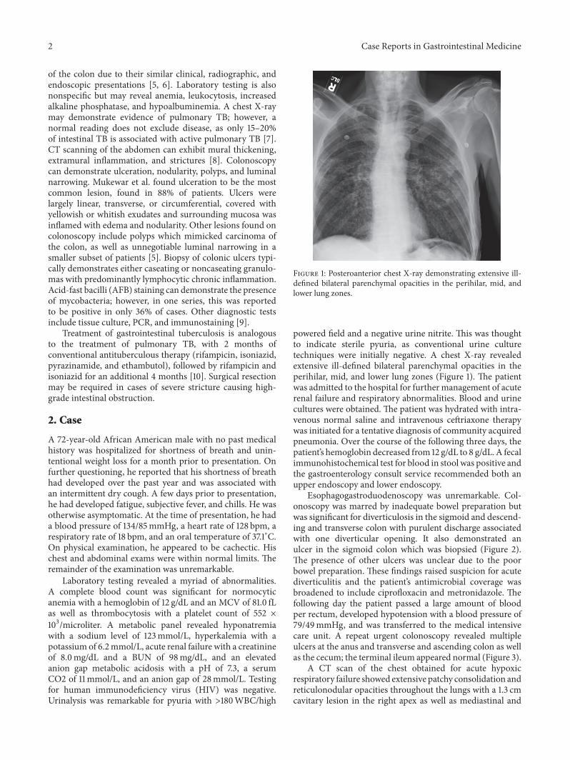
Figure 1: Posteroanterior chest X-ray demonstrating extensive ill-defined bilateral parenchymal opacities in the perihilar, mid, andlower lung zones.
powered field and a negative urine nitrite. This was thoughtto indicate sterile pyuria, as conventional urine culturetechniques were initially negative. A chest X-ray revealedextensive ill-defined bilateral parenchymal opacities in theperihilar, mid, and lower lung zones (Figure 1). The patientwas admitted to the hospital for further management of acuterenal failure and respiratory abnormalities. Blood and urinecultures were obtained. The patient was hydrated with intra-venous normal saline and intravenous ceftriaxone therapywas initiated for a tentative diagnosis of community acquiredpneumonia. Over the course of the following three days, thepatient’s hemoglobin decreased from 12 g/dL to 8 g/dL. A fecalimmunohistochemical test for blood in stool was positive andthe gastroenterology consult service recommended both anupper endoscopy and lower endoscopy.
Esophagogastroduodenoscopy was unremarkable. Col-onoscopy was marred by inadequate bowel preparation butwas significant for diverticulosis in the sigmoid and descend-ing and transverse colon with purulent discharge associatedwith one diverticular opening. It also demonstrated anulcer in the sigmoid colon which was biopsied (Figure 2).The presence of other ulcers was unclear due to the poorbowel preparation. These findings raised suspicion for acutediverticulitis and the patient’s antimicrobial coverage wasbroadened to include ciprofloxacin and metronidazole. Thefollowing day the patient passed a large amount of bloodper rectum, developed hypotension with a blood pressure of79/49mmHg, and was transferred to the medical intensivecare unit. A repeat urgent colonoscopy revealed multipleulcers at the anus and transverse and ascending colon as wellas the cecum; the terminal ileum appeared normal (Figure 3).
A CT scan of the chest obtained for acute hypoxicrespiratory failure showed extensive patchy consolidation andreticulonodular opacities throughout the lungs with a 1.3 cmcavitary lesion in the right apex as well as mediastinal and

Case Reports in Gastrointestinal Medicine 3
Figure 2: First colonoscopy: extensive diverticulosis and solitary ulcer in the sigmoid colon.
(a) (b)
Figure 3: Second colonoscopy: (a) cecal ulcer and (b) transverse colon ulcer.
hilar lymphadenopathy. These findings were suspicious foratypical infection (Figure 4). The patient underwent bron-choscopy with bronchoalveolar lavage and a lavage samplewas AFB stain positive. Pathology of the sigmoid ulcer biopsyrevealed focal active colitis with cryptitis, crypt abscesses, andmild stromal lymphoplasmacytic inflammation. AFB stain ofthe sigmoid ulcer biopsywas positive with findings consistentwith mycobacterium (Figure 5). AFB stain of a urine sampleobtained to explain sterile pyuria was also positive. Thepatient was placed under respiratory isolation andwas startedon appropriate antitubercular therapy.
Over the course of our patient’s hospitalization, hisclinical condition continued to decline. Multiorgan failurerequiring vasopressor support later ensued and the patientultimately passed away 29 days into admission.
3. Discussion
We present a case of diffuse systemic tuberculosis (TB)infection involving the colon, urinary tract, and lungs com-plicated by multiorgan failure and death. Unique to ourpatient was the lack of typical risk factors for TB infection.The patient had no history of travel to TB endemic areasand denied incarceration, homelessness, or exposure toinfected individuals. It remains unclearwhere the patientmay
have contracted the infection; however, earlier exposure toinfected individuals or family members that the patient doesnot recall is possible. Also unique in our patient’s case isthe presentation of colonic tuberculosis with hematocheziacausing hemodynamic instability. Rectal bleeding is a rarepresentation of colonic TB; only a handful of case reportshave documented this manifestation [11].
Our patient’s initial colonoscopy findings of a rectal ulcer,as well as diverticulosis showing signs of active infection,were highly suspicious for acute diverticulitis (Figure 2).However, initial colonoscopy was marred by poor bowelpreparation and did not demonstrate the extent of colonicinvolvement with ulceration and there was low suspicionof active tuberculosis as a potential cause for the patient’sdrop in hemoglobin. Given our patient’s advanced age aswell as the presence of multiple diverticulae, angiodysplasticchanges in colonic vessels were also part of our differentialfor lower gastrointestinal bleeding. However, massive hema-tochezia the following day prompted repeat colonoscopywhich showed multiple new ulcers involving all segments ofthe colon, raising concern for an infectious process (Figure 3).No biopsies were obtained during the second colonoscopydue to active bleeding. Acid-fast stain of the sigmoid ulcerbiopsy obtained during the first colonoscopy showed acid-fast organisms consistent with mycobacteria (Figure 5).

4 Case Reports in Gastrointestinal Medicine
(a) (b)
Figure 4: CT chest with IV contrast taken on day 13. (a) shows right upper lobe cavitary lesion. (b) shows multilobar consolidation.
(a) (b)
Figure 5: (a) 20x hematoxylin and eosin (H&E) stain demonstrating active colitis with crypt abscess and an ill-defined defined granulomatousarea. (b) 40x AFB stain of the splenic flexure with necrotizing granuloma and acid-fast bacilli (circled).
Tuberculosis remains a “great mimicker” and must con-tinue to be part of the differential diagnosis in cases ofcolitis especially in individuals who have additional findingssuspicious for infection on chest imaging. While stainingfor acid-fast bacilli is not routine practice in pathologylaboratories, this should be considered in select patients whopresent with concerns for infection, especially both in thelungs and in the gastrointestinal tract. As described abovehowever, only 15–20% of intestinal TB is associated withactive pulmonary TB, thus providers must maintain a highindex of suspicion to allow early diagnosis and management.
Our case also serves as a reminder for the importance ofpostexposure testing and prophylaxis in healthcare workers.Nosocomial transmission of TB in the healthcare setting iswell described; cases of multidrug resistant TB transmittedto providers and patients in hospitals have also been reported
[12, 13]. In hospitals that receivemore than 200TB admissionsa year, incidence of hospital worker infection has beenreported to be as high as 10% a year [14].
Hospital-based infection control programs are essentialin the control of such transmission. Airborne infection iso-lation rooms (previously termed negative pressure isolationrooms) should be utilized for patients with suspected orconfirmed tuberculosis. N95 masks that filter particles ≥1micrometer in diameter with at least 95 percent efficiencyshould be utilized by healthcare staff and offered to visi-tors of patients with suspected or confirmed tuberculosis.Nonessential invasive diagnostic procedures ought to bepostponed unless absolutely necessary [15].
Providers exposed to patients with active pulmonarytuberculosis should be assessed for symptoms suggestiveof active TB infection (hemoptysis, weight loss, and fever)

Case Reports in Gastrointestinal Medicine 5
and, if present, should undergo chest X-ray with sputumacid-fast stain and culture. Providers without symptoms ofactive infection are to undergo tuberculin skin testing or aninterferon-gamma release assay at baseline and again 8 to 12weeks postexposure [12].
In our case, all providers who may have been exposedto the patient underwent interferon-𝛾 release assay testing(QuantiFERON, Cellestis Ltd., Australia) at baseline and 8weeks postexposure. The patient’s family members were alsocontacted and offered screening and appropriate treatmentfor TB.
Conflicts of Interest
The authors declare that there are no conflicts of interestregarding the publication of this paper.
References
[1] J. L. Salinas, G. Mindra, M. B. Haddad, R. Pratt, S. F. Price, andA. J. Langer, “Leveling of tuberculosis incidence—United States,2013–2015,”Morbidity and Mortality Weekly Report, vol. 65, no.11, pp. 273–278, 2016.
[2] Department of Health and Human Services (US DHHS), Cen-ters for Disease Control and Prevention (CDC), and Divisionof TB Elimination, “Online Tuberculosis Information System(OTIS), National Tuberculosis Surveillance System, UnitedStates, 1993–2014,” CDC WONDER Online Database, January2016.Data for all years updated by June 5, 2015, http://wonder.cdc.gov/tb-v2014.html.
[3] T. A. Sheer and W. J. Coyle, “Gastrointestinal tuberculosis,”Current Gastroenterology Reports, vol. 5, no. 4, pp. 273–278,2003.
[4] K. D.Horvath and R. L.Whelan, “Intestinal tuberculosis: returnof an old disease,”TheAmerican Journal of Gastroenterology, vol.93, no. 5, pp. 692–696, 1998.
[5] S. Mukewar, S. Mukewar, R. Ravi, A. Prasad, and K. S. Dua,“Colon tuberculosis: endoscopic features and prospective endo-scopic follow-up after anti-tuberculosis treatment,” Clinical andTranslational Gastroenterology, vol. 3, article e24, 2012.
[6] Y. Lee, S. Yang, J. Byeon et al., “Analysis of colonoscopic findingsin the differential diagnosis between intestinal tuberculosis andcrohn’s disease,” Endoscopy, vol. 38, no. 6, pp. 592–597, 2006.
[7] B. Nagi, R. Kochhar, D. K. Bhasin, and K. Singh, “Colorectaltuberculosis,” European Radiology, vol. 13, no. 8, pp. 1907–1912,2003.
[8] W.-K. Lee, F. Van Tonder, C. J. Tartaglia et al., “CT appearancesof abdominal tuberculosis,”Clinical Radiology, vol. 67, no. 6, pp.596–604, 2012.
[9] K. M. Kim, A. Lee, K. Y. Choi, K. Y. Lee, and J. J. Kwak, “Intesti-nal tuberculosis: clinicopathologic analysis and diagnosis byendoscopic biopsy,” The American Journal of Gastroenterology,vol. 93, no. 4, pp. 606–609, 1998.
[10] V. K. Kapoor, “Abdominal tuberculosis,” Postgraduate MedicalJournal, vol. 74, no. 874, pp. 459–467, 1998.
[11] M. Kela, V. B. Agrawal, R. Sharma, R. Agarwal, and A.Agarwal, “Ileal tuberculosis presenting as a case of massiverectal bleeding,” Clinical and Experimental Gastroenterology,vol. 2, article 129, 2009.
[12] M. S. Bader and D. S. McKinsey, “Postexposure prophylaxis forcommon infectious diseases,” American Family Physician, vol.88, no. 1, pp. 25–32, 2013.
[13] Centers for Disease Control (CDC), “Nosocomial transmissionof multidrug-resistant tuberculosis to health-care workers andHIV-infected patients in an urban hospital—Florida,”Morbidityand Mortality Weekly Report, vol. 39, no. 40, pp. 718–722, 1990.
[14] D. Menzies, A. Fanning, L. Yuan, and M. Fitzgerald, “Tuber-culosis among health care workers,” New England Journal ofMedicine, vol. 332, no. 2, pp. 92–98, 1995.
[15] Centers for Disease Control and Prevention, “Guidelines forpreventing the transmission of Mycobacterium tuberculosis inhealth-care settings, 2005,” MMWr 54.RR-17, 2–107, 2005.

Submit your manuscripts athttps://www.hindawi.com
Stem CellsInternational
Hindawi Publishing Corporationhttp://www.hindawi.com Volume 2014
Hindawi Publishing Corporationhttp://www.hindawi.com Volume 2014
MEDIATORSINFLAMMATION
of
Hindawi Publishing Corporationhttp://www.hindawi.com Volume 2014
Behavioural Neurology
EndocrinologyInternational Journal of
Hindawi Publishing Corporationhttp://www.hindawi.com Volume 2014
Hindawi Publishing Corporationhttp://www.hindawi.com Volume 2014
Disease Markers
Hindawi Publishing Corporationhttp://www.hindawi.com Volume 2014
BioMed Research International
OncologyJournal of
Hindawi Publishing Corporationhttp://www.hindawi.com Volume 2014
Hindawi Publishing Corporationhttp://www.hindawi.com Volume 2014
Oxidative Medicine and Cellular Longevity
Hindawi Publishing Corporationhttp://www.hindawi.com Volume 2014
PPAR Research
The Scientific World JournalHindawi Publishing Corporation http://www.hindawi.com Volume 2014
Immunology ResearchHindawi Publishing Corporationhttp://www.hindawi.com Volume 2014
Journal of
ObesityJournal of
Hindawi Publishing Corporationhttp://www.hindawi.com Volume 2014
Hindawi Publishing Corporationhttp://www.hindawi.com Volume 2014
Computational and Mathematical Methods in Medicine
OphthalmologyJournal of
Hindawi Publishing Corporationhttp://www.hindawi.com Volume 2014
Diabetes ResearchJournal of
Hindawi Publishing Corporationhttp://www.hindawi.com Volume 2014
Hindawi Publishing Corporationhttp://www.hindawi.com Volume 2014
Research and TreatmentAIDS
Hindawi Publishing Corporationhttp://www.hindawi.com Volume 2014
Gastroenterology Research and Practice
Hindawi Publishing Corporationhttp://www.hindawi.com Volume 2014
Parkinson’s Disease
Evidence-Based Complementary and Alternative Medicine
Volume 2014Hindawi Publishing Corporationhttp://www.hindawi.com
![WallFlex Colonic Stent - Boston Scientific- US · WallFlex ™ Colonic Stent Visualization Expertise in combining stent materials has resulted ... (BTS). “The WallFlex™ [Colonic]](https://img.pdfslide.net/doc/110x75/5ae601bc7f8b9a8b2b8ca931/wallflex-colonic-stent-boston-scientific-us-colonic-stent-visualization-expertise.jpg)