Embed Size (px)
Citation preview

Christine Hinz, MS, MLS(ASCP)CM
Hematology Essentials: A Foundation for Accurate
Smear Reviews

Current Challenges
System Wide Approach
Standardization
Differential Training Program

Training Area

How does the program work?
Training Material Trainer and Trainee
Checklists
Reference guides
Actual patient slides
Case Study Power Point
Competency Checklist

“ I have to tell you I was dreading the diff training BUT it was AWESOME! Where I worked before I didn't have any formal diff training, you knew the basics and that was it. I love all the hand outs you provided. Things made more sense after the class.”
Trainee Feedback

“I was worried about the time commitment, but every employee came back saying how valuable the training was…”
“I was impressed to hear how excited my employees were about hematology after the training”
“Employees feel they really have the tools now to provide great patient care”
Manager Feedback

Follow up post training with Cellavision images for competency
Ongoing competency assessment
Adapted for smaller sites and/or affiliates
Post Training

smooth, homogenous film
1/2 to 3/4 the slide length
straight feather edge
at least 1/4 inch examination area
pink RBCs and appropriate WBC blues under gross examination (Rainbow feather edge)
Proper Slide Preparation

Bad slide prep

Good slide prep

The Good and Bad

Examine on 10X: Check for good cell distribution, free of precipitate
Examine extreme feather edge:
Platelet clumps
Look for abnormal cells: More dense and larger cells will be pushed to the feather edge
Starting your slide examination

Area between extreme feather edge and “Zone of Morphology” is the cobblestone area. DON’T do the morph or diff in this area.
“Zone of Morphology”-area where cells evenly distributed, RBC’s close but not touching. Diff and morphology should be performed here
Starting your slide examination

Zone of morphology

Make sure slide has been made correctly
If the slide has been pushed too hard when making the slide, WBC’s will be concentrated at extreme feather edge and estimate will not match instrument result.
WBC Estimate

Estimate the white count under 10x or 40X/50x.
Under low power 10X: 5 WBC's = 1,000/cumm
Under 40X/50X: 1 WBC = 2,500/cumm
The white count estimate may not be reported, but every manual differential white count is checked in this manner
WBC Estimate

In “Zone of Morphology”:
Switch to 40x/50X or 100X to count 100 WBC cells. Note: Perception at 100x can be distorted
Manual differential vs analyzer differential
Must drop to 100X for RBC morphology and Platelet estimate.
Platelet Estimate = (Total # of PLTs Counted in 10 Fields Using 100X ) X 15,000
Performing a manual differential

Morphology not reported: Anisocytosis, Macrocytosis, Microcytosis, Poikilocytosis, Stomatocytes
Morphology reported as present: Toxic Granulation, Dohle Bodies, Auer Rods, Hypersegmented Neutrophils, Hyposegmented Neutrophils, Vacuolated Neutrophils, Reactive Lymphocytes, Smudge Cells, Large Platelets, Agranular Platelets, Dwarf Megakaryocytes, Atypical Platelets, Basophillic Stippling, Pappenheimer Bodies, Howell Jolly Bodies, Sickle Cells, Rouleaux
Know appropriate morphology reporting
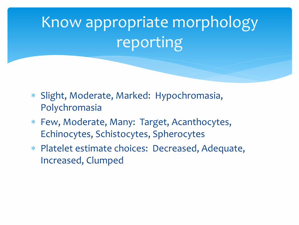
Slight, Moderate, Marked: Hypochromasia, Polychromasia
Few, Moderate, Many: Target, Acanthocytes, Echinocytes, Schistocytes, Spherocytes
Platelet estimate choices: Decreased, Adequate, Increased, Clumped
Know appropriate morphology reporting

Review Blood Maturation Chart

N/C Ratio
Chromatin pattern-clumped or fine
Nucleoli
Cytoplasm-Color of granules, inclusions
Size of cell
Myeloid Series-5 characteristics to look for
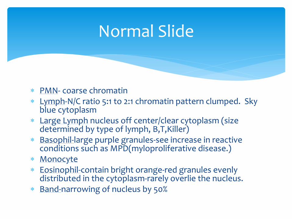
PMN- coarse chromatin Lymph-N/C ratio 5:1 to 2:1 chromatin pattern clumped. Sky
blue cytoplasm Large Lymph nucleus off center/clear cytoplasm (size
determined by type of lymph, B,T,Killer) Basophil-large purple granules-see increase in reactive
conditions such as MPD(myloproliferative disease.) Monocyte Eosinophil-contain bright orange-red granules evenly
distributed in the cytoplasm-rarely overlie the nucleus. Band-narrowing of nucleus by 50%
Normal Slide
Copyright ©2002 American Society of Hematology. Copyright restrictions may apply.
Maslak, P. ASH Image Bank 2002;2002:100360
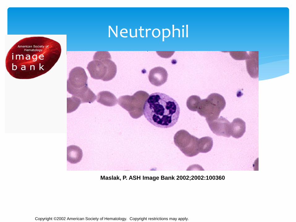
Neutrophil

Lymphocyte
7-16 µm, nucleus is the size of a normal
RBC, condensed chromatin, granules may
be present

Monocyte
12-20 µm, folded nucleus, lacy chromatin,
blue-gray cytoplasm, fine granules
http://library.med.utah.edu/WebPath/HEMEHTML/HEME003.html

Eosinophil and Basophil
12-15 µm, 2-3 lobed nucleus,
prominent reddish-orange
granules
10-15 µm, segmented nucleus,
prominent blue granules
Slides courtesy of
http://library.med.utah.edu/WebPath/HEMEHTML/H
EME003.html

Band Neutrophil
9-15 µm, horseshoe shaped nucleus,
chromatin present in any filaments

Leukemia is the uncontrollable growth of cells. Demonstrates a variety of immature cells, including blasts Basophilia and a left shift can be some of the first signs of CML Cells to be identified on slide:
Myelocyte Metamylocyte-Nucleus kidney bean shaped Promyelocyte-(granules can overlap nucleus) Basophilic
cytoplasm-Chromatin pattern is fine 1-2 nucleoli NRBC Myeloblast-Most immature cell in the myeloid series, N/C ratio
high-fine chromatin pattern, basophilic cytoplasm
Chronic Myeloid Leukemia

CML

Mononuclear cells seen on slide
Not seeing RBC’s overlapping on slide
Not seeing many platelets
Pancytopenia-All three cell lines are affected
Don’t see many neutrophils (neutropenia)
Large lymphs (clear cytoplasm/offset Nucleus)
Blasts: Note-If you see Auer Rods this indicates cell is in the myeloid lineage
Acute Myeloid Leukemia

RBC morphology sometimes seen on slide:
Basophilic stippling
Polychromatic
Elliptocytes (Ovalocytes)
Teardrops
NRBCs
Acute Myeloid Leukemia

AML

AML

Blast vs Lymph

4yr old, cough, fatigue
High WBC count, low Hgb-3.8g/dl, low Plt-20,000
Mononuclear cells with high N/C ratio, fine very fine, smooth chromatin pattern
Slide full of Blasts
ALL
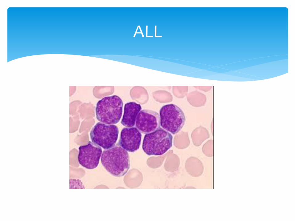
ALL

Affects B-cell lymphocytes
Typical Lymphocytosis >5.0 absolute
Characteristic nucleus that looks like “cracked earth” or a soccer ball
Cells are fragile, resulting in smudge cells present on smear
Albumin slides made to reduce smudge cells, diff should be performed on albumin slide, RBC/WBC morphology should be performed on the original slide
CLL

CLL

CLL vs ALL

Variability of cellular size and shape as well as nuclear size, shape and chromatin pattern
Seen in many viral illnesses-infectious mononucleosis
Nucleus attached to cell wall
Cytoplasm surrounding RBC’s
Reactive lymph vs Monocyte
Reactive Lymphs

Reactive Lymphs

Reactive Lymph

Used to boost WBC following chemo
Toxic granulation
Dohle Bodies-sometimes
Immature cells
GCSF: Neulasta, Neupogen

Toxic Granulation-Large, purple or dark blue azurophilicgranules, resembling the primary granules of promyelocytes, in the cytoplasm of neutrophils, bands and metamylocytes. Seen in severe infection, chemical poisoning, and other toxic states
Dohle Bodies-Appear as single or multiple light blue or gray staining area in the cytoplasm of neutrophil. RNA and represent failure of cytoplasm to mature. Seen in infections, poisoning, burns and following chemotheraphy
Vacuolated Neutrophils-seen in cytoplasm of neutrophils and bands and represent the sites of phagocytosed material. Seen in association with toxic granulation
Toxic gran, Dohle Bodies, Vacuolated Neutrophils

Toxic Granulation

Toxic Granules with Vacuoles

Toxic Granules + Dohle Bodies

Neutrophil with 5 or more lobes
Need to see a # of them to call
Seen in megaloblastic anemia, B12/Folate deficiency
Seeing macrocytosis-MCV is 130 on this patient
Hypersegmented Neutrophils

Hypersegmented Neutrophil

Unilobed neutrophil
Genetic Disorder (benign)
Cells will function fine
Pelger vs pseudo Pelger vs pyknotic
Pelger Huet

Pelger Huet vs Pyknotic

True hypogranular, hypolobulatedneutrophils

Case Study Time!

Case Study #1
22 yr old female presents at college health services
Patient complains of sore throat, fever, and swollen glands

Case Study #1
CBC results: Differential results:
WBC 16.0 thou/cu mm Neutrophils 26RBC 4.22 mil/cu mm Lymphocytes 63HGB 12.8 g/dL Monocytes 10HCT 37.5 % Eos 1 MCV 89 fL
MCH 30.4 pgMCHC 34.2 %RDW 12.6 %PLT 213 thou/cu mm

Case Study #1

Case Study #1

Case Study #1
Manual Differential reveals 3+ reactive lymphs
Heterophile Antibody Test confirms infectious mononucleosis diagnosis
Case Study #2
63 yr old female presents in ED
Left lower quadrant pain, fever, chills
History of diverticulitis, breast cancer
Patient is quadriplegic due to the effects of polio as a child

Case Study #2
CBC results: Differential results:
WBC 124.3 thou/cu mm Neutrophils 48RBC 4.31 mil/cu mm Lymphocytes 10HGB 13.3 g/dL Monocytes 5HCT 39.9 % Eos 2MCV 93 fL Baso 3MCH 30.9 pg Bands 14MCHC 33.3 % Meta 7RDW 17.1 % Myelo 11PLT 189 thou/cu mm

Case Study #2

Case Study #2
Blast-peripheral blood Bone marrow-ME slide

Case Study #2
Initial Hematology/Oncology consult determined increase in WBC was due to infection since Hgb and Plts were normal
Next step?

Case Study #2
Smear was referred to pathologist
Pathologist sent blood for BCR/ABL gene
Specific for Chronic Myelogenous Leukemia (CML)
Results are positive
Second Oncology consult results in bone marrow biopsy
Bone marrow confirms CML diagnosis

Case Study #3
Child Presented to clinic with cough and
fatigue Pediatrician ordered CBC/Differential CBC results revealed the following:
WBC 32,000Hgb 3.8 g/dlPlt 19,000

Case Study #3
Peripheral smear review:
High % mononuclear WBC’s
Irregular, clefted nuclei
Vacuoles present
Pediatrician informed of possible abnormal cells; requires confirmation by Pathologist
Slide sent STAT to hospital
Blasts confirmed by Pathology

Case Study #3

Case Study #3
Case Study #3
Pediatrician notified by Pathologist
Flow Cytometry: Lymphoid
B Cell ALL
Cytogenetics t(12;21)
Prognosis: favorable
5-year overall survival rate for childhood ALL 89%
Treatment: Induction/Consolidation

Case Study #4
Pre-op for total knee replacement
Routine labs included urinalysis, BMP, and CBC
CBC revealed low platelet count =86
Slide reviewed
No abnormalities revealed
Next day platelet count low
Slide reviewed (rule, Blast flag)

Case Study #4 Images
Blast w/ prominent nucleolus
Blast w/Auer Rod

Case Study #4
Slide review revealed 2-3 blast type cells with possible auer rods
Pathologist reviewed, contacted physician for further workup
Initial slide reviewed to see if we missed anything
Surgery delayed
Patient had bone marrow biopsy

Case Study #4
Morphology
Large blast cells
Basophillic cytoplasm/granules
Auer rods

Case Study #4
AML with t(8;21)
Prevalence ~25% adult AMLs
Prognosis: Good, 70% 5 year survival rate
Treatment: Patient starts induction chemo followed by consolidation therapy

Break!

Normal Red Blood Cells
Function
Size
Color
Central Palor
Note area of review
Stain quality

Polychromasia
Acute/chronic bleed
Hemolysis
Newborns
Hypochromasia
IDA
Thalassemias
Schrier, S. ASH Image Bank 2001;2001:100208
Maslak, P. ASH Image Bank 2004;2004:101122

Spherocytes and many times, polychromasia
Inherited hemolytic anemia
Defect in the protein that forms the outer membrane of RBC
RBC’s become spherical and lose central palor
Cells break down more quickly and are destroyed in spleen
Bone marrow will start producing more RBC
Hereditary Spherocytosis

Spherocytes

Same patient as previous slide after spleen removed
Seeing Howell-Jolly bodies in RBC’s
Round, purple nuclear fragments composed of DNA
Seen following splenectomy
Notice not seeing polychromasia because bone marrow doesn’t have to work as hard
Hereditary Spherocytosis

Post splenectomy

Marked increase in fragmented RBC (schistocytes)
May be of any size or shape including helmet cells, keratocyte and other irregular, unusual shapes
Look sheared or cut
Fragmented RBCs

Fragmented RBCs

Clinically significant and often seen in 3 conditions
Mechanical heart valve shearing RBCs
Burn victims
Microangiopathic anemias that includes disseminated intravascular coagulation (DIC), Hemolytic Uremic Syndrome (HUS), or Thrombotic thrombocytopenic Purpura (TTP)-these are hemeemergencies. A physician and/or pathologist should be notified immediately.
Fragmented RBCs

Extensive microscopic clots are formed in small blood vessels
Caused primarily by autoimmune inhibition of the ADAMTS13 enzyme that cleaves Von Willebrandfactor. The increase in vWF increases platelet adhesion
Treatment is plasma exchange to reduce circulating antibodies and increase the ADAMTS13 enzyme
TTP

TTP

RBC’s that lack central pallor with multiple oblong projections (rounded ends)
Form due to alteration in the lipid content of the RBC membrane
Seen in abetalipoproteinemia (genetic and rare disease)
Also seen in severe liver disease
Acanthocytes (Spur Cells)

Acanthocytes

Tear Drops
Myelofibrosis
Thalassemias
Maslak, P. ASH Image Bank 2002;2002:100453

Basophillic Stippling
Numerous fine or coarse granules
Evenly distributed
Composed of RNA
Lead Poisoning
ThalassemiasLazarchick, J. ASH Image Bank 2007;2007:7-00025

Pappenheimer Bodies
Fine, irregular granules
Usually in clusters
Composed of Iron
Splenectomy
Hemoglobinopathies
Hemolytic anemia
Sideroblastic anemia Lazarchick, J. ASH Image Bank 2007;2007:7-00013

RBC’s appearing in the shape of a sickle with two pointed ends
Can also appear as crescent-shaped, boat shaped and lack central pallor
Also see many target cells on this slide
Sickle Cell

Sickle cell
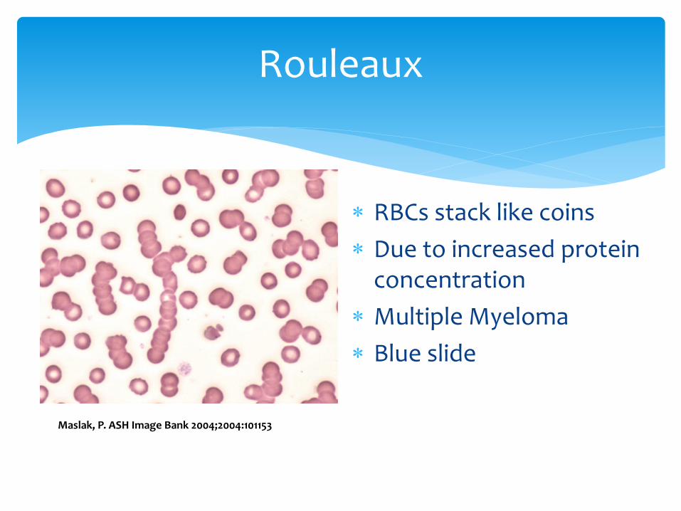
Rouleaux
RBCs stack like coins
Due to increased protein concentration
Multiple Myeloma
Blue slide
Maslak, P. ASH Image Bank 2004;2004:101153

See clumps of RBC’s
Caused by cold agglutinins
RBC Agglutination
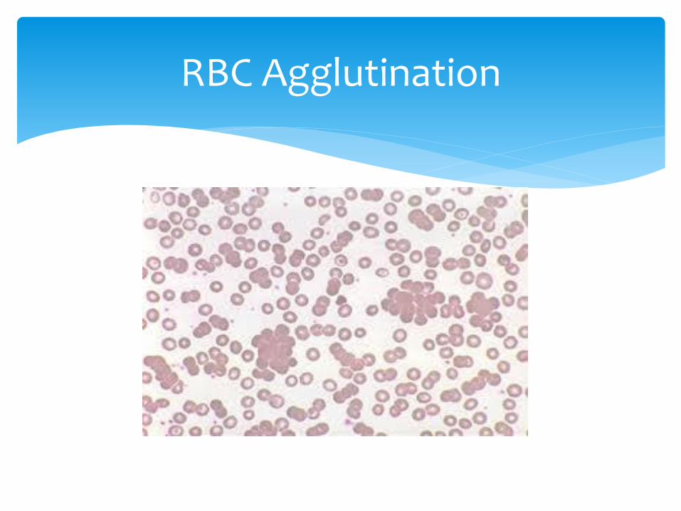
RBC Agglutination

Note Rouleaux (as compared to agglutination)
Plasma cells have eccentric nucleus, “clockface” nuclei
Plasma vs reactive lymphs
Plasma Cell Leukemia
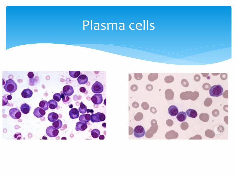
Plasma cells

Malaria

Look for maltase cross, often in rings
Unlike malaria, can have extracellular ring forms
Babesia

Babesia
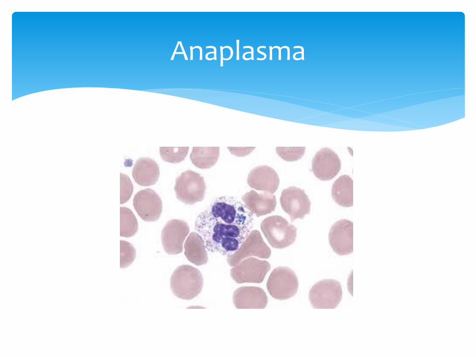
Anaplasma
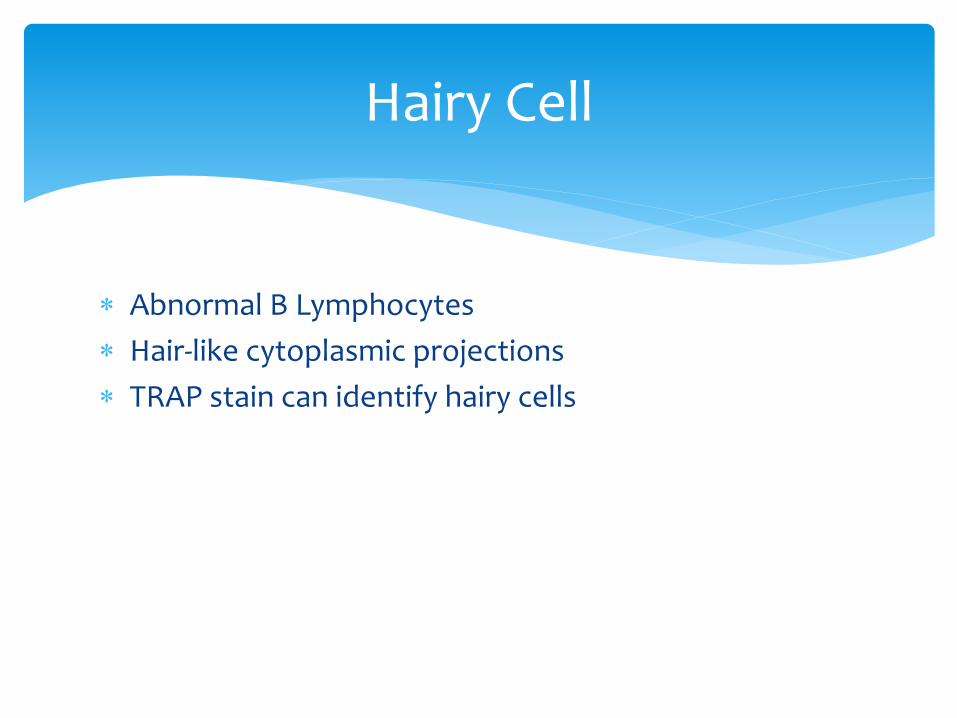
Abnormal B Lymphocytes
Hair-like cytoplasmic projections
TRAP stain can identify hairy cells
Hairy Cell
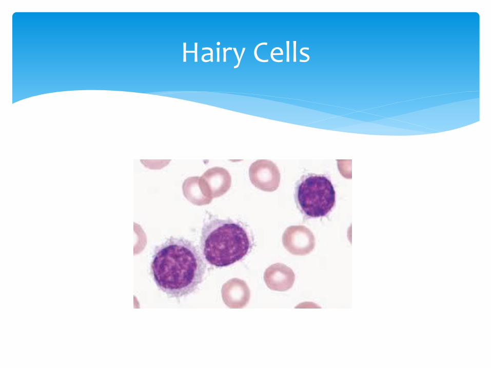
Hairy Cells

Case Study Time!

Case Study #1
47 yr old female presents at clinic
History of gastric bypass surgery
3 week history of fever with unknown origin 101.8 F
Experiencing sweats and chills 1-2 times/day
Recently was treated with amoxicillin for strep throat
Peripheral smear referred

Case Study #1
CBC results: Differential results:
WBC 4.3 thou/cu mm Neutrophils 33RBC 3.84 mil/cu mm Lymphocytes 57HGB 9.7 g/dL Monocytes 7HCT 31.8 % Eosinophils 2 MCV 83 fL Myelocytes 1MCH 25.3 pg NRBC 2MCHC 30.5 %RDW 16.7 %PLT 306 thou/cu mm

Case Study #1
Additional history reveals Patient underwent gastric bypass surgery 17 yrs ago
that was unsuccessful
Patient had corrective surgery but developed short bowel syndrome and subsequent chronic malnutrition
Patient had a Hickman catheter placed to receive nutrition (TPN) at night

Case Study #1
Peripheral smear: Neutrophil- What’s in it?

Case Study #1-Examine the entire slide
Don’t forget the
feathered edge!
Peripheral smear

Case Study #1
Yeast!
Pathologist notified and primary physician called immediately
Patient admitted
Catheter removed
Blood and catheter cultures revealed Rhodotorula Species

Case Study #2
68 yr old male presents in ER
2 week history of nausea, diarrhea, chills, weight loss, and mild confusion
Right upper quadrant pain

Case Study #2
CBC results: Differential results:
WBC 5.37 thou/cu mm Neutrophils 27RBC 3.64 mil/cu mm Lymphocytes 3HGB 11.1 g/dL Monocytes 4HCT 31.1 % Bands 62 MCV 85.4 fL Metas 4MCH 30.5 pgMCHC 35.7 %RDW 13.5 %PLT 18 thou/cu mm

Case Study #2

Case Study #2
Additional history reveals
One week prior to this episode he spent time at his cabin in Western Wisconsin with his wife

Case Study #2
Human Anaplasmosisrevealed on buffy coat smear
Present in neutrophils
Physician alerted immediately
Patient started on IV doxycycline
DNA by PCR was positive
Case Study #3
34 yr old male presents to the ED with the following:
Sternal chest pain
Back Pain
Groin Pain
Mild Shortness of Breath

Case Study #3
CBC:
WBC 13.8
HGB 8.1
PLT 370
Other labs:
Total Bilirubin: 15.3
ALT 52
AST 58

Case Study #3

Case Study #3

Case Study #3
Sickle Cell Crisis Homozygous Hemoglobin S Disease Present in 0.3-1.3 % of African Americans Sickle Cell Trait: 8-10% Deoxygenated state produces sickled cells Sickled cells jam in capillaries causing pain Anemia caused by hemolysis Lifespan of a sickle cell: 14 days Treatment: Hydroxyurea

Case Study #4
84 yr old female presents with the following:
Left hip fracture after a fall
Moderate fatigue
History of CAD

Case Study #4
CBC:HGB 10.5MCV 73MCH 18.5MCHC 28.3RDW 18.2PLT 320Other labs: Ferritin 31(normal 25-400)Soluable Transferrin Receptor 14.5 (normal 1.9-4.4)

Case Study #4

Case Study #4
Advanced Stage Iron Deficiency Anemia
HGB decreased
MCV <75
Ferritin 31 <15 is diagnostic
Increased STfR
Microcytic, hypochromic (MCH, MCHC)
Target cells
Therapy: Iron replacement

Case Study #5
67 yr old male presents with:
Fatigue
Shortness of Breath
Post aortic valve replacement

Case Study #5
CBC:
WBC 8.9
HGB 7.8
Retics 6.3%
Other labs:
LDH increased
Haptoglobin decreased

Case Study #5

Case Study #5
Microangiopathic Hemolytic Anemia (MAHA)
Secondary to a poorly functioning heart valve
Schistocytes present
Will probably have to have valve replaced

Case #6
34 yr old male presents with:
Fever
Fatigue
Chills
Sweats

Case Study #6

Case Study #6

Case Study #6
Babesia microti
Transmitted by the tick Ixodus scapularis (deer tick) present in the Minnesota
Important to distinguish babesia from other RBC inclusions or malaria
Often found in tetrads, vary in size,
Treatment: Clindamycin and quinine

Case Study #7
Middle aged patient
Symptoms-fatigue, general “ill” feelings
CBC results: WBC 25.6, RBC 5.90, HCT 58, RDW 26, PLT >750,000

Case Study #7
Polycythemia vera
WHO: Chronic Myeloproliferative Disease
Molecular on PB-JAK2
Treatment-hydroxyurea

Case Study #7
Same patient, 3 years later, presents with bone pain
CBC revealed pancytopenia, bizarre platelet morphology
Bone marrow biopsy-reticulin stain
Addition of chromosome 9
CMPD-Myelofibrosis
Treatment
Splenectomy
Continued hydroxyurea

Case Study #7

Case Study #7

Case Study #7

Case Study #7

Case Study #7
Same patient, 13 years later presents with continued bone pain, poor quality of life
CBC and Differential-PB, WBC >100,000, increased blasts ~20%
No bone marrow biopsy, confirmed by flow for CD34+ cells
Transformation to Acute Leukemia
WHO: AML w/multilineage displasia (w/prior MDS)

Thank you