Embed Size (px)
Citation preview
doi:10.1182/blood-2008-04-0781882008 112: 3927-3938
Alan N. Schechter Hemoglobin research and the origins of molecular medicine
http://bloodjournal.hematologylibrary.org/content/112/10/3927.full.htmlUpdated information and services can be found at:
(1174 articles)Red Cells � (1328 articles)Free Research Articles �
(34 articles)ASH 50th Anniversary Reviews �Articles on similar topics can be found in the following Blood collections
http://bloodjournal.hematologylibrary.org/site/misc/rights.xhtml#repub_requestsInformation about reproducing this article in parts or in its entirety may be found online at:
http://bloodjournal.hematologylibrary.org/site/misc/rights.xhtml#reprintsInformation about ordering reprints may be found online at:
http://bloodjournal.hematologylibrary.org/site/subscriptions/index.xhtmlInformation about subscriptions and ASH membership may be found online at:
Copyright 2011 by The American Society of Hematology; all rights reserved.Washington DC 20036.by the American Society of Hematology, 2021 L St, NW, Suite 900, Blood (print ISSN 0006-4971, online ISSN 1528-0020), is published weekly
For personal use only. at CAPES CONSORTIUM on January 19, 2012. bloodjournal.hematologylibrary.orgFrom
Hemoglobin research and the origins of molecular medicineAlan N. Schechter1
1Molecular Medicine Branch, National Institute of Diabetes, Digestive, and Kidney Diseases, National Institutes of Health, Bethesda, MD
Much of our understanding of humanphysiology, and of many aspects of pa-thology, has its antecedents in laboratoryand clinical studies of hemoglobin. Overthe last century, knowledge of the genet-ics, functions, and diseases of the hemo-globin proteins has been refined to themolecular level by analyses of their crys-tallographic structures and by cloningand sequencing of their genes and sur-
rounding DNA. In the last few decades,research has opened up new paradigmsfor hemoglobin related to processes suchas its role in the transport of nitric oxideand the complex developmental controlof the �-like and �-like globin gene clus-ters. It is noteworthy that this recent workhas had implications for understandingand treating the prevalent diseases ofhemoglobin, especially the use of hy-
droxyurea to elevate fetal hemoglobin insickle cell disease. It is likely that currentresearch will also have significant clinicalimplications, as well as lessons for otheraspects of molecular medicine, the originof which can be largely traced to thisresearch tradition. (Blood. 2008;112:3927-3938)
Introduction
During the past 60 years, the study of human hemoglobin, probablymore than any other molecule, has allowed the birth and maturationof molecular medicine. Laboratory research, using physical, chemi-cal, physiological, and genetic methods, has greatly contributed to,but also built upon, clinical research devoted to studying patientswith a large variety of hemoglobin disorders. During this period,the pioneering work of Linus Pauling, Max Perutz, Vernon Ingram,Karl Singer, Herman Lehmann, William Castle, Ruth and ReinholdBenesch, Titus Huisman, Ernst Jaffe, Ernest Beutler, and manyothers still active has been instrumental in these studies. Ourunderstanding of the molecular basis of hemoglobin developmentaland genetic control, structure-function relations, and its diseasesand their treatment is probably unparalleled in medicine. Indeed,this field, especially during the first 25 years of the existence of theAmerican Society of Hematology, provided the model for develop-ments in many other areas of research in hematology and othersubspecialities. This review attempts to highlight some recentdevelopments in hemoglobin research most relevant to the hema-tologist in the context of the current understanding of the functionsof these proteins and their genes. I am occasionally asked, “What’snew in hemoglobin?” I believe that this review will show that weare still learning much that is very relevant to our understanding ofhuman physiology and disease.
Hemoglobin structure
The human hemoglobin molecules are a set of very closely relatedproteins formed by symmetric pairing of a dimer of polypeptidechains, the �- and �-globins, into a tetrameric structural andfunctional unit. The �2�2 molecule forms the major adult hemoglo-bin. Their main function in mammals is to transport oxygen (O2)from the lungs to tissues, but they also specifically interact with the3 other gases, carbon dioxide (CO2), carbon monoxide (CO), andnitric oxide (NO), that have important biological roles.
The functional properties of hemoglobin molecules are primar-ily determined by the characteristic folds of the amino acid chains
of the globin proteins, including 7 stretches of the peptide �-helixin the �-chains and 8 in the �-chains (Figure 1).1,2 These helices arein turn folded into a compact globule that heterodimerizes and thenforms the tetramer structure.3 These 4 polypeptides of the hemoglo-bin tetramer each have a large central space into which a hemeprosthetic group, an iron-protoporphyrin IX molecule, is bound bynoncovalent forces, and thus the iron atom is protected from accessof the surrounding aqueous solution. The iron atoms in thisenvironment are primarily in the physiologic ferrous (FeII) chemi-cal valence state, coordinated to 4 pyrrole nitrogen atoms in oneplane, to an imidazole nitrogen atom of the invariant histidineamino acid at position 8 of the “F”-helix, and to a gas atom on theside opposite (with respect to the porphyrin plane) the histidineresidue. The reversible binding of gases to these 4 ferrous ironatoms in the tetramer of globin polypeptides allows hemoglobin totransport O2, CO, and NO.4 CO2 is transported in the blood insolution and by interactions with the amino-terminal residues ofhemoglobin as a weak carbamino complex and not by binding tothe iron atoms.
In recent years, knowledge of the properties of the character-istic folds of each of the globin polypeptides and their ability tobind heme prosthetic groups has led to the development of adetailed evolutionary tree to describe the ontogeny of thisfamily of genes from bacteria to vertebrates.5,6 In bacteria, theyare known as flavohemoglobins and appear to be primarily NOdioxygenases for detoxifying NO; in the protist and plant taxa,these single-chain globin proteins are largely involved withelectron transfer and O2 storage and scavenging. In inverte-brates, the O2 transport function of the globins develops as doseveral other biochemical functions. It is in the vertebrate taxathat the characteristic pattern of highly expressed intracellularglobins, frequently functioning as multimers, for oxygen trans-port over relatively long distances evolved (Figure 2). Theseseveral globin proteins also include, however, the single-chainmyoglobin, in high concentrations in many muscle tissues, aswell as the homologous (to myoglobin and to each other) �- and
Submitted April 22, 2008; accepted July 18, 2008. DOI 10.1182/blood- 2008-04-078188.
ASH 50th anniversary review
3927BLOOD, 15 NOVEMBER 2008 � VOLUME 112, NUMBER 10
For personal use only. at CAPES CONSORTIUM on January 19, 2012. bloodjournal.hematologylibrary.orgFrom

�-globins and their very stable �/� dimers that pair to formhemoglobin. In the highly specialized mammalian enucleatedcell, the erythrocyte, these molecules are expressed at very highconcentrations (Figure 1), resulting in a tremendously efficient
transport mechanism. The genes of myoglobin (and otherglobins) separated from the �- and �-globin genes duringvertebrate evolution, and these 2 genes themselves evolved intocomplex genetic loci on separate chromosomes. The numbers of
Figure 1. The X-ray determined structure of thehemoglobin molecule and a representation of itsvery high concentration in the erythrocyte. (A) Thearrangement of the �-helices (shown as tubes) in each�� unit—one on the left and one, 180° rotated, on theright—is shown, as are the 4 heme groups with theiriron atoms where gas molecules bind. The site of thesickle mutations on mutant �-chains as well as the �93conserved cysteine residues is also shown. Hemoglo-bin molecules in the red blood cell, shown in an inset onthe right, are very tightly packed (at a concentration ofapproximately 34 g/dL) and have little access to sol-vent; this allows efficient oxygen transport by each cellbut also affects the chemical behavior of the molecules,such as promoting sickle cell hemoglobin polymeriza-tion upon slight deoxygenation. (B) A representation ofthe quaternary structural changes in the hemoglobintetramer, in a top-down view, in the transition from theoxy conformation (left) to the deoxy conformation (right).The iron atoms shift relative to the planes of the hemegroups and a central cavity between the �-chainsopens, facilitating 2,3 BPG binding. These diagramsare based on drawings of Irving M. Geis. Illustration byAlice Y. Chen.
Figure 2. A diagram of the proposed evolutionaryrelationships of the human globin proteins as in-ferred from sequence analyses. NGB, neuroglobin;CYGB, cytoglobin; MB, myoglobin. Reprinted fromPesce et al (EMBO Rep. 2002;3:1146-1151) with per-mission. Illustration by Alice Y. Chen.
3928 SCHECHTER BLOOD, 15 NOVEMBER 2008 � VOLUME 112, NUMBER 10
For personal use only. at CAPES CONSORTIUM on January 19, 2012. bloodjournal.hematologylibrary.orgFrom
these genes, their chromosomal locations, and their developmen-tal control vary greatly among species; however, the basicglobin gene structure and protein folds are conserved inevolution among all mammals.
Myoglobin has a very high affinity for O2 compared withhemoglobin, but a detailed understanding of its function has stillnot been achieved. Mice with knockout of the myoglobin genehave almost normal physiology. Myoglobin is thought to servemore to facilitate oxygen diffusion in muscle, especially tomitochondria, than to act as a storage site, as previously thought.7
Myoglobin also appears to act as an NO dioxygenase and a nitritereductase. In the last decade, several other homologous proteins—neuroglobin and cytoglobin—have been detected in low amountsin certain tissues and appear to protect against hypoxia; again,however, there is much controversy about their functions.8,9
Hemoglobin function
The role of erythrocyte-encapsulated hemoglobin in transportingoxygen has been the focus of many of the greats of physiology,including Christian Bohr, August Krogh, J. B. Haldane, F. J. W.Roughton, and others in the last century and has been reviewed indetail.10,11 More recently elucidated was how this finely tunedsystem is regulated via heterotropic interactions with other mol-ecules, such as protons, anions, and bisphosphosphoglyceric acid(2,3 BPG or, in the older convention, 2,3 DPG),12 and byintramolecular, or homotropic, interactions for optimal normalrespiratory function.13 Cooperative oxygen binding can be ex-plained very precisely in terms of the allosteric model14 of proteinregulation of Monod, Wyman, and Changeux, but alternativemodels are still being developed.15 Understanding the physiologicfine-tuning of this function by proton binding (the Bohr effect) or2,3 BPG binding has been a triumph of basic protein chemistry andapplied physiology during the last 50 years.10,11
In the 1950s, the methods of protein sequence determinationand X-ray crystallography allowed the determination of the aminoacid sequences of various hemoglobins and the spatial arrange-ments of their atoms. This work, marked in particular by thehigh-resolution structural analysis—among the first for any pro-tein—by Nobelist Max Perutz (Figure 3) and his colleagues in thelate 1960s,2,16 soon resulted in a detailed explanation of therelationship of hemoglobin function as an oxygen transporter to itsmolecular structure. Furthermore, this information allowed for theexplanation of the clinical phenotypes of most of the manyhundreds of characterized mutations in the globin genes andproteins, which cause changes in function and include the many“hemoglobinopathy” diseases, in terms of this molecular structure.These correlations, initiated by Perutz and Lehmann17 and pio-neered in the United States by Ranney, Beutler, Nathan, Bunn,Forget, and others, remain among the landmark accomplishmentsof the then-new field of molecular medicine. Although much of thisinformation is now securely rooted in the textbooks, studies ofhemoglobin function have recently become quite active again.
In the last decade, there has been considerable attention tounderstanding the interactions of normal hemoglobin with CO, inrecognition of the fact that as well as being a toxic hazard, CO isproduced in the body from free heme by heme oxygenase and canitself activate soluble guanylyl cyclase.18 For this and otherreasons, it has potential pharmacological applications. This atten-tion to non–oxygen-related functions has been even more appli-cable to the study of NO/hemoglobin interactions since the
important realization in the mid-1980s that NO is a ubiquitouslyproduced cell signaling molecule, acting via both soluble guanylylcyclase production of cyclic GMP and other mechanisms, throughoutalmost all life forms. It is especially important in mammals in theregulation of vascular tone, cell interactions, and neural function.19
It has been known since before World War I that NO reacts withoxyhemoglobin to produce methemoglobin, with ferric (FeIII) ironand nitrate ions. Recent work suggests that most of the methemoglo-bin circulating in red blood cells is derived from this oxidationprocess,20 which is normally reversed by the erythrocytic methemo-globin reductase system. In the past 40 years, a second reaction ofNO with deoxyhemoglobin to form nitrosyl(heme)hemoglobin(NO-hemoglobin), with the NO liganded to the ferrous iron atom,has also been studied intensively. Like the reaction with oxyhemo-globin, this reaction had generally been assumed to be irreversible.However, there is now evidence that NO-hemoglobin in the circulatingred blood cell may be capable of releasing NO molecules—thuspotentially allowing a mechanism for hemoglobin-based, endocrine-liketransport of NO from one tissue to another within the body.21
Ten years ago, a third reaction of NO with oxyhemoglobin waspostulated to be physiologically important: the binding of NO tothe strongly conserved �-chain cysteine amino acid at position 93(Figure 1) to form S-nitrosylhemoglobin (SNO-hemoglobin).22 Itwas suggested that SNO-hemoglobin can physiologically dissoci-ate to release NO at low oxygen concentrations. Thus, this could bea mechanism for homeostatic control of blood flow to tissues,because the NO released would promote vascular dilatation andincrease blood flow and oxygen delivery. This hypothesis, althoughteleologically attractive, has been very controversial, with manystudies negating it, and very recent work with transgenic micelacking the �93 cysteine residues appears to disprove it.23 Morerecently, an alternate hypothesis to account for the transport of NOby erythrocytes has been advanced. It has been suggested that
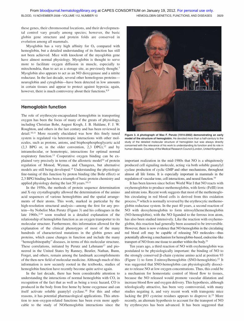
Figure 3. A photograph of Max F. Perutz (1914-2002) demonstrating an earlymodel of the structure of hemoglobin. He devoted more than a half-century to thestudy of the detailed molecular structure of hemoglobin but was always directlyconcerned with the relevance of his work to understanding its function and its role inhuman disease. Courtesy of the Medical Research Council (London, United Kingdom).
HEMOGLOBIN GENETICS, FUNCTIONS, AND DISEASES 3929BLOOD, 15 NOVEMBER 2008 � VOLUME 112, NUMBER 10
For personal use only. at CAPES CONSORTIUM on January 19, 2012. bloodjournal.hematologylibrary.orgFrom
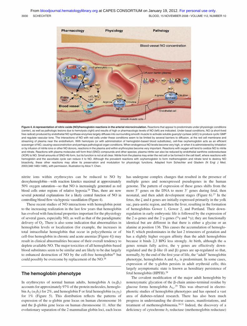
nitrite ions within erythrocytes can be reduced to NO bydeoxyhemoglobin—with reaction kinetics maximal at approximately50% oxygen saturation—so that NO is increasingly generated as redblood cells enter regions of relative hypoxia.24 Thus, there are nowseveral potential explanations for a likely central function of NO incontrolling blood flow via hypoxic vasodilation (Figure 4).
These recent studies of NO interactions with hemoglobin pointto the increasing realization in the last few years that hemoglobinhas evolved with functional properties important for the physiologyof several gases, especially NO, as well as that of the paradigmaticdelivery of O2. There is also some indication that abnormalities inhemoglobin levels or localization (for example, the increases intotal intracellular hemoglobin that occur in polycythemia or ofcell-free hemoglobin in chronic and acute anemias [Figure 4]) mayresult in clinical abnormalities because of their overall tendency todeplete available NO. The major toxicities of all hemoglobin-basedblood substitutes seem to be similar and are likely to be due largelyto enhanced destruction of NO by the cell-free hemoglobin25 butcould possibly be overcome by replacement of the NO.26
The hemoglobin phenotype
In erythrocytes of normal human adults, hemoglobin A (�2�2)accounts for approximately 97% of the protein molecules, hemoglo-bin A2 (�2�2) for 2%, and hemoglobin F or fetal hemoglobin (�2�2)for 1% (Figure 5). This distribution reflects the patterns ofexpression of the �-globin gene locus on human chromosome 16and the �-globin gene locus on human chromosome 11. After theevolutionary separation of the 2 mammalian globin loci, each locus
has undergone complex changes that resulted in the presence ofmultiple genes and nonexpressed pseudogenes in the humangenome. The pattern of expression of these genes shifts from themore 5� genes on the DNA to more 3� genes during fetal, thenneonatal, and then adult development stages (Figure 6).27 In thefetus, the � and ε genes are initially expressed primarily in the yolksac, para-aortic region, and then the liver, resulting in the formationof hemoglobins Gower 1, Gower 2, and Portland. Their down-regulation in early embryonic life is followed by the expression ofthe 2 �-genes and the 2 �-genes (G� and A�); they are functionallyidentical but are different in that there is either a glycine or analanine at position 136. This causes the accumulation of hemoglo-bin F, which predominates in the last 2 trimesters of gestation andhas a slightly higher oxygen affinity than the adult hemoglobinsbecause it binds 2,3 BPG less strongly. At birth, although the �genes remain fully active, the � genes are effectively down-regulated and the �-like (� and �) genes are up-regulated so that,normally, by the end of the first year of life, the “adult” hemoglobinphenotype, hemoglobins A and A2, is predominant. In some cases,expression of the �-globin persists in adult erythroid cells; thislargely asymptomatic state is known as hereditary persistence offetal hemoglobin (HPFH).28
The covalent modification of the major adult hemoglobin bynonenzymatic glycation of the �-chain amino-terminal residue byglucose forms hemoglobin A1c.29 This was observed in electro-phoretic studies of hemoglobin phenotypes and has opened a vastarea of diabetes-related research. There has also been muchprogress in understanding the diverse causes, manifestations, andtreatment of methemoglobinemia.30,31 Indeed, the discovery of adeficiency of cytochrome b5 reductase (methemoglobin reductase)
Figure 4. A representation of nitric oxide (NO)/hemoglobin reactions in the arterial microcirculation. Reactions that appear to predominate under physiologic conditions(center), as well as pathologic lesions due to hemolysis (right) and results of high or pharmacologic levels of NO (left) are indicated. Under basal conditions, NO (a short-livedfree radical) produced by endothelial NO synthase enzymes largely diffuses into surrounding smooth muscle to activate soluble guanylyl cyclase (sGC) to produce cyclic GMPand regulate vascular tone. The interactions of NO with red cells under these conditions seem to be limited by several barriers to diffusion, at the red cell membrane andstreaming of plasma near the endothelium. With hemolysis (or with administration of hemoglobin-based blood substitutes), cell-free oxyhemoglobin acts as an efficientscavenger of NO, causing vasoconstriction and perhaps pathological organ conditions. When endogenous NO levels become very high, or when it is administered by inhalationor by infusion of nitrite ions or other NO donors, reactions in the plasma and within erythrocytes become very important. Reactions with oxygen will tend to oxidize NO to nitriteand nitrate. Reactions with plasma molecules will form thiol (SNO) compounds and other species; plasma nitrite can also be reduced by endothelial xanthine oxidoreductase(XOR) to NO. Small amounts of SNO-Hb form, but its function is not at all clear. Nitrite from the plasma may enter the red cell or be formed in the cell itself, where reactions withhemoglobin and the ascorbate cycle can reduce it to NO. Although the prevalent reactions with oxyhemoglobin to form methemoglobin and nitrate tend to destroy NObioactivity, these other reactions may allow its preservation and modulation for physiologic functions. Adapted from Schechter and Gladwin (N Engl J Med.2003;348:1483-1485), with permission. Illustration by Alice Y. Chen.
3930 SCHECHTER BLOOD, 15 NOVEMBER 2008 � VOLUME 112, NUMBER 10
For personal use only. at CAPES CONSORTIUM on January 19, 2012. bloodjournal.hematologylibrary.orgFrom
as a cause of familial methemoglobinemia may be considered thefirst description of an enzyme defect in a hereditary disorder.32
There have also been significant advances in understanding thecomplex physiologic adaptive responses to acute and chronichypoxia, especially of populations at high altitudes.33
During the last 30 years, an enormous amount of effort has beendevoted to understanding the molecular and cellular mechanismsthat underlie these changes (called hemoglobin “switching”) inexpression of the �- and �-globin gene clusters.34 This has beenbecause of the intrinsic interest of this system as one of developmen-tal gene control but also because of the potential relevance of thisinformation to developing therapies for the 2 most common groupsof genetic diseases of hemoglobin, the sickle cell syndromes andthe thalassemia syndromes. Before reviewing these studies ofglobin developmental control, I note some of the relevant work—especially recent findings—on the pathophysiology of these 2 groupsof diseases and how altering the hemoglobin phenotype might beclinically beneficial.
Sickle cell disease
The discovery by Linus Pauling and his associates in 194935 thatthe molecular basis of sickle cell anemia is due to an abnormal
hemoglobin virtually created the field of molecular medicineand moved research hematology to its forefront. It is sometimesforgotten that this molecular medicine paradigm also requiredunderstanding of the inheritance pattern of this disease, whichwas supplied in the same year by J. V. Neel,36 whose publicationis also one of the founding articles of the field of medicalgenetics. We now have a detailed understanding of how a singlenucleotide change (A to T) in the �-globin gene leads to thevaline for glutamic acid substitution37 in the �-globin protein.This in turn allows the formation of stable intermolecularinteractions (linear polymers of the tetramers) in the concen-trated intracellular solutions of deoxyhemoglobin S (�2�2
S orsickle hemoglobin).38 This process is the basis for our understand-ing of the pathophysiology of this disease39,40 at the genetic,molecular and cellular levels. Sickle cell anemia pathophysiol-ogy is a consequence of this reduced solubility, causingpolymerization of hemoglobin S tetramers in red blood cellsupon partial deoxygenation and the impaired flow of these cellsin the microcirculation.38 Other mechanisms secondary tointracellular polymerization have been extensively studied,especially in animal models, but their relative importance tohuman pathophysiology remains unclear.
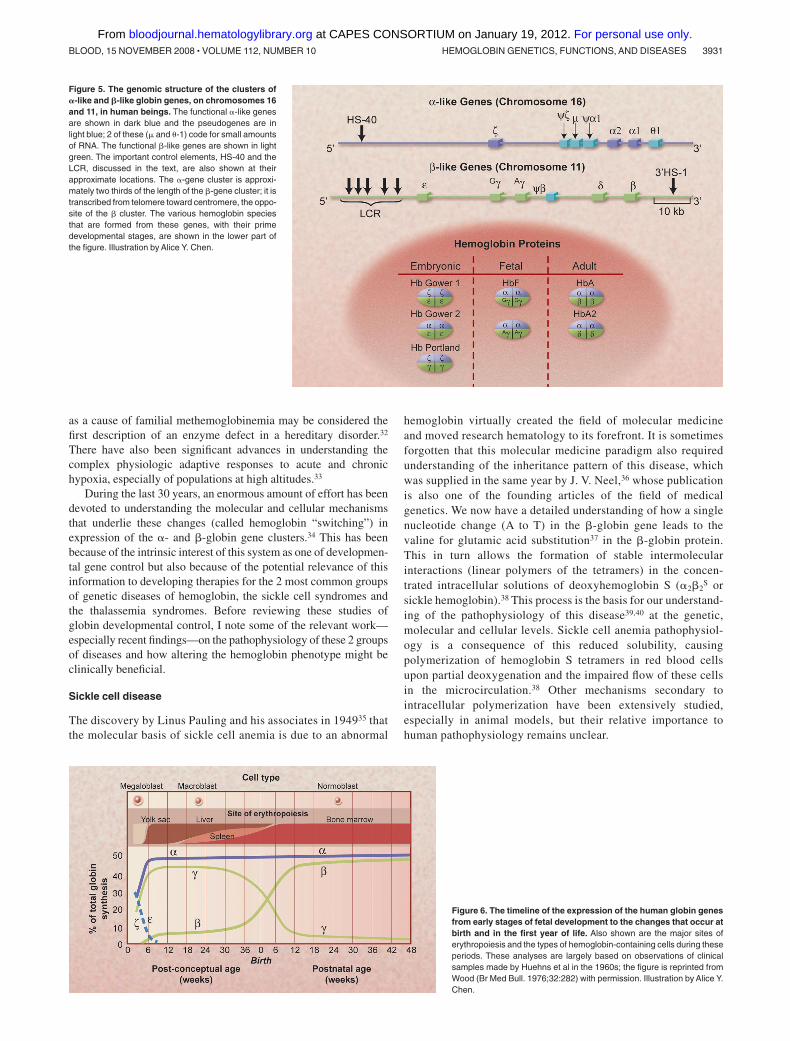
Figure 5. The genomic structure of the clusters of�-like and �-like globin genes, on chromosomes 16and 11, in human beings. The functional �-like genesare shown in dark blue and the pseudogenes are inlight blue; 2 of these (� and �-1) code for small amountsof RNA. The functional �-like genes are shown in lightgreen. The important control elements, HS-40 and theLCR, discussed in the text, are also shown at theirapproximate locations. The �-gene cluster is approxi-mately two thirds of the length of the �-gene cluster; it istranscribed from telomere toward centromere, the oppo-site of the � cluster. The various hemoglobin speciesthat are formed from these genes, with their primedevelopmental stages, are shown in the lower part ofthe figure. Illustration by Alice Y. Chen.
Figure 6. The timeline of the expression of the human globin genesfrom early stages of fetal development to the changes that occur atbirth and in the first year of life. Also shown are the major sites oferythropoiesis and the types of hemoglobin-containing cells during theseperiods. These analyses are largely based on observations of clinicalsamples made by Huehns et al in the 1960s; the figure is reprinted fromWood (Br Med Bull. 1976;32:282) with permission. Illustration by Alice Y.Chen.
HEMOGLOBIN GENETICS, FUNCTIONS, AND DISEASES 3931BLOOD, 15 NOVEMBER 2008 � VOLUME 112, NUMBER 10
For personal use only. at CAPES CONSORTIUM on January 19, 2012. bloodjournal.hematologylibrary.orgFrom
More than 50 years ago it was postulated (probably first byJ. B. S. Haldane) that the sickle mutation results in increasedresistance to malaria in heterozygotic persons or carriers41; subse-quent work indicates that this is true for thalassemia as well.42
Current research suggests the importance of redox and immuno-logic processes in this protection, but the exact cellular mecha-nisms are not yet clear.42,43 Again, as with so many other studies ofthe clinical biology of hemoglobin, this concept of selectiveadvantages for carriers of certain disease-causing (in the homozy-gous state) genes has been applied widely.
Another new concept from sickle cell anemia research quicklyextended to other diseases was the realization by Y. W. Kan and hiscolleagues in 197844 that restriction enzymes could be used todetect DNA polymorphisms linked to the abnormal �-globin geneto identify prenatally those fetuses who have one or both of themutant hemoglobin genes. These studies also initiated the gradualtransition of the molecular diagnosis of hemoglobin disorders fromprotein methods to the current wide range of extremely sensitiveand precise nucleic acid analyses.45
However, despite the detailed characterization of the abnormalgene and protein and the behavior of hemoglobin S in red cells, weunderstand relatively little of how these abnormalities affectspecific organs and the overall health of affected persons. The bestindicator of this conundrum is the unexplained heterogeneity in ageof onset and severity of disease in persons whose hemoglobingenotype and phenotype appear similar or identical.46 Unlike“classical” monozygotic diseases, even many of the thalassemicsyndromes, clinical progression and the need for treatment inpatients with sickle cell anemia patients can only be predicted inlimited circumstances, such as in children detected to haveabnormal blood flow in the large vessels of the brain as measuredby the transcranial-Doppler ultrasound method47 or in adults withpulmonary hypertension.48
Although many other measurements, such as globin clusterhaplotype analysis or white blood cell levels, have been suggestedto have explanatory and predictive value, only 2, the presence of�-thalassemia and the levels of hemoglobin F, have been validatedcomprehensively. Coexisting �-thalassemia leads to a reduction inMCHC, which inhibits hemoglobin S polymerization, but this beneficial
effect seems to be counter-balanced by the increase in total hemoglobinlevels, which may have some deleterious effects.49,50
The beneficial effects of hemoglobin F have been confirmed byclinical observations, epidemiologic studies, biophysical measure-ments, and therapeutic trials. In 1948, Janet Watson51 noted thatuntil adult hemoglobin displaces the form present at birth (hemoglo-bin F), manifestations of sickle cell disease are limited. Populationstudies among different groups of persons with sickle cell disease(eg, Saudi Arabs vs African populations) or within single geo-graphic areas, as well as a large “natural history” study in theUnited States confirmed that various measures of severity wereinversely related to hemoglobin F levels but suggested that veryhigh ( 25%) levels were needed for major benefit. At the sametime, diverse laboratory studies showed the mechanism by whichhemoglobin F had a sparing effect on intracellular polymerization(Figure 7) and confirmed clinical estimates of the levels ofhemoglobin F needed for benefit.52 Equally importantly, severaldrugs were found to increase hemoglobin F levels in nonhumanprimates. DeSimone and Heller53 and Letvin et al54 showed that5-azacytidine and hydroxyurea (now frequently designated ashydroxycarbamide) had such effects. This work was extended topatients by Platt, Charache, Dover, Nienhuis, Ley, Rodgers, andtheir colleagues (reviewed by Rodgers55). In a multicenter, double-blinded study of adults with frequent pain crises, led by Charache,56
hydroxyurea improved several clinical parameters compared withplacebo. In 1998, hydroxyurea was approved by the US Food andDrug Administration for treating these types of patients, and arecent systematic review has confirmed its efficacy in adult patientswith sickle cell disease.57
However, many patients do not respond at all to hydroxyureawith elevations of hemoglobin F, whereas some clinical manifes-tations seem to be little affected by even the 10% to 15% levelsof hemoglobin F obtained in some patients with the drug.Furthermore, there is yet limited evidence that the drug preventsdamage of crucial organs, such as the lungs, kidneys, and brain,or improves survival in adult patients.58 Long-term and con-trolled studies in children to assess the effects and safety ofhydroxyurea have only been recently initiated. Thus, in additionto further clinical outcome studies with hydroxyurea, there is a
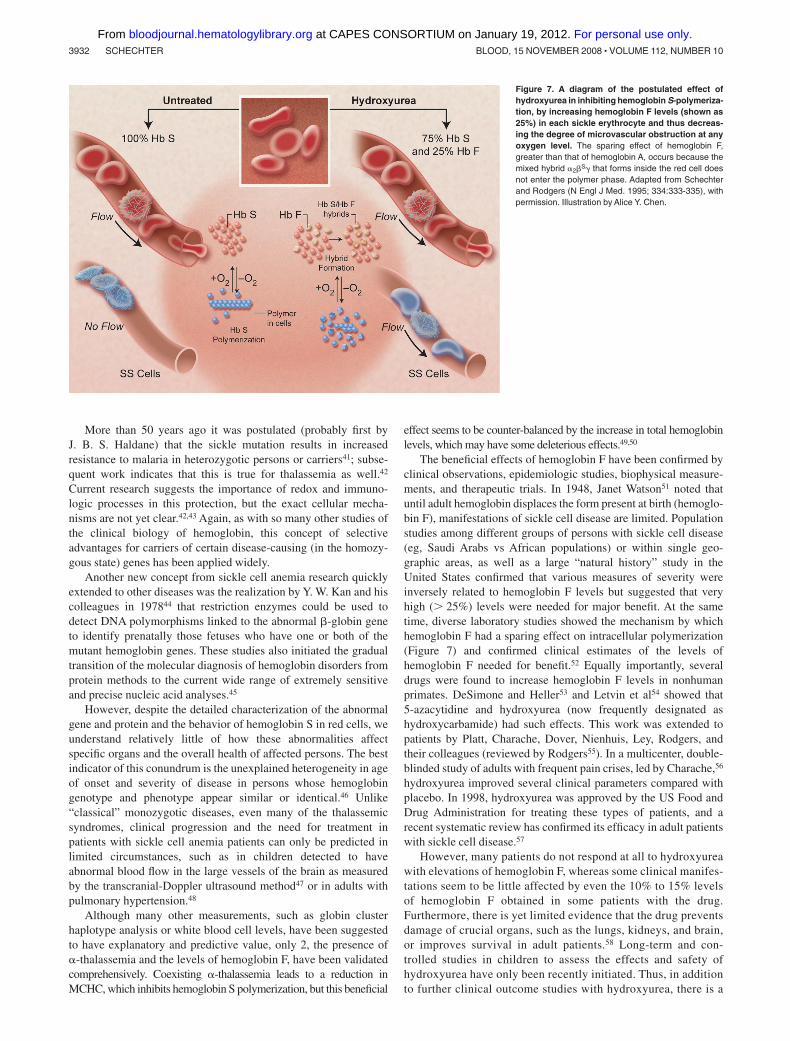
Figure 7. A diagram of the postulated effect ofhydroxyurea in inhibiting hemoglobin S-polymeriza-tion, by increasing hemoglobin F levels (shown as25%) in each sickle erythrocyte and thus decreas-ing the degree of microvascular obstruction at anyoxygen level. The sparing effect of hemoglobin F,greater than that of hemoglobin A, occurs because themixed hybrid �2�S� that forms inside the red cell doesnot enter the polymer phase. Adapted from Schechterand Rodgers (N Engl J Med. 1995; 334:333-335), withpermission. Illustration by Alice Y. Chen.
3932 SCHECHTER BLOOD, 15 NOVEMBER 2008 � VOLUME 112, NUMBER 10
For personal use only. at CAPES CONSORTIUM on January 19, 2012. bloodjournal.hematologylibrary.orgFrom
strong need to find other agents that may singly or in combina-tion with hydroxyurea have a more robust effect on hemoglobinF levels. To this end, studies with erythropoietin, butyratecompounds, and deoxyazacytidine are being pursued, but atpresent, none seems strongly promising. Clearly, much morework, both clinical and laboratory, is needed to test these drugsfurther and to find new agents if we are to improve on thispartially effective pharmacologic approach to this disease.
Although the utility of hydroxyurea in treating sickle celldisease has been generally accepted in the academic community,its more general use in the treatment of patients with sickle celldisease has been quite limited, even among patients who arelikely to benefit.59 Further complicating its evaluation has beenthat its clinical effects have been attributed by some tomechanisms other than inducing hemoglobin F, such as loweringneutrophil counts or adhesion molecules, generating NO, andothers. The evidence for these is minimal, and some of theseeffects may be indirect results of elevating hemoglobin F.Likewise, many pathophysiologic mechanisms have been pro-posed in sickle cell disease studies as alternatives or comple-ments to the intracellular polymerization of deoxyhemoglobin Sand its effects on the rheologic properties of the sickleerythrocyte. None has the weight of evidence that surrounds theprimary polymerization phenomenon, and they are likely to besecondary factors; none of the therapeutic approaches based onthese hypotheses has been promising up to now compared withinhibiting polymerization of deoxyhemoglobin S with hemoglobin F.
However, it has recently been proposed that as a consequenceof the fragility of the sickle erythrocyte (due to intracellularpolymerization, which results in intravascular hemolysis and thechronic anemia characteristic of this disease), circulating cell-free hemoglobin levels are increased, and this acts as a strongNO scavenger. This hypothesis60 postulates that some of theclinical manifestations of sickle cell disease (pulmonary hyper-tension, leg ulcers, and possibly stroke) relate to this NOdeficiency, whereas others (vaso-occlusive pain crises, acutechest syndrome) are due primarily to occlusion of microcircula-tory flow by red cells made rigid (not necessarily “sickled”) byintracellular polymer. Patients with sickle cell disease appear todiffer in the relative importance of the hemolytic and theocclusive mechanisms for reasons that are not clear. This modelsuggests possible treatment with NO to dilate vessels so as todiminish sickle cell entrapment, in addition to attempts to inhibitpolymerization itself, and thus should be amenable to clinicaltesting in the near future.
The thalassemia syndromes
Studies of the �- and �-thalassemia syndromes, especially byWeatherall, Wood, Higgs, Nathan, and their colleagues, duringthe past 50 years have also tremendously informed the basicunderstanding of the hemoglobin genes and proteins, as have thelaboratory studies informed the clinical description of patientswith these syndromes.61 The genetic mechanisms causing reduc-tion of �-globin synthesis in the �-thalassemias and of �-globinsynthesis in the �-thalassemias have been models for the studyof other genetic diseases. These have been reviewed in de-tail,61,62 as has been the explanation of pathophysiology as beingdue to chain imbalances within the thalassemic erythroidprecursors resulting in ineffective erythropoiesis and medullaryas well as intravascular hemolysis, perhaps as a result ofoxidative processes and resulting apoptosis-like events duringerythroid development.63 In recent years, studies of patients
with de novo �-thalassemia and mental retardation and with�-thalassemia and myelodysplastic syndrome have identified asomatic mutation in the gene ATRX, the role of which inchromatin remodeling has a strong effect on �-globin geneexpression.64 These studies again illustrate the continued impactof hemoglobin genetics on other aspects of molecular medicine.
Among the major unanswered questions in thalassemiaresearch has been the variability of clinical symptoms for somepatients with �-thalassemia intermedia65 but especially for thelarge numbers of patients who are doubly heterozygous forhemoglobin E and �-thalassemia.66 Recent studies have led tothe conceptualization of this variability in terms of primary,secondary and tertiary factors.67,68 The primary factors are thoseof the �-globin genotype, in particular the amount of globinmRNA and protein (eg, �o with no globin versus � withreduced globin levels of expression from each mutant gene).The secondary factors are other genetic changes in the globingene clusters that either contribute to levels of �-globin ordetermine �-globin levels in response to deficiencies of �-globinexpression. Among the latter are the Xmn1-G � polymorphism atposition �158 of the G�-gene, which up-regulates its expres-sion, as well as other less well understood genetic changes thatmay cause the markedly high levels of hemoglobin F levels seenin HPFH or increase these levels only slightly. The tertiaryfactors are much more diverse and include factors that affectiron absorption, bilirubin metabolism, and other factors knownand unknown. A protein that binds to free �-chains, the �hemoglobin stabilizing protein, has been identified as a molecu-lar chaperone (affecting the folding of the �-chains) in globinbiosynthesis69 and is postulated to influence the severity of�-thalassemia syndromes.70
These analyses confirm the likelihood that pharmacologic orgenetic methods to elevate hemoglobin F could have great therapeu-tic value for most cases of �-thalassemia, as with sickle celldisease. Unfortunately, the response to agents such as hydroxyureaand even the more potent 5-azacytidine that work in many patientswith sickle cell disease have been relatively disappointing inpatients with thalassemia.71 It has been suggested that recent bloodtransfusions, which are generally more necessary in patients withthalassemia than in those with sickle cell disease, blunt the effectsof these drugs because they seem to need rapidly proliferating bonemarrow to increase expression of hemoglobin F.72,73 Clinicalstudies, with respect to timing of therapies or the use of otherfactors, such as erythropoietin and iron, may establish regimenswith greater clinical benefit. With regard to other recent research in�-thalassemia, it may be noted that new understanding of thecontrol of iron absorption and metabolism and improved chelationagents may also allow amelioration of some of the clinicalcomplications that cause morbidity and mortality without changingthe fundamental genetic imbalance.74
Genetic testing and the availability of prenatal diagnosis hasgreatly reduced the numbers of �-thalassemia patients in certaincountries during the last 2 decades. Stem cell transplantation hasalso been very successful in treating many patients with thesesyndromes when the requisite medical facilities are available.Overall, survival and cure of the thalassemic syndrome approaches90% in several major centers.75 High-resolution human leukocyteantigen typing has extended this success to unrelated donors, andthe use of less intense conditioning regimens, with the goal ofachieving stable mixed chimerism, promises also to expand theutility of stem cell transplants.
HEMOGLOBIN GENETICS, FUNCTIONS, AND DISEASES 3933BLOOD, 15 NOVEMBER 2008 � VOLUME 112, NUMBER 10
For personal use only. at CAPES CONSORTIUM on January 19, 2012. bloodjournal.hematologylibrary.orgFrom
However, both transplants and prenatal diagnosis have beenmuch less used or successful in the sickle cell disease population. Stemcell transplants seem to be unfeasible for most patients for economicreasons or because of the lack of matched stem cell donors.
Treatment of the thalassemic and sickle cell syndromes by genetransfer therapy, the original goal of many gene therapy investiga-tors, has been beset by all of the tribulations that have affected thisfield in general. It has not been possible to achieve high-efficiencytransfer of globin gene vectors into erythroid cells with resultantprolonged robust expression of the normal globin gene. Equallyimportant, the recent cases of leukemia resulting from insertionalmutagenesis in children with X-linked severe combined immunode-ficiency who were being treated with gene therapy has greatlyincreased safety concerns with regard to viral vectors. Furthermroe,there are reasons to expect that because of the need for high levelsof globin protein production with precise chain balances, thesediseases will not be easy to treat. However, clinical advancesrelated to the stem cell transplants mentioned above, as well asimprovements in the design of globin gene vectors and transfectionmethods, suggest that clinical studies of gene therapy protocols for somegenetic diseases of hemoglobin will soon be undertaken.76-78 An entirelynew approach to therapy of genetic diseases, with induced pluripotentstem cells (iPS) generated from autologous skin, has just now beendemonstrated in a sickle cell anemia mouse model.79
Human globin genetics
The �-like globin gene clusters shown in Figure 5 are at the p13.3locus of chromosome 16 in a region of ubiquitously expressedgenes near the telomere. The �-like genes are in the p15.5 region ofchromosome 11, which contains multiple DNA sequences that actas strong tissue and developmental stage-specific enhancers oftranscription. These differences probably account for the fact thatthe 2 �-globin genes are expressed strongly and continuously inerythroid cells from a short time after early embryonic develop-ment to adulthood (Figure 6). However, the 5� �-2 gene isexpressed much more strongly than the more 3� but identical �-1gene. In contrast the �-globin gene cluster undergoes sequentialexpression of the ε-globin gene, then the 2 �-globin genes, and thenthe adult �- and �-globin genes, but with a marked preponderanceof �-globin compared with �-globin. The embryonic �-globin gene,which is expressed briefly in early fetal life, is 5� to the 2 �-globingenes on chromosome 16.
In the �-globin gene locus, a DNA region 5� to the cluster calledhypersensitivity site-40 (HS-40) acts as an erythroid-specific enhancerof transcription of closely linked genes.61,80 In contrast, on chromosome11, the �-globin gene is regulated by its proximity to a group of at least5 DNAsites, termed the locus control region (LCR), that are hypersensi-tive to cleavage by the DNAse I nuclease (for reviews, see Li et al81 andDean82). The LCR appears to contribute significantly to regulation of thesequential 5�-to-3� expression of the globin genes during development,as well as their very high level of expression—necessary to obtain, withmatched contributions from the �-locus, the very high hemoglobinlevels of the normal erythrocyte.
Each of the segments of DNAin the 2 globin gene clusters that codesfor an RNA transcript for a particular globin protein is by conventioncalled a globin “gene.” All of these genes, 8 in the human, have verysimilar structures. There are 3 coding exons and 2 intervening se-quences, or introns of DNA, the RNA copy of which is spliced from thepre-messenger RNA after transcription, that are subject to manymutations that affect splicing efficiency. The preservation of thisstructure of the globin genes, which may be related to the preservation ofthe protein folds, among the human genes and even among many
mammalian species, is in contrast to the much greater variability of thenumber and arrangement of the genes among species.
Immediately 5� to each gene are regulatory DNA sequences, termedpromoters. These, and other more distant DNA elements, contribute tothe regulation of expression of each of these genes (for reviews, seeStamatoyannopoulos et al,34 Weatherall and Clegg,61 Higgs et al,80
Orkin,83 Martin et al,84 Chakalova et al,85 and Mahajan et al86), andmutations in them also affect the transcription efficiency of each gene.Such mutations, as well as those within the coding sequence that affectpost-transcriptional modifications or translational efficiency, are mani-fest clinically as the thalassemia syndromes discussed above. Suchregulatory single nucleotide or point mutations, now numbering in thehundreds, are more common in the �-thalassemias than in the �-thalasse-mia syndromes, which are mainly caused by large deletions in the �-genecluster. A registry of these hemoglobin variants and much other informa-tion about these molecules and their genes, initiated by Titus Huisman, isavailable online at the database HbVar (http://globin.bx.psu.edu/hbvar).87,88
Globin gene regulatory mechanisms
Directly flanking each of the globin genes at its 5� end are regionsof DNA sequences designated the proximal promoter that regulatethe binding of a complex of proteins that control the initiation andrate of transcription of the mRNA.34,89,90 These cis-acting elementsinclude, in the immediate or proximal region, the ATA, CCAAT,and CACCC nucleotide sequences, frequently found in theseregions in many genes. More distant 5� sequences, up to 1 or2 kilobases from the gene and called the distal promoter, may alsoserve to contribute to activation or silencing of each of the globingenes. GATA sequences [(A/T)GATA(A/G)] in the DNA, whichbind GATA-1 transcription regulatory proteins, are found through-out the globin gene clusters and have strong erythroid specificity.The transcription regulatory protein (or trans-acting factor) EKLFwas originally thought to have strong specificity for positiveregulation of the �-globin gene but is now known to also haveeffects on many erythroid genes. Figure 8 shows a relatively simplemodel compatible with these results for how developmental controlof the �-globin gene cluster may occur via interactions with theLCR and these major factors, but a host of other factors (see below)clearly also play crucial roles.
In recent years, based on the pioneering studies of Felsenfeld,Orkin, Bieker, Engel, and others, evidence has been presented for arole of a variety of other transcription factors or complexes in thecontrol of individual globin genes or their sequential expression,including NF-E2, BP-1, SSP, FOG, FKLF, DRED, and PYR (forreviews, see Stamatoyannopoulos et al,34 Orkin,83 Martin et al,84
Chakalova et al,85 Mahajan et al,86 Giardino et al,88 and Stamatoy-annopoulos89). No simple picture of how any of these mightregulate the developmental pattern of the globin genes has emerged.DNA sequencing of the globin gene clusters has revealed anenormous number of other DNA motifs in the globin cluster, somewithin the LCR, that appear to be binding sites for many otherproteins that also contribute to enhancing or silencing transcription,revealing the enormous complexity of this system.
The DNA sequences coding for the pre-mRNA of each globingene includes that for the mRNA cap site, the initiator codon,splicing sites, terminator codons, and mRNA polyadenylatingsignals. Mutations in any of these can affect transcription effi-ciency; mRNA processing, stability, and transport from the nucleusto the cytoplasm; and the efficiency of translation into globinprotein on ribosomes. Impairment in any of these can lead to athalassemia syndrome, as can more distal mutations or deletions inthe globin cluster. Conversely, mutations affecting the binding of
3934 SCHECHTER BLOOD, 15 NOVEMBER 2008 � VOLUME 112, NUMBER 10
For personal use only. at CAPES CONSORTIUM on January 19, 2012. bloodjournal.hematologylibrary.orgFrom
transcription factors near the �-globin genes or DNA deletions thatchange the relationship of these genes to cis-acting sequences canup-regulate �-gene expression as in the HPFH syndromes.28
The molecular mechanisms of the �-globin LCR have beenpartially elucidated by 2 decades of research by the laboratories ofTuan, Grosveld, Groudine, and others, with in vitro, cellular, andgenetically modified animal assays. These closely spaced DNAsehypersensitivity sites were initially postulated to act singly or as agroup as a major regulator of globin gene expression, both as astrong general enhancer of transcription and as a specific mediatorof developmental control of the �-cluster. Both mechanisms arebelieved to occur by interactions of all or parts of the LCRsequentially with the embryonic, then the fetal, and then finally theadult-like globin genes during ontogeny and, in adult life, duringmaturation of marrow erythroblasts (Figure 6). They seem tosomehow “open” the chromatin of the relevant segments of DNA,allowing the transcription initiation complex and other trans-actingfactors to bind at the appropriate gene.91
Much effort has been extended to understand how the LCRaffects transcription of the entire �-globin gene cluster. Modelshave ranged from those based on binding of transcription factors tothe LCR and their subsequent migration or “tracking” to theindividual genes (compatible with observed intergene RNA tran-scripts) to the more generally accepted “looping” concepts. Re-cently, de Laat et al92 have refined these looping models byshowing the formation of “active chromatin hubs” or “transcriptionfactories” during erythroid maturation and stochastic interactions of theLCR with the individual globin genes. Current work also emphasizesthe possible importance of cell-cycle control and of intergenic transcrip-tion in the formation of these transcription domains.93
In some species, parts of the LCR seem to act as insulators,which can shield the �-cluster from the effects of other cis-actingelements or, conversely, limit the spread of the effects of chromatinchanges that occur within the globin gene cluster from affectinggenes outside of the cluster.94,95 A hypersensitive site 3� to the�-globin gene cluster has also been noted, but its role is not wellcharacterized. Recent data, however, have suggested that verystrong enhanced transcription of later genes may give the appear-ance of “switching” as early genes are diluted out.96 Finally, itshould be noted that virtually all the work just reviewed is focused
on initiation and rates of transcription. It is clear that post-transcriptionalfactors, such as stability of mRNA of the �-globin genes or translationrates (as affected by heme) are also very important in the regulation ofintracellular hemoglobin phenotypes.97,98 Unfortunately, relatively littleresearch has occurred in these areas in recent years because thetranscription paradigm has dominated the field.
Developmental biology of hemoglobin
Much information about the developmental modulation of hemoglo-bin initially came from clinical examination of human fetuses andnewborns. In recent years, most data have come from studies oferythroid cells in culture or from genetically modified animalmodels, none of which is a precise model of these processes inhumans.99 With that caveat, it should be noted that investigations inmice and, to a limited extent, in humans suggest that erythropoiesiscan be divided into a primitive phase or stage in the yolk sac andthen a definitive stage, initially in the liver and spleen and later inthe bone marrow of the fetus. Extensive delineation of the geneticsignals and hematopoietic factors that control primitive erythropoi-esis has been accomplished100; mechanisms of the control of globingene expression during the clinically important definitive erythro-poiesis stage have been much less tractable to these approaches.
It has been generally accepted that the specificity of expressionof the globin genes is not directly related to the site of hematopoi-esis, to clonal control of cell maturation, or to growth factors orother signaling molecules. However, recent work has providedevidence for cells of different phenotypes during ontogeny inhumans96 and, at least in vitro, the influence of certain cytokines,101
such as stem cell factor (SCF), on the hemoglobin phenotype.Erythropoietin, a very important cell growth factor, stimulatesproduction of erythroblasts that mature into hemoglobin producingcells and thus markedly increases the production of hemoglobin102
but does not seem to affect directly the expression of the globingenes or their developmental control.
Mechanisms of hemoglobin F inducers
In an era striving for “rational” therapeutics, our chances ofimproving the efficacy of hydroxyurea for sickle cell disease orobtaining agents that work in the thalassemia syndromes would
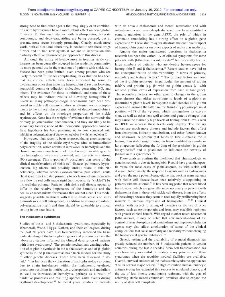
Figure 8. A simplified model of the developmentalcontrol of �-globin gene transcription at the embry-onic, fetal, and adult developmental stages (seeFigures 5 and 6). The sequential interaction of the LCRand trans-acting factors, such as GATA and EKLF, withthe promoters of the several globin genes are shown asare putative cis-acting silencers that may down-regulate expression for “switching.” Evidence for silenc-ers is stronger for the �-globin gene, but it has beenassumed that they would also affect the �-globingenes. Levels of expression are indicated by thelengths of the arrows above each gene. In 3 dimen-sions, these interactions would be represented byloops of DNA with bridging transcription and chromatinproteins between the LCR and the genes, now calledactive chromatin hubs. A more detailed model wouldinclude many other transcription factors, modificationsof chromatin structure, and the possible role of mi-croRNA molecules as well as many post-transcriptioncontrol mechanisms. Illustration by Alice Y. Chen.
HEMOGLOBIN GENETICS, FUNCTIONS, AND DISEASES 3935BLOOD, 15 NOVEMBER 2008 � VOLUME 112, NUMBER 10
For personal use only. at CAPES CONSORTIUM on January 19, 2012. bloodjournal.hematologylibrary.orgFrom
clearly be improved if we understood the mechanisms of the drugscurrently used. A major conceptual advance in this field was basedon the observations of Stamatoyannopoulos, Papayannopoulou,and their collaborators that adult erythroid cells in culture couldexpress fetal globins and their development of a model based onthis to separate cell differentiation and proliferation events.34,89
These concepts helped frame studies in the late 1970s of theimportance of methylation of CpG “islands” in the DNA ofpromoters in suppressing transcription103 and led to the use byothers of 5-azacytidine, a demethylating agent, to elevate hemoglo-bin F in baboons. However, controversy about this mechanism—and the suggestion that the hemoglobin F induction was due to aneffect of cytostasis on cell differentiation—led to trials withhydroxyurea and other agents. Although less potent then 5-azacyti-dine, hydroxyurea appeared safer and thus became the focus ofmost subsequent research. Later, butyrate compounds, which areelevated in the plasma of mothers with diabetes whose fetuses havedelayed “switching,” was tried, and its effect has been ascribed toanother gene regulatory mechanism, the acetylation of chromatinproteins.104 Thus attention focused on agents that appeared tochange the epigenetic expression of the globin genes directly orindirectly by affecting the cell cycle.
Unfortunately, despite 2 decades of such sophisticated mecha-nistic research, our understanding of how any of these compoundsaffects the hemoglobin phenotype of human erythroid cells is reallyquite limited.89,105,106 Nor do we have good assays for such agentsexcept for studies in nonhuman primates and in humans. Thus it islikely that the search for new drugs must remain both empiric andmechanistic, but now with very precise analytical tools formeasuring RNA and protein changes, for the immediate future.
Conclusions
Hemoglobin, perhaps the best studied of all macromolecules, hasnot revealed all its secrets even at the clinically relevant levels, tosay nothing of biophysical studies at the levels of its atoms andelectrons. In recent years, although unexpected new functions havebeen found, the central goal of most biomedical hemoglobinresearch has been the development of a mechanistic description ofthe developmental control of the �- and �-globin gene clusters.This field of research has been of great interest to those interested inthe whole range of hemoglobin studies—from the most basicmolecular genetics, to various “translational” models, to clinicalproblems in treating patients. It has been the hope that understand-ing these control mechanisms would lead to the discovery or designof drugs to treat the genetic hemoglobin diseases by efficientelevation of fetal hemoglobin and would also improve the effi-ciency of stem cell and gene transfer approaches to therapy.Although some of these therapies have progressed greatly duringthis period, we are still far from understanding the basic processescontrolling developmental changes in the globin gene clusters.Despite the enormous body of experimental data obtained fromcell, animal, and clinical studies, no predictive model has yet beenproposed to explain the control of this obviously complex system.
New experimental approaches to these problems are needed.Very recently genome-wide association studies have demonstrateda gene (BCL11A) encoding a zinc-finger protein on chromosome2p15 that modulates hemoglobin F levels.107,108 There is muchcurrent interest in the potential role of microRNA (miRNA)molecules, rather than more traditional protein trans-factors, incontrolling erythroid differentiation.109 It has recently been re-ported that the transcription factor GATA-1 activates transcriptionof 2 miRNA molecules that are essential for erythropoiesis.110 Thisnewly emerging paradigm for the control of much gene expressionby small, noncoding RNA molecules (the RNA “world”) is onlynow being investigated in the hemoglobin gene loci.
These and other totally new approaches suggest that ourconcepts of hemoglobin, and especially its genetics, will be asdifferent in 10 or 20 years as our current concepts differ from thoseof the investigators who made the first electrophoretic separationsof this abundant and convenient protein 60 years ago. However,studies of normal human beings and patients with hemoglobindiseases must continue as vigorously as that of the many modelsystems that complement clinical work in this field. If improvinghealth is the goal, results from model studies will only be useful ifthe reiterative processes of “bedside to bench to bedside” continueat all stages of the research.
Acknowledgments
I thank the physicians and scientists of virtually every discipline ofbiomedical research who have, for many decades, formed aninternational “college” devoted to hemoglobin and its diseases. Iapologize to those I could not name explicitly and for not beingable to cite hundreds of important articles because of spacelimitations. Indeed, except for some papers that I consider classicsin the relationship of hemoglobin research to the development ofthe field of molecular medicine, I have generally cited—wherepossible—recent review articles rather than the many originalpapers. In addition, I have emphasized studies clearly relevant tothe human hemoglobin molecules and have skirted the vast—butfrequently contradictory—literature on “model” systems. In addi-tion, I thank my mentors, my colleagues within and without theintramural National Institutes of Health research program, andespecially my “students,” from whom I have learned the most—and in whose accomplishments I have taken the most pleasure.
Authorship
Contribution: A.N.S. wrote the manuscript.Conflict-of-interest disclosure: A.N.S. is a coinventor of a
patent held by the National Institutes of Health for the use of nitritesalts for the treatment of cardiovascular diseases.
Correspondence: Alan N. Schechter, Molecular MedicineBranch, NIDDK, Building 10, 9N-314, National Institutes ofHealth, Bethesda, MD 20892; e-mail: [email protected].
References
1. Perutz MF. X-Ray Analysis of Haemoglobin.Stockholm: Les Prix Nobel; 1963.
2. Perutz MF. Science is not a quiet life: unravelingthe atomic mechanism of haemoglobin. London:Imperial College Press; 1997.
3. Dickerson RE, Geis I. Hemoglobin, structure,
function, evolution, and pathology. Menlo Park,CA: Benjamin/Cummings Publishing Company;1983.
4. Antonini E, Brunori M. Hemoglobin and myoglo-bin in their reactions with ligands. Amsterdam:North-Holland; 1971.
5. Hardison R. The evolution of hemoglobin. Am Sci.1999;87:126-130.
6. Lecomte JT, Videtich DA, Lesk AM. Structural di-vergence and distant relationships in proteins:evolution of the globins. Curr Opin Struct Biol.2005;15:290-301.
3936 SCHECHTER BLOOD, 15 NOVEMBER 2008 � VOLUME 112, NUMBER 10
For personal use only. at CAPES CONSORTIUM on January 19, 2012. bloodjournal.hematologylibrary.orgFrom
7. Garry DJ, Mammen PP. Molecular insights intothe functional role of myoglobin. Adv Exp MedBiol. 2007;618:181-193.
8. Fago A, Hundahl C, Malte H, Weber RE. Func-tional properties of neuroglobin and cytoglobin.Insights into the ancestral physiological roles ofglobins. IUBMB Life. 2004;56:689-696.
9. Brunori M, Vallone B. A globin for the brain.FASEB J. 2006;20:2192-2197.
10. Bunn HF, Forget BG. Hemoglobin: Molecular, Ge-netic and Clinical Aspects. Philadelphia: WBSaunders; 1986.
11. Edsall JT. Understanding blood and hemoglobin:an example of international relations in science.Perspect Biol Med. 1986;29:S107-S123.
12. Benesch RE, Benesch R. The mechanism of in-teraction of red cell organic phosphates with he-moglobin. Adv Protein Chem. 1974;28:211-237.
13. Perutz MF. Sterochemistry of cooperative effectsin haemoglobin. Nature. 1970;21:726-730.
14. Eaton WA, Henry ER, Hofrichter J, Bettati S,Viappiani C, Mozzarelli A. Evolution of allostericmodels of hemoglobin. IUBMB Life. 2007;59:586-599.
15. Lukin JA, Ho C. The structure-function relation-ship of hemoglobin in solution at atomic resolu-tion. Chem Rev. 2004;104:1219-1230.
16. Perutz MF, Muirhead H, Cox JM, Goaman LC.Three-dimensional Fourier synthesis of horseoxyhaemoglobin at 2.8 Å resolution: the atomicmodel. Nature. 1968;219:131-139.
17. Perutz M, Lehmann H. Molecular pathology ofhuman haemoglobin. Nature. 1968;219:902-909.
18. Ryter SW, Otterbein LE. Carbon monoxide in biologyand medicine. Bioessays. 2004;26:270-280.
19. Ignarro LJ. Nitric Oxide: Biology and Pathobiol-ogy. San Diego, CA: Academic Press; 2000.
20. Gladwin MT, Ognibene FP, Pannell LK, et al.Relative role of heme nitrosylation and beta-cys-teine 93 nitrosation in the transport and metabo-lism of nitric oxide by hemoglobin in the humancirculation. Proc Natl Acad Sci U S A. 2000;97:9943-9948.
21. Schechter AN, Gladwin MT. Hemoglobin and theparacrine and endocrine functions of nitric oxide.N Engl J Med. 2003;348:1483-1485.
22. Singel DJ, Stamler JS. Chemical physiology ofblood flow regulation by red blood cells: the roleof nitric oxide and S-nitrosohemoglobin. AnnuRev Physiol. 2005;67:99-145.
23. Isbell TS, Sun C-W, Wu L-C, et al. SNO-hemoglo-bin not essential for red blood cell dependent hy-poxic vasodilatation. Nat Med. 2008;14:773-777.
24. Gladwin MT, Schechter AN, Kim-Shapiro DB, etal. The emerging biology of the nitrite anion. NatChem Biol. 2005;1:308-314.
25. Natanson C, Kern SJ, Lurie P, Banks SM, WolfeSM. Cell-free hemoglobin- based blood substi-tutes and risk of myocardial infarction and death:a meta- analysis. JAMA. 2008;299:2304-2312.
26. Yu B, Raher MJ, Volpato GP, Bloch KD, IchinoseF, Zapol WM. Inhaled nitric oxide enables artificialblood transfusion without hypertension. Circula-tion. 2008;117:1982-1990.
27. Huehns ER, Dance N, Beaven GH, Hecht F,Motulsky AG. Human embryonic hemoglobins.Cold Spring Harb Symp Quant Biol. 1964;29:327-331.
28. Forget BG. Molecular basis of hereditary persis-tence of fetal hemoglobin. Ann NY Acad Sci.1998;850:38-44.
29. Bunn HF, Gabbay KH, Gallop PM. The glycosyla-tion of hemoglobin: relevance to diabetes melli-tus. Science. 1978;200:21-27.
30. Jaffe ER. Methaemoglobinaemia. Clin Haematol.1981;10:99-122.
31. Percy MJ, McFerran NV, Lappin TR. Disorders ofoxidized haemoglobin. Blood Rev. 2005;19:61-68.
32. Percy MJ, Lappin TR. Recessive congenital
methaemoglobinaemia: cytochrome b(5) reduc-tase deficiency. Br J Haematol. 2008;141:298-308.
33. Winslow RM. The role of hemoglobin oxygen af-finity in oxygen transport at high altitude. RespirPhysiol Neurobiol. 2007;158:121-128.
34. Stamatoyannopoulos G, Grosveld F. Hemoglobinswitching. In: Stamatoyannopoulos G, Majerus P,Perlmutter RM, Varmus H, eds. The MolecularBasis of Blood Diseases. 3rd ed. Philadelphia:WB Saunders; 2001:135-182.
35. Pauling L, Itano HA, Singer SJ, Wells I. Sickle cellanemia, a molecular disease. Science. 1949;110:543-548.
36. Neel JV. The inheritance of sickle cell anemia.Science. 1949;110:64-66.
37. Ingram VM. A specific chemical difference be-tween the globins of normal human and sickle-cellanemia haemoglobin. Nature. 1956;178:792-794.
38. Schechter AN, Noguchi CT, Rodgers GP. Sicklecell anemia. In: Stamatoyannopoulos G, NienhuisAW, Leder P, Majerus PW, eds. The MolecularBasis of Blood Diseases. Philadelphia: WBSaunders; 1987:179-218.
39. Serjeant GR, Serjeant BE. Sickle Cell Disease.3rd ed. New York: Oxford University Press; 2001.
40. Stuart MJ, Nagel RL. Sickle-cell disease. Lancet.2004;364:1343-1360.
41. Allison AC. Protection afforded by sickle cell traitagainst subtertian malareal infection. Br Med J.1954;1:290-294.
42. Weatherall DJ, Miller LH, Baruch DI, et al. Malariaand the red cell. Hematology Am Soc HematolEduc Program. 2002:35-57.
43. Roberts DJ, Williams TN. Hemoglobinopathiesand resistance to malaria. Redox Rep. 2003;8:304-310.
44. Kan YW, Dozy AM. Polymorphism of DNA se-quence adjacent to human beta-globin structuralgene: relationship to sickle mutation. Proc NatlAcad Sci U S A. 1978;75:5631-5635.
45. Clark BE, Thein SL. Molecular diagnosis of hae-moglobin disorders. Clin Lab Haematol. 2004;26:159-176.
46. Steinberg MH. Predicting clinical severity in sicklecell anaemia. Br J Haematol. 2005;129:465-481.
47. Switzer JA, Hess DC, Nichols FT, AdamsRJ. Pathophysiology and treatment of stroke insickle-cell disease: present and future. LancetNeurol. 2006;5:501-512.
48. Gladwin MT, Sachdev V, Jison ML, et al. Pulmo-nary hypertension as a risk factor for death in pa-tients with sickle cell disease. N Engl J Med.2004;350:886-895.
49. Embury SH, Dozy AM, Miller J, et al. Concurrentsickle-cell anemia and alpha- thalassemia: effecton severity of anemia. N Engl J Med. 1982;306:270-274.
50. Higgs DR, Aldridge BE, Lamb J, et al. The inter-action of alpha-thalassemia and homozygoussickle-cell disease. N Engl J Med. 1982;306:1441-1446.
51. Watson J. The significance of the paucity of sicklecells in newborn Negro infants. Am J Med Sci.1948;215:419-423.
52. Noguchi CT, Rodgers GP, Serjeant G, SchechterAN. Levels of fetal hemoglobin necessary fortreatment of sickle cell disease. N Engl J Med.1988;318:96-99.
53. DeSimone J, Heller P, Hall L, Zwiers D. 5-Azacyti-dine stimulates fetal hemoglobin synthesis inanemic baboons. Proc Natl Acad Sci U S A.1982;79:4428-4431.
54. Letvin NL, Linch DC, Beardsley GP, McIntyre KW,Nathan DG. Augmentation of fetal-hemoglobinproduction in anemic monkeys by hydroxyurea.N Engl J Med. 1984;310:869-873.
55. Rodgers GP. Pharmacological therapy. BaillieresClin Haematol. 1998;11:239-255.
56. Charache S, Terrin ML, Moore RD, et al. Effect of
hydroxyurea on the frequency of painful crises insickle cell anemia. Investigators of the MulticenterStudy of Hydroxyurea in Sickle Cell Anemia.N Engl J Med. 1995;332:1317-1322.
57. Lanzkron S, Strouse JJ, Wilson R, et al. System-atic review: Hydroxyurea for the treatment ofadults with sickle cell disease. Ann Intern Med.2008;148:939-955.
58. Platt OS. Hydroxyurea for the treatment of sicklecell anemia. N Engl J Med. 2008;358:1362-1369.
59. Brawley OW, Cornelius LJ, Edwards LR, et al.National Institutes of Health Consensus Develop-ment Conference statement: hydroxyurea treat-ment for sickle cell disease. Ann Int Med. 2008;148:932-938.
60. Kato GJ, Gladwin MT, Steinberg MJ. Decon-structing sickle cell disease: reappraisal of therole of hemolysis in the development of clinicalsubphenotypes. Blood Rev. 2007;21:37-47.
61. Weatherall DJ, Clegg JB. The Thalassemia Syn-dromes. 4th ed. Malden, MA: Blackwell Science,2001.
62. Rund D, Rachmilewitz E. Beta-thalassemia.N Engl J Med. 2005;353:1135-1146.
63. Schrier SL. Thalassemia: pathophysiology of redcell changes. Annu Rev Med. 1994;45:211-218.
64. Steensma DP, Gibbons RJ, Higgs DR. Acquiredalpha-thalassemia in association with myelodys-plastic syndrome and other hematologic malig-nancies. Blood. 2005;105:443-452.
65. Taher A, Isma’eel H, Cappellini MD. Thalassemiaintermedia: revisited. Blood Cells Mol Dis. 2006;37:12-20.
66. Fucharoen S, Ketyichit P, Pootrakul P,Siritanaratkul N, Piankijagum A, Wasi. Clinicalmanifestation of beta-thalassemia/hemoglobin Edisease. J Pediatric Hematol Oncol 2000;22:552-557.
67. Thein SL. Genetic insights into the clinical diver-sity of beta thalassemia. Br J Haematol. 2004;124:264-274.
68. Thein SL. Genetic modifiers of the beta-haemoglobi-nopathies. Br J Haematol. 2008;141:357-366.
69. Yu X, Kong Y, Dore LC, et al. An erythroid chaper-one that facilitates folding of alpha-globin sub-units for hemoglobin synthesis. J Clin Invest.2007;117:1856-1865.
70. Lai MI, Jiang J, Silver N, et al. Alpha-haemoglobinstabilising protein is a quantitative trait gene thatmodifies the phenotype of beta-thalassemia. Br JHaematol. 2006;133:675-682.
71. Quek L, Thein SL. Molecular therapies in beta-thalassemia. Br J Haematol. 2007;136:353-365.
72. Atweh GF, Schechter AN. Pharmacologic induc-tion of fetal hemoglobin: raising the therapeuticbar in sickle cell disease. Curr Opin Hematol.2001;8:123-130.
73. Atweh GF, Loukopoulos D. Pharmacological in-duction of fetal hemoglobin in sickle cell diseaseand beta-thalassemia. Semin Hematol. 2001;38:367-373.
74. Cunningham MJ, Nathan DG. New developments iniron chelators. Curr Opin Hematol. 2005;12:129-134.
75. Lucarelli G, Gaziev J. Advances in the allogeneictransplantation for thalassemia. Blood Rev. 2008;22:53-63.
76. Sadelain M. Recent advances in globin genetransfer for the treatment of beta-thalassemia andsickle cell anemia. Curr Opin Hematol. 2006;13:142-148.
77. Lisowski L, Sadelain M. Current status of globingene therapy for the treatment of beta-thalasse-mia. Brit J Haematol. 2008;141:335-345.
78. Nienhuis AW. Development of gene therapy forblood disorders. Blood. 2008;111:4431-4444.
79. Hanna J, Wernig M, Markoulaki S, et al. Treat-ment of sickle cell anemia mouse model with iPScells generated from autologous skin. Science.2007;318:1920-1923.
HEMOGLOBIN GENETICS, FUNCTIONS, AND DISEASES 3937BLOOD, 15 NOVEMBER 2008 � VOLUME 112, NUMBER 10
For personal use only. at CAPES CONSORTIUM on January 19, 2012. bloodjournal.hematologylibrary.orgFrom
80. Higgs DR, Garrick D, Anguita E, et al. Under-standing alpha-globin gene regulation: aiming toimprove the management of thalassemia. Ann NY Acad Sci. 2005;1054:92-102.
81. Li Q, Peterson KR, Fang X, StamatoyannopoulosG. Locus control regions. Blood. 2002;100:3077-3086.
82. Dean A. On a chromosome far, far away: LCRs andgene expression. Trends Genet. 2006;22:38-45.
83. Orkin SH. Regulation of globin gene expression inerythroid cells. Eur J Biochem. 1995;231:271-281.
84. Martin DI, Fiering S, Groudine M. Regulation ofbeta-globin gene expression: straightening outthe locus. Curr Opin Genet Dev. 1996;6:488-495.
85. Chakalova L, Carter D, Debrand E, et al. Devel-opmental regulation of the beta globin gene lo-cus. Prog Mol Subcell Biol. 2005;38:183-2006.
86. Mahajan MC, Karmakar S, Weissman SM. Con-trol of the beta globin genes. J Cell Biochem.2007;102:801-810.
87. Hardison RC, Chui DH, Riemer C, et al. Data-bases of human hemoglobin variants and otherresources at the globin gene server. Hemoglobin.2001;25:183-193.
88. Giardino B, van Baal S, Kalmakis, et al. HbVardatabase of human hemoglobin variants andthalassemia mutations:2007 update. Hum Mutat.2007;28:206.
89. Stamatoyannopoulos G. Control of globin geneexpression during development and erythroid dif-ferentiation. Exp Hematol. 2005;33:259-271.
90. Bank A. Regulation of human fetal hemoglobin:new players, new complexities. Blood. 2006;107:435-442.
91. Bresnick EH, Johnson KD, Kim SI, Im H. Estab-lishment and regulation of chromatin domains:mechanistic insights from studies of hemoglobinsynthesis. Prog Nucleic Acid Res Mol Biol. 2006;81:435-471.
92. de Laat W, Klous P, Kooren J, et al. Three-dimen-sional organization of gene expression in ery-throid cells. Curr Top Dev Biol. 2008;82:117-139.
93. Miles J, Mitchell JA, Chakalova L, et al. Intergenictranscription, cell-cycle and the developmentallyregulated epigenetic profile of the human beta-globin locus. PLos ONE. 2007;2:e630.
94. Felsenfeld G, Burgess-Beusse B, Farrell C, et al.Chromatin boundaries and chromatin domains. ColdSpring Harb Symp Quant Biol. 2004;69:245-250.
95. Gaszner M, Felsenfeld G. Insulators: exploitingtranscriptional and epigenetic mechanisms. NatRev Genet. 2006;7:703-713.
96. Oneal PA, Gantt NM, Schwartz JD, et al. Fetalhemoglobin silencing in humans. Blood. 2006;108:2081-2086.
97. Waggoner SA, Liebhaber SA. Regulation of al-pha-globin mRNA stability. Exp Biol Med (May-wood). 2003;228:387-395.
98. Russell JE. A post-transcriptional process contributesto efficient gamma-globin silencing in definitive ery-throid cells. Eur J Haematol. 2007;79:516-525.
99. McGrath K, Palis J. Ontogeny of erythropoiesis inthe mammalian embryo. Curr Top Dev Biol. 2008;82:1-22.
100. Shivdasani RA, Orkin SH. The transcriptional controlof hematopoiesis. Blood. 1996;87:4025-4039.
101. Bhanu NV, Trice TA, Lee YT, Miller JL. A signalingmechanism for growth- related expression of fetalhemoglobin. Blood. 2004;103:1929-1933.
102. Kaushansky K. Lineage specific hematopoieticgrowth factors. N Engl J Med. 2006;354:2034-2045.
103. Goren A, Simchen G, Fibach E, et al. Fine tuningof globin gene expression by DNA methylation.PLoS ONE. 2006;1:e46.
104. Yin W, Barkess G, Fang X, et al. Histone acetyla-tion at the human beta-globin locus changes withdevelopmental age. Blood. 2007;110:4101-4107.
105. Pace BS, Zein S. Understanding mechanisms ofgamma-globin regulation to develop strategies forpharmacological fetal hemoglobin induction. DevDyn. 2006;235:1727-1737.
106. Mabaera R, Greene MR, Richardson CA, et al.Neither DNA hypomethylation nor changes in thekinetics of erythroid differentiation explain 5-aza-cytidine’s ability to induce fetal hemoglobin.Blood. 2008;111:411-420.
107. Menzel S, Garner C, Gut I, et al. A QTL influenc-ing F cell production maps to a gene encoding azinc-finger protein on chromosome 2p15. NatGenet. 2007;39:1197-1199.
108. Uda M, Galanello R, Sanna S, et al. Genome-wide association study shows BCL 11A associ-ated with persistent fetal hemoglobin and amelio-ration of the phenotype of beta-thalassemia. ProcNatl Acad Sci U S A. 2008;105:1620-1625.
109. Zhan M, Miller CP, Papyannopoulou T,Stamatoyannopoulos G. MicroRNA expressiondynamics during murine and human differentia-tion. Exp Hematol. 2007;35:1015-1025.
110. Dore LC, Amigo JD, Dos Santos CO, et al. AGATA-1-regulated microRN locus essential forerythropoiesis. Proc Nat Acad Sci U S A. 2008;105:3333-3338.
Alan N. Schechter
I have been fortunate to have worked as an investigator in the Intramural Research Program at theNational Institutes of Health (NIH) in Bethesda, Maryland, for more than 40 years. The intellectualcaliber and diverse backgrounds and interests of its staff, and the freedom to range widely frombasic to clinical science, have been a beacon for me and many other medical investigators overthis period.
As an undergraduate at Cornell, I experienced for the first time the excitement of experimental biologicalscience, working in Howard A. Schneiderman’s insect physiology laboratory, and participated in agraduate seminar on the very new science that we now call molecular biology. In 1960, as a second-yearmedical student at Columbia, I was able to begin to work in the cancer laboratory of I. Bernard Wein-stein. Bernie had just come from a fellowship at MIT and was among the few scientists I met who wereexcited about the implications of molecular biology for medicine. We contributed one of the first papersshowing the universality of the genetic code. In my senior year at Columbia, I was fortunate to partici-pate in a weekly faculty seminar conducted by Vernon M. Ingram, commuting from Boston, in which Ilearned of the elegance of hemoglobin research. Although Irving M. London at Albert Einstein was notenthusiastic about my continuing my laboratory research as an intern and resident in Internal Medicine,I managed to read enough during those years to develop a certain skepticism about some of what wastaught as fact on the wards. This confirmed my already evolving career choice of a research career.
By July 1965 I was at NIH, and not in Vietnam, as a Commissioned Officer in the US Public Health Ser-vice, working with Charles J. Epstein in Christian B. Anfinsen’s Laboratory of Chemical Biology. The first7 years, working with Charlie on myoglobin and then with Chris on staphylococcal nuclease, were forme the equivalent of getting a PhD in protein chemistry and allowed me to contribute a bit to the studiesof protein refolding for which Chris shared the 1972 Nobel Prize in Chemistry. After that event I finallyhad to figure out what I wanted to do on my own and, after several false starts, began to work in the mid-1970s on normal and sickle hemoglobin. These studies have included the development of immunologi-cal assays, attempts to inhibit chemically sickle hemoglobin polymerization, and analyses of the bio-physics of intracellular polymerization and of the molecular genetics of globin gene expression.However, this laboratory work eventually drew me to studies of sickle cell patients in our attempts to
quantitate the severity of the disease, to evaluate the efficacy of 5-azacytidine and hydroxyurea in elevating fetal hemoglobin and—most recently in conjoint labo-ratory and clinical studies—to study the interaction of nitric oxide with hemoglobin. I have been most fortunate in the hundred or so research fellows with whom Ihave worked, especially my long-term colleague Constance T. Noguchi and many superb physician-scientists who came to the NIH for training and careers overthese decades.
In addition to these studies I have, to the puzzlement of many of my colleagues, been interested in the history, philosophy, and ethics of medical research. In addi-tion to my day job, I served for 2 years as Acting NIH Historian and I now serve as coeditor of Perspectives in Biology and Medicine. On the basis of my career ob-servations, I have recently been particularly concerned about the loss of clinical research in the entire NIH portfolio and continue to write on this subject.
In all of this, it has been the support of my family, especially the real hematologist, my wife, Geraldine P. Schechter (who considers me a “hemoglobinologist”), thathas allowed me to enjoy the wonderful opportunities of academic medicine, including collaborations and other interactions with many in the national and interna-tional community of hemoglobin investigators.
3938 SCHECHTER BLOOD, 15 NOVEMBER 2008 � VOLUME 112, NUMBER 10
For personal use only. at CAPES CONSORTIUM on January 19, 2012. bloodjournal.hematologylibrary.orgFrom


![⃝˄[danny schechter] the madoff moment a literary](https://img.pdfslide.net/doc/110x75/568ca9271a28ab186d9c4a1d/danny-schechter-the-madoff-moment-a-literary.jpg)