Embed Size (px)
Citation preview

Please cite this article in press as: Maetzel et al., Genetic and Chemical Correction of Cholesterol Accumulation and Impaired Autophagy inHepatic and Neural Cells Derived from Niemann-Pick Type C..., Stem Cell Reports (2014), http://dx.doi.org/10.1016/j.stemcr.2014.03.014
Stem Cell Reports
ArticleGenetic and Chemical Correction of Cholesterol Accumulation and ImpairedAutophagy in Hepatic and Neural Cells Derived from Niemann-Pick Type CPatient-Specific iPS Cells
Dorothea Maetzel,1,5 Sovan Sarkar,1,5 Haoyi Wang,1,5 Lina Abi-Mosleh,2 Ping Xu,1 Albert W. Cheng,1
Qing Gao,1 Maisam Mitalipova,1 and Rudolf Jaenisch1,3,4,*1Whitehead Institute for Biomedical Research, 9 Cambridge Center, Cambridge, MA 02142, USA2Department of Molecular Genetics, University of Texas Southwestern Medical Center, 5323 Harry Hines Boulevard, Dallas, TX 75390-9046, USA3Skolkovo Institute of Science and Technology (Skoltech), Novaya Street 100, Skolkovo 143025, Moscow Region, Russia4Department of Biology, Massachusetts Institute of Technology, 77 Massachusetts Avenue, Cambridge, MA 02142, USA5Co-first author
*Correspondence: [email protected]
http://dx.doi.org/10.1016/j.stemcr.2014.03.014
This is an open access article under the CC BY-NC-ND license (http://creativecommons.org/licenses/by-nc-nd/3.0/).
SUMMARY
Niemann-Pick typeC (NPC) disease is a fatal inherited lipid storage disorder causing severe neurodegeneration and liver dysfunctionwith
only limited treatment options for patients. Loss of NPC1 function causes defects in cholesterol metabolism and has recently been impli-
cated in deregulation of autophagy. Here, we report the generation of isogenic pairs of NPCpatient-specific induced pluripotent stem cells
(iPSCs) using transcription activator-like effector nucleases (TALENs).We observed decreased cell viability, cholesterol accumulation, and
dysfunctional autophagic flux in NPC1-deficient human hepatic and neural cells. Genetic correction of a disease-causing mutation
rescued these defects and directly linked NPC1 protein function to impaired cholesterol metabolism and autophagy. Screening for auto-
phagy-inducing compounds in disease-affected human cells showed cell type specificity. Carbamazepine was found to be cytoprotective
and effective in restoring the autophagy defects in both NPC1-deficient hepatic and neuronal cells and therefore may be a promising
treatment option with overall benefit for NPC disease.
INTRODUCTION
NPC disease is an inherited, autosomal recessive lysosomal
storage disorder caused by loss-of-function mutations pri-
marily in the NPC1 gene (�95%), leading to severe neuro-
degeneration and liver dysfunction (Carstea et al., 1997;
Millard et al., 2005; Vance and Peake, 2011; Vanier,
2010). NPC1 is a transmembrane protein located on the
late endosomal/lysosomal (LE/L) compartments where it
regulates cholesterol efflux (Abi-Mosleh et al., 2009;
Carstea et al., 1997; Millard et al., 2005). So far, more
than 250 different NPC1 mutations effecting protein
expression, function and stability have been identified.
The most common mutation associated with the classical
juvenile-onset phenotype, NPC1I1061T, promotes ER-medi-
ated degradation of the mutant protein (Gelsthorpe et al.,
2008). Characteristic for NPC disease is the sequestration
of low-density lipoprotein (LDL)-derived cholesterol and
other lipids in the cellular LE/L compartments due to defec-
tive export (Xie et al., 1999). Loss of NPC1 function causes
impaired cholesterol homeostasis that has a major impact
on liver and brain (Vance and Peake, 2011; Vanier, 2010;
Xie et al., 1999).
Autophagy, an intracellular degradation pathway for
damaged organelles and aggregation-prone proteins, is
essential for cellular homeostasis (Mizushima et al., 2008;
Ravikumar et al., 2010). Autophagy regulates lipid meta-
bolism and alterations in intracellular lipid content is likely
to impact the autophagy pathway (Singh and Cuervo,
2012; Singh et al., 2009). The degenerative phenotypes in
the liver and cerebellum observed in NPC patients
resemble those seen in the organs of autophagy-deficient
mice (Hara et al., 2006; Komatsu et al., 2006, 2007;Mizush-
ima et al., 2008; Rosenbaum and Maxfield, 2011), suggest-
ing a role of autophagy in the etiology of NPC disease. The
dynamic process of autophagy, defined as autophagic flux,
encompasses the generation of autophagosomes and its
fusion with late endosomes to form amphisomes, which
subsequently fuse with lysosomes forming autolysosomes
where the autophagic cargo is degraded (Ravikumar et al.,
2010). Although impaired autophagy has been shown to
contribute to neurodegenerative and liver disorders
(Komatsu, 2012; Mizushima et al., 2008; Sarkar, 2013)
and is implicated in NPC disease (Elrick et al., 2012; Ordo-
nez et al., 2012; Pacheco et al., 2007; Sarkar et al., 2013), the
exact nature of autophagy dysfunction in human NPC
disease-affected cells has not been clarified.
The generation of NPC patient-specific induced pluripo-
tent stem cells (iPSCs) provides access to unlimited
numbers of disease-affected cell types and a unique oppor-
tunity to gain mechanistic insights and screening for new
cell type-specific therapeutic compounds (Grskovic et al.,
2011; Hara et al., 2006; Kondo et al., 2013; Saha and Jae-
nisch, 2009; Soldner et al., 2009; Soldner and Jaenisch,
Stem Cell Reports j Vol. 2 j 1–15 j June 3, 2014 j ª2014 The Authors 1

Table 1. Overview of Generated NPC Patient-Specific iPS Cell Lines and Used ESCs
iPS/ESClone ID
ParentalCell Line Donor
Age ofBiopsy(years)
ReprogrammingFactors
Number ofFactor-freeiPS Clones
Number ofiPS ClonesCharacterized Cell Line Designation
NPC1-1
(#4, #13)
GM18453 Niemann-Pick
disease, type C NPC1
(I1061T/I1061T)
nk loxP-TetO-OKSM,
loxP-FUW-M2rtTA
13 2 WIBR-IPS-NPC1I1061T/ I1061T
NPC1-2
(#9, #26)
GM03123 Niemann-Pick
disease, type C NPC1
(P237S/I1061T)
9 loxP-TetO-OKSM,
loxP-FUW-M2rtTA
15 2 WIBR-IPS-NPC1P237S/ I1061T
NPC1-3
(#4, #47)
GM22870 Niemann-Pick
disease, type C NPC1
(1920 delG/1009G > A)
4 loxP-TetO-OKSM,
loxP-FUW-M2rtTA
15 2 WIBR-IPS-NPC11920 delG/1009G > A
NPC1-4
(#17, #20)
GM22871 Niemann-Pick
disease, type C NPC1
(1920 delG/1009G > A)
4 loxP-TetO-OKSM,
loxP-FUW-M2rtTA
9 2 WIBR-IPS-NPC11920 delG/1009G > A
Control-1 hES Cell Line WIBR3
Control-2
(#11, #13)
GM23151 Niemann-Pick
disease, type C NPC1
(1920 delG/wt)
39 loxP-TetO-OKSM,
loxP-FUW-M2rtTA
10 2 WIBR-IPS-NPC11920 delG/wt
nk, not known.
Stem Cell ReportsGenetic and Chemical Correction of NPC1 Deficiency
Please cite this article in press as: Maetzel et al., Genetic and Chemical Correction of Cholesterol Accumulation and Impaired Autophagy inHepatic and Neural Cells Derived from Niemann-Pick Type C..., Stem Cell Reports (2014), http://dx.doi.org/10.1016/j.stemcr.2014.03.014
2012; Yusa et al., 2011). Isogenic iPSCs that differ exclu-
sively in a single disease-causing genetic mutation enable
studying of the disease phenotypes under highly
controlled conditions and allow linking the observed
defects directly to disease-causing genetic alterations (Sold-
ner et al., 2011).
Here, we report the generation of patient-specific NPC1
iPSCs and isogenic mutant and control cell lines. NPC1
iPSC-derived hepatic and neuronal cells showed reduced
cell viability compared to their controls and displayed de-
fects in cholesterol metabolism and impairment in auto-
phagic flux. TALEN-mediated correction of the NPC1I1061T
mutation rescued these disease phenotypes, including
dysfunctional autophagic flux, thus implying that the
defect in autophagy is directly linked to loss of NPC1 pro-
tein function. Screening of small molecule autophagy
inducers identified compounds that could rescue the block
in autophagy, leading to increased cell viability in NPC1-
deficient hepatic and neuronal cells.
RESULTS
Generation and Characterization of NPC Patient-
Specific iPSCs
We generated transgene-free iPSCs from fibroblasts of NPC
patients (Table 1) using Cre-excisable lentiviruses (Fig-
ure S1A available online) (Soldner et al., 2009; Sommer
andMostoslavsky, 2010) and derived up to 15 independent
2 Stem Cell Reports j Vol. 2 j 1–15 j June 3, 2014 j ª2014 The Authors
NPC1 iPSC lines from each patient sample (Table 1). We
chose those with the lowest number of viral integrations
for Cre-recombinase-mediated vector excision, which was
confirmed by Southern blot analysis (Figures S1B and
S1C). NPC1 iPSC lines expressed transcripts of endogenous
pluripotency-related genes, stained positive for pluri-
potency markers, displayed a normal karyotype and were
capable of forming teratomas with contribution to all three
embryonic germ layers (Figures S1D–S1G). NPC1 protein
levels were markedly reduced in NPC1 iPS-derived cells
compared to control cells (Figure S1H). To generate
disease-affected cell types, we induced hepatic (Si-Tayeb
et al., 2010) and neuronal differentiation (Marchetto
et al., 2010). Hepatic-like cells showed characteristic
morphology, stained positive for lineage-specific markers
such as a-fetoprotein (AFP), HNF4-a (HNF4a) and human
albumin (ALB), and expressed lineage-specific genes (Fig-
ures 1A, S1I, and S1J). Neurons expressed specific markers
such as class III b-tubulin (TUJ1) and microtubule-associ-
ated protein 2 (MAP2) (Figure 1B). Cell viability was signif-
icantly reduced inNPC1 iPSC-derived hepatic-like cells and
aged neuronal cultures as compared to control iPSC and
hESC-derived cells (Figures 1C and 1D).
Generation of Isogenic Mutant and Control NPC1
iPSCs
Recent progress in human gene targeting using zinc finger
nuclease and TALENs allows for the correction of a single
disease-causing point mutation in iPSCs, and thereby the

Figure 1. Generation and Characterization of Patient-Specific NPC1 iPSCs(A) Immunofluorescence staining of hepatic cultures derived from representative NPC1 iPSC lines 21 days after induction of hepatocytedifferentiation for Alpha-fetoprotein (AFP; green) and HNF4a (red). Nuclei were stained with DAPI (blue). Scale bar, 100 mm.(B) Immunofluorescence staining of neuronal cultures derived from representative NPC1 iPSC lines 14 days after induction of differen-tiation for neuron-specific microtubule-associated protein 2 (MAP2; green) and class III b-tubulin (TUJI; red). Nuclei were stained withDAPI (blue). Scale bar, 100 mm.(C) FACS analysis of cell viability and apoptosis in control and NPC1 iPSC-derived hepatic cultures measuring FITC-Annexin V and propidiumiodide staining. Graphical data (right panel) represent mean ± SE (n = 3).(D) Analysis of cell death in control and NPC1 iPSC-derived 5-week-old TUJI positive neurons. Nuclei stained with DAPI. Arrow showsapoptotic nuclei. Scale bar, 10 mm. Graphical data represent mean ± SE (n = 3).Results shown are representative of at least three independent experiments using two different clones of each line unless otherwiseindicated. ***p < 0.001; **p < 0.01; *p < 0.05; ns, nonsignificant.
Stem Cell Reports j Vol. 2 j 1–15 j June 3, 2014 j ª2014 The Authors 3
Stem Cell ReportsGenetic and Chemical Correction of NPC1 Deficiency
Please cite this article in press as: Maetzel et al., Genetic and Chemical Correction of Cholesterol Accumulation and Impaired Autophagy inHepatic and Neural Cells Derived from Niemann-Pick Type C..., Stem Cell Reports (2014), http://dx.doi.org/10.1016/j.stemcr.2014.03.014

Figure 2. Correction of NPC1I1061T Mutation in Patient-Specific iPSCs(A) Schematic overview of specific TALENs cutting site in the NPC1 gene. Blue letters are indicating the wild-type base and amino acid,respectively; red indicates the mutation.(B) Schematic overview depicting the NPC1I1061T targeting strategy showing piggyBac (PB) donor plasmid design with homologous 50- and30-arms, PB terminal repeats (PB-TR), and selection cassette (PGK-puroTK, pGH-pA). Exons (white boxes), restriction sites, and location ofexternal 50 and 30 Southern blot probes (red bars) are indicted. Enlarged sequence indicates I1061T mutation in exon 21 (red base).Introduced changes are labeled in green.
(legend continued on next page)
4 Stem Cell Reports j Vol. 2 j 1–15 j June 3, 2014 j ª2014 The Authors
Stem Cell ReportsGenetic and Chemical Correction of NPC1 Deficiency
Please cite this article in press as: Maetzel et al., Genetic and Chemical Correction of Cholesterol Accumulation and Impaired Autophagy inHepatic and Neural Cells Derived from Niemann-Pick Type C..., Stem Cell Reports (2014), http://dx.doi.org/10.1016/j.stemcr.2014.03.014
generation of isogenic disease and control cell lines (Sold-
ner et al., 2011; Yusa et al., 2011). To repair the NPC1I1061T
mutation, we designed TALEN pairs introducing a DNA
double-strand break close to nt 3181C (Figures 2A, 2B,
and S2A; see Supplemental Information) (Cermak et al.,
2011). The donor construct contained a puromycin selec-
tion cassette (puroDtk) flanked by piggyBac terminal re-
peats (Yusa et al., 2011) (Figure 2B) allowing for correction
of the I1061T mutation and the complete removal of the
selection cassette. We targeted a NPC patient line that is
compound heterozygous and carries the NPC1I1061T muta-
tion on one allele (NPC1-2) (Table 1). Integration of the
piggyBac cassette was confirmed by Southern blot analysis
and PCR (Figure S2B; data not shown). Out of 146 NPC1-2
iPSC-derived clones analyzed, five were targeted on the
allele carrying the I1061Tmutation (Table S1). In addition,
we targeted the control line (control-2) that has one NPC1
mutant and one wild-type allele (Table 1). Two clones had
the selection cassette integrated on the wild-type allele
(Table S1). Integration of the piggyBac cassette on the
wild-type allele in this cell line disrupted exon 21 and
thereby generated a second mutant allele (Control-2-
Mut). Overall, we observed a targeting efficiency of 6%.
Transient expression of transposase in the targeted clones
led to removal of the piggyBac selection cassette, which
was confirmed by Southern blot (Figure 2C). We did not
detect any reintegration of the piggyBac element (Fig-
ure S2C). Correction of the I1061T mutation in the
NPC1-2-Corr line and restoration of the wild-type allele
in the Control-2-Corr line were further confirmed by
sequence analysis and PCR (Figures 2D and S2D). Analysis
of the genomic DNA of corrected clones at the top ten
predicted off-target cutting loci of TALEN pair 1 revealed
no mutations (see Supplemental Information; data not
shown). Genome-wide comparison of copy number varia-
tions (CNVs) of independent NPC1 iPSC lines and the
isogenic pairs using Illumina sequencing showed nomajor
changes (Figures S2E and S2F). Corrected NPC1 iPSC lines
expressed pluripotency markers, were capable of forming
teratomas and efficiently differentiated into hepatic-like
and neuronal cells (Figures 2E, S2G, and S2H). Loss and
restoration of NPC1 protein levels in the targeted NPC1
iPSCs was confirmed by western blot analysis (Figure 2F).
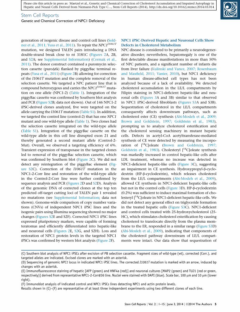
(C) Southern blot analysis of NPC1 iPSCs after excision of PB selectiotargeted alleles are indicated. Excised clones are marked with an aste(D) Sequencing of genomic NPC1 locus in indicated NPC1 iPSC lines. Tchanges with an asterisk.(E) Immunofluorescence staining of hepatic [AFP (green) and HNF4arespectively)] derived from representative NPC1-2-Corr#36 line. Nucleright).(F) Immunoblot analysis of indicated control and NPC1 iPSCs lines deResults shown in (C)–(F) are representative of at least three indepen
NPC1 iPSC-Derived Hepatic and Neuronal Cells Show
Defects in Cholesterol Metabolism
NPC disease is considered to be primarily a neurodegener-
ative disorder; nevertheless, hepatomegaly is one of the
first detectable disease manifestations in more than 50%
of NPC patients, and a significant number of infants die
from liver failure (Kulinski and Vance, 2007; Rosenbaum
and Maxfield, 2011; Vanier, 2010), but NPC1 deficiency
in human disease-affected cell types has not been
analyzed because of a lack of availability. We detected
cholesterol accumulation in the LE/L compartments by
Filipin staining in NPC1-deficient hepatic-like and neu-
ronal cells (Figures 3A and 3B) similar to that observed
in NPC1 iPSC-derived fibroblasts (Figures S3A and S3B).
Sequestration of cholesterol in the LE/L compartments
consequently affects downstream pathways, such as
cholesterol ester (CE) synthesis (Abi-Mosleh et al., 2009;
Brown and Goldstein, 1997; Goldstein et al., 1983),
prompting us to analyze cholesterol esterification and
the cholesterol sensing machinery in mutant hepatic
cells. Defects in acetyl-CoA acetyltransferase-mediated
synthesis of CE were detected by measuring the incorpo-
ration of [14C]oleate (Brown and Goldstein, 1997;
Goldstein et al., 1983). Cholesteryl [14C]oleate synthesis
was markedly increased in control hepatic-like cells after
LDL treatment, whereas no increase was detected in
NPC1-deficient hepatic-like cells (Figure 3C), suggesting
an impairment in CE synthesis. Hydroxypropyl-b-cyclo-
dextrin (HP-b-cyclodextrin), which releases cholesterol
from the LE/L compartments (Abi-Mosleh et al., 2009),
allowed CE synthesis in NPC1-deficient hepatic-like cells
but not in the control cells (Figure 3D). HP-b-cyclodextrin
(0.2%) was sufficient to induce maximal formation of cho-
lesteryl [14C]oleate in NPC1-deficient hepatic-like cells. We
did not detect any general effect on triglyceride formation
in the treated hepatic cells (Figure S3C). NPC1-deficient
and control cells treated with 25-hydroxycholesterol (25-
HC), which stimulates cholesterol esterification by causing
cholesterol to translocate directly from the plasma mem-
brane to the ER, responded in a similar range (Figure S3D)
(Abi-Mosleh et al., 2009), indicating that components of
the cholesterol pathway downstream of LE/L compart-
ments were intact. Our data show that sequestration of
n cassette. Fragment sizes of wild-type (wt), corrected (Corr.), andrisk.he corrected I1061T mutation is marked with an arrow, induced bp
(red)] and neuronal cultures [MAP2 (green) and TUJ1 (red or green,i were stained with DAPI (blue). Scale bar, 100 mm and 10 mm (lower
tecting NPC1 and actin protein levels.dent experiments using two different clones of each line.
Stem Cell Reports j Vol. 2 j 1–15 j June 3, 2014 j ª2014 The Authors 5

Figure 3. Analysis of Cholesterol Meta-bolism in Isogenic NPC1 iPSC-DerivedCell Types(A and B) Immunofluorescence staining ofrepresentative control, mutant, and cor-rected NPC1 iPSC-derived hepatic (A) andneuronal (B) cells with lineage markers[AFP, green; HNF4a, red (A); TUJI, green(B)], respectively. Endogenous cholesterolwas detected by Filipin staining (white) inthe same samples as the lineage markerstaining (A) or in duplicate samples of thesame experiment (B). Scale bar, (A) 100 mmand (B) 10 mm. Results shown are repre-sentative of at least three independentexperiments using two different clones ofeach line.(C) Cholesterol ester formation in NPC1-deficient and control hepatic-like culturesafter exposure to different concentrationsof low-density lipoprotein (LDL). Meanvariation for each of the duplicate in-cubations for control, NPC1-1, and NPC1-2were in a range between 9% and 20% for theuntreated samples and between 1%–31%for the data points at the different LDLconcentrations, respectively.(D) Cholesterol ester formation in NPC1iPSC-derived hepatic cultures after expo-sure to different concentrations of HP-b-cyclodextrin (HP-b-CD [%w/v]). Meanvariations for each of the duplicateincubations for control, NPC1-1, and NPC1-2 were in a range between 1% and 37%for the untreated samples and between0.2% and 24% for the data points at thedifferent HP-b-CD concentrations, respec-tively.
(C and D) Results shown are the mean of duplicates of each cell line and representative for three independent experiments using differentclones of indicated cell lines.(E and F) Immunoblot analysis detecting the activation of sterol regulatory element-binding protein 2 (SREBP2) cleavage in NPC1 iPSC-derived hepatic (E) and neuronal (F) cells after incubation with indicated serum concentrations. pSREBP2 (precursor SREBP2) in thecytoplasmic and nSREBP2 in the nuclear fraction of cell lysates were detected. Protein sizes are indicated. Results shown are representativeof at least three independent experiments using two different clones of each line.
Stem Cell ReportsGenetic and Chemical Correction of NPC1 Deficiency
Please cite this article in press as: Maetzel et al., Genetic and Chemical Correction of Cholesterol Accumulation and Impaired Autophagy inHepatic and Neural Cells Derived from Niemann-Pick Type C..., Stem Cell Reports (2014), http://dx.doi.org/10.1016/j.stemcr.2014.03.014
LE/L-resident cholesterol impairs cholesterol esterification
in NPC1-deficient hepatic-like cells.
Rescue of Cholesterol Defects by Treatment with HP-
b-Cyclodextrin or by Genetic Correction of the
NPC1I1061T Mutation
A block in cholesterol transport to the ER impairs tran-
scriptional regulation caused by constant activation of
proteolytic cleavage of the sterol regulatory element-
binding proteins (SREBPs) (Horton et al., 2002). Because
cleaved SREBP translocates to the nucleus and activates
6 Stem Cell Reports j Vol. 2 j 1–15 j June 3, 2014 j ª2014 The Authors
transcription, we analyzed the formation of nuclear
SREBP-2 (nSREBP-2) in NPC1-deficient hepatic cells.
Although in control cells nSREBP-2 was decreased in the
nuclear fraction in response to treatment with 10% serum,
we did not detect any changes in NPC1-deficient cells
(Figure 3E). Similar observations were made in NPC1
iPSC-derived neurons after treatment with 2% serum
(Figure 3F). In addition, increased nSREBP-2 levels in
NPC1-deficient hepatic cells lead to upregulation of
transcription of specific target genes, such as SREBPs
themselves. We detected an increase in SREBP RNA levels

Stem Cell ReportsGenetic and Chemical Correction of NPC1 Deficiency
Please cite this article in press as: Maetzel et al., Genetic and Chemical Correction of Cholesterol Accumulation and Impaired Autophagy inHepatic and Neural Cells Derived from Niemann-Pick Type C..., Stem Cell Reports (2014), http://dx.doi.org/10.1016/j.stemcr.2014.03.014
in NPC1-deficient hepatic cells after treatment with 10%
serum, whereas RNA levels in controls were downregu-
lated (Figure S3E). Upon combined treatment with 10%
serum and 0.2% HP-b-cyclodextrin, transcript levels of
SREBP and low-density lipoprotein receptor (LDL-R)
decreased in NPC1-deficient hepatic cultures and were
comparable to those in control cells after treatment with
10% serum only (Figures S3F and S3G). After correction of
the NPC1I1061T mutation, NPC iPSC-derived hepatic and
neuronal cells showed normal cholesterol distribution (Fig-
ures 3A and 3B) and responded to serum treatment by sup-
pressing SREBP2 cleavage, as evident by a reduction in
nSREBP2 protein and SREBP transcript levels (Figures 3E,
3F, and S3E). These results confirmed defects in cholesterol
sensing in NPC1 iPSC-derived hepatic and neuronal cells
that could be rescued by genetic correction of NPC1 I1061T.
Impairment of Autophagic Flux inNPC1 iPSC-Derived
Human Disease-Affected Cell Types
Emerging evidence indicates that impaired autophagy
contributes to neurodegenerative and liver disorders
(Hara et al., 2006; Komatsu, 2012; Mizushima et al.,
2008; Ravikumar et al., 2010) and is thought to be
involved in NPC1 disease (Elrick et al., 2012; Ordonez
et al., 2012; Sarkar et al., 2013). We assessed perturbation
in autophagic flux in NPC1-deficient hepatic and
neuronal cells. To monitor autophagic flux in NPC
patient-specific iPSC-derived cells, we measured the levels
of the specific autophagosome maker LC3-II (light chain 3
of microtubule-associated protein 1) and the pathway-
specific substrate, p62. Levels of LC3-II directly correlate
with the steady-state number of autophagosomes,
whereas the levels of p62 reflect autophagic turnover
(Klionsky et al. 2012, Bjørkøy et al., 2009). Both LC3-II
and p62 levels were significantly increased in NPC1-defi-
cient hepatic-like cells compared to the control cells (Fig-
ure 4A), suggesting impaired autophagic flux. However,
p62 transcript levels in control and NPC1-deficient hepat-
ic cells were comparable (Figure S4A), implying that accu-
mulation of p62 protein is due to reduced clearance rather
than diminished transcription. Similar observations were
made in NPC1 iPSC-derived neuronal cultures (Figure 4B).
We further analyzed autophagosome synthesis with a
saturating concentration of bafilomycin A1 (BafA1), which
inhibits lysosomal acidification and prevents LC3-II degra-
dation, thus causing its accumulation (Klionsky et al.,
2012). After BafA1 treatment, the accumulation of LC3-II
in NPC1-deficient hepatic-like and neuronal cells was
abrogated compared to controls (Figures 4C and 4D).
These data suggest that accumulation of autophagosomes
is not due to increased autophagosome synthesis but
rather occurs because of a block in autophagic flux in
NPC1-deficient cells. We did not find alterations in the up-
stream events regulating autophagosome synthesis, such
as the levels of Beclin 1 and Atg5-Atg12 conjugation, in
NPC1-deficient cells (Figures S4B and S4C). These data
are consistent with our recent findings related to a block
in autophagic flux seen in mouse NPC1 mutant cells
that is attributed to impaired formation of amphisomes,
resulting from defects in the SNARE machinery that regu-
lates autophagosome maturation (Sarkar et al., 2013).
Impaired SNARE functioning and autophagic flux are
also reported in other lysosomal storage disorders, such
as multiple sulphatase deficiency and mucopolysacchari-
dosis type IIIA, where LE/L-resident cholesterol accumula-
tion is observed (Fraldi et al., 2010). We were not able to
detect impaired autophagy in NPC1-deficient iPSCs, he-
patic and neuronal progenitor cells, or early neurons
(data not shown). Our data argue against previous findings
that attributed the increase in steady-state LC3-II levels in
human cells to an induction of autophagy (Ordonez et al.,
2012; Pacheco et al., 2007).
Defect in Autophagy Is Directly Linked to the
NPC1I1061T Mutation
To assess whether the defect in autophagic flux is caused
by NPC1 deficiency, we compared LC3-II and p62 levels in
hepatic and neuronal cells carrying the NPC1I1061T
mutation and their isogenic control. We found that basal
autophagy was restored in the mutant cells after genetic
correction (Figures 4E and 4F). In contrast, disruption of
the NPC1 wild-type allele in control hepatic cells, which
led to loss of NPC1 expression (Figure 2F), resulted in
marked increase of LC3-II and p62 levels, which phenocop-
ied the effects found in NPC1 iPSC-derived hepatic cells
(Figure S4D). Electronmicroscopic analysis confirmedaccu-
mulation of autophagic vacuoles and lipid droplets (LDs) in
NPC1mutant hepatic cells, a phenotype thatwas abolished
after correction of theNPC1I1061Tmutation (Figures 4G and
S4E). Thus, genetic correction of the NPC1 mutation
rescued both the cholesterol and autophagy abnormalities.
Treatment with HP-b-cyclodextrin (Vance and Peake,
2011) did not rescue the perturbation in autophagic flux.
Instead, we observed an increased block in autophagy,
which was dose dependent as evident by elevation of
LC3-II and p62 levels (Figure S5A). A lower dose of HP-
b-cyclodextrin (0.2%), sufficient to restore defects in
cholesterol metabolism as shown by induction of choles-
terol esterification (Figure 3D), did not further impair auto-
phagy in NPC1 iPSC-derived hepatic-like cells (Figure S5A).
In summary, we show a block in autophagic flux in NPC1
disease-affected human cells. This defect was abrogated in
hepatic-like and neuronal cells by genetic correction of
the NPC1I1061T mutation but not by depleting cholesterol,
thus implying that impaired autophagy is directly linked to
loss of NPC1 protein function.
Stem Cell Reports j Vol. 2 j 1–15 j June 3, 2014 j ª2014 The Authors 7

Figure 4. Genetic Correction of Autophagy Phenotype in NPC1 iPSC-Derived cells(A and B) Immunoblot analysis and quantifications of p62 and LC3-II levels using anti-p62, anti-LC3, and anti-GAPDH antibodies inhepatic (A) and neuronal cultures (B) derived from indicated control and NPC1 iPSC lines. Graphical data represent mean ± SE (n = 4).(C and D) Immunoblot analysis and quantification of LC3-II levels using anti-LC3 and anti-GAPDH antibodies in hepatic (C) and neuronalcultures (D) derived from control and NPC1 iPSC lines, treated with or without 400 nM bafilomycin A1 (BafA1) for 4 hr. Graphical datarepresent mean ± SE (n = 3).
(legend continued on next page)
8 Stem Cell Reports j Vol. 2 j 1–15 j June 3, 2014 j ª2014 The Authors
Stem Cell ReportsGenetic and Chemical Correction of NPC1 Deficiency
Please cite this article in press as: Maetzel et al., Genetic and Chemical Correction of Cholesterol Accumulation and Impaired Autophagy inHepatic and Neural Cells Derived from Niemann-Pick Type C..., Stem Cell Reports (2014), http://dx.doi.org/10.1016/j.stemcr.2014.03.014
Induction of Autophagy Rescues the Defect in Basal
Autophagy and Increases Cell Viability in NPC1-
Deficient Hepatic-like and Neuronal Cells
Upregulation of autophagy is beneficial in transgenic
models of several neurodegenerative and certain liver
disorders (Rubinsztein et al., 2012; Sarkar, 2013). Using
NPC iPSC-derived cells may allow the identification of
potent compounds that can rescue the autophagy defects
in human disease-affected cells. We found that induction
of autophagy by inhibiting the mammalian target of rapa-
mycin (mTOR) pathway with rapamycin (Rap) or starva-
tion (Ravikumar et al., 2010) significantly reduced p62
levels in NPC1 iPSC-derived hepatic cells to amounts com-
parable to the basal levels in control cells (Figures 5A and
S5B). Because long-term treatment with mTOR inhibitors
may have unfavorable side effects due to critical functions
of mTOR in growth and translation (Sarkar, 2013), we
performed a small-scale candidate drug screen testing
different concentrations ofmTOR-independent autophagy
enhancers, such as carbamazepine (CBZ), verapamil (Ver),
trehalose (Tre), and SMER28 (Figures 5B and S5C; see Sup-
plemental Information). These compounds are reported
to be protective in Drosophila or mouse models of Alz-
heimer’s, Parkinson’s, and Huntington’s disease (Rubinsz-
tein et al., 2012; Sarkar, 2013) or a-antitrypsin deficiency
(Hidvegi et al., 2010; Sarkar, 2013). Bymeasuring p62 clear-
ance, we found CBZ to be the most potent drug in rescuing
the defective autophagy phenotype in NPC1 iPSC-derived
hepatic cells (Figure 5B). CBZ, which induces mTOR-inde-
pendent autophagy by lowering inositol and IP3 levels (Sar-
kar et al., 2005), reduced p62 levels to a similar extent as
observed with rapamycin treatment (Figure 5B). Our data
suggest that CBZ-mediated restoration of autophagic flux
was sufficient to overcome the block observed in NPC1-
deficient hepatic cells, whichmay be due to a recently char-
acterized bypass mechanism (Sarkar et al., 2013).
We assessed the possibility of using CBZ in combination
with HP-b-cyclodextrin (0.2%, sufficient to induce choles-
terol ester formation without perturbing autophagy; Fig-
ures 3D and S5A) to simultaneously restore both the
cholesterol and autophagy defects. We found a significant
reduction in p62 levels after dual treatment comparable
to the effects of CBZ alone (Figure 5C), as well as a partial
rescue of the defective cholesterol metabolism as observed
by downregulation of SREBP protein, and SREBP and LDL-R
(E and F) Immunoblot analysis and quantifications of p62 and LC3-II le2 iPSC line after correction of the NPC1I1061T mutation. Graphical dat(G) Electron microscopy images of representative NPC1 iPSC-deriveindicating autophagic vacuoles. Graphical data represent mean ± SEchondria (M), Golgi (G). Scale bar, 500 nm.Results shown are representative for at least three independent exper0.01; *p < 0.05; ns, nonsignificant.
transcript levels (Figures S5D and S5E). p62 transcript levels
were not affected by the compound treatment (Figure S5F),
suggesting that the clearance of p62 protein levels is due
to increased autophagic degradation. Chemical induction
of autophagy with Rap or CBZ, or CBZ in combination
with a low dose of HP-b-cyclodextrin, significantly
increased cell viability of NPC1-deficient hepatic cells (Fig-
ures 5D and 5E).
We further evaluated the effects of autophagy-inducing
compounds using NPC1 iPSC-derived neuronal cultures.
In addition to Rap and CBZ, Tre and Ver significantly
rescued the defective autophagy phenotype in these
cells, as assessed by p62 clearance (Figures 5F and S5G).
Notably, cell viability in NPC1-deficient neuronal cultures
was significantly increased with all the chemical inducers
of autophagy alone, as well as in combination with
0.2% HP-b-cyclodextrin (Figure 5G). Although our data
suggest that induction of autophagy by itself is cyto-
protective in the context of NPC disease, combining
this treatment with a partial release of the LE/L-resident
cholesterol can restore the autophagy defects as well
as improve cholesterol homeostasis in the liver and the
brain.
DISCUSSION
In summary, our data show that patient-specific NPC1
iPSC-derived hepatic and neuronal cells are less viable
and develop disease-relevant defects, such as in cholesterol
metabolism and autophagic flux (Figure 6). In contrast to
previous reports, we do not observe similar defects in
NPC1 iPSCs or in neuronal and liver progenitor cells (Trilck
et al., 2013). Both these defects are abrogated after correc-
tion of the NPC1I1061T mutation, implying that the NPC1
protein has a functional role in both the processes. Recent
studies provide evidence that regulation of lipolysis and
autophagy are interrelated and controlled by similar
regulatory mechanisms (Singh and Cuervo, 2012). We
speculate that disturbance of this interrelation may trap
NPC1-deficient cells in a vicious cycle where inhibition of
autophagy will cause increased intracellular lipid content
(Ouimet, 2013; Sarkar et al., 2013; Singh and Cuervo,
2012; Singh et al., 2009), a condition thatmay further dete-
riorate by the release of high levels of cholesterol (Peake
vels in hepatic (E) and neuronal cultures (F) derived from the NPC1-a represent mean ± SE (n = 3).d hepatic-like cells before and after genome editing. Arrows are(n = 3). Nucleus (N), rough endoplasmatic reticulum (rER), mito-
iments using two different clones of each line. ***p < 0.001; **p <
Stem Cell Reports j Vol. 2 j 1–15 j June 3, 2014 j ª2014 The Authors 9

Figure 5. Chemical Correction of Autophagy Phenotype in NPC1 iPSC-Derived cells(A) Immunoblot analysis and quantification of p62 levels using anti-p62 and anti-GAPDH antibodies in control and NPC1 iPSC-derivedhepatic cultures, treated with either DMSO (vehicle control) or rapamycin (Rap). Graphical data represent mean ± SE (n = 3).
(legend continued on next page)
10 Stem Cell Reports j Vol. 2 j 1–15 j June 3, 2014 j ª2014 The Authors
Stem Cell ReportsGenetic and Chemical Correction of NPC1 Deficiency
Please cite this article in press as: Maetzel et al., Genetic and Chemical Correction of Cholesterol Accumulation and Impaired Autophagy inHepatic and Neural Cells Derived from Niemann-Pick Type C..., Stem Cell Reports (2014), http://dx.doi.org/10.1016/j.stemcr.2014.03.014

Figure 6. Schematic Overview of Correction of Functional Defects in NPC Disease-Affected Cell TypesLeft panel shows cholesterol distribution and autophagic flux under normal conditions. Middle panel shows the effects due to loss of NPC1protein function on cholesterol metabolism and autophagic flux. Mutations in the NPC1 gene on both alleles lead to accumulation ofcholesterol in the LE/L compartments by inhibiting its efflux, and to a block in autophagic flux causing accumulation of autophagosomesand autophagy substrate arising due to impaired formation of amphisomes. Chemical correction of disease related phenotypes areachieved by HP-b-cyclodextrin-mediated cholesterol release and CBZ-mediated autophagy induction (green arrows, right panel).Restoration of autophagic flux by autophagy inducer is possibly through a bypass mechanism.
Stem Cell ReportsGenetic and Chemical Correction of NPC1 Deficiency
Please cite this article in press as: Maetzel et al., Genetic and Chemical Correction of Cholesterol Accumulation and Impaired Autophagy inHepatic and Neural Cells Derived from Niemann-Pick Type C..., Stem Cell Reports (2014), http://dx.doi.org/10.1016/j.stemcr.2014.03.014
and Vance, 2012) or by blocking autophagy (Meske et al.,
2014; Sarkar et al., 2013). Brain and liver-specific deletion
of essential autophagy genes (such as Atg5 or Atg7) in
normal mice causes neurodegeneration and liver injury,
respectively (Hara et al., 2006; Komatsu et al., 2006,
2007), similar to those observed in NPC1 patients. These
observations further support our assumption that impair-
ment in autophagy is a crucial contributing factor for
NPC disease.
Our data argue that induction of autophagy might be
a plausible treatment option for NPC disease. Such a
strategy is supported by our recent study in NPC1 mutant
mouse cells showing that a block in autophagic flux due
to defects in amphisome formation can be bypassed by
stimulating autophagy (Sarkar et al., 2013). Induction of
autophagy has been shown to be protective in several
models of neurodegenerative and liver diseases (Hidvegi
et al., 2010; Ravikumar et al., 2010; Rubinsztein et al.,
2012; Sarkar, 2013). Because mTOR governs critical cellular
function such as translation and cell growth, small
molecule autophagy enhancers acting independently of
(B) Immunoblot analysis and quantification of p62 levels using antihepatic cultures after treatment with autophagy inducing compounds(Ver), trehelose (Tre), and SMER28. Graphical data represent mean ±(C) Immunoblot analysis and quantification of p62 levels using anti-p6treated with CBZ and HP-b-CD as indicated. Graphical data represent(D and E) FACS analysis of cell viability and apoptosis in NPC1 iPSC-dmeasuring FITC-Annexin V and propidium iodide staining. (E) Graphi(F) Immunoblot analysis and quantification of p62 levels using ancultures after treatment with autophagy inducing compounds: untreat(n = 3).(G) Analysis of cell death in NPC1 iPSC-derived 5-week-old neurons brepresent mean ± SE (n = 6).Results shown are representative for at least two independent experi0.01; *p < 0.05; ns, nonsignificant.
mTOR might be more suitable for long-term treatment
of patients. Screening mTOR-independent autophagy
inducers that are known to increase autophagic flux in
various in vitro and in vivo transgenic disease models (Sar-
kar, 2013) revealed cell type specificity, because only CBZ
could overcome the impairment in autophagy in both
NPC1 iPSC-derived hepatic and neuronal cells. These
findings underline the unique value of using human
iPSC-derived disease-relevant cells for identifying potent
compounds of biomedical relevance (Grskovic et al.,
2011). We show that higher concentrations of HP-b-cyclo-
dextrin treatment further perturb autophagic flux in NPC1
iPSC-derived hepatic cells with likely deleterious cellular
consequences. These observations are consistent with
earlier findings describing neurotoxic effects of HP-b-cyclo-
dextrin treatment in neurons from NPC1 mutant mice
(Peake and Vance, 2012).
Our data indicate that induction of autophagy itself
is beneficial, and in combination with a cholesterol-
depleting agent (such as low dose of HP-b-cyclodextrin)
that does not impact on autophagy will rescue both the
-p62 and anti-GAPDH antibodies in control and NPC1 iPSC-derived: untreated (UT), rapamycin (Rap), carbamazepine (CBZ), verapamilSE (n = 3).2 and anti-GAPDH antibodies in NPC1 iPSC-derived hepatic cultures,mean ± SE (n = 3).erived hepatic cultures after treatment with indicated compoundscal data represent mean ± SE (n = 3).ti-p62 and anti-GAPDH antibodies in NPC1 iPSC-derived neuronaled (UT), Rap, CBZ, Ver, and Tre. Graphical data represent mean ± SE
y assessing fragmented and TUNNEL positive nuclei. Graphical data
ments using two different clones of each line. ***p < 0.001; **p <
Stem Cell Reports j Vol. 2 j 1–15 j June 3, 2014 j ª2014 The Authors 11

Stem Cell ReportsGenetic and Chemical Correction of NPC1 Deficiency
Please cite this article in press as: Maetzel et al., Genetic and Chemical Correction of Cholesterol Accumulation and Impaired Autophagy inHepatic and Neural Cells Derived from Niemann-Pick Type C..., Stem Cell Reports (2014), http://dx.doi.org/10.1016/j.stemcr.2014.03.014
autophagy and cholesterol defects in NPC1 patients. Our
study suggests that inducing autophagy with CBZ may
have overall benefit for the treatment of NPC disease as it
might be effective in liver and brain.
EXPERIMENTAL PROCEDURES
HiPSC Derivation, Cultivation, and DifferentiationTransgene-free iPSCs were generated from fibroblasts of NPC
patients (Table 1) using Cre-excisable lentiviruses encoding a poly-
cistronic doxcycycline-inducible (DOX) expression cassette con-
taining all four reprogramming factors Oct4, Klf4, Sox2, c-Myc
(OKSM) (pHAGE2-TetOminiCMV-hSTEMCCA-loxP) (Sommer
and Mostoslavsky, 2010) and a modified tetracycline-controlled
trans-activator (FUW-M2rtTA-loxP) (Soldner et al., 2009). All
pluripotent cell lines have been characterized using standard
pluripotency assays (see Supplemental Experimental Procedures).
All protocols were approved by the relevant Institutional Review
Board (Massachusetts Institute of Technology) and the Embryonic
Stem Cell Research Oversight Committees (Whitehead Institute).
Differentiation of iPSCs into hepatic-like cells was induced after
single-cell replating of iPSCs on matrigel following published pro-
tocols (Si-Tayeb et al., 2010). Neuronal progenitors (NPs) and neu-
rons were derived using an embryoid body (EB)-based protocol
(Marchetto et al., 2010).
TALEN and Donor Design and GenerationTALENs were designed according to previously published princi-
ples (Cermak et al., 2011; Miller et al., 2011). All TALENs used
the +63 truncation point for fusion to the obligate heterodimeric
FokI cleavage domain (Doyon et al., 2011; Miller et al., 2011).
The tandem arrays of TALE repeats were assembled as described
previously (Marchetto et al., 2010) (see Supplemental Informa-
tion). Their editing activity was assayed using the Surveyor
nuclease (Transgenomic) (Guschin et al., 2010). The targeted
loci were PCR amplified using the following primer pairs: Cel-I-
NPC1-F (50-atgctgcctatagttctgcag-30) and Cel-I-NPC1-R (50-tcacagagactttagattctg-30) for the NPC1 locus. PCR products were then
denatured, rehybridized, digested with the Surveyor nuclease,
and analyzed. NPC1I1061T-specific TALENS pairs introducing a
DNA double-strand break in exon 21 (nt 3181C) were designed
according to previously published principles (Cermak et al.,
2011). To increase targeting efficiency, we used a donor construct
containing a puromycin selection cassette (PGK-puroDtk-pGH-
pA cassette) flanked by piggyBac terminal repeats (Yusa et al.,
2011). The 700 bp 30 homology arm introduced the ACA (Thr) to
ATA (Ile) switch correcting for the mutation. In addition, a silent
CTT to TTA (Leu) codon switch 3 bp upstream of the mutation
was introduced, generating the TTAA sequence necessary for the
piggyBac excision (Figures 2A and 2B).
TALEN-Mediated Gene Targeting and Transposon
ExcisionCells were prepared as described previously (Kondo et al., 2013;
Soldner et al., 2009). Cells (1 3 107) were electroporated with
35 mg of donor plasmid (designed and assembled by H.W.) and
12 Stem Cell Reports j Vol. 2 j 1–15 j June 3, 2014 j ª2014 The Authors
7.5 mg of each TALEN expression vector (Gene Pulser Xcell System,
Bio-Rad: 250 V, 500 mF, 0.4 cm cuvettes) and subsequently plated
on DR4 MEF feeder layers in hESC medium supplemented
with ROCK inhibitor for the first 24 hr. Puromycin selection
(0.5 mg/ml) was started 72 hr after electroporation. Individual col-
onies were picked 10–14 days after electroporation. Correctly
targeted clones were confirmed by Southern blot (AseI, EcoRI
digested) and used for transposon removal. Cells (1 3 107) were
electroporated with 10 mg of hyperactive piggyBac transposase
expression vector (pCMV-hypBase) as described previously (Sold-
ner et al., 2009). On day 4, medium was changed to hESC
medium containing 0.25 mM FIAU. Individual colonies were
picked and expanded. Genotype and deletion of the piggyBac
transposon were analyzed by Southern blot (AseI, EcoRI digest).
Additionally PCR and sequencing mutation analysis was per-
formed. Genomic DNA was amplified with primers NPC1-Wt-F
(50-cctttgattatacatgaaaccag-30), NPC1-I1061T-Mut-F (50-gaagaaagcccgacttac-30), NPC1-I1061T-Corr-F (50-gaagaaagcccgattaat-30), re-
spectively, andNPC1-R (50-gagcattccacagcattctg-30) under standardPCR conditions. PCR products were sequenced (Applied Bio-
systems Model 3730 capillary DNA sequencer with Big Dye Termi-
nator Cycle Sequencing Kit).
Prediction of NPC1 TALENs Off-Target SitesThe position weight matrices (PWMs) of NPC1 TALEN pairs (see
Supplemental Information) were constructed using model 3 as
described previously (Moscou and Bogdanove, 2009) (see Supple-
mental Information).
ImmunostainingImmunostaining was performed according to standard protocols
using primary antibodies listed in the Supplemental Information.
Cholesterol was detected by Filipin staining (50 mg/ml Filipin com-
plex, Streptomyces filipinensis; Sigma-Aldrich).
Acyl-CoA Acetyltransferase AssayDefects in cholesterol metabolism were assessed using the Acyl-
CoA Acetyltransferase Assay. The rate of incorporation of
[14C]oleate into cholesterol [14C]oleate and [14C]triglycerides by
intact cell monolayers was measured as previously described
(Goldstein et al., 1983).
Immunoblot AnalysisCell pellets were lysed on ice in Lysis Buffer (10 mM Tris-HCl
[pH 7.4], 2% SDS, 1 mM DTT, 10% glycerol, and 120 mg/ml
urea) for 30 min in presence of Complete EDTA-free Protease
Inhibitor Cocktail (Roche Diagnostics) and subjected to SDS-
PAGE and standard immunoblot analysis. Primary antibodies
used are listed in the Supplemental Information. Amersham ECL
Western Blotting Detection Reagent (GE Healthcare) was used for
visualization.
Chemical CompoundsAutophagic flux in NPC1 iPSC-derived cells was assessed using
a saturating concentration of bafilomycin A1 (BafA1) clamping
LC3-II/autophagosome degradation (Klionsky et al., 2012). To

Stem Cell ReportsGenetic and Chemical Correction of NPC1 Deficiency
Please cite this article in press as: Maetzel et al., Genetic and Chemical Correction of Cholesterol Accumulation and Impaired Autophagy inHepatic and Neural Cells Derived from Niemann-Pick Type C..., Stem Cell Reports (2014), http://dx.doi.org/10.1016/j.stemcr.2014.03.014
test the possibility of inducing autophagy, we performed a
candidate drug screen including described mTOR-independent
autophagy inducers (Sarkar, 2013) (Figure S5G; Supplemental In-
formation). Their effect was analyzed by measuring p62 levels
and cell viability by FITC-Annexin V/ PI staining. Compounds
used for treatment of hepatic-like cells were bafilomycin A1
(Enzo Life Sciences), rapamycin (LC Laboratories), carbamazepine,
verapamil, trehalose, SMER28, and HP-b-cyclodextrin (all Sigma-
Aldrich). Cells were treated with compounds as indicated.
Statistical AnalysesDensitometry analyses on the immunoblots were done by ImageJ
software (NIH) by measuring levels of the protein of interest rela-
tive to the loading control, as previously described (Williams
et al., 2008). The control condition was set to 100%, and the
data were represented as mean ± SEM. The y axis values are shown
in percentages (%). Experiments were performed in triplicates at
least twice. The p values for densitometry analyses, vesicle number,
and aggregate formation were determined by Student’s t test
(unpaired) using Prism 6 software (GraphPad), as previously
described (Korolchuk et al., 2009). ***p < 0.001; **p < 0.01; *p <
0.05; ns, nonsignificant.
ACCESSION NUMBERS
The NCBI SRA accession number for the CMV analysis reported in
this paper is SRP026624.
SUPPLEMENTAL INFORMATION
Supplemental Information includes Supplemental Experimental
Procedures, five figures, and one table and can be found with
this article online at http://dx.doi.org/10.1016/j.stemcr.2014.
03.014.
AUTHOR CONTRIBUTIONS
D.M., S.S., H.W., and R.J. designed the experiments and wrote the
paper. D.M. and H.W. designed and performed the TALEN-medi-
ated gene correction experiments. D.M. and S.S. designed and per-
formed the autophagy experiments. L.A.-M. performed the ACAT
assay. M.M. and P.X. provided technical assistance. A.W.C. per-
formed computational analysis for TALEN specificity and for
TALEN off-target cutting sides. Q.G. analyzed teratomas. All other
experiments were performed by D.M.
ACKNOWLEDGMENTS
We thank J.L. Goldstein, M.S. Brown, and D.F. Voytas for their
helpful advice; R. Alagappan, T. Lungjangwa, K. Ganz, R. Flannery,
D. Fu, T. DiCesare, T. Volkert, N. Watson, and W. Salmon for tech-
nical assistance. We thank Keck Microscopy Facility; Whitehead
Technology Core; G. Bell of Whitehead Bioinformatics & Research
Computing. We thank all Jaenisch lab members for helpful discus-
sion. D.M. was a Peter G. Pentchev Research Fellow of the NNPD
Foundation. S.S. is a Former Fellow at Hughes Hall, University of
Cambridge, UK. R.J. was supported by US NIH grant R01-
CA084198 and Skoltech Center; J.L. Goldstein and M.S. Brown
were supported by the US NIH grant HL20948. R.J. is an advisor
to Stemgent and Fate Therapeutics.
Received: January 3, 2014
Revised: March 25, 2014
Accepted: March 31, 2014
Published: May 15, 2014
REFERENCES
Abi-Mosleh, L., Infante, R.E., Radhakrishnan, A., Goldstein, J.L.,
and Brown, M.S. (2009). Cyclodextrin overcomes deficient
lysosome-to-endoplasmic reticulum transport of cholesterol in
Niemann-Pick type C cells. Proc. Natl. Acad. Sci. USA 106,
19316–19321.
Bjørkøy, G., Lamark, T., Pankiv, S., Øvervatn, A., Brech, A., and
Johansen, T. (2009). Monitoring autophagic degradation of p62/
SQSTM1. Methods Enzymol. 452, 181–197.
Brown,M.S., and Goldstein, J.L. (1997). The SREBP pathway: regu-
lation of cholesterol metabolism by proteolysis of a membrane-
bound transcription factor. Cell 89, 331–340.
Carstea, E.D., Morris, J.A., Coleman, K.G., Loftus, S.K., Zhang, D.,
Cummings, C., Gu, J., Rosenfeld, M.A., Pavan, W.J., Krizman,
D.B., et al. (1997). Niemann-Pick C1 disease gene: homology to
mediators of cholesterol homeostasis. Science 277, 228–231.
Cermak, T., Doyle, E.L., Christian, M., Wang, L., Zhang, Y.,
Schmidt, C., Baller, J.A., Somia, N.V., Bogdanove, A.J., and Voytas,
D.F. (2011). Efficient design and assembly of custom TALEN and
other TAL effector-based constructs for DNA targeting. Nucleic
Acids Res. 39, e82.
Doyon, Y., Vo, T.D., Mendel, M.C., Greenberg, S.G., Wang, J., Xia,
D.F., Miller, J.C., Urnov, F.D., Gregory, P.D., and Holmes, M.C.
(2011). Enhancing zinc-finger-nuclease activity with improved
obligate heterodimeric architectures. Nat. Methods 8, 74–79.
Elrick, M.J., Yu, T., Chung, C., and Lieberman, A.P. (2012).
Impaired proteolysis underlies autophagic dysfunction in Nie-
mann-Pick type C disease. Hum. Mol. Genet. 21, 4876–4887.
Fraldi, A., Annunziata, F., Lombardi, A., Kaiser, H.J., Medina, D.L.,
Spampanato, C., Fedele, A.O., Polishchuk, R., Sorrentino, N.C.,
Simons, K., and Ballabio, A. (2010). Lysosomal fusion and SNARE
function are impaired by cholesterol accumulation in lysosomal
storage disorders. EMBO J. 29, 3607–3620.
Gelsthorpe, M.E., Baumann, N., Millard, E., Gale, S.E., Langmade,
S.J., Schaffer, J.E., and Ory, D.S. (2008). Niemann-Pick type C1
I1061T mutant encodes a functional protein that is selected for
endoplasmic reticulum-associated degradation due to protein mis-
folding. J. Biol. Chem. 283, 8229–8236.
Goldstein, J.L., Basu, S.K., and Brown, M.S. (1983). Receptor-medi-
ated endocytosis of low-density lipoprotein in cultured cells.
Methods Enzymol. 98, 241–260.
Grskovic, M., Javaherian, A., Strulovici, B., and Daley, G.Q. (2011).
Induced pluripotent stem cells—opportunities for disease model-
ling and drug discovery. Nat. Rev. Drug Discov. 10, 915–929.
Guschin,D.Y.,Waite, A.J., Katibah, G.E.,Miller, J.C., Holmes,M.C.,
and Rebar, E.J. (2010). A rapid and general assay for monitoring
endogenous gene modification. Methods Mol. Biol. 649, 247–256.
Stem Cell Reports j Vol. 2 j 1–15 j June 3, 2014 j ª2014 The Authors 13

Stem Cell ReportsGenetic and Chemical Correction of NPC1 Deficiency
Please cite this article in press as: Maetzel et al., Genetic and Chemical Correction of Cholesterol Accumulation and Impaired Autophagy inHepatic and Neural Cells Derived from Niemann-Pick Type C..., Stem Cell Reports (2014), http://dx.doi.org/10.1016/j.stemcr.2014.03.014
Hara, T., Nakamura, K., Matsui, M., Yamamoto, A., Nakahara, Y.,
Suzuki-Migishima, R., Yokoyama, M., Mishima, K., Saito, I.,
Okano, H., and Mizushima, N. (2006). Suppression of basal auto-
phagy in neural cells causes neurodegenerative disease in mice.
Nature 441, 885–889.
Hidvegi, T., Ewing, M., Hale, P., Dippold, C., Beckett, C., Kemp, C.,
Maurice, N.,Mukherjee, A., Goldbach, C.,Watkins, S., et al. (2010).
An autophagy-enhancing drug promotes degradation of mutant
alpha1-antitrypsin Z and reduces hepatic fibrosis. Science 329,
229–232.
Horton, J.D., Goldstein, J.L., and Brown, M.S. (2002). SREBPs: acti-
vators of the complete program of cholesterol and fatty acid syn-
thesis in the liver. J. Clin. Invest. 109, 1125–1131.
Klionsky, D.J., Abdalla, F.C., Abeliovich, H., Abraham, R.T.,
Acevedo-Arozena, A., Adeli, K., Agholme, L., Agnello, M., Agosti-
nis, P., Aguirre-Ghiso, J.A., et al. (2012). Guidelines for the use
and interpretation of assays for monitoring autophagy. Autophagy
8, 445–544.
Komatsu, M. (2012). Liver autophagy: physiology and pathology.
J. Biochem. 152, 5–15.
Komatsu, M., Waguri, S., Chiba, T., Murata, S., Iwata, J., Tanida, I.,
Ueno, T., Koike, M., Uchiyama, Y., Kominami, E., and Tanaka, K.
(2006). Loss of autophagy in the central nervous system causes
neurodegeneration in mice. Nature 441, 880–884.
Komatsu, M., Waguri, S., Koike, M., Sou, Y.S., Ueno, T., Hara, T.,
Mizushima, N., Iwata, J., Ezaki, J., Murata, S., et al. (2007). Homeo-
static levels of p62 control cytoplasmic inclusion body formation
in autophagy-deficient mice. Cell 131, 1149–1163.
Kondo, T., Asai, M., Tsukita, K., Kutoku, Y., Ohsawa, Y., Sunada, Y.,
Imamura, K., Egawa, N., Yahata, N., Okita, K., et al. (2013).
Modeling Alzheimer’s disease with iPSCs reveals stress phenotypes
associated with intracellular Ab and differential drug responsive-
ness. Cell Stem Cell 12, 487–496.
Korolchuk, V.I., Mansilla, A., Menzies, F.M., and Rubinsztein, D.C.
(2009). Autophagy inhibition compromises degradation of ubiqui-
tin-proteasome pathway substrates. Mol. Cell 33, 517–527.
Kulinski, A., and Vance, J.E. (2007). Lipid homeostasis and lipopro-
tein secretion in Niemann-Pick C1-deficient hepatocytes. J. Biol.
Chem. 282, 1627–1637.
Marchetto,M.C., Carromeu, C., Acab, A., Yu, D., Yeo, G.W., Mu, Y.,
Chen, G., Gage, F.H., and Muotri, A.R. (2010). A model for neural
development and treatment of Rett syndrome using human
induced pluripotent stem cells. Cell 143, 527–539.
Meske, V., Erz, J., Priesnitz, T., and Ohm, T.G. (2014). The autopha-
gic defect in Niemann-Pick disease type C neurons differs from
somatic cells and reduces neuronal viability. Neurobiol. Dis. 64,
88–97.
Millard, E.E., Gale, S.E., Dudley, N., Zhang, J., Schaffer, J.E., and
Ory, D.S. (2005). The sterol-sensing domain of the Niemann-Pick
C1 (NPC1) protein regulates trafficking of low density lipoprotein
cholesterol. J. Biol. Chem. 280, 28581–28590.
Miller, J.C., Tan, S., Qiao, G., Barlow, K.A., Wang, J., Xia, D.F.,
Meng, X., Paschon, D.E., Leung, E., Hinkley, S.J., et al. (2011). A
TALE nuclease architecture for efficient genome editing. Nat.
Biotechnol. 29, 143–148.
14 Stem Cell Reports j Vol. 2 j 1–15 j June 3, 2014 j ª2014 The Authors
Mizushima,N., Levine, B., Cuervo, A.M., and Klionsky, D.J. (2008).
Autophagy fights disease through cellular self-digestion. Nature
451, 1069–1075.
Moscou, M.J., and Bogdanove, A.J. (2009). A simple cipher governs
DNA recognition by TAL effectors. Science 326, 1501.
Ordonez, M.P., Roberts, E.A., Kidwell, C.U., Yuan, S.H., Plaisted,
W.C., and Goldstein, L.S. (2012). Disruption and therapeutic
rescue of autophagy in a human neuronal model of Niemann
Pick type C1. Hum. Mol. Genet. 21, 2651–2662.
Ouimet, M. (2013). Autophagy in obesity and atherosclerosis: In-
terrelationships between cholesterol homeostasis, lipoprotein
metabolism and autophagy in macrophages and other systems.
Biochim. Biophys. Acta 1831, 1124–1133.
Pacheco, C.D., Kunkel, R., and Lieberman, A.P. (2007). Autophagy
inNiemann-PickCdisease is dependent uponBeclin-1 and respon-
sive to lipid trafficking defects. Hum. Mol. Genet. 16, 1495–1503.
Peake, K.B., and Vance, J.E. (2012). Normalization of cholesterol
homeostasis by 2-hydroxypropyl-b-cyclodextrin in neurons and
glia from Niemann-Pick C1 (NPC1)-deficient mice. J. Biol. Chem.
287, 9290–9298.
Ravikumar, B., Sarkar, S., Davies, J.E., Futter, M., Garcia-Arencibia,
M., Green-Thompson, Z.W., Jimenez-Sanchez, M., Korolchuk, V.I.,
Lichtenberg, M., Luo, S., et al. (2010). Regulation of mammalian
autophagy in physiology and pathophysiology. Physiol. Rev. 90,
1383–1435.
Rosenbaum, A.I., and Maxfield, F.R. (2011). Niemann-Pick type C
disease: molecular mechanisms and potential therapeutic ap-
proaches. J. Neurochem. 116, 789–795.
Rubinsztein, D.C., Codogno, P., and Levine, B. (2012). Autophagy
modulation as a potential therapeutic target for diverse diseases.
Nat. Rev. Drug Discov. 11, 709–730.
Saha, K., and Jaenisch, R. (2009). Technical challenges in using
human induced pluripotent stem cells to model disease. Cell
Stem Cell 5, 584–595.
Sarkar, S. (2013). Regulation of autophagy by mTOR-dependent
and mTOR-independent pathways: autophagy dysfunction in
neurodegenerative diseases and therapeutic application of auto-
phagy enhancers. Biochem. Soc. Trans. 41, 1103–1130.
Sarkar, S., Floto, R.A., Berger, Z., Imarisio, S., Cordenier, A., Pasco,
M., Cook, L.J., and Rubinsztein, D.C. (2005). Lithium induces
autophagy by inhibiting inositol monophosphatase. J. Cell Biol.
170, 1101–1111.
Sarkar, S., Carroll, B., Buganim, Y., Maetzel, D., Ng, A.H., Cassady,
J.P., Cohen, M.A., Chakraborty, S., Wang, H., Spooner, E., et al.
(2013). Impaired autophagy in the lipid-storage disorder Nie-
mann-Pick type C1 disease. Cell Rep. 5, 1302–1315.
Si-Tayeb, K., Noto, F.K., Nagaoka, M., Li, J., Battle, M.A., Duris, C.,
North, P.E., Dalton, S., and Duncan, S.A. (2010). Highly efficient
generation of human hepatocyte-like cells from induced pluripo-
tent stem cells. Hepatology 51, 297–305.
Singh, R., and Cuervo, A.M. (2012). Lipophagy: connecting auto-
phagy and lipid metabolism. Int. J. Cell Biol. 2012, 282041.
Singh, R., Kaushik, S., Wang, Y., Xiang, Y., Novak, I., Komatsu, M.,
Tanaka, K., Cuervo, A.M., and Czaja, M.J. (2009). Autophagy regu-
lates lipid metabolism. Nature 458, 1131–1135.

Stem Cell ReportsGenetic and Chemical Correction of NPC1 Deficiency
Please cite this article in press as: Maetzel et al., Genetic and Chemical Correction of Cholesterol Accumulation and Impaired Autophagy inHepatic and Neural Cells Derived from Niemann-Pick Type C..., Stem Cell Reports (2014), http://dx.doi.org/10.1016/j.stemcr.2014.03.014
Soldner, F., and Jaenisch, R. (2012). Medicine. iPSC disease
modeling. Science 338, 1155–1156.
Soldner, F., Hockemeyer, D., Beard, C., Gao, Q., Bell, G.W., Cook,
E.G., Hargus, G., Blak, A., Cooper, O., Mitalipova, M., et al.
(2009). Parkinson’s disease patient-derived induced pluripotent
stem cells free of viral reprogramming factors. Cell 136, 964–977.
Soldner, F., Laganiere, J., Cheng, A.W., Hockemeyer, D., Gao, Q.,
Alagappan, R., Khurana, V., Golbe, L.I., Myers, R.H., Lindquist,
S., et al. (2011). Generation of isogenic pluripotent stem cells
differing exclusively at two early onset Parkinson pointmutations.
Cell 146, 318–331.
Sommer, C.A., and Mostoslavsky, G. (2010). Experimental
approaches for the generation of induced pluripotent stem cells.
Stem Cell Res. Ther. 1, 26.
Trilck,M., Hubner, R., Seibler, P., Klein, C., Rolfs, A., and Frech,M.J.
(2013). Niemann-Pick type C1 patient-specific induced pluripo-
tent stem cells display disease specific hallmarks. Orphanet J.
Rare Dis. 8, 144.
Vance, J.E., and Peake, K.B. (2011). Function of the Niemann-Pick
type C proteins and their bypass by cyclodextrin. Curr. Opin.
Lipidol. 22, 204–209.
Vanier,M.T. (2010). Niemann-Pick disease typeC.Orphanet J. Rare
Dis. 5, 16.
Williams, A., Sarkar, S., Cuddon, P., Ttofi, E.K., Saiki, S., Siddiqi,
F.H., Jahreiss, L., Fleming, A., Pask, D., Goldsmith, P., et al.
(2008). Novel targets for Huntington’s disease in an mTOR-inde-
pendent autophagy pathway. Nat. Chem. Biol. 4, 295–305.
Xie, C., Turley, S.D., Pentchev, P.G., and Dietschy, J.M. (1999).
Cholesterol balance and metabolism in mice with loss of
function of Niemann-Pick C protein. Am. J. Physiol. 276, E336–
E344.
Yusa, K., Rashid, S.T., Strick-Marchand, H., Varela, I., Liu, P.Q.,
Paschon, D.E., Miranda, E., Ordonez, A., Hannan, N.R., Rou-
hani, F.J., et al. (2011). Targeted gene correction of a1-antitryp-
sin deficiency in induced pluripotent stem cells. Nature 478,
391–394.
Stem Cell Reports j Vol. 2 j 1–15 j June 3, 2014 j ª2014 The Authors 15