Embed Size (px)
Citation preview
Hepatic morphology: variations and its clinical importance
ORIGINAL ARTICLE Eur. J. Anat. 22 (3): 195-201 (2018)
Justin Chin, Patrick O’Toole, Jun Lin, Sumathilatha S. Velavan
Department of Clinical Anatomy and Embryology, Touro College of Osteopathic Medicine, New York, USA
SUMMARY
Emergent technologies and advances in the fields of diagnostic radiology and gastroenterology have created a need to better understand the mor-phological features of the liver. Variations in these features are a potential source for diagnostic er-rors, which can lead to costly follow-up testing and detrimental health outcomes. In the present study, the morphological features of human cadaveric liver specimens were evaluated via macroscopic examination and measurements to asses for varia-tions in accessory fissures/sulci, accessory lobes, and the pons hepatis. The study was conducted on 33 specimens obtained from cadavers utilized for routine dissection for first year medical students in the 2016-2017 academic year in the Department of Clinical Anatomy and Embryology at the Touro College of Osteopathic Medicine. Out of 33 speci-mens, 12 were considered normal without any ac-cessory fissures, lobes, or presence of a pons hepatis. 21 livers had 1 or more morphological var-iations, which included but were not limited to: mul-tiple accessory fissures, Riedel’s lobe, and varying degrees of pons hepatis. The study aims to throw greater light to the field of hepatic morphology and its variations.
Key words: Liver – Hepatic variation – Hepatic morphology – Riedel’s lobe – Pons hepatis – Ac-cessory lobe – Fissure – Sulci INTRODUCTION
The liver is the largest viscera in the abdominal cavity as it occupies the right hypochondriac, epi-gastric, and left hypochondriac regions. Under non-pathological conditions, the liver has a homoge-nous parenchyma and is divided into 4 anatomical lobes by peritoneal and ligamentous attachments (Patil et al., 2014). Age, body size, and sex con-tribute to the vast variations in liver size and weight, with adult livers weighing approximately 2% of the total body weight (Vinnakota and Jayasree, 2013).
Divisions of the liver in functional anatomy are based on Couinaud’s classification, utilizing an imaginary plane and hepatic vasculature distribu-tion to divide the liver into 8 segments (Couinaud, 1957; Joshi, et al. 2009; Patil et al., 2014; Vinnako-ta and Jayasree, 2013). While segmental liver anatomy research receives the greatest attention, there are also studies that focus on common/rare morphological variants. With increasing depend-ence on radiological imaging for disease diagnosis and laparoscopic procedures, knowledge of com-mon anatomical surface variations of the liver is critical for the best patient outcomes (Rumack et al., 2016; Sato et al., 1998). Furthermore, although most hepatic variants are quiescent, there have been documented cases of clinical manifestations caused by variant morphology (Vinnakota and Jayasree, 2013; Glenisson et al., 2014; Kudo, 2000; Akbulut et al., 2011; Fitzgerald et al., 1993).
The aim of this study was to examine common gross surface variations of liver and review the literature on its clinical impact and implications.
MATERIALS AND METHODS
Dissections were performed on 33 cadavers in
195
Submitted: 1 September, 2017. Accepted: 13 February, 2018.
Corresponding author: Justin Chin. Touro College of Osteo-
pathic Medicine, Department of Clinical Anatomy and Embryo-
logy, 230 West 125th Street, 3rd Floor, 10027 New York, USA
E-mail: [email protected]
Hepatic morphology variations
196
the gross anatomy lab at Touro College of Osteo-pathic Medicine. Age range of the cadavers was 54 to 96 years old with a gender distribution of 9 males and 24 females, with the presence of Riedel’s lobe being a gender specific variation seen on initial examination. All the cadavers were of Caucasian origin, with no associated gross pathological changes or surgical scars present in the cadavers.
Utilizing standard dissection methods (Detton and Tank, 2012), the coronary, falciform, and trian-gular ligaments of the liver were detached at their attachments to the liver. Surrounding connective tissue and hepatic nerve plexus were removed to clear the dissection plane. The inferior vena cava was cut at its entry to and exit from the liver, with care taken to preserve the pons hepatis if present. Gross measurements of liver size and weight were taken to ensure that all specimens adhered to a standard average adult liver size. The gallbladder, biliary system, and hepatic vasculature were dis-sected away from the surrounding liver.
The lobes of the liver —right, left, caudate, and quadrate— were studied in detail and photo-graphed, with attention paid to size, shape, acces-sory fissures, and accessory lobes.
RESULTS
In the present study of the 33 liver specimens, the average liver size was approximately 15.5 cm in width and the average weight was 1.1 kg. Even distribution of liver variations was present across genders with the exception of Riedel’s lobe (Table 1). No gross anatomical variations were noted in the gallbladder, biliary system, and surrounding hepatic vasculature. 12 livers were observed with normal surfaces, fissures, and borders without any additional accessory fissures or malformations (36%). Of the remaining 21 specimens, hepatic variations were documented and broadly grouped as having accessory fissures, accessory lobes, and/or the presence of a pons hepatis (hepatic bridge or ‘pont hepatique’). In several of the speci-mens, multiple anatomical variations were docu-mented (i.e. having an accessory lobe and pons hepatis).
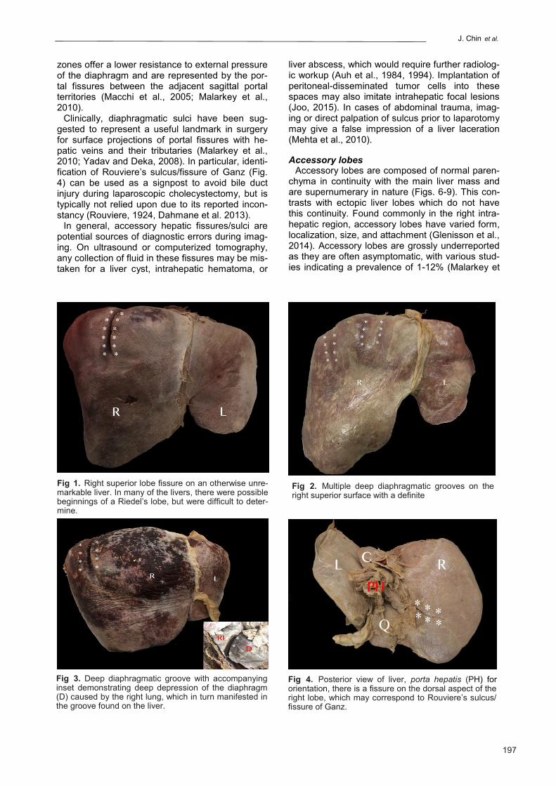
Accessory sulci/fissures were present in 9 livers (27%), with 7 having fissures on the superior sur-
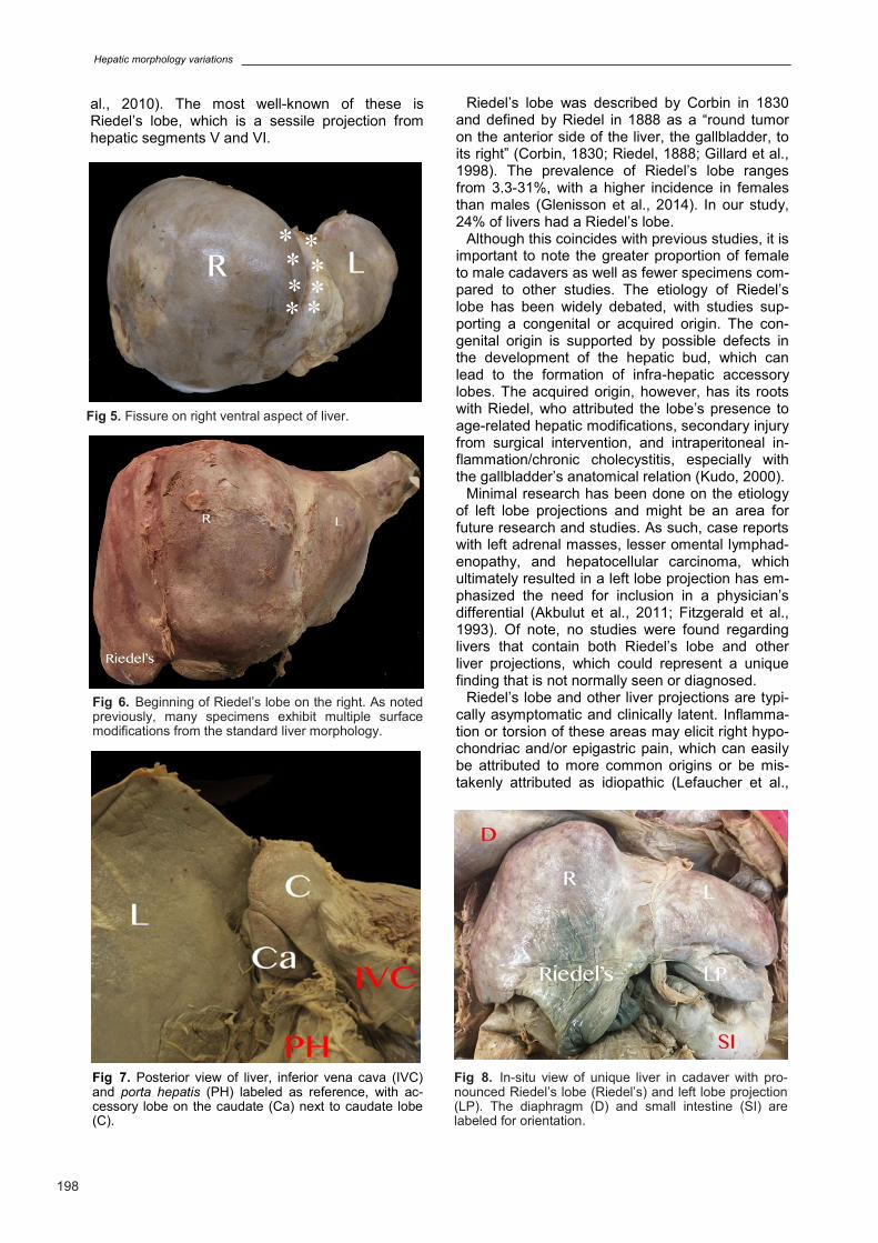
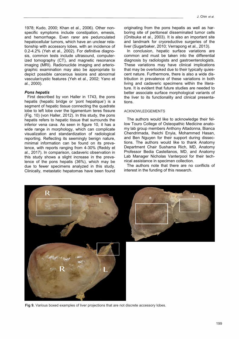
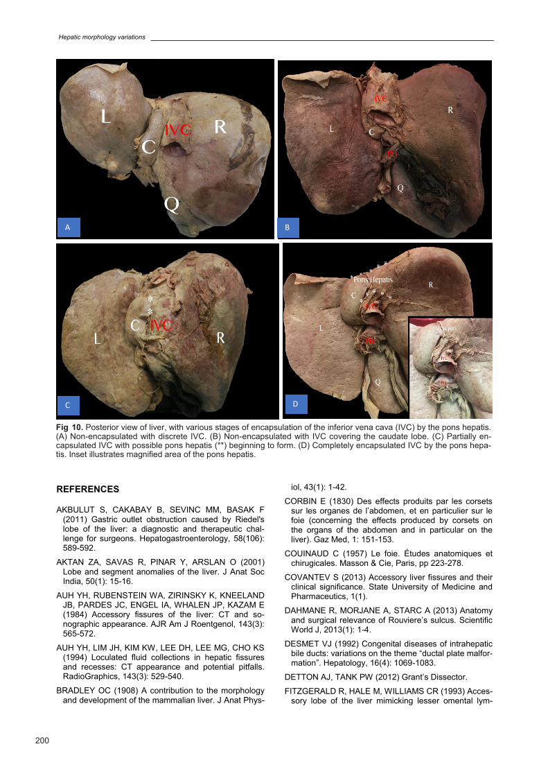
face of the right lobe (Fig. 1). Of the superior sulci, 4 appeared to be deep diaphragmatic grooves while 3 had multiple fissures (Figs. 2 and 3). One liver was noted to have fissures on the right ventral lobe surface while another had a fissure on the right dorsal lobe surface (Figs. 4 and 5). Accessory lobes were identified on 8 specimens (24%), with 6 indicating the presence or establishment of Riedel’s lobe and 2 livers having miniature acces-sory lobes on the caudate (Figs. 6 and 7). One liver had a prominent Riedel’s lobe extending infe-riorly as well as a left liver lobe projection (Fig. 8). Other various liver projections were also docu-mented on 4 livers, but were not representative of true accessory lobes (Fig. 9). Pons hepatis with variable levels of inferior vena cava encapsulation were seen in 12 specimens (36%) (Fig. 10). DISCUSSION
External morphology of the liver is highly varied,
creating a wide array of presentations on physical examination, radiologic imaging, and post-mortem cadaveric studies (Bradley, 1908; Kudo, 1918; Loth, 1931; Thomson, 1985; Sato et al., 1998; Joshi et al., 2009; Patil et al., 2014). These varia-tions are broadly defined as acquired versus con-genital malformations, each with their own clinical presentation and impact (Ruge, 1907; Joshi et al., 2009; Covantev, 2013). Congenital liver defects that affect the external morphology are largely ra-re, but tend to have a more predictable clinical presentation as they often impact the biliary sys-tem (Sato et al., 1998; Aktan et al., 2001). Ac-quired liver defects will also affect the external morphology, but their clinical effects are typically quiescent unless there are inciting stimuli such as torsion, trauma, or tumors (Aktan et al., 2001; Co-vantev, 2013). Accessory hepatic fissures/sulci, accessory lobes, and the pons hepatis are some of the most common hepatic variations that are most likely to be seen on clinical examination (Vinnakota and Jayasree, 2013; Sato et al., 1998).
Accessory hepatic fissures/sulci
Various studies have described diaphragmatic sulci, which is the primary hepatic sulci that can be found in 40% of all liver observations (Figs. 1-5) (Thomson, 1899; Kanchan et al., 2014; Lim et al., 1987). In comparison, only 27% of the livers in this study contained a measurable fissure or sulci, which could be attributed to the small sample size. Traditionally, it was understood that diaphragmatic sulci resulted from hypertrophic diaphragm muscle bands, which created variable resistances and thus promoted uneven hepatic parenchymal growth (Kanchan et al., 2014). Recent radiological and corrosion cast studies, however, have also attributed the formation of sulci to the existence of weakened zones of hepatic parenchyma. These
Table 1 Liver variations across genders.
Classification Male Female Total
Normal 6 6 12
Accessory Fissures 2 7 9
Accessory Lobes 2 6 8
Pons Hepatis 1 11 12
J. Chin et al.
197
zones offer a lower resistance to external pressure of the diaphragm and are represented by the por-tal fissures between the adjacent sagittal portal territories (Macchi et al., 2005; Malarkey et al., 2010).
Clinically, diaphragmatic sulci have been sug-gested to represent a useful landmark in surgery for surface projections of portal fissures with he-patic veins and their tributaries (Malarkey et al., 2010; Yadav and Deka, 2008). In particular, identi-fication of Rouviere’s sulcus/fissure of Ganz (Fig. 4) can be used as a signpost to avoid bile duct injury during laparoscopic cholecystectomy, but is typically not relied upon due to its reported incon-stancy (Rouviere, 1924, Dahmane et al. 2013).
In general, accessory hepatic fissures/sulci are potential sources of diagnostic errors during imag-ing. On ultrasound or computerized tomography, any collection of fluid in these fissures may be mis-taken for a liver cyst, intrahepatic hematoma, or
liver abscess, which would require further radiolog-ic workup (Auh et al., 1984, 1994). Implantation of peritoneal-disseminated tumor cells into these spaces may also imitate intrahepatic focal lesions (Joo, 2015). In cases of abdominal trauma, imag-ing or direct palpation of sulcus prior to laparotomy may give a false impression of a liver laceration (Mehta et al., 2010).
Accessory lobes
Accessory lobes are composed of normal paren-chyma in continuity with the main liver mass and are supernumerary in nature (Figs. 6-9). This con-trasts with ectopic liver lobes which do not have this continuity. Found commonly in the right intra-hepatic region, accessory lobes have varied form, localization, size, and attachment (Glenisson et al., 2014). Accessory lobes are grossly underreported as they are often asymptomatic, with various stud-ies indicating a prevalence of 1-12% (Malarkey et
Fig 1. Right superior lobe fissure on an otherwise unre-markable liver. In many of the livers, there were possible beginnings of a Riedel’s lobe, but were difficult to deter-mine.
Fig 2. Multiple deep diaphragmatic grooves on the right superior surface with a definite
Fig 3. Deep diaphragmatic groove with accompanying inset demonstrating deep depression of the diaphragm (D) caused by the right lung, which in turn manifested in the groove found on the liver.
Fig 4. Posterior view of liver, porta hepatis (PH) for orientation, there is a fissure on the dorsal aspect of the right lobe, which may correspond to Rouviere’s sulcus/fissure of Ganz.
Hepatic morphology variations
198
al., 2010). The most well-known of these is Riedel’s lobe, which is a sessile projection from hepatic segments V and VI.
Riedel’s lobe was described by Corbin in 1830 and defined by Riedel in 1888 as a “round tumor on the anterior side of the liver, the gallbladder, to its right” (Corbin, 1830; Riedel, 1888; Gillard et al., 1998). The prevalence of Riedel’s lobe ranges from 3.3-31%, with a higher incidence in females than males (Glenisson et al., 2014). In our study, 24% of livers had a Riedel’s lobe.
Although this coincides with previous studies, it is important to note the greater proportion of female to male cadavers as well as fewer specimens com-pared to other studies. The etiology of Riedel’s lobe has been widely debated, with studies sup-porting a congenital or acquired origin. The con-genital origin is supported by possible defects in the development of the hepatic bud, which can lead to the formation of infra-hepatic accessory lobes. The acquired origin, however, has its roots with Riedel, who attributed the lobe’s presence to age-related hepatic modifications, secondary injury from surgical intervention, and intraperitoneal in-flammation/chronic cholecystitis, especially with the gallbladder’s anatomical relation (Kudo, 2000).
Minimal research has been done on the etiology of left lobe projections and might be an area for future research and studies. As such, case reports with left adrenal masses, lesser omental lymphad-enopathy, and hepatocellular carcinoma, which ultimately resulted in a left lobe projection has em-phasized the need for inclusion in a physician’s differential (Akbulut et al., 2011; Fitzgerald et al., 1993). Of note, no studies were found regarding livers that contain both Riedel’s lobe and other liver projections, which could represent a unique finding that is not normally seen or diagnosed.
Riedel’s lobe and other liver projections are typi-cally asymptomatic and clinically latent. Inflamma-tion or torsion of these areas may elicit right hypo-chondriac and/or epigastric pain, which can easily be attributed to more common origins or be mis-takenly attributed as idiopathic (Lefaucher et al.,
Fig 5. Fissure on right ventral aspect of liver.
Fig 6. Beginning of Riedel’s lobe on the right. As noted previously, many specimens exhibit multiple surface modifications from the standard liver morphology.
Fig 7. Posterior view of liver, inferior vena cava (IVC) and porta hepatis (PH) labeled as reference, with ac-cessory lobe on the caudate (Ca) next to caudate lobe (C).
Fig 8. In-situ view of unique liver in cadaver with pro-nounced Riedel’s lobe (Riedel’s) and left lobe projection (LP). The diaphragm (D) and small intestine (SI) are labeled for orientation.
J. Chin et al.
199
1978; Kudo, 2000; Khan et al., 2006). Other non-specific symptoms include constipation, emesis, and hemorrhage. Even rarer are pedunculated hepatocellular tumors, which have an unclear rela-tionship with accessory lobes, with an incidence of 0.2-4.2% (Yeh et al., 2002). For definitive diagno-sis, common tests include ultrasound, computer-ized tomography (CT), and magnetic resonance imaging (MRI). Radionuclide imaging and arterio-graphic examination may also be appropriate to depict possible cancerous lesions and abnormal vascular/cystic features (Yeh et al., 2002; Yano et al., 2000).
Pons hepatis
First described by von Haller in 1743, the pons hepatis (hepatic bridge or ‘pont hepatique’) is a segment of hepatic tissue connecting the quadrate lobe to left lobe over the ligamentum teres fissure (Fig. 10) (von Haller, 2012). In this study, the pons hepatis refers to hepatic tissue that surrounds the inferior vena cava. As seen in figure 10, it has a wide range in morphology, which can complicate visualization and standardization of radiological reporting. Reflecting its seemingly benign nature, minimal information can be found on its preva-lence, with reports ranging from 4-30% (Reddy et al., 2017). In comparison, cadaveric observation in this study shows a slight increase in the preva-lence of the pons hepatis (36%), which may be due to fewer specimens analyzed in this study. Clinically, metastatic hepatomas have been found
originating from the pons hepatis as well as har-boring site of peritoneal disseminated tumor cells (Onitsuka et al., 2003). It is also an important site and landmark for cryoreductive surgeries of the liver (Sugarbaker, 2010; Verrapong et al., 2013).
In conclusion, hepatic surface variations are common and must be taken into the differential diagnosis by radiologists and gastroenterologists. These variations may have clinical implications that may be overlooked due to their typically quies-cent nature. Furthermore, there is also a wide dis-tribution in prevalence of these variations in both living and cadaveric specimens within the litera-ture. It is evident that future studies are needed to better associate surface morphological variants of the liver to its functionality and clinical presenta-tions.
ACKNOWLEDGEMENTS
The authors would like to acknowledge their fel-low Touro College of Osteopathic Medicine anato-my lab group members Anthony Altadonna, Bianca Chendrimada, Iheichi Enyia, Mohammed Hasan, and Ben Nguyen for their support during dissec-tions. The authors would like to thank Anatomy Department Chair Sushama Rich, MD, Anatomy Professor Bedia Castellanos, MD, and Anatomy Lab Manager Nicholas Vanterpool for their tech-nical assistance in specimen collection.
The authors note that there are no conflicts of interest in the funding of this research.
Fig 9. Various boxed examples of liver projections that are not discrete accessory lobes.
Hepatic morphology variations
200
REFERENCES AKBULUT S, CAKABAY B, SEVINC MM, BASAK F
(2011) Gastric outlet obstruction caused by Riedel's lobe of the liver: a diagnostic and therapeutic chal-lenge for surgeons. Hepatogastroenterology, 58(106): 589-592.
AKTAN ZA, SAVAS R, PINAR Y, ARSLAN O (2001) Lobe and segment anomalies of the liver. J Anat Soc India, 50(1): 15-16.
AUH YH, RUBENSTEIN WA, ZIRINSKY K, KNEELAND JB, PARDES JC, ENGEL IA, WHALEN JP, KAZAM E (1984) Accessory fissures of the liver: CT and so-nographic appearance. AJR Am J Roentgenol, 143(3): 565-572.
AUH YH, LIM JH, KIM KW, LEE DH, LEE MG, CHO KS (1994) Loculated fluid collections in hepatic fissures and recesses: CT appearance and potential pitfalls. RadioGraphics, 143(3): 529-540.
BRADLEY OC (1908) A contribution to the morphology and development of the mammalian liver. J Anat Phys-
iol, 43(1): 1-42.
CORBIN E (1830) Des effects produits par les corsets sur les organes de l’abdomen, et en particulier sur le foie (concerning the effects produced by corsets on the organs of the abdomen and in particular on the liver). Gaz Med, 1: 151-153.
COUINAUD C (1957) Le foie. Études anatomiques et chirugicales. Masson & Cie, Paris, pp 223-278.
COVANTEV S (2013) Accessory liver fissures and their clinical significance. State University of Medicine and Pharmaceutics, 1(1).
DAHMANE R, MORJANE A, STARC A (2013) Anatomy and surgical relevance of Rouviere’s sulcus. Scientific World J, 2013(1): 1-4.
DESMET VJ (1992) Congenital diseases of intrahepatic bile ducts: variations on the theme “ductal plate malfor-mation”. Hepatology, 16(4): 1069-1083.
DETTON AJ, TANK PW (2012) Grant’s Dissector.
FITZGERALD R, HALE M, WILLIAMS CR (1993) Acces-sory lobe of the liver mimicking lesser omental lym-
A B
C D
Fig 10. Posterior view of liver, with various stages of encapsulation of the inferior vena cava (IVC) by the pons hepatis. (A) Non-encapsulated with discrete IVC. (B) Non-encapsulated with IVC covering the caudate lobe. (C) Partially en-capsulated IVC with possible pons hepatis (**) beginning to form. (D) Completely encapsulated IVC by the pons hepa-tis. Inset illustrates magnified area of the pons hepatis.

J. Chin et al.
201
phadenopathy. Br J Radiol, 66(789): 839-841.
GILLARD JH, PATEL MC, ABRAHAMS PH, DIXON AK (1998) Riedel’s lobe of the liver: fact or fiction? Clin Anat, 11(1): 47-49.
GLENISSON M, SALLOUM C, LIM C, LACAZE L, MA-LEK A, ENRIQUEZ A, COMPAGNON P, LAURENT A, AZOULAY D (2014) Accessory liver lobes: anatomical description and clinical implications. J Visc Surg, 151(6): 451-455.
JOO I (2015) The role of intraoperative ultrasonography in the diagnosis and management of focal hepatic lesions. Ultrasonography, 34(4): 246-257.
JOSHI SD, JOSHI SS, ATHAVALE SA (2009) Some interesting observations on the surface features of the liver and their clinical implications. Singapore Med J, 50(7): 715-719.
KANCHAN T, ACHARYA J, NAIK R (2014) Grooves on the hepatic surface. J Forensic Leg Med, 21(1): 24-25.
KHAN AM, HUNDAL R, MANZOOR K, DHUPER S, KORSTEN MA (2006) Accessory liver lobes: A diag-nostic and therapeutic challenge of their torsions. Scand J Gastroenterol, 41(2): 125-130.
KUDO M (2000) Riedel’s lobe of the liver and its clinical implication. Intern Med, 39(2): 87-88.
KUDO T (1918) Die Leber der Japaner. Acta Scholae Med, Univ. Kyoto 2: 459-469.
LEFAUCHER C, DUPUIS E, MULLER J, LAHAM A (1978) Torsion of riedel’s lobe two cases. J Chir, 115: 25.
LIM JH, KO YT, HAN MC, KIM CW, CHOI BI, IM JG (1987) The inferior accessory hepatic fissure: so-nographic appearance. AJR Am J Roentgenol, 149(3): 495-497.
LOTH E (1931) Anthropologie des partes molles. Mas-son & Cie, Paris, pp 297-331.
MACCHI V, PORZIONATO A, PARENTI A, MACCHI C, NEWELL R, DE CARO R (2005) Main accessory sul-cus of the liver. Clin Anat, 18(1): 39-45.
MALARKEY DE, JOHNSON K, RYAN L, BOORMAN G, MARONPOT RR (2010) New insights into functional aspects of liver morphology. Toxicol Pathol, 33(1): 27-34.
MEHTA V, ARORA J, MANIK P, SURI RK, RATH G (2010) Clinico-anatomical aspects of accessory fis-sures obscuring the normal hepatic morphology. Clin Ter, 161(3): 259-260.
ONITSUKA A, KATAGIRI Y, MIYAUCHI T, SHIMAMO-TO T, MIMOTO H, OZEKI Y (2003) Metastatic hepato-ma originating from the pons hepatis presenting extra-hepatic growth-- classification of different patterns covering REX’s recessus. Hepatogastroenterology, 50(49): 235-237.
PATIL S, SETHI M, KAKAR S (2014) Morphological study of human liver and its importance. Int J Anat Res, 2(2): 310-314.
REDDY N, JOSHI SS, MITTAL PS, JOSHI SD (2017) Morphology of caudate and quadrate lobes of liver. J Evolution Med Dentl Sci, 6(11): 897-901.
RIEDEL BMKL (1888) Ueber den Zungenförmigen fortsatz des rechten Leberlappens und seine pathog-nostische Bedeutung für die Erkrankung der Gallen-blase nebst Bemerkungen über Gallensteinopera-tionen. Berliner klinische Wochenschrift, 25: 577-581, 602-607.
ROUVIERE (1924) Sur la configuration et la signification du sillon du processus caudé. Bulletin et Memoires de la Societé Anatomique de Paris, 94: 335-358.
RUGE G (1907) Die äußeren formverhältnisse der Leber bei den Primaten. Morph Jahr, 37.
RUMACK CM, WILSON S, WITHERS C (2016) Diag-nostic Ultrasound. In: The Liver. (please complete with editor/s, editorial, place of edition and pages)
SATO S, WATANABE M, NAGASAWA S, NIIGAKI M, SAKAI S, AKAGI S (1998) Laparoscopic observations of congenital anomalies of the liver. Gastrointest En-dosc, 47(2): 136-140.
SUGARBAKER PH (2010) Pont hepatique (hepatic bridge), an important anatomic structure in cytoreduc-tive surgery. J Surg Oncol, 101(3): 251-252.
THOMSON A (1885) Some variations in the anatomy of the human liver. J Anat Physiol, 19(3): 303-306.
THOMSON A (1899) The morphological significance of certain fissures in the human liver. J Anat Physiol, 33(4): 546-564.
VERRAPONG J, SOLOMON H, HELM CW (2013) Divi-sion of the pont hepatique of the liver in cytoreductive surgery for peritoneal malignancy. Gynecol Oncol, 128(1): 133.
VINNAKOTA S, JAYASREE N (2013) A new insight into the morphology of the human liver: a cadaveric study. ISRN Anatomy, 1(1): 1-6.
VON HALLER A (2012) Iconum Anatomicarum Quibus Praecipuae Partes Corporis Humani Exquisita Cura Delineatae Continentur Fasciculus.
YADAV GD, DEKA P (2008) Accessory sulcus of the liver – an incidental laparotomy finding. Indian J Surg, 70(2): 92-93.
YANO K, OHTSUBO M, MIZOTA T, KATO H, HAYASHIDA Y, MORITA S, FURUKAWA R, HAYAKAWA A (2000) Riedel’s lobe of the liver evalu-ated by multiple imaging modalities. Internal Med, 39(2): 136-138.
YEH CN, LEE WC, JENG LB, CHEN M (2002) Pedun-culated hepatocellular carcinoma: clinicopathologic study of 18 surgically resected cases. World J Surg, 26(9): 1133-1138.