Embed Size (px)
Citation preview

Herpes Simplex Virus 1 Ubiquitin-Specific Protease UL36 InhibitsBeta Interferon Production by Deubiquitinating TRAF3
Shuai Wang,a,b Kezhen Wang,a,b Jie Li,a Chunfu Zhenga,b
Institutes of Biology and Medical Sciences, Soochow University, Suzhou, Chinaa; Molecular Virology and Viral Immunology Research Group, Wuhan Institute of Virology,Chinese Academy of Sciences, Wuhan, Chinab
Interferon (IFN)-mediated innate immune defense is a potent antiviral mechanism. Viruses evade innate immunity and limitsecretion of beta interferon (IFN-�) to replicate and survive in the host. The largest tegument protein of herpes simplex virus 1(HSV-1), UL36, contains a novel deubiquitinase (DUB) motif embedded in its N terminus, denoted UL36 ubiquitin-specific pro-tease (UL36USP). In the present study, we demonstrate that HSV-1 UL36USP inhibits Sendai virus (SeV)-induced interferonregulatory factor 3 (IRF3) dimerization, promoter activation, and transcription of IFN-�. The DUB activity of UL36USP is es-sential to block IFN-� production. UL36USP also inhibited IFN-� promoter activity induced by overexpression of the N termi-nus of RIG-I (RIG-IN) and MAVS, but not TBK-1, I�B kinase � (IKK�), and IRF3/5D. UL36USP was subsequently shown to deu-biquitinate TRAF3 and prevent the recruitment of the downstream adaptor TBK1. The recombinant HSV-1 lacking UL36USPDUB activity was generated. Cells infected with the mutant virus produced more IFN-� than wild-type (WT) HSV-1-infectedcells. These findings demonstrate HSV-1 UL36USP removes polyubiquitin chains on TRAF3 and counteracts the IFN-� pathway.
Innate immunity is the first line of host defense against pathogeninvasion. The type I interferon (IFN-I) system plays a crucial
role for vertebrates in controlling viral infections. Pattern recog-nition receptors (PRRs) of the host cells mediate the innate recog-nition of viruses and initiate a series of signaling cascades, activat-ing the transcription factors NF-�B and interferon regulatoryfactors 3 and 7 (IRF3 and IRF7). The activated NF-�B and IRF3/7collaborate to trigger the expression of IFN-I, which upregulates adiverse set of interferon-stimulated genes (ISGs) and protects hostcells against the invading virus (1–4).
Ubiquitination is a widely used posttranslational protein mod-ification that regulates many physiological processes, includingimmune responses (5, 6). Ubiquitination has a crucial role in reg-ulating the RIG-I signaling pathway. It is reported that ubiquitin(Ub) ligase tripartite motif-containing protein 25 (TRIM25) andRNF135 catalyze K63-linked polyubiquitination of RIG-I, andthis enhances the binding of RIG-I to MAVS (IPS-1/Cardiff/VISA) (7, 8). Downstream of RIG-I, K63-linked polyubiquitina-tion of TRAF3 recruits the kinases TBK1 and I�B kinase ε (IKKε),leading to IRF3 phosphorylation and subsequent IFN-I produc-tion (9–13). TRIM56 stimulates K63-linked polyubiquitination ofSTING, helping to recruit TBK1 (14, 15). TBK1 and IKKε alsoundergo polyubiquitination, which has been suggested to pro-mote IRF3 activation (16, 17). Furthermore, TRIM23 is involvedin polyubiquitination of NEMO, enhancing beta interferon(IFN-�) production (18).
Herpes simplex virus 1 (HSV-1) is the archetypal member ofthe Alphaherpesvirinae subfamily, with a large, linear double-stranded DNA (dsDNA) virus genome of about 152 kb. HSV-1 isan extremely successful human pathogen and has evolved multi-ple immune evasion strategies that allow it to exist for the lifetimeof its host. For example, HSV-1 ICP0 targets IRF3 and blocks IFNproduction (19–22). Previous studies from our lab have demon-strated that varicella-zoster virus (VZV) immediate early proteinopen reading frame 61 (ORF61), the homologue of HSV-1 ICP0,antagonizes the IFN-� pathway by degradation of activated IRF3(23), and HSV-1 US11 serves as a novel antagonist of the IFN-�
pathway via direct binding to RIG-I and MDA-5 (24). HSV-1ICP34.5 binds and sequesters TBK-1 to inhibit IFN production(25, 26). The virion host shutoff (vhs) protein of HSV-2 sup-presses IFN and ISG induction by degrading cellular mRNA (27,28). ICP27 was also suggested to inhibit IFN production, andHSV-1 lacking functional ICP27 induces higher levels of IFN-�and IFN-� in macrophages than wild-type (WT) virus does (29).HSV-1 US3 is suggested to play an important role in immuneevasion during HSV-1 infection, and US3 null HSV-1 resulted instrong activation of IRF3 and IFN-I responses (30).
The largest tegument protein of HSV-1, VP1/2, the product ofthe UL36 gene, is essential for HSV-1 replication and is conservedacross the Herpesviridae family. VP1/2, a large multifunctionalprotein, plays crucial roles in HSV-1 entry, capsid transport, andvirion assembly, formation of mature virions, microtubule trans-port of capsids, neuroinvasion, pathogenesis, etc. (31–41). Kat-tenhorn et al. have identified an approximately 500-amino-acidpeptide that exhibits unique deubiquitinase (DUB) activity (de-noted as UL36USP, for UL36 ubiquitin-specific protease), whichis embedded within the N-terminal region of HSV-1 VP1/2 (42).UL36USP is detectable as early as 12 h postinfection and only aftercleavage of UL36USP from full-length UL36. HSV-1 UL36USP ishighly specific for ubiquitin and cleaves K48- and K63-linkedpolyubiquitin chains but not ubiquitin-like proteins, such asSUMO 1, Nedd8, or ISG15 (38, 42–44). A purified UL36USP ex-pressed in Escherichia coli has also been shown to specifically bindto ubiquitin and cleave ubiquitin-based substrates. UL36USPcontains the core catalytic residues, including C65 (in HSV), thatare required for its deubiquitinase activity (42).
Received 3 May 2013 Accepted 20 August 2013
Published ahead of print 28 August 2013
Address correspondence to Chunfu Zheng, [email protected].
Copyright © 2013, American Society for Microbiology. All Rights Reserved.
doi:10.1128/JVI.01211-13
November 2013 Volume 87 Number 21 Journal of Virology p. 11851–11860 jvi.asm.org 11851

Homologues of HSV-1 UL36USP have been confirmed in sev-eral other members of herpesviruses, including pseudorabies virus(PRV) (45), Marek’s disease virus (46), human cytomegalovirus(HCMV) (44, 47), murine cytomegalovirus (MCMV) (48), sim-ian cytomegalovirus (47), Epstein-Barr virus (EBV) (38), Kaposi’ssarcoma-associated herpesvirus (KSHV), (49), and mouse her-pesvirus strain 68 (MHV68) (50). Although the precise role ofUL36USP remains unclear, its strict conservation through all Herpes-viridae subfamilies suggests an important role during viral infection.
Ubiquitination has a crucial role in regulating the innate im-mune response. Thus, it is not surprising that viruses have evolvedstrategies to manipulate the Ub ligation process to thwart hostdefense mechanisms (51, 52). It was recently reported that EBV-encoded BPLF1 deubiquitinates TRAF6 to inhibit NF-�B signal-ing during lytic infection, leading to promotion of viral lytic DNAreplication (53). However, the mechanism by which HSV-1UL36USP is involved in immune evasion is still poorly under-stood. In this study, we demonstrate that ectopic expression ofUL36USP significantly downregulates Sendai virus (SeV)-acti-vated IFN-� promoter activity and that the deubiquitinase activityof UL36USP is indispensable for the inhibitory activity. Addition-ally, UL36USP is demonstrated to cleave both the K63- and K48-linked polyubiquitin chains of TRAF3 and abrogate TRAF3 me-diation of IFN-� production. Finally, a recombinant HSV-1lacking deubiquitinase activity, denoted C40A HSV-1, was gener-ated, and infection by mutant HSV-1 resulted in higher produc-tion of IFN-�. These findings reveal a novel mechanism for HSV-1to evade the host’s antiviral immunity.
MATERIALS AND METHODSCells, viruses, and antibodies. HEK293T cells, Vero cells, and HeLa cellswere grown in Dulbecco’s modified Eagle medium (DMEM) (Gibco-BRL) supplemented with 10% fetal bovine serum (FBS) and 100 U/ml ofpenicillin and streptomycin. The WT HSV-1 F strain and its derivativeUL36USP mutant HSV-1 strain were propagated in Vero cells and titratedas described previously (54). Sendai virus (SeV) was propagated andtitrated as previously described (24). The protease inhibitor cocktail mix-ture was purchased from CST (Boston, MA). Mouse anti-Myc, anti-Flag,and antihemagglutinin (anti-HA) monoclonal antibodies (MAbs) werepurchased from ABmart (Shanghai, China). Mouse monoclonal IgG1 andIgG2b isotype control antibodies were purchased from eBioscience, Inc.(San Diego, CA). Rabbit anti-TRAF3 polyclonal antibody (pAb), rabbitanti-Ub pAb, and mouse anti-�-actin MAb were purchased from SantaCruz Biotechnology (Santa Cruz, CA). Rabbit antibody against IRF3-S396was previously described (23).
Plasmid construction. All enzymes used for cloning procedures, ex-cept for T4 DNA ligase (New England BioLabs, MA), were purchasedfrom TaKaRa (Dalian, China). To construct UL36USP-Flag, the N-termi-nal 500-amino-acid (aa) region of UL36 was amplified from plasmidUL36-EYFP (expressing enhanced yellow fluorescent protein [EYFP]) aspreviously described (55) and cloned into the HindIII and EcoRI sites ofthe pCMV-Flag vector. Commercial reporter plasmids include NF-�B-Luc (expressing luciferase (Luc) (Stratagene, La Jolla, CA) and pRL-TKplasmid (expressing thymidine kinase [TK]) (Promega). Gift plasmidsinclude the following: (PRDIII-I)4-Luc (56), pcDNA3.1-FlagTBK1 andpcDNA3.1/Zeo-MAVS (57), pcDNA3.1-FlagIKKε (58), pEF-Flag-RIG-IN (59), IRF3/5D (60), pCAGGS-NS1 (61), and IFN-� promoterreporter plasmid p125-luc (62).
RNA isolation, semiquantitative RT-PCR, and quantification of gelimage. Total RNA was extracted from HEK293T cells with TRIzol (Invit-rogen, CA) according to the manufacturer’s manual. Samples were di-gested with DNase I and subjected to reverse transcription (RT) (63). ThecDNA was used as a template for semiquantitative PCR to investigate the
expression patterns of human IFN-�, ISG54, and ISG56. The details of theprotocols have been described previously (23).
Transfection and dual luciferase reporter (DLR) assay. HEK293Tcells were plated on 24-well dishes (Corning, NY) in DMEM (Gibco-BRL,MD) with 10% FBS at a density of 1 � 105 cells per well overnight beforetransfection, as previously described (23). Cells were then cotransfectedwith 1 �g expression plasmid, 500 ng reporter plasmid, such as thoseexpressing IFN-�-Luc, NF-�B-Luc, or (PRDIII-I)4-Luc, and 50 ng ofpRL-TK Renilla luciferase reporter plasmid to normalize transfection ef-ficiency, as indicated by standard calcium phosphate precipitation (64,65). At 24 h posttransfection, cells were infected with SeV (100 hem-agglutination units [HAU]/ml) for 16 h, and then luciferase assayswere performed as previously described (23) with a luciferase assay kit(Promega, Madison, WI). Poly(I · C) and poly(dA-dT) were purchasedfrom InvivoGen.
Co-IP and WB analysis. Coimmunoprecipitation (co-IP) assays wereperformed as previously described (54). Briefly, HEK293T cells(�5 �106) were cotransfected with 10 �g of the indicated expressionplasmids. Transfected cells were harvested at 24 h posttransfection andlysed on ice with 500 �l of lysis buffer. The lysates were incubated with theindicated antibodies and 30 �l of a 1:1 slurry of protein A/G PLUS-agarose(Santa Cruz Biotechnology, Santa Cruz, CA) overnight at 4°C. The beadswere washed four times with 1 ml of lysis buffer containing 500 mM NaCl,and Western blot (WB) analysis was performed to detect the interaction ofproteins. The co-IP assays were repeated twice; a typical blot is shown.
WB analysis was performed as previously described (54). Briefly, theprotein samples were subjected to 10% SDS-PAGE and transferred topolyvinylidene difluoride (PVDF) or nitrocellulose membranes, followedby blocking with 5% nonfat milk in Tris-buffered saline-Tween (TBST)and probed with the indicated primary antibodies at 37°C for 2 h. Afterbeing washed with TBST, the membrane was incubated with alkalinephosphatase (AP)-conjugated goat anti-rabbit IgG or goat anti-mouseIgG. Protein bands specific to the antibody were developed by 5-bromo-4-chloro-3-indolylphosphate (BCIP)-nitroblue tetrazolium (NBT) andterminated by distilled water.
Native PAGE. Native PAGE was carried out using ReadyGels (7.5%;Bio-Rad). The gel was prerun with 25 mM Tris and 192 mM glycine (pH8.4), with 1% deoxycholate (DOC) in the cathode chamber for 30 min at40 mA. Samples in native sample buffer (10 �g protein, 62.5 mM Tris-Cl[pH 6.8], 15% glycerol, 1% DOC) were size fractionated by electropho-resis for 60 min at 25 mA and transferred to nitrocellulose membranes forWB analysis as previously described (66).
Recombinant virus construction. Two-step Red-mediated recombi-nation was applied to construct UL36(C40A) HSV-1 (67). The Kanr cas-sette was amplified by PCR with a pair of primers containing 40-bp-homology flanking sequence of mutant sites. Then the PCR product wastransformed into E. coli GS1783 competent cells carrying the pHSV-1bacterial artificial chromosome (BAC) via electroporation. PCR assayswere used to identify the positive clones. L-Arabinose was used to inducethe second Red recombination to delete the Kanr cassette (see Fig. 5). Toanalyze the integrity of the BAC clones, 15 �l of BAC DNA was digestedwith HindIII or BamHI, and the restriction pattern of BAC DNA wascompared to that of WT BAC (data not shown). The recombinant viruseswere validated by PCR and sequencing using primers upstream or down-stream of the UL36 mutant sites (data not shown). Then viruses wereharvested, and the growth kinetics of the recombinant viruses were char-acterized by both traditional plaque assay and luciferase activity assay inVero cells at a multiplicity of infection (MOI) of 0.1 or 1. The luciferaseactivity assay was performed with a luciferase assay kit (Promega, Madi-son, WI).
ELISA for IFN-�. An enzyme-linked immunosorbent assay (ELISA)to quantify secreted IFN-� was carried out with culture supernatants col-lected from infected cells as previously described (24). Briefly, cell culturemedium was collected and centrifuged to remove cell debris. A humanIFN-� ELISA kit (PBL InterferonSource, Piscataway, NJ) was used to
Wang et al.
11852 jvi.asm.org Journal of Virology

detect the IFN-� according to the manufacturer’s instructions. Four-week-old female C57BL/6 mice were purchased from the ExperimentalAnimal Center, Wuhan Institute of Virology, Chinese Academy of Sci-ences. The mice were injected with 106 PFU of the indicated virus. After 24h, the mice were sacrificed, and a Legend Max mouse IFN-� ELISA kit(BioLegend, San Diego, CA) was used to detect the IFN-� in serum. Theanimal study proposal was approved by the Institutional Animal Care andUse Committee (IACUC) of the Experimental Animal Center, WuhanInstitute of Virology, Chinese Academy of Sciences. The approved proto-col no. is IACUC2013012.
RESULTSUL36USP inhibits the SeV-mediated activation of IFN-� andinterferon-stimulated response element promoter activity. Toinvestigate the function of UL36USP in the regulation of virus-mediated activation of IFN-� promoter, UL36USP was coex-pressed in HEK293T cells in the presence of IFN-� reporter, andthe pRL-TK Renilla luciferase reporter plasmid. DLR assaysshowed that Sendai virus (SeV) infection led to an �170-foldinduction of the IFN-�-Luc reporter activity. Ectopic expressionof UL36USP significantly suppressed SeV-mediated activation ofthe IFN-� promoter activity (Fig. 1A). The NS1 served as a posi-tive control. Furthermore, UL36USP also inhibited the poly(I ·C)-induced IFN-� promoter activity (Fig. 1B). It has been re-ported that the core catalytic residues, including C65 (in HSV-1),are important for the deubiquitinase activity of UL36USP, and themutation of Cys65 to Ala abolishes its deubiquitinase activity (42).However, in the HSV-1 F strain, we found that residue Cys40 isrequired for the DUB activity of UL36USP. In Fig. 1C, ubiquiti-nation level of total protein was significantly reduced by transfec-tion of UL36USP but not the C40A mutant. In order to determinewhether the DUB activity of UL36USP is required for the inhibi-tion of IFN production, C40A mutants were generated. DLR as-says showed that ectopic expression of C40A did not affect theSeV-mediated activation of the IFN-� promoter activity (Fig. 1D).
IRF3 is a key transcription factor in the IFN-� signaling path-way, and its dimerization is a hallmark of the early activation of theantiviral response. Native PAGE assays were performed to exam-ine whether UL36USP inhibited IRF3 dimerization. As shown inFig. 1E, IRF3 existed as a monomer in mock-treated cells, and SeVinfection induced the dimerization of IRF3. However, the pres-ence of UL36USP inhibited the SeV-mediated IRF3 dimer for-mation. Ectopic expression of C40A failed to inhibit dimeriza-tion of IRF3.
The IFN-I induces a diverse set of IFN-stimulated genes (ISGs)to mediate the innate antiviral response. Whether UL36USP in-hibited SeV-induced transcription of ISG54 and ISG56 was de-tected by semiquantitative PCR. Expression of wild-typeUL36USP significantly inhibited SeV-induced ISG54 and ISG56mRNA expression, whereas the presence of the C40A mutant didnot affect the expression of ISG54 and ISG56 (Fig. 1F). Takentogether, these results indicate that UL36USP was sufficient toinhibit SeV-mediated activation of IFN-� activities and that theDUB activity of UL36USP was required for its inhibitory activity.
UL36 inhibits the IFN-� signaling pathway at the level be-tween MAVS and TBK1. The transcription of the IFN-� generequired transcription factor IRF3 and NF-�B binding to distinctregulatory domains in the IFN-� promoter. To investigatewhether UL36USP inhibited the SeV-induced activation of IRF3and NF-�B, reporter gene assays were performed in HEK293 cellsusing the luciferase reporter plasmid driven by the tandem IRF
binding sites PRD(III-I) and the NF-�B regulatory element fromthe IFN-� promoter. The result showed that SeV infection in-duces strong IRF-responsive PRD(III-I) and NF-�B promoter ac-tivities, and transfection of UL36USP remarkably inhibited bothSeV-induced PRD(III-I) (Fig. 2A) and NF-�B (Fig. 2B) reporteractivities.
To determine at what level in the pathway UL36USP blockedIFN-� production, UL36USP, and expression plasmids of RIG-Isignaling pathway components, including the active CARD do-main-containing form of RIG-I (RIG-IN), MAVS, IKKε kinase,TBK1 kinase, or the active form of IRF3 (IRF3/5D), were cotrans-fected into HEK293T cells. All expression constructs resulted in a110- to 800-fold induction of the IFN-�-Luc reporter activity (Fig.3A to E). IFN-� promoter activation driven by RIG-IN or MAVSwas inhibited about 90% by UL36USP (Fig. 3A and B). WhereasUL36USP could not significantly suppress the IFN-� promoteractivation driven by TBK1, IKKε, or IRF3/5D (Fig. 3C to E). Theseresults suggested that UL36USP inhibited the IFN antiviral re-sponse at the level between MAVS and TBK1.
UL36USP deubiquitinates TRAF3 and inhibits recruitmentof TBK1. In the RLR-mediated signaling pathway, TRAF3 linksupstream IFN signaling responses of IPS-1 (MAVS/Cardiff/VISA)to TBK1. The K63-linked polyubiquitination of TRAF3 is crucialfor signaling by MAVS and recruitment of the kinases TBK1 andIKKε (9, 11). Thus, we investigated the possibility that UL36USPcleaved the polyubiquitin chain of TRAF3. In the co-IP experi-ment, TRAF3-Flag, Ub-HA, and UL36USP or the C40A expres-sion plasmid were cotransfected into HEK293T cells. FollowingSeV infection, TRAF3 was immunoprecipitated by anti-Flag anti-body, and the Western blot assay was performed to detect theubiquitination of TRAF3. As shown in Fig. 4A, the presence ofUL36USP reduced the SeV-induced ubiquitination of TRAF3,whereas the C40A mutant did not affect the ubiquitination level ofTRAF3. Then co-IP assays were carried out to investigate whetherUL36USP removed the K63- or K48-linked polyubiquitin chain ofTRAF3. TRAF3-Flag was transfected with the linkage-specificubiquitin HA-K63-Ub or HA-K48-Ub expression plasmid intoHEK293T cells. The results showed that UL36USP could inhibitboth K48- and K63-linked ubiquitination of TRAF3 (Fig. 4B).
We further investigated whether deubiquitinating activity ofUL36USP impaired the interaction between TRAF3 and TBK1. Asshown in Fig. 4C, more TBK1 was coimmunoprecipitated byTRAF3 after SeV stimulation than that shown in the lane withoutSeV infection. Cotransfection with UL36USP, but not the C40Amutant, inhibited the SeV-induced recruitment of TBK1 byTRAF3. Taken together, the results indicated that UL36USP deu-biquitinated TRAF3 and abrogated the recruitment of TBK1.
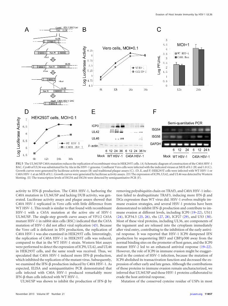
UL36 C40A mutant virus induced a larger amount of IFN-�than WT virus. To investigate the physiological functions ofUL36USP in context with HSV-1 infection, the UL36 C40A pointmutant, denoted C40A HSV-1, was generated using our HSV-1BAC system as previously described (Fig. 5A) (68). RecombinantHSV-1 BAC contains a firefly luciferase cassette, and the luciferaseactivity could be easily quantified in vitro. Luciferase activity as-says were used to determine the replication kinetics of C40AHSV-1 and WT HSV-1. When Vero cells were infected with thecorresponding viruses at an MOI of 0.1, mutation of the key cys-teine residue of UL36USP did not affect the viral replication (Fig.5B). Traditional plaque assays were also performed at an MOI of1.0 in Vero cells, and the results correlated with the luciferase
Evasion of Host Innate Immunity by HSV-1 UL36
November 2013 Volume 87 Number 21 jvi.asm.org 11853

activity (Fig. 5C). Because Vero cells are deficient in IFN produc-tion, we examined the replication of C40A HSV-1 in HEK293Tcells to further study the function of UL36USP. Interestingly,the luciferase activity assay showed that the replication ofC40A HSV-1 was remarkably impaired compared with that ofWT HSV-1 (Fig. 5D). ICP0, UL42, and UL46 expression inC40A HSV-1-infected HEK293T cells was also reduced (Fig. 5E),
confirming the reduced replication of C40A HSV-1. Semiquanti-tative PCR was performed to detect the transcription of ISG54 andISG56 in C40A HSV-1-infected HEK293 cells. The results showedthat a remarkably higher level of transcription of ISG54 and ISG56was observed in C40A HSV-1-infected HEK293T cells at 12 and 24h postinfection (Fig. 5F), implying that the replication of C40AHSV-1 was suppressed by the higher level of ISGs.
FIG 1 HSV-1 UL36USP inhibits SeV-mediated IFN-� induction. UL36USP inhibits activation of IFN-� promoter, dimerization of IRF3, and transcription ofISGs. (A) HEK293T cells were cotransfected with IFN-�-Luc reporter plasmid, the pRL-TK control plasmid along with empty vector, and plasmids encodingUL36USP or influenza virus NS1 protein. Twenty-four hours after transfection, cells were infected with 100 HAU/ml SeV or mock infected, luciferase activity wasmeasured 16 h postinfection, and fold activation was determined compared to that of the empty vector with mock infection. (B) UL36USP inhibited poly(I ·C)-induced IFN-� promoter activity. IFN-�-Luc, pRL-TK, TLR3, UL36USP, and empty vector were transfected as indicated. After 24 h, 100 ng/ml poly(I · C)was transfected as indicated. Luciferase activity was measured as in panel A. (C) HEK293T cells were cotransfected with HA-Ub and control vector, UL36USP,or UL36USP with the C40A mutant. Western blotting was performed to examine the ubiquitination of total cell lysates. (D) DLR assays showed that the C40Amutant did not inhibit the SeV-mediated activation of the IFN-� promoter activity. (E) HEK293T cells were transfected with the UL36USP and C40A expressionplasmid. Twenty-four hours posttransfection, cells were mock infected or infected with SeV for 16 h. Whole-cell extracts were subjected to native PAGE andprobed with anti-IRF3 antibody to detect IRF3 dimerization. (F) Semiquantitative RT-PCR analysis was then performed to detect the mRNA levels of ISG54and ISG56.
Wang et al.
11854 jvi.asm.org Journal of Virology

To detect the ubiquitination of TRAF3 in the context of viralinfection, HEK293T cells were transfected with TRAF3-Flag andHA-Ub expression plasmid and subsequently infected with SeV,WT HSV-1, or C40A HSV-1 at an MOI of 5 for 16 h. Then co-IPexperiments were performed to detect the ubiquitination ofTRAF3. As expected, WT HSV-1 infection abrogated TRAF3ubiquitination, whereas C40A HSV-1 infection did not (Fig. 6A).Co-IP assays were also performed using endogenous TRAF3 andUb, and the results demonstrated that WT HSV-1 infection, butnot C40A HSV-1 infection, abrogates the ubiquitination ofTRAF3 (Fig. 6B).
Semiquantitative PCR was performed to measure the IFN-�mRNA expression in HEK293T cells, mouse embryonic fibroblast(MEF) cells, and HeLa cells infected with wild-type or C40AHSV-1. SeV induced a high level of IFN-� mRNA as a positivecontrol. The WT HSV-1 infection induced only a trace amount ofIFN-� mRNA. The C40A mutant viruses induced a significantlyhigher level of IFN-� mRNA than WT HSV-1 did in all three celllines (Fig. 6C). Then ELISAs were also performed to measure thesecretion of IFN-� when HEK293T cells were infected by thoseviruses at an MOI of 5 for 10 h. The results indicated that C40AHSV-1 induced a remarkably larger amount of IFN-� secretion
FIG 2 UL36USP inhibits SeV-mediated IRF3 and NF-�B promoter activation. HEK293T cells were cotransfected with either pRDIII-I-Luc (A) or NF-�Breporter plasmid (B) along with pRL-TK control plasmid and empty vector or plasmids encoding the indicated viral proteins. Twenty-four hours aftertransfection, cells were infected with 100 HAU/ml SeV or mock infected for 16 h, and luciferase activity was measured and fold activation was determinedcompared to those of the empty vector with mock infection.
FIG 3 UL36USP inhibits the IFN-� promoter activity between the level of MAVS and TBK1/IKKε. HEK293T cells were cotransfected with IFN-�-Lucreporter, PRL-TK and RIG-IN (A), IPS-1 (B), TBK1 (C), IKKε (D), or IRF3/5D (E) expression plasmids along with UL36USP expression plasmid.Luciferase activity was analyzed as described for Fig. 1A. The data represent means � standard deviations for three replicates. Statistical analysis wasperformed using the t test. *, P � 0.05.
Evasion of Host Innate Immunity by HSV-1 UL36
November 2013 Volume 87 Number 21 jvi.asm.org 11855

than the wild type (Fig. 6D). In addition, C57BL/6 mice wereinfected with 106 PFU of the indicated virus for 24 h, and serumIFN-� was detected with an ELISA kit. WT HSV-1 only induced alittle IFN-� production. C40A HSV-1 infection induced the pro-duction of approximately 12 ng/ml IFN-�, which was remarkablehigher than the level in WT virus infection (Fig. 6E).
Taken together, these pieces of evidence demonstrated thatHSV-1 UL36USP deubiquitinates TRAF3 and suppresses IFN-�production, contributing to immune evasion during HSV-1 infec-tion.
DISCUSSION
Innate immunity is a conserved, rapid response mechanismagainst pathogen invasion. IFN-� plays a crucial role in mediatingantiviral response through the induction of a diverse set of ISGs.The PRRs of host cells recognize a pathogen-associated molecularpattern, which usually is viral nucleic acid. PRRs subsequentlyinitiate a series of signaling cascades and finally activate IRF3 andNF-�B, inducing the transcription of IFN-� (69, 70). Ubiquitina-tion is a crucial regulatory mechanism in the innate antiviral re-sponse, and many viruses encode DUBs to counteract the innateimmunity. A number of viruses encode DUBs to manipulate cel-lular processes, and several viral DUBs have been shown to playcrucial roles in immune evasion. HSV-1 UL36USP and the homo-logues in other herpesviruses, including MCMV M48, HCMVUL48, EBV BPLF1, and KSHV and MHV68 ORF64 (44, 47–49,53), could cleave both K48- and K63-linked polyubiquitin chains.It is recently reported that the N-terminal 325-aa region of EBVBPLF1 carries DUB activity, interacts with, and deubiquitinatesTRAF6 to inhibit NF-�B signaling during lytic infection (53).KSHV ORF64 reduces the ubiquitination of RIG-I, counteractingRIG-I-mediated IFN signaling (71). The papain-like protease do-main 2 (PLP2) of murine hepatitis virus A59 (MHV-A59) deubiq-
uitinated TBK1 and reduced its kinase activity (72). Papain-likeprotease (PLpro) of foot-and-mouth disease virus (FMDV), a pa-pain-like proteinase which acts as a viral DUB, was reported todecrease IRF3/7 expression and inhibit activation of NF-�B, sup-pressing dsRNA-induced IFN-I production (73). Hepatitis B virus(HBV) X protein cleaves Lys63-linked polyubiquitin chains ofmany proteins to negatively regulate IFN-I production (74). Thecysteine protease domain of porcine reproductive and respiratorysyndrome virus (PRRSV) nonstructural protein 2 possesses deu-biquitinating and interferon antagonism functions (75). PLprofrom the severe acute respiratory syndrome coronavirus (SARS-CoV) removes K48-linked polyubiquitin chains; PLpro mutationsenhanced innate immune signaling (76, 77). Arterivirus papain-like protease 2 (PLP2) can also remove ubiquitin from cellularproteins and is also involved in innate immune evasion (78). All ofthese facts indicate that encoding of viral DUBs is one of the pre-ferred mechanisms to subvert the host’s innate immune response.
We found that ectopic expression of UL36USP was sufficientto downregulate SeV-activated IFN-� promoter activity.UL36USP inhibited the activities of RIG-IN and MAVS, but notTBK1, IKKε, and IRF3/5D-mediated IFN-� promoter activities,suggesting UL36 inhibits IFN-� production at a level betweenthose of MAVS and TBK1. Subsequently, UL36USP was identifiedas deubiquitinating TRAF3 and inhibiting the recruitment ofTBK1 by TRAF3. Also, a recombinant HSV-1 lacking DUB activ-ity of UL36USP induced more IFN-� than WT HSV-1.
HSV-1 UL36USP is embedded within the N-terminal region ofthe VP1/2 HSV-1 large tegument protein, and the core catalyticresidue, including C65 (in HSV), is required for its deubiquitinaseactivity (42). Whereas the Cys65 did not exist in VP1/2 of HSV-1F strain, Cys40 was further identified to be required for the DUBactivity. The C40A mutation in UL36USP abolished its inhibitory
FIG 4 UL36USP deubiquitinates TRAF3. (A) HEK293T cells were transfected with HA-Ub, TRAF3-Flag along with empty vector, UL36USP-Myc, or C40A-Myc. Twenty-four hours posttransfection, cells were infected with SeV or mock infected. Flag-tagged TRAF3 was immunoprecipitated using an anti-FLAGantibody and subjected to immunoblot (IB) analysis using an anti-HA antibody to detect ubiquitination of TRAF3. (B) UL36 deubiquitinates both K48- andK63-linked polyubiquitin chains of TRAF3. TRAF3-Flag, UL36USP-Myc, and HA-Ub or the linkage-specific ubiquitin HA-K63-Ub or HA-K48-Ub expressionplasmid was transfected as indicated. After SeV infection, co-IP assays were performed to detect the ubiquitination of TRAF3. (C) Co-IP experiments indicatedthat UL36USP inhibited the SeV-mediated recruitment of TBK1 by TRAF3.
Wang et al.
11856 jvi.asm.org Journal of Virology
activity to IFN-� production. The C40A HSV-1, harboring theC40A mutation in UL36USP and lacking DUB activity, was gen-erated. Luciferase activity assays and plaque assays showed thatC40A HSV-1 replicated in Vero cells with little difference fromWT HSV-1. This result is similar to that found with recombinantHSV-1 with a C65A mutation at the active site of HSV-1UL36USP. The single-step growth curve assays of VP1/2 C65Amutant HSV-1 in rabbit skin cells (RSC) indicated that the C65Amutation of HSV-1 did not affect viral replication (43). Becausethe Vero cell is deficient in IFN production, the replication ofC40A HSV-1 was also examined in HEK293T cells. Interestingly,the replication of C40A HSV-1 in HEK293T cells was reduced,compared to that in the WT HSV-1 strain. Western blot assayswere performed to detect the expression of ICP0, UL42, and UL46in HEK293T cells, and the same result was received. Thus, wespeculated that C40A HSV-1 induced more IFN-� production,which inhibited the replication of the mutant virus. Subsequently,we examined the IFN-� production induced by C40A HSV-1. Asexpected, ELISA and semiquantitative PCR demonstrated thatcells infected with C40A HSV-1 produced remarkably moreIFN-� than cells infected with WT HSV-1.
UL36USP was shown to inhibit the production of IFN-� by
removing polyubiquitin chain on TRAf3, and C40A HSV-1 infec-tion failed to deubiquitinate TRAF3, inducing more IFN-� andISGs expression than WT virus did. HSV-1 evolves multiple im-mune evasion strategies, and several HSV-1 proteins have beendemonstrated to inhibit IFN-� production and contribute to im-mune evasion at different levels, including ICP0 (19–22), US11(24), ICP34.5 (25, 26), vhs (27, 28), ICP27 (29), and US3 (30).Most of these viral proteins, including UL36, are components ofthe tegument and are released into the cytoplasm immediatelyafter viral entry, contributing to the inhibition of the early antivi-ral response. It was reported that HSV-1 ICP0 dampened IFNproduction by sequestering IRF3 and CBP/p300 away from thenormal binding sites on the promoter of host genes, and the ICP0mutant HSV-2 led to an enhanced antiviral response (19–22).However, the role of ICP0 in immune evasion might be exagger-ated in the context of HSV-1 infection, because the mutation ofICP0 abolished its transactivation function and decreased the ex-pression of other early and late genes. Although the contributionsof those proteins to immune evasion remain uncharacterized, weinferred that UL36USP and those HSV-1 proteins collaborated toevade the host antiviral response.
Mutation of the conserved cysteine residue of USPs in most
FIG 5 The UL36USP C40A mutation reduces the replication of recombinant virus in HEK293T cells. (A) Schematic diagram of construction of the C40A HSV-1BAC. Cys40 of UL36 was substituted for by Ala in the HSV-1 genome. Confluent Vero cells were infected with the indicated viruses at MOI of 0.1 (B) and 1.0 (C).Growth curves were generated by luciferase activity assays (B) and traditional plaque assays (C). (D, E, and F) HEK293T cells were infected with WT HSV-1 orC40A HSV-1 at an MOI of 0.1. Growth curves were generated by luciferase activity assays. (D) The expression of ICP0, UL42, and UL46 was detected by Westernblotting. (E) The transcription levels of ISG54 and ISG56 were detected by semiquantitative PCR (F).
Evasion of Host Innate Immunity by HSV-1 UL36
November 2013 Volume 87 Number 21 jvi.asm.org 11857

herpesviruses impaired viral replication in vitro. HEK293 cells in-fected by BPLF1-deficient recombinant EBV exhibited poor viralDNA replication compared with the wild type. Knockdown of p65in cells restored DNA replication of BPLF1-deficient viruses (53).MHV68 carrying an enzymatically inactive ORF64 protein wascleared faster than revertant viruses in an in vivo mouse infectionmodel (50). HCMV harboring a mutation in the pUL48 USP do-main (C24I or H162A) replicated more slowly and produced 10-fold-lower level of progeny virus (36, 44). PRV with a VP1/2 USPC26S or C26A mutation resulted in about a 20- to 30-fold or 10- to20-fold reduction in virus replication, respectively (45, 79).
In recent years, herpesvirus tegument USPs have attracted
much interest. Several USPs have been shown to play an impor-tant role in virus pathogenesis and have been suggested to be pos-sible targets for antiviral therapy (48). For example, in the absenceof USP activity, PRV is deficient for neuroinvasion properties inthe mouse model system and exhibits delayed onset of pathogenicfeatures after intranasal infection of mice (45). In this study,UL36USP was identified to deubiquitinate TRAF3 and suppressIFN-� production. These findings reveal a novel mechanism forHSV-1 to evade host antiviral immunity and will help to developnew drug targets for anti-HSV-1 therapy.
ACKNOWLEDGMENTS
This work was supported by grants from the National Natural ScienceFoundation of China (81371795, 81171584, 81101263, and 31300886),the Program for Changjiang Scholars and Innovative Research Team inSoochow University (PCSIRT and IRT1075), and the Jiangsu ProvincialInnovative Research Team.
We thank Yi-Ling Lin for the gift of plasmid IRF3/5D, S. Ludwig for(PRDDIII-)4-Luc, and Takashi Fujita for pEF-Flag-RIG-IN, pMyc-MAVS, and IFN-�-Luc. We thank You Li for help with constructingHSV-1 mutant viruses. We thank Karen Mossman and Rongtuan Lin forhelp with this study. We greatly appreciate the comments from anony-mous reviewers for improving our manuscript.
REFERENCES1. Kadowaki N, Antonenko S, Lau JY, Liu YJ. 2000. Natural interferon
alpha/beta-producing cells link innate and adaptive immunity. J. Exp.Med. 192:219 –226.
2. Randall RE, Goodbourn S. 2008. Interferons and viruses: an interplaybetween induction, signalling, antiviral responses and virus countermea-sures. J. Gen. Virol. 89:1– 47.
3. Sarkar SN, Sen GC. 2004. Novel functions of proteins encoded by viralstress-inducible genes. Pharmacol. Ther. 103:245–259.
4. Takeuchi O, Akira S. 2009. Innate immunity to virus infection. Immunol.Rev. 227:75– 86.
5. Bhoj VG, Chen ZJ. 2009. Ubiquitylation in innate and adaptive immu-nity. Nature 458:430 – 437.
6. Jiang X, Chen ZJ. 2012. The role of ubiquitylation in immune defenceand pathogen evasion. Nat. Rev. Immunol. 12:35– 48.
7. Gack MU, Shin YC, Joo CH, Urano T, Liang C, Sun L, Takeuchi O,Akira S, Chen Z, Inoue S, Jung JU. 2007. TRIM25 RING-finger E3ubiquitin ligase is essential for RIG-I-mediated antiviral activity. Nature446:916 –920.
8. Oshiumi H, Matsumoto M, Hatakeyama S, Seya T. 2009. Riplet/RNF135, a RING finger protein, ubiquitinates RIG-I to promote interfer-on-beta induction during the early phase of viral infection. J. Biol. Chem.284:807– 817.
9. Hacker H, Redecke V, Blagoev B, Kratchmarova I, Hsu LC, Wang GG,Kamps MP, Raz E, Wagner H, Hacker G, Mann M, Karin M. 2006.Specificity in Toll-like receptor signalling through distinct effector func-tions of TRAF3 and TRAF6. Nature 439:204 –207.
10. Kayagaki N, Phung Q, Chan S, Chaudhari R, Quan C, O’Rourke KM,Eby M, Pietras E, Cheng G, Bazan JF, Zhang Z, Arnott D, Dixit VM.2007. DUBA: a deubiquitinase that regulates type I interferon production.Science 318:1628 –1632.
11. Oganesyan G, Saha SK, Guo B, He JQ, Shahangian A, Zarnegar B, PerryA, Cheng G. 2006. Critical role of TRAF3 in the Toll-like receptor-dependent and -independent antiviral response. Nature 439:208 –211.
12. Saha SK, Pietras EM, He JQ, Kang JR, Liu SY, Oganesyan G, Shah-angian A, Zarnegar B, Shiba TL, Wang Y, Cheng G. 2006. Regulation ofantiviral responses by a direct and specific interaction between TRAF3 andCardif. EMBO J. 25:3257–3263.
13. Seth RB, Sun L, Ea CK, Chen ZJ. 2005. Identification and characteriza-tion of MAVS, a mitochondrial antiviral signaling protein that activatesNF-kappaB and IRF 3. Cell 122:669 – 682.
14. Barber GN. 2011. Innate immune DNA sensing pathways: STING, AIMIIand the regulation of interferon production and inflammatory responses.Curr. Opin. Immunol. 23:10 –20.
15. Tsuchida T, Zou J, Saitoh T, Kumar H, Abe T, Matsuura Y, Kawai T,
FIG 6 Cells and C57BL/6 mice infected with C40A HSV-1 produce moreIFN-� than those infected with WT HSV-1. (A and B) WT HSV-1 infection,but not C40A HSV-1 infection, affected the ubiquination of TRAF3. (A)TRAF3-Flag and HA-Ub-expressing plasmids were cotransfected intoHEK293T cells, and the cells were infected with SeV, WT HSV-1, or C40AHSV-1 at an MOI of 5 for 16 h. Co-IP experiments were performed to detectthe ubiquitination of TRAF3. (B) HEK293T cells were infected as in panel A,and co-IP experiments were performed using TRAF3 and Ub pAb to detect theubiquitination of endogenous TRAF3. (C) HEK293T cells, MEF cells, andHeLa cells were infected with the indicated viruses for 16 h, and semiquanti-tative PCR assays were performed to detect the mRNA level of IFN-�. (D)HEK293T cells in a 24-cell plate were infected with WT HSV-1 or C40A HSV-1at an MOI of 5 for 20 h. Medium from the cells was analyzed by ELISA forIFN-� secretion. (E) C57BL/6 mice were infected with 106 PFU of the indi-cated virus. After 24 h, blood serum was collected and subjected to ELISA todetect IFN-� production. The data represent means � standard deviations forthree replicates. Statistical analysis was performed using the t test. *, P � 0.05.
Wang et al.
11858 jvi.asm.org Journal of Virology

Akira S. 2010. The ubiquitin ligase TRIM56 regulates innate immuneresponses to intracellular double-stranded DNA. Immunity 33:765–776.
16. Friedman CS, O’Donnell MA, Legarda-Addison D, Ng A, CardenasWB, Yount JS, Moran TM, Basler CF, Komuro A, Horvath CM, XavierR, Ting AT. 2008. The tumour suppressor CYLD is a negative regulator ofRIG-I-mediated antiviral response. EMBO Rep. 9:930 –936.
17. Wang C, Chen T, Zhang J, Yang M, Li N, Xu X, Cao X. 2009. The E3ubiquitin ligase Nrdp1 ‘preferentially’ promotes TLR-mediated produc-tion of type I interferon. Nat. Immunol. 10:744 –752.
18. Arimoto K, Funami K, Saeki Y, Tanaka K, Okawa K, Takeuchi O, AkiraS, Murakami Y, Shimotohno K. 2010. Polyubiquitin conjugation toNEMO by triparite motif protein 23 (TRIM23) is critical in antiviral de-fense. Proc. Natl. Acad. Sci. U. S. A. 107:15856 –15861.
19. Lin R, Noyce RS, Collins SE, Everett RD, Mossman KL. 2004. Theherpes simplex virus ICP0 RING finger domain inhibits IRF3- and IRF7-mediated activation of interferon-stimulated genes. J. Virol. 78:1675–1684.
20. Melroe GT, DeLuca NA, Knipe DM. 2004. Herpes simplex virus 1 hasmultiple mechanisms for blocking virus-induced interferon production. J.Virol. 78:8411– 8420.
21. Mossman K. 2005. Analysis of anti-interferon properties of the herpessimplex virus type I ICP0 protein. Methods Mol. Med. 116:195–205.
22. Paladino P, Collins SE, Mossman KL. 2010. Cellular localization of theherpes simplex virus ICP0 protein dictates its ability to block IRF3-mediated innate immune responses. PLoS One 5:e10428. doi:10.1371/journal.pone.0010428.
23. Zhu H, Zheng C, Xing J, Wang S, Li S, Lin R, Mossman KL. 2011.Varicella-zoster virus immediate-early protein ORF61 abrogates theIRF3-mediated innate immune response through degradation of activatedIRF3. J. Virol. 85:11079 –11089.
24. Xing J, Wang S, Lin R, Mossman KL, Zheng C. 2012. Herpes simplexvirus 1 tegument protein US11 downmodulates the RLR signaling path-way via direct interaction with RIG-I and MDA-5. J. Virol. 86:3528 –3540.
25. Mossman KL, Smiley JR. 2002. Herpes simplex virus ICP0 and ICP34.5counteract distinct interferon-induced barriers to virus replication. J. Vi-rol. 76:1995–1998.
26. Verpooten D, Ma Y, Hou S, Yan Z, He B. 2009. Control of TANK-binding kinase 1-mediated signaling by the gamma(1)34.5 protein of her-pes simplex virus 1. J. Biol. Chem. 284:1097–1105.
27. Elgadi MM, Hayes CE, Smiley JR. 1999. The herpes simplex virus vhsprotein induces endoribonucleolytic cleavage of target RNAs in cell ex-tracts. J. Virol. 73:7153–7164.
28. Yao XD, Rosenthal KL. 2011. Herpes simplex virus type 2 virion hostshutoff protein suppresses innate dsRNA antiviral pathways in humanvaginal epithelial cells. J. Gen. Virol. 92:1981–1993.
29. Melchjorsen J, Siren J, Julkunen I, Paludan SR, Matikainen S. 2006.Induction of cytokine expression by herpes simplex virus in humanmonocyte-derived macrophages and dendritic cells is dependent on virusreplication and is counteracted by ICP27 targeting NF-kappaB and IRF-3.J. Gen. Virol. 87:1099 –1108.
30. Peri P, Mattila RK, Kantola H, Broberg E, Karttunen HS, Waris M,Vuorinen T, Hukkanen V. 2008. Herpes simplex virus type 1 Us3 genedeletion influences Toll-like receptor responses in cultured monocyticcells. Virol. J. 5:140. doi:10.1186/1743-422X-5-140.
31. Abaitua F, O’Hare P. 2008. Identification of a highly conserved, func-tional nuclear localization signal within the N-terminal region of herpessimplex virus type 1 VP1-2 tegument protein. J. Virol. 82:5234 –5244.
32. Chou J, Roizman B. 1989. Characterization of DNA sequence-commonand sequence-specific proteins binding to cis-acting sites for cleavage ofthe terminal a sequence of the herpes simplex virus 1 genome. J. Virol.63:1059 –1068.
33. Desai PJ. 2000. A null mutation in the UL36 gene of herpes simplex virustype 1 results in accumulation of unenveloped DNA-filled capsids in thecytoplasm of infected cells. J. Virol. 74:11608 –11618.
34. Fuchs W, Klupp BG, Granzow H, Mettenleiter TC. 2004. Essentialfunction of the pseudorabies virus UL36 gene product is independent ofits interaction with the UL37 protein. J. Virol. 78:11879 –11889.
35. Lee JI, Luxton GW, Smith GA. 2006. Identification of an essential do-main in the herpesvirus VP1/2 tegument protein: the carboxy terminusdirects incorporation into capsid assemblons. J. Virol. 80:12086 –12094.
36. Luxton GW, Lee JI, Haverlock-Moyns S, Schober JM, Smith GA. 2006.The pseudorabies virus VP1/2 tegument protein is required for intracel-lular capsid transport. J. Virol. 80:201–209.
37. Roberts AP, Abaitua F, O’Hare P, McNab D, Rixon FJ, Pasdeloup D.2009. Differing roles of inner tegument proteins pUL36 and pUL37 duringentry of herpes simplex virus type 1. J. Virol. 83:105–116.
38. Schlieker C, Korbel GA, Kattenhorn LM, Ploegh HL. 2005. A deubiq-uitinating activity is conserved in the large tegument protein of the Her-pesviridae. J. Virol. 79:15582–15585.
39. Shanda SK, Wilson DW. 2008. UL36p is required for efficient transportof membrane-associated herpes simplex virus type 1 along microtubules.J. Virol. 82:7388 –7394.
40. Zaichick SV, Bohannon KP, Hughes A, Sollars PJ, Pickard GE, SmithGA. 2013. The herpesvirus VP1/2 protein is an effector of dynein-mediated capsid transport and neuroinvasion. Cell Host Microbe 13:193–203.
41. Zhou ZH, Chen DH, Jakana J, Rixon FJ, Chiu W. 1999. Visualization oftegument-capsid interactions and DNA in intact herpes simplex virus type1 virions. J. Virol. 73:3210 –3218.
42. Kattenhorn LM, Korbel GA, Kessler BM, Spooner E, Ploegh HL. 2005.A deubiquitinating enzyme encoded by HSV-1 belongs to a family ofcysteine proteases that is conserved across the family Herpesviridae. Mol.Cell 19:547–557.
43. Bolstad M, Abaitua F, Crump CM, O’Hare P. 2011. Autocatalyticactivity of the ubiquitin-specific protease domain of herpes simplex virus1 VP1-2. J. Virol. 85:8738 – 8751.
44. Kim ET, Oh SE, Lee YO, Gibson W, Ahn JH. 2009. Cleavage specificityof the UL48 deubiquitinating protease activity of human cytomegalovirusand the growth of an active-site mutant virus in cultured cells. J. Virol.83:12046 –12056.
45. Bottcher S, Maresch C, Granzow H, Klupp BG, Teifke JP, MettenleiterTC. 2008. Mutagenesis of the active-site cysteine in the ubiquitin-specificprotease contained in large tegument protein pUL36 of pseudorabies virusimpairs viral replication in vitro and neuroinvasion in vivo. J. Virol. 82:6009 – 6016.
46. Jarosinski K, Kattenhorn L, Kaufer B, Ploegh H, Osterrieder N. 2007. Aherpesvirus ubiquitin-specific protease is critical for efficient T cell lym-phoma formation. Proc. Natl. Acad. Sci. U. S. A. 104:20025–20030.
47. Wang J, Loveland AN, Kattenhorn LM, Ploegh HL, Gibson W. 2006.High-molecular-weight protein (pUL48) of human cytomegalovirus is acompetent deubiquitinating protease: mutant viruses altered in its active-site cysteine or histidine are viable. J. Virol. 80:6003– 6012.
48. Schlieker C, Weihofen WA, Frijns E, Kattenhorn LM, Gaudet R, PloeghHL. 2007. Structure of a herpesvirus-encoded cysteine protease reveals aunique class of deubiquitinating enzymes. Mol. Cell 25:677– 687.
49. Gonzalez CM, Wang L, Damania B. 2009. Kaposi’s sarcoma-associatedherpesvirus encodes a viral deubiquitinase. J. Virol. 83:10224 –10233.
50. Gredmark-Russ S, Isaacson MK, Kattenhorn L, Cheung EJ, Watson N,Ploegh HL. 2009. A gammaherpesvirus ubiquitin-specific protease is in-volved in the establishment of murine gammaherpesvirus 68 infection. J.Virol. 83:10644 –10652.
51. Shackelford J, Pagano JS. 2005. Targeting of host-cell ubiquitin pathwaysby viruses. Essays Biochem. 41:139 –156.
52. Sulea T, Lindner HA, Menard R. 2006. Structural aspects of recentlydiscovered viral deubiquitinating activities. Biol. Chem. 387:853– 862.
53. Saito S, Murata T, Kanda T, Isomura H, Narita Y, Sugimoto A,Kawashima D, Tsurumi T. 2013. Epstein-Barr virus deubiquitinasedownregulates TRAF6-mediated NF-kappaB signaling during productivereplication. J. Virol. 87:4060 – 4070.
54. Xing J, Wang S, Lin F, Pan W, Hu CD, Zheng C. 2011. Comprehensivecharacterization of interaction complexes of herpes simplex virus type 1ICP22, UL3, UL4, and UL20.5. J. Virol. 85:1881–1886.
55. Xing J, Wang S, Li Y, Guo H, Zhao L, Pan W, Lin F, Zhu H, Wang L,Li M, Zheng C. 2011. Characterization of the subcellular localization ofherpes simplex virus type 1 proteins in living cells. Med. Microbiol. Im-munol. 200:61– 68.
56. Ehrhardt C, Kardinal C, Wurzer WJ, Wolff T, von Eichel-Streiber C,Pleschka S, Planz O, Ludwig S. 2004. Rac1 and PAK1 are upstream ofIKK-epsilon and TBK-1 in the viral activation of interferon regulatoryfactor-3. FEBS Lett. 567:230 –238.
57. Paz S, Vilasco M, Arguello M, Sun Q, Lacoste J, Nguyen TL, Zhao T,Shestakova EA, Zaari S, Bibeau-Poirier A, Servant MJ, Lin R, Meurs EF,Hiscott J. 2009. Ubiquitin-regulated recruitment of IkappaB kinase epsi-lon to the MAVS interferon signaling adapter. Mol. Cell. Biol. 29:3401–3412.
58. Zhao T, Yang L, Sun Q, Arguello M, Ballard DW, Hiscott J, Lin R. 2007.
Evasion of Host Innate Immunity by HSV-1 UL36
November 2013 Volume 87 Number 21 jvi.asm.org 11859
The NEMO adaptor bridges the nuclear factor-kappaB and interferonregulatory factor signaling pathways. Nat. Immunol. 8:592– 600.
59. Yoneyama M, Kikuchi M, Natsukawa T, Shinobu N, Imaizumi T,Miyagishi M, Taira K, Akira S, Fujita T. 2004. The RNA helicase RIG-Ihas an essential function in double-stranded RNA-induced innate antivi-ral responses. Nat. Immunol. 5:730 –737.
60. Chang TH, Liao CL, Lin YL. 2006. Flavivirus induces interferon-betagene expression through a pathway involving RIG-I-dependent IRF-3 andPI3K-dependent NF-kappaB activation. Microbes Infect. 8:157–171.
61. Kochs G, Garcia-Sastre A, Martinez-Sobrido L. 2007. Multiple anti-interferon actions of the influenza A virus NS1 protein. J. Virol. 81:7011–7021.
62. Lin R, Lacoste J, Nakhaei P, Sun Q, Yang L, Paz S, Wilkinson P,Julkunen I, Vitour D, Meurs E, Hiscott J. 2006. Dissociation of aMAVS/IPS-1/VISA/Cardif-IKKepsilon molecular complex from the mi-tochondrial outer membrane by hepatitis C virus NS3-4A proteolyticcleavage. J. Virol. 80:6072– 6083.
63. Wang S, Liu N, Chen AJ, Zhao XF, Wang JX. 2009. TRBP homologinteracts with eukaryotic initiation factor 6 (eIF6) in Fenneropenaeus chi-nensis. J. Immunol. 182:5250 –5258.
64. Jordan M, Schallhorn A, Wurm FM. 1996. Transfecting mammaliancells: optimization of critical parameters affecting calcium-phosphate pre-cipitate formation. Nucleic Acids Res. 24:596 – 601.
65. Zhong B, Yang Y, Li S, Wang YY, Li Y, Diao F, Lei C, He X, Zhang L,Tien P, Shu HB. 2008. The adaptor protein MITA links virus-sensingreceptors to IRF3 transcription factor activation. Immunity 29:538 –550.
66. Iwamura T, Yoneyama M, Yamaguchi K, Suhara W, Mori W, Shiota K,Okabe Y, Namiki H, Fujita T. 2001. Induction of IRF-3/-7 kinase andNF-kappaB in response to double-stranded RNA and virus infection:common and unique pathways. Genes Cells 6:375–388.
67. Tischer BK, von Einem J, Kaufer B, Osterrieder N. 2006. Two-stepRed-mediated recombination for versatile high-efficiency markerlessDNA manipulation in Escherichia coli. Biotechniques 40:191–197.
68. Li Y, Wang S, Zhu H, Zheng C. 2011. Cloning of the herpes simplex virustype 1 genome as a novel luciferase-tagged infectious bacterial artificialchromosome. Arch. Virol. 156:2267–2272.
69. Alexopoulou L, Holt AC, Medzhitov R, Flavell RA. 2001. Recognition ofdouble-stranded RNA and activation of NF-kappaB by Toll-like receptor3. Nature 413:732–738.
70. Takahasi K, Yoneyama M, Nishihori T, Hirai R, Kumeta H, Narita R,Gale M, Jr, Inagaki F, Fujita T. 2008. Nonself RNA-sensing mechanismof RIG-I helicase and activation of antiviral immune responses. Mol. Cell29:428 – 440.
71. Inn KS, Lee SH, Rathbun JY, Wong LY, Toth Z, Machida K, Ou JH,Jung JU. 2011. Inhibition of RIG-I-mediated signaling by Kaposi’s sarco-ma-associated herpesvirus-encoded deubiquitinase ORF64. J. Virol. 85:10899 –10904.
72. Wang G, Chen G, Zheng D, Cheng G, Tang H. 2011. PLP2 of mousehepatitis virus A59 (MHV-A59) targets TBK1 to negatively regulate thecellular type I interferon signaling pathway. PLoS One 6:e17192. doi:10.1371/journal.pone.0017192.
73. Wang D, Fang L, Li P, Sun L, Fan J, Zhang Q, Luo R, Liu X, Li K, ChenH, Chen Z, Xiao S. 2011. The leader proteinase of foot-and-mouthdisease virus negatively regulates the type I interferon pathway by acting asa viral deubiquitinase. J. Virol. 85:3758 –3766.
74. Jiang J, Tang H. 2010. Mechanism of inhibiting type I interferon induc-tion by hepatitis B virus X protein. Protein Cell 1:1106 –1117.
75. Sun Z, Chen Z, Lawson SR, Fang Y. 2010. The cysteine protease domainof porcine reproductive and respiratory syndrome virus nonstructuralprotein 2 possesses deubiquitinating and interferon antagonism func-tions. J. Virol. 84:7832–7846.
76. Barretto N, Jukneliene D, Ratia K, Chen Z, Mesecar AD, Baker SC.2005. The papain-like protease of severe acute respiratory syndrome coro-navirus has deubiquitinating activity. J. Virol. 79:15189 –15198.
77. Lindner HA, Fotouhi-Ardakani N, Lytvyn V, Lachance P, Sulea T,Menard R. 2005. The papain-like protease from the severe acute respira-tory syndrome coronavirus is a deubiquitinating enzyme. J. Virol. 79:15199 –15208.
78. van Kasteren PB, Bailey-Elkin BA, James TW, Ninaber DK, BeugelingC, Khajehpour M, Snijder EJ, Mark BL, Kikkert M. 2013. Deubiquiti-nase function of arterivirus papain-like protease 2 suppresses the innateimmune response in infected host cells. Proc. Natl. Acad. Sci. U. S. A.110:E838 –E847.
79. Lee JI, Sollars PJ, Baver SB, Pickard GE, Leelawong M, Smith GA. 2009.A herpesvirus encoded deubiquitinase is a novel neuroinvasive determi-nant. PLoS Pathog. 5:e1000387. doi:10.1371/journal.ppat.1000387.
Wang et al.
11860 jvi.asm.org Journal of Virology