Embed Size (px)
Citation preview
Heterogeneity of Astrocyte Resting Membrane Potentials andIntercellular Coupling Revealed by Whole-Cell and Gramicidin-Perforated Patch Recordings from Cultured Neocortical andHippocampal Slice Astrocytes
Guy M. McKhann II,1 Raimondo D’Ambrosio,1 and Damir Janigro1,2
1Departments of Neurological Surgery and 2Environmental Health, University of Washington School of Medicine,Seattle, Washington 98104
Astrocytes are thought to regulate the extracellular potassiumconcentration by mechanisms involving both voltage-dependentand transport-mediated ion fluxes combined with intercellularcommunication via gap junctions. Mechanisms regulating restingmembrane potential (RMP) play a fundamental role in determiningglial contribution to buffering of extracellular potassium and up-take of potentially toxic neurotransmitters. We have investigatedthe passive electrophysiological properties of cultured neocorticalastrocytes and astrocytes recorded in hippocampal slices from18–25 d postnatal rats. These experiments revealed a wide rangeof astrocyte RMPs that were independent of developmentalfactors, length of culturing, cellular morphology, the electrophys-iological techniques used (whole-cell vs perforated recording),cell-specific expression of Na1/2HCO3
2 co-transporters, orvoltage-dependent Na1 channels. Exposure of cultured astro-cytes to differentiation-inducing factors (such as cAMP) or inhibi-tion of proliferation (by serum deprivation) did not significantly
influence RMP. Expression of ATP-sensitive potassium channelswas absent in these glia; thus, K(ATP)-related mechanisms did notcontribute to cell resting potential. In both cultured and sliceastrocytes, spontaneous electrophysiological changes were com-monly observed. These reversible events, which resulted in differ-ential sensitivity to potassium channel blockers (cesium and bar-ium) and sudden current–voltage profile changes, wereattributable to dynamic changes in cell-to-cell coupling, as con-firmed by recordings from isolated pairs of cells. We conclude thatthe heterogeneity of astrocytic RMP and intercellular couplingboth in culture and in situ are intrinsic properties of glia that maycontribute to transcellular transport of potassium. We propose amodel in which spatial buffering may be facilitated by heteroge-neous mechanisms controlling glial RMP in combination withdynamic changes in intercellular coupling.
Key words: spatial buffering; ion channel; excitability; inwardrectifier; glia/neuronal interactions; resting membrane potential
Astrocytes have traditionally been described as a relatively uni-form population of cells characterized by a highly negative restingmembrane potential (RMP), low input resistance, and extensiveintercellular coupling via gap junctions (Kuffler et al., 1966; Bal-lanyi et al., 1987; Casullo and Krnjevic, 1987; Dermietzel et al.,1991; Giaume et al., 1991). Because of this intercellular commu-nication network and the ubiquitous expression of large andpredominant IK, astrocytes have often been modeled as a homog-enous syncytium of coupled cells (Joyner and Somjen, 1973)adapted for the uptake of potassium in response to neuronalactivity. In addition to removing excess [K1]out , astrocytes canpotentially transport potassium from areas of accumulation toregions where potassium is low or to the proximity of capillaries(Paulson and Newman, 1987). From a purely theoretical stand-point, the coexistence of rapid transmembrane transport togetherwith a syncytium adapted for topographic regulation of [K1]out
constitutes an ideal mechanism for potassium homeostasis. Direct
evidence for such a mechanism in the mammalian cortex has yetto be found.
Physiological investigations have revealed that astrocytes arenot homogeneous (Black et al., 1993; Sontheimer, 1994; Guatteoet al., 1996). Astrocytes from different areas of the CNS expressdifferent ion channels and neurotransmitter receptors (Stein-hauser, 1993; Sontheimer, 1994). Furthermore, astrocytes cul-tured from different regions display different levels of intercellu-lar coupling (Lee et al., 1994). Both gap junction and ion channelexpression are developmentally regulated (Sontheimer et al.,1992; Kressin et al., 1995; Giaume and McCarthy, 1996).
Despite the observed electrophysiological heterogeneity of as-trocytes, it is assumed that these CNS glia are uniform with respectto RMP. This is perhaps surprising, given the variability in currentexpression reported for glia. Intracellular recording studies haveidentified astrocytes by criteria that include highly negative RMPand lack of “active” responses (Kuffler, 1967; Sontheimer andWaxman, 1993). This criterion has been established since thepioneering work by Nicholls and Kuffler (1964), who reported amean RMP of 267 mV. Assuming that astrocytic currents wereexclusively permeant to potassium, recordings from putative glia(“unresponsive cells”) with RMPs positive to 260 mV were dis-carded because these “sharp” electrode penetrations were thoughtto represent recordings from cell processes or injured cells. Be-cause of sharp electrode technical limitations, it was impossible todetermine whether a depolarized cell was healthy (e.g., Alger et al.,
Received May 15, 1997; revised June 23, 1997; accepted June 26, 1997.This manuscript was supported by Grants NS10217–01 (G.M.M.) and 51614 (D.J.)
from the National Institutes of Health, Grant ES 07033 from the National Instituteof Environmental Health Sciences (D.J.), and National Institutes of Health GrantNS 21076 (D.J.), and by the Research Foundation of the American Association ofNeurological Surgeons (G.M.M.). We acknowledge Kathe A. Stanness for help withthe tissue culture and Philip A. Schwartzkroin for helpful comments on thismanuscript.
Correspondence should be addressed to Damir Janigro, Department of Neuro-logical Surgery, 325 9th Avenue, Box 359914, Seattle, WA 98104.Copyright © 1997 Society for Neuroscience 0270-6474/97/176850-14$05.00/0
The Journal of Neuroscience, September 15, 1997, 17(18):6850–6863

1983). Kuffler and colleagues (1966) subsequently excluded all glialcells with RMP positive to 285 mV.
The advent of patch clamping greatly improved the control ofthe electrophysiological properties of these glia; thus, INa expres-sion became apparent, and subsets of potassium and mixed cationcurrents have been reproducibly recorded (Sontheimer, 1994).Nevertheless, the axiom of highly negative glial RMP establishedduring the sharp microelectrode era has survived largely undis-turbed through almost two decades of patch-clamp investigations.
Considering the importance that glial RMP and intercellularcoupling have in modulating neuronal physiology, we studied themechanisms involved in the regulation of astrocyte RMP. Wefound that astrocytes have a wide range of RMP and are dynam-ically coupled; these changes in cell-to-cell coupling impact theelectrophysiological and pharmacological behavior of a given cell.
MATERIALS AND METHODSAll the experiments involving animals were performed in accordancewith the guidelines for maintenance and care as put forth by the NationalInstitutes for Health. The protocols for primary cell cultures and hip-pocampal slice preparation were approved by the University of Wash-ington Animal Care Committee.
Cortical astrocyte cultures. Pregnant rats were anesthetized with halo-thane and decapitated. Primary cultures of rat astrocytes were obtained asdescribed previously (Guatteo et al., 1996). Briefly, 21-d-old rat fetuseswere removed from the uterus, and the heads were separated and immersedin cold HBSS without Ca 21 or Mg 21 (BioWhittaker, Walkersville, MD).Neocortices were isolated from the brain and subsequently minced inHBSS. After trituration the tissue was incubated for 15 min at 37°C intrypsin–versene mixture (BioWhittaker) containing trypsin (0.5 mg/ml)and EDTA (0.2 mg/ml). The proteolytic reaction was stopped with DMEM(BioWhittaker) plus 10% FBS (HyClone, Logan, UT). After an initialcentrifugation (8–10 min at 1000 rpm) the cells were resuspended, vortexedat maximum speed, and centrifuged a total of three times at the same rate.After a final trituration to break up all aggregates, the cells were filteredthrough a 74 mm nitex mesh (Tetko, Inc., Elmsford, NY). The mixed glialcells so obtained were then resuspended and plated in previously preparedflasks. Flasks (75 cm 2; Corning, Corning, NY) were coated with poly-D-lysine (200 mg/flask; Sigma, St. Louis, MO) for at least 1 hr, washed well,and allowed to air dry. Growth medium was DMEM plus 10% FBSsupplemented with 1.8 g/l glucose, 2 mM glutamine, 10 mM HEPES, MEMessential vitamin mixture, nonessential amino acids (100 mM each), 1 mMsodium pyruvate, and PSF (100 U/ml penicillin, 100 mg/ml streptomycin,and 0.25 mg/ml Fungizone). After 24 hr, the flasks were placed on a rotaryshaker for up to 5 hr to release unattached cells and microglia, which weredecanted, and fresh media were added. This procedure was repeated every3 d until cells reached confluence (5–10 d). When confluent, flasks weretrypsinized, and cells were expanded to uncoated flasks for further growthor to 35 mm tissue culture dishes (Falcon) for patch-clamp or stainingprocedures. For experiments at early time points, cells were plated directlyonto 35 mm tissue culture dishes. The dishes contained glass coverslips andhad been coated with 2% gelatin (Kodak, Rochester, NY) in Medium 199(Life Technologies, Gaithersburg, MD) and 2% FBS.
Immunocytochemical staining of cells. Before immunocytochemicalstaining the cells were fixed in 4% paraformaldehyde for $1 hr. Afterseveral washes the cells were placed in a blocking buffer containing 3%goat serum, 1.5–3% BSA, and 0.1% Triton X-100 in 0.1 M TBS, pH 7.4,for 1 hr to prevent nonspecific binding. Primary GFAP antibodies werediluted in the same buffer and allowed to react from 1 hr to overnight.After several washes the cells were placed in a fluorescent and anti-rabbitIgG (Sigma) secondary antibody for 1–3 hr in the dark. After severalwashes the dishes could be stored in 0.1 M TBS in the dark.
Hippocampal slice preparation. Hippocampal slices were prepared from18- to 25-d-old male Wistar rats (Janigro et al., 1997a). Briefly, halothane-anesthetized rats were decapitated, and the heads were kept in ice-cold,oxygenated-modified artificial CSF composed of (in mM): 120 NaCl, 3.1KCl, 4 MgCl2 , 1 CaCl2 , 1.25 KH2PO4 , 26 NaHC03 , and 10 dextrose. Thewhole brain was rapidly dissected out and glued on the stage of a vibratome,and 400-mm-thick slices were cut perpendicular to the longitudinal axis ofthe hippocampus. Slices were then stored at room temperature (usually;24°C) in a recovery chamber containing the following oxygenated salinesolution (in mM): 120 NaCl, 3.1 KCl, 1 MgCl2 , 2 CaCl2 , 1.25 KH2PO4 , 26
NaHCO3 , and 10 dextrose. Both solutions were equilibrated with 95% O2plus 5% CO2 to a final pH of 7.4.
Perforated and whole-cell patch-clamp recordings f rom hippocampal sliceor cultured glial cells. The experiments performed to elucidate the elec-trical behavior of in situ glial cells were performed with either a whole-cell patch or perforated patch technique (Janigro et al., 1997a) fromvisually identified cells in the stratum radiatum of the hippocampal CA1and CA3 regions. Astrocytes were initially identified morphologicallybased on the characteristic size and shape of their soma; this identifica-tion was further confirmed by histochemical analysis on biocytin-filledcells (R. D’Ambrosio, G. M. McKhann II, and D. Janigro, unpublishedresults). After at least 1 hr spent in the holding chamber, slices weregently transferred to a submersion recording chamber where they werecontinuously perfused at a rate of 2–3 ml/min with freshly oxygenatedsolution. Recordings were performed at room temperature (range, 22–25°C) in either the whole-cell or perforated patch configuration using anAxopatch 200A or an Axopatch 1C (Axon Instruments); temperaturefluctuations allowed within the same experiment were ,1°C. Seal for-mation was established under visual control, maintaining positive pres-sure in the patch electrode when entering into the slice. Slices werecontinuously perfused with a solution containing (in mM): 120 NaCl, 3KCl, 1.0 MgSO4 , 1.25 KH2PO4 , 26 NaHCO3 , 2 CaCl2 , and 10 dextrose,equilibrated with 95% O2 plus 5% CO2 , pH 7.4. Whole-cell patchpipettes were filled with (in mM): 140 potassium gluconate, 1 MgCl2 , 2Na2ATP, 0.3 NaGTP, 10 HEPES, and 0.5 EGTA, final pH 7.2. Forperforated patch recordings, the antibiotic gramicidin was used at aconcentration of 15 mg/ml in a solution containing (in mM): 35 HEPES,70 KCl, 70 KF, 10 NaCl, and 1 EGTA, pH 7.30, with KOH. We routinelyused KF to monitor patch-rupturing events. Accidental rupture of theseal was characterized by a large and sudden depolarization attributableto the blocking action of intracellular KF on potassium currents (Janigroet al., 1997a). Pipettes had a resistance of ;5 MV. Cell and pipettecapacitance compensation, signal filtering (at 2 kHz), and series resis-tance compensation were routinely performed. Astrocytes were distin-guished from neurons because of their electrical properties. Undercurrent-clamp conditions, the differences between neuronal and glialcells is most evident; both pyramidal and interneuronal cells fire regulartrains or bursts of action potentials after depolarizing current injectionsor at rest and are characterized by RMP close to 265 mV. In contrast,spontaneous or depolarization-induced action potentials were never ob-served in astrocytes. Similarly, spontaneous and evoked postsynapticpotentials, a hallmark of neuronal cells, were never encountered duringglial recordings.
The intracellular solution used for whole-cell recordings from culturedneocortical astrocytes contained (in mM): 120 potassium aspartate, 2MgCl2 , 1 CaCl2 , 5 EGTA-KOH, 10 HEPES, 2 Na2ATP, and 0.5Na2GTP, pH 7.30, with KOH. The estimated free calcium concentrationwas ;10 28 M. Perforated recordings were obtained with the same solu-tion used for slice experiments (see above). Cells were continuouslybathed with a solution containing (in mM): 125 NaCl, 3 KCl, 2 MgCl2 , 1CaCl2 , 10 HEPES (whole-cell) or 35 HEPES (peforated patch), and 10dextrose, pH 7.3, with NaOH, at a flow rate of 2 ml/min. Solutions wereexchanged through small diameter rigid plastic tubing positioned close(100 mm) to the cell. Detectable effects after switching to a drug-containing solution are usually evident within 15 sec.
Pair patch-clamp recordings were performed from neighboring astro-cytes in culture. Combinations of perforated/perforated and whole cell /whole cell were used. Two patch-clamp amplifiers (Axopatch 200A andAxopatch 1C) were grounded to a common ground point and were usedfor both voltage- and current-clamp recordings. Coupling ratios wereobtained during simultaneous pair recordings from morphologically (andmost often electrically) coupled cells and were determined as the ratiobetween V2 and V1 , where V1 and V2 represent the current deflectionsmeasured in response to current injection in cell 1.
All resting membrane potentials are reported after correction of tippotentials; tip potential (usually ,3 mV) was measured after withdrawalof the pipette. Input resistance was determined by measuring in voltageclamp the steady-state current evoked by brief (10 msec) hyperpolarizingpulses (from a holding potential of 260 mV); alternatively, brief hyper-polarizing currents were injected during current-clamp experiments.Cell capacitance was analogically determined by Axopatch 1C. Statisticalanalysis was performed by ANOVA. Function fitting and data interpo-lation were performed by Origin (version 4.1, Microcal).
McKhann et al. • Electrophysiological Heterogeneity of Astrocytes J. Neurosci., September 15, 1997, 17(18):6850–6863 6851
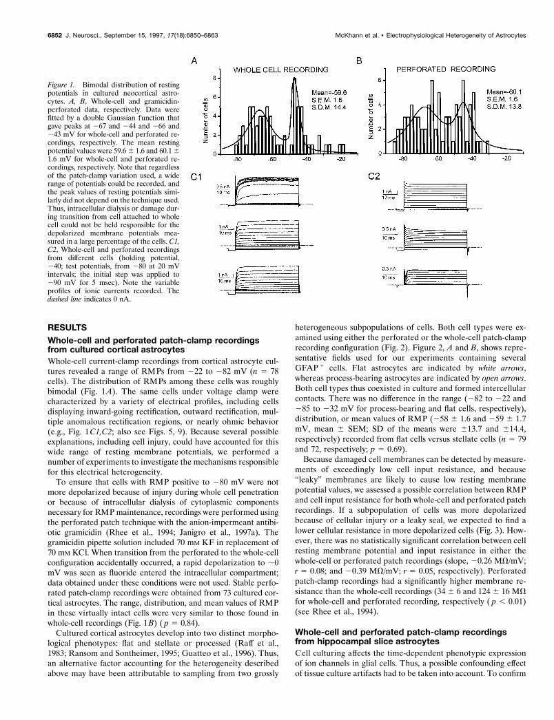
RESULTSWhole-cell and perforated patch-clamp recordingsfrom cultured cortical astrocytesWhole-cell current-clamp recordings from cortical astrocyte cul-tures revealed a range of RMPs from 222 to 282 mV (n 5 78cells). The distribution of RMPs among these cells was roughlybimodal (Fig. 1A). The same cells under voltage clamp werecharacterized by a variety of electrical profiles, including cellsdisplaying inward-going rectification, outward rectification, mul-tiple anomalous rectification regions, or nearly ohmic behavior(e.g., Fig. 1C1,C2; also see Figs. 5, 9). Because several possibleexplanations, including cell injury, could have accounted for thiswide range of resting membrane potentials, we performed anumber of experiments to investigate the mechanisms responsiblefor this electrical heterogeneity.
To ensure that cells with RMP positive to 280 mV were notmore depolarized because of injury during whole cell penetrationor because of intracellular dialysis of cytoplasmic componentsnecessary for RMP maintenance, recordings were performed usingthe perforated patch technique with the anion-impermeant antibi-otic gramicidin (Rhee et al., 1994; Janigro et al., 1997a). Thegramicidin pipette solution included 70 mM KF in replacement of70 mM KCl. When transition from the perforated to the whole-cellconfiguration accidentally occurred, a rapid depolarization to ;0mV was seen as fluoride entered the intracellular compartment;data obtained under these conditions were not used. Stable perfo-rated patch-clamp recordings were obtained from 73 cultured cor-tical astrocytes. The range, distribution, and mean values of RMPin these virtually intact cells were very similar to those found inwhole-cell recordings (Fig. 1B) ( p 5 0.84).
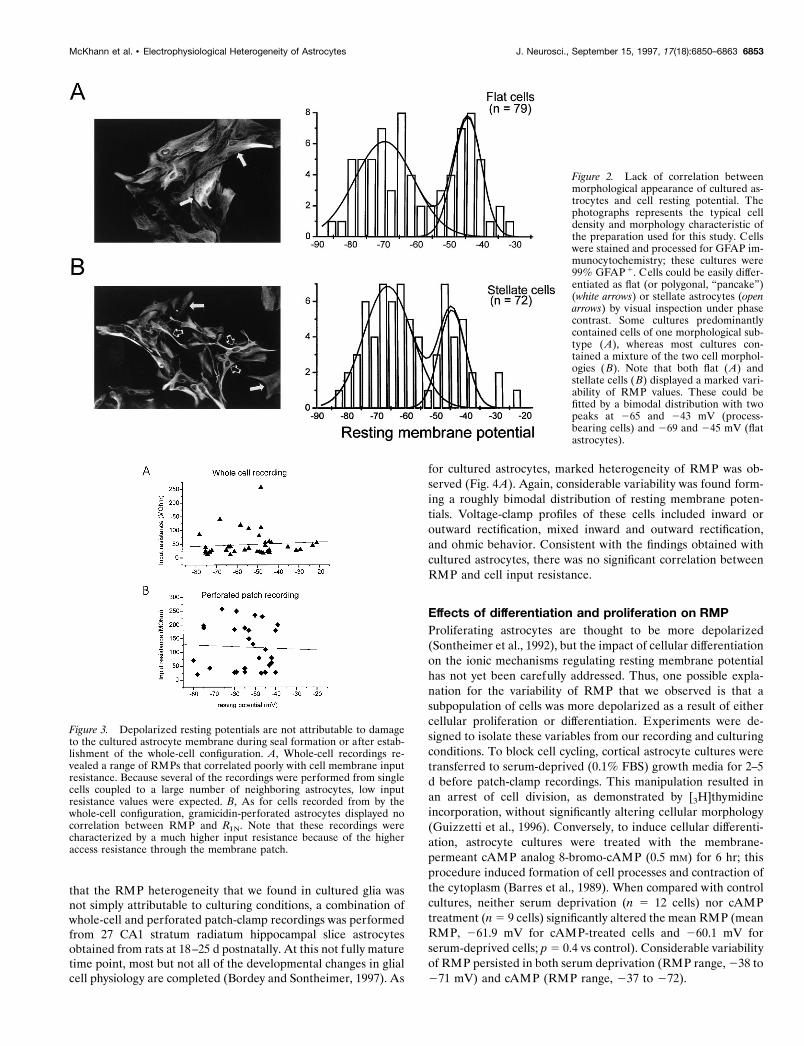
Cultured cortical astrocytes develop into two distinct morpho-logical phenotypes: flat and stellate or processed (Raff et al.,1983; Ransom and Sontheimer, 1995; Guatteo et al., 1996). Thus,an alternative factor accounting for the heterogeneity describedabove may have been attributable to sampling from two grossly
heterogeneous subpopulations of cells. Both cell types were ex-amined using either the perforated or the whole-cell patch-clamprecording configuration (Fig. 2). Figure 2, A and B, shows repre-sentative fields used for our experiments containing severalGFAP1 cells. Flat astrocytes are indicated by white arrows,whereas process-bearing astrocytes are indicated by open arrows.Both cell types thus coexisted in culture and formed intercellularcontacts. There was no difference in the range (282 to 222 and285 to 232 mV for process-bearing and flat cells, respectively),distribution, or mean values of RMP (258 6 1.6 and 259 6 1.7mV, mean 6 SEM; SD of the means were 613.7 and 614.4,respectively) recorded from flat cells versus stellate cells (n 5 79and 72, respectively; p 5 0.69).
Because damaged cell membranes can be detected by measure-ments of exceedingly low cell input resistance, and because“leaky” membranes are likely to cause low resting membranepotential values, we assessed a possible correlation between RMPand cell input resistance for both whole-cell and perforated patchrecordings. If a subpopulation of cells was more depolarizedbecause of cellular injury or a leaky seal, we expected to find alower cellular resistance in more depolarized cells (Fig. 3). How-ever, there was no statistically significant correlation between cellresting membrane potential and input resistance in either thewhole-cell or perforated patch recordings (slope, 20.26 MV/mV;r 5 0.08; and 20.39 MV/mV; r 5 0.05, respectively). Perforatedpatch-clamp recordings had a significantly higher membrane re-sistance than the whole-cell recordings (34 6 6 and 124 6 16 MVfor whole-cell and perforated recording, respectively ( p , 0.01)(see Rhee et al., 1994).
Whole-cell and perforated patch-clamp recordingsfrom hippocampal slice astrocytesCell culturing affects the time-dependent phenotypic expressionof ion channels in glial cells. Thus, a possible confounding effectof tissue culture artifacts had to be taken into account. To confirm
Figure 1. Bimodal distribution of restingpotentials in cultured neocortical astro-cytes. A, B, Whole-cell and gramicidin-perforated data, respectively. Data werefitted by a double Gaussian function thatgave peaks at 267 and 244 and 266 and243 mV for whole-cell and perforated re-cordings, respectively. The mean restingpotential values were 59.6 6 1.6 and 60.1 61.6 mV for whole-cell and perforated re-cordings, respectively. Note that regardlessof the patch-clamp variation used, a widerange of potentials could be recorded, andthe peak values of resting potentials simi-larly did not depend on the technique used.Thus, intracellular dialysis or damage dur-ing transition from cell attached to wholecell could not be held responsible for thedepolarized membrane potentials mea-sured in a large percentage of the cells. C1,C2, Whole-cell and perforated recordingsfrom different cells (holding potential,240; test potentials, from 280 at 20 mVintervals; the initial step was applied to290 mV for 5 msec). Note the variableprofiles of ionic currents recorded. Thedashed line indicates 0 nA.
6852 J. Neurosci., September 15, 1997, 17(18):6850–6863 McKhann et al. • Electrophysiological Heterogeneity of Astrocytes
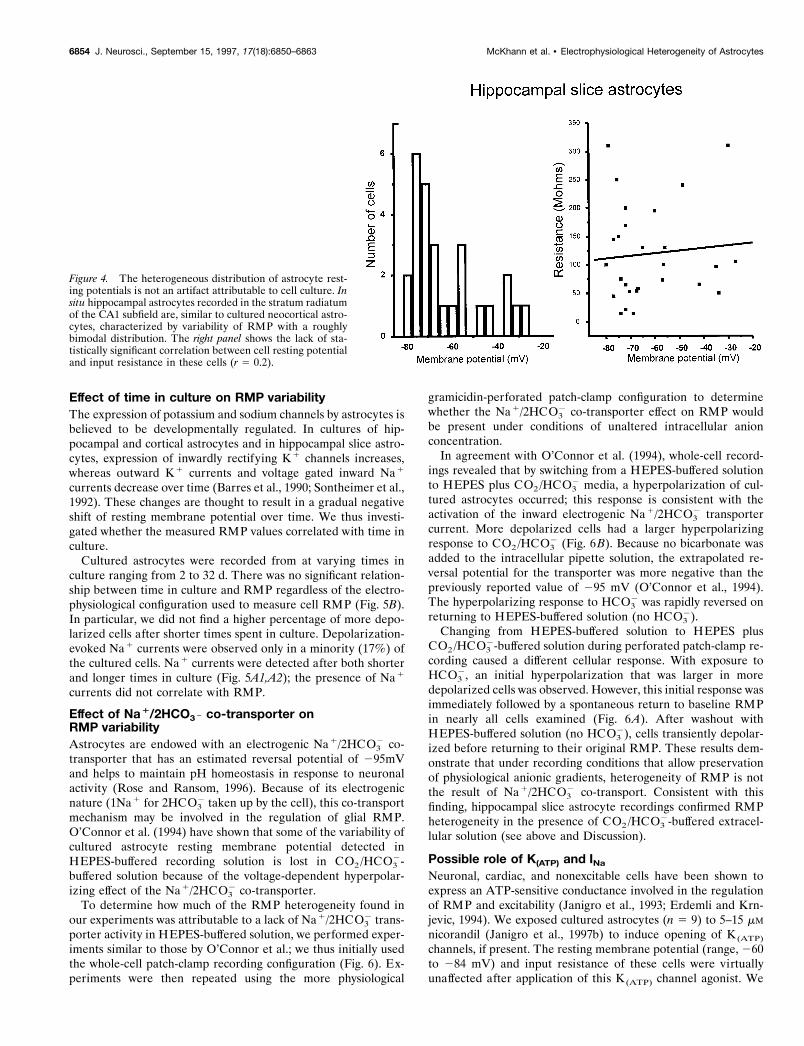
that the RMP heterogeneity that we found in cultured glia wasnot simply attributable to culturing conditions, a combination ofwhole-cell and perforated patch-clamp recordings was performedfrom 27 CA1 stratum radiatum hippocampal slice astrocytesobtained from rats at 18–25 d postnatally. At this not fully maturetime point, most but not all of the developmental changes in glialcell physiology are completed (Bordey and Sontheimer, 1997). As
for cultured astrocytes, marked heterogeneity of RMP was ob-served (Fig. 4A). Again, considerable variability was found form-ing a roughly bimodal distribution of resting membrane poten-tials. Voltage-clamp profiles of these cells included inward oroutward rectification, mixed inward and outward rectification,and ohmic behavior. Consistent with the findings obtained withcultured astrocytes, there was no significant correlation betweenRMP and cell input resistance.
Effects of differentiation and proliferation on RMPProliferating astrocytes are thought to be more depolarized(Sontheimer et al., 1992), but the impact of cellular differentiationon the ionic mechanisms regulating resting membrane potentialhas not yet been carefully addressed. Thus, one possible expla-nation for the variability of RMP that we observed is that asubpopulation of cells was more depolarized as a result of eithercellular proliferation or differentiation. Experiments were de-signed to isolate these variables from our recording and culturingconditions. To block cell cycling, cortical astrocyte cultures weretransferred to serum-deprived (0.1% FBS) growth media for 2–5d before patch-clamp recordings. This manipulation resulted inan arrest of cell division, as demonstrated by [3H]thymidineincorporation, without significantly altering cellular morphology(Guizzetti et al., 1996). Conversely, to induce cellular differenti-ation, astrocyte cultures were treated with the membrane-permeant cAMP analog 8-bromo-cAMP (0.5 mM) for 6 hr; thisprocedure induced formation of cell processes and contraction ofthe cytoplasm (Barres et al., 1989). When compared with controlcultures, neither serum deprivation (n 5 12 cells) nor cAMPtreatment (n 5 9 cells) significantly altered the mean RMP (meanRMP, 261.9 mV for cAMP-treated cells and 260.1 mV forserum-deprived cells; p 5 0.4 vs control). Considerable variabilityof RMP persisted in both serum deprivation (RMP range, 238 to271 mV) and cAMP (RMP range, 237 to 272).
Figure 2. Lack of correlation betweenmorphological appearance of cultured as-trocytes and cell resting potential. Thephotographs represents the typical celldensity and morphology characteristic ofthe preparation used for this study. Cellswere stained and processed for GFAP im-munocytochemistry; these cultures were99% GFAP 1. Cells could be easily differ-entiated as flat (or polygonal, “pancake”)(white arrows) or stellate astrocytes (openarrows) by visual inspection under phasecontrast. Some cultures predominantlycontained cells of one morphological sub-type ( A), whereas most cultures con-tained a mixture of the two cell morphol-ogies ( B). Note that both flat ( A) andstellate cells (B) displayed a marked vari-ability of RMP values. These could befitted by a bimodal distribution with twopeaks at 265 and 243 mV (process-bearing cells) and 269 and 245 mV (flatastrocytes).
Figure 3. Depolarized resting potentials are not attributable to damageto the cultured astrocyte membrane during seal formation or after estab-lishment of the whole-cell configuration. A, Whole-cell recordings re-vealed a range of RMPs that correlated poorly with cell membrane inputresistance. Because several of the recordings were performed from singlecells coupled to a large number of neighboring astrocytes, low inputresistance values were expected. B, As for cells recorded from by thewhole-cell configuration, gramicidin-perforated astrocytes displayed nocorrelation between RMP and RIN. Note that these recordings werecharacterized by a much higher input resistance because of the higheraccess resistance through the membrane patch.
McKhann et al. • Electrophysiological Heterogeneity of Astrocytes J. Neurosci., September 15, 1997, 17(18):6850–6863 6853
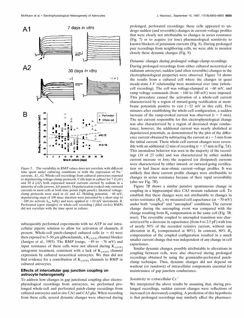
Effect of time in culture on RMP variabilityThe expression of potassium and sodium channels by astrocytes isbelieved to be developmentally regulated. In cultures of hip-pocampal and cortical astrocytes and in hippocampal slice astro-cytes, expression of inwardly rectifying K1 channels increases,whereas outward K1 currents and voltage gated inward Na1
currents decrease over time (Barres et al., 1990; Sontheimer et al.,1992). These changes are thought to result in a gradual negativeshift of resting membrane potential over time. We thus investi-gated whether the measured RMP values correlated with time inculture.
Cultured astrocytes were recorded from at varying times inculture ranging from 2 to 32 d. There was no significant relation-ship between time in culture and RMP regardless of the electro-physiological configuration used to measure cell RMP (Fig. 5B).In particular, we did not find a higher percentage of more depo-larized cells after shorter times spent in culture. Depolarization-evoked Na1 currents were observed only in a minority (17%) ofthe cultured cells. Na1 currents were detected after both shorterand longer times in culture (Fig. 5A1,A2); the presence of Na1
currents did not correlate with RMP.
Effect of Na1/2HCO3 2 co-transporter onRMP variabilityAstrocytes are endowed with an electrogenic Na1/2HCO3
2 co-transporter that has an estimated reversal potential of 295mVand helps to maintain pH homeostasis in response to neuronalactivity (Rose and Ransom, 1996). Because of its electrogenicnature (1Na1 for 2HCO3
2 taken up by the cell), this co-transportmechanism may be involved in the regulation of glial RMP.O’Connor et al. (1994) have shown that some of the variability ofcultured astrocyte resting membrane potential detected inHEPES-buffered recording solution is lost in CO2/HCO3
2-buffered solution because of the voltage-dependent hyperpolar-izing effect of the Na1/2HCO3
2 co-transporter.To determine how much of the RMP heterogeneity found in
our experiments was attributable to a lack of Na1/2HCO32 trans-
porter activity in HEPES-buffered solution, we performed exper-iments similar to those by O’Connor et al.; we thus initially usedthe whole-cell patch-clamp recording configuration (Fig. 6). Ex-periments were then repeated using the more physiological
gramicidin-perforated patch-clamp configuration to determinewhether the Na1/2HCO3
2 co-transporter effect on RMP wouldbe present under conditions of unaltered intracellular anionconcentration.
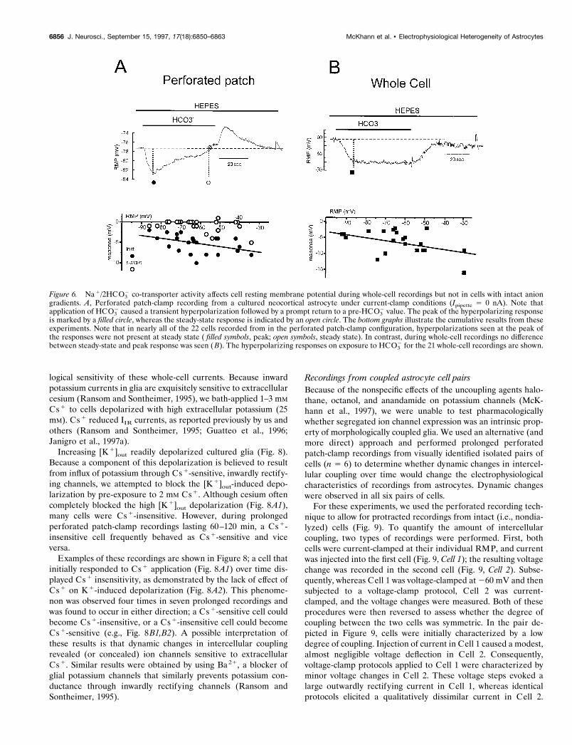
In agreement with O’Connor et al. (1994), whole-cell record-ings revealed that by switching from a HEPES-buffered solutionto HEPES plus CO2/HCO3
2 media, a hyperpolarization of cul-tured astrocytes occurred; this response is consistent with theactivation of the inward electrogenic Na1/2HCO3
2 transportercurrent. More depolarized cells had a larger hyperpolarizingresponse to CO2/HCO3
2 (Fig. 6B). Because no bicarbonate wasadded to the intracellular pipette solution, the extrapolated re-versal potential for the transporter was more negative than thepreviously reported value of 295 mV (O’Connor et al., 1994).The hyperpolarizing response to HCO3
2 was rapidly reversed onreturning to HEPES-buffered solution (no HCO3
2).Changing from HEPES-buffered solution to HEPES plus
CO2/HCO32-buffered solution during perforated patch-clamp re-
cording caused a different cellular response. With exposure toHCO3
2, an initial hyperpolarization that was larger in moredepolarized cells was observed. However, this initial response wasimmediately followed by a spontaneous return to baseline RMPin nearly all cells examined (Fig. 6A). After washout withHEPES-buffered solution (no HCO3
2), cells transiently depolar-ized before returning to their original RMP. These results dem-onstrate that under recording conditions that allow preservationof physiological anionic gradients, heterogeneity of RMP is notthe result of Na1/2HCO3
2 co-transport. Consistent with thisfinding, hippocampal slice astrocyte recordings confirmed RMPheterogeneity in the presence of CO2/HCO3
2-buffered extracel-lular solution (see above and Discussion).
Possible role of K(ATP) and INa
Neuronal, cardiac, and nonexcitable cells have been shown toexpress an ATP-sensitive conductance involved in the regulationof RMP and excitability (Janigro et al., 1993; Erdemli and Krn-jevic, 1994). We exposed cultured astrocytes (n 5 9) to 5–15 mM
nicorandil (Janigro et al., 1997b) to induce opening of K (ATP)
channels, if present. The resting membrane potential (range, 260to 284 mV) and input resistance of these cells were virtuallyunaffected after application of this K (ATP) channel agonist. We
Figure 4. The heterogeneous distribution of astrocyte rest-ing potentials is not an artifact attributable to cell culture. Insitu hippocampal astrocytes recorded in the stratum radiatumof the CA1 subfield are, similar to cultured neocortical astro-cytes, characterized by variability of RMP with a roughlybimodal distribution. The right panel shows the lack of sta-tistically significant correlation between cell resting potentialand input resistance in these cells (r 5 0.2).
6854 J. Neurosci., September 15, 1997, 17(18):6850–6863 McKhann et al. • Electrophysiological Heterogeneity of Astrocytes
subsequently performed experiments with no ATP in our intra-cellular pipette solution to allow for activation of channels, ifpresent. Whole-cell patch-clamped cultured cells (n 5 6) werethen exposed to 5–50 mM glibenclamide, a K(ATP) channel blocker(Janigro et al., 1993). The RMP (range, 249 to 276 mV) andinput resistance of these cells were not altered during K (ATP)
antagonist treatment, consistent with a lack of K(ATP) channelexpression by cultured neocortical astrocytes. We thus did notfind evidence for a contribution of K (ATP) channels to RMP incultured astrocytes.
Effects of intercellular gap junction coupling onastrocyte heterogeneityTo address how changes in gap junctional coupling alter electro-physiological recordings from astrocytes, we performed pro-longed whole-cell and perforated patch-clamp recordings fromcultured astrocytes and in situ CA1 and CA3 glia. When recordingfrom these cells, several dynamic changes were observed during
prolonged, perforated recordings; these cells appeared to un-dergo sudden (and reversible) changes in current–voltage profilesthat were clearly not attributable to changes in series resistance(Fig. 7) or to acquire (or lose) pharmacological sensitivity toknown blockers of potassium currents (Fig. 8). During prolongedpair recordings from neighboring cells, we were able to monitorclosely these dynamic changes (Fig. 9).
Dynamic changes during prolonged voltage-clamp recordingsDuring prolonged recordings from either cultured neocortical orradiatum astrocytes, sudden (and often reversible) changes in theelectrophysiological properties were observed. Figure 7A showsthe results from a cultured cell where the changes in quasisteady-state I–V relationship were monitored over time (whole-cell recording). The cell was voltage-clamped at 260 mV, andramp voltage commands (from 2160 to 100 mV) were imposed.This procedure caused the activation of a whole-cell currentcharacterized by a region of inward-going rectification at mem-brane potentials positive to rest (252 mV in this cell). Fiveminutes after establishing the whole-cell configuration, a suddenincrease of the ramp-evoked current was observed (t 5 5 min).The net current responsible for this electrophysiological changewas also characterized by a region of decreased slope conduc-tance; however, the additional current was nearly abolished atdepolarized potentials, as demonstrated by the plot of the differ-ence current obtained by subtracting the current at t 5 5 min fromthe initial current. These whole-cell current changes were revers-ible with an additional 12 min of recording (t 5 17 min in Fig. 7A).This anomalous behavior was seen in the majority of the record-ings (18 of 23 cells) and was characterized by either suddencurrent increase or loss; the acquired (or dissipated) currentswere characterized by either inward- or outward-going rectifica-tion or had linear near-ohmic current–voltage profiles. It wasunlikely that these current profile changes were attributable tochanges in series resistance because of their rapid reversibility(also see Fig. 7B).
Figure 7B shows a similar putative spontaneous change incoupling in a hippocampal slice CA3 stratum radiatum cell. Toestablish that these changes were not attributable to changes inseries resistance (RS), we measured cell capacitance (at 270 mV)under both “coupled” and “uncoupled” conditions. The currentchange during the uncoupling event was compared with thechange resulting from RS compensation in the same cell (Fig. 7B,inset). The reversible coupled to uncoupled transition was char-acterized by a decrease in capacitance (from 8 to 2.5 pF) with lossof nearly 50% of the recorded resistive current, without anyalteration in RS (compensated at 80%). In contrast, 80% RS
compensation of the coupled configuration resulted in a muchsmaller current change that was independent of any change in cellcapacitance.
Similar dynamic changes, possibly attributable to alterations incoupling between cells, were also observed during prolongedrecordings obtained by using the gramicidin-perforated patch-clamp technique. Thus, dynamic changes did not depend onwashout (or rundown) of intracellular components essential formaintenance of gap junction conductance.
Sensitivity to extracellular Cs1
We interpreted the above results by assuming that, during pro-longed recordings, sudden current changes were reflections ofincreased (or decreased) coupling. A prediction of this hypothesisis that prolonged recordings may similarly affect the pharmaco-
Figure 5. The variability in RMP values does not correlate with differenttime spent under culturing conditions or with the expression of Na 1
currents. A1, A2, Whole-cell recordings from cultured astrocytes exposedto depolarizing voltage-clamp protocols. Cells kept in culture for 7 d (A1)and 30 d (A2) both expressed inward currents carried by sodium in aminority of cells (arrows, lef t panels). Depolarization evoked only outwardcurrents in most cells at both time points (right panels). Identical voltage-clamp protocols were used in A1 and A2. Holding potential, 260 mV;depolarizing steps of 100 msec duration were preceded by a short step to2100 (to activate INa fully) and were applied at 110 mV increments. B,Perforated (open triangles) or whole-cell recording ( filled circles) RMPsdid not correlate with the time spent in culture.
McKhann et al. • Electrophysiological Heterogeneity of Astrocytes J. Neurosci., September 15, 1997, 17(18):6850–6863 6855
logical sensitivity of these whole-cell currents. Because inwardpotassium currents in glia are exquisitely sensitive to extracellularcesium (Ransom and Sontheimer, 1995), we bath-applied 1–3 mM
Cs1 to cells depolarized with high extracellular potassium (25mM). Cs1 reduced IIR currents, as reported previously by us andothers (Ransom and Sontheimer, 1995; Guatteo et al., 1996;Janigro et al., 1997a).
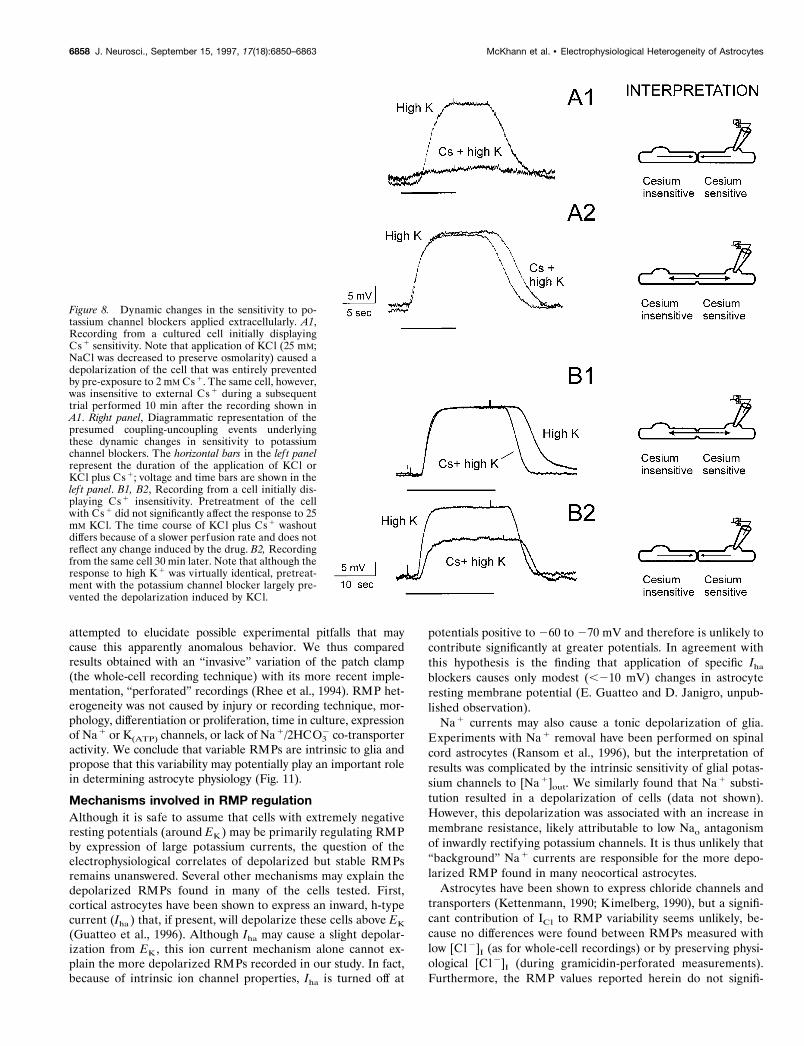
Increasing [K1]out readily depolarized cultured glia (Fig. 8).Because a component of this depolarization is believed to resultfrom influx of potassium through Cs1-sensitive, inwardly rectify-ing channels, we attempted to block the [K1]out-induced depo-larization by pre-exposure to 2 mM Cs1. Although cesium oftencompletely blocked the high [K1]out depolarization (Fig. 8A1),many cells were Cs1-insensitive. However, during prolongedperforated patch-clamp recordings lasting 60–120 min, a Cs1-insensitive cell frequently behaved as Cs1-sensitive and viceversa.
Examples of these recordings are shown in Figure 8; a cell thatinitially responded to Cs1 application (Fig. 8A1) over time dis-played Cs1 insensitivity, as demonstrated by the lack of effect ofCs1 on K1-induced depolarization (Fig. 8A2). This phenome-non was observed four times in seven prolonged recordings andwas found to occur in either direction; a Cs1-sensitive cell couldbecome Cs1-insensitive, or a Cs1-insensitive cell could becomeCs1-sensitive (e.g., Fig. 8B1,B2). A possible interpretation ofthese results is that dynamic changes in intercellular couplingrevealed (or concealed) ion channels sensitive to extracellularCs1. Similar results were obtained by using Ba 21, a blocker ofglial potassium channels that similarly prevents potassium con-ductance through inwardly rectifying channels (Ransom andSontheimer, 1995).
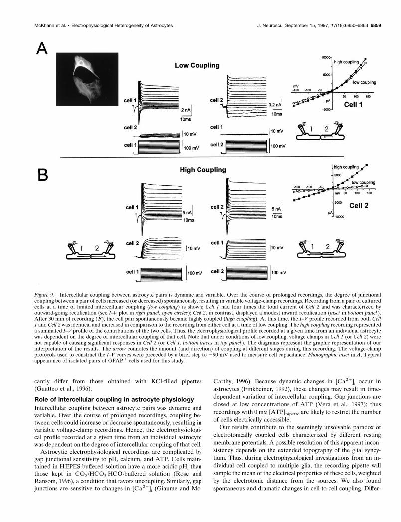
Recordings from coupled astrocyte cell pairsBecause of the nonspecific effects of the uncoupling agents halo-thane, octanol, and anandamide on potassium channels (McK-hann et al., 1997), we were unable to test pharmacologicallywhether segregated ion channel expression was an intrinsic prop-erty of morphologically coupled glia. We used an alternative (andmore direct) approach and performed prolonged perforatedpatch-clamp recordings from visually identified isolated pairs ofcells (n 5 6) to determine whether dynamic changes in intercel-lular coupling over time would change the electrophysiologicalcharacteristics of recordings from astrocytes. Dynamic changeswere observed in all six pairs of cells.
For these experiments, we used the perforated recording tech-nique to allow for protracted recordings from intact (i.e., nondia-lyzed) cells (Fig. 9). To quantify the amount of intercellularcoupling, two types of recordings were performed. First, bothcells were current-clamped at their individual RMP, and currentwas injected into the first cell (Fig. 9, Cell 1); the resulting voltagechange was recorded in the second cell (Fig. 9, Cell 2). Subse-quently, whereas Cell 1 was voltage-clamped at 260 mV and thensubjected to a voltage-clamp protocol, Cell 2 was current-clamped, and the voltage changes were measured. Both of theseprocedures were then reversed to assess whether the degree ofcoupling between the two cells was symmetric. In the pair de-picted in Figure 9, cells were initially characterized by a lowdegree of coupling. Injection of current in Cell 1 caused a modest,almost negligible voltage deflection in Cell 2. Consequently,voltage-clamp protocols applied to Cell 1 were characterized byminor voltage changes in Cell 2. These voltage steps evoked alarge outwardly rectifying current in Cell 1, whereas identicalprotocols elicited a qualitatively dissimilar current in Cell 2.
Figure 6. Na 1/2HCO32 co-transporter activity affects cell resting membrane potential during whole-cell recordings but not in cells with intact anion
gradients. A, Perforated patch-clamp recording from a cultured neocortical astrocyte under current-clamp conditions (Ipipette 5 0 nA). Note thatapplication of HCO3
2 caused a transient hyperpolarization followed by a prompt return to a pre-HCO32 value. The peak of the hyperpolarizing response
is marked by a filled circle, whereas the steady-state response is indicated by an open circle. The bottom graphs illustrate the cumulative results from theseexperiments. Note that in nearly all of the 22 cells recorded from in the perforated patch-clamp configuration, hyperpolarizations seen at the peak ofthe responses were not present at steady state ( filled symbols, peak; open symbols, steady state). In contrast, during whole-cell recordings no differencebetween steady-state and peak response was seen ( B). The hyperpolarizing responses on exposure to HCO3
2 for the 21 whole-cell recordings are shown.
6856 J. Neurosci., September 15, 1997, 17(18):6850–6863 McKhann et al. • Electrophysiological Heterogeneity of Astrocytes
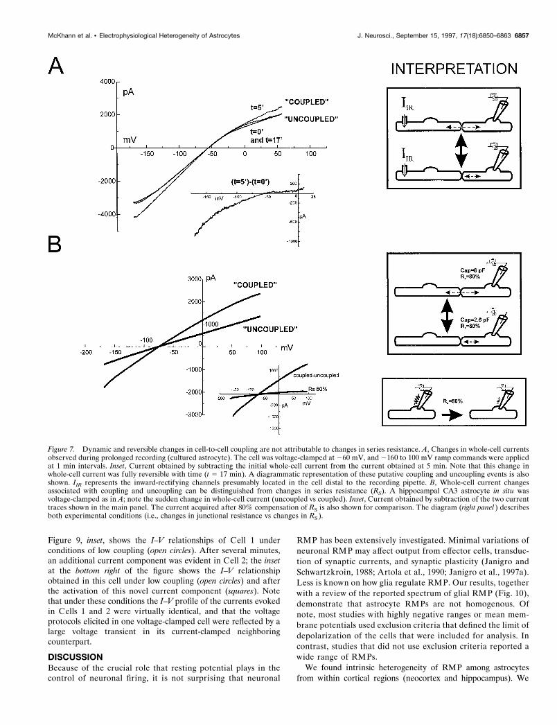
Figure 9, inset, shows the I–V relationships of Cell 1 underconditions of low coupling (open circles). After several minutes,an additional current component was evident in Cell 2; the insetat the bottom right of the figure shows the I–V relationshipobtained in this cell under low coupling (open circles) and afterthe activation of this novel current component (squares). Notethat under these conditions the I–V profile of the currents evokedin Cells 1 and 2 were virtually identical, and that the voltageprotocols elicited in one voltage-clamped cell were reflected by alarge voltage transient in its current-clamped neighboringcounterpart.
DISCUSSIONBecause of the crucial role that resting potential plays in thecontrol of neuronal firing, it is not surprising that neuronal
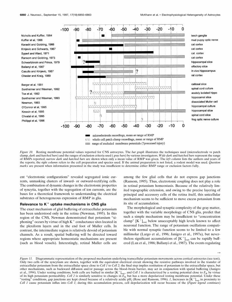
RMP has been extensively investigated. Minimal variations ofneuronal RMP may affect output from effector cells, transduc-tion of synaptic currents, and synaptic plasticity (Janigro andSchwartzkroin, 1988; Artola et al., 1990; Janigro et al., 1997a).Less is known on how glia regulate RMP. Our results, togetherwith a review of the reported spectrum of glial RMP (Fig. 10),demonstrate that astrocyte RMPs are not homogenous. Ofnote, most studies with highly negative ranges or mean mem-brane potentials used exclusion criteria that defined the limit ofdepolarization of the cells that were included for analysis. Incontrast, studies that did not use exclusion criteria reported awide range of RMPs.
We found intrinsic heterogeneity of RMP among astrocytesfrom within cortical regions (neocortex and hippocampus). We
Figure 7. Dynamic and reversible changes in cell-to-cell coupling are not attributable to changes in series resistance. A, Changes in whole-cell currentsobserved during prolonged recording (cultured astrocyte). The cell was voltage-clamped at 260 mV, and 2160 to 100 mV ramp commands were appliedat 1 min intervals. Inset, Current obtained by subtracting the initial whole-cell current from the current obtained at 5 min. Note that this change inwhole-cell current was fully reversible with time (t 5 17 min). A diagrammatic representation of these putative coupling and uncoupling events is alsoshown. IIR represents the inward-rectifying channels presumably located in the cell distal to the recording pipette. B, Whole-cell current changesassociated with coupling and uncoupling can be distinguished from changes in series resistance (RS). A hippocampal CA3 astrocyte in situ wasvoltage-clamped as in A; note the sudden change in whole-cell current (uncoupled vs coupled). Inset, Current obtained by subtraction of the two currenttraces shown in the main panel. The current acquired after 80% compensation of RS is also shown for comparison. The diagram (right panel ) describesboth experimental conditions (i.e., changes in junctional resistance vs changes in RS ).
McKhann et al. • Electrophysiological Heterogeneity of Astrocytes J. Neurosci., September 15, 1997, 17(18):6850–6863 6857
attempted to elucidate possible experimental pitfalls that maycause this apparently anomalous behavior. We thus comparedresults obtained with an “invasive” variation of the patch clamp(the whole-cell recording technique) with its more recent imple-mentation, “perforated” recordings (Rhee et al., 1994). RMP het-erogeneity was not caused by injury or recording technique, mor-phology, differentiation or proliferation, time in culture, expressionof Na1 or K(ATP) channels, or lack of Na1/2HCO3
2 co-transporteractivity. We conclude that variable RMPs are intrinsic to glia andpropose that this variability may potentially play an important rolein determining astrocyte physiology (Fig. 11).
Mechanisms involved in RMP regulationAlthough it is safe to assume that cells with extremely negativeresting potentials (around EK ) may be primarily regulating RMPby expression of large potassium currents, the question of theelectrophysiological correlates of depolarized but stable RMPsremains unanswered. Several other mechanisms may explain thedepolarized RMPs found in many of the cells tested. First,cortical astrocytes have been shown to express an inward, h-typecurrent (Iha ) that, if present, will depolarize these cells above EK
(Guatteo et al., 1996). Although Iha may cause a slight depolar-ization from EK, this ion current mechanism alone cannot ex-plain the more depolarized RMPs recorded in our study. In fact,because of intrinsic ion channel properties, Iha is turned off at
potentials positive to 260 to 270 mV and therefore is unlikely tocontribute significantly at greater potentials. In agreement withthis hypothesis is the finding that application of specific Iha
blockers causes only modest (,210 mV) changes in astrocyteresting membrane potential (E. Guatteo and D. Janigro, unpub-lished observation).
Na1 currents may also cause a tonic depolarization of glia.Experiments with Na1 removal have been performed on spinalcord astrocytes (Ransom et al., 1996), but the interpretation ofresults was complicated by the intrinsic sensitivity of glial potas-sium channels to [Na1]out. We similarly found that Na1 substi-tution resulted in a depolarization of cells (data not shown).However, this depolarization was associated with an increase inmembrane resistance, likely attributable to low Nao antagonismof inwardly rectifying potassium channels. It is thus unlikely that“background” Na1 currents are responsible for the more depo-larized RMP found in many neocortical astrocytes.
Astrocytes have been shown to express chloride channels andtransporters (Kettenmann, 1990; Kimelberg, 1990), but a signifi-cant contribution of ICl to RMP variability seems unlikely, be-cause no differences were found between RMPs measured withlow [Cl2]I (as for whole-cell recordings) or by preserving physi-ological [Cl2]I (during gramicidin-perforated measurements).Furthermore, the RMP values reported herein do not signifi-
Figure 8. Dynamic changes in the sensitivity to po-tassium channel blockers applied extracellularly. A1,Recording from a cultured cell initially displayingCs 1 sensitivity. Note that application of KCl (25 mM;NaCl was decreased to preserve osmolarity) caused adepolarization of the cell that was entirely preventedby pre-exposure to 2 mM Cs 1. The same cell, however,was insensitive to external Cs 1 during a subsequenttrial performed 10 min after the recording shown inA1. Right panel, Diagrammatic representation of thepresumed coupling-uncoupling events underlyingthese dynamic changes in sensitivity to potassiumchannel blockers. The horizontal bars in the lef t panelrepresent the duration of the application of KCl orKCl plus Cs 1; voltage and time bars are shown in thelef t panel. B1, B2, Recording from a cell initially dis-playing Cs 1 insensitivity. Pretreatment of the cellwith Cs 1 did not significantly affect the response to 25mM KCl. The time course of KCl plus Cs 1 washoutdiffers because of a slower perfusion rate and does notreflect any change induced by the drug. B2, Recordingfrom the same cell 30 min later. Note that although theresponse to high K 1 was virtually identical, pretreat-ment with the potassium channel blocker largely pre-vented the depolarization induced by KCl.
6858 J. Neurosci., September 15, 1997, 17(18):6850–6863 McKhann et al. • Electrophysiological Heterogeneity of Astrocytes
cantly differ from those obtained with KCl-filled pipettes(Guatteo et al., 1996).
Role of intercellular coupling in astrocyte physiologyIntercellular coupling between astrocyte pairs was dynamic andvariable. Over the course of prolonged recordings, coupling be-tween cells could increase or decrease spontaneously, resulting invariable voltage-clamp recordings. Hence, the electrophysiologi-cal profile recorded at a given time from an individual astrocytewas dependent on the degree of intercellular coupling of that cell.
Astrocytic electrophysiological recordings are complicated bygap junctional sensitivity to pH, calcium, and ATP. Cells main-tained in HEPES-buffered solution have a more acidic pHi thanthose kept in CO2/HCO3
2HCO-buffered solution (Rose andRansom, 1996), a condition that favors uncoupling. Similarly, gapjunctions are sensitive to changes in [Ca21]i (Giaume and Mc-
Carthy, 1996). Because dynamic changes in [Ca 21]i occur inastrocytes (Finkbeiner, 1992), these changes may result in time-dependent variation of intercellular coupling. Gap junctions areclosed at low concentrations of ATP (Vera et al., 1997); thusrecordings with 0 mM [ATP]pipette are likely to restrict the numberof cells electrically accessible.
Our results contribute to the seemingly unsolvable paradox ofelectrotonically coupled cells characterized by different restingmembrane potentials. A possible resolution of this apparent incon-sistency depends on the extended topography of the glial syncy-tium. Thus, during electrophysiological investigations from an in-dividual cell coupled to multiple glia, the recording pipette willsample the mean of the electrical properties of these cells, weightedby the electrotonic distance from the sources. We also foundspontaneous and dramatic changes in cell-to-cell coupling. Differ-
Figure 9. Intercellular coupling between astrocyte pairs is dynamic and variable. Over the course of prolonged recordings, the degree of junctionalcoupling between a pair of cells increased (or decreased) spontaneously, resulting in variable voltage-clamp recordings. Recording from a pair of culturedcells at a time of limited intercellular coupling (low coupling) is shown; Cell 1 had four times the total current of Cell 2 and was characterized byoutward-going rectification (see I–V plot in right panel, open circles); Cell 2, in contrast, displayed a modest inward rectification (inset in bottom panel ).After 30 min of recording (B), the cell pair spontaneously became highly coupled (high coupling). At this time, the I–V profile recorded from both Cell1 and Cell 2 was identical and increased in comparison to the recording from either cell at a time of low coupling. The high coupling recording representeda summated I–V profile of the contributions of the two cells. Thus, the electrophysiological profile recorded at a given time from an individual astrocytewas dependent on the degree of intercellular coupling of that cell. Note that under conditions of low coupling, voltage clamps in Cell 1 (or Cell 2) werenot capable of causing significant responses in Cell 2 (or Cell 1, bottom traces in top panel ). The diagrams represent the graphic representation of ourinterpretation of the results. The arrow connotes the amount (and direction) of coupling at different stages during this recording. The voltage-clampprotocols used to construct the I–V curves were preceded by a brief step to 290 mV used to measure cell capacitance. Photographic inset in A, Typicalappearance of isolated pairs of GFAP 1 cells used for this study.
McKhann et al. • Electrophysiological Heterogeneity of Astrocytes J. Neurosci., September 15, 1997, 17(18):6850–6863 6859
ent “electrotonic configurations” revealed segregated ionic cur-rents, unmasking clusters of inward- or outward-rectifying cells.The combination of dynamic changes in the electrotonic propertiesof syncytia, together with the segregation of ion currents, are thebases for a theoretical framework to understanding the electricalsubstrates of heterogeneous expression of RMP in glia.
Relevance to K1 uptake mechanisms in CNS gliaThe exact mechanism of glial buffering of extracellular potassiumhas been understood only in the retina (Newman, 1995). In thisregion of the CNS, Newman demonstrated that potassium “si-phoning” occurs by virtue of high K1 conductance sites located inthe plexiform layers and in the end foot of Muller cells. Incontrast, the intermediate region is relatively devoid of potassiumchannels. As a result, spatial buffering will be directed towardregions where appropriate homeostatic mechanisms are present(such as blood vessels). Interestingly, retinal Muller cells are
among the few glial cells that do not express gap junctions(Ransom, 1995). Thus, electrotonic coupling does not play a rolein retinal potassium homeostasis. Because of the relatively lim-ited topographic extension, and owing to the precise layering ofprincipal and accessory cells of the retina itself, this unicellularmechanism seems to be sufficient to move excess potassium fromits site of accumulation.
The morphological and synaptic complexity of the gray matter,together with the variable morphology of CNS glia, predict thatsuch a simple mechanism may be insufficient to “concentrationclamp” [K1]out below unacceptably high levels known to affectneuronal function. The range of potassium oscillations compati-ble with normal synaptic function seems to be limited to a fewmillimolar (Largo et al., 1996; Janigro et al., 1997a), but never-theless significant accumulations of [K1]out can be rapidly buff-ered (Lux et al., 1986; Ballanyi et al., 1987). The events regulating
Figure 10. Resting membrane potential values reported for CNS astrocytes. The bar graph illustrates the techniques used (microelectrode vs patchclamp, dark and hatched bars) and the ranges of exclusion criteria used ( gray bars) by various investigators. Wide dark and hatched bars represent the rangeof RMPs reported; narrow dark and hatched bars are shown when only a mean value of RMP was given. The lef t column lists the authors and years ofthe reports; the right column refers to the cell preparation and species used. If the animal preparation is not listed, a rodent model was used. Questionmarks are present when information presented in the study was insufficient to determine either RMP range or exclusion factors fully.
3
Figure 11. Diagrammatic representation of the proposed mechanism underlying transcellular potassium movements across cortical astrocytes (see text).Only two cells of the syncytium are shown, together with the equivalent electrical circuit showing the resistive pathways involved in the transfer ofextracellular potassium from the extracellular space close to Cell 1 to Cell 2; the final step implies restitution of potassium to the extracellular space, butother mechanisms, such as backward diffusion and/or passage across the blood–brain barrier, may act in conjunction with spatial buffering (Janigroet al., 1994). Under resting conditions, both cells are bathed in similar [K 1]out , and Cell 1 is characterized by a resting potential close to EK by virtueof its high potassium permeability. Cell 2 is similarly sensitive to changes in EK but has a more depolarized resting membrane potential. Under these“resting” conditions, gap junctions are kept closed because of a relatively acidic pHi (Rose and Ransom, 1996). 1, Increases in [K 1]out in proximity toCell 1 cause potassium influx into Cell 1; during this accumulation process, cell depolarization will occur because of the (Figure legend continues)
6860 J. Neurosci., September 15, 1997, 17(18):6850–6863 McKhann et al. • Electrophysiological Heterogeneity of Astrocytes
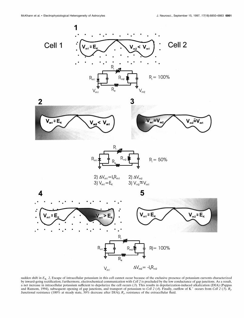
sudden shift in EK. 2, Escape of intracellular potassium in this cell cannot occur because of the exclusive presence of potassium currents characterizedby inward-going rectification; furthermore, electrochemical communication with Cell 2 is precluded by the low conductance of gap junctions. As a result,a net increase in intracellular potassium sufficient to depolarize the cell occurs (3). This results in depolarization-induced alkalization (DIA) (Pappasand Ransom, 1994), subsequent opening of gap junctions, and transport of potassium to Cell 2 (4). Finally, outflow of K 1 occurs from Cell 2 (5). Rj,Junctional resistance (100% at steady state, 50% decrease after DIA); Re, resistance of the extracellular fluid.
McKhann et al. • Electrophysiological Heterogeneity of Astrocytes J. Neurosci., September 15, 1997, 17(18):6850–6863 6861
potassium transport in gray matter remain, however, largely amatter of speculation. Our results shed some light on the possiblemechanisms involved. First, we have demonstrated that dynamicchanges in cell-to-cell coupling occur in the absence of anyexogenous stimulus. These apparently untriggered reorganiza-tions of cellular coupling may be attributable to intracellular ionicshifts that cannot be effectively controlled during patch-clampexperiments, particularly during perforated recordings. We haveshown that uncoupling of cells unmasks what appears to be asegregated localization of inward- or outward-rectifying currents.This, in addition to the bimodal distribution of cortical glialRMPs, prompted us to develop a model for potassium transportacross the glial syncytium (Fig. 11).
This qualitative model is based on two theoretical and exper-imentally tested assumptions: (1) based on observations that,during recordings from morphologically coupled cells, one cellwas characterized by a resting membrane potential more negative(by 5–30 mV) than its neighbor, we assumed that more depolar-ized cells are coupled to cells characterized by a more negativeresting membrane potential; and (2) given that basal intracellularpH favors gap junction closure, we hypothesized that, at rest,these glia are weakly coupled. Events occurring after neuronalactivity facilitate gap junction openings (Marrero and Orkand,1996). We thus incorporated in the model a stimulus (or[K1]out )-induced increase in intercellular K1 mobility. One as-pect of our working hypothesis rests on the fact that cells in whichsignificant (and early) potassium accumulation occurs (Fig. 11,Cell 1) are characterized by selective expression of inwardlyrectifying potassium channels, whereas cells adapted for potas-sium release (Fig. 11, Cell 2) selectively express more outwardpotassium channels. This hypothesis is supported by the recentlocalization of the delayed rectifier Kv1.5 potassium channel toastrocytic end foot processes surrounding the microvasculature inthe hippocampus (Roy et al., 1996), a location ideally situated forpotassium efflux. Further evidence awaits the development ofspecific tools to morphologically and functionally study potassiumchannels in astrocytes in situ.
ConclusionsIn conclusion, we have shown that both cultured and hippocampalslice astrocytes are characterized by a wide range of restingpotentials. More depolarized cells were not injured glia, andseveral chemical and developmental factors have been ruled outas possible determinants of astrocytic RMP. We have also shownthat Na1/2HCO3
2 co-transporters and K(ATP) channels play littleor no role in the steady-state regulation of glial RMP. In addition,we have studied the effects of intercellular coupling on glial cellphysiological properties with particular emphasis on mechanismsinvolved in clearance and transcellular transport of potassium.We describe dynamic coupling between cultured and hippocam-pal slice glia and differential recorded pharmacological sensitivityof cultured astrocytes, likely as a result of changes in intercellularcoupling. We propose that spatial buffering may be facilitated byheterogeneous mechanisms controlling glial resting membranepotential in combination with dynamic changes in intercellularcoupling.
REFERENCESAlger BE, McCarren M, Fisher RS (1983) On the possibility of simul-
taneously recording from two cells with a single microelectrode in thehippocampal slice. Brain Res 270:137–141.
Artola A, Brocher S, Singer W (1990) Different voltage-dependent
thresholds for inducing long-term depression and long-term potentia-tion in slices of rat visual cortex. Nature 347:69–72.
Ballanyi K, Grafe P, Bruggencate GT (1987) Ion activities and potas-sium uptake mechanisms of glial cells of guinea-pig olfactory cortexslices. J Physiol (Lond) 382:159–174.
Barres BA, Chun LLY, Corey DP (1989) Calcium current in corticalastrocytes: induction by cAMP and neurotransmitters and permissiveeffect of serum factors. J Neurosci 9:3169–3175.
Barres BA, Chun LLY, Corey DP (1990) Ion channels in vertebrate glia.Annu Rev Neurosci 13:441–474.
Berger T, Schnitzer J, Kettenmann H (1991) Developmental changes inthe membrane current pattern, K 1 buffer capacity, and morphology ofglial cells in the corpus callosum slice. J Neurosci 11:3008–3024.
Black JA, Sontheimer H, Waxman SG (1993) Spinal cord astrocytes invitro: phenotypic diversity and sodium channel immunoreactivity. Glia7:272–285.
Bordey A, Sontheimer H (1997) Post-natal development of ionic cur-rents in rat hippocampal astrocytes in situ, J Neurophysiol, in press.
Casullo J, Krnjevic K (1987) Glial potentials in hippocampus. CanJ Physiol Pharmacol 65:847–855.
Chesler M, Kraig RP (1989) Intracellular pH transients of mammalianastrocytes. J Neurosci 9:2011–2019.
Chvatal A, Pastor A, Mauch M, Sykov’a E, Kettenmann H (1995) Dis-tinct populations of identified glial cells in the developing rat spinalcord slice: ion channel properties and cell morphology. Eur J Neurosci7:129–142.
Dermietzel R, Hertberg EL, Kessler JA, Spray DC (1991) Gap junctionsbetween cultured astrocytes: immunocytochemical, molecular, andelectrophysiological analysis. J Neurosci 11:1421–1432.
Erdemli G, Krnjevic K (1994) Guanosine diphosphate is required foractivation of a glyburide, ATP and cromakalim-sensitive outward cur-rent in rat hippocampal neurons. NeuroReport 5:1362–1364.
Finkbeiner S (1992) Calcium waves in astrocytes-filling in the gaps.Neuron 8:1101–1108.
Giaume C, McCarthy KD (1996) Control of gap-junctional communica-tion in astrocytic networks. Trends Neurosci 19:319–325.
Giaume C, Fromaget C, el Aoumari A, Cordier J, Glowinski J, Gros D(1991) Gap junctions in cultured astrocytes: single-channel currentsand characterization of channel-forming protein. Neuron 6:133–143.
Guatteo E, Stanness KA, Janigro D (1996) Hyperpolarization-activatedcurrents in cultured rat cortical and spinal cord astrocytes. Glia16:196–209.
Guizzetti M, Costa P, Peters J, Costa LG (1996) Acetylcholine as amitogen: muscarinic receptor-mediated proliferation of rat astrocytesand human astrocytoma cells. Eur J Pharmacol 297:265–273.
Janigro D, Schwartzkroin PA (1988) Effects of GABA and baclofen onpyramidal cells in the developing rabbit hippocampus: an “in vitro”study. Brain Res 469:171–184.
Janigro D, West GA, Gordon EL, Winn HR (1993) ATP-sensitive K 1
channels in rat aorta and brain microvascular endothelial cells. Am JPhysiol 265:812–821.
Janigro D, West GA, Nguyen T, Winn HR (1994) Regulation of blood-brain barrier endothelial cells by nitric oxide. Circ Res 75:528–538.
Janigro D, Gasparini S, D’Ambrosio R, McKhann II GM, DiFrancesco D(1997a) Reduction of K 1 uptake in glia prevents LTD maintenanceand causes epileptiform activity. J Neurosci 17:2813–2824.
Janigro D, Nguyen T, Meno J, West GA, Winn HR (1997b)Endothelium-dependent regulation of cerebrovascular tone by extra-cellular and intracellular ATP. Am J Physiol 42:H878–H885.
Joyner R, Somjen GG (1973) A model simulating the hypothetical con-tribution of glia cells to extracellular potentials. Prog Neurobiol1:227–237.
Karahashi Y, Goldring S (1966) Intracellular potentials from “idle” cellsin cerebral cortex of cat. Electroencephalogr Clin Neurophysiol20:600–607.
Kettenmann H (1990) Chloride channels and carriers in cultured glialcells. In: Chloride channels and carriers in nerve, muscle, and glial cells.(Alvarez-Leefmans FJ, Russel JM, eds), pp 193–208. New York:Plenum.
Kimelberg HK (1990) Chloride transport across glial cell membrane. In:Chloride channels and carriers in nerve, muscle, and glial cells(Alvarez-Leefmans FJ, Russel JM, eds), pp 159–191. New York:Plenum.
Kressin K, Kuprijanova E, Jabs R, Seifert G, Steinhauser C (1995)
6862 J. Neurosci., September 15, 1997, 17(18):6850–6863 McKhann et al. • Electrophysiological Heterogeneity of Astrocytes
Developmental regulation of Na 1 and K 1 conductances in glial cells ofmouse hippocampal brain slices. Glia 15:173–187.
Krnjevic K, Schwartz S (1967) Some properties of unresponsive cells inthe cerebral cortex. Exp Brain Res 3:306–319.
Kuffler SW (1967) Neuroglial cells: physiological properties and apotassium-mediated effect of neuronal activity on the glial membranepotential. Proc R Soc Lond [Biol] 168:1–21.
Kuffler SW, Nichols JG, Orkand RK (1966) Physiological properties ofglial cells in the central nervous system of amphibia. J Neurophysiol29:768–787.
Largo C, Cuevas P, Somjen GG, Martin del Rio R, Herreras O (1996)The effect of depressing glial function in rat brain in situ on ionhomeostasis, synaptic transmission, and neuron survival. J Neurosci16:1219–1229.
Lee SH, Kim WT, Cornell Bell AH, Sontheimer H (1994) Astrocytesexhibit regional specificity in gap-junction coupling. Glia 11:315–325.
Lux HD, Heinemann U, Dietzel I (1986) Ionic changes and alterationsin the size of extracellular space during epileptic activity. In: Advancesin Neurology. (Delgado-Escueta AV, Ward AA, eds), pp 619–639. NewYork: Raven.
Marrero H, Orkand RK (1996) Nerve impulses increase glial intercellu-lar permeability. Glia 16:285–289.
McKhann GM, D’Ambrosio R, Janigro D (1997) Potential pitfalls in thepharmacological investigation of astrocyte ion channels and gap junc-tions. Soc Neurosci Abstr. vol. 22.
Newman EA (1995) Glial cell regulation of extracellular potassium. In:Neuroglia. (Kettenmann H, Ransom BR, eds), pp 717–731. New York:Oxford University.
Nicholls JG, Kuffler SW (1964) Extracellular space as a pathway forexchange between blood and neurons in the CNS of the leech: ioniccomposition of glial cells and neurons. J Neurophysiol 27:645–671.
O’Connor ER, Sontheimer H, Ransom BR (1994) Rat hippocampalastrocytes exhibit electrogenic sodium-bicarbonate co-transport. J Neu-rophysiol 72:2580–2589.
Pappas CA, Ransom BR (1994) Depolarization-induced alkalinization(DIA) in rat hippocampal astrocytes. J Neurophysiol 72:2816–2826.
Paulson OB, Newman EA (1987) Does the release of potassium from theendfeet regulate cerebral blood flow? Science 237:896–898.
Philippi M, Vyklicky L, Orkand RK (1996) Potassium currents in cul-tured glia of the frog optic nerve. Glia 17:72–82.
Raff MC, Abney ER, Cohen J, Linsday R, Noble M (1983) Two types ofastrocytes in cultures of developing white matter. J Neurosci3:1289–1300.
Ransom BR (1995) Gap junctions. In: Neuroglia. (Kettenmann H, Ran-som BR, eds), pp 299–318. New York: Oxford University.
Ransom BR, Goldring S (1973a) Ionic determinants of membrane po-tential of cells presumed to be glia in cerebral cortex of cat. J Neuro-physiol 36:855–868.
Ransom BR, Goldring S (1973b) Slow depolarization in cells presumedto be glia in cerebral cortex of cat. J Neurophysiol 36:869–878.
Ransom BR, Goldring S (1973c) Slow hyperpolarization in cells pre-sumed to be glia in cerebral cortex of cat. J Neurophysiol 36:879–892.
Ransom CB, Sontheimer H (1995) Biophysical and pharmacologicalcharacterization of inwardly rectifying potassium currents in rat spinalcord astrocytes. J Neurophysiol 73:333–346.
Ransom CB, Sontheimer H, Janigro D (1996) Astrocytic inwardly-rectifying potassium currents are dependent on extracellular sodiumions. J Neurophysiol 76:626–630.
Rhee J, Ebihara S, Akaike N (1994) Gramicidin perforated patch-clamp technique reveals glycine-gated outward chloride currents indissociated nucleus solitarii neurons of the rat. J Neurophysiol72:1103–1108.
Rose CR, Ransom BR (1996) Mechanism of H 1 and Na 1 changesinduced by glutamate, kainate, and D-Aspartate in rat hippocampalastrocytes. J Neurosci 16:5393–5404.
Roy ML, Saal D, Perney T, Sontheimer H, Waxman SG, Kaczmarek LK(1996) Manipulation of the delayed rectifier Kv1.5 potassium channelin glial cells by antisense oligonucleotides. Glia 18:177–184.
Schwartzkroin PA, Prince DA (1979) Recordings from presumed glialcells in the hippocampal slice. Brain Res 161:533–538.
Sontheimer H (1994) Voltage-dependent ion channels in glial cells. Glia11:156–172.
Sontheimer H, Waxman SG (1993) Expression of voltage-activated ionchannels by astrocytes and oligodendrocytes in the hippocampal slice.J Neurophysiol 70:1863–1873.
Sontheimer H, Black JA, Ransom BR, Waxman SG (1992) Ion channelsin spinal cord astrocytes in vitro I. Transient expression of high levelsof Na1 and K1 channels. J Neurophysiol 68:985–1000.
Steinhauser C (1993) Electrophysiologic characteristics of glial cells.Hippocampus 3:113–124.
Sypert GW, Ward AA (1971) Unidentified neuroglia potentials duringpropagated seizures in neocortex. Exp Neurol 33:239–255.
Tse FW, Fraser DD, Duffy S, MacVicar BA (1992) Voltage-activatedK 1 currents in acutely isolated hippocampal astrocytes. J Neurosci12:1781–1788.
Vera B, Sanchez Abarca LI, Bolanos JP, Medina JM (1997) Inhibition ofastrocyte gap junctional communication by ATP depletion is reversedby calcium sequestration. FEBS Lett 392:225–228.
McKhann et al. • Electrophysiological Heterogeneity of Astrocytes J. Neurosci., September 15, 1997, 17(18):6850–6863 6863