Embed Size (px)
Citation preview
8/7/2019 heterologous prot
http://slidepdf.com/reader/full/heterologous-prot 1/8
REVIEW ARTICLE
CURRENT SCIENCE, VOL. 80, NO. 9, 10 MAY 2001 1121
Expression systems for production of heterologous proteins
Meena Rai* and Harish Padh#,†
*Biochemistry Department, M.S. University of Baroda, Vadodara 390 002, India#B.V. Patel Pharmaceutical Education and Research Development Centre, Thaltej–Gandhinagar Highway, Thaltej, Ahmedabad 380 054, India
With the advent of our ability to clone and express aforeign gene in the heterologous host, came a remarka-ble capability to make almost any protein in abundantquantity to be used as therapeutic or diagnosticagents. It quickly led to the realization that proteinsmade in different hosts are different in many ways,
particularly in their post-translation modifications. Inthis review a variety of available expression host sys-tems are evaluated for heterologous production of proteins. Factors affecting the stability and expressionof heterologous genes are also discussed. Eventualobjective of producing a desired protein in an eco-nomical heterologous host is influenced by a variety of factors discussed in this review. Subsequent to theproduction, stabilization and formulation of proteinswill pose significant hurdles in utilizing the naturalbiological catalysts and other proteins for therapeuticand industrial purposes.
ADVANCES in genetic engineering have made possible theproduction of therapeutics and vaccines for human and
animals in the form of recombinant proteinsl,2. These bio-
technology-derived recombinant proteins form a new
class of drugs for many ailments like genetic disorders,
cancer, hypertension and AIDS for which we have no
better treatment or cure. Unlike chemical drugs, biologi-
cals are our own molecules and hence more compatible
with biological systems. At present there are more than
100 biotechnology-derived therapeutics and vaccines
approved by US FDA for medical use and over 1000
additional drugs and vaccines are in various phases of
clinical trials. In addition, use of DNA, proteins andenzymes in diagnostics is increasing exponentially. Indus-
trial uses of enzymes in food, textile, leather, detergent,
medicinal chemistry sectors are also increasing rapidly.
The growing need of therapeutic and other applications of
enzymes and proteins could only be met by heterologous
synthesis of recombinant proteinsl,2
. Table 1 indicates key
therapeutic recombinant biotech products with their recent
market sales and names of key manufacturers. Table 2
shows how rapidly many new therapeutic products have
recently entered the market and the packed pipeline of
product development and clinical trials. Many of these
products will be approved in the near future, adding to the
growing biotech product line. Indian share in consump-
tion of biotech products is very insignificant, totalling
only about 1.2 billion USD1, most of which is by trading
of imported goods (Table 3). Recently new companies
like Shanta Biotech and Bharat Biotech have come up inHyderabad, producing hepatitis B vaccine and with few
other products in the pipeline. Shanta Biotech is using
yeast as an expression system. Similarly few pharma
companies like Torrent, Cadila Pharma and Zydus Cadila
have also initiated work in production of recombinant
therapeutic proteins.
In this review we outline steps involved in the produc-
tion of heterologous proteins and then evaluate in detail
available expression systems and factors affecting hetero-
logous protein expression.
Heterologous production of proteins
Protein over-expression refers to the directed synthesis of
large amounts of desired proteins. The heterologous pro-
duction of proteins and enzymes involves two major
steps:
(1) Introduction of foreign DNA into the host cells. This
step has three major considerations. (a) Identification and
isolation of the DNA to be introduced; (b) Identification
of the vector and construction of recombinant vector;
(c) Identification of the suitable expression system to re-
ceive rDNA.
(2) Factors affecting the expression of foreign DNA for
protein synthesis in the chosen expression system.
Points (1a) and (1b) are topics in themselves and will
not be dealt with in this review. Briefly, at present a
variety of vectors are available to ferry DNA in and out of
cells: plasmids, lambda phage, cosmids, phagmids, artifi-
cial chromosomes from bacteria, yeast or human origin
(BAC, YAC and HAC3, respectively). The vectors could
either be integrating (becomes part of the host’s chromo-
somes) or extrachromosomal. They could be in copies
varying from one to several hundreds.
In general, expression vectors have the following attri-butes (Figure 1):
†For correspondence. (e-mail: [email protected])
REVIEW ARTICLE
8/7/2019 heterologous prot
http://slidepdf.com/reader/full/heterologous-prot 2/8
REVIEW ARTICLE
CURRENT SCIENCE, VOL. 80, NO. 9, 10 MAY 20011122
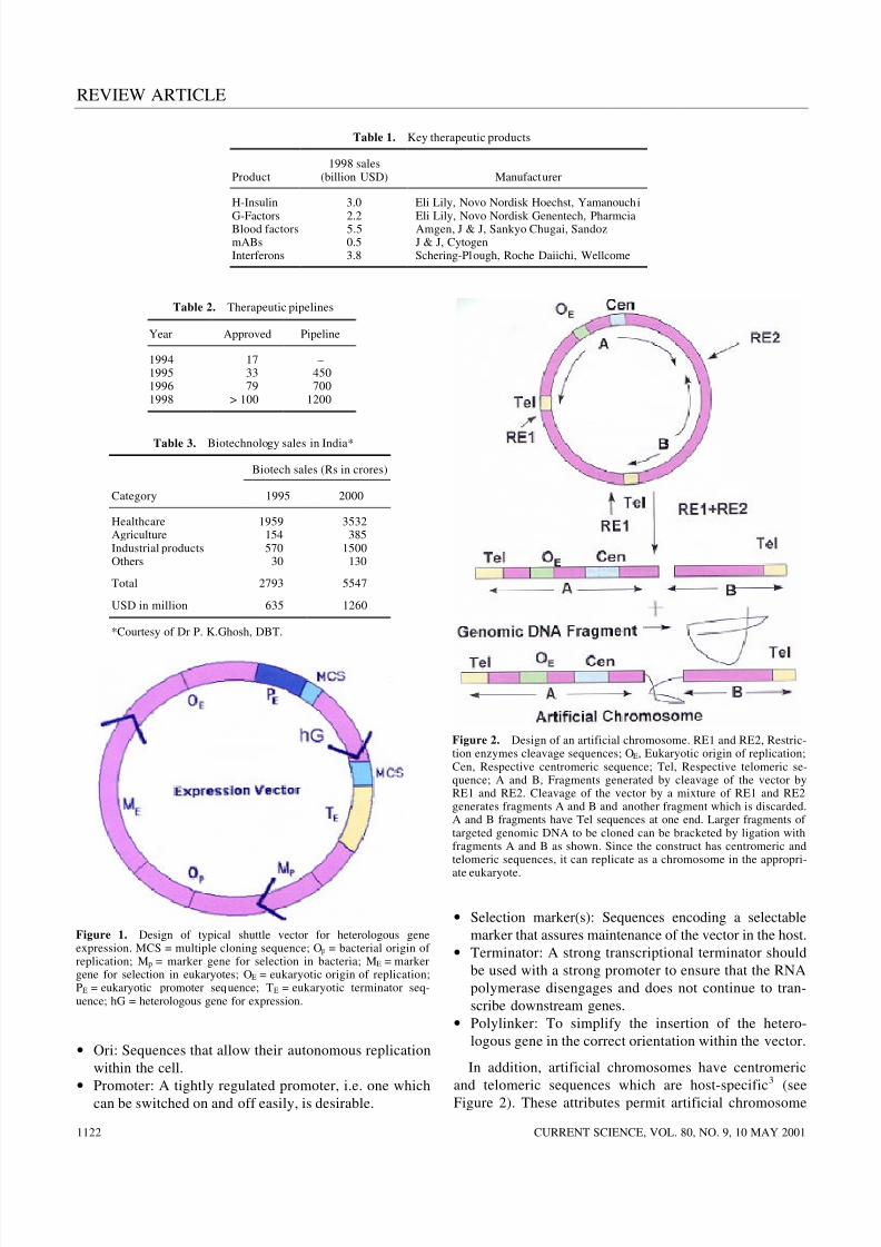
• Ori: Sequences that allow their autonomous replication
within the cell.
•Promoter: A tightly regulated promoter, i.e. one whichcan be switched on and off easily, is desirable.
• Selection marker(s): Sequences encoding a selectable
marker that assures maintenance of the vector in the host.
• Terminator: A strong transcriptional terminator should
be used with a strong promoter to ensure that the RNA
polymerase disengages and does not continue to tran-
scribe downstream genes.
• Polylinker: To simplify the insertion of the hetero-
logous gene in the correct orientation within the vector.
In addition, artificial chromosomes have centromeric
and telomeric sequences which are host-specific
3
(seeFigure 2). These attributes permit artificial chromosome
Table 1. Key therapeutic products
Product1998 sales
(billion USD) Manufacturer
H-Insulin 3.0 Eli Lily, Novo Nordisk Hoechst, Yamanouchi
G-Factors 2.2 Eli Lily, Novo Nordisk Genentech, PharmciaBlood factors 5.5 Amgen, J & J, Sankyo Chugai, SandozmABs 0.5 J & J, CytogenInterferons 3.8 Schering-Plough, Roche Daiichi, Wellcome
Table 3. Biotechnology sales in India*
Biotech sales (Rs in crores)
Category 1995 2000
HealthcareAgricultureIndustrial productsOthers
1959154570
30
3532385
1500130
Total 2793 5547
USD in million 635 1260
*Courtesy of Dr P. K.Ghosh, DBT.
Table 2. Therapeutic pipelines
Year Approved Pipeline
1994 17 –1995 33 4501996 79 7001998 > 100 1200
Figure 2. Design of an artificial chromosome. RE1 and RE2, Restric-tion enzymes cleavage sequences; OE, Eukaryotic origin of replication;Cen, Respective centromeric sequence; Tel, Respective telomeric se-quence; A and B, Fragments generated by cleavage of the vector byRE1 and RE2. Cleavage of the vector by a mixture of RE1 and RE2generates fragments A and B and another fragment which is discarded.A and B fragments have Tel sequences at one end. Larger fragments of targeted genomic DNA to be cloned can be bracketed by ligation withfragments A and B as shown. Since the construct has centromeric andtelomeric sequences, it can replicate as a chromosome in the appropri-
ate eukaryote.
Figure 1. Design of typical shuttle vector for heterologous geneexpression. MCS = multiple cloning sequence; Op = bacterial origin of replication; Mp = marker gene for selection in bacteria; ME = markergene for selection in eukaryotes; OE = eukaryotic origin of replication;PE = eukaryotic promoter sequence; TE = eukaryotic terminator seq-uence; hG = heterologous gene for expression.
8/7/2019 heterologous prot
http://slidepdf.com/reader/full/heterologous-prot 3/8
REVIEW ARTICLE
CURRENT SCIENCE, VOL. 80, NO. 9, 10 MAY 2001 1123
vectors to be faithfully replicated and distributed to
daughter cells during cell division. Use of artificial chro-
mosomes in commercial production of heterologous
proteins remains unexplored.
Available expression system
Prokaryotic and eukaryotic systems are the two general
categories of expression systems. Prokaryotic systems are
generally easier to handle and are satisfactory for most
purposes. However, there are serious limitations in using
prokaryotic cells for the production of eukaryotic pro-
teins. For example, many of the eukaryotic proteins un-
dergo a variety of post-translational modifications like
proper folding, glycosylation, phosphorylation, formation
of disulphide bridges, etc. There is no universal expres-
sion system for heterologous proteins. All expression sys-
tems have some advantages as well as some disadvantages
that should be considered in selecting which one to use.
Choosing the best one requires evaluating the options –
from yield to glycosylation, to proper folding, to econo-
mics of scale-up.
Bacterial system
E. coli is by far the most widely employed host, provided
post-translational modifications of the product are not
essential. Its popularity is due to the vast body of know-
ledge about its genetics, physiology and complete geno-
mic sequence which greatly facilitates gene cloning and
cultivation4,5
. High growth rates combined with the ability
to express high levels of heterologous proteins, i.e. strains
producing up to 30% of their total protein as the ex-
pressed gene product, result in high volumetric producti-
vity. Furthermore, E. coli can grow rapidly to high densities
in simple and inexpensive media. Strains used for recom-
binant production have been genetically manipulated so
that they are generally regarded as safe for large-scale
fermentation. Purification has been greatly simplified by
producing recombinant fusion proteins which can be
affinity-purified, e.g. glutathione-S-transferase and maltose-
binding fusion proteins6. However, expression in bacteriadoes have some serious disadvantages. It poses significant
problems in post-translational modifications of proteins.
Common bacterial expression systems such as E. coli
have no capacity to glycosylate proteins in either N- or
O-linked conformation. Although other bacterial strains
such as Neisseria meningirulls have recently been shown
to O-glycosylate some of their endogenous proteins, the
trisaccharide added is different from O-linked sugars
found in eukaryotes. Protein expressed in large amounts
often precipitates into insoluble aggregates called inclu-
sion bodies, from which it can only be recovered in an
active form by solubilization in denaturing agentsfollowed by careful renaturation
7. Lysis to recover the
cytoplasmic proteins often results in the release of endo-
toxins, which must be removed from the final product.
Currently, strategies to secrete the target proteins by trans-
location into the periplasmic space or to release the target
proteins by linking to existing excretory systems are being
developed8. Additionally, the efficiency of expression willalso depend on differences of codon utilization by bacteria9.
At times the original sequence of the heterologous gene
has to be modified to reflect the codon usage by the cho-
sen expression system. E . coli has toxic cell wall pyrogens
and hence products need to be tested more extensively
before use.
Yeast
Yeast is the favoured alternative host for expression of
foreign proteins for research, industrial or medical use10
.
As a food organism, it is highly acceptable for the produc-tion of pharmaceutical proteins. In contrast, E . coli has
toxic cell wall pyrogens and mammalian cells may contain
oncogenic or viral DNA. Compared to mammalian cells,
yeast can be grown relatively rapidly (doubling time
90 min) on simple media and to high cell density and its
genetics is more advanced than any other eukaryote, so
that it can be manipulated as readily. Added advantages
are the availability of complete genomic sequence, the
nuclear stable high copy plasmids and ability to secrete
the target protein11. Saccharomyces strains have high
copy stably-inherited plasmid of 6.3 kb known as 2-
micron plasmid, which codes for 4 genes FLP, REP1,
REP2 and D. It also contains an open reading frame
(ORF), STB locus (required in cis for stabilization) and
two 599 bp inverted repeat sequences. FLP encodes a
site-specific recombinase which promotes flipping about
the FLP recombination targets (FRT) within the inverted
repeats, so that cells contain two forms of 2-micron plas-
mids, A and B. The simple 2-micron shuttle vectors con-
tain the 2-micron ORI-STB, a yeast selectable marker and
bacterial plasmid sequence (ori and selection markers)
and are used in host strains which supply REP1 and REP2
proteins.
These lower eukaryotic systems are able to glycosylate
the target proteins, but it has been shown that both N- andO-linked oligosaccharide structures are however, signifi-
cantly different from their mammalian counterpartsl2,13
.
Hypermannosylation (addition of a large number of man-
nose residues to the core oligosaccharide) is a common
feature in yeast, hindering proper folding and therefore
the activity of the protein. At the moment yeast provides a
good compromise between bacteria on one side and
mammalian cell lines on the other.
Insect
The baculoviruses have emerged as a popular system foroverproducing recombinant proteins in eukaryotic
8/7/2019 heterologous prot
http://slidepdf.com/reader/full/heterologous-prot 4/8
REVIEW ARTICLE
CURRENT SCIENCE, VOL. 80, NO. 9, 10 MAY 20011124
cellsl4,15
. Several factors have contributed to their popu-
larity. Being eukaryotes, they use many of the protein
modifications, processing, and transport systems present
in higher eukaryotic cellsl6. They use a helper-indepen-
dent virus that can be propagated to high titers in insect
cells adapted for growth in suspension cultures, making itpossible to obtain large amounts of recombinant proteins
with relative ease. Expressed proteins are usually ex-
pressed in the proper cellular compartment, i.e. membrane
proteins are usually localized to the membrane,
nuclear proteins to the nucleus and secreted proteins
secreted into the medium. Majority of the overproduced
proteins remain soluble in insect cells. Viral genome is
large (130 kb) and thus can accommodate large fragments
of foreign DNA. Baculoviruses are non-infectious to
vertebrates and their promoters have been shown to be
inactive in mammalian cells, which gives them a possible
advantage over other systems when expressing oncogenesor potentially toxic proteins. Also the process develop-
ment time is short. Expression using baculoviral vectors
also has some limitations. Since baculoviruses infect
invertebrates, it is possible that the processing of proteins
produced by vertebrates is different and this seems to be
the case for some post-translational modifications, e.g.
internal proteolytic cleavages at arginine- or lysine-rich
sequences are highly inefficient. The glycosylation capa-
bility is generally limited to producing only high mannose
type and not processed to complex type oligosaccharides
containing fucose, galactose and sialic acid.
Mammalian cells17,18
Ideally, proteins requiring mammalian post-translational
modifications should be expressed in mammalian cells. If
product authenticity is absolutely essential for clinical
efficacy, then despite the many shortcomings, a mammalian
host is the only choice, as it offers the greatest degree of
product fidelity. It should, however, be noted that oligo-
saccharide processing is species- and cell type-dependent
among mammalian cells. Differences in glycosylation
pattern are reported in rodent cell lines and human tissues.
Even the use of human cell line is not perfect, since thetransformation event required in most cases to produce a
stable cell line may itself result in altered glycosylation
profiles. Also mammalian expression techniques are time
consuming and much more difficult to perform on a large
scale. Complex nutrient requirement and low product
concentration have meant that the end product must be
highly value-added for this approach to be commercially
viable.
Dictyostelium discoideum
Recently, the cellular slime mold Dictyostelium dis-coideum has been developed as an alternative eukaryotic
system for expressing recombinant proteins19,20
. Circular
plasmids are common in prokaryotes, but only a few
eukaryotes have been identified and studied for having
circular nuclear plasmids and Dictyostelium is one of
them21,22. The cellular slime molds have families of
similar plasmids which are found in the nuclei of differentspecies. They are organized differently from the yeast 2-
micron like plasmids. Dictyostelium plasmids are pack-
aged in a nucleosomal structure similar to the chromatin
organization of higher eukaryotes. The best characterized
family is that of the Ddp2-like plasmids. These plasmids
are small (4.4–5.8 kb), encode ORF and contain an
inverted repeat. The product of ORF is required in trans
for plasmid maintenance, while an approximately 600 bp
fragment, presumably containing the origin of replication
is required in cis. The second best characterized plasmid
family is Ddp1, found in wild type isolates like NC4 and
V12. Ddp1 is a 13.7 kb plasmid which is present at anestimated copy number of 50–100 per cell and encodes at
least 5 growth-specific and 5 development-specific tran-
scripts. Although Ddp1 is larger and highly transcribed
than the plasmids of the Ddp2-like family, none of the
known transcripts seems to be essential for its replication.
However, while approximately 1.2 kb region appeared
to carry all the elements necessary for extra chromosomal
replication, in the absence of selection, this fragment is
quickly lost from the sub-population. In addition, deletion
of plasmid sequences outside the region results in reduc-
tion of plasmid copy number. Therefore at least some of
the Ddp1-encoded genes are required for the long-term
maintenance of the plasmid at its normal copy number.
The development of reliable transformation systems for
D. discoideum has provided the possibility of expressing
heterologous genes in this microbe23,24. Dictyostelium is a
simple eukaryotic micro-organism with a haploid genome
of 5 × 10⁷ bp and a lifecycle that alternates between
single-celled and multicellular stages. They grow to a
high cell density without the serum factors or special aera-
tion needed by animal cell cultures. There is no cell wall
and the high copy number plasmid vectors allow the
expression of protein in cell-associated, membrane-
attached or secreted form under the control of regulatable
promoters25. The cells of Dictyostelium can carry out bothO- as well as N-glycosylation
26,27. The major advantages
of this system include a very simple and cheap growth
medium and the potential for large-scale production of
proteins.
Factors affecting intracellular expression
Having the target DNA in an appropriate vector in the
expression system of choice is the first step in optimizing
production of the heterologous proteins. Within a given
system the transcription and translation processes leadingto the heterologous protein production are a complex set
8/7/2019 heterologous prot
http://slidepdf.com/reader/full/heterologous-prot 5/8
REVIEW ARTICLE
CURRENT SCIENCE, VOL. 80, NO. 9, 10 MAY 2001 1125
of reactions. Each process is carried out and controlled by
several enzymes/factors. In recent years we have learnt
that the following few key steps or reactions are critical in
determining the ultimate outcome.
Initiation of transcription
Gene expression is most frequently regulated at the level
of transcription and it is generally assumed that the
steady-state mRNA level is a primary determinant of the
final yield of a foreign protein. The mRNA level is deter-
mined both by the rate of initiation and the rate of turn-
over. In most cases the yield of a foreign protein
expressed using a host promoter has been much lower
than the yield of the homologous protein, using the same
promoter (~ 50%) (refs 28, 29). Many factors could
account for these differences, e.g. downstream activating
sequences (DAS) and upstream activating sequences
(UAS)30
. If DAS are characterized, it may be possible to
incorporate them into upstream promoter fragments, in
order to create more efficient expression vectors.
Alternatively, if DAS prove to be strongly position-
dependent, they could be placed within an intron which
could be excised prior to translation. If neither of these
options works, then maximal transcription will only be
possible using fusion proteins.
RNA elongation
The elongation of transcripts is not thought normally to
affect the overall rate of transcription, but the yield of full
length transcripts could be affected by fortuitous
sequences in foreign genes which cause pausing or termi-
nation. These could either act in the same way as natural
host terminator or else by a different mechanism, e.g. in
yeast, though not widely recognized, this problem could
be a very common reason for low yields or complete fail-
ure of expression of foreign genes. At present the only
solution is to increase the AT/GC of the offending section
of genes by chemical synthesis.
RNA stability31,32
There is evidence that subtle changes in mRNA sequence
affect the stability of mRNA, with low mRNA stability
being a primary factor in poor yields of foreign proteins.
Where mRNA instability is diagnosed as a problem, over-
all yield might be improved by (i) using a more powerful
promoter, (ii) using a promoter with more rapid induction
kinetics or (iii) chemically synthesizing the gene with
altered codons or deleting the 3′ untranslated region in the
hope that the instability determinant will be removed.
Degradation of mRNA is also more pronounced underadverse growth conditions.
Gene dosage
Since the target gene is often incorporated into a plasmid
vector system, gene dosage is dependent on plasmid copy
number. As can be expected, an increase in copy number
results in concomitantly higher recombinant proteinproductivity, but not indefinitely. Plasmid copy number is
affected by plasmid and host genetics and also by cultivation
conditions such as growth rates, media and temperature33.
Initiation of translation34
Translational efficiency is a function of either transla-
tional initiation or elongation rate. Translational effici-
ency is controlled primarily by the rate of initiation.
Initiation in eukaryotes is thought to follow a scanning
mechanism, whereby the 40 S ribosomal subunit plusco-factors bind the 5′ cap of the mRNA and then migrate
down the untranslated leader scanning for the first AUG
codon. Any part of this process, which is affected by the
structure of the leader and the AUG content, could limit
the initiation rate. AUG is recognized efficiently as initia-
tion codon only when it is in the right context and optimal
content is found to be GCC(A or G)CCAUGG. The
purines (A or G) three bases before AUG and G immedi-
ately following it are found to be the most important,
influencing translation to the tune of 10-fold. The follow-
ing factors have also been found to be important for pro-
karyotes: (i) the ribosome-binding nucleotide sequence or
Shine–Dalgarno (S–D) sequence; (ii) the distance between
the initiation codon and S–D, and (iii) the secondary
structure of mRNA.
Translational elongation
Translational elongation does not effect the yield or quality
of polypeptide normally, but it can become limiting with
very high mRNA levels. Codon usage is considered a
potential factor affecting product yield. Despite the
degeneracy of the genetic code, a non-random codon
usage is found in most organisms. The codon usage of most genes reflects the nucleotide composition of the
genome; highly-expressed genes show a strong bias to-
wards a subset of codons35. This major codon bias, which
can vary greatly between organisms, is thought to be a
growth optimization strategy such that only a subset of
tRNAs and aminoacyl-tRNA synthetases are needed at high
concentration for efficient translation of highly-expressed
genes at fast growth rates36
. Rare codons, for which the
cognate tRNA is less abundant, are translated at a slower
rate, but this will not normally affect the level of product
from an mRNA, since initiation is usually rate-limiting37
.
A ribosome finishing translation of one mRNA moleculeis most likely to initiate translation of a different mRNA

8/7/2019 heterologous prot
http://slidepdf.com/reader/full/heterologous-prot 6/8
REVIEW ARTICLE
CURRENT SCIENCE, VOL. 80, NO. 9, 10 MAY 20011126
species, unless the original species comprises a large pro-
portion of the total mRNA. Thus, the overall rate of trans-
lation of an mRNA is not usually affected by a slower
elongation rate unless ribosomes become limiting, which
would affect all transcripts in the cell. In contrast to the
normal situation, there is evidence that codon usage mayaffect both the yield and quality of a protein when a gene
is transcribed to very high levels. With very high levels of
mRNA containing rare codons, aminoacyl-tRNAs may
become limiting, increasing the probability of mistransla-
tion38, which is the incorporation of an amino acid which
does not correspond to the codon being translated, and
possibly causing ribosomes to drop off. Thus the codon
content of a foreign gene may influence the yield of pro-
tein where the mRNA is produced at very high levels.
This may more likely occur on growth in minimal
medium, when the cell produces a wide variety of biosyn-
thetic enzymes encoded by genes containing rare codons.The effect on product quality has been difficult to meas-
ure, but requires further attention since it has further
implications for therapeutic proteins. Proteins containing
amino acid mis-incorporation are difficult to separate and
may affect the activity and antigenicity of the product.
Since small genes are now frequently synthesized chemi-
cally, they may be easily and perhaps profitably engi-
neered to contain optimal codons for high-level expres-
sion. mRNA secondary structure, in addition to codon
usage may affect translational elongation39.
Polypeptide folding
During or following translation, the polypeptide must fold
so as to adopt its functionally-active conformation. Since
many denatured proteins can be refolded in vitro, it
appears that the information for correct folding is con-
tained in the primary polypeptide structure40. However,
folding comprises rate-limiting steps during which some
molecules may aggregate, particular!y at high rates of
synthesis and at higher temperatures. There is evidence
that certain heat shock proteins act as molecular chaper-
ones in preventing the formation and accumulation of
unfolded aggregates, while accelerating the folding reac-tions. Due to the intrinsic nature of polypeptide folding
and low specificity of chaperones, it is very unlikely that
foreign cytosolic proteins will accumulate in non-native
conformations, but when fragments of proteins or fusion
proteins are expressed, normal folding domains may
be perturbed, resulting in an insoluble product. Insolu-
ble proteins can often be renatured in vitro, though
the techniques for this can be complex and unpredict-
able41
. In contrast to intracellular proteins, naturally
secreted proteins encounter an abnormal environment in
the cytoplasm; disulphide bond formation is not favoured
and glycosylation cannot occur. In E . coli, foreign pro-teins are frequently insoluble, but low temperature has
been found to increase solubility in some cases42
. This
may be due to a decreased translation rate or to the fact
that hydrophobic interactions, such as those occurring in
aggregates, become less favourable. A dramatic increase
in the yield of active, soluble protein is observed on
reducing the rate of induction43.
Post-translational processing
Prokaryotic expression systems are generally useful for
producing heterologous proteins from cloned eukaryotic
cDNA. In some cases however, eukaryotic proteins that
have been synthesized in bacteria are either unstable or
lack biological activity. The inability of prokaryotic
organisms to produce authentic versions of proteins is for
the most part due to the absence of appropriate mecha-
nisms for generating certain post-translational modifica-tions. In eukaryotes there are a number of modifications
that may occur at the post-translational stage, after protein
synthesis is complete.
Amino-terminal modifications of polypeptides: These
are the most common processing events and occur on
most cytosolic proteins44
. Two types of events normally
occur – removal of the N-terminal Met residue, catalysed
by Met aminopeptidase (MAP) and acetylation of the N-
terminal residue, catalysed by N-acetyltransferase (NAT).
Both enzymes are associated with ribosomes and act on
nascent polypeptide. In most cases the structure of the N-
terminus should not affect the biological activity of a pro-
tein, but there may be exceptions, e.g. the response of
haemoglobin to physiological modifiers involves the N-
terminus and correct folding of alpha and beta globins
is therefore advantageous. Similarly N-acetylation of
melanocyte-stimulating hormone is required for full bio-
logical activity.
Disulphide bond formation: In eukaryotes, formation of
disulphide bond (cys-s-s-cys) occurs in the lumen of RER
and is mediated by an enzyme called disulphide isom-
erase45. Disulphide bond is confined to secretory proteins
and exoplasmic membrane proteins. This is important instabilization of tertiary structure. An improperly folded
protein is unstable and lacks activity.
Proteolytic cleavage of a precursor form: This is required
in some cases. Selected segments of amino acid sequences
are removed to yield a functional protein46
.
Glycosylation: This is the most extensive of all the post-
translational modifications and has important function in
secretion, antigenicity and clearance of glycoproteins47
.
Oligosaccharides can attach to proteins in three ways:
(i) Via an N-glycosidic bond to the R-group of anAsn-residue within the consensus sequence Asn-X-Ser/
8/7/2019 heterologous prot
http://slidepdf.com/reader/full/heterologous-prot 7/8
REVIEW ARTICLE
CURRENT SCIENCE, VOL. 80, NO. 9, 10 MAY 2001 1127
Thr (N-glycosylation)48
. All mature N-linked glycan
structures have a common core of Man.GlcNAc, which
can form part of simple oligomannose structures or be
extensively modified by other residues such as fucose,
galactose and sialic acid. Hybrid structures also exist
where one or more arms of the glycan are modified andthe remaining arms contain only mannose. (ii) Via an
O-gIycosidic bond to the R group of the Ser or Thr
(O-glycosylation). O-linked glycosylation is extensive in
structural proteins such as proteoglycans. Small glycan
structures can also be O-linked to the side chain of
hydroxylysine or hydroxyproline. (iii) Carbohydrates are
also components of the glycophosphotidylinositol anchor
used to secure some proteins to the cell membrane. The
presence of these consensus sequences by no means guar-
antees their glycosylation. They show varying degrees of
occupancy with oligosaccharides (macroheterogeneity)
depending on their position within the protein and its con-formation, the host cell type used for expression and its
physiological status. These three factors also determine
the extent of variation in the type of sugar residues found
within each oligosaccharide (microheterogeneity). Glyco-
sylation is both organism- and cell type-specific and
therefore expression of a protein in a heterologous system
will almost certainly result in a product with different
modification from the native protein. This may affect the
function and immunogenicity of the protein49,50.
Modification of amino acid within proteins: Modifications
of this type include phosphorylation, acetylation, sulphation,acylation (carboxylation, myristylation and palmitylation)
51.
Of these modifications, prokaryotic host cells are least
likely to carry out either proper glycosylation or additions
to specific amino acids within the heterologous protein.
Moreover, no single eukaryotic host cell system is capable
of performing all the possible post-translational modifica-
tions for every potential heterologous protein. Therefore,
if a particular protein requires a specific set of modifica-
tions, then it may be necessary to examine different
eukaryotic expression systems to find the one that can
produce a biologically authentic product.
Stability of intracellular proteins
So far processes affecting the rate of synthesis of proteins
have been considered, but the ultimate yield is equally
affected by the rate of degradation52
. Yields might logi-
caIly be improved by the following measures: (i) fusion to
a stable protein53,54
; (ii) secretion to segregate the product
from intracellular proteases55
; (iii) using a more rapidly
induced promoter; (iv) using additional protease inhibi-
tors to minimize degradation during extraction; (v) induc-
ing at lower temperature; and (vi) harvesting cells in theexponential growth phase.
Stability of plasmid
Plasmid instability is a major problem in continuous and
large-scale fermentation, since these cultures go through
many generations. The resulting effects are lower produc-
tivity and increased production cost, because of the build-upof non-productive plasmid-free cells. Plasmid instability
is categorized as segregational instability56,57
and struc-
tural instability58,59. Segregational instability is the loss of
plasmid from one of the daughter cells during division
because of defective partitioning. Structural instability is
attributed to deletions, insertions and rearrangements in
the plasmid structure, resulting in the loss of the desired
gene function. Plasmid stability is influenced by the vec-
tor and host genotypes; the same plasmid in different
hosts exhibits different degrees of stability and vice versa.
The origin and size of foreign DNA have been observed
to affect the plasmid stability. Plasmid stability is also afunction of physiological parameters that affect the
growth rate of the host cell, which include pH, tempera-
ture, aeration rate, medium components and heterologous
protein accumulation. Mathematically structured and un-
structured kinetic models of plasmid stability have been
developed which are ultimately useful for the design of
recombinant processes.
Summary and conclusion
The eventual objective of producing a desired protein inan economical heterologous host is influenced by a vari-
ety of factors. However, maximizing production of
heterologous proteins for commercial application is
still an art. We have begun to understand factors influ-
encing the eventual production. These factors, described
in detail in this article are varied and at times poorly
understood. Largely the approach remains empirical.
However, our collective experience will permit us to
rationalize our approach in designing heterologous pro-
duction of commercially important proteins in a variety of
expression systems. Subsequent to production, stabiliza-
tion and formulation of proteins will pose significant
hurdles in utilizing the natural biological catalysts and
other proteins for therapeutic and industrial purposes.
1. Ghosh, P. K., Aust. Biotechnol., 1998, 3, 214–222.
2. Padh, H., BioPharma (USA), 1999, 12, 18–19.
3. Ikeno, M. et al., Nat . Biotechnol., 1998, 16, 431–439.
4. Shatzman, A. R. and Rosenberg, M., Methods Enzymol., 1987,
152, 661–673.
5. Casadaban, M., Martinez-Arias, A., Shapira, S. and Chou, J.,
Methods Enzymol., 1983, 100, 293–308.
6. Maina, C. V. et al., Gene, 1988, 74, 365–373.
7. Marston, F. A. O. and Hartley, D. L., Methods Enzymol., 1990,
182, 264–276.8. Robinson, M. et al., Nucleic Acids Res., 1984, 12, 6663–6671.
8/7/2019 heterologous prot
http://slidepdf.com/reader/full/heterologous-prot 8/8
REVIEW ARTICLE
CURRENT SCIENCE, VOL. 80, NO. 9, 10 MAY 20011128
9. Schein, C. H., Bio/Technology, 1989, 7, 1141–1148.
10. Hitzeman, R. A., Hagie, F. F., Levine, H. L., Goeddel, D. W.,
Ammerer, G. and Hall, B. D., Nature, 1981, 293, 717–732.
11. Hitzeman, R. A. et al., Methods Enzymol., 1990, 185, 421–
440.
12. Kukuruzinska, M. A., Bergh, M. L. E. and Jackson, B. L., Annu.
Rev. Biochem., 1987, 56, 915–944.13. Kornfeld, R. and Kornfeld, S., Annu. Rev. Biochem., 1985, 54,
631–664.
14. Kitt, P. A. and Possee, R. D., BioTechniques, 1993, 14, 810–817.
15. O’Reilly, D. R., Miller, L. K. and Lucknow, V. A., Baculovirus
Expression System – A Laboratory Manual, Oxford University
Press, New York, 1992.
16. Matsuura, Y., Possee, R. D. and Bishop, D. H. L., J . Gen. Virol.,
1987, 68, 1233–1250.
17. Kaufman, R. J., Methods Enzymol., 1990, 185, 537–566.
18. Kaufman, R. J., Methods Enzymol., 1990, 185, 487–511.
19. Dittrich, W., Williams, K. L. and Slade, M. B., Biotechnology
(NY ), 1994, 12, 614–618.
20. Tiltscher, H. and Storr, M., Appl. Microbiol. Biotechnol., 1993,
40, 246–250.
21. Ashktorab, H. and WeIker, D. L., Gene, 1988, 65, 41–49.22. Ahern, K. G., Howard, P. K. and Firtel, R. A., Nucleic Acids Res.,
1988, 16, 6825–6837.
23. Firtel, R. A., Silan, C., Ward, T. E., Howard, P., Metz, B. A.,
NelIen, W. and Jacobson, A., Mol. Cell. Biol., 1985, 5, 3241–
3250.
24. Hughes, J. E., Podgorski, G. J. and WeIker, D. L., Plasmid , 1992,
28, 37.
25. Manstein, D. J., Schuster, H. P., Morandins, P. and Hint, D. M.,
Gene, 1995, 162, 129–134.
26. Jung, E. and Williams, K. L., Biotechnol. Appl. Biochem., 1997,
25, 3–8.
27. Jung, E., Gooley, A. A., Packer, N. H., Slade, M. B., Williams,
K. L. and Dittrich, W., Biochemistry, 1997, 36, 4034–4040.
28. Mellor, J., Dobson, M., Roberts, N. A., Kingsman, A. J. and
Kingsman, S. M., Gene, 1985, 33, 215–226.
29. Chen, C. Y., Oppermann, H. and Mitgeenan, R. A., Nucleic Acids
Res., 1984, 12, 8951–8970.
30. Purvis, I. J., Betlany, A. J. E., Loughlin, L. and Brown, A. J. P.,
Nucleic Acids Res., 1987, 15, 7963–7974.
31. Humphrey, T., Sadhale, P., Platt, T. and Proudfoot, N., EMBO J .,
1991, 10, 3503–3511.
32. Romanos, M. A. et al., Nucleic Acids Res., 1991, 19, 1461–1467.
33. Hughes, J. E. and WeIker, D. L., Plasmid , 1989, 22, 215–223.
34. Kozak, M., J . Cell Biol., 1989, 108, 229–241.
35. Bennetzen, J. L. and Hall, B. D., J . Biol. Chem., 1982, 257, 3026–
3031.
36. Kurland, C. G., Trends Biochem. Sci., 1987, 12, 126–128.
37. Pedersen, S., EMBO J ., 1984, 3, 2895–2898.
38. Scorer, C. A., Carrier, M. J. and Rosenberger, R. F., Nucleic Acids
Res., 1991, 19, 3511–3516.
39. Baim, S. B., Pietras, D. F., Eustice, D. C. and Sherman, F., Mol. Cell. Biol., 1985, 5, 1839–1846.
40. Gething, M. J. and Sambrook, J., Nature, 1992, 355, 33–45.
41. Marston, F. A. O., Biochem. J ., 1986, 240, 1–12.
42. Schein, C. H. and Noteborn, M. H. N., Bio/Technology, 1988, 6,
291–294.
43. Kopetzki, E., Schumacher, G. and Buckel, P., Mol. Gen. Genet .,
1989, 216, 149–155.
44. Kendall, R. L., Yamada, R. and Bradshaw, R. A., Methods Enzy-
mol., 1990, 185, 398–407.
45. Freedman, R. B., Cell, 1989, 57, 1069–1072.
46. Thim, L. et al., Proc. Natl. Acad . Sci. USA, 1986, 83, 6766–6770.
47. Rademacher, T. W., Parekh, R. B. and Dwek, R. A., Annu. Rev.
Biochem., 1988, 57, 785–838.
48. Komfeld, R. and Komfeld, S., Annu. Rev. Biochem., 1985, 54,
631–664.49. Parekh, R. B. et al., Biochemistry, 1989, 28, 7670–7679.
50. Kukuruzinska, M. A., Bergh, M. L. E. and Jackson, B. L., Annu.
Rev. Biochem., 1987, 56, 915–944.
51. Tames, G. and Olson, E. N., Biochemistry, 1990, 29, 2623–2634.
52. Dice, F., FASEB J ., 1987, 1, 349–357.
53. Lees, N., Cozzjkorto, J., Wainwright, N. and Testa, D., Nucleic
Acids Res., 1984, 12, 6797–6812.
54. Cousens, L. S. et al., Gene, 1987, 61, 265–275.
55. Itoh, Y. and Fujisawa, Y., Biochem. Biophys. Res. Commun.,
1986, 141, 942–948.
56. Cashmore, A. M., Aibury, M. S., Hadfield, C. and Meacock,
P. M., Mol. Gen. Genet ., 1986, 203, 154–162.
57. Scott, J. R., Microbiol. Rev., 1984, 48, 1–23.
58. Novick, R. P., Microbiol. Rev., 1987, 51, 381–395.
59. Ahern, K. G., Howard, P. K. and Firtel, R. A., Nucleic Acids Res.,
1988, 16, 6825–6837.
ACKNOWLEDGEMENTS. We thank the Council of Scientific and
Industrial Research (project 38(973)99-EMR-II) for supporting re-
search work in H.P.’s laboratory and award of Senior Research Fellow-
ship to M.R.
Received 5 July 2000; revised accepted 15 December 2000