Embed Size (px)
Citation preview

www.cell-research.com | Cell Research
Qing Wei et al.949npg
ORIGINAL ARTICLE
Heterotrimeric G-protein is involved in phytochrome A-mediated cell death of Arabidopsis hypocotylsQing Wei1, Wenbin Zhou1, Guangzhen Hu1, Jiamian Wei1, Hongquan Yang1, Jirong Huang1
1National Key Laboratory of Plant Molecular Genetics, Institute of Plant Physiology and Ecology, Shanghai Institutes for Biological Sciences, Chinese Academy of Sciences, 300 Fenglin Road, Shanghai 200032, China
The heterotrimeric guanine nucleotide-binding protein (G-protein) has been demonstrated to mediate various signal-ing pathways in plants. However, its role in phytochrome A (phyA) signaling remains elusive. In this study, we discover a new phyA-mediated phenotype designated far-red irradiation (FR) preconditioned cell death, which occurs only in the hypocotyls of FR-grown seedlings following exposure to white light (WL). The cell death is mitigated in the Gα mutant gpa1 but aggravated in the Gβ mutant agb1 in comparison with the wild type (WT), indicative of antagonistic roles of GPA1 and AGB1 in the phyA-mediated cell-death pathway. Further investigation indicates that FR-induced accumulation of nonphotoconvertible protochlorophyllide (Pchlide633), which generates reactive oxygen species (ROS) on exposure to WL, is required for FR-preconditioned cell death. Moreover, ROS is mainly detected in chloroplasts us-ing the fluorescent probe. Interestingly, the application of H2O2 to dark-grown seedlings results in a phenotype similar to FR-preconditioned cell death. This reveals that ROS is a critical mediator for the cell death. In addition, we observe that agb1 is more sensitive to H2O2 than WT seedlings, indicating that the G-protein may also modify the sensitivity of the seedlings to ROS stress. Taking these results together, we infer that the G-protein may be involved in the phyA signaling pathway to regulate FR-preconditioned cell death of Arabidopsis hypocotyls. A possible mechanism underlying the involvement of the G-protein in phyA signaling is discussed in this study. Keywords: heterotrimeric G protein, phytochrome A, ROS, cell death, PchlideCell Research (2008) 18:949-960. doi: 10.1038/cr.2008.271; published online 12 August 2008
Correspondence: Jirong HuangTel: +86-21-54924145; Fax: +86-21-54924015E-mail: [email protected] 19 November 2007; revised 19 February 2008; accepted 7 March 2008; published online 12 August 2008
npgCell Research (2008) 18:949-960.© 2008 IBCB, SIBS, CAS All rights reserved 1001-0602/08 $ 30.00 www.nature.com/cr
Introduction
Heterotrimeric guanine nucleotide-binding proteins (G-proteins) are signaling switches that are highly conserved in eukaryotes. These proteins precisely transduce extracellular signals from the ligand-activated G-protein-coupled recep-tors (GPCR) to the downstream effectors. In vertebrates, G-proteins are involved in the recognition and transduction of diverse signals such as light, calcium, odorants, small molecules, and proteins [1, 2]. In contrast to mammalian cells that contain 23 Gα, 6 Gβ, and 12 Gγ genes, plant cells have a smaller number of genes encoding G-protein subunits. For example, the Arabidopsis genome encodes single canonical α (GPA1) and β (AGB1) subunits and
two γ (AGG1 and AGG2) subunits [3-6]. The other com-ponents central to G-protein signaling, such as GPCR and regulators of G-protein signaling (RGS), are also sparsely found in plants [7]. Nevertheless, the G-protein signaling pathway has been demonstrated to mediate a wide range of plant responses to light, phytohormones, sugar, and various stresses [7, 8]. Further, there are an increasing number of physiological processes that are mediated by the G-protein signaling pathway [9, 10].
Light is an important signal that dramatically influences plant growth and development as well as photosynthesis [11]. Plants possess several photoreceptors including the blue/ultraviolet A (UV-A)-absorbing cryptochromes and the red/far-red irradiation (FR)-absorbing photochromes to monitor the light quality, intensity, and directionality [12]. Since the visual transduction cascade in rod photoreceptor cells represents a classical model system for GPCR signal-ing [13], this well-understood signaling pathway has pro-moted researchers to investigate a similar role of G-proteins in the light sensing of plants. The early experiments to test this hypothesis were conducted with two adenosine diphos-
Cell Research | Vol 18 No 9 | September 2008
G-protein involvement in phyA-mediated cell death950npg
phate (ADP)-ribosylating drugs, namely, the cholera toxin that constitutively activates the G-protein pathway and the pertussis toxin that switches off the G-protein signaling pathway [14-17]. Similarly, two chemical agents, GTPγs (a Gα-subunit activator) and GDPβs (a Gα-subunit inhibitor), have also been used in the study of the G-protein signaling pathway [18, 19]. The results derived from these experi-ments supported the hypothesis that the G-protein on the plasma membrane is involved in light signaling pathways. The most interesting results reflecting G-protein mediation of light signaling were derived from the study on a tomato phytochrome A (phyA)-deficient mutant known as aurea, in which chloroplast development is severely suppressed [18, 19]. Microinjection of exogenous GTPγs and cholera toxin into aurea hypocotyl cells restored chloroplast de-velopment and anthocyanin biosynthesis, suggesting that G-protein activation triggers almost a complete cellular response initiated by phyA.
Recently, two papers have published conflicting data on G-protein involvement in phyA-mediated signal transduc-tion using genetic and molecular approaches [20, 21]. Oka-moto et al. [20] showed that the overexpression of either a native or a constitutively active form of GPA1 driven by a glucocorticoid-inducible promoter resulted in hypersensi-tivity to light, including red, FR, and blue light that affected hypocotyl elongation. It was found that phyA was essential for the enhanced response of the Gα overexpressors after exposure to FR; this result supported the involvement of the G-protein in the phyA signaling pathway. In contrast, using the null mutants and various overexpressors of G-protein subunits, Jones’ group [21] eliminated the possibility of a direct role for the G-protein in red- and FR-light signaling with regard to hypocotyl growth in Arabidopsis. These conflicting results raise questions regarding the involve-ment of the G-protein in phyA signaling.
In this study, we discovered a new phyA-dependent phenotype designated FR-preconditioned cell death. Re-cent reports have revealed that the G-protein is involved in cell death induced by oxidative stress (such as ozone) and pathogenic invasion [22-24]. Thus, we took advantage of the phyA-dependent cell death system to examine whether the G-protein is involved in this process. Our results dem-onstrate that gpa1 and agb1 mutants had opposite effects on phyA-dependent cell death, and support the hypothesis that the G-protein is involved in the phyA signaling pathway.
Results
FR-preconditioned cell death is phyA-dependentWhen wild-type (WT) seedlings were continuously
grown in FR (fluence rate 5 µmol/m2/s) for 3 d followed by continuous exposure to white light (WL) (fluence rate
70 µmol/m2/s) for an additional 3 d in half-strength MS medium containing 1% sucrose, we observed that most hypocotyls were dehydrated, and they then wilted. This occurred despite the fact that their cotyledons were able to partially turn green (Figure 1A). A careful examination showed that cell death initiated from the middle section of the hypocotyls 1 d after the seedlings were transferred from FR to WL. However, the seedlings that were grown in blue or red light for 3 d and then transferred to WL did not exhibit this phenotype (data not shown). Thus, we designated this phenotype as FR-preconditioned cell death. As far as we know, such cell death in hypocotyls has not been reported to date. Cell death was not visible if FR-grown seedlings were transferred to the dark; this
Figure 1 FR-preconditioned cell death of hypocotyls is phyA-dependent. (A) Morphology of WT, phyA, and phyB seedlings. Seedlings were grown in darkness for 1.5 d, then FR for 3 d followed by a 3-d exposure to WL. Arrows indicate light-induced wilting of the middle section of the hypocotyls. Scale bar, 1 mm. (B) Percentage of WT, phyA, and phyB seedlings exhibiting hypocotyl cell death. FR-WL indicates that seedlings grown in darkness for 1.5 d followed by FR for 3 d were transferred to WL for 3 d. Dark-WL indicates that seedlings grown in the dark for 5 d were transferred to WL for 3 d. The bars indicate the standard deviations from 3 independent experiments. Asterisks above the bar indicate a significant difference in cell death (P < 0.01) between phyA and WT or phyB seedlings.
A
B
WT phyA phyB
WT phyA phyB
FR-WL Dark-WL
See
dlin
gs w
ith h
ypoc
otyl
cell
deat
h (%
)
100
80
60
40
20
0
**
www.cell-research.com | Cell Research
Qing Wei et al.951npg
indicated that light was required for cell death. We also observed that the longer the seedlings were incubated in FR, the more the hypocotyls wilted (data not shown). In addition, unlike FR-blocked cotyledon greening that can be rescued by addition of sugar to the growth medium [25], FR-preconditioned cell death of hypocotyls occurred in the presence of sugar. It was noteworthy that seedlings with wilted hypocotyls could develop new green leaves in a plate (Figure 1A); this indicated that the shoot meristem was insensitive to FR.
Because only the photoreceptor phyA senses FR, we questioned whether cell death was specifically mediated by phyA. Therefore, we assayed the hypocotyl response of the phyA and phyB mutants together with the WT under similar conditions. In order to minimize the difference in the rapidity of seed germination, seeds stratified at 4 ºC for 4 d were germinated for 1.5 d in the dark. The etiolated seedlings were then incubated in FR for 3 d and subse-quently in WL for 3 d. As expected, wilted hypocotyls were
not visible in the phyA mutant but were clearly observed in the phyB mutant (Figure 1A). In order to quantitatively determine the degree of cell death, the seedlings were stained with Evans blue solution to specifically stain the dead cells [26]. The results showed that 72.6% and 71.2% of phyB and WT seedlings, respectively, were stained by Evans blue, whereas only 7.8% of the phyA plants were stained (Figure 1B). The seedlings grown in the dark for 5 d and subsequently exposed to WL continuously for 3 d were used as a control; these seedlings were barely stained by Evans blue (Figure 1B). Thus, FR-preconditioned cell death was specifically mediated by phyA.
FR-preconditioned cell death is affected by the G-proteinBased on increasing evidence indicating that the G-pro-
tein signaling pathway is involved in the process of cell death induced by various stresses [22-24], we explored whether the G-protein also modulated FR-preconditioned cell death. As shown in Figure 2A, among the hypocotyls
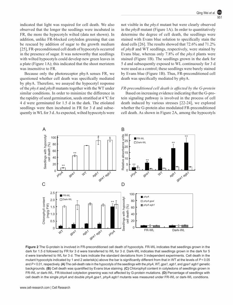
Figure 2 The G-protein is involved in FR-preconditioned cell death of hypocotyls. FR-WL indicates that seedlings grown in the dark for 1.5 d followed by FR for 3 d were transferred to WL for 3 d. Dark-WL indicates that seedlings grown in the dark for 5 d were transferred to WL for 3 d. The bars indicate the standard deviations from 3 independent experiments. Cell death in the mutant hypocotyls indicated by 1 and 2 asterisk(s) above the bar is significantly different from that in WT at the levels of P < 0.05 and P < 0.01, respectively. (A) The cell-death rate in the hypocotyls of the seedlings with the phyA, WT, gpa1, agb1, and gpa1 agb1 genetic backgrounds. (B) Cell death was quantified by Evans blue staining. (C) Chlorophyll content in cotyledons of seedlings grown in FR-WL or dark-WL. FR-blocked cotyledon greening was not affected by G-protein mutations. (D) Percentage of seedlings with cell death in the single phyA and double phyA gpa1, phyA agb1 mutants was measured under FR-WL or dark-WL conditions.
A
C
B
D
See
dlin
gs w
ith h
ypoc
otyl
cell
deat
h (%
)
100
80
60
40
20
0
30
25
20
15
10
5
0
**FR-WL
Dark-WL
See
dlin
gs w
ith h
ypoc
otyl
cell
deat
h (%
)
100
80
60
40
20
0FR-WL Dark-WL
FR-WL
Dark-WLphyA
phyA gpal
phyA agb1
Chl
orop
hyII
cont
ent
(mg/
g FW
)
2.5
2.0
1.5
1.0
0.5
0
Rel
ativ
e ab
sorb
ance
uni
ts
phyAgpa1 WT
agb1
gpa1 agb1
WTgpa1
agb1
gpa1 agb1phyA
phyA
gpa1 agb1agb1WT
gpa1
****
**
**
**
**
**

Cell Research | Vol 18 No 9 | September 2008
G-protein involvement in phyA-mediated cell death952npg
evaluated by Evans blue staining, the Gα null mutant gpa1 exhibited a lower cell-death rate (45.2%) while the Gβ null mutant agb1 exhibited a higher rate (90.1%) compared to the WT. This suggested that GPA1 was a positive player while AGB1 was a negative player in FR-preconditioned cell death. The antagonistic action of the GPA1 and AGB1 subunits was also demonstrated during cell death caused by pathogens and ozone stress [22-24]. In addition, the gpa1 agb1 double mutant exhibited a cell-death rate similar to that of the agb1 mutant, indicating that AGB1 was the main transducer of the G-protein signaling pathway.
To quantify FR-preconditioned cell death, the Evans blue stain was extracted from the dyed hypocotyls and measured by spectrometry. Consistent with the other results, the gpa1 mutant exhibited lower intensity Evans blue staining while the agb1 and gpa1 agb1 mutants exhibited higher intensity staining as compared to the WT (Figure 2B). There was no difference observed in cell death between the WT and the mutant seedlings that were grown in the dark for 5 d and then exposed to WL (Figure 2B). In addition, we examined the effect of the G-protein on FR-blocked cotyledon green-ing, which is a typical phenotype mediated by the phyA signaling pathway. Our results showed that the chlorophyll contents in the cotyledons of WT, gpa1, and agb1 seedlings grown either in FR or in the dark, was very similar (Figure 2C); however, the chlorophyll content of these seedlings grown in FR was significantly lower than that of phyA (Fig-ure 2C). This indicated that the G-protein was not involved in the process of FR-suppressed cotyledon greening. Taken together, the results suggested that the G-protein-mediated phyA signaling pathway was tissue specific.
To obtain genetic evidence, we constructed phyA gpa1 and phyA agb1 double mutants in order to examine whether phyA and G-protein were localized in the same pathway during FR-preconditioned cell death. Our results showed that both phyA gpa1 and phyA agb1 double mutants ex-hibited the same rate of hypocotyl cell death as that of the
phyA mutant (Figure 2D); this suggested that the sole FR receptor phyA is epistatical to the G-protein.
Thylakoid formation 1 (THF1) protein is involved in FR-preconditioned cell death
THF1 is identified as a GPA1 interacting protein that is highly conserved in oxygenic photosynthetic organisms
Figure 3 THF1 is involved in FR-preconditioned cell death. Results from WT, single (thf1 and gpa1) and double (gpa1 thf1) mutant seedlings are presented. The values are the mean ± SD (n = 3). Asterisks above the bar indicate p < 0.01 vs WT.
See
dlin
gs w
ith h
ypoc
otyl
cell
deat
h (%
)
80
60
40
20
0 WT thf1 gpa1 gpa1 thf1
Figure 4 FR-preconditioned cell death is not associated with POR expression. (A) Levels of PORA and PORB transcripts were ana-lyzed by RT-PCR after 28 cycles in hypocotyls of seedlings. The actin transcript was used as a quantitative control. (B) The total level of POR protein in hypocotyls of seedlings was analyzed by western blotting. The band marked with a star represents the POR protein. Other bands are the degraded forms of POR.
α-POR
A
B
Dark
WT phyA gpa1 agb1
WT phyA gpa1 agb1
FR
PORA
PORB
Actin
PORA
PORB
Actin
phyA WT gpa1 agb1
★
Dark
FR
**
**
**
★

www.cell-research.com | Cell Research
Qing Wei et al.953npg
is not associated with FR-preconditioned cell death of hypocotyls
Previous studies on the FR-blocked cotyledon greening have revealed that POR is a critical determiner of seedling greening and survival [25, 28]. The phyA mutant may have escaped cell death because the expression of PORA is not suppressed under FR, and PORA-bound protochlorophyl-lide (Pchlide) is converted to non-toxic chlorophyllide (Chlide) in the presence of light [28]. We therefore stud-ied the role of POR in FR-preconditioned cell death. Our RT-PCR analysis data with gene-specific primers showed that both PORA and PORB were expressed in the dark (Figure 4A). After FR exposure, PORA was only detected in the hypocotyls of phyA (Figure 4A), whereas PORB was expressed in all genetic backgrounds (Figure 4A). A comparison of the PORB expression levels in FR-grown hypocotyls demonstrated that the expression in agb1 was
[27]. Although it has been shown that THF1 functions in the sugar signaling pathway mediated by the G-protein, it is not clear whether THF1 is also involved in FR-precondi-tioned cell death. We adopted a genetic approach to answer this question using the thf1 null mutant. The result showed that the death rate of the thf1 seedlings was 18.7% (Figure 3), which was lower than that of WT and gpa1 seedlings; this suggested that THF1, like GPA1, positively regulates FR-preconditioned cell death. The genetic relationship between THF1 and GPA1 was further examined using the thf1 gpa1 double mutant. The death rate of thf1 gpa1 hypocotyls was lower than that of gpa1 but higher than that of thf1; this implied that a complex genetic relation-ship exists between GPA1 and THF1 in the regulation of FR-preconditioned cell death.
Expression of protochlorophyllide oxidoreductase (POR)
Figure 5 Nonphotoconvertible Pchlide (Pchlide633) is required for FR-preconditioned cell death. Pchlide was extracted from 50 hypocotyls of seedlings grown in the dark for 5 d (Dark-WL) or in the dark for 1.5 d followed by FR for 3 d (FR-WL) and then exposure to WL for 30 min. (A) Room temperature fluorescence spectra of Pchlide extracted from hypocotyls of WT, phyA, gpa1, and agb1 seedlings. (B) The level of Pchlide633 quantified from (A). The bars indicate standard deviations from three independent experiments. Significant differences (P < 0.01) in Pchlide633 levels between mutants and WT are indicated by two asterisks above the bar.
B
A
Rel
ativ
e flu
ores
cenc
eR
elat
ive
fluor
esce
nce
(pea
k 63
3nm
)
FR-WL Dark-WL400
300
200
100
0
400
300
200
100
0
160
120
80
40
0
160
120
80
40
0
FR-WL Dark-WL
600 620 640 660 680 700 600 620 640 660 680 700Wavelength (nm) Wavelength (nm)
671nm
633nm
agb1
gpa1
phyA
WT
agb1
gpa1
phyA
WT
phyA gpa1 WT agb1 phyA gpa1 WT agb1
**
**
Rel
ativ
e flu
ores
cenc
eR
elat
ive
fluor
esce
nce
(pea
k 63
3nm
)
Cell Research | Vol 18 No 9 | September 2008
G-protein involvement in phyA-mediated cell death954npg
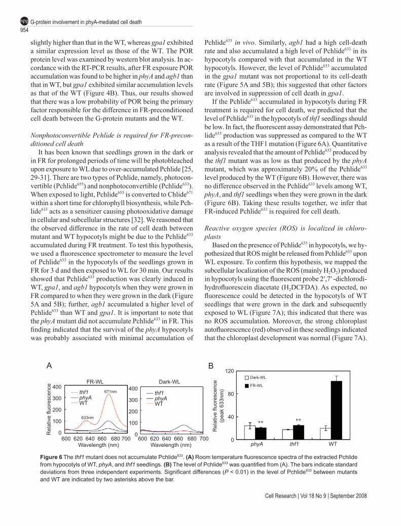
Figure 6 The thf1 mutant does not accumulate Pchlide633. (A) Room temperature fluorescence spectra of the extracted Pchlide from hypocotyls of WT, phyA, and thf1 seedlings. (B) The level of Pchlide633 was quantified from (A). The bars indicate standard deviations from three independent experiments. Significant differences (P < 0.01) in the level of Pchlide633 between mutants and WT are indicated by two asterisks above the bar.
BA
FR-WL Dark-WL400
300
200
100
0
400
300
200
100
0Rel
ativ
e flu
ores
cenc
e
600 620 640 660 680 700 600 620 640 660 680 700Wavelength (nm) Wavelength (nm)
thf1phyAWT
thf1phyAWT
671nm
633nm
Rel
ativ
e flu
ores
cenc
e(p
eak
633n
m)
Dark-WL
FR-WL
120
80
40
0phyA thf1 WT
slightly higher than that in the WT, whereas gpa1 exhibited a similar expression level as those of the WT. The POR protein level was examined by western blot analysis. In ac-cordance with the RT-PCR results, after FR exposure POR accumulation was found to be higher in phyA and agb1 than that in WT, but gpa1 exhibited similar accumulation levels as that of the WT (Figure 4B). Thus, our results showed that there was a low probability of POR being the primary factor responsible for the difference in FR-preconditioned cell death between the G-protein mutants and the WT.
Nonphotoconvertible Pchlide is required for FR-precon-ditioned cell death
It has been known that seedlings grown in the dark or in FR for prolonged periods of time will be photobleached upon exposure to WL due to over-accumulated Pchlide [25, 29-31]. There are two types of Pchlide, namely, photocon-vertible (Pchlide655) and nonphotoconvertible (Pchlide633). When exposed to light, Pchlide655 is converted to Chlide671 within a short time for chlorophyll biosynthesis, while Pch-lide633 acts as a sensitizer causing photooxidative damage in cellular and subcellular structures [32]. We reasoned that the observed difference in the rate of cell death between mutant and WT hypocotyls might be due to the Pchlide633 accumulated during FR treatment. To test this hypothesis, we used a fluorescence spectrometer to measure the level of Pchlide633 in the hypocotyls of the seedlings grown in FR for 3 d and then exposed to WL for 30 min. Our results showed that Pchlide633 production was clearly induced in WT, gpa1, and agb1 hypocotyls when they were grown in FR compared to when they were grown in the dark (Figure 5A and 5B); further, agb1 accumulated a higher level of Pchlide633 than WT and gpa1. It is important to note that the phyA mutant did not accumulate Pchlide633 in FR. This finding indicated that the survival of the phyA hypocotyls was probably associated with minimal accumulation of
Pchlide633 in vivo. Similarly, agb1 had a high cell-death rate and also accumulated a high level of Pchlide633 in its hypocotyls compared with that accumulated in the WT hypocotyls. However, the level of Pchlide633 accumulated in the gpa1 mutant was not proportional to its cell-death rate (Figure 5A and 5B); this suggested that other factors are involved in suppression of cell death in gpa1.
If the Pchlide633 accumulated in hypocotyls during FR treatment is required for cell death, we predicted that the level of Pchlide633 in the hypocotyls of thf1 seedlings should be low. In fact, the fluorescent assay demonstrated that Pch-lide633 production was suppressed as compared to the WT as a result of the THF1 mutation (Figure 6A). Quantitative analysis revealed that the amount of Pchlide633 produced by the thf1 mutant was as low as that produced by the phyA mutant, which was approximately 20% of the Pchlide633 level produced by the WT (Figure 6B). However, there was no difference observed in the Pchlide633 levels among WT, phyA, and thf1 seedlings when they were grown in the dark (Figure 6B). Taking these results together, we infer that FR-induced Pchlide633 is required for cell death.
Reactive oxygen species (ROS) is localized in chloro-plasts
Based on the presence of Pchlide633 in hypocotyls, we hy-pothesized that ROS might be released from Pchlide633 upon WL exposure. To confirm this hypothesis, we mapped the subcellular localization of the ROS (mainly H2O2) produced in hypocotyls using the fluorescent probe 2′,7′ -dichlorodi-hydrofluorescein diacetate (H2DCFDA). As expected, no fluorescence could be detected in the hypocotyls of WT seedlings that were grown in the dark and subsequently exposed to WL (Figure 7A); this indicated that there was no ROS accumulation. Moreover, the strong chloroplast autofluorescence (red) observed in these seedlings indicated that the chloroplast development was normal (Figure 7A).
** **

www.cell-research.com | Cell Research
Qing Wei et al.955npg
Figure 7 Fluorescence microscopic image of ROS localization in the cells of WT hypocotyls. (A) H2DCFDA fluorescence was not detectable in the hypocotyls grown in the dark for 5 d and then transferred to WL for an additional day. Scale bar, 1 mm. (B) Strong H2DCFDA fluorescence was observed in the cells of the hypocotyls that were grown in FR for 3 d followed by expo-sure to WL for an additional day. Scale bar, 1 mm. (C) The H2DCFDA fluorescence shown in (B) was mainly emitted from the plastids/chloroplasts. Insets show H2DCFDA fluorescence in the chloroplasts of guard cells (white square) and dermal cells (red square). Scale bar, 200 µm.
C
A BDIC Merged DIC Merged
H2DCFHfluorescence
H2DCFHfluorescence
ChlorophyIIfluorescence
ChlorophyIIfluorescence
Figure 8 H2O2 treatment mimics FR-preconditioned cell death of hypocotyls. Seedlings grown in the dark for 5 d were treated with 1 mM H2O2 for 2 h, washed with deionized water, and put in WL for 1 d. (A) Typical morphology of a seedling with hypocotyl cell death 6 h after H2O2 treatment. Scale bar, 2 mm. (B) The effect of exogenous H2O2 on cell death of WT, phyA, gpa1, agb1, and thf1 seedlings. Cell death was measured with Evans blue staining. Error bars indicate SD (n = 3). Significant differences (P < 0.01) in cell death between mutants and WT are indicated by two asterisks above the bar.
In the FR-grown hypocotyls, however, the H2DCFDA-stained fluorescent signal (green) was strong in the middle section of hypocotyls (Figure 7B); this implied that ROS
was generated in these cells. Interestingly, chloroplast auto-fluorescence was emitted mainly from the upper section of the hypocotyls (Figure 7B). A detailed inspection revealed
BA5
4
3
2
1
0
Rel
ativ
e ab
sorb
ance
uni
ts
phyA WT gpa1 agb1 thf1
****
Cell Research | Vol 18 No 9 | September 2008
G-protein involvement in phyA-mediated cell death956npg
that the green fluorescent region coincided with the position of the plastids/chloroplasts (Figure 7C). Thus, this result was consistent with the idea that ROS was derived from plastid-localized Pchlide633.
H2O2 treatment mimics FR-preconditioned cell deathBased on the above data, we questioned whether exog-
enous oxidative stress could mimic FR-preconditioned cell death. Therefore, we treated 5-d-old dark-grown seedlings with 1 mM of H2O2 under WL for 2 h; H2O2 was then removed from the plates. As expected, the H2O2 exposure induced a similar phenotype as that observed in FR-pre-conditioned cell death approximately 6 h after treatment (Figure 8A). We then quantified cell death by Evans blue staining. Our result showed that there was no difference in the cell-death rate among the gpa1, phyA, and WT seed-lings. However, the cell death rate was more severe in the agb1 and thf1 mutants than that in the WT (Figure 8B); this suggested that agb1 and thf1 are more sensitive to oxida-tive stress than gpa1, phyA, and WT. These results provide further evidence supporting the notion that oxidative stress mediates FR-preconditioned cell death.
Discussion
FR-preconditioned cell death of hypocotyls is regulated by phyA signaling
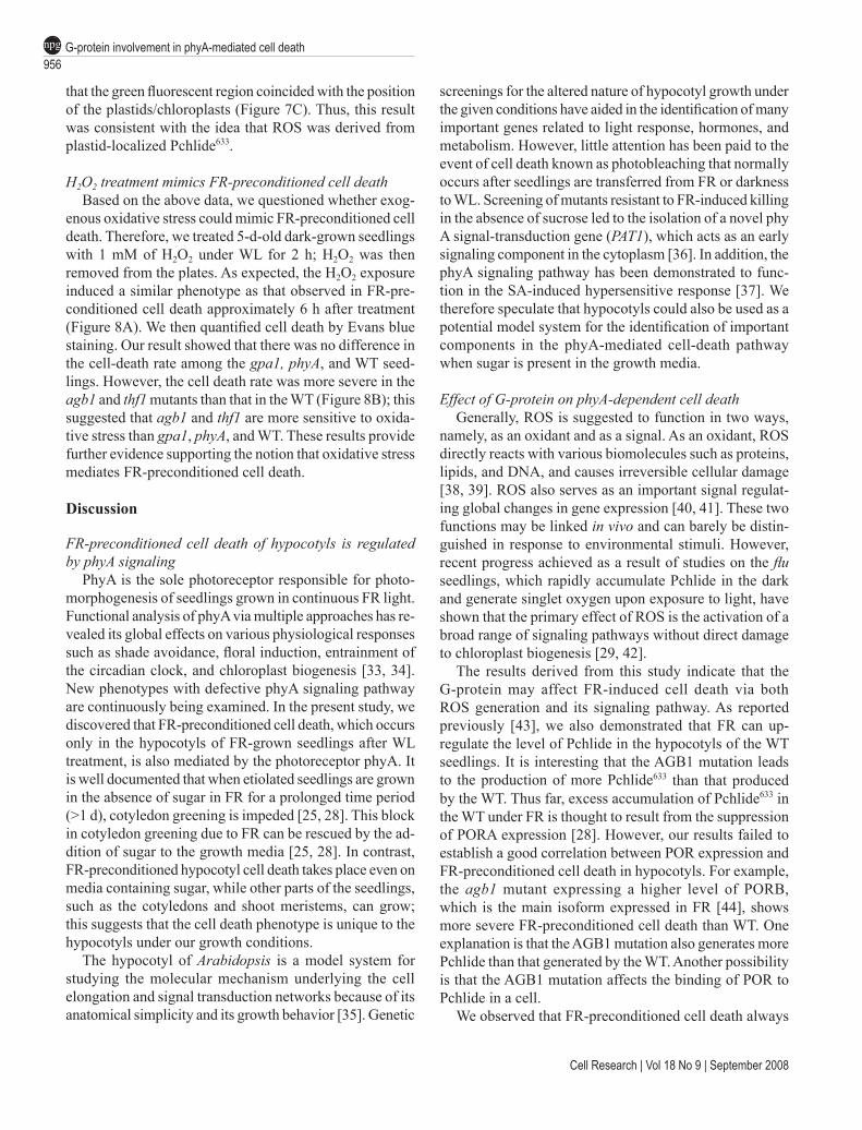
PhyA is the sole photoreceptor responsible for photo-morphogenesis of seedlings grown in continuous FR light. Functional analysis of phyA via multiple approaches has re-vealed its global effects on various physiological responses such as shade avoidance, floral induction, entrainment of the circadian clock, and chloroplast biogenesis [33, 34]. New phenotypes with defective phyA signaling pathway are continuously being examined. In the present study, we discovered that FR-preconditioned cell death, which occurs only in the hypocotyls of FR-grown seedlings after WL treatment, is also mediated by the photoreceptor phyA. It is well documented that when etiolated seedlings are grown in the absence of sugar in FR for a prolonged time period (>1 d), cotyledon greening is impeded [25, 28]. This block in cotyledon greening due to FR can be rescued by the ad-dition of sugar to the growth media [25, 28]. In contrast, FR-preconditioned hypocotyl cell death takes place even on media containing sugar, while other parts of the seedlings, such as the cotyledons and shoot meristems, can grow; this suggests that the cell death phenotype is unique to the hypocotyls under our growth conditions.
The hypocotyl of Arabidopsis is a model system for studying the molecular mechanism underlying the cell elongation and signal transduction networks because of its anatomical simplicity and its growth behavior [35]. Genetic
screenings for the altered nature of hypocotyl growth under the given conditions have aided in the identification of many important genes related to light response, hormones, and metabolism. However, little attention has been paid to the event of cell death known as photobleaching that normally occurs after seedlings are transferred from FR or darkness to WL. Screening of mutants resistant to FR-induced killing in the absence of sucrose led to the isolation of a novel phy A signal-transduction gene (PAT1), which acts as an early signaling component in the cytoplasm [36]. In addition, the phyA signaling pathway has been demonstrated to func-tion in the SA-induced hypersensitive response [37]. We therefore speculate that hypocotyls could also be used as a potential model system for the identification of important components in the phyA-mediated cell-death pathway when sugar is present in the growth media.
Effect of G-protein on phyA-dependent cell deathGenerally, ROS is suggested to function in two ways,
namely, as an oxidant and as a signal. As an oxidant, ROS directly reacts with various biomolecules such as proteins, lipids, and DNA, and causes irreversible cellular damage [38, 39]. ROS also serves as an important signal regulat-ing global changes in gene expression [40, 41]. These two functions may be linked in vivo and can barely be distin-guished in response to environmental stimuli. However, recent progress achieved as a result of studies on the flu seedlings, which rapidly accumulate Pchlide in the dark and generate singlet oxygen upon exposure to light, have shown that the primary effect of ROS is the activation of a broad range of signaling pathways without direct damage to chloroplast biogenesis [29, 42].
The results derived from this study indicate that the G-protein may affect FR-induced cell death via both ROS generation and its signaling pathway. As reported previously [43], we also demonstrated that FR can up-regulate the level of Pchlide in the hypocotyls of the WT seedlings. It is interesting that the AGB1 mutation leads to the production of more Pchlide633 than that produced by the WT. Thus far, excess accumulation of Pchlide633 in the WT under FR is thought to result from the suppression of PORA expression [28]. However, our results failed to establish a good correlation between POR expression and FR-preconditioned cell death in hypocotyls. For example, the agb1 mutant expressing a higher level of PORB, which is the main isoform expressed in FR [44], shows more severe FR-preconditioned cell death than WT. One explanation is that the AGB1 mutation also generates more Pchlide than that generated by the WT. Another possibility is that the AGB1 mutation affects the binding of POR to Pchlide in a cell.
We observed that FR-preconditioned cell death always

www.cell-research.com | Cell Research
Qing Wei et al.957npg
occurred in the middle section of the hypocotyls. Erdei et al. [31] observed a similar light response in the epicotyls of dark-germinated pea seedlings. In their model, the photo-sensitivity of the epicotyls was explained by the balance between POR and Pchlide that is crucial for survival of dark-grown epicotyls, along which the Pchlide gradient can be detected [45]. Pchlide633 was thought to bind to unknown proteins [46]. The AGB1 mutation appears to enhance the ratio of Pchlide633 to photoactive Pchlide655. Until date, little information is available regarding the regulation of this ratio in plants. Our data (unpublished) indicated that hypocotyls of dark-germinated seedlings of the agb1 mutant were more resistant to light-induced wilting than those of the WT. This result can be explained by the higher level of POR expression in agb1 than that in the WT. AGB1 mutation may influence the binding of POR to Pchlide and lead to the production of nontransformable Pchlide in FR. Moreover, AGB1 might also have an impact on the sensitivity of hypocotyls to ROS because agb1 suf-fers more severe cell death than that incurred by the WT when treated with the same concentration of H2O2. A similar role of Gβ in oxidative stress has also been demonstrated in mammalian cells, where H2O2 directly activates the G-protein signaling pathway and the Gβγ subunit is the main signal transducer involved in the protection of the cells against oxidative stress via downstream activation of the mitogen-activated protein kinase cascade. In the case of gpa1, although the mechanism underlying reduction of FR-preconditioned cell death as a result of the mutation is unclear because there is no difference observed in the accumulation of Pchlide and sensitivity to H2O2 between the gpa1 and WT seedlings, our data are in agreement with previous reports on GPA1-mutation-induced reduction of cell damage caused by ROS stress [23]. As Joo et al. [23] reported, in FR light the pattern of ROS generation in gpa1 might be altered as compared to that in the WT. We specu-late that since the double gpa1 agb1 mutant exhibits the same level of cell death as that of the single agb1 mutant, it is possible that the GPA1 mutation resulted in a lower level of FR-preconditioned cell death than that of WT due to the presence of free AGB1 in the gpa1 seedlings. This hypothesis regarding G-protein-regulated FR-precondi-tioned cell death is similar to that of lateral root formation, where AGB1 may act downstream of GPA1 to negatively regulate lateral root formation.
G-protein may finely regulate the phyA signaling path-way
The early studies via a pharmacological approach showed that the G-protein was an important transducer in the phyA signaling pathway for chloroplast development in the hy-pocotyls of aurea. Recently, the gene controlling the pale
green phenotype of the aurea mutant was isolated and found to be the phytochromobilin synthase that catalyzes the conversion of biliverdin IXα into 3Z-phytochromobi-lin during phytochrome chromophore biosynthesis [47]. The chlorophyll-deficient phenotype of the aurea mutant is due to the suppression of 5-aminolevulinic acid (ALA) synthesis caused by a high level of heme and subsequent reduction in Pchlide synthesis [48]. Thus, we reasoned that the GPA1-promoted chloroplast development observed in aurea might partially release the inhibition of ALA syn-thesis caused by heme. It would be interesting to examine whether ALA accumulation is altered in the gpa1 mutant. Besides ALA, chlorophyll biosynthesis can be modulated by the G-protein via another key factor, namely, POR, whose expression in FR is always slightly higher in the agb1 mutant than that in the WT. Thus, the G-protein may finely tune the regulation of tetrapyrrole biosynthesis.
Further investigation is required to determine the mechanism underlying the mediation of the phyA signaling pathway by the G-protein. However, the involvement of the G-protein in FR-preconditioned cell death in hypocotyls provides a new opportunity to unravel the link between phyA and the G-protein at a molecular level. FR appears to trigger a process that is extremely harmful for plant growth because it suppresses POR expression and simultaneously promotes Pchlide accumulation. The genetic dissection of the molecular mechanism underlying the plant’s response to FR has revealed the involvement of many positive regu-lators, such as PAT1, FHY1, FHY3, FIN2, and FAR1 [36, 49-51], as well as negative regulators, such as PSI2 and SPA1 [52, 53]. They are involved in the phyA signaling pathway or phytochrome chromophore biosynthesis, sug-gesting that the phyA signaling pathway is tightly regulated and integrated in the whole cellular signaling network. We propose that the G-protein is one of the switches involved in finely tuning the regulation of the phyA signaling pathway. Okamoto et al. [20] demonstrated that FHY1 played a role in GPA1-promoted hypocotyl elongation under FR. FHY1 is also involved in FR-suppressed chlorophyll biosynthesis since the expression of HEMA1 markedly decreased in the mutant [54]. Thus, FHY1 might be an appropriate candidate linking the G-protein and one branch of the phyA signaling pathway. Recent research has determined that the interac-tion of activated phyA with FHY1, FHL1, NADPK2, and PSK1 is an early event in cytoplasmic phyA signaling. Among them, FHY1 and FHL1 were required for phyA translocation from the cytoplasm to the nucleus [55-57], and NADPK2 was suggested to potentially activate the G-protein by increasing the level of GTP [20]. We sug-gest two possibilities for crosstalk between G-protein and phyA signaling in regulation of chlorophyll biosynthesis. One possibility is that the G-protein affects the process of
Cell Research | Vol 18 No 9 | September 2008
G-protein involvement in phyA-mediated cell death958npg
phyA nuclear translocation via FHY1 or FHL1. Another is that the G-protein signaling pathway activated by phyA in the cytoplasm regulates nuclear gene expression. However, in phyA-mediated FR responses, only the inhibition of gravitropism was found to be regulated by the cytoplasmic phyA signaling pathway [58]. Thus, the latter possibility of G-protein involvement in the phyA signaling pathway may be excluded. It would be interesting to investigate whether the G-protein has an impact on the rapidity of the phyA movement from the cytoplasm to the nucleus using a combination of genetic and molecular approaches.
Materials and Methods
Plant materials, growth conditions, and light sourcesArabidopsis thaliana WT (ecotype Columbia, Col-0) plants were
used in this study. All the mutants used were in the Col-0 background. The phyA (phyA-211) and phyB (phyB-9) mutants were obtained from the Arabidopsis Biological Resources Center (Columbus, OH). The other mutants are gpa1 (gpa1-4), agb1 (agb1-2) and thf1 (thf1-1) [27]. The double mutants used in the experiment were created by using the abovementioned single mutants and confirmed by plant genotyping in the F2 generation.
Surface-sterilized seeds stratified at 4 ºC for 4 d were germinated on half-strength Murashige and Skoog (MS) medium with 1% of sucrose for 1.5 d in darkness before FR irradiation at 22 ºC. FR light was provided by the far-red diodes (model E-30, Percival Scientific, Boone, IA) at the peak wavelength of 730 nm with a plexiglas filter (FRF 700, Westlake Plastics, Lenni, PA). The light strength was determined using a spectroradiometer (ASD, Boulder, CO). After FR irradiation, the seedlings were subsequently transferred to WL (TL-D, 30W, Philip, Shanghai). WL fluence rates were measured using a ZDS-10 Luxmeter (Xuelian Instrument Factory, Shanghai). At least 50 seedlings from each genotype were sampled for further analysis in the same experiment.
Qualitative and quantitative determinations of cell death in hypocotyls
More than 50 seedlings were examined for each qualitative or quantitative assay of cell death in hypocotyls. For the qualitative assay of cell death, hypocotyls showing a visible wilted phenotype or Evans blue staining under the FR-induced cell death condition were recorded. For quantitative measurement of cell death, seedlings were completely submerged in 2.5% (w/v) solution of Evans blue dye for 10 min, then washed three times with distilled water. The dye bound to the dead cells in hypocotyls was extracted using a solu-tion containing 50% (v/v) methanol and 1% (w/v) sodium dodecyl sulfate (SDS) at 60 ºC for 30 min and then quantified by measuring the absorbance at 600 nm [26].
Chlorophyll and Pchlide measurementChlorophyll was extracted using 80% acetone at 4 ºC for 24 h in
the dark. The chlorophyll content was determined using a spectropho-tometer (UNIC UV-2102 PCS, Shanghai) [59]. Protochlorophyllide was extracted and measured by a previously described method [60]. In each replicate experiment, 50 hypocotyls were sampled during the analysis.
Fluorescence microscopic imaging of ROS and H2O2 treat-ment
Subcellular localization of ROS was examined by fluorescence microscopic imaging [23]. Fluorescence was observed with a LSM 510 META laser scanning confocal microscope (ZEISS, Germany), with excitation at 488 nm and emission at 530 nm. For H2O2 treat-ment, seedlings grown for 5 d in the dark were shifted to WL and immediately submerged in 1 mM H2O2 solution for 2 h. They were then washed with deionized water and placed in WL for 1 d; thereafter, the cell death was quantitatively measured.
Semiquantitative reverse transcription-polymerase chain reaction
Total RNA was isolated using the RNAgents total RNA isolation system (Promega). Reverse transcription was performed according to the manufacturer’s instructions (Reverse transcription system, Promega). Gene expression was determined using gene-specific primers by semiquantitative reverse transcription-polymerase chain reaction (RT-PCR) or quantitative real-time RT-PCR. Primers for the POR genes were as follows: PORA-F: CAT TAC ACT CTT TAA GTC TTA AAC G; PORA-R: AAC CAC GTT TCC TTT TCT AAG T; PORB-F: CTC TCT CTT TGT TCG AGT ACC AAC; and PORB-R: GTA TTC GTG TTC CCG GTA ATG G. The amplified DNA fragments were separated by agarose gel electrophoresis and analyzed using GIS-2010 (Tanon, Shanghai).
Protein extraction and western blot analysisTotal protein was extracted and quantified by a previously de-
scribed method [25]. Protein expression was detected by immunoblot analysis. Total protein (40 µg) was loaded in each well. Signals were detected using a medical film processor (SRX-101A, Konica, Minolta, Tokyo). Anti-POR antibody was obtained from Agrisera, and used at a dilution of 1:2 000.
Acknowledgements
This work was supported by the National Natural Sci-ence Foundation of China (30570131), by the Pujiang Program of Shanghai Municipality (06PJ14103), and by the ‘100 Talents Project’ of Chinese Academy of Sciences to JH.
References
1 Morris AJ, Malbon CC. Physiological regulation of G protein-linked signaling. Physiol Rev 1999; 79:1373-1430.
2 Spiegel AM, Weinstein LS. Inherited diseases involving G proteins and G protein-coupled receptors. Annu Rev Med 2004; 55:27-39.
3 Ma H, Yanofsky MF, Meyerowitz EM. Molecular cloning and characterization of GPA1, a G protein α subunit gene from Arabi-dopsis thaliana. Proc Natl Acad Sci USA 1990; 87:3821-3825.
4 Weiss CA, Garnaat CW, Mukai K, Hu Y, Ma H. Isolation of cDNAs encoding guanine nucleotide-binding protein β-subunit homologues from maize (ZGB1) and Arabidopsis (AGB1). Proc Natl Acad Sci USA 1994; 91:9554-9558.
5 Mason MG, Botella JR. Completing the heterotrimer: isolation

www.cell-research.com | Cell Research
Qing Wei et al.959npg
and characterization of an Arabidopsis thaliana G protein γ-sub-unit cDNA. Proc Natl Acad Sci USA 2000; 97:14784-14788.
6 Mason MG, Botella JR. Isolation of a novel G protein γ-subunit from Arabidopsis thaliana and its interaction with Gβ. Biochim Biophys Acta 2001; 1520:147-153.
7 Assmann SM. G proteins go green: a plant G protein signaling FAQ sheet. Science 2005; 310:71-73.
8 Jones AM. G-protein-coupled signaling in Arabidopsis. Curr Opin Plant Biol 2002; 5:402-407.
9 Wang L, Xu YY, Ma QB, Li D, Xu ZH, Chong K. Heterotrimeric G protein alpha subunit is involved in rice brassinosteroid re-sponse. Cell Res 2006; 16:916-922.
10 Wang S, Narendra S, Fedoroff N. Heterotrimeric G protein sig-naling in the Arabidopsis unfolded protein response. Proc Natl Acad Sci USA 2007; 104:3817-3822.
11 Schepens I, Duek P, Fankhauser C. Phytochrome-mediated light signalling in Arabidopsis. Curr Opin Plant Biol 2004; 7:564-569.
12 Quail PH. Phytochrome photosensory signaling networks. Nat Rev Mol Cell Biol 2002; 3:85-93.
13 Arshavsky VY, Lamb TD, Pugh EN. G proteins and phototrans-duction. Annu Rev Physiol 2002; 64:53-87.
14 Romero LC, Sommer D, Gotor C, Song PS. G-proteins in etio-lated Avena seedlings. Possible phytochrome regulation. FEBS Lett 1991; 282:341-346.
15 Romero LC, Biswal B, Song PS. Protein phosphorylation in isolated nuclei from etiolated Avena seedlings. Effects of red/far-red light and cholera toxin. FEBS Lett 1991; 282:347-350.
16 Romero LC, Lam E. Guanine nucleotide binding protein involve-ment in early steps of phytochrome-regulated gene expression. Proc Natl Acad Sci USA 1993; 90:1465-1469.
17 Warpeha KM, Hamm HE, Rasenick MM, Kaufman LS. A blue-light-activated GTP-binding protein in the plasma membranes of etiolated peas. Proc Natl Acad Sci USA 1991; 88:8925-8929.
18 Neuhaus G, Bowler C, Kern R, Chua NH. Calcium/calmodulin-dependent and -independent phytochrome signal transduction pathways. Cell 1993; 73:937-952.
19 Bowler C, Neuhaus G, Yamagata H, Chua NH. Cyclic GMP and calcium mediate phytochrome phototransduction. Cell 1994; 77:73-81.
20 Okamoto H, Matsui M, Deng XW. Overexpression of the hetero-trimeric G-protein α-subunit enhances phytochrome-mediated inhibition of hypocotyl elongation in Arabidopsis. Plant Cell 2001; 13:1639-1652.
21 Jones AM, Ecker JR, Chen JG. A reevaluation of the role of the heterotrimeric G protein in coupling light responses in Arabi-dopsis. Plant Physiol 2003; 131:1623-1627.
22 Llorente F, Alonso-Blanco C, Sanchez-Rodriguez C, Jorda L, Molina A. ERECTA receptor-like kinase and heterotrimeric G protein from Arabidopsis are required for resistance to the necrotrophic fungus Plectosphaerella cucumerina. Plant J 2005; 43:165-180.
23 Joo JH, Wang S, Chen JG, Jones AM, Fedoroff NV. Different signaling and cell death roles of heterotrimeric G protein alpha and beta subunits in the Arabidopsis oxidative stress response to ozone. Plant Cell 2005; 17:957-970.
24 Trusov Y, Rookes JE, Chakravorty D, Armour D, Schenk PM, Botella JR. Heterotrimeric G proteins facilitate Arabidopsis re-sistance to necrotrophic pathogens and are involved in jasmonate
signaling. Plant Physiol 2006; 140:210-220.25 Barnes SA, Nishizawa NK, Quaggio RB, Whitelam GC, Chua
NH. Far-red light blocks greening of Arabidopsis seedlings via a phytochrome A-mediated change in plastid development. Plant Cell 1996; 8:601-615.
26 Ochsenbein C, Przybyla D, Danon A, et al. The role of EDS1 (enhanced disease susceptibility) during singlet oxygen-mediated stress responses of Arabidopsis. Plant J 2006; 47:445-456.
27 Huang J, Taylor JP, Chen JG, et al. The plastid protein THY-LAKOID FORMATION1 and the plasma membrane G-protein GPA1 interact in a novel sugar-signaling mechanism in Arabi-dopsis. Plant Cell 2006; 18:1226-1238.
28 Sperling U, van Cleve B, Frick G, Apel K, Armstrong GA. Over-expression of light-dependent PORA or PORB in plants depleted of endogenous POR by far-red light enhances seedling survival in white light and protects against photooxidative damage. Plant J 1997; 12:649-658.
29 Op den Camp RGL, Przybyla D, Ochsenbein C, et al. Rapid induction of distinct stress responses after the release of singlet oxygen in Arabidopsis. Plant Cell 2003; 15:2320-2332.
30 Reinbothe S, Reinbothe C, Apel K, Lebedev N. Evolution of chlorophyll biosynthesis - the challenge to survive photooxida-tion. Cell 1996; 86:703-705.
31 Erdei N, Barta C, Hideg E, Boddi B. Light-induced wilting and its molecular mechanism in epicotyls of dark-germinated pea (Pisum sativum L.) seedlings. Plant Cell Physiol 2005; 46:185-191.
32 Lebedev NN, Krasnovsky Jr AA, Litvin FF. Phosphorescence of protochlorophyll(ide) and chlorophyll(ide) in etiolated and greening bean leaves. Photosynth Res 1991; 30:7-14.
33 Wang H, Deng XW. Dissecting the phytochrome A-dependent signaling network in higher plants. Trends Plant Sci 2003; 8:172-178.
34 Chen M, Chory J, Fankhauser C. Light signal transduction in higher plants. Annu Rev Genet 2004; 38:87-117.
35 Gendreau E, Traas J, Desnos T, Grandjean O, Caboche M, Hofte H. Cellular basis of hypocotyl growth in Arabidopsis thaliana. Plant Physiol 1997; 114:295-305.
36 Bolle C, Koncz C, Chua NH. PAT1, a new member of the GRAS family, is involved in phytochrome A signal transduction. Genes Dev 2000; 14:1269-1278.
37 Genoud T, Buchala AJ, Chua NH, Metraux JP. Phytochrome signaling modulates the SA-perceptive pathway in Arabidopsis. Plant J 2002; 31:87-95.
38 Apel K, Hirt H. Reactive oxygen species: metabolism, oxida-tive stress, and signal transduction. Annu Rev Plant Biol 2004; 55:373-399.
39 Girotti AW. Photosensitized oxidation of membrane lipids: reac-tion pathways, cytotoxic effects and cytoprotective mechanisms. J Photochem Photobiol B 2001; 63:103-113.
40 Gechev TS, Hille J. Hydrogen peroxide as a signal controlling plant programmed cell death. J Cell Biol 2005; 168:17-20.
41 Dalton TP, Shertzer HG, Puga A. Regulation of gene expression by reactive oxygen. Annu Rev Pharmacol Toxicol 1999; 39:67-101.
42 Lee KP, Kim C, Landgraf F, Apel K. EXECUTER1- and EX-ECUTER2-dependent transfer of stress-related signals from the plastid to the nucleus of Arabidopsis thaliana. Proc Natl Acad Sci USA 2007; 104:10270-10275.
Cell Research | Vol 18 No 9 | September 2008
G-protein involvement in phyA-mediated cell death960npg
43 Sineshchekov V, Belyaeva O, Sudnitsin A. Up-regulation by phytochrome A of the active protochlorophyllide, Pchlide655, biosynthesis in dicots under far-red light. J Photochem Photobiol B 2004; 74:47-54.
44 Runge S, Sperling U, Frick G, Apel K, Armstrong GA. Distinct roles for light-dependent NADPH:protochlorophyllide oxidore-ductases (POR) A and B during greening in higher plants. Plant J 1996; 9:513-523.
45 Boddi B, McEwen B, Ryberg M, Sundqvist C. Protochlorophyl-lide forms in non-greening epicotyls of dark-grown pea (Pisum sativum). Physiol Plant 1994; 92:31-39.
46 Schoefs B. Protochlorophyllide reduction - what is new in 2005? Photosynthetica 2005; 43:329-343.
47 Muramoto T, Kami C, Kataoka H, et al. The tomato photomor- phogenetic mutant, aurea, is deficient in phytochromobilin synthase for phytochrome chromophore biosynthesis. Plant Cell Physiol 2005; 46:661-665.
48 Terry MJ, Kendrick RE. Feedback inhibition of chlorophyll synthesis in the phytochrome chromophore-deficient aurea and yellow-green-2 mutants of tomato. Plant Physiol 1999; 119:143-152.
49 Whitelam GC, Johnson E, Peng J, et al. Phytochrome A null mutants of Arabidopsis display a wild-type phenotype in white light. Plant Cell 1993; 5:757-768.
50 Soh MS, hong SH, Hanzawa H, Furuya M, Nam HG. Genetic identification of FIN2, a far red light specific signaling compo-nent of Arabidopsis thaliana. Plant J 1998; 16:411-419.
51 Hudson M, Ringli C, Boylan MT, Quail PH. The FAR1 locus encodes a novel nuclear protein specific to phytochrome A sig-naling. Genes Dev 1999; 13:2017-2027.
52 Genoud T, Millar AJ, Nishizawa N, et al. An Arabidopsis mutant
hypersensitive to red and far-red light signals. Plant Cell 1998; 10:889-904.
53 Hoecker U, Tepperman JM, Quail PH. SPA1, a WD-repeat protein specific to phytochrome A signal transduction. Science 1999; 284:496-499.
54 McCormack AC, Terry MJ. Light-signaling pathways leading to the coordinated expression of HEMA1 and Lhcb during chloroplast development in Arabidopsis thaliana. Plant J 2002; 32:549-559.
55 Zeidler M, Zhou Q, Sarda X, Yau CP, Chua NH. The nuclear localization signal and the C-terminal region of FHY1 are re-quired for transmission of phytochrome A signals. Plant J 2004; 40:355-365.
56 Zhou Q, Hare PD, Yang SW, Zeidler M, Huang LF, Chua NH. FHL is required for full phytochrome A signaling and shares overlapping functions with FHY1. Plant J 2005; 43:356-370.
57 Hiltbrunner A, Tscheuschler A, Viczian A, Kunkel T, Kircher S, Schafer E. FHY1 and FHL act together to mediate nuclear accumulation of the phytochrome A photoreceptor. Plant Cell Physiol 2006; 47:1023-1034.
58 Rosler J, Klein I, Zeidler M. Arabidopsis fhl/fhy1 double mutant reveals a distinct cytoplasmic action of phytochrome A. Proc Natl Acad Sci USA 2007; 104:10737-10742.
59 Arnon DI. Copper enzymes in isolated chloroplasts: polyphenol oxidase in Beta vulgaris. Plant Physiol 1949; 24:1-15.
60 Goslings D, Meskauskiene R, Kim C, Lee KP, Nater M, Apel K. Concurrent interactions of heme and FLU with Glu tRNA reductase (HEMA1), the target of metabolic feedback inhibition of tetrapyrrole biosynthesis, in dark- and light-grown Arabidopsis plants. Plant J 2004; 40:957-967.


![Phytochromes and Phytochrome Interacting Factors1[OPEN] · Update on Phytochromes and Phytochrome Interacting Factors Phytochromes and Phytochrome Interacting Factors1[OPEN] Vinh](https://img.pdfslide.net/doc/110x75/5e9224c5cbd0a85457462c45/phytochromes-and-phytochrome-interacting-factors1open-update-on-phytochromes-and.jpg)