Embed Size (px)
Citation preview
Journal Pre-proof
Hierarchical design of hyaluronic acid-peptide constructs forglioblastoma targeting: Combining insights from NMR andmolecular dynamics simulations
Maria Mendes, Tânia Cova, João Basso, M. Luísa Ramos, RuiVitorino, João Sousa, Alberto Pais, Carla Vitorino
PII: S0167-7322(20)32331-X
DOI: https://doi.org/10.1016/j.molliq.2020.113774
Reference: MOLLIQ 113774
To appear in: Journal of Molecular Liquids
Received date: 15 April 2020
Revised date: 3 July 2020
Accepted date: 7 July 2020
Please cite this article as: M. Mendes, T. Cova, J. Basso, et al., Hierarchical design ofhyaluronic acid-peptide constructs for glioblastoma targeting: Combining insights fromNMR and molecular dynamics simulations, Journal of Molecular Liquids (2020),https://doi.org/10.1016/j.molliq.2020.113774
This is a PDF file of an article that has undergone enhancements after acceptance, suchas the addition of a cover page and metadata, and formatting for readability, but it isnot yet the definitive version of record. This version will undergo additional copyediting,typesetting and review before it is published in its final form, but we are providing thisversion to give early visibility of the article. Please note that, during the productionprocess, errors may be discovered which could affect the content, and all legal disclaimersthat apply to the journal pertain.
© 2020 Published by Elsevier.
Jour
nal P
re-p
roof
1
Hierarchical design of hyaluronic acid-peptide constructs for glioblastoma targeting:
combining insights from NMR and molecular dynamics simulations
Maria Mendesa,b,c,*
, Tânia Covac,*
, João Bassoa,b,c
, M. Luísa Ramosc, Rui Vitorino
d, João
Sousaa,c
, Alberto Paisc,, Carla Vitorino
a,b,c,#
*These authors contributed equally to this work and should be regarded as co-first authors
aFaculty of Pharmacy, University of Coimbra, Polo das Ciências da Saúde, Azinhaga de Santa
Comba, 3000-548 Coimbra, Portugal.
bCentre for Neurosciences and Cell Biology (CNC), University of Coimbra, Faculty of
Medicine, Polo I, 1st floor, Rua Larga, 3004-504 Coimbra, Portugal
cCoimbra Chemistry Center, Department of Chemistry, University of Coimbra, Rua Larga,
3004-535 Coimbra, Portugal
dDepartment of Medical Sciences and Institute of Biomedicine – iBiMED, University of
Aveiro, Aveiro, Portugal
#To whom correspondence should be addressed. (e–mail: [email protected])
Abstract
The main bottleneck of glioblastoma still relies on the existence of the blood
brain-blood brain tumor dual barrier, along with the lack of therapy specificity. The present
work deals with the question of whether (and how) different targeting hyaluronic acid (HA)-
peptide [c(RGDfK) and/or H7K(R2)2] moieties hierarchically interact with each other, to ensure
a unique entity with specificity to glioblastoma. A dual experimental-computational approach,
encompassing nuclear magnetic resonance and molecular dynamics simulations is enclosed.
Relevant contact patterns based on the identification of the stabilizing/destabilizing
noncovalent interactions within the constructs are detailed. The synthesis pathway requires the
HA-c(RGDfK)-H7k(R2)2 association hierarchy, stemming from the size and amino acid residue
rearrangement, in the 1:1 molar ratio, to obtain a stable conjugate ultimately able to interact
with the tumor cell membrane. To our knowledge, the structural and mechanistic rationale for
the formation of hybrid polymer-peptide constructs, including HA-c(RGDfK)-H7k(R2)2, for
glioblastoma has not been addressed so far.
Keywords: Hyaluronic acid; c(RGDfK; H7k(R2)2; polymer-peptide conjugates; glioblastoma
Journal Pre-proof
Jour
nal P
re-p
roof
2
List of abbreviations
HA - Hyaluronic acid
GBM - Glioblastoma
BBB – Blood-brain barrier
ECM- Extracellular matrix
c(RGDfK) - Cyclo (arginine(R)–glycine(G)–aspartic acid(D)–phenylalanine–lysine)
H7K(R2)2 - arginine(R)-rich peptide that possesses the pH trigger sequence H7
CPP - Cell-penetrating peptide
NMR - Nuclear magnetic resonance
MD - Molecular dynamics
EDC - N-(3-Dimethylaminopropyl)-N′-ethylcarbodiimide hydrochloride
sulfo-NHS - N-Hydroxysulfosuccinimide sodium salt
PME - Particle mesh Ewald
IGM - Independent gradient method
RMSD - Root mean square deviation
NCI - Noncovalent interactions
TOCSY - Total correlated spectroscopy
COSY - Correlation spectroscopy
ROESY - Rotating-frame Overhauser spectroscopy
HSQC - Heteronuclear single quantum coherence spectroscopy
HMBC - Heteronuclear Multiple Bond Correlation
HA-c(RGDfK) – Conjugate comprising HA coupled to the c(RGDfK) peptide
HA-c(RGDfK)-H7k(R2)2 - Conjugate comprising HA coupled to the H7k(R2)2 peptideHA-
c(RGDfK)-H7k(R2)2 - Conjugate comprising HA coupled to the c(RGDfK) and H7k(R2)2
peptides
HA:c(RGDfK) - Complex established between HA and the c(RGDfK) peptide
HA:H7K(R2)2 - Complex established between HA and the H7K(R2)2 peptide
Journal Pre-proof
Jour
nal P
re-p
roof
3
HA-c(RGDfK):H7k(R2)2 - Complex established between HA-c(RGDfK) conjugate and the
H7K(R2)2 peptide
1. Introduction
Brain tumors, as heterogeneous malignant neoplasms in the central nervous system, are
considered deleterious diseases responsible for incurring a low survival rate. The global
estimated number of new cases of brain and other nervous system cancers in 2018 was 3.5 per
100,000 people, while the number of deaths was 2.8 per 100,000 people, which accounts for an
80% mortality rate. Glioblastoma (GBM), a grade IV astrocytoma, arises as the most common,
aggressive and lethal malignant brain tumor in humans1. The current therapies rely on the
surgical resection, followed by radiotherapy and adjuvant chemotherapy2. However, the diffuse
nature of the tumor hampers an efficient surgical resection, also associated to the inability of
removing brainstem structures. The most challenging issues in GBM therapy include the (i)
complexity and heterogeneity molecular biology of the tumor, (ii) high growth rate due to the
marked angiogenesis contribution, (iii) presence of the blood brain barrier (BBB), a severely
restrictive layer, and (iv) structural complexity of the brain, which limits the amount of drugs
that achieve therapeutic concentrations in the invasive regions3. The armamentarium of
strategies currently under investigation, envisioning glioblastoma treatment, is still poorly
understood and far from consensual4–6
. These include, among others, the use of nanosystems,
which have been designed aiming at overcoming the limitations and improving the treatment
efficacy, ultimately on the basis of a personalized medicine perspective. For that, the
functionalization of particle surface to actively targeting the tumor cells and disrupting the
tumor microenvironment is mandatory7. In this context, and taking advantage of tumor brain
related molecular processes, such as transmembrane receptor overexpression (e.g. epidermal
growth factor receptors), and the physicochemical tumor features, i.e. lower pH, hypoxia and
enzymatic expression, different supramolecular assemblies have been proposed8–15
. Peptide
engineering, involving the residue- and/or site-specific modification of peptides with polymer
backbones have been endorsed to design hybrid constructs potentially addressed to fit this
purpose.
This work aims at synthesizing a triple targeting polymer-peptide conjugate for the
treatment of GBM, mechanistically hypothesized to gather the suitable properties to enhance
both tumor targeting and anti-tumor activity. Hyaluronic acid (HA), a component of the
extracellular matrix (ECM), and synthesized at the inner face of the plasma membrane as a free
linear polymer, is proposed as biopolymer backbone16
. HA is a biodegradable, biocompatible
and a non-immunogenic glycosaminoglycan, composed of repeating disaccharides of
Journal Pre-proof
Jour
nal P
re-p
roof
4
glucuronic acid and N-acetylglucosamine, which has been largely used for tumor targeting17
.
The strategy is supported by the fact that many types of tumor cells, including GBM ones,
overexpress HA receptors (e.g. CD44). However, poor selectivity is evidenced, especially due
to the facile saturation of CD44. HA:peptide conjugates, including HA:c(RGDfK) and/or
HA-c(RGDfK):H7k(R2)2 combinations, arise as an interesting approach to improve tumor-
specific targeting18
. Cyclo (arginine(R)–glycine(G)–aspartic acid(D)–phenylalanine–lysine),
c(RGDf) peptide is a promising ligand due to the respective high-binding selectivity for
αvβ3 and αvβ5 integrins, heterodimeric receptors that mediate tumor growth, metastasis and
tumor angiogenesis and are overexpressed on the endothelial cells of tumor angiogenic vessels,
as well as in GBM cells (e.g. U-87 MG cell line)19–21
. In turn, H7K(R2)2, an arginine(R)-rich
peptide that possesses the pH trigger sequence H7, can respond to the acidic pH
microenvironment in glioma tissues due to the ionization of polyHis switching from
hydrophobic to hydrophilic under acid conditions, thus being considered a tumor-specific pH-
responsive peptide22,23
. On the other hand, this peptide presents cell-penetrating peptide (CPP)
characteristics, which endow the ability to cross the BBB and to accumulate in the brain in a
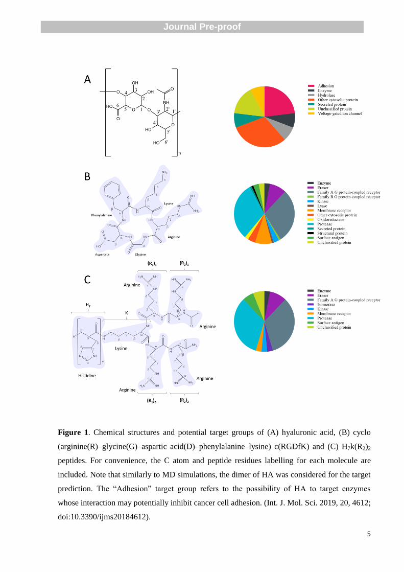
seemingly energy independent manner. Figure 1 shows the chemical structures of hyaluronic
acid and both peptides, c(RGDfK) and H7K(R2)2, as well as potential targeting receptor groups
identified through the SwissTargetPrediction server24
.
Journal Pre-proof
Jour
nal P
re-p
roof
5
Figure 1. Chemical structures and potential target groups of (A) hyaluronic acid, (B) cyclo
(arginine(R)–glycine(G)–aspartic acid(D)–phenylalanine–lysine) c(RGDfK) and (C) H7k(R2)2
peptides. For convenience, the C atom and peptide residues labelling for each molecule are
included. Note that similarly to MD simulations, the dimer of HA was considered for the target
prediction. The “Adhesion” target group refers to the possibility of HA to target enzymes
whose interaction may potentially inhibit cancer cell adhesion. (Int. J. Mol. Sci. 2019, 20, 4612;
doi:10.3390/ijms20184612).
Journal Pre-proof
Jour
nal P
re-p
roof
6
The synthesis and characterization of polymer-peptide conjugates raise several
questions in what concerns the hierarchical topology of such structures. How can the synthesis
pathway be tailored to provide a unique stable system? Novelty consists on explaining
polymer-peptide interactions based on a dual experimental-computational framework,
encompassing nuclear magnetic resonance (NMR) and molecular dynamics (MD) simulations.
1. Materials and methods
2.1.Materials
N-(3-Dimethylaminopropyl)-N′-ethylcarbodiimide hydrochloride (EDC) and
N-Hydroxysulfosuccinimide sodium salt (sulfo-NHS) were obtained from Sigma-Aldrich
(USA). Hyaluronic acid (MW 14800Da) was acquired from Life Biomedical, LLC (UK).
C(RGDfK) and H7K(R2)2 were purchased from GL Biochem Ltd (China) and Beijing SciLight
Biotechnology LLC (China), respectively.
2.2.Synthesis of peptide-polymer conjugates
Hyaluronic acid (HA) modification was achieved by amine coupling. First, carboxylic
groups of HA were activated by EDC/sulfo-NHS method as previously reported25
. HA (5x10⁻⁴
mmol) was dissolved in ultrapurified water followed by the addition of EDC (10x10-4
mmol)
and sulfo-NHS (10x10-4
mmol). The reaction mixture was stirred gently for 2 h followed by the
addition of c(RGDfK) (5x10⁻⁴
mmol) and/or H7K(R2)2 (5x10⁻⁴
mmol) and stirring for 3 h. The
reaction mixture was dialyzed against ultrapurified water using a 14 kDa cutoff membrane for
24 h to remove unreacted reagents.
2.3.Structural analysis based on NMR
The 1H and
13C NMR spectra were obtained on a Bruker Avance III HD 500 MHz NMR
spectrometer. The 13
C spectra were recorded using proton decoupling techniques taking
advantage of the nuclear Overhauser effect. The methyl signal of tert-butyl alcohol was used as
internal reference for 1H (δ 1.2) and
13C (δ 31.2) shifts. The homo- and heteronuclear 2D NMR
spectra, DQCOSY, TOCSY, ROESY, HSQC and HMBC were recorded on the same
spectrometer. Solutions were prepared in D2O or in a mixture 70% H2O/30% D2O and the pH*
values quoted are the direct pH-meter readings (room temperature) after standardization with
aqueous buffers26
.
Journal Pre-proof
Jour
nal P
re-p
roof
7
2.4.Molecular dynamics simulations
MD simulations combined with the analysis of noncovalent interactions have proven
valuable in decoupling the type and strengths of intermolecular interactions, endowing a
powerful predictive complement to experiments27–29
. Aiming at inspecting the experimental
synthesis pathway at a molecular level, hierarchical constructs were sequentially analysed,
including HA, HA:c(RGDfK), and HA-c(RGDfK):H7K(R2)2, based on the topological features
of the electronic charge densities of the binding partners. Single solvated peptide molecules
were also considered as system references. For HA backbone, two units of glucuronic acid/N-
acetylglucosamine were considered. The starting geometries of HA, c(RGDfK), H7K(R2)2 and
the respective conjugates were constructed in Avogadro and Pymol and optimized with the
semi-empirical Antechamber/SQM method. Partial charges for each molecule were obtained
from AM1-BCC30
.
A cubic box of 8.5×8.5×8.5 nm3 was employed for each system containing one
molecule of each binding partner HA:c(RGDfK), HA:H7K(R2)2, and HA-c(RGDfK):H7K(R2)2,
solvated with approximately 20200 water molecules.
2.4.1. Simulation details
MD simulations were performed using the GROMACS package (version 4.6.5)31
and
the all atom amber99sb force field32
, under periodic boundary conditions, and with a NPT
ensemble, see33
. TIP3P waters were employed for modelling aqueous solvation. A constant
temperature and pressure of 300 K and 1 bar, respectively, were imposed in all simulations, by
the coupling constants of 0.5 ps and 1 ps, respectively. An equilibration run of 40 ns was
performed prior to each production run, maintaining the pressure at 1 bar. No pressure coupling
was imposed during the production runs, allowing to keep the size of the simulation box
constant. Lennard–Jones interactions and electrostatic interactions were assessed using a
cut-off of 0.9 nm and the particle mesh Ewald (PME) method, respectively34
. The constraints in
the binding partners were imposed by the LINCS algorithm35
.
2.4.2. Analysis of noncovalent interactions
Evaluation of the noncovalent interactions (NCI) within HA:c(RGDfK), HA:H7K(R2)2,
c(RGDfK):H7K(R2)2, and HA-c(RGDfK):H7K(R2)2 associations was performed based on the
Independent Gradient Method (IGM)28,36
, and using the IGMPlot software (version 1.0). This
method is based on the analysis of the electronic charge density of the binding partners and the
respective gradients, also enabling the visualization and quantification of those regions of
Journal Pre-proof
Jour
nal P
re-p
roof
8
stabilizing/destabilizing noncovalent interactions. IGM is governed by the topological
characteristics of the electronic charge density, ρ, of each system. In addition to ρ, this method
uses quantities corresponding to the first and second derivatives of the density. δginter
refers to
the IGM descriptor estimated by the difference between the first derivatives of the charge
densities for the final system and the respective single components,
𝛿𝑔𝑖𝑛𝑡𝑒𝑟 = |𝛻𝜌𝐼𝐺𝑀,𝑖𝑛𝑡𝑒𝑟| − |𝛻𝜌| (Eq. 1)
The occurrence and strength of noncovalent interactions are inferred, respectively, when
δginter
> 0, and by the magnitude of the descriptor at a point in space.
∇ρIGM,inter is obtained from the sum of the N atoms in the different components, referred
as A and B, in the x-direction,
(𝛿𝜌
𝛿𝑥)𝐼𝐺𝑀,𝑖𝑛𝑡𝑒𝑟
= |∑𝛿𝜌𝑖
𝛿𝑥
𝑁𝐴𝑖=1 | + |∑
𝛿𝜌𝑖
𝛿𝑥
𝑁𝐵𝑖=1 | (Eq. 2)
IGMPlot employs the pre-computed atomic charge densities for estimating a
pro-molecular density that produces a minimal effect on the noncovalent interactions. δginter
allows identifying NCI regions, and ∇ 2ρ, a second derivative (Laplacian) of the density, is used
for discriminating favorable/unfavorable NCIs. The decomposition of the Laplacian term into
∇2ρ=λ1+λ2+λ3 (λ1≤λ2≤λ3), the three eigenvalues of maximal variation, provides information on
the stabilizing (λ2 < 0) or destabilizing (λ2 > 0) interactions. Larger (negative) values of
sign(λ2)ρ indicate stronger interactions, e.g. hydrogen bonds, while values close to zero reflect
weak NCI, including van der Waals forces.
The coordinates of the HA:c(RGDfK), HA:H7K(R2)2, and HA-c(RGDfK):H7K(R2)2 conjugates
were extracted from the ensemble of structures at the equilibrium state, sampled during the MD
simulations (100 ns of production run), using the clustering procedure described in33
.
Geometric clusters were established based on the calculation of the root mean square deviation
(RMSD of 0.25 nm) of the atom positions between all pairs of each polymer-peptide structure.
Also, water molecules were discarded and the conjugates were separated into the respective
binding components33
. NCI are represented by isosurface volumes for the δginter
, coloured
according to the sign(λ2)ρ values. These values provide a quantitative evaluation of the types
and strengths of NCI within each system, reflecting both the extent and (de)stabilizing nature of
the interactions.
Visual Molecular Dynamics software version 1.9.2 was used to visualize the polymer-
peptide systems and the respective isosurfaces.
Journal Pre-proof
Jour
nal P
re-p
roof
9
3. Results and discussion
The rationale behind polymer-peptide conjugate synthesis is assuming increasingly
importance, in particular for tumour targeting purposes. Betting on supramolecular structures
that entail a certain level of complexity, associated to e.g. the size, nature and type of amino
acid residues, and also to variations in conformational behavior must be supported by a deep
understanding of the mechanistic aspects involved in the binding process and stability of
polymer-peptide conjugates. In this context, several questions are raised: How can
polymer-peptide supramolecular constructs be formed? Is the peptide order addition relevant?
What governs these hierarchical constructs at the molecular level? Which are the main
interaction forces established? These challenging aspects are explored in what follows,
combining insights from NMR and MD simulations.
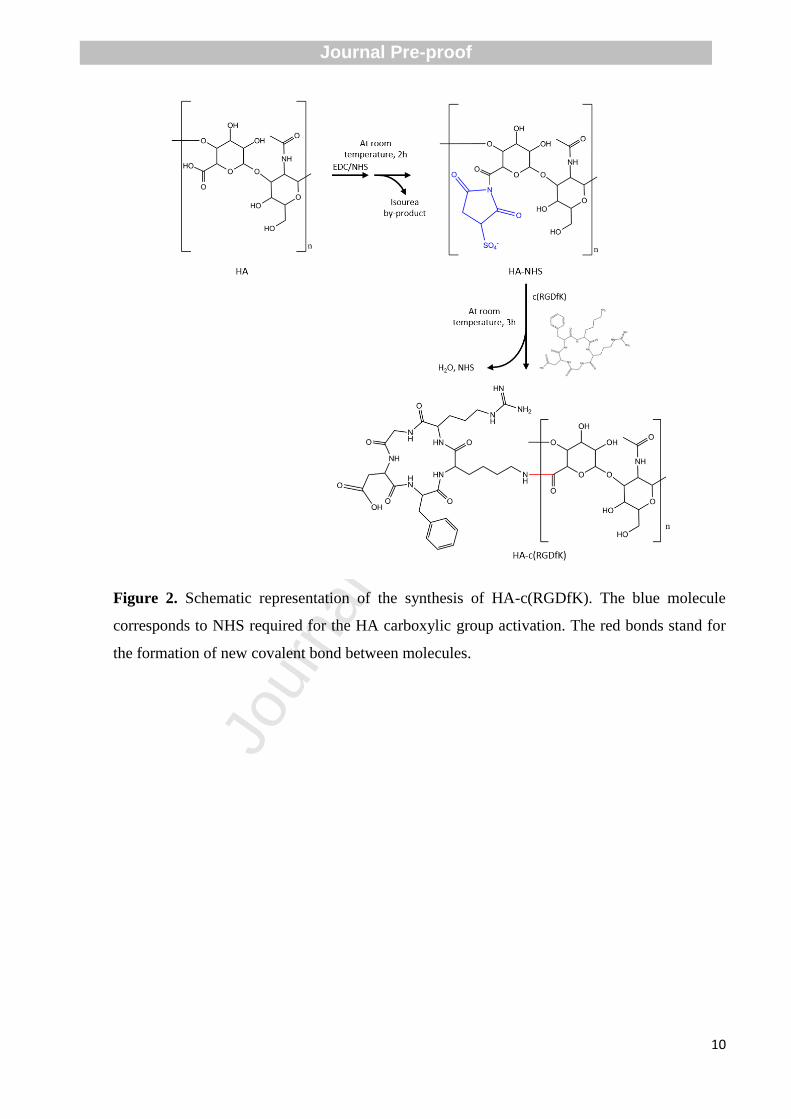
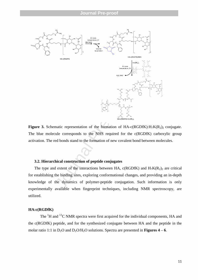
3.1. Synthetic route proposed for polymer:peptide contructs
The hierarchical design of hyaluronic acid-peptide constructs for glioblastoma targeting
is hypothesized in Figures 2 and 3. Such proposal results from the successful attachment of
c(RGDfK) and H7K(R2)2 peptides onto the HA backbone, which enables their further
utilization in the surface shaping of ultra-small lipid nanoparticles tailored for glioblastoma
targeting. Systems corresponding to HA:c(RGDfK), HA:H7K(R2)2, and HA-
c(RGDfK):H7K(R2)2 interaction pairs will be duly discussed, so as to establish the assumptions
for the suggested synthetic route.
Journal Pre-proof
Jour
nal P
re-p
roof
10
Figure 2. Schematic representation of the synthesis of HA-c(RGDfK). The blue molecule
corresponds to NHS required for the HA carboxylic group activation. The red bonds stand for
the formation of new covalent bond between molecules.
Journal Pre-proof
Jour
nal P
re-p
roof
11
Figure 3. Schematic representation of the formation of HA-c(RGDfK):H7K(R2)2 conjugate.
The blue molecule corresponds to the NHS required for the c(RGDfK) carboxylic group
activation. The red bonds stand to the formation of new covalent bond between molecules.
3.2. Hierarchical construction of peptide conjugates
The type and extent of the interactions between HA, c(RGDfK) and H7K(R2)2 are critical
for establishing the binding sites, exploring conformational changes, and providing an in-depth
knowledge of the dynamics of polymer-peptide conjugation. Such information is only
experimentally available when fingerprint techniques, including NMR spectroscopy, are
utilized.
HA:c(RGDfK)
The 1H and
13C NMR spectra were first acquired for the individual components, HA and
the c(RGDfK) peptide, and for the synthesized conjugate between HA and the peptide in the
molar ratio 1:1 in D2O and D2O/H2O solutions. Spectra are presented in Figures 4 – 6.
Journal Pre-proof
Jour
nal P
re-p
roof
12
Figure 4. 1H NMR spectra of the NH region (expansion 6.5 – 9.0 ppm) of the solutions in
H2O/D2O (70%:30%) of (a) hyaluronic acid (HA) 5.0 mmol dm-3
, pH* 6.61; (b) c(RGDfK)
peptide, 5.0 mmol dm-3
, pH* 3.21 and (c) HA-c(RGDfK) conjugate 5.0 mmol dm-3
, pH* 3.15.
Journal Pre-proof
Jour
nal P
re-p
roof
13
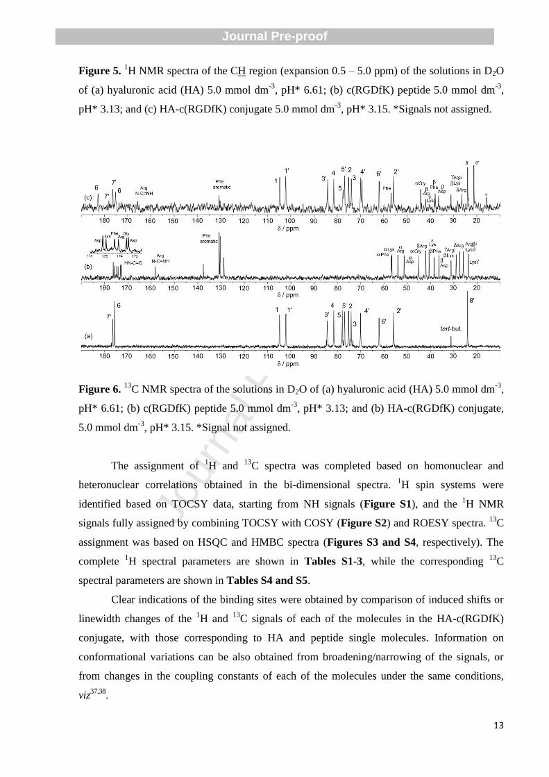
Figure 5. 1H NMR spectra of the CH region (expansion 0.5 – 5.0 ppm) of the solutions in D2O
of (a) hyaluronic acid (HA) 5.0 mmol dm-3
, pH* 6.61; (b) c(RGDfK) peptide 5.0 mmol dm-3
,
pH* 3.13; and (c) HA-c(RGDfK) conjugate 5.0 mmol dm-3
, pH* 3.15. *Signals not assigned.
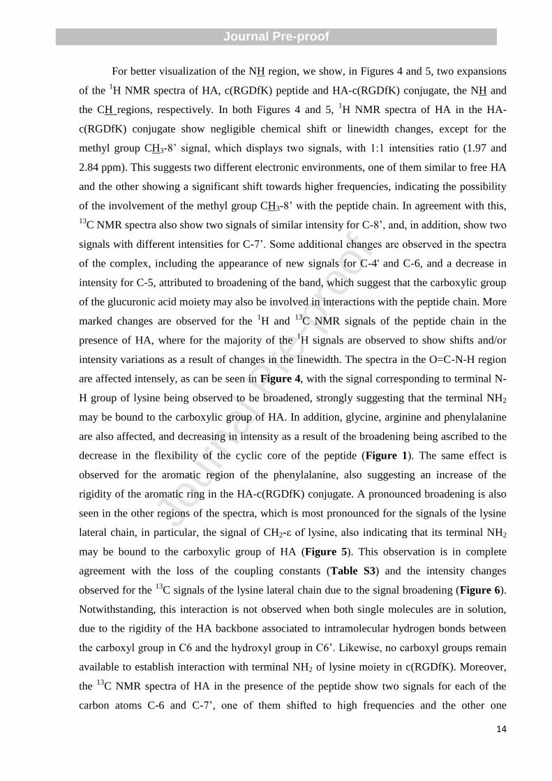
Figure 6. 13
C NMR spectra of the solutions in D2O of (a) hyaluronic acid (HA) 5.0 mmol dm-3
,
pH* 6.61; (b) c(RGDfK) peptide 5.0 mmol dm-3
, pH* 3.13; and (b) HA-c(RGDfK) conjugate,
5.0 mmol dm-3
, pH* 3.15. *Signal not assigned.
The assignment of 1H and
13C spectra was completed based on homonuclear and
heteronuclear correlations obtained in the bi-dimensional spectra. 1H spin systems were
identified based on TOCSY data, starting from NH signals (Figure S1), and the 1H NMR
signals fully assigned by combining TOCSY with COSY (Figure S2) and ROESY spectra. 13
C
assignment was based on HSQC and HMBC spectra (Figures S3 and S4, respectively). The
complete 1H spectral parameters are shown in Tables S1-3, while the corresponding
13C
spectral parameters are shown in Tables S4 and S5.
Clear indications of the binding sites were obtained by comparison of induced shifts or
linewidth changes of the 1H and
13C signals of each of the molecules in the HA-c(RGDfK)
conjugate, with those corresponding to HA and peptide single molecules. Information on
conformational variations can be also obtained from broadening/narrowing of the signals, or
from changes in the coupling constants of each of the molecules under the same conditions,
viz37,38
.
Journal Pre-proof
Jour
nal P
re-p
roof
14
For better visualization of the NH region, we show, in Figures 4 and 5, two expansions
of the 1H NMR spectra of HA, c(RGDfK) peptide and HA-c(RGDfK) conjugate, the NH and
the CH regions, respectively. In both Figures 4 and 5, 1H NMR spectra of HA in the HA-
c(RGDfK) conjugate show negligible chemical shift or linewidth changes, except for the
methyl group CH3-8’ signal, which displays two signals, with 1:1 intensities ratio (1.97 and
2.84 ppm). This suggests two different electronic environments, one of them similar to free HA
and the other showing a significant shift towards higher frequencies, indicating the possibility
of the involvement of the methyl group CH3-8’ with the peptide chain. In agreement with this,
13C NMR spectra also show two signals of similar intensity for C-8’, and, in addition, show two
signals with different intensities for C-7’. Some additional changes are observed in the spectra
of the complex, including the appearance of new signals for C-4' and C-6, and a decrease in
intensity for C-5, attributed to broadening of the band, which suggest that the carboxylic group
of the glucuronic acid moiety may also be involved in interactions with the peptide chain. More
marked changes are observed for the 1H and
13C NMR signals of the peptide chain in the
presence of HA, where for the majority of the 1H signals are observed to show shifts and/or
intensity variations as a result of changes in the linewidth. The spectra in the O=C-N-H region
are affected intensely, as can be seen in Figure 4, with the signal corresponding to terminal N-
H group of lysine being observed to be broadened, strongly suggesting that the terminal NH2
may be bound to the carboxylic group of HA. In addition, glycine, arginine and phenylalanine
are also affected, and decreasing in intensity as a result of the broadening being ascribed to the
decrease in the flexibility of the cyclic core of the peptide (Figure 1). The same effect is
observed for the aromatic region of the phenylalanine, also suggesting an increase of the
rigidity of the aromatic ring in the HA-c(RGDfK) conjugate. A pronounced broadening is also
seen in the other regions of the spectra, which is most pronounced for the signals of the lysine
lateral chain, in particular, the signal of CH2-ε of lysine, also indicating that its terminal NH2
may be bound to the carboxylic group of HA (Figure 5). This observation is in complete
agreement with the loss of the coupling constants (Table S3) and the intensity changes
observed for the 13
C signals of the lysine lateral chain due to the signal broadening (Figure 6).
Notwithstanding, this interaction is not observed when both single molecules are in solution,
due to the rigidity of the HA backbone associated to intramolecular hydrogen bonds between
the carboxyl group in C6 and the hydroxyl group in C6’. Likewise, no carboxyl groups remain
available to establish interaction with terminal NH2 of lysine moiety in c(RGDfK). Moreover,
the 13
C NMR spectra of HA in the presence of the peptide show two signals for each of the
carbon atoms C-6 and C-7’, one of them shifted to high frequencies and the other one
Journal Pre-proof
Jour
nal P
re-p
roof
15
presenting very similar resonance to the respective nuclei in free HA; in addition, the signals
corresponding to nuclei C-5 and C-4’ appear broadened and duplicated, respectively (Figure
6). These observations, in particular, the intensities of the bound and unbound signals, suggest
that spatially alternating moieties of HA are involved in interactions with the peptide by the
carboxylic acid, which is bound to the terminal NH2 group of lysine. This alternating state
allows the formation of other NH…
O hydrogen bonds between other moieties of c(RGDfK),
such as C6=O …
NH (D-Phe), see Table S1.
Additional interactions of the methyl group of the N-acetyl glucosamine moiety with the
aromatic group of phenylalanine cannot be ruled out, based on the changes in the
corresponding signals, as has previously been described for peptides and HA39
.
HA:H7K(R2)2
The 1H NMR spectra of HA in the HA-H7K(R2)2 conjugate indicate, when compared to
free HA, negligible chemical shift or linewidth changes, except for the methyl group CH3-8’
signal, which displays two signals, (1.97 and 2.83 ppm) with 2:1 and 4:1 intensities ratio, for
conjugates synthesised starting from mixtures containing HA-H7K(R2)2 with molar ratios 1:1
and 4:1, respectively. Two different electronic environments are suggested, one of them
containing two and four units, respectively, similar to free HA, while the other corresponds to
one unit, showing a significant shift towards higher frequencies, indicating the possibility of the
involvement of the methyl group CH3-8’ with the peptide. In agreement with this, 13
C NMR
spectra also show two signals with similar relative intensities for C-8’. Some minor additional
changes are observed for HA in the spectra of the conjugate, including a decrease in intensity
for C-5, attributed to broadening of the band, which also suggests that the D-glucuronic moiety
may also be involved with the H7K(R2)2 peptide. Moreover, significant changes of intensity are
observed for the carboxyl groups of HA, suggesting the involvement in the peptide binding.
More marked changes are also observed for the 1H and
13C NMR signals of the peptide chain in
the presence of HA, where for the majority of the 1H and
13C signals are observed significant
variations. For most of the signals, the severe broadening precludes their detection, suggesting
the increasing of the rigidity of the chain. The conjugate is established probably via the linkage
between the carboxylic group of HA and the α-NH group of the terminal histidine residue. In
addition, the loss of the 1H and
13C signals should indicate the decrease of the flexibility of the
peptide chain, also suggesting an increase of hydrophobic interactions together with important
steric effects arising from the HA backbone in the conjugate when compared with the peptide
alone. These observations, in particular, the intensities of the bound and unbound signals of
Journal Pre-proof
Jour
nal P
re-p
roof
16
methyl group CH3-8’, suggest that in the conjugate obtained from the solution having a HA to
peptide molar ratio 1:1, considering three consecutive moieties of HA, two are free and one is
involved in interactions with the peptide.
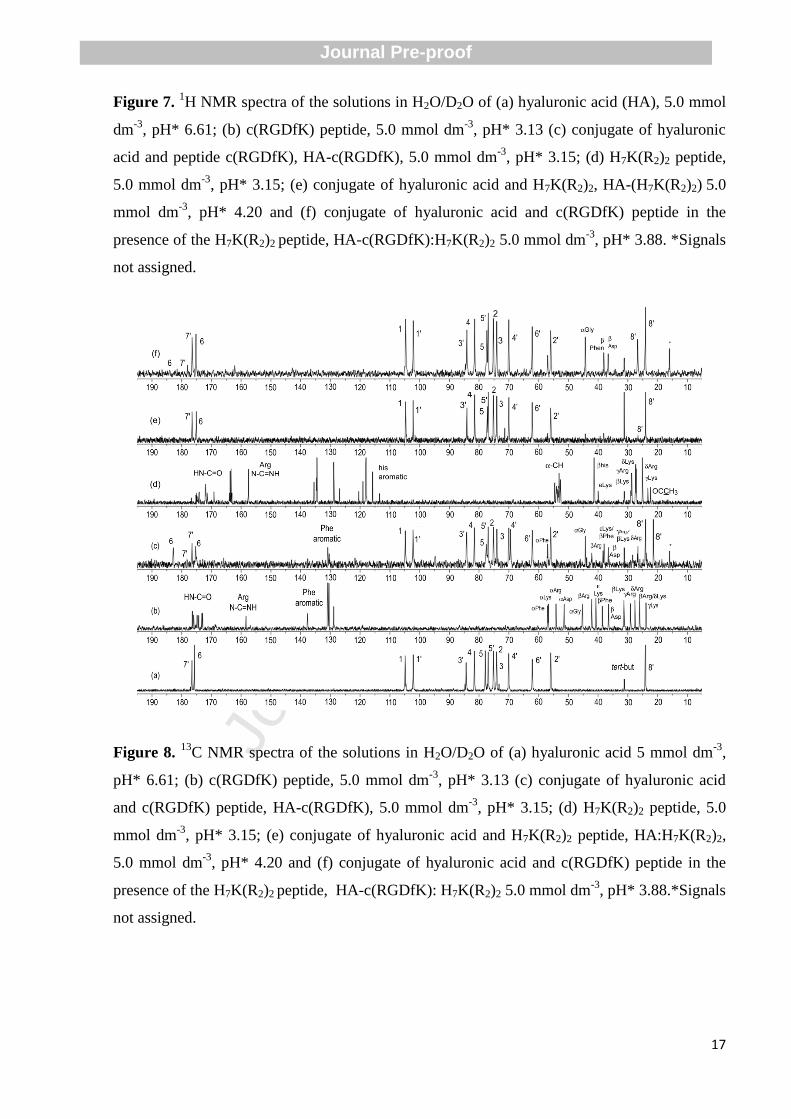
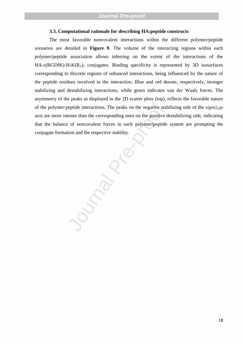
HA-c(RGDfK):H7K(R2)2
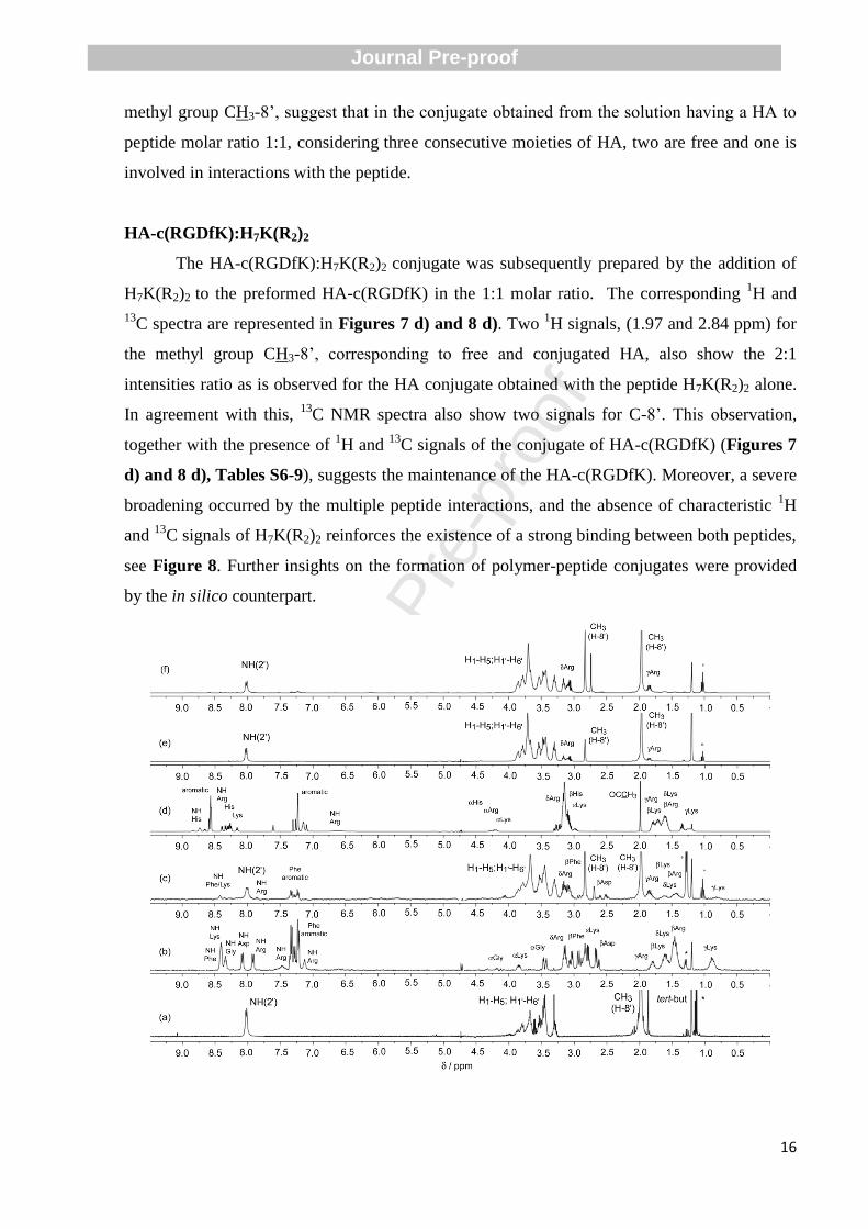
The HA-c(RGDfK):H7K(R2)2 conjugate was subsequently prepared by the addition of
H7K(R2)2 to the preformed HA-c(RGDfK) in the 1:1 molar ratio. The corresponding 1H and
13C spectra are represented in Figures 7 d) and 8 d). Two
1H signals, (1.97 and 2.84 ppm) for
the methyl group CH3-8’, corresponding to free and conjugated HA, also show the 2:1
intensities ratio as is observed for the HA conjugate obtained with the peptide H7K(R2)2 alone.
In agreement with this, 13
C NMR spectra also show two signals for C-8’. This observation,
together with the presence of 1H and
13C signals of the conjugate of HA-c(RGDfK) (Figures 7
d) and 8 d), Tables S6-9), suggests the maintenance of the HA-c(RGDfK). Moreover, a severe
broadening occurred by the multiple peptide interactions, and the absence of characteristic 1H
and 13
C signals of H7K(R2)2 reinforces the existence of a strong binding between both peptides,
see Figure 8. Further insights on the formation of polymer-peptide conjugates were provided
by the in silico counterpart.
Journal Pre-proof
Jour
nal P
re-p
roof
17
Figure 7. 1H NMR spectra of the solutions in H2O/D2O of (a) hyaluronic acid (HA), 5.0 mmol
dm-3
, pH* 6.61; (b) c(RGDfK) peptide, 5.0 mmol dm-3
, pH* 3.13 (c) conjugate of hyaluronic
acid and peptide c(RGDfK), HA-c(RGDfK), 5.0 mmol dm-3
, pH* 3.15; (d) H7K(R2)2 peptide,
5.0 mmol dm-3
, pH* 3.15; (e) conjugate of hyaluronic acid and H7K(R2)2, HA-(H7K(R2)2) 5.0
mmol dm-3
, pH* 4.20 and (f) conjugate of hyaluronic acid and c(RGDfK) peptide in the
presence of the H7K(R2)2 peptide, HA-c(RGDfK):H7K(R2)2 5.0 mmol dm-3
, pH* 3.88. *Signals
not assigned.
Figure 8. 13
C NMR spectra of the solutions in H2O/D2O of (a) hyaluronic acid 5 mmol dm-3
,
pH* 6.61; (b) c(RGDfK) peptide, 5.0 mmol dm-3
, pH* 3.13 (c) conjugate of hyaluronic acid
and c(RGDfK) peptide, HA-c(RGDfK), 5.0 mmol dm-3
, pH* 3.15; (d) H7K(R2)2 peptide, 5.0
mmol dm-3
, pH* 3.15; (e) conjugate of hyaluronic acid and H7K(R2)2 peptide, HA:H7K(R2)2,
5.0 mmol dm-3
, pH* 4.20 and (f) conjugate of hyaluronic acid and c(RGDfK) peptide in the
presence of the H7K(R2)2 peptide, HA-c(RGDfK): H7K(R2)2 5.0 mmol dm-3
, pH* 3.88.*Signals
not assigned.
Journal Pre-proof
Jour
nal P
re-p
roof
18
3.3. Computational rationale for describing HA:peptide constructs
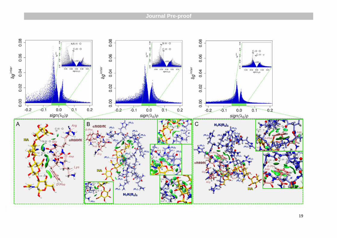
The most favorable noncovalent interactions within the different polymer/peptide
scenarios are detailed in Figure 9. The volume of the interacting regions within each
polymer/peptide association allows inferring on the extent of the interactions of the
HA-c(RGDfK)-H7K(R2)2 conjugates. Binding specificity is represented by 3D isosurfaces
corresponding to discrete regions of enhanced interactions, being influenced by the nature of
the peptide residues involved in the interaction. Blue and red denote, respectively, stronger
stabilizing and destabilizing interactions, while green indicates van der Waals forces. The
asymmetry of the peaks as displayed in the 2D scatter plots (top), reflects the favorable nature
of the polymer:peptide interactions. The peaks on the negative stabilizing side of the sign(λ2)ρ
axis are more intense than the corresponding ones on the positive destabilizing side, indicating
that the balance of noncovalent forces in each polymer/peptide system are prompting the
conjugate formation and the respective stability.
Journal Pre-proof
Journal P
re-proof
19
Journal Pre-proof
Journal P
re-proof
20
Figure 9. Interaction patterns underlying polymer/peptides association (A) HA-c(RGDfK), (B) HA-c(RGDfK)-H7K(R2)2, and (C)
HA-c(RGDfK):H7K(R2)2, represented in the 2D scatter plots (top) and in the 3D IGMPlot isosurfaces (bottom). The coordinates
corresponding to the final conjugate structures were extracted from the last time step of MD simulations. Isosurfaces display the total IGM
interaction points for δginter≤0.1 and in the region −0.2≤sign(λ2)ρ≤0.2. and are colored based on a red-green-blue gradient over the electron
density range −0.05<sign(λ2)ρ<0.05 a.u.. Blue/red reflect stabilizing/destabilizing noncovalent interactions, while green represents van der
Waals-type forces. HA and c(RGDfK) backbones are represented in yellow and pink, respectively, as licorice, while H7K(R2)2 is featured in
ball-and-stick and colored in iceblue. Nitrogen, oxygen and hydrogen atoms are represented in blue, red and white, respectively.
Journal Pre-proof
Jour
nal P
re-p
roof
21
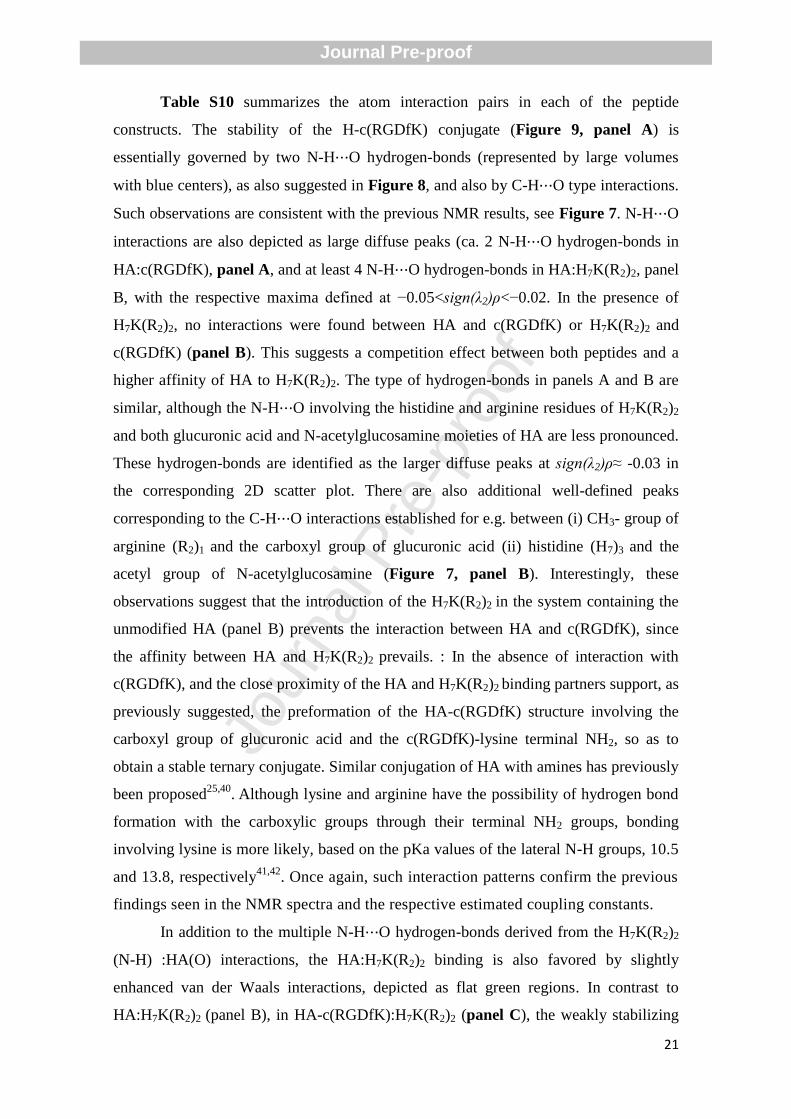
Table S10 summarizes the atom interaction pairs in each of the peptide
constructs. The stability of the H-c(RGDfK) conjugate (Figure 9, panel A) is
essentially governed by two N-H⋯O hydrogen-bonds (represented by large volumes
with blue centers), as also suggested in Figure 8, and also by C-H⋯O type interactions.
Such observations are consistent with the previous NMR results, see Figure 7. N-H⋯O
interactions are also depicted as large diffuse peaks (ca. 2 N-H⋯O hydrogen-bonds in
HA:c(RGDfK), panel A, and at least 4 N-H⋯O hydrogen-bonds in HA:H7K(R2)2, panel
B, with the respective maxima defined at −0.05<sign(λ2)ρ<−0.02. In the presence of
H7K(R2)2, no interactions were found between HA and c(RGDfK) or H7K(R2)2 and
c(RGDfK) (panel B). This suggests a competition effect between both peptides and a
higher affinity of HA to H7K(R2)2. The type of hydrogen-bonds in panels A and B are
similar, although the N-H⋯O involving the histidine and arginine residues of H7K(R2)2
and both glucuronic acid and N-acetylglucosamine moieties of HA are less pronounced.
These hydrogen-bonds are identified as the larger diffuse peaks at sign(λ2)ρ≈ -0.03 in
the corresponding 2D scatter plot. There are also additional well-defined peaks
corresponding to the C-H⋯O interactions established for e.g. between (i) CH3- group of
arginine (R2)1 and the carboxyl group of glucuronic acid (ii) histidine (H7)3 and the
acetyl group of N-acetylglucosamine (Figure 7, panel B). Interestingly, these
observations suggest that the introduction of the H7K(R2)2 in the system containing the
unmodified HA (panel B) prevents the interaction between HA and c(RGDfK), since
the affinity between HA and H7K(R2)2 prevails. : In the absence of interaction with
c(RGDfK), and the close proximity of the HA and H7K(R2)2 binding partners support, as
previously suggested, the preformation of the HA-c(RGDfK) structure involving the
carboxyl group of glucuronic acid and the c(RGDfK)-lysine terminal NH2, so as to
obtain a stable ternary conjugate. Similar conjugation of HA with amines has previously
been proposed25,40
. Although lysine and arginine have the possibility of hydrogen bond
formation with the carboxylic groups through their terminal NH2 groups, bonding
involving lysine is more likely, based on the pKa values of the lateral N-H groups, 10.5
and 13.8, respectively41,42
. Once again, such interaction patterns confirm the previous
findings seen in the NMR spectra and the respective estimated coupling constants.
In addition to the multiple N-H⋯O hydrogen-bonds derived from the H7K(R2)2
(N-H) :HA(O) interactions, the HA:H7K(R2)2 binding is also favored by slightly
enhanced van der Waals interactions, depicted as flat green regions. In contrast to
HA:H7K(R2)2 (panel B), in HA-c(RGDfK):H7K(R2)2 (panel C), the weakly stabilizing
Journal Pre-proof
Jour
nal P
re-p
roof
22
van der Waals-type interactions are established mainly between the H7K(R2)2-lysine (K)
moiety and the c(RGDfK)-phenylalanine phenyl CH groups. Stabilization of the
HA-c(RGDfK):H7K(R2)2 construct is also assured by weak hydrogen bonds between the
H7K(R2)2-arginine (R1 and R2) carbonyl groups, and the c(RGDfK)-lysine CεH2, CβH2,
CγH2 and c(RGDfK)-aspartate CβH2 groups.
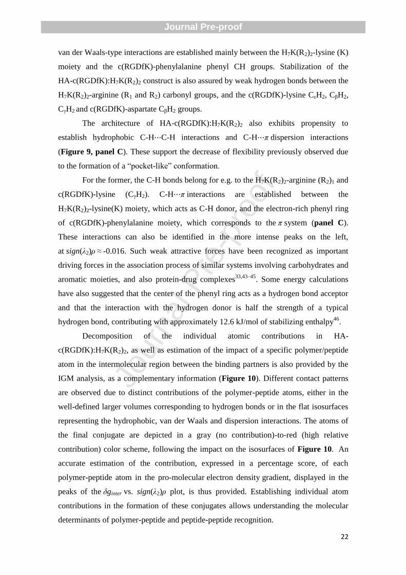
The architecture of HA-c(RGDfK):H7K(R2)2 also exhibits propensity to
establish hydrophobic C-H⋯C-H interactions and C-H⋯π dispersion interactions
(Figure 9, panel C). These support the decrease of flexibility previously observed due
to the formation of a “pocket-like” conformation.
For the former, the C-H bonds belong for e.g. to the H7K(R2)2-arginine (R2)1 and
c(RGDfK)-lysine (CγH2). C-H⋯π interactions are established between the
H7K(R2)2-lysine(K) moiety, which acts as C-H donor, and the electron-rich phenyl ring
of c(RGDfK)-phenylalanine moiety, which corresponds to the π system (panel C).
These interactions can also be identified in the more intense peaks on the left,
at sign(λ2)ρ ≈ -0.016. Such weak attractive forces have been recognized as important
driving forces in the association process of similar systems involving carbohydrates and
aromatic moieties, and also protein-drug complexes33,43–45
. Some energy calculations
have also suggested that the center of the phenyl ring acts as a hydrogen bond acceptor
and that the interaction with the hydrogen donor is half the strength of a typical
hydrogen bond, contributing with approximately 12.6 kJ/mol of stabilizing enthalpy46
.
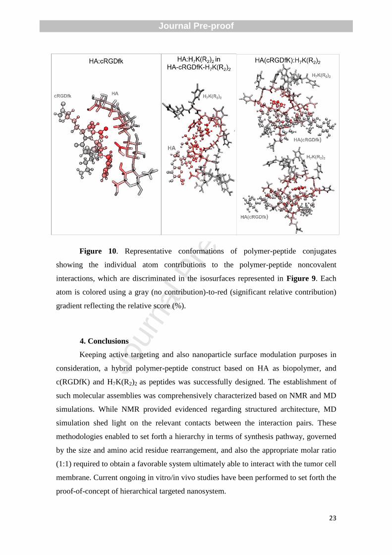
Decomposition of the individual atomic contributions in HA-
c(RGDfK):H7K(R2)2, as well as estimation of the impact of a specific polymer/peptide
atom in the intermolecular region between the binding partners is also provided by the
IGM analysis, as a complementary information (Figure 10). Different contact patterns
are observed due to distinct contributions of the polymer-peptide atoms, either in the
well-defined larger volumes corresponding to hydrogen bonds or in the flat isosurfaces
representing the hydrophobic, van der Waals and dispersion interactions. The atoms of
the final conjugate are depicted in a gray (no contribution)-to-red (high relative
contribution) color scheme, following the impact on the isosurfaces of Figure 10. An
accurate estimation of the contribution, expressed in a percentage score, of each
polymer-peptide atom in the pro-molecular electron density gradient, displayed in the
peaks of the δginter vs. sign(λ2)ρ plot, is thus provided. Establishing individual atom
contributions in the formation of these conjugates allows understanding the molecular
determinants of polymer-peptide and peptide-peptide recognition.
Journal Pre-proof
Jour
nal P
re-p
roof
23
Figure 10. Representative conformations of polymer-peptide conjugates
showing the individual atom contributions to the polymer-peptide noncovalent
interactions, which are discriminated in the isosurfaces represented in Figure 9. Each
atom is colored using a gray (no contribution)-to-red (significant relative contribution)
gradient reflecting the relative score (%).
4. Conclusions
Keeping active targeting and also nanoparticle surface modulation purposes in
consideration, a hybrid polymer-peptide construct based on HA as biopolymer, and
c(RGDfK) and H7K(R2)2 as peptides was successfully designed. The establishment of
such molecular assemblies was comprehensively characterized based on NMR and MD
simulations. While NMR provided evidenced regarding structured architecture, MD
simulation shed light on the relevant contacts between the interaction pairs. These
methodologies enabled to set forth a hierarchy in terms of synthesis pathway, governed
by the size and amino acid residue rearrangement, and also the appropriate molar ratio
(1:1) required to obtain a favorable system ultimately able to interact with the tumor cell
membrane. Current ongoing in vitro/in vivo studies have been performed to set forth the
proof-of-concept of hierarchical targeted nanosystem.
Journal Pre-proof
Jour
nal P
re-p
roof
24
Acknowledgments
The authors acknowledge the Portuguese Agency for Scientific Research,
“Fundação para a Ciência e a Tecnologia (FCT)”, for the financial support through the
projects POCI-01-0145-FEDER-016648, PEst-UID/NEU/04539/2013 and COMPETE
(Ref. POCI-01-0145-FEDER-007440). The Coimbra Chemistry Centre (CQC) is also
supported by FCT through the Project UID/QUI/00313/2020. RV also acknowledges
FCT for the financial support of the projects IF/00286/2015, iBiMED
(UID/BIM/04501/2019 and POCI-01-0145-FEDER-007628), and UnIC
(UID/IC/00051/2019). Maria Mendes and João Basso acknowledge the PhD research
Grants SFRH/BD/133996/2017 and SFRH/BD/149138/2019, respectively, assigned by
FCT.
NMR data were obtained at the UC-NMR facility which is supported in part by
the FEDER – European Regional Development Fund through the COMPETE
Programme (Operational Programme for Competitiveness) and by the National Funds
through FCT with the grants REEQ/481/QUI/2006, RECI/QEQ- QFI/0168/2012,
CENTRO-07-CT62-FEDER-002012, and the Rede Nacional de Ressonância Magnética
Nuclear (RNRMN).
References
1 T. Kazda, A. Dziacky, P. Burkon, P. Pospisil, M. Slavik, Z. Rehak, R. Jancalek,
P. Slampa, O. Slaby and R. Lakomy, Radiol. Oncol., 2018, 52, 121–128.
2 R. Stupp, W. P. Mason, M. J. Van Den Bent, M. Weller, B. Fisher, M. J. B.
Taphoorn, K. Belanger, A. A. Brandes, C. Marosi and U. Bogdahn, N. Engl. J.
Med., 2005, 352, 987–996.
3 H. P. Ellis, M. Greenslade, B. Powell, I. Spiteri, A. Sottoriva and K. M. Kurian,
Front. Oncol., 2015, 5, 251.
4 O. Van Tellingen, B. Yetkin-Arik, M. C. De Gooijer, P. Wesseling, T. Wurdinger
and H. E. De Vries, Drug Resist. Updat., 2015, 19, 1–12.
5 K. B. Johnsen, A. Burkhart, F. Melander, P. J. Kempen, J. B. Vejlebo, P. Siupka,
M. S. Nielsen, T. L. Andresen and T. Moos, Sci. Rep., 2017, 7, 10396.
6 M. Mendes, J. Sousa, A. Pais and C. Vitorino, Pharmaceutics, 2018, 10, 181.
7 K. K. Jain, Front. Oncol.
8 L. Zhang, Y. Zhang, L. Tai, K. Jiang, C. Xie, Z. Li, Y.-Z. Lin, G. Wei, W. Lu and
W. Pan, Acta Biomater., 2016, 42, 90–101.
9 H. Gao, S. Zhang, S. Cao, Z. Yang, Z. Pang and X. Jiang, Mol. Pharm., 2014, 11,
2755–2763.
Journal Pre-proof
Jour
nal P
re-p
roof
25
10 W. Ke, Z. Zha, J. F. Mukerabigwi, W. Chen, Y. Wang, C. He and Z. Ge,
Bioconjug. Chem., 2017, 28, 2190–2198.
11 T. J. Harris, G. von Maltzahn, M. E. Lord, J. Park, A. Agrawal, D. Min, M. J.
Sailor and S. N. Bhatia, small, 2008, 4, 1307–1312.
12 J. Yao, Y. Ma, W. Zhang, L. Li, Y. Zhang, L. Zhang, H. Liu, J. Ni and R. Wang,
PeerJ, 2017, 5, e3429.
13 K. Shi, Y. Long, C. Xu, Y. Wang, Y. Qiu, Q. Yu, Y. Liu, Q. Zhang, H. Gao and
Z. Zhang, ACS Appl. Mater. Interfaces, 2015, 7, 21442–21454.
14 Y. Wang, K. Shi, L. Zhang, G. Hu, J. Wan, J. Tang, S. Yin, J. Duan, M. Qin and
N. Wang, Autophagy, 2016, 12, 949–962.
15 T. Jiang, Z. Zhang, Y. Zhang, H. Lv, J. Zhou, C. Li, L. Hou and Q. Zhang,
Biomaterials, 2012, 33, 9246–9258.
16 H. Zhou, H. Xu, X. Li, Y. Lv, T. Ma, S. Guo, Z. Huang, X. Wang and P. Xu,
Mater. Sci. Eng. C, 2017, 81, 261–270.
17 M. Swierczewska, H. S. Han, K. Kim, J. H. Park and S. Lee, Adv. Drug Deliv.
Rev., 2016, 99, 70–84.
18 L. I. Qin, C. Wang, H. Fan, C. Zhang, H. Zhang, M. Lv and S. Cui, Oncol. Lett.,
2014, 8, 2000–2006.
19 S. Song, G. Mao, J. Du and X. Zhu, Drug Deliv., 2016, 23, 1404–1408.
20 Y. Miura, T. Takenaka, K. Toh, S. Wu, H. Nishihara, M. R. Kano, Y. Ino, T.
Nomoto, Y. Matsumoto and H. Koyama, ACS Nano, 2013, 7, 8583–8592.
21 H. Hyun, Y. Yoo, S. Y. Kim, H. S. Ko, H. J. Chun and D. H. Yang, J. Ind. Eng.
Chem., 2020, 81, 178–184.
22 M. Zhou, N. Jiang, J. Fan, S. Fu, H. Luo, P. Su, M. Zhang, H. Shi, J. Zeng and Y.
Huang, J. Control. Release, 2019, 310, 24–35.
23 Y. Zhao, W. Ren, T. Zhong, S. Zhang, D. Huang, Y. Guo, X. Yao, C. Wang, W.-
Q. Zhang and X. Zhang, J. Control. Release, 2016, 222, 56–66.
24 D. Gfeller, A. Grosdidier, M. Wirth, A. Daina, O. Michielin and V. Zoete,
Nucleic Acids Res., 2014, 42, W32–W38.
25 Z. R. Cohen, S. Ramishetti, N. Peshes-Yaloz, M. Goldsmith, A. Wohl, Z. Zibly
and D. Peer, ACS Nano, 2015, 9, 1581–1591.
26 P. K. Glasoe and F. A. Long, J. Phys. Chem., 1960, 64, 188–190.
27 T. F. G. G. Cova, D. J. Bento and S. C. C. Nunes, Pharmaceutics, 2019, 11, 119.
28 A. Aviñó, A. F. Jorge, C. S. Huertas, T. F. G. G. Cova, A. Pais, L. M. Lechuga,
R. Eritja and C. Fabrega, Biochim. Biophys. Acta (BBA)-General Subj., 2019,
1863, 1619–1630.
29 M. Mendes, A. Miranda, T. Cova, L. Gonçalves, A. J. Almeida, J. J. Sousa, M. L.
C. do Vale, E. F. Marques, A. Pais and C. Vitorino, Eur. J. Pharm. Sci., 2018,
117, 255–269.
30 A. Jakalian, B. L. Bush, D. B. Jack and C. I. Bayly, J. Comput. Chem., 2000, 21,
132–146.
31 D. Van Der Spoel, E. Lindahl, B. Hess, G. Groenhof, A. E. Mark and H. J. C.
Berendsen, J. Comput. Chem., 2005, 26, 1701–1718.
32 V. Hornak, R. Abel, A. Okur, B. Strockbine, A. Roitberg and C. Simmerling,
Proteins Struct. Funct. Bioinforma., 2006, 65, 712–725.
33 T. F. Cova, B. F. Milne and A. A. C. C. Pais, Carbohydr. Polym., 2019, 205, 42–
54.
34 J. Wong-ekkabut and M. Karttunen, Biochim. Biophys. Acta (BBA)-
Biomembranes, 2016, 1858, 2529–2538.
35 B. Hess, H. Bekker, H. J. C. Berendsen and J. G. E. M. Fraaije, J. Comput.
Journal Pre-proof
Jour
nal P
re-p
roof
26
Chem., 1997, 18, 1463–1472.
36 C. Lefebvre, G. Rubez, H. Khartabil, J.-C. Boisson, J. Contreras-García and E.
Hénon, Phys. Chem. Chem. Phys., 2017, 19, 17928–17936.
37 L. M. P. Verissimo, J. M. M. Teigão, M. L. Ramos, H. D. Burrows, M. A. Esteso
and A. C. F. Ribeiro, J. Chem. Thermodyn., 2016, 101, 245–250.
38 M. C. F. Barros, M. L. Ramos, H. D. Burrows, M. A. Esteso, D. G. Leaist and A.
C. F. Ribeiro, J. Chem. Thermodyn., 2015, 90, 169–173.
39 H. Tian, L. Lin, J. Chen, X. Chen, T. G. Park and A. Maruyama, J. Control.
release, 2011, 155, 47–53.
40 R. D. Dubey, R. Klippstein, J. T.-W. Wang, N. Hodgins, K.-C. Mei, J.
Sosabowski, R. C. Hider, V. Abbate, P. N. Gupta and K. T. Al-Jamal,
Nanotheranostics, 2017, 1, 59.
41 M. J. Harms, J. L. Schlessman, M. S. Chimenti, G. R. Sue, A. Damjanović and B.
García‐ Moreno E, Protein Sci., 2008, 17, 833–845.
42 C. A. Fitch, G. Platzer, M. Okon, B. Garcia‐ Moreno E and L. P. McIntosh,
Protein Sci., 2015, 24, 752–761.
43 T. F. G. G. Cova, S. C. C. Nunes and A. A. C. C. Pais, Phys. Chem. Chem. Phys.,
2017, 19, 5209–5221.
44 M. Nishio, Phys. Chem. Chem. Phys., 2011, 13, 13873–13900.
45 V. Spiwok, Molecules, 2017, 22, 1038.
46 M. Levitt and M. F. Perutz, J. Mol. Biol., 1988, 201, 751–754.
Journal Pre-proof
Jour
nal P
re-p
roof
27
Author statement
Maria Mendes: Investigation, Writing - Original Draft Tânia Cova: Data Curation,
Writing - Original Draft, Formal Analysis, João Basso: Writing - Original Draft,
Validation, Maria Luísa Ramos: Investigation, Data Curation, Writing - Original Draft,
Formal Analysis, Rui Vitorino: Software, Reviewing, João Sousa: Resources,
Supervision, Alberto Pais: Resources, Supervision, Editing & Reviewing Carla
Vitorino: Conceptualization, Writing- Reviewing and Editing
Journal Pre-proof
Jour
nal P
re-p
roof
28
Declaration of interests
☒ The authors declare that they have no known competing financial interests or personal
relationships that could have appeared to influence the work reported in this paper.
☐The authors declare the following financial interests/personal relationships which may be considered as potential competing interests:
Journal Pre-proof
Jour
nal P
re-p
roof
29
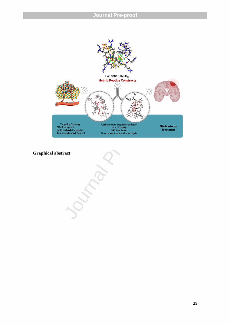
Graphical abstract
Journal Pre-proof
Jour
nal P
re-p
roof
30
Highlights
A hybrid construct based on HA, c(RGDfK) and H7K(R2)2 is successfully designed.
A dual experimental-computational framework is provided for guiding conjugate
formation.
MD enlightens the interactions governing the formation and stability of the constructs.
Synthesis pathway requires a hierarchical conjugation of the HA-c(RGDfK)-H7k(R2)2.
HA-c(RGDfK)-H7k(R2)2 conjugate displays a promising tumor targeting signature.
Journal Pre-proof