Embed Size (px)
Citation preview
CLINICAL RESEARCH www.jasn.org
High Prevalence of Autoantibodies to hLAMP-2 inAnti–Neutrophil Cytoplasmic Antibody–AssociatedVasculitis
Renate Kain,* Henko Tadema,† Eoin F. McKinney,‡ Alexandra Benharkou,*Ricarda Brandes,* Andrea Peschel,* Virginie Hubert,* Tjerk Feenstra,* Gürkan Sengölge,§
Coen Stegeman,| Peter Heeringa,¶ Paul A. Lyons,‡ Kenneth G.C. Smith,‡
Cees Kallenberg,† and Andrew J. Rees*
*Clinical Institute of Pathology and §Clinical Division of Nephrology and Dialysis, Medical University of Vienna, Vienna,Austria; Departments of †Rheumatology and Clinical Immunology and |Internal Medicine, Division of Nephrology, and¶Medical Biology, University Medical Centre, University of Groningen, Groningen, The Netherlands; and‡Department of Medicine and Cambridge Institute for Medical Research, Addenbrooke’s Hospital, University ofCambridge School of Clinical Medicine, Cambridge, United Kingdom
ABSTRACTThe involvement of autoantibodies to human lysosome-associated membrane protein-2 (hLAMP-2) inanti–neutrophil cytoplasmic antibody (ANCA)–associated vasculitis is controversial because of the ab-sence of confirmatory data subsequent to the initial reports of their high prevalence in this disease. Wecharacterized three assays for anti-hLAMP-2 antibodies: ELISA and Western blotting assays usingunglycosylated recombinant hLAMP-2 expressed in Escherichia coli, and an indirect immunofluores-cence assay using stably transfected ldlD cells that expressed glycosylated full-length hLAMP-2 on theplasma membrane. The assays detected autoantibodies to hLAMP-2 in human sera reproducibly andwith comparable sensitivity and the assays gave the same results in 80.5% of the test panel of 40 selectedpositive and negative sera. In untreated patients at presentation, the frequencies of autoantibodies toLAMP-2 were 89%, 91%, and 80%, respectively, among three groups of patients with ANCA-associatedvasculitis from Vienna, Austria (n=19); Groningen, the Netherlands (n=50) and Cambridge, United Kingdom(n=53). Prevalence of LAMP-2 autoantibodies was similar in both those with myeloperoxidase-ANCA andproteinase 3-ANCA. Furthermore, we detected LAMP-2 autoantibodies in two ANCA-negative patients.LAMP-2 autoantibodies rapidly became undetectable after the initiation of immunosuppressive treat-ment and frequently became detectable again during clinical relapse. We conclude that when robustassays are used, circulating autoantibodies to hLAMP-2 can be detected in most European patients withANCA-associated vasculitis. Large-scale prospective studies are now needed to determine whether theyare pathogenic or merely an epiphenomenon.
J Am Soc Nephrol 23: 556–566, 2012. doi: 10.1681/ASN.2011090920
Pauci-immune focal necrotizing GN (piFNGN) isa severe inflammatory disease that occurs in anti–neutrophil cytoplasmic antibody (ANCA)–associatedvasculitis (AAV), such as microscopic polyangiitis(MPA) or granulomatosis with polyangiitis (GPA,formerly Wegener’s granulomatosis).1,2 AAV has arelapsing course and over a quarter of those affecteddie within 5 years either fromuncontrolled disease orcomplications of treatment,3 which emphasizes theneed for more specific immunosuppressive therapytailored to the underlying pathogenic mechanisms.
Between 85% and 90% of those with piFNGNhave antibodies to neutrophil cytoplasmic antigens
Received September 15, 2011. Accepted December 2, 2011.
Published online ahead of print. Publication date available atwww.jasn.org.
Correspondence: Dr. Renate Kain, Clinical Institute of PathologyMedical University of Vienna, Waehringer Guertel 18-20, A-1090Vienna, Austria. Email: [email protected]
Copyright © 2012 by the American Society of Nephrology
556 ISSN : 1046-6673/2303-556 J Am Soc Nephrol 23: 556–566, 2012

(ANCA) that recognize either myeloperox-idase (MPO) or proteinase 3 (PR3).1,2 Thisprovides strong evidence for their involve-ment in pathogenesis, which is supportedby in vitro studies4 and experimental models(at least for MPO-ANCA).5–7 Despite this,MPO and PR3 are not normally expressedin kidney although MPO released from in-filtrating neutrophils can decorate glomer-ular endothelium,8 and additional factorsare needed for anti-MPO antibodies tocause severe injury in rodent models.6,8,9
The weak correlation between ANCA titersand clinical disease activity suggests that thesame is true in humans.10–12 We identifiedautoantibodies to lysosome-associatedmembrane protein-2 (LAMP-2) in activepiFNGN and proposed that they mightcontribute to injury because the antigenis expressed in the plasma membrane ofglomerular endothelial cells.13,14
Antibodies to hLAMP-2 were originallydiscovered in 16 of 17 patients withpiFNGNbyWesternblotting in a systematicsearch for autoantibodies to neutrophil orglomerular membrane proteins.13 Wefound a similarly high prevalence in a sub-sequent cohort of 84 patients with activepiFNGN.14 Patients’ autoantibodies com-monly bind two epitopes, one of which(P41-49) is shared with the bacterial adhesinFimH with which they cross-react. Injec-tion of antibodies to the LAMP-2 extracel-lular domain induced piFNGN in WKYrats as did immunization with FimH thatacted as molecular mimic and provokedsynthesis of antibodies to rat LAMP-2.Thus, antibodies to LAMP-2 cause piFNGNin rodents, which raises the issue whetherthey are similarly pathogenic in humans.Robust assays are required to investigatethis further, and development of suitableassays for antibodies to hLAMP-2 has beenchallenging because of the difficulty in ob-taining pure preparations of appropriatelyglycosylated native or recombinant anti-gen,15,16 a problem shared with other glycosylated membraneproteins such as the membranous nephropathy antigen,phospholipase A2 receptor.17 Recombinant membrane pro-teins often need modification to produce soluble substratesfor ELISA, and inappropriate glycosylation can affect acces-sibility of epitopes recognized by patients’ autoantibodies.Only one other group has reported the development of assaysfor anti-hLAMP-2 antibodies and they have challenged ourconclusions.18
In this study, we characterize three assays for antibodiesto hLAMP-2 in human sera and show that they give highlyconcordant results. In applying them to new European cohortsfrom three different centers, we confirm that antibodies tohLAMP-2 are highly prevalent in patients with piFNGN bothat presentation and during clinical relapse. Results of sequentialmeasurements after the start of treatment provide a possibleexplanation for the disparity between our findings and thoseof Roth et al.18
Figure 1. cDNA constructs, generation, and quality control of recombinant hLAMP-2. (A)Representation of cDNA encoding hLAMP-2A with the 28 amino acid leader peptide (LP),347 amino acid extracellular domain, 24 amino acid transmembrane domain (TM), and 11amino acid cytoplasmic domain (Cytopl). The two extracellular domain constructs wereutilized to express soluble hLAMP-2 in E. coli (hLAMP-2/GST) and mammalian cells(hLAMP-2sol). Both contain the leader peptide but not the transmembrane domain orcytoplasmic tail. hLAMP-2sol expressed in mammalian cells results in an appropriatelyglycosylated soluble protein exported into the culture supernatant via the default secretorypathway in mammalian cells. The hLAMP-2 cytoplasmic tail contains the signal that directsits retrieval from the plasma membrane to lysosomes. The critical tyrosine was mutated toa histidine in hLAMP-2H (Y/H), which targets it to the plasma membrane when expressed inldlD cells. (B) Purified hLAMP-2/GST runs as a single 65-kD band on SDS-PAGE and silverstain. Fractions of high purity were pooled and identity of hLAMP-2 was confirmed with anantibody reactive with hLAMP-2 only. (C) Purified hLAMP-2/GST (10 mg/ml) was separatedby SDS-PAGE and transferred onto PVDF before probing with specific antibodies tohLAMP-2 (932b), which bound exclusively to the fusion protein. Antibody to GST (anti-GST) recognized both fusion protein and free GST. A human serum (1:100 dilution) con-taining anti-hLAMP-2 antibodies (Pat. 6) also bound the fusion protein, whereas serumfrom a healthy control (Healthy Co 50) did not. Serum from a patient with renal disease(Dis. Co 8) contained antibodies to GST. Secondary antibodies alone were negative (anti-human, anti-rabbit). (D) Sensitivity of the Western blot was assessed from doubling dilu-tions (1:50 to 1:400) of the standard positive control (Stand pos Co) used for all assays. Thisalso confirmed the optimal binding/background ratio was 1:100.
J Am Soc Nephrol 23: 556–566, 2012 Anti-hLAMP-2 Antibodies in AAV 557
www.jasn.org CLINICAL RESEARCH

RESULTS
Recombinant Escherichia coliExpressed hLAMP-2 for WesternBlotting and ELISAMost patients’ autoantibodies bind epitopesin the protein backbone of the extracellulardomain not occluded by glycosylation in na-tive neutrophil and glomerular hLAMP-2.13,14
Consequently, we induced recombinantunglycosylated hLAMP-2 truncated to 342amino acids of the full extracellular domainas GST fusion protein in E. coli (Figure 1A).After purification on Glutathione-Sepharose,hLAMP-2/GST fusion protein runs as a singleband of approximately 65 kD on SDS-PAGE(Figure 1B), whose identity was confirmedby immunoblot with antibodies to hLAMP-2and GST. It also binds IgG in sera from pa-tients with antibodies to hLAMP-2 but notcontrols (Figure 1C). Patients’ sera werediluted 1:100 to give the best binding/background ratio (Figure 1D).
hLAMP-2/GSTwasprepared inbatchesof,10 mg and used within 3 months becauselarge-scale cultures and pre-purificationstorage of pellets increased degradation andcontamination with other proteins (Supple-mental Figure 1, A and C) and the recombi-nant protein degrades rapidly at220°C andeven 280°C after 6 months (SupplementalFigure 1, B and D).
Measuring Antibodies to hLAMP-2by ELISAELISA plates were coated with 5 mg/ml ofhLAMP-2/GST for 1 hour, optimal condi-tions for distinguishing between positiveand negative sera (Figure 2A). Coated plateswere stable for 4 weeks at 4°C (Figure 2B).When diluted, 1:100 moderately strong pos-itive sera gave an OD of approximately 0.9compared with a mean OD of 0.27 for nor-mal sera (Figure 2A). Minor degrees of sub-strate degradation profoundly affected assay performance(Supplemental Figure 1, B andD), necessitating rigorous qualitycontrol of hLAMP-2/GST batches by SDS-PAGE and immuno-blotting (Supplemental Figure 1C) and testing ELISA plates withstandard sera to ensure consistency (Figure 2C). Serawere testedfor contamination with FimH-expressing bacteria because theseinhibit binding, as does repeated freezing and thawing.
The ELISAwas highly reproducible and our standard positivecontrol gave ameanODof0.95860.159 (coefficient of variation,16.5%) when assayed 24 times over 6 months using three dif-ferent hLAMP-2/GST preparations. Anti-hLAMP-2 antibody
concentrations were calculated from a standard curve generatedwith this serum (Figure 2, C and D) with 100 U equating to theOD of the 1:100 dilution. Sera from 80 healthy controls had amean6 SD of 15.9166.68 U, giving a 95% confidence limit ofthe upper limit of normal of 29 units for the ELISA: higher valueswere scored positive.
Expression of hLAMP-2 in ldlD Cells forImmunofluorescence AssayBinding of autoantibodies to glycosylated LAMP-2 was testedusing an indirect immunofluorescence assay (IIF) assay in
Figure 2. Development of anti-hLAMP-2 ELISA. (A) Comparison of ELISA plates coatedwith 0.5 and 5.0 mg/ml of recombinant hLAMP-2/GST fusion protein (FP). Plates coatedwith 5.0 mg/ml gave much better separation between positive and negative sera. (B)ELISA plates coated with hLAMP-2/GST FP were stored at 4°C and tested after differenttime points. Serial dilutions of a standard positive control serum gave similar results usingplates stored for 1–33 days, but there was a considerable increase in background bindingafter 41 days. (C) Comparison of ELISA plates coated with two different batches ofhLAMP-2/GST FP demonstrates comparable binding of serial dilutions of a standardpositive control serum. (D) The standard curve derived from the results of assaying serialdilutions of the standard control serum was used to calculate anti-hLAMP-2 antibodyconcentrations with 100 U equating to OD of the 1:100 dilution of the positive control.
558 Journal of the American Society of Nephrology J Am Soc Nephrol 23: 556–566, 2012
CLINICAL RESEARCH www.jasn.org
which hLAMP-2 was expressed exclusively on the surface ofldlD cells—a Chinese hamster ovary (CHO) cell subline usedfor studying glycoproteins.19
First, we assessed the complexity of glycosylation of CHOcell expressed hLAMP-2. The expressed extracellular domainhad molecular mass of 110 kD similar to native human glomer-ular hLAMP-2 but less than neutrophil hLAMP-2 (Supplemen-tal Figure 2, A and B). As expected, the soluble extracellulardomain was secreted into the supernatant and could not bedetected in transfected cells using hLAMP-2–specific antibodies(Supplemental Figure 2, A and D). By contrast, most commer-cially available anti-hLAMP-2 antibodies cross-reacted withhamster LAMP-2 and stained lysosomes of transfected anduntransfected cells, particularly after permeabilization withsaponin (Supplemental Figure 2, C and D), negating theiruse as positive controls for transfection.
We then stably transfected ldlD cells with full-lengthhLAMP-2 with a tyrosine to histidine mutation in the cy-toplasmic motif responsible for retrieval from the plasmamembrane (hLAMP-2H). Transfected cells expressed abun-dant hLAMP-2 on the cell surface, where it was visualizedwithout permeabilization with six different antibodies; how-ever, none was detected in lysosomes with human-specificantibodies (Figure 3, A and B). Test sera containing anti-hLAMP-2 antibodies bound specifically to transfected ldlDcells, whereas control sera did not (Supplemental Figure 3).Sera were routinely diluted to 1:40 but binding of moderatelystrong positive sera way seen at 1:400 dilution (Figure 3C),indicating equivalent sensitivity to the other two assays.
Comparison of Western, ELISA, and IIF AssaysWe compared assay performance using a panel of sera selectedfor their range of positive and negative results: 16 from AAV(11 active disease; 5 remission), 15 renal disease controls, and10 healthy controls. Overall, 14 sera (all from the AAV group)were positive in $1 assay. Two-way comparisons confirmedthe consistency between results of the different assays (Fig-ure 4A) with interassay concordance rates of 83%, 90% and92% for ELISA versus IIF, ELISA versus Western blot, andIIF versusWestern blot, respectively. In all three assays, 33 ofthe 41 sera gave the same result, yielding an overall concor-dance rate of 80.5% (Figure 4B). The concordance rates withingroups were as follows: 82%, 9 of 11 patients with active AAV;40%, 2 of 5 patients with AAV in remission; 86% in 13 of 15controls with renal disease; and 100% in 10 of 10 healthycontrols.
Discrepancies between the assays were most frequent in theinactivepiFNGNgroup,which likely reflectsminordifferences inassay sensitivity. Notably, the ELISA did not always distinguishlow concentrations of specific antibody from high backgroundbinding of normal IgG. Consequently, we introduced a border-line range (22–29 U) into the ELISA results. Three of the 41 seragave false positive ELISA results alongwith negative results in theother two assays; however, the reasons are unclear because im-munoblots excluded binding to GSTor contaminating proteins.
Consequently, autoantibodies were considered present onlywhen two of the three hLAMP-2 assays were positive.
Prevalence of Anti-hLAMP-2 AntibodiesThe three assays were then used to ascertain the prevalence ofanti-hLAMP-2 antibodies in three new groups of patients withAAV from Vienna, Austria; Groningen, the Netherlands; andCambridge, United Kingdom. The Vienna group comprised19 patients (23 sera: 12 with AAVat presentation, 2 with clin-ical AAV relapse, and 9 with clinically inactive AAV). TheGroningen group comprised 50 patients (50 sera: 35 with un-treated AAV at presentation, 9 with AAV at presentation andreceiving treatment, and 6 with AAV clinical relapse). TheCambridge group comprised 53 patients (53 sera: 33 withAAVat presentation and 20 with AAV relapse) (SupplementalTables 1–3). The assays were performed without knowledge ofwhether sera were from AAV or control groups, or from pre-sentation or follow-up. All assays were performed withoutknowledge of the other assay results and the data are summa-rized in Figure 5.
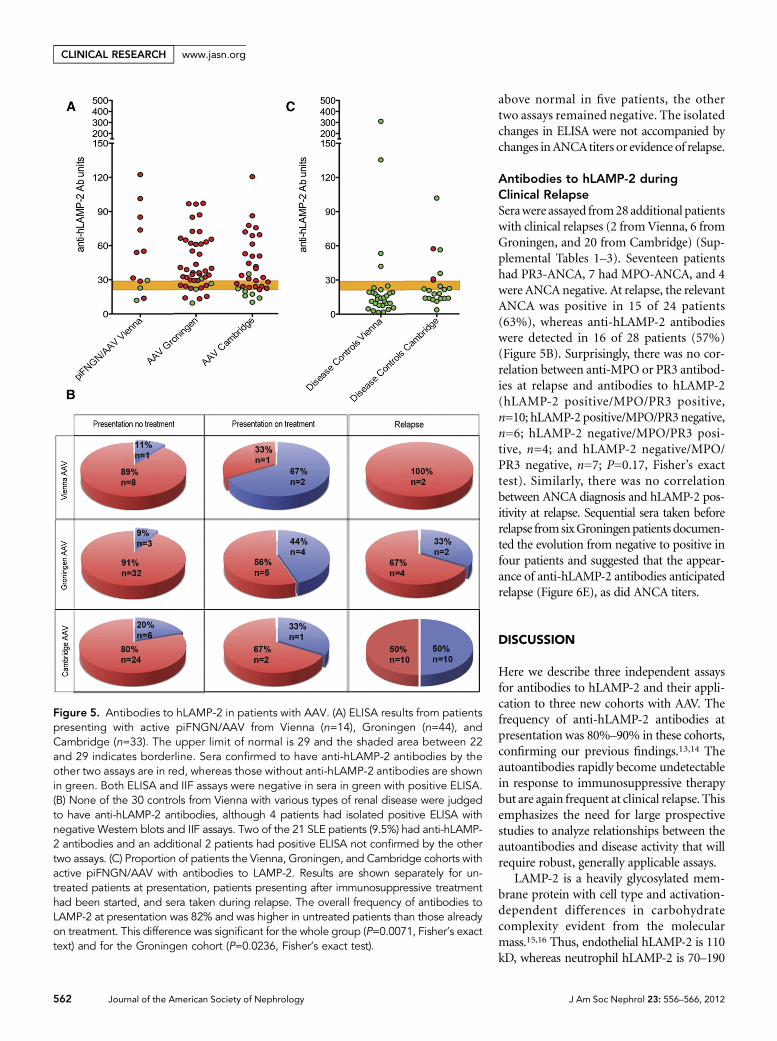
Of the individuals presenting with piFNGN, 9 of 12 Viennapatients (75%), 37 of 44 Groningen patients (84%), and 26 of33 Cambridge patients (79%) had positive results (Figure 5).This gives an overall frequency of 81% compared with 93% inour original study,14 which was restricted to patients presentingwithout prior treatment to avoid confounding effects of im-munosuppression. Applying the same restriction to this studyfurther increased the frequency of anti-hLAMP-2 antibodies in8 of 9 Vienna patients (89%), 32 of 35 Groningen patients(91%), and 24 of 30 Cambridge patients (80%). The differencein frequency of anti-hLAMP-2 antibodies between treated anduntreated patients was highly significant (all patients,P=0.0071; Groningen cohort, P=0.0236; Fisher’s exact test).
There was a high degree of agreement between the assays, withthe three assays giving identical results in 80%, 67%, and 67% ofthe patients from Vienna, Groningen, and Cambridge, respec-tively (Supplemental Tables 1–3). Thus, we can be confident ofthe high prevalence of anti-hLAMP-2 antibodies in patientspresenting with untreated piFNGN in this series (86.5%).
Autoantibodies tohLAMP-2were equally frequent inMPO-and PR3-ANCA–associated disease and were independent ofthe clinical diagnosis of GPA or MPA. We also detected theautoantibodies in both ANCA-negative patients presentingwith piFNGN (Supplemental Tables 1–3). Other than priortreatment, patients without detectable anti-hLAMP-2 anti-bodies had no special characteristics. Specifically, they hadsimilar titers of antibodies to MPO or PR3, and equally se-vere renal involvement and extent of organ involvement. Tenof 11 patients without evidence of renal involvement in theGroningen group had detectable antibodies to hLAMP-2.
We assayed 51 additional participants as disease controls:30 from Vienna with renal disease and 21 fromCambridgewithSLE (Figure 5C).No renal controls had detectable anti-hLAMP-2 antibodies although four had isolated positive or borderlineELISA results. Two of the 21 participants with SLE (9.5%) had
J Am Soc Nephrol 23: 556–566, 2012 Anti-hLAMP-2 Antibodies in AAV 559
www.jasn.org CLINICAL RESEARCH

Figure 3. Indirect immunofluorescence on ldlD cells stably expressing hLAMP-2 on the cell surface (ldlD/hLAMP-2H). (A) ldlD cells weretransfected with full-length hLAMP-2 cDNA with a tyrosine to histidine mutation in the cytoplasmic lysosomal retrieval signal. Stabletransfectants were sorted by flow cytometry for uniform expression of the transgene and stained with monoclonal (CD3) and polyclonal(932B) antibodies to hLAMP-2 generated by M. Fukuda as well as commercially available monoclonal (H4B4) and polyclonal anti-LAMP-2antibodies (SCN17 and Abnova). All of the antibodies bound to hLAMP-2 on the surface of nonpermeabilized transfected cells (np). Thecommercial anti-hLAMP-2 cross-reacted with hamster LAMP-2 in transfected and untransfected cells especially after permeabilization(p). CD3 and 932B were the only antibodies that did not cross-react with hamster LAMP-2. A polyclonal antibody to rat LAMP-2 (ZymedLaboratories) and secondary antibody alone fail to detect either human or hamster LAMP-2. (B) FACS analysis of ldlD/hLAMP-2H cellsand control ldlD cells stained with H4B4, a mAb to hLAMP-2 that cross-reacts with hamster LAMP-2. Surface expression of hLAMP-2 isdetected only in transfected cells, whereas intracellular staining of native hamster LAMP-2 is apparent after permeabilization withsaponin. (C) The sensitivity of the IIF assay for antibodies to hLAMP-2 assessed from doubling dilutions (1:50 to 1:800) of the standardpositive control (Stand Co) compared with binding of monoclonal anti-hLAMP-2 antibody (H4B4; pos Co) and negative control serum(neg Co). IgG binding to ldlD/hLAMP-2H cells decreases progressively in titers.1:100 and becomes negative at 1:400, which is 10-foldhigher than the standard assay dilution of 1:40.
560 Journal of the American Society of Nephrology J Am Soc Nephrol 23: 556–566, 2012
CLINICAL RESEARCH www.jasn.org

anti-hLAMP-2 antibodies and two morehad isolated positive ELISA results. Accord-ingly, the interassay concordance in theELISA was lower for SLE sera, which em-phasizes the need to confirm ELISA resultswith other assays.
Response of Anti-hLAMP-2Antibodies to ImmunosuppressiveTherapyThe Groningen patients were managed ac-cording to a standard protocol of corticoste-roids and cyclophosphamide, together withplasma exchange for those most severely af-fected; eight patients also received rituximabaspartof theRituximabforANCA-AssociatedVasculitis trial.20 They were followed withclinical and serological assessment at 1, 2, 3,6, and 12 months after presentation. Oncetreatment was started, disease activity de-creased rapidly accompanied by a decreasein ANCA titers that eventually became nega-tive, at least transiently, in 22patients and verylow titer (1:20) in an additional 9 patients(Figure 6, A and B). However, ANCA titersremained above 1:640 in two patients. Con-centrations of anti-hLAMP-2 antibodies fellmore rapidly and they became undetectablein 36 of 37 positive patients by 1 month(Figure 6, C and D). Thereafter, antibodiesto hLAMP-2 were judged negative in allbut one serum throughout the 12-monthstudy—although ELISA increased to just
Figure 4. Evaluation of assays for antibodies to LAMP-2. (A) ELISA results from a panelof sera from 78 healthy controls were used to derive the mean 6 SD for this group andthe 95% confidence limit of the upper limit of normal for the assay, which was es-tablished at 29 U. Six controls had positive ELISA with negative Western blots and IIFassays. (B) ELISA results of the data shown in A. The upper limit of normal in the assayis 29 U. Sera confirmed to have anti-hLAMP-2 antibodies by the other two assays are in
red,whereas those inwhichpositiveELISAwasnot confirmed because ELISA and IIF assayswere negative are in green. (C) Measurementofantibodies tohLAMP-2byELISAandWesternblot using hLAMP-2/GST FP and IIF on ldlD/hLAMP-2H cells was compared using a panelof 41 sera selected to cover a range of positiveand negative values. The panel consisted of16 sera from patients with AAV (11 with activedisease and 5 in remission), 15 controls withother renal diseases, and 10 healthy controls.The assays were graded positive (red), lowpositive (Western and IIF), borderline (ELISA)(orange), or negative (green). The figure illus-trates the strong concordance among resultsfrom the three assays. Sera were consideredto have antibodies to LAMP-2 when $2 assayswerepositive.Abbreviations:TX, renal transplant;MGN, membranous nephropathy; IgA GN, IgAnephropathy; MPGN, membranoproliferativeGN; TIN, tubulointerstitial nephritis.
J Am Soc Nephrol 23: 556–566, 2012 Anti-hLAMP-2 Antibodies in AAV 561
www.jasn.org CLINICAL RESEARCH
above normal in five patients, the othertwo assays remained negative. The isolatedchanges in ELISA were not accompanied bychanges inANCAtiters or evidence of relapse.
Antibodies to hLAMP-2 duringClinical RelapseSerawere assayed from28additional patientswith clinical relapses (2 fromVienna, 6 fromGroningen, and 20 from Cambridge) (Sup-plemental Tables 1–3). Seventeen patientshad PR3-ANCA, 7 had MPO-ANCA, and 4were ANCA negative. At relapse, the relevantANCA was positive in 15 of 24 patients(63%), whereas anti-hLAMP-2 antibodieswere detected in 16 of 28 patients (57%)(Figure 5B). Surprisingly, there was no cor-relation between anti-MPO or PR3 antibod-ies at relapse and antibodies to hLAMP-2(hLAMP-2 positive/MPO/PR3 positive,n=10; hLAMP-2 positive/MPO/PR3negative,n=6; hLAMP-2 negative/MPO/PR3 posi-tive, n=4; and hLAMP-2 negative/MPO/PR3 negative, n=7; P=0.17, Fisher’s exacttest). Similarly, there was no correlationbetween ANCA diagnosis and hLAMP-2 pos-itivity at relapse. Sequential sera taken beforerelapse fromsixGroningenpatients documen-ted the evolution from negative to positive infour patients and suggested that the appear-ance of anti-hLAMP-2 antibodies anticipatedrelapse (Figure 6E), as did ANCA titers.
DISCUSSION
Here we describe three independent assaysfor antibodies to hLAMP-2 and their appli-cation to three new cohorts with AAV. Thefrequency of anti-hLAMP-2 antibodies atpresentation was 80%–90% in these cohorts,confirming our previous findings.13,14 Theautoantibodies rapidly become undetectablein response to immunosuppressive therapybut are again frequent at clinical relapse. Thisemphasizes the need for large prospectivestudies to analyze relationships between theautoantibodies and disease activity that willrequire robust, generally applicable assays.
LAMP-2 is a heavily glycosylated mem-brane protein with cell type and activation-dependent differences in carbohydratecomplexity evident from the molecularmass.15,16 Thus, endothelial hLAMP-2 is 110kD, whereas neutrophil hLAMP-2 is 70–190
Figure 5. Antibodies to hLAMP-2 in patients with AAV. (A) ELISA results from patientspresenting with active piFNGN/AAV from Vienna (n=14), Groningen (n=44), andCambridge (n=33). The upper limit of normal is 29 and the shaded area between 22and 29 indicates borderline. Sera confirmed to have anti-hLAMP-2 antibodies by theother two assays are in red, whereas those without anti-hLAMP-2 antibodies are shownin green. Both ELISA and IIF assays were negative in sera in green with positive ELISA.(B) None of the 30 controls from Vienna with various types of renal disease were judgedto have anti-hLAMP-2 antibodies, although 4 patients had isolated positive ELISA withnegative Western blots and IIF assays. Two of the 21 SLE patients (9.5%) had anti-hLAMP-2 antibodies and an additional 2 patients had positive ELISA not confirmed by the othertwo assays. (C) Proportion of patients the Vienna, Groningen, and Cambridge cohorts withactive piFNGN/AAV with antibodies to LAMP-2. Results are shown separately for un-treated patients at presentation, patients presenting after immunosuppressive treatmenthad been started, and sera taken during relapse. The overall frequency of antibodies toLAMP-2 at presentation was 82% and was higher in untreated patients than those alreadyon treatment. This difference was significant for the whole group (P=0.0071, Fisher’s exacttext) and for the Groningen cohort (P=0.0236, Fisher’s exact test).
562 Journal of the American Society of Nephrology J Am Soc Nephrol 23: 556–566, 2012
CLINICAL RESEARCH www.jasn.org

Figure 6. ANCA and anti-hLAMP-2 antibody titers in patients with AAV. (A and B) Sequential ANCA titers expressed as median andinterquartile range in the 43 patients in the Groningen cohort followed for 12 months. ANCA titers decreased rapidly after the induction oftreatment. The patients are separated into those who had PR3-ANCA (n=25; A and C) and MPO-ANCA (n=18; B and D). (C and D) Sequentialtiters of antibodies to hLAMP-2 measured by ELISA in the 43 patients in the Groningen cohort followed for 12 months. The upper limit ofnormal is 29 U and the yellow bar indicates the borderline positive. The patients are separated into those who had PR3-ANCA (n=25) andMPO-ANCA (n=18). Anti-hLAMP-2 antibodies became undetectable in,1 month in all but one of the patients. They became undetectable inall 37 positive patients, irrespective of the presence or absence of antibodies to either (C) PR3 or (D) MPO. (E) Sequential titers of antibodies tohLAMP-2 measured by ELISA in six patients from Groningen in the months preceding clinical relapse. The upper limit of normal is 29 U andthe yellow bar indicates the borderline positive. Sera from four patients had detectable antibodies to hLAMP-2 before clinical relapse.
J Am Soc Nephrol 23: 556–566, 2012 Anti-hLAMP-2 Antibodies in AAV 563
www.jasn.org CLINICAL RESEARCH

kD. Most patients’ autoantibodies bind epitopes in the proteinbackbone of the extracellular domain that are accessible innative neutrophil and endothelial hLAMP-2 as well as recombi-nant human antigen expressed in CHO cells.13,14 This deter-mined our selection of the following hLAMP-2 preparations asassay substrates: recombinant human unglycosylated extracel-lular domain expressed in E. coli for ELISA and Western blot-ting, and glycosylated full-length hLAMP-2 targeted to theplasma membrane of ldlD cells for IIF.
Recombinant hLAMP-2/GST fusion protein is simple toproduce in standard laboratory E. coli that do not express FimHunder standard conditions,21 but is susceptible to degradation.Degradationmarkedly reduces specific binding in the ELISAandthe effect is amplified in sera from patients with chronic disease,such as lupus, and after repeated freezing and thawing. Precau-tions to minimize degradation included truncating hLAMP-2extracellular domain to 324 amino acids and preparing antigenin small batches. We tested for antibodies that bind glycosylatedhLAMP-2 by IIF on ldlD cells stably transfected with full-lengthhLAMP-2 because the isolated extracellular domain is, like othersoluble proteins,22 secreted and not retained within the cell. Incontrast, full-length hLAMP-2 traffics to the plasma membraneand is then retrieved to lysosomes. The cytoplasmic retrievalsignalwasmutated in the full-length construct weused, resultingin exclusive hLAMP-2 cell surface expression. This enabled us toassay antibodies that bind it without cell permeabilization, thusavoiding the potential confusion caused by co-existing autoanti-bodies that recognize hamster LAMP-2 or intracellular antigensfound in autoimmune disease.23,24
The three anti-hLAMP-2 antibody assays gave remarkablysimilar results, with three-way concordance rates of 80.5% and74%intheevaluationandtestcohorts, respectively.Theproportionofpositive serawas similar foreachassayandwecan thusbecertainthat the prevalence of anti-hLAMP-2 antibodies at presentation isbetween 80% and 90% in these new AAV cohorts and that theepitopes recognized are not occluded by carbohydrate in mam-malian expressed antigen. This replicates our previous reports13,14
but contrasts sharply with that of Roth et al.,18 possibly because ofcritical differences in the patients studied and assays used.
We have always analyzed sera from patients with carefullydefined disease activity, whereas sera in the study by Roth et al.were for the most part not segregated by disease activity. This isimportant because anti-hLAMP-2 antibodies are confined tothose with active disease. Both groups used ELISA and immu-noblotting assays but with markedly hLAMP-2 extracellular do-main preparations. Roth et al. used complexly glycosylatedhLAMP-2 expressed in HEK293 cells,18 whereas we used ungly-cosylated hLAMP-2 that, in our hands, gives identical results tothe less complexly glycosylated ldlD expressed hLAMP-2.14 TheELISA results in the study by Roth et al. are most like our ownwhen direct comparisons can be made. Thus, 7 of 15 (47%) oftheir presenting patients tested positive compared with 80%–
90% in our cohorts. By contrast, results of their other two assayswere categorically different by being uniformly negative even insera that were positive by ELISA, whereas our immunoblotting
and IIF assays gave similar results to the ELISA. The difference ishighlighted by the four Viennese positive controls provided tovalidate the Roth ELISA: These were positive in all three Viennaassays and in the Roth ELISA, but were negative by immuno-blotting andwere not stated for the IIF assay. Possible reasons forthese disparities include failure of autoantibodies to bind morecomplexly glycosylated hLAMP-2 by immunoblotting and usetransiently expressed soluble hLAMP-2 extracellular domainthat, in our hands, is not retained in the cells.
Anti-hLAMP-2 autoantibodies become undetectable strik-ingly quickly after starting steroids and immunosuppressivedrugs and remained undetectable in the absence of clinicalevidence of relapse until the end of the study at 12months. Thisexplains the significantly lower frequency of anti-hLAMP-2antibodies in patients presenting after the start of treatment. TheANCA titers decreasedmore slowly and often remained positivein those without clinical evidence of disease activity. The rapiddisappearance of anti-hLAMP-2 antibodies is reminiscent of thefate of anti-MPO antibodies in Churg-Strauss Syndrome.25 Thisimplies a short t1/2 of the autoantibody,which iswhatwe observedin rats injected with antibodies to LAMP-2.14 We attribute thisto the well documented rapid internalization of anti-LAMP-2antibodies after they bind at the cell surface.26,27
In conclusion, results from three new European cohortsconfirm the high frequencyof antibodies to hLAMP-2 in patientspresenting with AAV and show that they rapidly become un-detectable once treatment is started but recur during clinicalrelapses. The critical remaining question is whether they arepathogenic or merely an epiphenomenon.
CONCISE METHODS
Patients and ControlsSera were studied from three independent groups of patients whose
details are summarized in Supplemental Tables 1–3. The groups com-
prised 19 patients from Vienna, Austria, with piFNGNwith or without
AAVeither at presentation or during follow-up; 50 patients from Gro-
ningen, the Netherlands, presenting with AAV and followed prospec-
tively for 12 months, and 6 patients with AAV in relapse in whom
sequential serum samples were available leading up to the relapse; 53
patients from Cambridge, United Kingdom with AAV with or without
piFNGN either at presentation (33 individuals) or with clinical relapse
(20 individuals)). Clinical diagnoses of GPA and MPA were in accor-
dance with the Chapel Hill Consensus criteria.28 Diagnosis of relapse
was made on clinical grounds. In addition, we studied a panel of 80
apparently healthy controls aged 21–84 years (mean 48.15 years) from
Vienna; 30 controls with various renal diseases from Vienna; and 21
patients presenting with SLE from Cambridge. All sera were assayed
without knowledge of whether they were from the AAVor disease con-
trol groups, or from presentation or follow-up. The performance of the
assays was compared using a test panel of 41 positive and negative sera.
Permission to use patients’ sera for autoantibody testing was granted
by the relevant ethics committees of the Medical University of Vienna
and the Universities of Groningen and Cambridge.
564 Journal of the American Society of Nephrology J Am Soc Nephrol 23: 556–566, 2012
CLINICAL RESEARCH www.jasn.org

Antibodies and ReagentsThe following primary antibodieswere used:monoclonalmouse anti-
hLAMP-2, clone H4B4 (Developmental Studies Hybridoma Bank,
University of Iowa); polyclonal rabbit (PAB12956; Abnova, Taipei,
Taiwan); goat anti-hLAMP-2 (sc-8101; Santa Cruz biotechnology, Santa
Cruz,CA);andrabbitanti-rLAMP-2(ZymedLaboratories,SanFrancisco,
CA). Monoclonal mouse anti-hLAMP-2 (clone CD3) and rabbit
anti-hLAMP-2 (932b) were kind gifts from Professor Minoru Fukuda
(The Burnham Institute, La Jolla, CA). Secondary antibodies used in IIF
were Alexa Fluor 488 conjugated F(ab`)2 fragment of goat anti-mouse
IgG (H+L) and goat anti-rabbit IgG (H+L) (Invitrogen, Carlsbad, CA),
and FITC conjugated sheep Ig to human IgG (INOVA Diagnostics,
San Diego, CA). Peroxidase-conjugated AffiniPure Goat Anti-Human
IgG, F(ab’) 2 Fragment Specific (Jackson Immuno Research, West
Grove, PA) was used in ELISA and alkaline phosphatase conjugated
anti-mouse, anti-human, and anti-rabbit IgG (Promega) were used
for Western blotting with their respective chromogenic substrates 1,2-
phenylendiamine-hydrochloride (Fluka AG, Buchs, Switzerland) and
nitro-blue tetrazolium and 5-bromo-4-chloro-39-indolyphosphate
(Pierce, Thermo Fischer Scientific, Rockford, IL).
Standard ANCA and Autoantibody AssaysSerial dilutions of all sera were routinely tested for ANCA, antinuclear
antibodies, and anti-glomerular basement membrane antibodies (The
Binding Site, Birmingham,UK), as described previously.13,14 One ormore
ANCA-positive serum fromeachpatient was tested for ANCAspecificity
using commercially available ELISA systems. For follow-up studies, all
sera from individual patients were re-tested by ELISA in the same assay.
Generation of Recombinant and Mutant hLAMP-2AGST-tagged hLAMP-2 fusion protein (which is more stable that the
His-tagged equivalent) corresponding to 342 amino acids of the extra-
cellular domain of hLAMP-2 was amplified from bases 283 to 1026,
cloned in frame into pGEX6p1 (pGEX6p1/hl2), and expressed in E. coli.
The extracellular domain of hLAMP-2 was amplified from bases 23
to 1119 by 59 and cloned into pCDNA1 to allow expression of a soluble
373 amino acid protein in mammalian cells (pCDNAI/hl2sol). A con-
struct to express hLAMP-2 on the surface of mammalian cells was gen-
erated by mutating tyrosine in position 378 of hLAMP-2 sequence to
histidine. Themutated hLAMP-2 cDNAwas cloned into pSVK3 (pSVK3/
LAMP-2H). All cloning procedures have been described previously.14
Purification of Recombinant hLAMP-2and Western BlottingE. coli BL21(DE3)pLysS (CLONTECH laboratories, Palo Alto, CA) were
used to avoid toxicity of the pGEX6p1/hl2 construct that had been ob-
served in other host strains. Toxicity and degradation of the fusion pro-
tein were also limited by reducing the induction time to a maximum of 2
hours. The cells of 1-L cultures were harvested, kept overnight at280°C,
and solubilized in PBS with 0.05% NaN3 and protease inhibitors (Com-
plete, Roche) from the periplasmic fraction by freezing in liquid N2 fol-
lowed by sonication. The recombinant protein was purified from cleared
supernatant using fresh batches of Glutathione-Sepharose (GEHealthcare
Europe, Vienna, Austria). Purity of individual batches of fusion protein
was analyzed by SDS-PAGE and silver stain. Only fractions free of con-
taminating proteins were pooled, dialyzed against PBS, snap frozen, and
stored at280°C for up to 4 months. Production of soluble hLAMP-2 in
CHO DG44 cells was induced using methods described previously.14
For Western blot analysis, 10 mg hLAMP-2/GST fusion protein per
lane were subjected to SDS-Page, with a subsequent transfer onto nitro-
cellulose followed by immuno-overlay with patients’ sera in 1:50 and
1:100 dilutions. Size and immunoreactivity of E. coli expressed hLAMP-
2were confirmedwith antibodies specific forhLAMP-2and theGST tag.
ELISA for Antibodies to hLAMP-2NUNCMaxisorp F ELISA plates were coated with 5 mg/ml of LAMP-
2/GST for 1 hour—conditions shown in exploratory experiments to
discriminate best between standard positive and negative sera after
testing different substrate concentrations (0.2–22 mg/ml) and incu-
bation times (1 hour to overnight). Newly coated ELISA plates were
tested to ensure the consistency of binding with standard positive and
negative control sera.
A standard positive control fromapatient’s plasma exchangebagwas
used in serial dilutions to generate a standard curve from which anti-
hLAMP-2 antibody concentrationswere determined. The normal range
in the ELISAwas established by testing a panel of 80 apparently healthy
controls aged 21–84 years (mean 48.15 years) at a dilutionof 1:100. Each
sample was measured three times and the average of these values was
used to derive the mean and SD for this group. All serum samples were
analyzed at least twice in triplicate. Sera of healthy participants were
used as controls in all experiments.
IIF Assay for Antibodies to hLAMP-2ldlD/hLAMP-2H cells were sorted by FACS for homogenous expression
of hLAMP-2H on the cell surface. A pool of cells were expanded for 2
days and subsequently frozen in aliquots of 106 cells/ml. Each newly
thawed batch went through a maximum of five passages before seeding
onto eight-well chamber slides to avoid loss of transgene that occurs in
around 10% of the cells per passage. ldlD parental or ldlD/hLAMP-2H
cells in chamber slides were fixed in freshly prepared 4% parafor-
maldehyde 24–36 hours after seeding before washing in PBS and in-
cubating for 1 hour at room temperature with patients’ sera diluted 1:40
in PBS. The slides were then incubatedwith FITC conjugated secondary
antibodies. Specific binding of test sera to hLAMP-2 was assessed by
comparisonwith reactivity and staining pattern of antibodies specific for
hLAMP-2. The staining pattern and its intensity were scored indepen-
dently by two observers. Representative photographs of IgG binding to
ldlD parental and ldlD/hLAMP-2H cells were taken as a record using a
fixed exposure time, based on the average of the optimal exposure time
for a standard low binding serum and a standard high binding one.
Statistical AnalysesMedian and interquartile ranges were calculated using GraphPad
Prism software (GraphPad Software Inc), as were statistical compar-
isons by Fisher’s exact test.
ACKNOWLEDGMENTS
We thank ProfessorDontschoKerajaschki for continual support and for
helpful discussions, as well as Régis Dieckmann for helpful discussion.
J Am Soc Nephrol 23: 556–566, 2012 Anti-hLAMP-2 Antibodies in AAV 565
www.jasn.org CLINICAL RESEARCH
A.J.R. was supported by an EU Marie Curie Excellence Chair
(MACRORIEN), V.H. and T.F. are Marie Curie Fellows in the EU FP7
funded Initial Training Network TranSVIR, and the work in Vienna has
been funded by the Vienna Science and Technology Fund (WWTF)
through Project LS09-075. Work in Cambridge was funded by the
Wellcome Trust, Medical Research Council, and the NIHR Cambridge
BioMedical Research Centre. K.G.C.S. is a Lister Prize Fellow. The
Cambridge Institute for Medical Research is in receipt of a Wellcome
Trust Strategic Award (079895). Work in Groningen was supported by
a grant from the Dutch Arthritis Association (06-1-401).
DISCLOSURESNone.
REFERENCES
1. Jennette JC, Falk RJ: Small-vessel vasculitis. N Engl J Med 337: 1512–1523, 1997
2. Morgan MD, Harper L, Williams J, Savage C: Anti-neutrophil cytoplasm-associated glomerulonephritis. J Am Soc Nephrol 17: 1224–1234, 2006
3. Flossmann O, Berden A, de Groot K, Hagen C, Harper L, Heijl C,Höglund P, Jayne D, Luqmani R, Mahr A, Mukhtyar C, Pusey C,Rasmussen N, Stegeman C, Walsh M, Westman K European VasculitisStudyGroup: Long-term patient survival in ANCA-associated vasculitis.Ann Rheum Dis 70: 488–494, 2011
4. Jennette JC, Xiao H, Falk RJ: Pathogenesis of vascular inflammation byanti-neutrophil cytoplasmic antibodies. J Am Soc Nephrol 17: 1235–1242, 2006
5. Xiao H, Heeringa P, Hu P, Liu Z, Zhao M, Aratani Y, Maeda N, Falk RJ,Jennette JC: Antineutrophil cytoplasmic autoantibodies specific formyeloperoxidase cause glomerulonephritis and vasculitis in mice. JClin Invest 110: 955–963, 2002
6. Heeringa P, Little MA: In vivo approaches to investigate ANCA-associatedvasculitis: Lessonsand limitations.ArthritisResTher13:204,2011
7. van der Geld YM, Hellmark T, Selga D, Heeringa P, Huitema MG,Limburg PC, Kallenberg CG: Rats and mice immunised with chimerichuman/mouse proteinase 3 produce autoantibodies to mouse Pr3 andrat granulocytes. Ann Rheum Dis 66: 1679–1682, 2007
8. Ruth AJ, Kitching AR, Kwan RY, Odobasic D, Ooi JD, Timoshanko JR,Hickey MJ, Holdsworth SR: Anti-neutrophil cytoplasmic antibodies andeffector CD4+ cells play nonredundant roles in anti-myeloperoxidasecrescentic glomerulonephritis. J Am Soc Nephrol 17: 1940–1949, 2006
9. Huugen D, Xiao H, van Esch A, Falk RJ, Peutz-Kootstra CJ, Buurman WA,Tervaert JW,JennetteJC,HeeringaP:Aggravationofanti-myeloperoxidaseantibody-induced glomerulonephritis by bacterial lipopolysaccharide:Role of tumor necrosis factor-alpha. Am J Pathol 167: 47–58, 2005
10. De’Oliviera J, Gaskin G, Dash A, Rees AJ, Pusey CD: Relationship be-tween disease activity and anti-neutrophil cytoplasmic antibody con-centration in long-term management of systemic vasculitis. Am J KidneyDis 25: 380–389, 1995
11. Schmitt WH, van der Woude FJ: Clinical applications of antineutrophilcytoplasmic antibody testing. Curr Opin Rheumatol 16: 9–17, 2004
12. Kallenberg CG, Stegeman CA, Bootsma H, Bijl M, Limburg PC:Quantitation of autoantibodies in systemic autoimmune diseases:Clinically useful? Lupus 15: 397–402, 2006
13. Kain R, Matsui M, Exner M, Binder S, Schaffner G, Sommer EM,Kerjaschki D: A novel class of autoantigens of anti-neutrophil cyto-plasmic antibodies in necrotizing and crescentic glomerulonephritis:The lysosomal membrane glycoprotein h-lamp-2 in neutrophil granulocytesand a related membrane protein in glomerular endothelial cells. J ExpMed 181: 585–597, 1995
14. Kain R, Exner M, Brandes R, Ziebermayr R, Cunningham D, Alderson CA,Davidovits A, Raab I, Jahn R, Ashour O, Spitzauer S, Sunder-Plassmann G,Fukuda M, Klemm P, Rees AJ, Kerjaschki D: Molecular mimicry in pauci-immune focal necrotizingglomerulonephritis.NatMed14: 1088–1096, 2008
15. Carlsson SR, Roth J, Piller F, FukudaM: Isolation and characterization ofhuman lysosomal membrane glycoproteins, h-lamp-1 and h-lamp-2.Major sialoglycoproteins carrying polylactosaminoglycan. J Biol Chem263: 18911–18919, 1988
16. Gough NR, Fambrough DM: Different steady state subcellular dis-tributions of the three splice variants of lysosome-associated membraneprotein LAMP-2 are determined largely by the COOH-terminal aminoacid residue. J Cell Biol 137: 1161–1169, 1997
17. Beck LH Jr, Bonegio RG, LambeauG, BeckDM, Powell DW, Cummins TD,Klein JB, Salant DJ: M-type phospholipase A2 receptor as target antigenin idiopathic membranous nephropathy. N Engl J Med 361: 11–21, 2009
18. Roth AJ, Brown MC, Smith RN, Badhwar AK, Parente O, Chung HC,Bunch DO, McGregor JG, Hogan SL, Hu Y, Yang JJ, Berg EA, Niles J,Jennette JC, Preston GA, Falk RJ: Anti-LAMP-2 antibodies are notprevalent in patients with antineutrophil cytoplasmic autoantibodyglomerulonephritis [published online ahead of print]. J Am SocNephrol doi:10.1681/ASN.2011030273
19. Remaley AT, Ugorski M, WuN, Litzky L, Burger SR, Moore JS, FukudaM, Spitalnik SL: Expression of human glycophorin A in wild type andglycosylation-deficient Chinese hamster ovary cells. Role of N- andO-linked glycosylation in cell surface expression. J Biol Chem 266:24176–24183, 1991
20. StoneJH,MerkelPA,SpieraR,SeoP, LangfordCA,HoffmanGS,KallenbergCG, St Clair EW, Turkiewicz A, Tchao NK, Webber L, Ding L,Sejismundo LP, Mieras K, Weitzenkamp D, Ikle D, Seyfert-MargolisV, Mueller M, Brunetta P, Allen NB, Fervenza FC, Geetha D, Keogh KA,Kissin EY, Monach PA, Peikert T, Stegeman C, Ytterberg SR, Specks URAVE-ITN Research Group: Rituximab versus cyclophosphamide forANCA-associated vasculitis. N Engl J Med 363: 221–232, 2010
21. Schwan WR, Seifert HS, Duncan JL: Growth conditions mediate dif-ferential transcription of fimgenes involved in phase variation of type 1pili. J Bacteriol 174: 2367–2375, 1992
22. Rothman JE, Wieland FT: Protein sorting by transport vesicles. Science272: 227–234, 1996
23. ArbuckleMR,McClainMT, RubertoneMV, Scofield RH,DennisGJ, JamesJA, Harley JB: Development of autoantibodies before the clinical onsetof systemic lupus erythematosus. N Engl J Med 349: 1526–1533, 2003
24. Isenberg DA, Manson JJ, Ehrenstein MR, Rahman A: Fifty years of anti-ds DNA antibodies: Are we approaching journey’s end? Rheumatology(Oxford) 46: 1052–1056, 2007
25. Keogh KA, Specks U: Churg-Strauss syndrome: Clinical presentation,antineutrophil cytoplasmic antibodies, and leukotriene receptor an-tagonists. Am J Med 115: 284–290, 2003
26. Kobayashi T, Vischer UM, Rosnoblet C, Lebrand C, Lindsay M, PartonRG, Kruithof EK, Gruenberg J: The tetraspanin CD63/lamp3 cyclesbetween endocytic and secretory compartments in human endothelialcells. Mol Biol Cell 11: 1829–1843, 2000
27. Janvier K, Bonifacino JS: Role of the endocytic machinery in the sortingof lysosome-associated membrane proteins. Mol Biol Cell 16: 4231–4242, 2005
28. Jennette JC, Falk RJ, Andrassy K, Bacon PA, Churg J, GrossWL, HagenEC, Hoffman GS, Hunder GG, Kallenberg CGM, McCluskey RT, SinicoRA, Rees AJ, van Es LA, Waldherr R, Wiik A: Nomenclature of systemicvasculitides. Proposal of an international consensus conference. Ar-thritis Rheum 37: 187–192, 1994
See related editorial, “Anti–LAMP-2 Autoantibodies in ANCA-AssociatedPauci-Immune Glomerulonephritis,” on pages 378–380.
This article contains supplemental material online at http://jasn.asnjournals.org/lookup/suppl/doi:10.1681/ASN.2011090920/-/DCSupplemental.
566 Journal of the American Society of Nephrology J Am Soc Nephrol 23: 556–566, 2012
CLINICAL RESEARCH www.jasn.org