Embed Size (px)
DESCRIPTION
Clinical study
Citation preview
High-Resolution CT of the Temporal Bone in Dysplasia of the Auricleand External Auditory Canal
T. E. Mayer, H. Brueckmann, R. Siegert, A. Witt, and H. Weerda
PURPOSE: To determine CT findings in the external, middle, and inner ear of patients with microtiaand external auditory canal dysplasia.METHODS:We used high-resolution CT, with multiplanar oraxial 1-mm continuous sections, coronal or sagittal reformations, or low-dose spiral acquisitions,to examine 184 temporal bones of children with microtia. RESULTS: In cases of minor microtia,auditory canal stenosis was the most common associated abnormality; in those with majormicrotia, atresia was predominant. Middle ear malformations depended on the severity of theauricular anomalies. Inner ear changes could also be noted. Ossicle dysplasias occurred in 98% ofpatients (stapes, 72%), absence of the oval window in 36%, labyrinthine malformations in 13%,closed round window in 6%, facial canal displacement in up to 75%, and aberrations of the vascularcanal in 38% of patients with third-grade auricular deformity. CONCLUSION: A variety of external,middle, and, less frequently, inner ear changes were detected in connection with microtia.
Index terms: Ear, abnormalities and anomalies; Temporal bone, computed tomography
AJNR Am J Neuroradiol 18:53–65, January 1997
3
Congenital aural dysplasias occur in one of3300 to 10 000 births (1, 2), except in the era ofthalidomide embryopathy (1959–1962), whenit was diagnosed in one of 900 neonates born inGermany (3). Microtia has been described evenon Babylonian tablets (4) and has been seen inprehistoric skulls (5). The classification of au-ricular deformities was proposed by Marx (6)and described by Rogers (7). Microtia is oftenassociated with abnormalities of other organs asthe result of genetic disorders, chromosomaldefects, intrauterine infections, or environmen-tal teratogens.Surgery for congenital aural atresia is one of
the most difficult ear operations, as it requiresreconstruction of the auricle, often done by us-ing grafts of skin and rib cartilage (8, 9), and, ifnecessary, restoration of hearing by drilling a
Received March 21, 1995; accepted after revision July 8, 1996.From the Institute of Radiology and Neuroradiology (T.E.M., H.B.,
A.W.) and the Clinic of Ear, Nose, Throat, and Plastic Surgery (R.S., H.W.),Medical University, Lubeck, Germany.
Address reprint requests to Dr Thomas Mayer, Abteilung Neuroradiolo-gie, Klinikum Grosshadern, 81377 Munchen, Germany.
AJNR 18:53–65, Jan 1997 0195-6108/97/1801–0053
© American Society of Neuroradiology
5
new auditory canal and reconstructing the os-sicular chain (10–12). Surgery is also per-formed for congenital cholesteatoma, labyrin-thine fistula, infection, or facial nerve palsy fromprevious surgery (13). Even if there is labyrin-thine involvement by the malformation, innerear function is preserved in most cases. Endo-scopic viewing is not possible in stenotic oratretic auditory canals. Staging by computedtomography (CT) is necessary to avoid the riskof facial nerve lesions, worsening of hearing,and bleeding, and to obtain a valid prognosis ofsuccess.We present a review of the CT findings in 92
patients with microtia and external auditory ca-nal dysplasia.
Subjects and MethodsAll 92 patients had high-resolution CT, 89 of them with
1- mm-thick sections and three with 2-mm-thick sections.Fifty-five patients only had axial CT, sometimes with ref-ormations of the coronal, oblique, or sagittal planes; 37patients were scanned in axial and coronal planes, and fiveof these were additionally scanned in the sagittal orienta-tion with low-dose (85 mAs) spiral CT. In these cases,
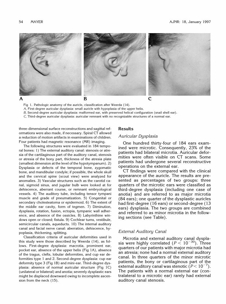
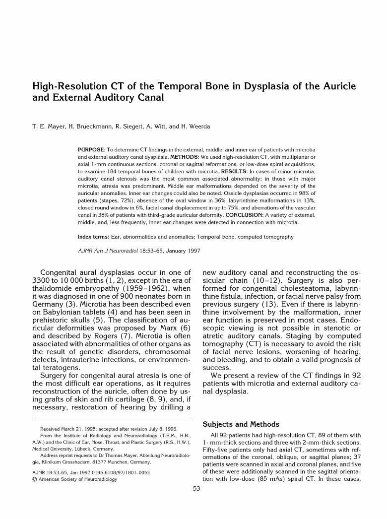
Fig 1. Pathologic anatomy of the auricle, classification after Weerda (14).A, First-degree auricular dysplasia: small auricle with hypoplasia of the upper helix.B, Second-degree auricular dysplasia: malformed ear, with preserved helical configuration (snail shell ear).C, Third-degree auricular dysplasia: auricular remnant with no recognizable structures of a normal ear.
54 MAYER AJNR: 18, January 1997
three-dimensional surface reconstructions and sagittal ref-ormations were also made, if necessary. Spiral CT alloweda reduction of motion artifacts in examinations of children.Four patients had magnetic resonance (MR) imaging.
The following structures were evaluated in 184 tempo-ral bones: 1) The external auditory canal: stenosis or atre-sia of the cartilaginous part of the auditory canal, stenosisor atresia of the bony part, thickness of the atresia plate(smallest dimension at the level of the hypotympanum). 2)Dysplasia or defects of the temporal bone, zygomaticbone, and mandibular condyle; if possible, the whole skulland the cervical spine (scout view) were analyzed foranomalies. 3) Vascular structures such as the carotid ca-nal, sigmoid sinus, and jugular bulb were looked at fordehiscence, aberrant course, or remnant embryologicalvessels. 4) The auditory tube, including tensor tympanimuscle and grade of pneumatization. 5) Congenital orsecondary cholesteatoma or epidermoid. 6) The extent ofthe middle ear cavity, form of tegmen. 7) Diminution,dysplasia, rotation, fusion, ectopia, tympanic wall adher-ence, and absence of the ossicles. 8) Labyrinthine win-dows open or closed; fistula. 9) Cochlear turns, vestibule,semicircular canals, aqueducts. 10) The internal auditorycanal and facial nerve canal: aberration, dehiscence, hy-poplasia, thickening, splitting.
Classification criteria of auricular deformities used inthis study were those described by Weerda (14), as fol-lows. First-degree dysplasia: macrotia, prominent ear,pocket ear, absence of the upper helix (Fig 1A), absenceof the tragus, clefts, lobular deformities, and cup ear de-formities type 1 and 2. Second-degree dysplasia: cup eardeformity type 3 (Fig 1B) and mini-ear. Third-degree dys-plasia: absence of normal auricular structure (Fig 1C)(unilateral or bilateral) and anotia; severely dysplastic earsmight be displaced downward owing to incomplete ascen-sion from the neck (15).
Results
Auricular Dysplasia
One hundred thirty-four of 184 ears exam-ined were microtic. Consequently, 23% of thepatients had bilateral microtia. Auricular defor-mities were often visible on CT scans. Somepatients had undergone several reconstructiveoperations on the external ear.CT findings were compared with the clinical
appearance of the auricle. The results are pre-sented as percentages of two groups: threequarters of the microtic ears were classified asthird-degree dysplasia (including one case ofanotia) and are referred to as major microtia(84 ears); one quarter of the dysplastic auricleshad first-degree (16 ears) or second-degree (13ears) dysplasia. The two groups are combinedand referred to as minor microtia in the follow-ing sections (see Table).
External Auditory Canal
Microtia and external auditory canal dyspla-sia were highly correlated (P , 10230). Threequarters of our patients with major microtia hadan atresia; none had a normal external auditorycanal. In three quarters of the minor microticpatients, the bony or cartilaginous part of theexternal auditory canal was stenotic (P , 1027).The patients with a normal external ear (con-tralateral to a microtic ear) rarely had externalauditory canal stenosis.
Petrous bone malformations correlated to the degree of auricular dysplasia (relative rate in each group of microtia)
Auricular Dysplasia, n 5 184Normal
(Contralateral) Ear,n 5 71
Minor Microtia(First and Second Grade),
n 5 16 1 13
Major Microtia(Third Grade),
n 5 84
External auditory canalStenosis 5.6 72.4 21.4Atresia 0 10.3 78.6
Malformations of:Temporal bone 9.9 55.2 89.3Mandible* 1.4 20.7 64.3Zygomatic arch† 0 13.8 44.0Others 14.1 17.2 29.8
Carotid canalSlight aberrations 1.4 17.2 15.5Hypoplasia 1.4 3.4 0
Sigmoid sinus, jugular bulb aberration, highriging 1.4 17.2 38.1Eustachian tube, bony dysplasia 0 20.7 44.0PneumatizationReduced‡ 7.0 10.3 19.0Little or none‡ 0 17.2 47.6
Middle ear space epitympanumReduced§ 1.4 24.1 20.2Minimal§ 0 3.4 41.7
MesotympanumReduced§ 0 37.9 46.4Minimal§ 0 6.9 41.7
HypotympanumReduced§ 1.4 24.1 17.9Minimal§ 0 13.8 73.8
Malleoincudal jointDysplastic, adherent 7.0 51.7 63.1Absent 0 3.4 34.5
Incudostapedial joint, dysplastic/absent 4.2 68.9 89.3StapesDyplastic 1.4 17.2 15.5Absent 0 34.5 56.0
Oval window, occluded 0 41.4 35.7Fallopian canal abnormalitiesHypoplasia of the labyrinthine segment 0 10.3 9.5Aberration of the labyrinthine segment 0 3.4 4.8Tympanic dehiscence 0 24.1 26.2Severe anomalies of the tympanic segment 0 0 2.4Mastoid segment anterior dislocation 0 55.2 61.9Severe aberration of the mastoid segment 0 3.4 11.9
Internal auditory canal, inclined/widened/agenesis 5.6 13.8 16.7Round window, occluded 0 3.4 6.0Inner ear dysplasiaCochlea 0 0 3.6Vestibulum 0 3.4 4.8Lateral semicircular canal 0 6.9 13.1
Note.—Values given in percentages.* n 5 137.† n 5 122.‡ Reduced indicates 75% less than contralateral side or only periantral cells bilaterally; little or none, complete loss of mastoid pneumatisation;
tympanum may or may not be air containing.§ Reduced indicates 33% less; minimal, 67% less.
AJNR: 18, January 1997 AUDITORY CANAL DYSPLASIA 55
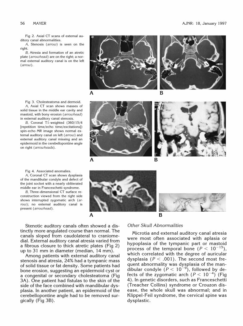
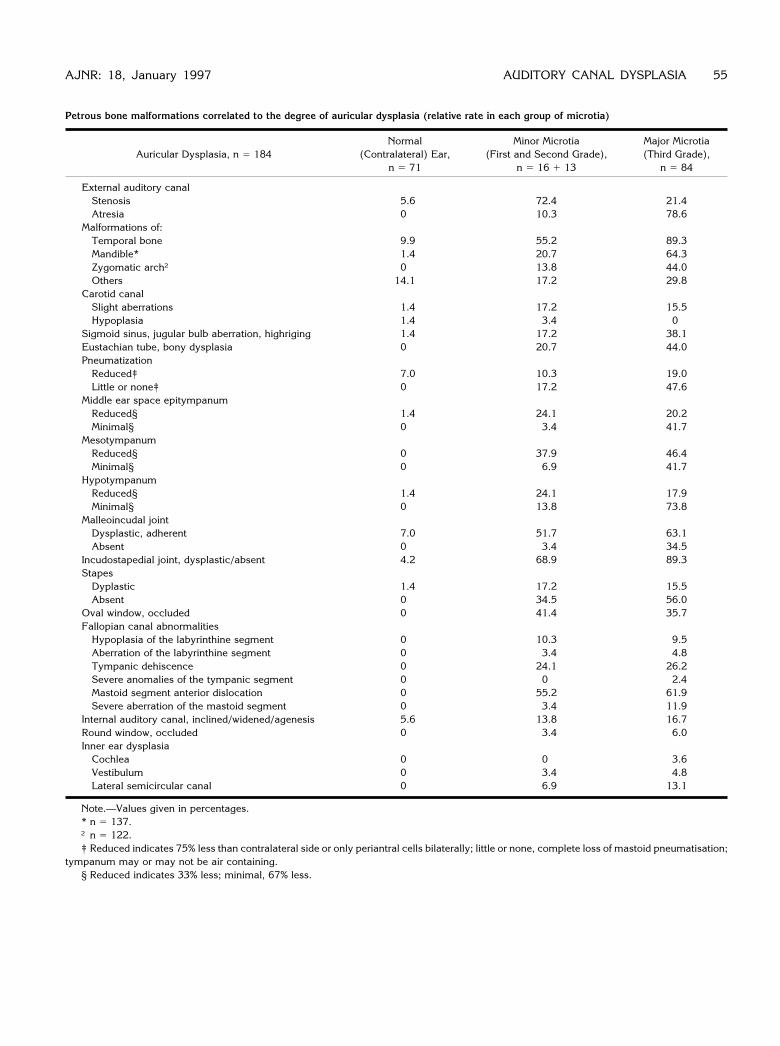
Fig 2. Axial CT scans of external au-ditory canal abnormalities.
A, Stenosis (arrow) is seen on theright.
B, Atresia and formation of an atreticplate (arrowhead) are on the right; a nor-mal external auditory canal is on the left(arrow).
Fig 3. Cholesteatoma and dermoid.A, Axial CT scan shows masses of
solid tissue in the middle ear cavity andmastoid, with bony erosion (arrowhead)in external auditory canal stenosis.
B, Coronal T1-weighted (360/15/4[repetition time/echo time/excitations])spin-echo MR image shows normal ex-ternal auditory canal on left (arrow) andexternal auditory canal missing and anepidermoid in the cerebellopontine angleon right (arrowheads).
Fig 4. Associated anomalies.A, Coronal CT scan shows dysplasia
of the mandibular condyle and defect ofthe joint socket with a nearly obliteratedmiddle ear in Franceschetti syndrome.
B, Three-dimensional CT surface re-construction viewed from the right sideshows interrupted zygomatic arch (ar-row); no external auditory canal ispresent (arrowhead).
56 MAYER AJNR: 18, January 1997
Stenotic auditory canals often showed a dis-tinctly more angulated course than normal. Thecanals sloped from caudolateral to craniome-dial. External auditory canal atresia varied froma fibrous closure to thick atretic plates (Fig 2)up to 31 mm in diameter (median, 14 mm).Among patients with external auditory canal
stenosis and atresia, 24% had a tympanic massof solid tissue or fat density. Some patients hadbone erosion, suggesting an epidermoid cyst ora congenital or secondary cholesteatoma (Fig3A). One patient had fistulas to the skin of theside of the face combined with mandibular dys-plasia. In another patient, an epidermoid of thecerebellopontine angle had to be removed sur-gically (Fig 3B).
Other Skull Abnormalities
Microtia and external auditory canal atresiawere most often associated with aplasia orhypoplasia of the tympanic part or mastoidprocess of the temporal bone (P , 10215),which correlated with the degree of auriculardysplasia (P , .001). The second most fre-quent abnormality was dysplasia of the man-dibular condyle (P , 1029), followed by de-fects of the zygomatic arch (P , 1024) (Fig4). In genetic disorders, such as Franceschetti(Treacher Collins) syndrome or Crouzon dis-ease, the whole skull was abnormal; and inKlippel-Feil syndrome, the cervical spine wasdysplastic.
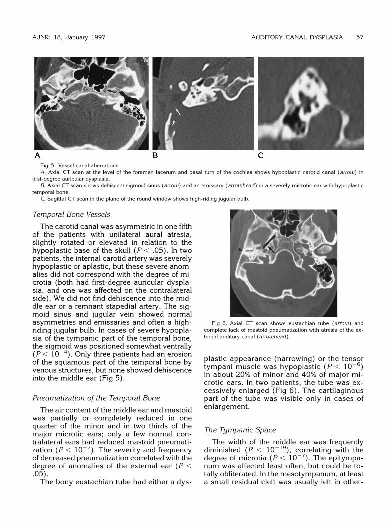
Fig 5. Vessel canal aberrations.A, Axial CT scan at the level of the foramen lacerum and basal turn of the cochlea shows hypoplastic carotid canal (arrow) in
first-degree auricular dysplasia.B, Axial CT scan shows dehiscent sigmoid sinus (arrow) and an emissary (arrowhead) in a severely microtic ear with hypoplastic
temporal bone.C, Sagittal CT scan in the plane of the round window shows high-riding jugular bulb.
AJNR: 18, January 1997 AUDITORY CANAL DYSPLASIA 57
Temporal Bone Vessels
The carotid canal was asymmetric in one fifthof the patients with unilateral aural atresia,slightly rotated or elevated in relation to thehypoplastic base of the skull (P , .05). In twopatients, the internal carotid artery was severelyhypoplastic or aplastic, but these severe anom-alies did not correspond with the degree of mi-crotia (both had first-degree auricular dyspla-sia, and one was affected on the contralateralside). We did not find dehiscence into the mid-dle ear or a remnant stapedial artery. The sig-moid sinus and jugular vein showed normalasymmetries and emissaries and often a high-riding jugular bulb. In cases of severe hypopla-sia of the tympanic part of the temporal bone,the sigmoid was positioned somewhat ventrally(P , 1024). Only three patients had an erosionof the squamous part of the temporal bone byvenous structures, but none showed dehiscenceinto the middle ear (Fig 5).
Pneumatization of the Temporal Bone
The air content of the middle ear and mastoidwas partially or completely reduced in onequarter of the minor and in two thirds of themajor microtic ears; only a few normal con-tralateral ears had reduced mastoid pneumati-zation (P , 1027). The severity and frequencyof decreased pneumatization correlated with thedegree of anomalies of the external ear (P ,.05).The bony eustachian tube had either a dys-
plastic appearance (narrowing) or the tensortympani muscle was hypoplastic (P , 1026)in about 20% of minor and 40% of major mi-crotic ears. In two patients, the tube was ex-cessively enlarged (Fig 6). The cartilaginouspart of the tube was visible only in cases ofenlargement.
The Tympanic Space
The width of the middle ear was frequentlydiminished (P , 10219), correlating with thedegree of microtia (P , 1027). The epitympa-num was affected least often, but could be to-tally obliterated. In the mesotympanum, at leasta small residual cleft was usually left in other-
Fig 6. Axial CT scan shows eustachian tube (arrow) andcomplete lack of mastoid pneumatization with atresia of the ex-ternal auditory canal (arrowhead).
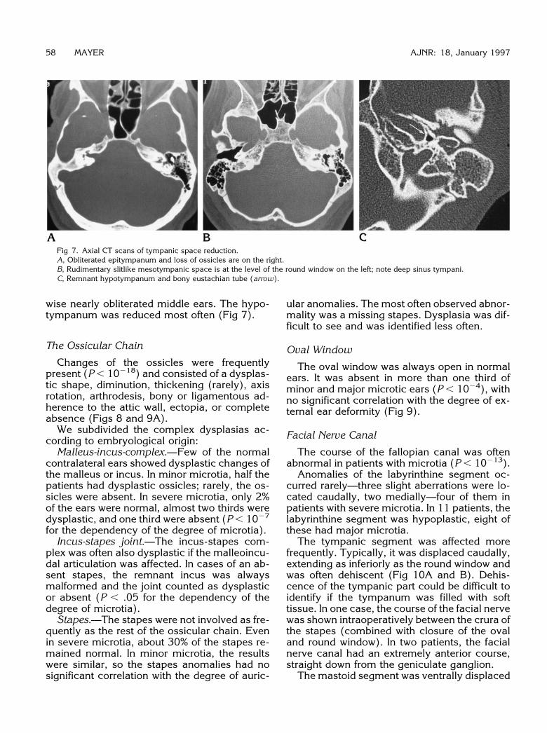
Fig 7. Axial CT scans of tympanic space reduction.A, Obliterated epitympanum and loss of ossicles are on the right.B, Rudimentary slitlike mesotympanic space is at the level of the round window on the left; note deep sinus tympani.C, Remnant hypotympanum and bony eustachian tube (arrow).
58 MAYER AJNR: 18, January 1997
wise nearly obliterated middle ears. The hypo-tympanum was reduced most often (Fig 7).
The Ossicular Chain
Changes of the ossicles were frequentlypresent (P , 10218) and consisted of a dysplas-tic shape, diminution, thickening (rarely), axisrotation, arthrodesis, bony or ligamentous ad-herence to the attic wall, ectopia, or completeabsence (Figs 8 and 9A).We subdivided the complex dysplasias ac-
cording to embryological origin:Malleus-incus-complex.—Few of the normal
contralateral ears showed dysplastic changes ofthe malleus or incus. In minor microtia, half thepatients had dysplastic ossicles; rarely, the os-sicles were absent. In severe microtia, only 2%of the ears were normal, almost two thirds weredysplastic, and one third were absent (P , 1027
for the dependency of the degree of microtia).Incus-stapes joint.—The incus-stapes com-
plex was often also dysplastic if the malleoincu-dal articulation was affected. In cases of an ab-sent stapes, the remnant incus was alwaysmalformed and the joint counted as dysplasticor absent (P , .05 for the dependency of thedegree of microtia).Stapes.—The stapes were not involved as fre-
quently as the rest of the ossicular chain. Evenin severe microtia, about 30% of the stapes re-mained normal. In minor microtia, the resultswere similar, so the stapes anomalies had nosignificant correlation with the degree of auric-
ular anomalies. The most often observed abnor-mality was a missing stapes. Dysplasia was dif-ficult to see and was identified less often.
Oval Window
The oval window was always open in normalears. It was absent in more than one third ofminor and major microtic ears (P , 1024), withno significant correlation with the degree of ex-ternal ear deformity (Fig 9).
Facial Nerve Canal
The course of the fallopian canal was oftenabnormal in patients with microtia (P , 10213).Anomalies of the labyrinthine segment oc-
curred rarely—three slight aberrations were lo-cated caudally, two medially—four of them inpatients with severe microtia. In 11 patients, thelabyrinthine segment was hypoplastic, eight ofthese had major microtia.The tympanic segment was affected more
frequently. Typically, it was displaced caudally,extending as inferiorly as the round window andwas often dehiscent (Fig 10A and B). Dehis-cence of the tympanic part could be difficult toidentify if the tympanum was filled with softtissue. In one case, the course of the facial nervewas shown intraoperatively between the crura ofthe stapes (combined with closure of the ovaland round window). In two patients, the facialnerve canal had an extremely anterior course,straight down from the geniculate ganglion.The mastoid segment was ventrally displaced
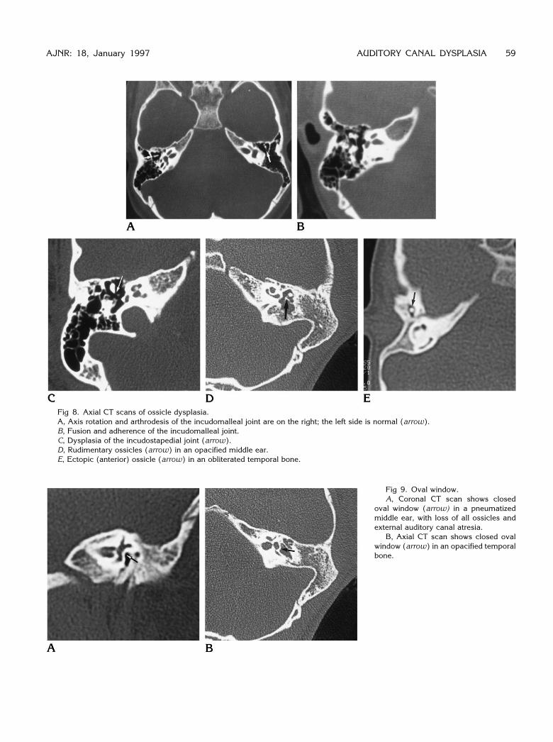
Fig 8. Axial CT scans of ossicle dysplasia.A, Axis rotation and arthrodesis of the incudomalleal joint are on the right; the left side is normal (arrow).B, Fusion and adherence of the incudomalleal joint.C, Dysplasia of the incudostapedial joint (arrow).D, Rudimentary ossicles (arrow) in an opacified middle ear.E, Ectopic (anterior) ossicle (arrow) in an obliterated temporal bone.
Fig 9. Oval window.A, Coronal CT scan shows closed
oval window (arrow) in a pneumatizedmiddle ear, with loss of all ossicles andexternal auditory canal atresia.
B, Axial CT scan shows closed ovalwindow (arrow) in an opacified temporalbone.
AJNR: 18, January 1997 AUDITORY CANAL DYSPLASIA 59
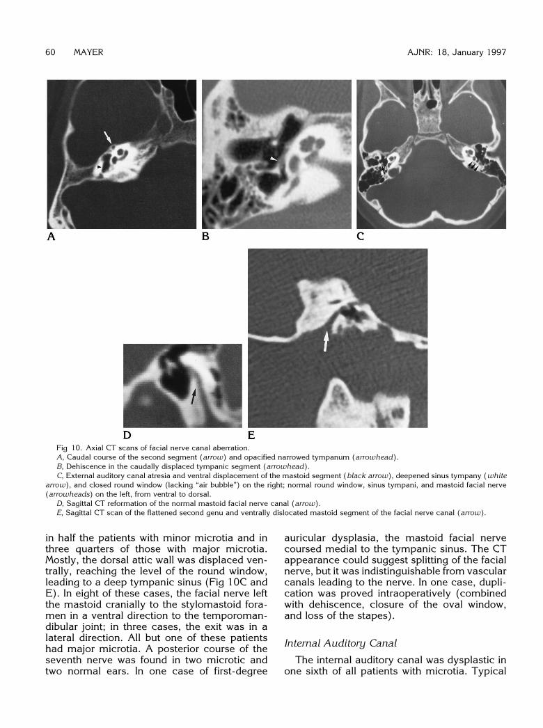
Fig 10. Axial CT scans of facial nerve canal aberration.A, Caudal course of the second segment (arrow) and opacified narrowed tympanum (arrowhead).B, Dehiscence in the caudally displaced tympanic segment (arrowhead).C, External auditory canal atresia and ventral displacement of the mastoid segment (black arrow), deepened sinus tympany (white
arrow), and closed round window (lacking “air bubble”) on the right; normal round window, sinus tympani, and mastoid facial nerve(arrowheads) on the left, from ventral to dorsal.
D, Sagittal CT reformation of the normal mastoid facial nerve canal (arrow).E, Sagittal CT scan of the flattened second genu and ventrally dislocated mastoid segment of the facial nerve canal (arrow).
60 MAYER AJNR: 18, January 1997
in half the patients with minor microtia and inthree quarters of those with major microtia.Mostly, the dorsal attic wall was displaced ven-trally, reaching the level of the round window,leading to a deep tympanic sinus (Fig 10C andE). In eight of these cases, the facial nerve leftthe mastoid cranially to the stylomastoid fora-men in a ventral direction to the temporoman-dibular joint; in three cases, the exit was in alateral direction. All but one of these patientshad major microtia. A posterior course of theseventh nerve was found in two microtic andtwo normal ears. In one case of first-degree
auricular dysplasia, the mastoid facial nervecoursed medial to the tympanic sinus. The CTappearance could suggest splitting of the facialnerve, but it was indistinguishable from vascularcanals leading to the nerve. In one case, dupli-cation was proved intraoperatively (combinedwith dehiscence, closure of the oval window,and loss of the stapes).
Internal Auditory Canal
The internal auditory canal was dysplastic inone sixth of all patients with microtia. Typical
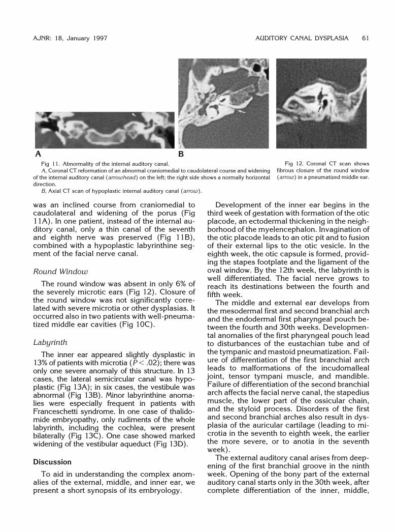
Fig 11. Abnormality of the internal auditory canal.A, Coronal CT reformation of an abnormal craniomedial to caudolateral course and widening
of the internal auditory canal (arrowhead) on the left; the right side shows a normally horizontaldirection.
B, Axial CT scan of hypoplastic internal auditory canal (arrow).
Fig 12. Coronal CT scan showsfibrous closure of the round window(arrow) in a pneumatized middle ear.
AJNR: 18, January 1997 AUDITORY CANAL DYSPLASIA 61
was an inclined course from craniomedial tocaudolateral and widening of the porus (Fig11A). In one patient, instead of the internal au-ditory canal, only a thin canal of the seventhand eighth nerve was preserved (Fig 11B),combined with a hypoplastic labyrinthine seg-ment of the facial nerve canal.
Round Window
The round window was absent in only 6% ofthe severely microtic ears (Fig 12). Closure ofthe round window was not significantly corre-lated with severe microtia or other dysplasias. Itoccurred also in two patients with well-pneuma-tized middle ear cavities (Fig 10C).
Labyrinth
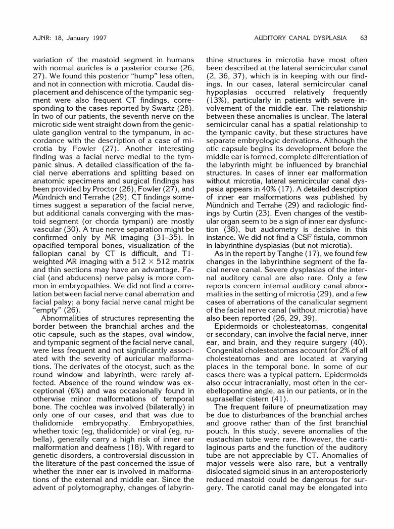
The inner ear appeared slightly dysplastic in13% of patients with microtia (P , .02); there wasonly one severe anomaly of this structure. In 13cases, the lateral semicircular canal was hypo-plastic (Fig 13A); in six cases, the vestibule wasabnormal (Fig 13B). Minor labyrinthine anoma-lies were especially frequent in patients withFranceschetti syndrome. In one case of thalido-mide embryopathy, only rudiments of the wholelabyrinth, including the cochlea, were presentbilaterally (Fig 13C). One case showed markedwidening of the vestibular aqueduct (Fig 13D).
Discussion
To aid in understanding the complex anom-alies of the external, middle, and inner ear, wepresent a short synopsis of its embryology.
Development of the inner ear begins in thethird week of gestation with formation of the oticplacode, an ectodermal thickening in the neigh-borhood of the myelencephalon. Invagination ofthe otic placode leads to an otic pit and to fusionof their external lips to the otic vesicle. In theeighth week, the otic capsule is formed, provid-ing the stapes footplate and the ligament of theoval window. By the 12th week, the labyrinth iswell differentiated. The facial nerve grows toreach its destinations between the fourth andfifth week.The middle and external ear develops from
the mesodermal first and second branchial archand the endodermal first pharyngeal pouch be-tween the fourth and 30th weeks. Developmen-tal anomalies of the first pharyngeal pouch leadto disturbances of the eustachian tube and ofthe tympanic andmastoid pneumatization. Fail-ure of differentiation of the first branchial archleads to malformations of the incudomallealjoint, tensor tympani muscle, and mandible.Failure of differentiation of the second branchialarch affects the facial nerve canal, the stapediusmuscle, the lower part of the ossicular chain,and the styloid process. Disorders of the firstand second branchial arches also result in dys-plasia of the auricular cartilage (leading to mi-crotia in the seventh to eighth week, the earlierthe more severe, or to anotia in the seventhweek).The external auditory canal arises from deep-
ening of the first branchial groove in the ninthweek. Opening of the bony part of the externalauditory canal starts only in the 30th week, aftercomplete differentiation of the inner, middle,
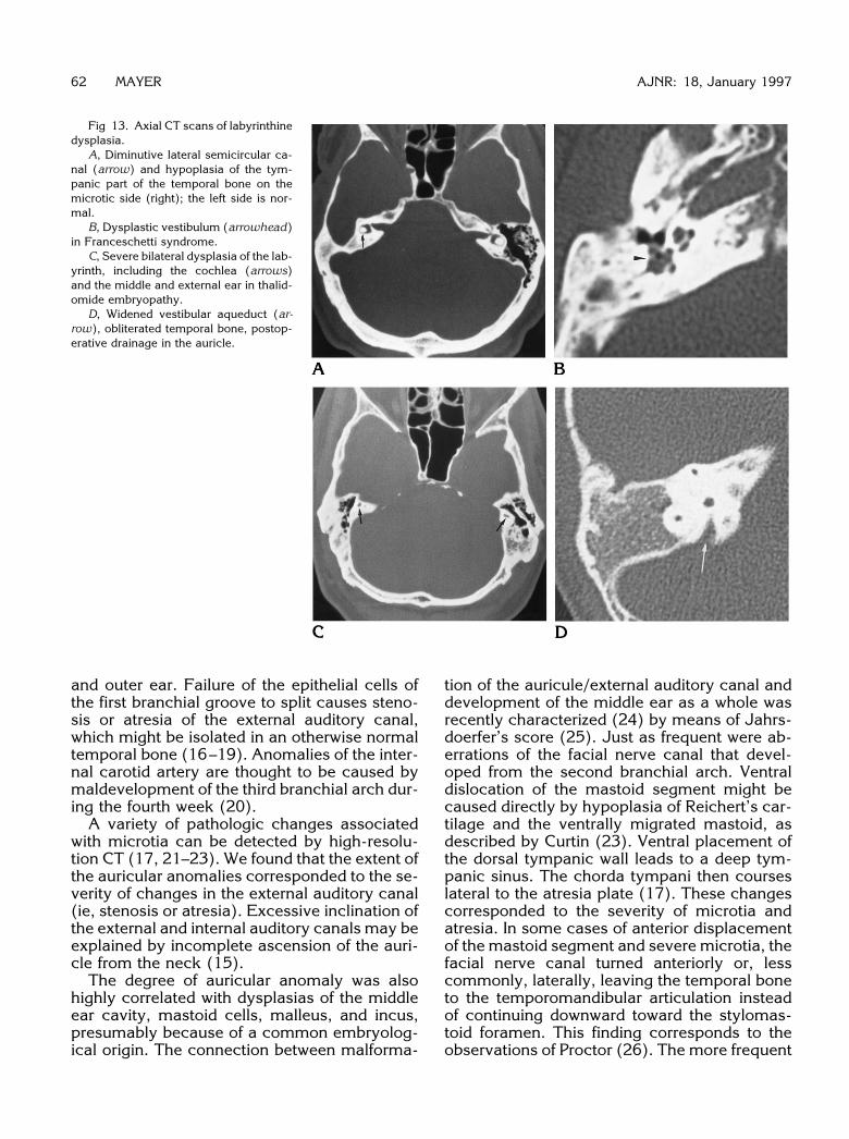
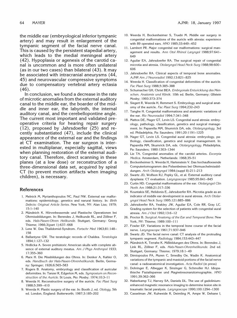
Fig 13. Axial CT scans of labyrinthinedysplasia.
A, Diminutive lateral semicircular ca-nal (arrow) and hypoplasia of the tym-panic part of the temporal bone on themicrotic side (right); the left side is nor-mal.
B, Dysplastic vestibulum (arrowhead)in Franceschetti syndrome.
C, Severe bilateral dysplasia of the lab-yrinth, including the cochlea (arrows)and the middle and external ear in thalid-omide embryopathy.
D, Widened vestibular aqueduct (ar-row), obliterated temporal bone, postop-erative drainage in the auricle.
62 MAYER AJNR: 18, January 1997
and outer ear. Failure of the epithelial cells ofthe first branchial groove to split causes steno-sis or atresia of the external auditory canal,which might be isolated in an otherwise normaltemporal bone (16–19). Anomalies of the inter-nal carotid artery are thought to be caused bymaldevelopment of the third branchial arch dur-ing the fourth week (20).A variety of pathologic changes associated
with microtia can be detected by high-resolu-tion CT (17, 21–23). We found that the extent ofthe auricular anomalies corresponded to the se-verity of changes in the external auditory canal(ie, stenosis or atresia). Excessive inclination ofthe external and internal auditory canals may beexplained by incomplete ascension of the auri-cle from the neck (15).The degree of auricular anomaly was also
highly correlated with dysplasias of the middleear cavity, mastoid cells, malleus, and incus,presumably because of a common embryolog-ical origin. The connection between malforma-
tion of the auricule/external auditory canal anddevelopment of the middle ear as a whole wasrecently characterized (24) by means of Jahrs-doerfer’s score (25). Just as frequent were ab-errations of the facial nerve canal that devel-oped from the second branchial arch. Ventraldislocation of the mastoid segment might becaused directly by hypoplasia of Reichert’s car-tilage and the ventrally migrated mastoid, asdescribed by Curtin (23). Ventral placement ofthe dorsal tympanic wall leads to a deep tym-panic sinus. The chorda tympani then courseslateral to the atresia plate (17). These changescorresponded to the severity of microtia andatresia. In some cases of anterior displacementof the mastoid segment and severe microtia, thefacial nerve canal turned anteriorly or, lesscommonly, laterally, leaving the temporal boneto the temporomandibular articulation insteadof continuing downward toward the stylomas-toid foramen. This finding corresponds to theobservations of Proctor (26). The more frequent
AJNR: 18, January 1997 AUDITORY CANAL DYSPLASIA 63
variation of the mastoid segment in humanswith normal auricles is a posterior course (26,27). We found this posterior “hump” less often,and not in connection with microtia. Caudal dis-placement and dehiscence of the tympanic seg-ment were also frequent CT findings, corre-sponding to the cases reported by Swartz (28).In two of our patients, the seventh nerve on themicrotic side went straight down from the genic-ulate ganglion ventral to the tympanum, in ac-cordance with the description of a case of mi-crotia by Fowler (27). Another interestingfinding was a facial nerve medial to the tym-panic sinus. A detailed classification of the fa-cial nerve aberrations and splitting based onanatomic specimens and surgical findings hasbeen provided by Proctor (26), Fowler (27), andMundnich and Terrahe (29). CT findings some-times suggest a separation of the facial nerve,but additional canals converging with the mas-toid segment (or chorda tympani) are mostlyvascular (30). A true nerve separation might beconfirmed only by MR imaging (31–35). Inopacified temporal bones, visualization of thefallopian canal by CT is difficult, and T1-weighted MR imaging with a 512 3 512 matrixand thin sections may have an advantage. Fa-cial (and abducens) nerve palsy is more com-mon in embryopathies. We did not find a corre-lation between facial nerve canal aberration andfacial palsy; a bony facial nerve canal might be“empty” (26).Abnormalities of structures representing the
border between the branchial arches and theotic capsule, such as the stapes, oval window,and tympanic segment of the facial nerve canal,were less frequent and not significantly associ-ated with the severity of auricular malforma-tions. The derivates of the otocyst, such as theround window and labyrinth, were rarely af-fected. Absence of the round window was ex-ceptional (6%) and was occasionally found inotherwise minor malformations of temporalbone. The cochlea was involved (bilaterally) inonly one of our cases, and that was due tothalidomide embryopathy. Embryopathies,whether toxic (eg, thalidomide) or viral (eg, ru-bella), generally carry a high risk of inner earmalformation and deafness (18). With regard togenetic disorders, a controversial discussion inthe literature of the past concerned the issue ofwhether the inner ear is involved in malforma-tions of the external and middle ear. Since theadvent of polytomography, changes of labyrin-
thine structures in microtia have most oftenbeen described at the lateral semicircular canal(2, 36, 37), which is in keeping with our find-ings. In our cases, lateral semicircular canalhypoplasias occurred relatively frequently(13%), particularly in patients with severe in-volvement of the middle ear. The relationshipbetween these anomalies is unclear. The lateralsemicircular canal has a spatial relationship tothe tympanic cavity, but these structures haveseparate embryologic derivations. Although theotic capsule begins its development before themiddle ear is formed, complete differentiation ofthe labyrinth might be influenced by branchialstructures. In cases of inner ear malformationwithout microtia, lateral semicircular canal dys-pasia appears in 40% (17). A detailed descriptionof inner ear malformations was published byMundnich and Terrahe (29) and radiologic find-ings by Curtin (23). Even changes of the vestib-ular organ seem to be a sign of inner ear dysfunc-tion (38), but audiometry is decisive in thisinstance. We did not find a CSF fistula, commonin labyrinthine dysplasias (but not microtia).As in the report by Tanghe (17), we found few
changes in the labyrinthine segment of the fa-cial nerve canal. Severe dysplasias of the inter-nal auditory canal are also rare. Only a fewreports concern internal auditory canal abnor-malities in the setting of microtia (29), and a fewcases of aberrations of the canalicular segmentof the facial nerve canal (without microtia) havealso been reported (26, 29, 39).Epidermoids or cholesteatomas, congenital
or secondary, can involve the facial nerve, innerear, and brain, and they require surgery (40).Congenital cholesteatomas account for 2% of allcholesteatomas and are located at varyingplaces in the temporal bone. In some of ourcases there was a typical pattern. Epidermoidsalso occur intracranially, most often in the cer-ebellopontine angle, as in our patients, or in thesuprasellar cistern (41).The frequent failure of pneumatization may
be due to disturbances of the branchial archesand groove rather than of the first branchialpouch. In this study, severe anomalies of theeustachian tube were rare. However, the carti-laginous parts and the function of the auditorytube are not appreciable by CT. Anomalies ofmajor vessels were also rare, but a ventrallydislocated sigmoid sinus in an anteroposteriorlyreduced mastoid could be dangerous for sur-gery. The carotid canal may be elongated into
the middle ear (embryological inferior tympanicartery) and may result in enlargement of thetympanic segment of the facial nerve canal.This is caused by the persistent stapedial artery,which leads to the medial meningeal artery(42). Hypoplasia or agenesis of the carotid ca-nal is uncommon and is more often unilateral(as in our two cases) than bilateral (43). It maybe associated with intracranial aneurysms (44,45) and neurovascular compressive symptomsdue to compensatory vertebral artery ectasia(46).In conclusion, we found a decrease in the rate
of microtic anomalies from the external auditorycanal to the middle ear, the boarder of the mid-dle and inner ear, the labyrinth, the internalauditory canal, and the cerebellopontine angle.The current most important and validated pre-operative criteria for hearing repair surgery(12), proposed by Jahrsdoerfer (25) and re-cently substantiated (47), include the clinicalappearance of the external ear and the findingsat CT examination. The ear surgeon is inter-ested in multiplanar, especially sagittal, viewswhen planning restoration of the external audi-tory canal. Therefore, direct scanning in theseplanes (at a low dose) or reconstruction of athree-dimensional data set, acquired by spiralCT (to prevent motion artifacts when imagingchildren), is necessary.
References1. Melnick M, Myrianthopoulos NC, Paul NW. External ear malfor-
mations: epidemiology, genetics and narural history. In: BirthDefects: Original Article Series. New York, NY: Alan Liss; 1979;15:1–140
2. Mundnich K. Horverbessernde und Plastische Operationen beiOhrmissbildungen. In: Berendes J, Heilkunde RL, and Zollner F,eds. Hals-Nasen-Ohren Heilkunde. Stuttgart, Germany: GeorgThieme; 1966;3(part 1):637–668
3. Lenz W. Das Thalidomid-Syndrom. Fortschr Med 1963;81:148–158
4. Ballantyne GW. The teratologic records of Chaldea. Teratologia1894;1:127–132
5. Hrdlicka A. Seven prehistoric American skulls with complete ab-sence of external auditory meatus. Am J Phys Anthropol 1933;17:355–360
6. Marx H. Die Missbildungen des Ohres. In: Denker A, Kahler O,eds. Handbuch der Hals-Nasen-Ohrenheilkunde. Berlin, Germa-ny: Springer; 1926;6:565–583
7. Rogers B. Anatomy, embryology and classification of auriculardeformities. In: Tanzer R, Edgerton M, eds. Symposium on Recon-struction of the Auricle. St Louis, Mo: Mosby; 1974;10:3–11
8. Weerda H. Reconstructive surgery of the auricle. Fac Plast Surg1988;5:399–410
9. Weerda H. Plastic surgery of the ear. In: Booth J, ed. Otology. 5thed. London, England: Butterworth; 1987;3:185–202
64 MAYER
10. Weerda H, Bockenheimer S, Truebi M. Middle ear surgery incongenital malformations of the auricle with atresia: experiencewith 89 operated ears. HNO 1985;33:449–452
11. Lambert PR. Major congenital ear malformations: surgical man-agement and results. Ann Otol Rhinol Laryngol 1988;97:641–649
12. Aguilar EA, Jahrsdoerfer RA. The surgical repair of congenitalmicrotia and atresia. Otolaryngol Head Neck Surg 1988;98:600–606
13. Jahrsdoerfer RA. Clinical aspects of temporal bone anomalies.AJNR Am J Neuroradiol 1992;13:821–825
14. Weerda H. Classification of congenital deformities of the auricle.Fac Plast Surg 1988;5:385–388
15. Schumacher GH, Christ BEA. Embryonale Entwicklung des Men-schen. Anatomie und Klinik. 10th ed. Berlin, Germany: UllsteinMosby; 1993:373–374
16. Siegert R, Weerda H, Remmert S. Embryology and surgical anat-omy of the auricle. Fac Plast Surg 1994;232–243
17. Tanghe H. Congenital malformations and anatomic variations ofthe ear. Riv Neuroradiol 1994;7:341–348
18. Mattox DE, Nager GT, Levin LS. Congenital aural atresia: embry-ology, pathology, classification, genetics and surgical manage-ment. In: Paparella MM, Shumrick DA, eds. Otolaryngology. 3rded. Philadelphia, Pa: Saunders; 1991;20:1191–1225
19. Nager GT, Levin LS. Congenital aural atresia: embryology, pa-thology, classification, genetics and surgical management. In:Paparella MM, Skumrick DA, eds. Otolaryngology. Philadelphia,Pa: Saunders; 1980:1303–1344
20. Lie TA. Congenital anomalies of the carotid arteries. ExcerptaMedica. Amsterdam, Netherlands: 1968;35–51
21. Bockenheimer S, Weerda H, Hartenstein V. Das hochaufloesendeComputertomogramm des Felsenbeines bei Ohrmuschelmissbil-dungen. Arch Otolaryngol 1984;(suppl II):211–213
22. Swartz JD, Wolfson RJ, Popky GL, et al. External auditory canaldysplasia: CT evaluation. Laryngoscope 1985;95:841–845
23. Curtin HD. Congenital malformations of the ear. Otolaryngol ClinNorth Am 1988;21:317–336
24. Kountakis SE, Helidonis E, Jahrsdoerfer RA. Microtia grade as anindicator of middle ear development in aural atresia. Arch Otolar-yngol Head Neck Surg 1995;121:885–886
25. Jahrsdoerfer RA, Yeakley JW, Aguilar EA, Cole RR, Gray LC.Grading system for the selection of patients with congenital auralatresia. Am J Otol 1992;13:6–12
26. Proctor B. Surgical Anatomy of the Ear and Temporal Bone. NewYork, NY: Thieme; 1989:100–111
27. Fowler EP. Variations in the temporal bone course of the facialnerve. Laryngoscope 1961;71:937–946
28. Swartz JD. The facial nerve canal: CT analysis of the protrudingtympanic segment. Radiology 1984;153:443–447
29. Mundnich K, Terrahe K. Mißbildungen des Ohres. In: Berendes J,Link RL, Zollner F, eds. Hals-Nasen-Ohrenheilkunde. 2nd ed.Stuttgart, Germany: Thieme; 1979;18:1–49
30. Dimiopoulos PA, Muren C, Smedby Oe, Wadin K. Anatomicalvariations of the tympanic and mastoid portions of the facial nervecanal: a radioanatomical investigation. Acta Radiol (in press)
31. Dohringer E, Albegger K, Sinzinger G, Schmoller HJ. Idiopa-thische Fazialisparese und Magnetresonanztomographie. HNO1991;39:362–366
32. Haberkamp TJ, Harvey SA, Daniels DL. The use of gadolinium-enhanced magnetic resonance imaging to determine lesion site intraumatic facial paralysis. Laryngoscope 1990;100:1294–1300
33. Casselman JW, Kuhweide R, Deimling M, Ampe W, Dehane I,
AJNR: 18, January 1997
AJNR: 18, January 1997 AUDITORY CANAL DYSPLASIA 65
Meeus L. Constructive interference in steady state-3DFT MR im-aging of the inner ear and cerebellopontine angle. AJNR Am JNeuroradiol 1993;14:47–57
34. Casselman JW, Kuhweide R, Ampe W, Meeus L, Steyaert L.Pathology of the membranous labyrinth: comparison of T1- andT2-weighted and gadolinium-enhanced spin-echo and 3DFT-CISS imaging. AJNR Am J Neuroradiol 1993;14:59–69
35. Roos EH, Lufkin RB. Magnetic resonance imaging of the temporalbone. In: Swartz JD, ed. Imaging of the Temporal Bone. Philadel-phia, Pa: Saunders; 1986;9:203–211
36. Franceschetti A, Klein D. Mandibulofacial dysostosis: a new he-reditary syndrome. Acta Ophthalmol 1949;27:143–148
37. Hutchinson JC, Caldarelli DD, Valvassori GE, Pruzansky S, ParrisPJ. The otologic manifestation of mandibulofacial dysostosis.Trans Am Acad Ophthalmol Otolaryngol 1977;84:520–528
38. Murata K, Isono M, Ohta F, Yoshida A, Ishida O. Quantitativeanalysis of inner ear anomalies by high resolution CT scanning ofthe temporal bone. J Otolaryngol 1987;16:133–136
39. Weissman JL, Arriga M, Curtin C, Hirsch B. Duplication anomalyof the internal auditory canal. AJNR Am J Neuroradiol 1991;12:867–869
40. Mehra YN, Dubey SP, Mann SBS, Suri S. Correlation betweenhigh-resolution computed tomography and surgical findings in
congenital aural atresia. Arch Otolaryngol Head Neck Surg 1988;114:137–141
41. Swartz JD. Middle ear and mastoid. In: Swartz DJ, ed. Imaging ofthe Temporal Bone. Philadelphia, Pa: Saunders; 1992:33–87
42. Harnsberger HR. Vascular aberrations. In: Swartz DJ, ed. Imagingof the Temporal Bone. Philadelphia, Pa: Saunders; 1992:154–191
43. Grand CM, Louryan S, Bank WO, Baleriaux D, Brotchi J, RaybaudC. Agenesis of the internal carotid artery and cavernous sinushypoplasia with contralateral cavernous sinus meningioma. Neu-roradiology 1993;35:588–590
44. Waga S, Okada M, Kojima T. Saccular aneurysm associated withabsence of the left cervical carotid arteries. Neurosurgery 1978;3:208–212
45. Kunishio K, Sunami N, Yamamoto Y, Asari S. Agenesis of the leftinternal carotid artery, common carotid artery and the main trunkof the external carotid artery associated with multiple cerebralaneurysms: case report. Neurol Surg (Tokyo) 1986;14:873–879
46. Kiuchi K, Kowada M, Kojima H. Hypoplasia of the internal carotidartery associated with spasmodic torticollis: the possible role ofaltered vertebrobasilar hemodynamics. Neuroradiology 1995;37:362–364
47. Schwager K, Helms J. Mikrochirurgie grosser Mittelohrfehlbildun-gen. HNO 1995;43:427–431