Embed Size (px)
Citation preview
High resolution ultrasound in equine ophthalmology to display the anterior segment of the eye
M. Cronau, H. Gerhards
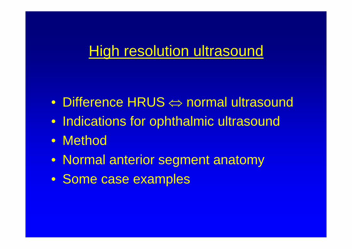
High resolution ultrasound
• Difference HRUS ⇔ normal ultrasound• Indications for ophthalmic ultrasound• Method• Normal anterior segment anatomy• Some case examples
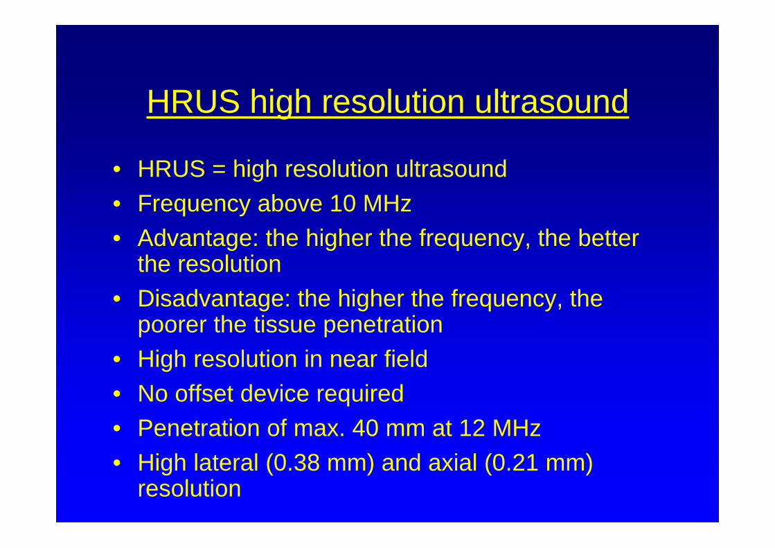
HRUS high resolution ultrasound
• HRUS = high resolution ultrasound• Frequency above 10 MHz• Advantage: the higher the frequency, the better
the resolution • Disadvantage: the higher the frequency, the
poorer the tissue penetration • High resolution in near field• No offset device required• Penetration of max. 40 mm at 12 MHz • High lateral (0.38 mm) and axial (0.21 mm)
resolution
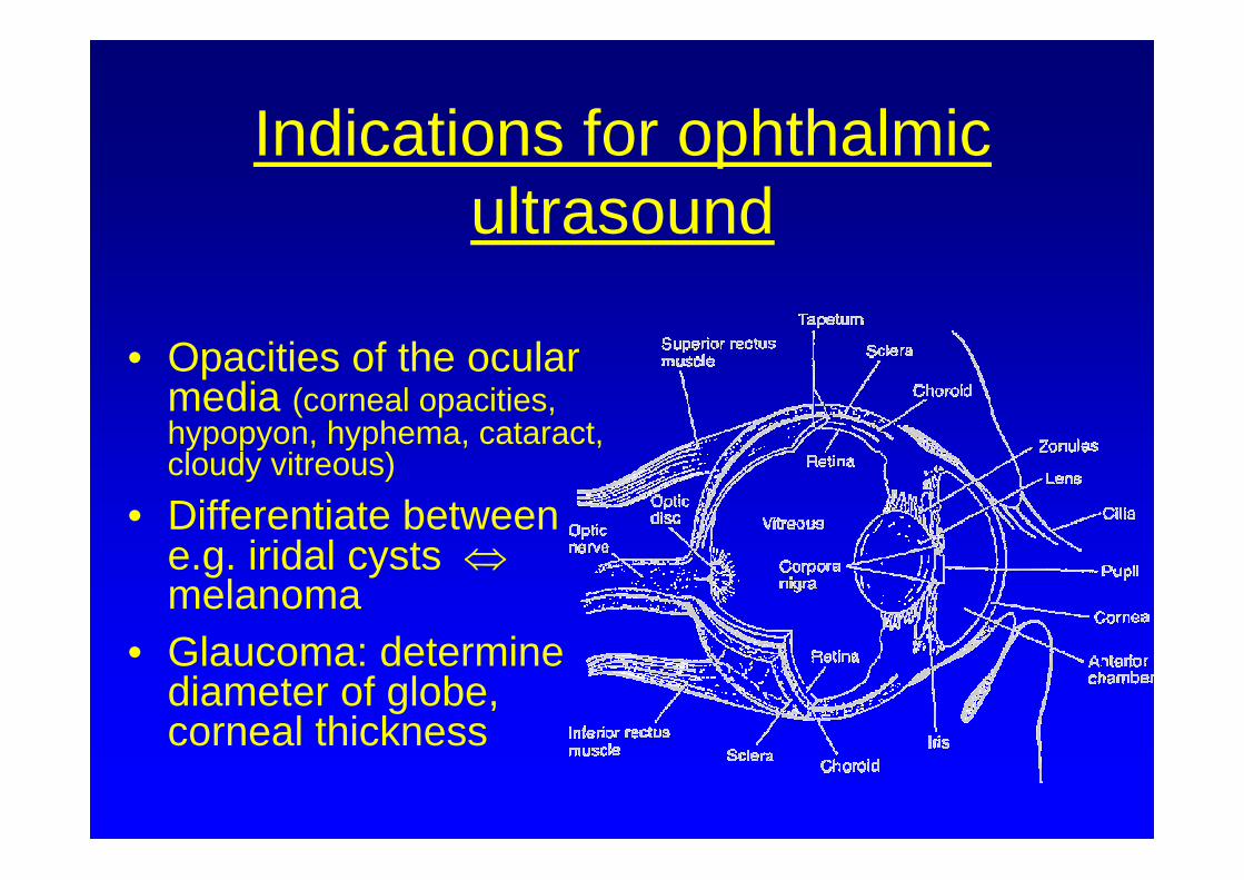
Indications for ophthalmic ultrasound
• Opacities of the ocular media (corneal opacities, hypopyon, hyphema, cataract, cloudy vitreous)
• Differentiate between e.g. iridal cysts ⇔melanoma
• Glaucoma: determine diameter of globe, corneal thickness
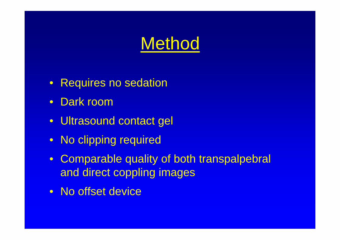
Method
• Requires no sedation
• Dark room
• Ultrasound contact gel
• No clipping required
• Comparable quality of both transpalpebraland direct coppling images
• No offset device
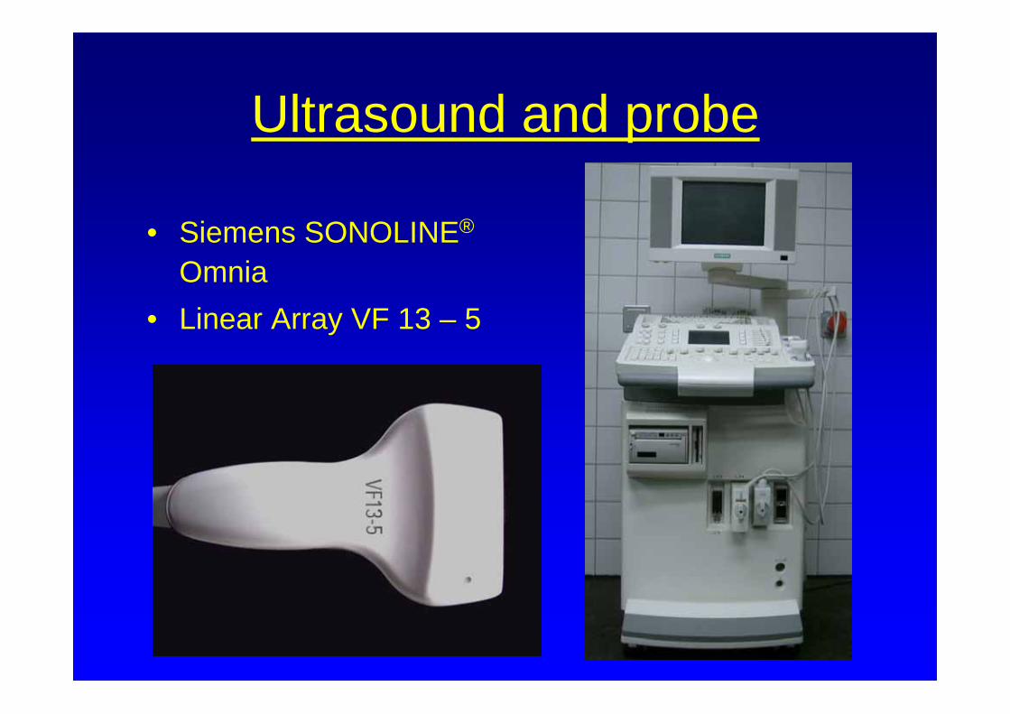
Ultrasound and probe
• Siemens SONOLINE®
Omnia• Linear Array VF 13 – 5
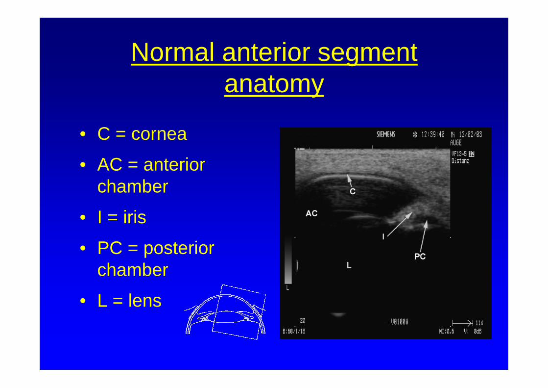
Normal anterior segment anatomy
• C = cornea
• AC = anterior chamber
• I = iris
• PC = posterior chamber
• L = lens
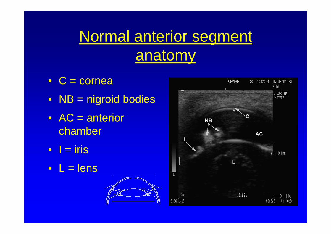
Normal anterior segment anatomy
• C = cornea
• NB = nigroid bodies
• AC = anterior chamber
• I = iris
• L = lens
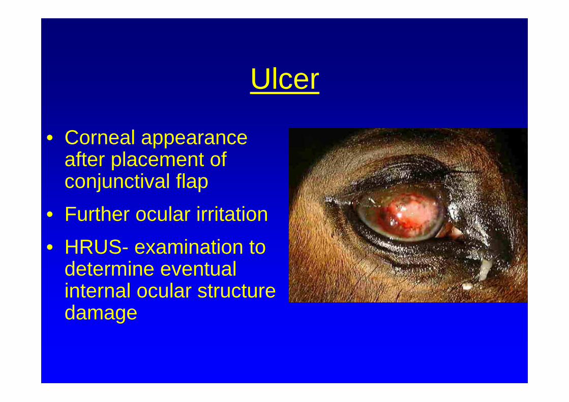
Ulcer
• Corneal appearance after placement of conjunctival flap
• Further ocular irritation• HRUS- examination to
determine eventual internal ocular structure damage
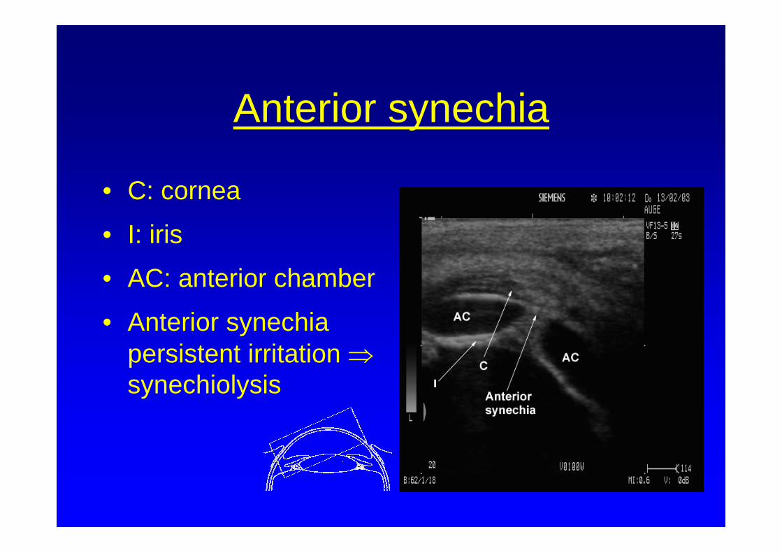
Anterior synechia
• C: cornea
• I: iris
• AC: anterior chamber
• Anterior synechiapersistent irritation ⇒synechiolysis
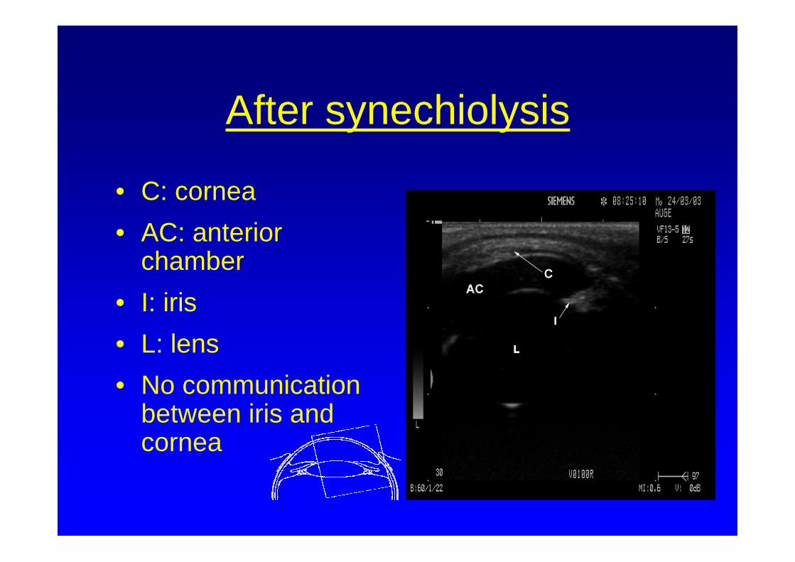
After synechiolysis
• C: cornea• AC: anterior
chamber• I: iris• L: lens• No communication
between iris and cornea
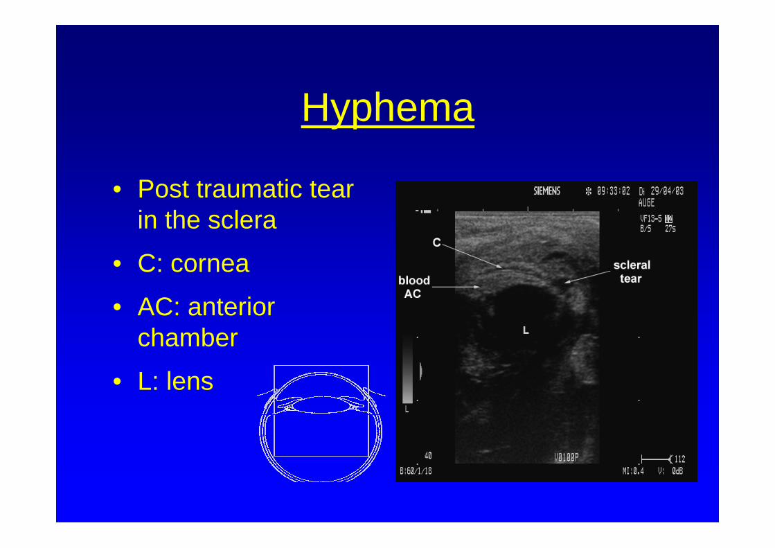
Hyphema
• Post traumatic tear in the sclera
• C: cornea
• AC: anterior chamber
• L: lens
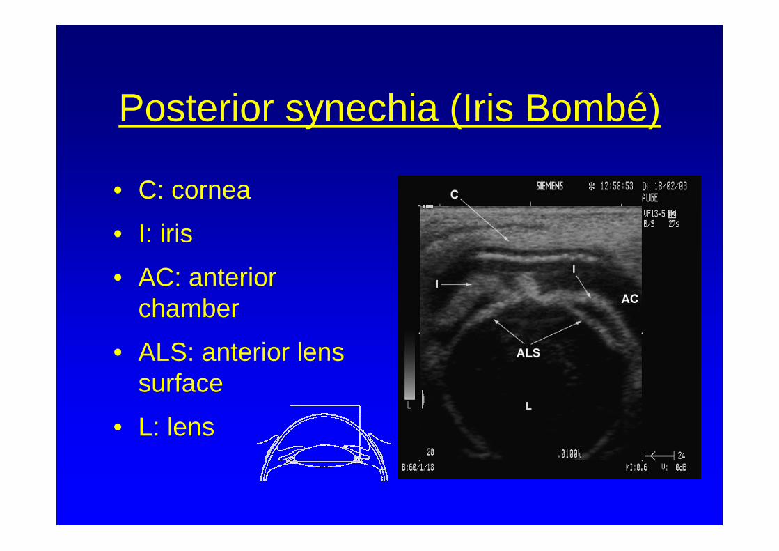
Posterior synechia (Iris Bombé)
• C: cornea
• I: iris
• AC: anterior chamber
• ALS: anterior lens surface
• L: lens
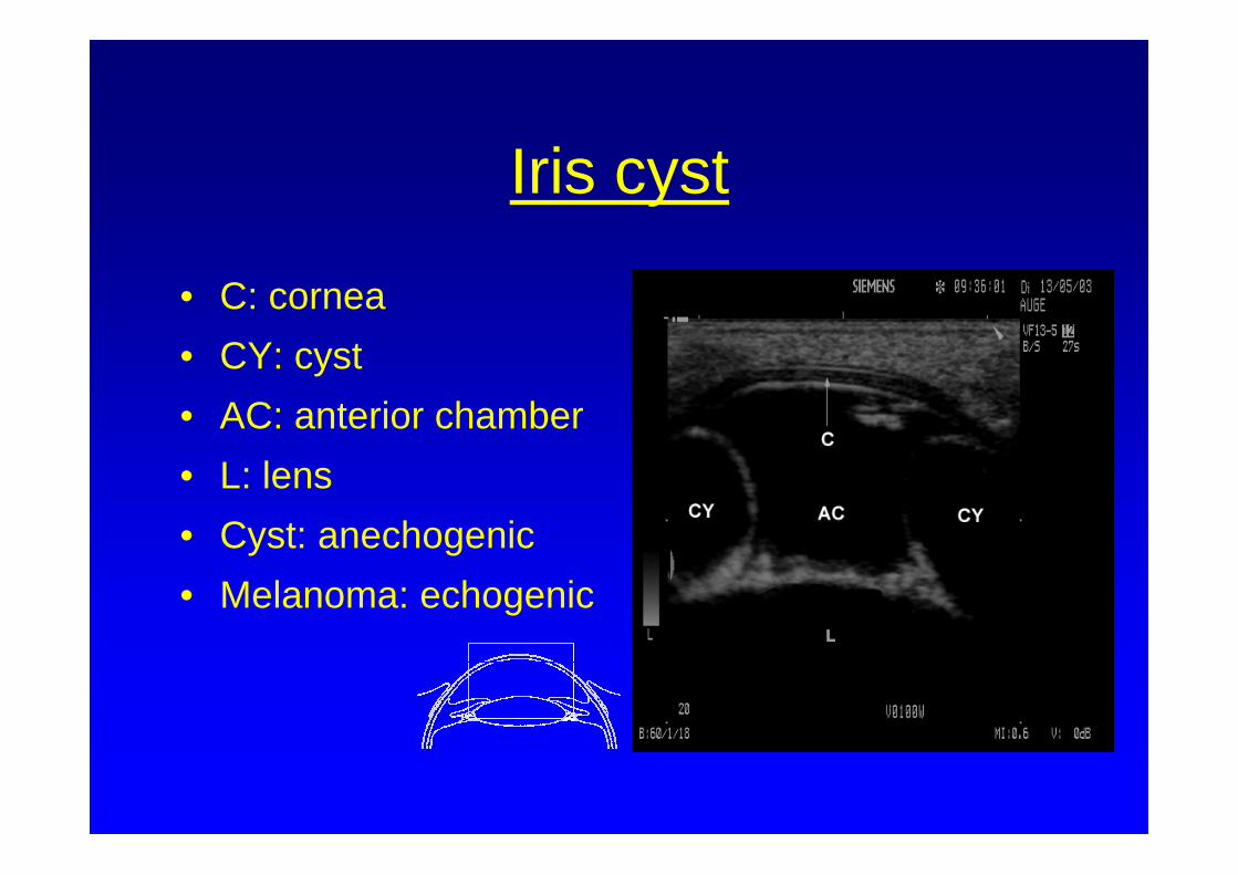
Iris cyst
• C: cornea• CY: cyst• AC: anterior chamber• L: lens• Cyst: anechogenic• Melanoma: echogenic
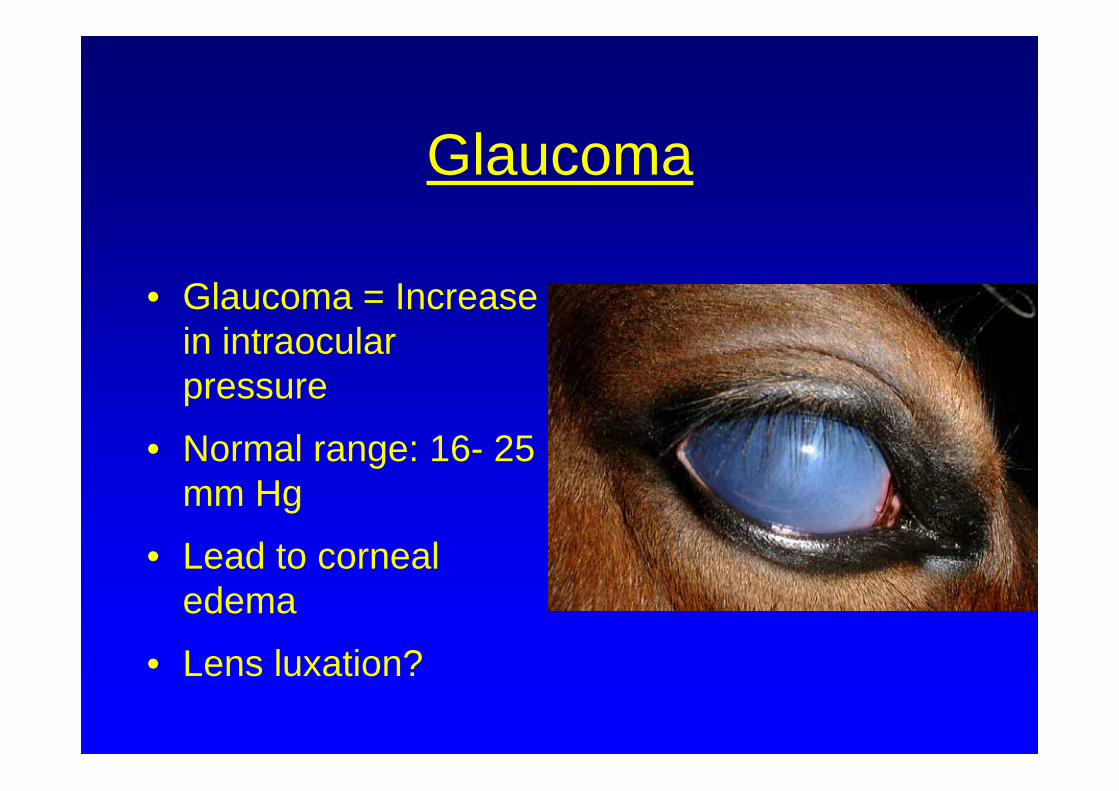
Glaucoma
• Glaucoma = Increase in intraocular pressure
• Normal range: 16- 25 mm Hg
• Lead to corneal edema
• Lens luxation?
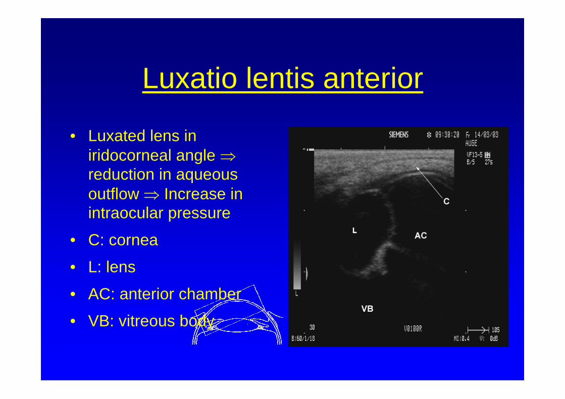
Luxatio lentis anterior
• Luxated lens in iridocorneal angle ⇒reduction in aqueous outflow ⇒ Increase in intraocular pressure
• C: cornea
• L: lens
• AC: anterior chamber
• VB: vitreous body
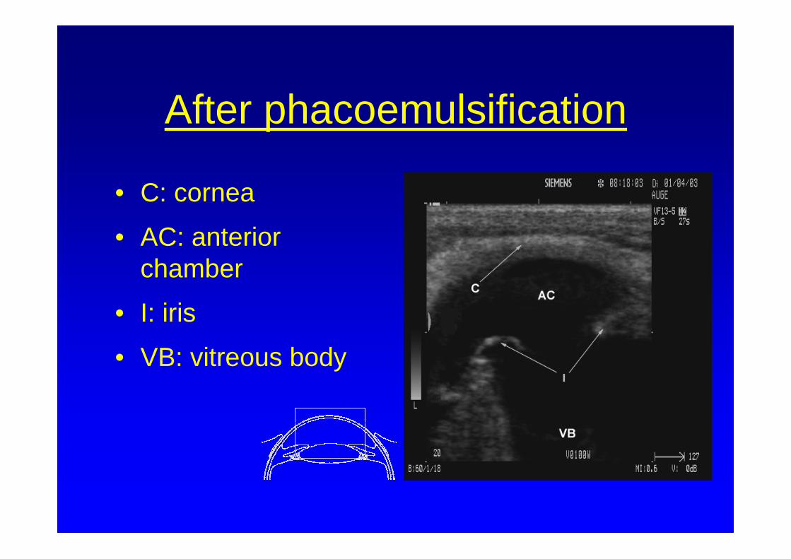
After phacoemulsification
• C: cornea
• AC: anterior chamber
• I: iris
• VB: vitreous body
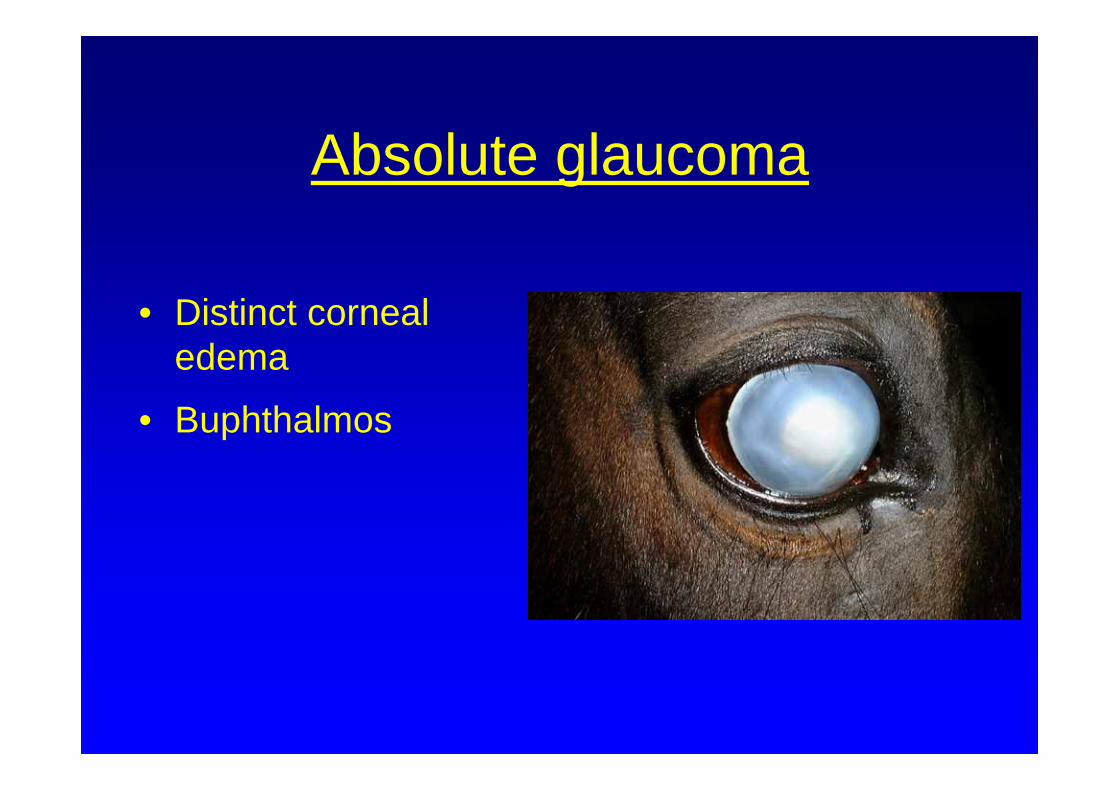
Absolute glaucoma
• Distinct corneal edema
• Buphthalmos
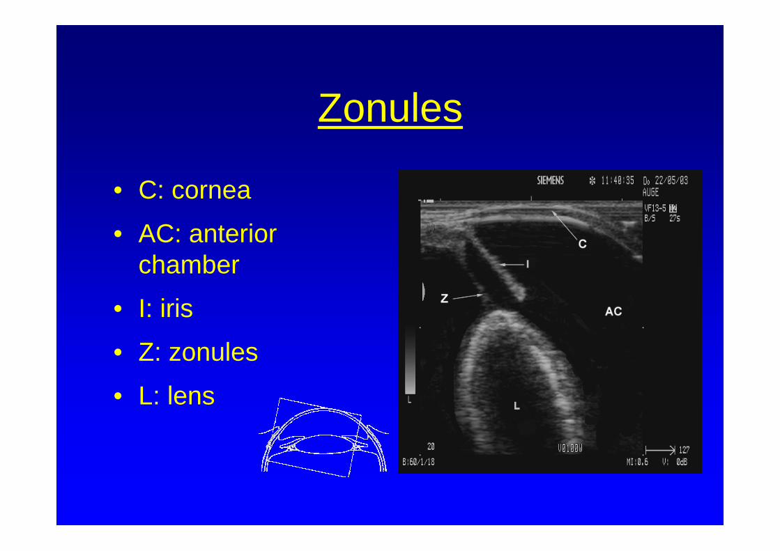
Zonules
• C: cornea
• AC: anterior chamber
• I: iris
• Z: zonules
• L: lens
Conclusion
• Ultrasonic examination is a simple and efficient means of evaluating eyes with opaque optic media
• Improved presentation of individual ocular structures when compared to conventional ultrasound, especially those located within the anterior segment