Embed Size (px)
Citation preview

ORIGINAL PAPER
Evangelos Christodoulou Æ Wojciech R. Rypniewski
Constantinos E. Vorgias
High-resolution X-ray structure of the DNA-binding protein HUfrom the hyper-thermophilic Thermotoga maritimaand the determinants of its thermostability
Received: 3 June 2002 /Accepted: 16 October 2002 / Published online: 12 December 2002� Springer-Verlag 2002
Abstract The histone-like DNA-binding proteins (HU)are a convenient model for studying factors affectingthermostability because of their relatively simple, easilycomparable structures, their common function, and theirpresence in organisms of widely differing thermostabili-ty. We report the determination of the high-resolutionstructure (1.53 A) at 273 K and 100 K of the HU pro-tein from the hyper-thermophilic eubacterium Thermo-toga maritima (HUTmar, Tm=80.5 �C). The structuraldata presented clearly show that the HUTmar has a foldsimilar to its thermophilic homologue HU from Bacillusstearothermophilus (HUBst). Based on primary structureanalysis, as well as on the results of mutational analysisof HUBst (Tm=61.6 �C) and Bacillus subtilis (HUBsu,Tm=39.7 �C), we have designed and produced severalsingle and combined mutations to study their effect onthe thermostability of the recombinant HUTmar.Among others, the triplet mutant HUTmar-G15E/E34D/V42I (Tm=35.9 �C) has converted the extremethermophilic protein HUTmar to mesophilic, like HUBsu. In an attempt to analyze the various mutants ofHUTmar, we crystallized the point mutation HUTmar-E34D, in which Glu34 was replaced by Asp, similar tothe mesophilic HUBsu. The mutant has Tm=72.9 �C, asmeasured by circular dichroism, 7.6 �C lower than the
wild type. The crystal structure of HUTmar-E34D wasdetermined at 100 K and refined at 1.72 A resolution. Acomparison with the wild-type structures clearly showsthat two hydrogen bonds have been disrupted betweenGlu34 from one subunit and Thr13 from the othersubunit, and vice versa. Our analysis points to this as theprime cause of the destabilization compared to the wildtype. The three new structures were compared, togetherwith the X-ray structure of a similar protein, HUBst,with the aim of relating their structural propertiesand different thermal stability. The presented resultsshow that the HUTmar protein achieves its stability byemploying a dual strategy. On the one hand, we observelocal hydrophobic interactions, which stabilize thesecondary structure elements, and on the other hand,electrostatic interactions between side chains.
Keywords Thermotoga maritima Æ Hyper-thermostablehistone-like protein HU Æ X-ray structure Æ Mutants
Introduction
The eubacterial cell nucleoid contains a number ofabundant, small, basic proteins classified as histone-likeDNA-binding proteins. Among these proteins, HU hasbeen identified as the major and ubiquitous proteincomponent of the bacterial nucleoid. In E. coli, HU(HUab) is themost abundantDNA-binding protein, with�30,000 dimers per cell. It is a heterodimer consisting oftwo (70% identity) subunits, a and b, each 90 amino acidslong, encoded by the genes hupA and hupB, respectively.HU appears to be a homodimer in nearly all bacterialspecies where it has been studied, except inE. coli,Serratiamarcescens, and Salmonella typhimurium. A comprehen-sive review on HU has been published (Drlica and Rou-viere-Yaniv 1987; Pettijohn 1988). Nearly 100 HU geneshave been identified and deposited in the databanks.
HU binds with low sequence specificity to both single-stranded and double-stranded DNA as well as RNA(Rouviere-Yaniv andGros 1975).HUbinds preferentially
Extremophiles (2003) 7:111–122DOI 10.1007/s00792-002-0302-7
Communicated by G. Antranikian
PDB references: HUTmar-wt (277 K) 1b8z; HUTmar-wt (100 K)2b8z; HUTmar-E34D (100 K) 3b8z.
E. Christodoulou Æ C.E. Vorgias (&)National and Kapodistrian University of Athens,Faculty of Biology, Department of Biochemistryand Molecular Biology, Panepistimiopolis-Zographou,157 84 Athens, GreeceE-mail: [email protected].: +30-210-7274514Fax: +30-210-7274158
W.R. RypniewskiInstitute of Biochemistry and Molecular Biology,UKE, c/o DESY, Build.22a, Notkestrasse 85,22603 Hamburg, Germany

to cruciform DNA and DNA-specific structures inducedby supercoiling, nicks, and gaps and causes DNAbending and negative supercoiling. Furthermore, HUinteracts with DNA, forming condensed nucleosome-like particles, and can introduce negative supercoilinginto a relaxed circular plasmid DNA in the presence oftopoisomerase I (Rouviere-Yaniv et al. 1979; Broylesand Pettijohn 1986).
In its accessory function, HU is involved in a numberof other protein-DNA interactions such as binding ofthe lac repressor and facilitating the binding of thecAMP receptor protein to the lac promoter (Flashnerand Gralla 1988). HU is a required factor in the trans-position by bacteriophage Mu (Craigie et al. 1985) andplays, in vitro, a regulatory role in k DNA replication(Mensa-Wilmot et al. 1989). HU is an important com-ponent of transposons and forms tight complexes infour-way junction DNA. E. coli HUa, and HUb areregulated by CRP and FIS proteins (Claret and Rou-viere-Yaniv 1996). It has also been reported that HUbinds specifically to DNA that contains single-strandbreaks or gaps (Castaing et al. 1995) and, recently, thatHU binds to DNA, forming multiple complexes, andbends DNA (Wojtuszewski et al. 2001; Grove andLynette 2001).
The crystal structure of HU from B. stearothermo-philus (HUBst) has been solved (Tanaka et al. 1984) andrecently refined at 2 A (White et al. 1999). The solutionstructure of the recombinant HU from B. stearother-mophilus expressed in E. coli (Padas et al. 1992) has alsobeen determined by NMR (Vis et al. 1995; Boelens et al.1996). HUBst protein has been used as a model systemto study protein-DNA interaction(s) of the histone-likeprotein family that includes the integration host factor(IHF) protein (Rice et al. 1996; White et al. 1989).
The structural properties responsible for the ther-mostability of HU proteins from mesophilic and thermo-philic microorganisms attracted attention in the past(Wilson et al. 1990). Meanwhile, the HU proteins fromB. stearothermophilus and B. subtilis have been analyzedwith respect to their sequence characteristics in correla-tion with their thermostability (Christodoulou andVorgias 2002; Kawamura et al. 1996, 1998). We want toexpand our studies on the HU protein to extremethermophilic organisms, such as the eubacteriumT. maritima (growth temperature 80–85 �C), whichshows 61% and 51% identity to HU from the thermo-philic B. stearothermophilus and the mesophilic B. sub-tiliis, respectively. The small size of the HU moleculeand the existence of homologous proteins in variousbacteria, from mesophilic to extreme thermophilic, makeit an attractive model to address questions of thermo-stability using the structure-mutation approach.
Engineering proteins for thermostability is a particu-larly exciting and challenging field, as it is crucial forbroadening the industrial use of recombinant proteins.Many experimental approaches have been applied toidentify determinants of thermostability (Zuber 1988;Serrano et al. 1993; Shih and Kirsch 1995; Spector et al.
2000; Sriprapundh et al. 2000). The structure-mutationapproach was applied predominantly, but it is time-consuming and expensive and requires proteins that arehighly conserved in their primary structure and are pre-sent in organisms that grow at low and high temperatures(Steen et al. 2001). Therefore, only a limited number ofproteins have been studied based on this approach(Salminen et al. 1996; Lehmann and Wyss 2001).
The comparison of homologous proteins with differ-ent thermostabilities offers a unique opportunity to elu-cidate strategies for thermal adaptation. Despite theirwidely different thermostabilities, thermophilic proteinsand their mesophilic counterparts often share the samefunction, high-sequence homology, and similar three-dimensional structure (Kumar et al. 2000). Thermosta-bility in various thermostable proteins seems not to beachieved by a single universal mechanism but by a com-bination of individual strategies, such as an increasednumber of hydrogen bonds and salt bridges, an opti-mized packing of the hydrophobic core, shortened sur-face loops, increased number of proline residues, and anincrease in buried hydrophobic residues (Querol et al.1996; Jaenicke and Bohm 1998; Ladenstein and Antra-nikian 1998; Sterner and Leibl 2001).
The present work is based on the principle of rationaldesign and focuses on studies of structure-thermosta-bility using X-ray structural analysis in combinationwith primary structure analysis and targeted site-directed mutagenesis using as a model system the DNA-binding protein HU from microorganisms living in awide range of temperatures. The purpose of our study isto identify the molecular determinants responsible forthe hyperthermostability of the HUTmar protein.
Materials and methods
Cloning, expression, and purification
The cloning, expression, and purification of the HUTmar has beendescribed previously (Christodoulou and Vorgias 1998). Site-spe-cific mutagenesis, using asymmetric PCR with a single mutagenicprimer and two flanking primers, was performed to produce theHUTmar-E34D mutation as described by Perrin and Gilliland(1990). The synthetic oligonucleotides used were a 28-mer 5’ACATATGAACAAGAAGGAACTCATCGAC 3’ HUTmar(C),a 29-mer 5’ AGGGATCCTCACTTGACCTTCTCTTTGAG 3’HUTmar(N), and a 44-mer 5’ AATCCAACGATCTGAACCTT-TTCACCCTTTGCGAGAGCGTCTGT 3’ HUTmar(C)E/D. Allprocedures used for cloning, expression, and purification of theHUTmar-E34D mutant protein were the same as for the wild type.
Protein sequence alignment
The sequences of several HUs, selected according to growth tem-perature of the parent organisms, were aligned using Clustal X(Thompson et al. 1997).
Amino acid analysis
For ease of presentation, each amino acid was assigned to one ofthree categories: charged (Asp, Glu, Arg, and Lys), uncharged
112

polar (Ser, Thr, Asn, and Gln), and non-polar (Gly, Ala, Val, Leu,Ile, Phe, Trp, Tyr, Pro, Met, Cys, and His) (Haney et al. 1999).
Crystallization of HUTmar wild type and mutant
The crystallization and production of high-quality crystals ofHUTmar wild-type (wt) and mutant E34D were carried out underidentical conditions using the vapor diffusion method as describedpreviously (Christodoulou and Vorgias 1998). HUTmar-wt andHUTmar-E34D formed crystals in 80% saturated ammoniumsulfate, at room temperature, after 3–5 months, and the obtainedcrystals have tetragonal symmetry.
Circular dichroism spectroscopy
Circular dichroism (CD) spectroscopy experiments were conductedusing a JASCO 715 spectropolarimeter with a Peltier-type cellholder (model PTC-348 from Jasco Corporation), which permitsaccurate temperature control. Wavelength scans were performedusing 0.2 mg/ml protein concentration in a 2-mm rectangular cellat a number of discrete temperatures. The proteins were dissolvedin 10 mM MOPS pH 7.0. Each spectrum was obtained by aver-aging four spectra recorded from 250 to 190 nm with 2-nm inter-vals at the rate of 50 nm min–1. A response time for each point was5 s and the bandwidth was 2 nm. Buffer scans were accumulatedand subtracted from the sample scans, and the mean residueellipticity was calculated. CD temperature scans were performed byvarying the temperature from 20 to 95 �C at a rate of 50 �C h–1,and the mean ellipticity was measured at 222 nm with 0.5 �Cintervals, 5 s response time, and 2 nm bandwidth. The proteinconcentration was 0.2 mg/ml. Both wild type and mutants wereexamined reversibly under these experimental conditions. Thefraction of native protein was calculated from the CD values bylinearly extrapolating the pre- and post-transition baselines,respectively, based on the assumption that the unfolding equilib-rium of these proteins follows a two-state mechanism. The tem-perature of the midpoint of the transition, Tm, at which half of theprotein is unfolded, was determined using the sigmoidal fitting ofBoltzmann’s equation.
X-ray data collection and processing
X-ray diffraction data were collected using synchrotron radiationon the EMBL beam lines X11 and BW7B (van Silfhout and Her-mes 1995) at the DORIS storage ring, DESY, Hamburg, on aMAR Research imaging plate scanner. Datasets were collectedfrom single crystals at 277 K and at 100 K for HUTmar-wt and at100 K for the mutant HUTmar-E34D. The oscillation angle wasvaried to minimize the overlapping of reflections. A range ofreciprocal space of 100� was covered in two separate sweeps, atdifferent exposure times, for both HUTmar-wt and HUTmar-E34D, to record the full range of intensities. The programsDENZO and SCALEPACK (Otwinowski and Minor 1997) wereused for data reduction and scaling. Initial scaling showed that nosignificant radiation damage had taken place during data collectionand that the images were scaled without a relative temperaturefactor. Outliers were rejected based on the v2 test implemented inSCALEPACK. The post-refinement option was used to refine thecell parameters. The intensities were converted to structure factoramplitudes, and a correction was applied to weak or negativemeasurements (French and Wilson 1978). Data collection and thefinal statistics are summarized in Table 4.
Structure determination and refinement
The structure of HUTmar-wt at 277 K was determined by molec-ular replacement using the program AMORE (Navaza 1994) fromthe CCP4 program suite (Collaborative Computational ProjectNumber 4 1994). The rotation function was calculated using terms
between 8 and 3 A with a Patterson search radius of 20 A. Usingthe structure of HU from B. stearothermophilus refined at 1.9 A asthe starting model, a solution was obtained (Dauter Z., personalcommunication) and placed in a P1 cell of dimensions 80·80·80 A.A peak in the rotation function was obtained, giving a correlationcoefficient of 0.199, while the other peaks had height less than 55%of this peak. It was not clear at that stage whether the space groupwas P41 or P43, and the translation function was calculated forboth space groups using the orientation corresponding to thehighest peak found in the rotation function. The translation func-tion gave a peak with a correlation coefficient of 0.208 and an Rfactor (=S|Fo|Fc|/SFo) of 0.566 for space group P41, while P43 gavea correlation coefficient of 0.382 and an R factor of 0.49. This wasfurther improved by rigid body refinement, as implemented inAMORE, to give a correlation coefficient of 0.425, with an R factor0.476. The model was refined by the conventional stereochemicallyrestrained maximum-likelihood method (Murshudov et al. 1999) asimplemented in the program REFMAC from the CCP4 programsuite. Data were used between 20 and 1.6 A, without a r cutoff,with 5% of the dataset aside for Rfree (Brunger 1992). Solventmolecules were inserted and refined using the program ARP(Lamzin and Wilson 1993) with real space positional refinementand automatic determination of statistically significant electrondensity level. Manual rebuilding of the model was based on the(2Fo-Fc) and (Fo-Fc) electron density maps, using an SGI graphicsstation and O (Jones et al. 1991).
The model of HUTmar-wt at 100 K also was refined by usingthe refined coordinates of HUTmar-wt at 277 K as a startingmodel, and the refinement was performed as described above.Diffraction data used were between 20 and 1.53 A.
The model of HUTmar-E34D at 100 K was also refined byusing the structure of HUTmar-wt at 277 K as the starting model.Data used were between 20 and 1.72 A.
Results
Comparison of HU proteins from microorganismsliving at various temperatures
HU proteins from four bacteria and the first archaeonthat contains HU protein have been selected for com-parison studies based on their growth temperature. Thecharacteristics of the microorganisms and some avail-able biochemical data concerning the HU proteins aresummarized in Table 1.
Primary and secondary structure comparisonof the HU proteins
As a first step to understanding and explaining themolecular basis of the thermostability of the selectedHU proteins, primary and secondary structure com-parisons were performed among the mesophilic HUBsu,thermophilic HUBst and HUTvo (Kawashima et al.2000), and extreme thermophilic HUTth and HUTmar.The topology of the HUBst protein is described in Whiteet al. (1999). The topology of HUTmar is described laterin this report in the section on X-ray structure deter-mination.
In a previous publication (Christodoulou and Vor-gias 2002), we proposed to divide the topology of theHU molecule into three ‘‘domains’’ based on functionalconsiderations rather than structural. For each monomer
113
of the HU molecule, we can distinguish the helix-turn-helix (HTH) domain, the dimerization signal (DS), andthe DNA-binding domain (DBD), which is comprised ofthe flexible arm and a small a-helix. Figure 1 presentsthe primary structure alignment of the HUs described inTable 1. The secondary structure elements described inFig. 1 are derived from the X-ray structure of HUBsu,HUBst, and HUTmar but cannot be assigned accuratelyfor HUTvo and HUTth.
The HU proteins of the five organisms used in thisstudy (Table 1) and their HTH and DBD parts werecompared and expressed as percent of identity. Theresulting calculations are presented in Table 2. The DSsignal was not included in Table 2, since it is practicallyidentical in all HU proteins.
Mutational analysis
An extensive mutational analysis has been carried inthe HUBsu, HUBst, and HUTmar proteins in order toassess the contribution of certain highly conservedamino acids and shed light on the mechanism of ther-mostabilization of these proteins (Christodoulou andVorgias 2002).
The study points to three amino acids being primarilyresponsible for the thermal stability of these HU pro-teins. They are Gly15, Glu34, and Val42 in HUBsu,HUBst, and HUTmar. However, these results cannot be
extrapolated for HUTth and HUTvo, as there are noavailable data for their structures. Gly15, Glu34, andVal42 in HUTmar were mutated to their mesophiliccounterparts, individually and in combination. The mu-tated HUTmar proteins were overexpressed in E. coliand purified to homogeneity (Christodoulou and Vor-gias 1998), and their melting temperature was deter-mined by CD spectroscopy. Fig. 2a presents the fullCD-spectrum of HUTmar-wt at various temperatures,and Fig. 2b shows the melting curves of the HUTmar-wtand the mutants described in Table 3. The experimen-tally determined Tm of the HUTmar-wt and variousmutants and the localization of the mutated amino acidsonto the three-dimensional structure of HUTmar aresummarized in Table 3.
Crystallization experiments
As the next step, we decided to determine the X-raystructure of the HUTmar-wt and the available mutantsin order to understand the mechanism of HUTmarstabilization at the molecular level.
HUTmar-wt was crystallized and the crystals werediffracted to high resolution. The mutant HUTmar-E34D was also crystallized under the same conditions.Crystallization trials are underway to obtain high-qual-ity crystals of HUTmar-G15E, HUTmar-V42I, andthe triplet mutant HUTmar-G15E/E34D/V42I to gain
Table 1 Comparison of the five HU proteins selected according to their growth temperatures (several other statistical data are alsopresented)
Parameter HUBsu HUBst HUTvo HUTth HUTmarOrganism Bacillus subtilis Bacillus
stearothermophilusThermoplasmavolcanium
Thermusthermophilus
Thermotogamaritima
Growth temperature (�C) 30 55 60 70 80Databank entry O31946 P02346 BAB59303 P19436 P36206Number of amino acids in the monomer 92 90 90 95 90Tm of the protein (�C) 39.7 61.6 60.0 n.d. 80.5Charged residues (%) 32.5 33.4 28.9 31.6 38.9Uncharged residues (%) 17.3 16.7 24.5 14.8 11.1Nonpolar residues (%) 50.2 49.9 46.6 53.6 50.0Homodimer (Da) 19.782 19.420 20.056 20.312 19.972X-ray structure available - 1HUUA, 1HUUB, 1HUUC - - 1B8ZA, 1B8ZB
Table 2 Identity scores amongthe HUBsu, HUBst, HUTth,and HUTmar proteins and theirdomains a
a Helix-turn-helix body (HTH):(residues: 1–45) and DNAbinding domain (DBD): (resi-dues: 51–90). The DS peptide isnot included since it is identicalamong all known HUs
HUBsu HUBst HUTvo HUTth HUTmar
HUBsu 100.0%HUBst 87.7% 100.0%
HTH: 77.8%DBD: 100%
HUTvo 35.9% 32.6% 100.0%HTH: 28.9% HTH: 24.4%DBD: 37.5% DBD: 35.0%
HUTth 51.1% 55.5% 24.4% 100.0%HTH: 35.5% HTH: 44.4% HTH: 17.8%DBD: 65.0% DBD: 90 6% DBD: 25.0%
HUTmar 51.1% 61.1% 33.3% 55.5% 100.0%HTH: 33.0% HTH: 53.3% HTH: 26.7% HTH: 44.4%DBD: 67.5% DBD: 67.5% DBD: 35.0% DBD: 65.0%
114

additional insight into the structural rearrangementsresponsible for their reduced thermostability.
Both HUTmar-wt and HUTmar-E34D proteinsformed bi-pyramidal crystals in the space group P43 witha unit cell containing eight HU polypeptide chainsarranged as four dimers around the 43 axis. Table 4summarizes the unit cell parameter of the measuredcrystals of HUTmar-wt and E34D mutant.
The model of HUTmar-wt at 277 K and 100 Kand HUTmar-E34D at 100 K
The statistics of data collection for HUTmar-wt at277 K and 100 K, as well as for HUTmar-E34D at100 K, are summarized in Table 4. The models ofHUTmar-wt based on data collected at 277 K and100 K consist of 1028 and 978 protein atoms and 67 and148 solvent molecules, respectively. In the case ofHUTmar-E34D mutant, the data were collected at100 K and the model was built using 975 protein atomsand 89 solvent molecules. Table 5 summarizes the sta-tistics of the three models. The quality and geometry ofall three final models, HUTmar-wt at 277 K and 100 Kand E34D mutant at 100 K, were analyzed usingPROCHECK (Laskowski et al. 1993). The three modelsdo not deviate significantly and are within the acceptedlimits of various geometrical criteria as summarized inTable 6.
The Ramachandran plot for the HUTmar-wt(Ramachandran and Sasisekharan 1968) is well clusteredwithin the accepted regions and has 98.5% of residues inthe most favored region and 0.5% in the additional al-lowed region, as defined in the program PROCHECK.The only outlying residue is the conserved Phe47, whichis involved in aromatic inter-subunit stacking interac-tions (White et al. 1999). The stereochemical restrainsfor the three models and the final standard deviationsare listed in Table 6.
The structure of HUTmar and the molecular contactsin the homodimer
As mentioned above, the monomer of the HU moleculeconsists of three parts, the HTH domain, the DS, and
the flexible arm (DBD). In the homodimer, the twoHTH domains, the two dimerization signals, and a smallthree-stranded b-sheet comprise the main body of themolecule. Two short a3-helices at the C-end of themolecule are also associated with the main body (Fig. 3).The two flexible arms of the molecule are extended,forming a U-shaped path that is responsible for thebinding of the protein to DNA.
The HTH domain is comprised of two helices: a1(residues 3–14) and a2 (residues 18–38) connected via athree-residue short loop (residues 16–17). The primarystructure of the HTH domain is not as highly conservedas the DNA-binding domain of the molecule, among all
Fig. 2 a UV-CD spectra of HUTmar at various temperatures asindicated on the figure. b Melting curves of HUTmar-wt andmutants indicated on the figure. The experimental conditions aredescribed in Materials and methods
Fig. 1 Alignment of the amino acid sequences of HUBst, HUBgl,HUTvo, HUTth, and HUTmar. The positions of the secondarystructure elements derived from the three-dimensional structure ofHUBst are shown. a1, a2, and a3 are a-helices; b1, b2, and b3 areb-sheets; and DS is dimerization signal
115
known HUs, as shown in Table 2. In the dimer ofHUTmar, the secondary structure elements are highlyintertwined between the subunits. The intersubunitinteractions are summarized in Table 7. The intersub-unit interactions in HUTmar-E34D and HUBst are alsodescribed for comparison.
The dimerization signal is a small part of the mole-cule (residues 46–50), located partly in the loop betweenstrands b1 and b2 and partly on the b2-strand, and ishighly conserved among the known HU proteins. Phe29, 47, 50, and 79 from each monomer are involved in theformation of an aromatic hydrophobic core involving
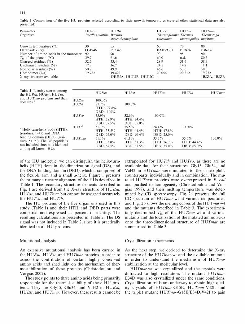
Table 3 Summary of the wt and mutated HUTmar proteins and their effects on the Tm as determined by CD and their localization on thestructure
From HUTmar to HUBsu Tm (�C) DTm (�C) Secondary structure occurrence of the mutant(s)
HUTmar –wt 80.5G 15 E 55.8 –24.7 Turn between a1-helix and a1-helixE 34 D 72.7 –7.8 a2-helixV 42 I 70.9 –9.6 b1-strandG 15 E / E 34 D 52.1 –28.4 Turn between a1-helix and a1-helix, b2-strandE 34 D / V 42 I 63.4 –17.1 Turn between a1-helix and a1-helix, a2-helixG 15 E / E 34 D / V42 I 35.9 –44.6 Turn between a1-helix and a1-helix, a2-helix, b2-strand
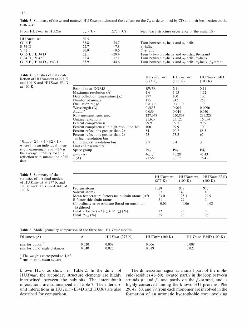
Table 4 Statistics of data col-lection of HUTmar-wt at 277 Kand 100 K and HUTmar-E34Dat 100 K
aRmerge=S|Ii|<I>|/S<I>,where Ii is an individual inten-sity measurement and <I>isthe average intensity for thisreflection with summation of alldata
HUTmar -wt HUTmar-wt HUTmar-E34D(277 K) (100 K) (100 K)
Beam line at DORIS BW7B X11 X11Maximum resolution (A) 1.6 1.53 1.72Data collection temperature (K) 277 100 100Number of images 175 242 258Oscillation range 0.8–1.6 0.7–1.0 1.0Wavelength (A) 0.8833 0.905 0.9096Rmerge
a 0.056 0.044 0.050Raw measurements used 127,048 220,803 239,228Unique reflections 21,639 23,127 16,554Percent completeness 99.9 99.7 99.9Percent completeness in high-resolution bin 100 99.9 100Percent reflections greater than 2r 84 90.7 88.5Percent reflections greater than 2rin high-resolution bin
55 73.3 65
I/r in highest resolution bin 2.7 3.4 3Unit cell parametersSpace group P43 P43 P43a=b (A) 46.12 45.28 45.43c (A) 77.56 76.17 76.45
Table 5 Summary of thestatistics of the final modelsof HUTmar-wt at 277 K and100 K and HUTmar-E34D at100 K
HUTmar-wt HUTmar-wt HUTmar-E34D(277 K) (100 K) (100 K)
Protein atoms 1028 978 975Solvent atoms 67 148 89Mean temperature factors main-chain atoms (A2) 23.0 25.3 29.9B factor side-chain atoms 31 29 34Co-ordinate error estimate Based on maximumlikelihood
0.06 0.06 0.08
Final R factor (=S|Fo|Fc|/SFo) (%) 22 23 23Final Rfree (%) 25 26 26
Table 6 Model geometry comparison of the three final HUTmar models
Distances (A) ra HUTmar (277 K) HUTmar (100 K) HUTmar -E34D (100 K)
rms for bonds b 0.020 0.008 0.006 0.008rms for bond angle distances 0.040 0.025 0.019 0.021
a The weights correspond to 1/r2b rms = root mean square
116
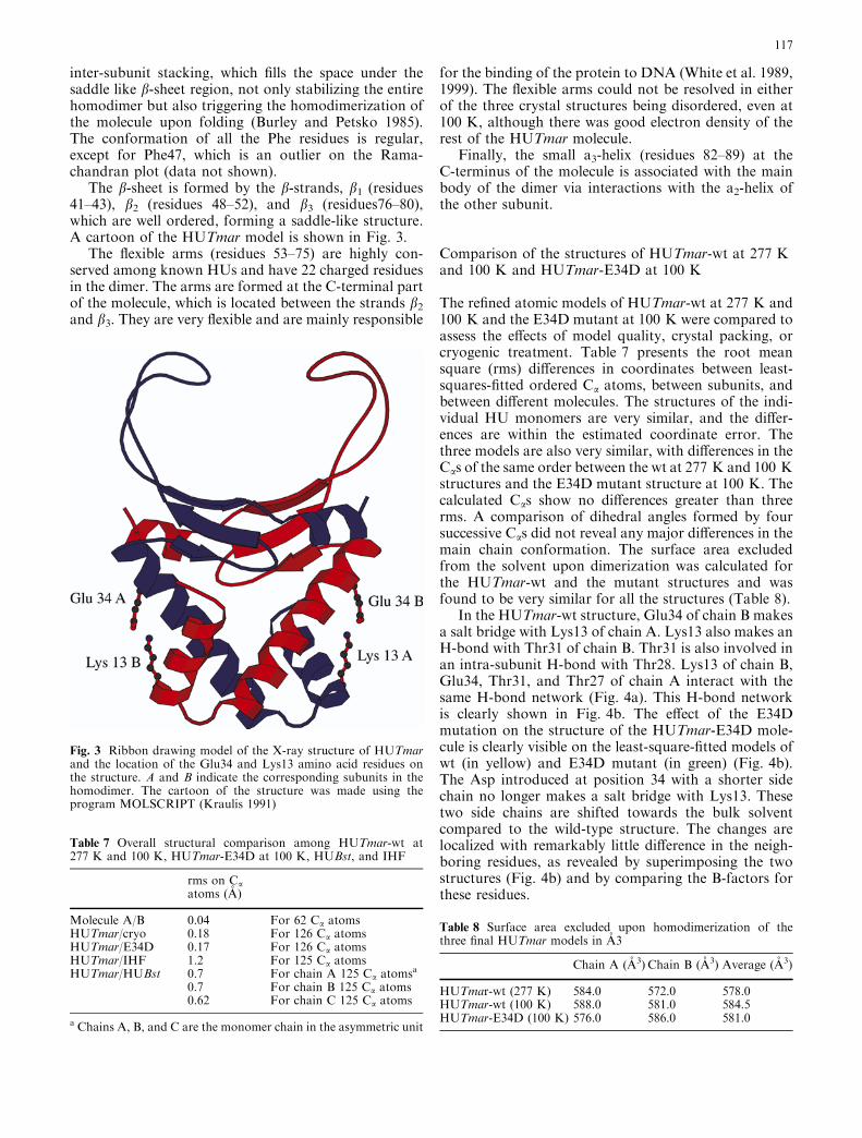
inter-subunit stacking, which fills the space under thesaddle like b-sheet region, not only stabilizing the entirehomodimer but also triggering the homodimerization ofthe molecule upon folding (Burley and Petsko 1985).The conformation of all the Phe residues is regular,except for Phe47, which is an outlier on the Rama-chandran plot (data not shown).
The b-sheet is formed by the b-strands, b1 (residues41–43), b2 (residues 48–52), and b3 (residues76–80),which are well ordered, forming a saddle-like structure.A cartoon of the HUTmar model is shown in Fig. 3.
The flexible arms (residues 53–75) are highly con-served among known HUs and have 22 charged residuesin the dimer. The arms are formed at the C-terminal partof the molecule, which is located between the strands b2
and b3. They are very flexible and are mainly responsible
for the binding of the protein to DNA (White et al. 1989,1999). The flexible arms could not be resolved in eitherof the three crystal structures being disordered, even at100 K, although there was good electron density of therest of the HUTmar molecule.
Finally, the small a3-helix (residues 82–89) at theC-terminus of the molecule is associated with the mainbody of the dimer via interactions with the a2-helix ofthe other subunit.
Comparison of the structures of HUTmar-wt at 277 Kand 100 K and HUTmar-E34D at 100 K
The refined atomic models of HUTmar-wt at 277 K and100 K and the E34D mutant at 100 K were compared toassess the effects of model quality, crystal packing, orcryogenic treatment. Table 7 presents the root meansquare (rms) differences in coordinates between least-squares-fitted ordered Ca atoms, between subunits, andbetween different molecules. The structures of the indi-vidual HU monomers are very similar, and the differ-ences are within the estimated coordinate error. Thethree models are also very similar, with differences in theCas of the same order between the wt at 277 K and 100 Kstructures and the E34D mutant structure at 100 K. Thecalculated Cas show no differences greater than threerms. A comparison of dihedral angles formed by foursuccessive Cas did not reveal any major differences in themain chain conformation. The surface area excludedfrom the solvent upon dimerization was calculated forthe HUTmar-wt and the mutant structures and wasfound to be very similar for all the structures (Table 8).
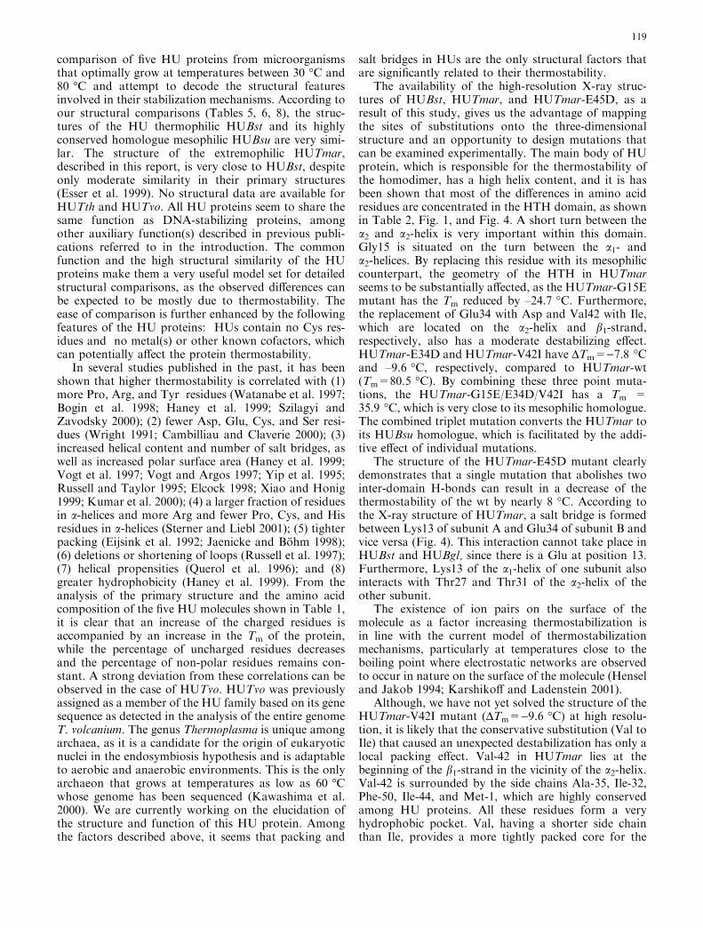
In the HUTmar-wt structure, Glu34 of chain B makesa salt bridge with Lys13 of chain A. Lys13 also makes anH-bond with Thr31 of chain B. Thr31 is also involved inan intra-subunit H-bond with Thr28. Lys13 of chain B,Glu34, Thr31, and Thr27 of chain A interact with thesame H-bond network (Fig. 4a). This H-bond networkis clearly shown in Fig. 4b. The effect of the E34Dmutation on the structure of the HUTmar-E34D mole-cule is clearly visible on the least-square-fitted models ofwt (in yellow) and E34D mutant (in green) (Fig. 4b).The Asp introduced at position 34 with a shorter sidechain no longer makes a salt bridge with Lys13. Thesetwo side chains are shifted towards the bulk solventcompared to the wild-type structure. The changes arelocalized with remarkably little difference in the neigh-boring residues, as revealed by superimposing the twostructures (Fig. 4b) and by comparing the B-factors forthese residues.
Fig. 3 Ribbon drawing model of the X-ray structure of HUTmarand the location of the Glu34 and Lys13 amino acid residues onthe structure. A and B indicate the corresponding subunits in thehomodimer. The cartoon of the structure was made using theprogram MOLSCRIPT (Kraulis 1991)
Table 8 Surface area excluded upon homodimerization of thethree final HUTmar models in A3
Chain A (A3) Chain B (A3) Average (A3)
HUTmar-wt (277 K) 584.0 572.0 578.0HUTmar-wt (100 K) 588.0 581.0 584.5HUTmar-E34D (100 K) 576.0 586.0 581.0
Table 7 Overall structural comparison among HUTmar-wt at277 K and 100 K, HUTmar-E34D at 100 K, HUBst, and IHF
rms on Caatoms (A)
Molecule A/B 0.04 For 62 Ca atomsHUTmar/cryo 0.18 For 126 Ca atomsHUTmar/E34D 0.17 For 126 Ca atomsHUTmar/IHF 1.2 For 125 Ca atomsHUTmar/HUBst 0.7 For chain A 125 Ca atomsa
0.7 For chain B 125 Ca atoms0.62 For chain C 125 Ca atoms
a Chains A, B, and C are the monomer chain in the asymmetric unit
117

Comparison of the HUTmar and HUBstand IHF structures
In addition to the currently presented crystal structure ofHUTmar and its mutant, there are two more solvedstructures from closely related proteins, i.e., HU proteinfrom B. stearothermophilus (HUBst, PDB code 1HUU)and the IHF from E. coli (1IHF). At the level of primarystructure, the main body of the HUBst proteins can beclosely aligned with the HU protein from T. maritima,sequence identity 58%, while the sequence identity toIHF is only 36%. The three structures give a reasonablefit despite the moderate sequence identity. Table 7summarizes the obtained data from the structural com-parison among the three proteins.
Discussion
A comprehensive comparison between homologousproteins that display different thermostability is a well-accepted approach to the elucidation of the mechanismof protein thermostability (Kimura et al. 1992; Akasako
et al. 1995; Lehman and Wyss 2001). The structure-mutation approach, despite its limitations, namely, costand time requirements, remains the predominant meth-od to decipher the mechanism of a protein of interest. Asstated in the introduction, our model proteins fulfill therequirements for such an approach. In our previousstudy, we systematically compared the HU proteinsfrom the thermophilic bacterium B. stearothermophilusand the mesophile B. subtilis, which differ in 11 out of 90residues and display a considerable difference in thermalstability. The DTm between the melting temperatures ofHUBst and HUBsu was determined to be 21.9 �C. Astep-by-step, site-directed mutagenesis of all 11 residuesin HUBst revealed that the difference in thermostabilitycan be fully accounted for by only three amino acids:Gly15, Glu34, and Val42, which are located in keypositions of the molecule (Christodoulou and Vorgias2002). These three residues are conserved in thermo-philic HUs, and their mesophilic counterparts are Glu,Asp, and Ile, respectively. By mutating them to theirmesophilic counterparts from B. subtilis, we can convertthe thermophilic HUBst (Tm=61.9 �C) to its mesophilichomologue HUBsu (Tm=39.7 �C). The same aminoacid residues in the mesophilic HUBsu can be replacedby their thermophilic counterparts, resulting in a therm-ophilic HUBst-like protein (Christodoulou and Vorgias2002).
We have extended our studies to the extremophilicHU from T. maritima, and in this report we give a
Fig. 4 a Electron-density map of HUTmar-E34D mutant. Thesubstitution of the Glu34 with Asp is clearly visible in the electrondensity. b Least-square fit of residues 12–14 of chain A and 27–34of chain B from the HUTmar-wt structure presented in yellow withresidues 12–14 of chain A and 27–34 of chain B from the HUTmar-E34D presented in green
118
comparison of five HU proteins from microorganismsthat optimally grow at temperatures between 30 �C and80 �C and attempt to decode the structural featuresinvolved in their stabilization mechanisms. According toour structural comparisons (Tables 5, 6, 8), the struc-tures of the HU thermophilic HUBst and its highlyconserved homologue mesophilic HUBsu are very simi-lar. The structure of the extremophilic HUTmar,described in this report, is very close to HUBst, despiteonly moderate similarity in their primary structures(Esser et al. 1999). No structural data are available forHUTth and HUTvo. All HU proteins seem to share thesame function as DNA-stabilizing proteins, amongother auxiliary function(s) described in previous publi-cations referred to in the introduction. The commonfunction and the high structural similarity of the HUproteins make them a very useful model set for detailedstructural comparisons, as the observed differences canbe expected to be mostly due to thermostability. Theease of comparison is further enhanced by the followingfeatures of the HU proteins: HUs contain no Cys res-idues and no metal(s) or other known cofactors, whichcan potentially affect the protein thermostability.
In several studies published in the past, it has beenshown that higher thermostability is correlated with (1)more Pro, Arg, and Tyr residues (Watanabe et al. 1997;Bogin et al. 1998; Haney et al. 1999; Szilagyi andZavodsky 2000); (2) fewer Asp, Glu, Cys, and Ser resi-dues (Wright 1991; Cambilliau and Claverie 2000); (3)increased helical content and number of salt bridges, aswell as increased polar surface area (Haney et al. 1999;Vogt et al. 1997; Vogt and Argos 1997; Yip et al. 1995;Russell and Taylor 1995; Elcock 1998; Xiao and Honig1999; Kumar et al. 2000); (4) a larger fraction of residuesin a-helices and more Arg and fewer Pro, Cys, and Hisresidues in a-helices (Sterner and Liebl 2001); (5) tighterpacking (Eijsink et al. 1992; Jaenicke and Bohm 1998);(6) deletions or shortening of loops (Russell et al. 1997);(7) helical propensities (Querol et al. 1996); and (8)greater hydrophobicity (Haney et al. 1999). From theanalysis of the primary structure and the amino acidcomposition of the five HU molecules shown in Table 1,it is clear that an increase of the charged residues isaccompanied by an increase in the Tm of the protein,while the percentage of uncharged residues decreasesand the percentage of non-polar residues remains con-stant. A strong deviation from these correlations can beobserved in the case of HUTvo. HUTvo was previouslyassigned as a member of the HU family based on its genesequence as detected in the analysis of the entire genomeT. volcanium. The genus Thermoplasma is unique amongarchaea, as it is a candidate for the origin of eukaryoticnuclei in the endosymbiosis hypothesis and is adaptableto aerobic and anaerobic environments. This is the onlyarchaeon that grows at temperatures as low as 60 �Cwhose genome has been sequenced (Kawashima et al.2000). We are currently working on the elucidation ofthe structure and function of this HU protein. Amongthe factors described above, it seems that packing and
salt bridges in HUs are the only structural factors thatare significantly related to their thermostability.
The availability of the high-resolution X-ray struc-tures of HUBst, HUTmar, and HUTmar-E45D, as aresult of this study, gives us the advantage of mappingthe sites of substitutions onto the three-dimensionalstructure and an opportunity to design mutations thatcan be examined experimentally. The main body of HUprotein, which is responsible for the thermostability ofthe homodimer, has a high helix content, and it is hasbeen shown that most of the differences in amino acidresidues are concentrated in the HTH domain, as shownin Table 2, Fig. 1, and Fig. 4. A short turn between thea2 and a2-helix is very important within this domain.Gly15 is situated on the turn between the a1- anda2-helices. By replacing this residue with its mesophiliccounterpart, the geometry of the HTH in HUTmarseems to be substantially affected, as the HUTmar-G15Emutant has the Tm reduced by –24.7 �C. Furthermore,the replacement of Glu34 with Asp and Val42 with Ile,which are located on the a2-helix and b1-strand,respectively, also has a moderate destabilizing effect.HUTmar-E34D and HUTmar-V42I have DTm=)7.8 �Cand –9.6 �C, respectively, compared to HUTmar-wt(Tm=80.5 �C). By combining these three point muta-tions, the HUTmar-G15E/E34D/V42I has a Tm =35.9 �C, which is very close to its mesophilic homologue.The combined triplet mutation converts the HUTmar toits HUBsu homologue, which is facilitated by the addi-tive effect of individual mutations.
The structure of the HUTmar-E45D mutant clearlydemonstrates that a single mutation that abolishes twointer-domain H-bonds can result in a decrease of thethermostability of the wt by nearly 8 �C. According tothe X-ray structure of HUTmar, a salt bridge is formedbetween Lys13 of subunit A and Glu34 of subunit B andvice versa (Fig. 4). This interaction cannot take place inHUBst and HUBgl, since there is a Glu at position 13.Furthermore, Lys13 of the a1-helix of one subunit alsointeracts with Thr27 and Thr31 of the a2-helix of theother subunit.
The existence of ion pairs on the surface of themolecule as a factor increasing thermostabilization isin line with the current model of thermostabilizationmechanisms, particularly at temperatures close to theboiling point where electrostatic networks are observedto occur in nature on the surface of the molecule (Henseland Jakob 1994; Karshikoff and Ladenstein 2001).
Although, we have not yet solved the structure of theHUTmar-V42I mutant (DTm=)9.6 �C) at high resolu-tion, it is likely that the conservative substitution (Val toIle) that caused an unexpected destabilization has only alocal packing effect. Val-42 in HUTmar lies at thebeginning of the b1-strand in the vicinity of the a2-helix.Val-42 is surrounded by the side chains Ala-35, Ile-32,Phe-50, Ile-44, and Met-1, which are highly conservedamong HU proteins. All these residues form a veryhydrophobic pocket. Val, having a shorter side chainthan Ile, provides a more tightly packed core for the
119

molecule. Introduction of an Ile at this position pushesresidue Ile-32, and repulsion between Leu-42 (b1-strand)and Ile-32 (a2-helix) might occur with a negative effecton the thermostability (DTm=-9.6 �C). This is an inte-rior apolar-to-apolar substitution that alters the packingwithout an accompanying hydrophobicity change andsubstantially destabilizes the protein (Sandberg andTerwillinger 1989).
Comparison of the high-resolution three-dimensionalstructures of HUBst, HUTmar, and HUTmar-E34Dshows that the overall differences between the threestructures are within the expected coordinate error.Therefore, we looked in detail at the side chain inter-actions. The differences in the number of salt bridgesbetween the thermophilic and mesophilic homologuesappear to correlate with the Tm, while other factors suchas compactness and hydrophobicity do not correlateconsistently (Karshikoff and Ladenstein 1998).
The information accumulated to date indicates thatnature does not rely on a single strategy for thermalstabilization. As a result, many publications in this areaarrive at different and sometimes inconsistent conclu-sions (Wintrode and Arnold 2001). The availability ofmore complete genome sequences may eventually resultin more reliable and accurate estimations of the factorsinvolved, but sequence statistics alone are still unlikelyto allow accurate predictions of thermostabilizing mu-tations (Van den Burg et al. 1998; Haney et al. 1999;Kumar et al. 2000; Lehmann and Wyss 2001).
The somehow puzzling results coming from variousanalyses lead to the question. Are there general rules toadaptation at high temperature? The answer might bepositive, but it is possible that no unambiguous rule canbe established and only general principles can be stated.In general, several distinct strategies can be distin-guished by which proteins achieve thermostability, butthe choice of the strategy employed varies from proteinto protein. Our model system concerns small proteinsthat exhibit high homology and a pronounced differencein thermostability and shows that less than 30% of theamino acid differences in the primary structure are in-volved in thermostabilization. This is less than 5% of thetotal residues. This observation highlights the problemof identifying the relevant thermostabilizing mutations,particularly in bigger and more complex proteins.
References
Akasako A, Haruki M, Oobatake M, Kanaya S (1995) High re-sistance of Escherichia coli ribonuclease HI variant with quin-tuple thermostabilizing mutations to thermal denaturation, aciddenaturation, and proteolytic degradation. Biochemistry34:8115–8122
Boelens R, Vis H, Vorgias CE, Wilson KS, Kaptein R (1996)Structure and dynamics of the DNA binding protein HU fromBacillus stearothermophilus by NMR spectroscopy. Biopoly-mers 40:553–559
Bogin O, Peretz M, Hacham Y, Korkhin Y, Frolow F, Kalb(Gilboa) AJ, Burstein Y (1998) Enhanced thermal stability ofClostridium beijerinckii alcohol dehydrogenase after strategic
substitution of amino acid residues with prolines from the ho-mologous thermophilic Thermoanaerobacter brockii alcoholdehydrogenase. Protein Sci 7:1156–1163
Broyles SS, Pettijohn DE (1986) Interaction of the Escherichia coliHU protein with DNA. Evidence for formation of nucleosome-like structures with altered DNA helical pitch. J Mol Biol187:47–60
Brunger AT (1992) Free R value: a novel statistical quantity forassessing the accurancy of crystal structures. Nature 355:472–475
Burley SK, Petsko GA (1985) Aromatic-aromatic interaction: amechanism of protein structure stabilization. Science 229:23–28
Cambilliau C, Claverie JM (2000) Structural and genomic corre-lates of hyperthermostability. J Biol Chem 275:32383–32386
Castaing BC, Zelwer C, Laval J, Boiteux S (1995) HU protein ofEscherichia coli binds specifically to DNA that contains single-strand breaks or gaps. J Biol Chem 270:10291–10296
Christodoulou E, Vorgias CE (1998) Cloning, overproduction,purification and crystallization of the DNA binding protein HUfrom the hyperthermophilic eubacterium Thermotoga maritima.Acta Crystallogr D Biol Crystallogr 54:1043–1045
Christodoulou E, Vorgias CE (2002) The thermostability ofDNA-binding protein HU from mesophilic, thermophilic, andextreme thermophilic bacteria. Extremophiles 6:21–31
Claret L, Rouviere-Yaniv J (1996) Regulation of HUa and HUb byCRP and FIS in Escherichia coli. J Mol Biol 263:126–139
Collaborative Computational Project Number 4 (1994) The CCP4suite: Programs for protein crystallography. Acta Crystallogr DBiol Crystallogr 50:760–763
Craigie R, Arndt-Jovin, DJ, Mizuuchi D (1985) A defined systemfor the DNA strand-transfer reaction at the initiation ofbacteriophage Mu transposition: protein and DNA substraterequirements. Proc Natl Acad Sci USA 82:7570–7574
Drlica K, Rouviere-Yaniv J (1987) Histonelike proteins of bacteria.Microbiol Rev 51:301–319
Eijsink VG, Dijkstra BW, Vriend G, van der Zee J, Veltman OR,van der Vinne B, van den Burg B, Kempe S, Venema G (1992)The effect of cavity-filling mutations on the thermostability ofBacillus stearothermophilus neutral protease. Protein Eng5:421–426
Elcock AH (1998) The stability of salt bridges at high temperatures:implications for hyperthermophilic proteins. J Mol Biol284:489–502
Esser D, Rudolph R, Jaeicke R, Bohm G (1999) The HU proteinfromThermotogamaritima: recombinant expression, purificationand physicochemical characterization of an extremely hyper-thermophilic DNA-binding protein. J Mol Biol 291:1135–1146
Flashner Y, Gralla JD (1988) DNA dynamic flexibility and proteinrecognition: differential stimulation by bacterial histone-likeprotein HU. Cell 54:713–721
French S, Wilson KS (1978) On treatment of negative intensityobservations. Acta Crystallogr A 34:517–525
Grove A, Lynette L (2001) High-affinity DNA binding of HUprotein from the hyperthermophile Thermotoga maritima. J MolBiol 311:491–502
Haney PJ, Badger JH, Buldak GL, Reich CI, Woese CR, Olsen GJ(1999) Thermal adaptation analyzed by comparison of proteinsequences from mesophilic and extremely thermophilic Met-hanococcus species. Proc Natl Acad Sci USA 96:3578–3583
Hensel R, Jakob I (1994) Stability of glycer-aldehyde-3-phosphatedehydrogenase from hyperthermophilic archaea at high tem-perature. Syst Appl Microbiol 16:742–745
Jaenicke R, Bohm G (1998) The stablility of proteins in extremeenvironments. Curr Opin Struct Biol 8:738–748
Jones TA, Zou JY, Kjeldgaard M (1991) Improved methods forbinding protein models in electron density maps and the loca-tion of errors in these models. Acta Crystallogr A 47:110–119
Karshikoff A, Ladenstein R (1998) Proteins from thermophilic andmesophilic organisms essentially do not differ in packing. Pro-tein Eng 11:867–872
Karshikoff A, Ladenstein R (2001) Ion pairs and the thermotol-erance of proteins from hyperthermophiles: a ‘‘traffic rule’’ forhot roads. Trends Biochem Sci 26:550–556
120

Kawamura S, Kakuta Y, Tanaka I, Hikichi K, Kuhara S,Yamasaki N, Kimura M (1996) Glycine-15 in the bend betweentwo a-helices can explain the thermostability of DNA bindingprotein HU from Bacillus stearothermophilus. Biochemistry35:1195–1200
Kawamura S, Abe Y, Ueda T, Masumoto K, Imoto T, YamasakiN, Kimura M (1998) Investigation of the Structural Basis forThermostability of DNA-binding Protein HU from Bacillusstearothermophilus. J Biol Chem 273:19982–19987
Kawashima T, Amano N, Koike H, Makino S, Higuchi S, Ka-washima-Ohya Y, Watanabe K, Yamazaki M, Kanehori K,Kawamoto T, Nunoshiba T, Yamamoto Y, Aramaki H,Makino K, Suzuki M (2000) Archaeal adaptation to highertemperatures revealed by genomic sequence of Thermoplasmavolcanium. Proc Natl Acad Sci USA 97:14257–14262
Kimura S, Nakamura H, Hashimoto T, Oobatake M, Kanaya S(1992) Stabilization of escherichia coli ribonuclease hi by stra-tegic replacement of amino acid residues with those from thethermophilic counterpart. J Biol Chem 267:21535–21542
Kraulis PJ (1991) MOLSCRIPT: a program to produce both de-tailed and schematic plots of protein structures. J Appl Crys-tallogr 24:946–960
Kumar S, Tsai CJ, Nussinov R (2000) Factors enhancing proteinthermostability. Protein Eng 13:179–191
Ladenstein R, Antranikian G (1998) Proteins from hyperthermo-philes: stability and enzymatic catalysis close to the boilingpoint of water. Adv Biochem Eng Biotechnol 61:37–85
Lamzin VS, Wilson KS (1993) Automated refinement of proteinmodels. Acta Crystallogr D Biol Crystallogr 49:129–147
Laskowski RA, MacArthur MW, Moss DS, Thornton JM (1993)PROCHECK: a program to check the stereochemical quality ofprotein structures. J Appl Crystallogr 26:283–291
Lehmann M, Wyss M (2001) Engineering proteins for thermosta-bility: the use of sequence alignments versus rational design anddirected evolution. Curr Opin Biotech 12:371–375
Mensa-Wilmot K, Carroll K, McMacken R (1989) Transcriptionalactivation of bacteriophage lambda DNA replication in vitro:regulatory role of histone-like protein HU of Escherichia coli.EMBO J 8:2393–2402
Murshudov GN, Vagin AA, Lebedev A, Wilson KS, Dodson EJ(1999) Efficient anisotropic refinement of macromolecularstructures using FFT. Acta Crystallogr D Biol Crystallogr55:(1):247–255
Navaza J (1994) AMoRe: an automated package for molecularreplacement. Acta Crystallogr A 50:157–163
Otwinowski Z, Minor W (1997) Processing of X-ray diffractiondata collected in oscillation mode. Academic Press, London
Padas PM, Wilson KS, Vorgias CE (1992) The DNA-bindingprotein HU from mesophilic and thermophilic bacilli: genecloning, overproduction and purification. Gene 117:39–44
Perrin S, Gilliland G (1990) Site-specific mutagenesis using asym-metric polymerase chain reaction and a single mutant primer.Nucleic Acids Res 18:7433–7438
Pettijohn DE (1988) Histone-like proteins and bacterial chromo-some structure. J Biol Chem 263:12793–12796
Querol E, Perez-Pons JA, Mozo-Villarias A (1996) Analysis ofprotein conformational characteristics related to thermostabil-ity. Protein Eng 9:265–271
Ramachandran GN, Sasisekharan V (1968) Conformation ofpolypeptides and proteins. Adv Protein Chem 23:283–437
Rice PA, Yang SW, Mizuuchi K, Nash H (1996) Crystal structureof an IHF-DNA complex: a protein-induced DNA U-turn. Cell87:1295–1306
Rouviere-Yaniv J, Gros F (1975) Characterization of a novel, low-molecular-weight DNA-binding protein from Escherichia coli.Proc Natl Acad Sci USA 72:3428–3432
Rouviere-Yaniv J, Yaniv M, Germond J (1979) E. coli DNAbinding protein HU forms nucleosomelike structures withdouble-stranded DNA. Cell 17:265–274
Russell RJ, Taylor GL (1995) Engineering thermostability: lessonsfrom thermophilic proteins. Curr Opin Biotech 6:370–374
Russell RJ, Ferguson JM, Hough DW, Danson MJ, Taylor GL(1997) The crystal structure of citrate synthase from the hy-perthermophilic archaeon Pyrococcus furiosus at 1.9 A resolu-tion. Biochemistry 36:9983–9994
Salminen T, Teplyakov A, Kankare J, Cooperman BS, Lahti R,Goldman A (1996) An unusual route to thermostabilitydisclosed by the comparison of Thermus thermophilus andEscherichia coli inorganic pyrophosphatases. Protein Sci 5:1014–1025
Sandberg WS, Terwilliger TC (1989) Influence of interior packingand hydrophobicity on the stability of a protein. Science245:54–57
Serrano L, Day AG, Fersht AR (1993) Step-wise mutation ofbarnase to binase. A procedure for engineering increased sta-bility of proteins and an experimental analysis of the evolutionof protein stability. J Mol Biol 233:305–312
Shih P, Kirsch JF (1995) Design and structural analysis of anengineered thermostable chicken lysozyme. Protein Sci 4:1063–2072
Spector S, Wang A, Carp SA, Robblee J, Hendsch ZS, Fairman R,Tidor B, Raleigh DP (2000) Rational modification of proteinstability by the mutation of charged surface residues. Bio-chemistry 39:872–879
Sriprapundh D, Vieille C, Zeikus JG (2000) Molecular determi-nants of xylose isomerase thermal stability and activity: analysisof thermozymes by site-directed mutagenesis. Protein Eng13:259–265
Steen IH, Madern D, Karlstrom M, Lien T, Ladenstein R, Birke-land NK (2001) Comparison of isocitrate dehydrogenase fromthree hyperthermophiles reveals differences in thermostability,cofactor specificity, oligomeric state, and phylogenetic affilia-tion. J Biol Chem 276:43924–43931
Sterner R, Liebl W (2001) Thermophilic adaptation of proteins.Crit Rev Biochem Mol Biol 36:39–106
Szilagyi A, Zavodszky P (2000) Structural differences betweenmesophilic, moderately thermophilic and extremely thermo-philic protein subunits: results of a comprehensive survey.Struct Fold Des 8:493–504
Tanaka I, Appelt K, Dijk J, White SW, Wilson KS (1984) 3-Aresolution structure of a protein with histone-like properties inprokaryotes. Nature 310:376–381
Thompson JD, Gibson TJ, Plewniak F, Jeanmougin F, HigginsDG (1997) The Clustal_X windows interface flexible strategiesfor multiple sequence alignment aided by quality analysis tools.Nucleic Acids Res 25:4876–4882
Van den Burg B, Vriend G, Veltman OR, Venema G, Eijsink VG(1998) Engineering an enzyme to resist boiling. Proc Natl AcadSci USA 95:2056–2060
Van Silfhout RG, Hermes C (1995) X-ray instrumentation for afocused wiggler beamline at the EMBL Outstation Hamburg.Rev Sci Instrum 66:1818–1820
Vis H, Mariani M, Vorgias CE, Wilson KS, Kaptein R, Boelens R(1995) Solution structure of the HU Protein from Bacillus ste-arothermophilus. J Mol Biol 254:692–703
Vogt G, Argos P (1997) Protein thermal stability: hydrogen bondsor internal packing? Fold Des 2:40–46
Vogt G, Woell S, Argos P (1997) Protein thermal stability,hydrogen bonds, and ion pairs. J Mol Biol 269:631–643
Watanabe K, Hata Y, Kizaki H, Katsube Y, Suzuki Y (1997) Therefined crystal structure of Bacillus cereus oligo-1,6-glucosidaseat 2.0 A resolution: structural characterization of proline-sub-stitution sites for protein thermostabilization. J Mol Biol269:142–153
White SW, Appelt K, Wilson KS, Tanaka I (1989) A proteinstructural motif that bends DNA. Proteins 5:281–288
White SW, Wilson KS, Appelt K, Tanaka I (1999) The high-reso-lution structure of DNA-binding protein HU from Bacillus ste-arothermophilus. Acta CrystallogrDBiol Crystallogr 55:801–809
Wilson KS, Vorgias CE, Tanaka I, White SW, Kimura M (1990)The thermostability of DNA-binding protein HU from Bacilli.Protein Eng 4:11–22
121

Wintrode PL, Arnold FH (2001) Temperature adaptation of en-zymes: lessons from laboratory evolution. Adv Protein Chem55:161–225
Wojtuszewski K, Hawkins ME, Cole JL, Mukerji I (2001) HUbinding to DNA: evidence for multiple complex formation andDNA bending. Biochemistry 27:2588–2598
Wright HT (1991) Nonenzymatic deamidation of asparaginyl andglutaminyl residues in proteins. Crit Rev Biochem Mol Biol26:4632–4636
Xiao L, Honig B (1999) Electrostatic contributions to the stabilityof hyperthermophilic proteins. J Mol Biol 289:1435–1444
Yip KS, Stillman TJ, Britton KL., Artymiuk PJ, Baker PJ, Sedel-nikova SE, Engel PC, Pasquo A, Chiaraluce R, Consalvi V(1995) The structure of Pyrococcus furiosus glutamate dehy-drogenase reveals a key role for ion-pair networks in main-taining enzyme stability at extreme temperatures. Structure3:1147–1158
Zuber H (1988) Temperature adaptation of lactate dehydrogenase.Structural, functional and genetic aspects. Biophys Chem29:171–179
122