Embed Size (px)
Citation preview
J CATARACT REFRACT SURG - VOL 32, NOVEMBER 2006
Application: Corneal Refractive Surgery
High-speed optical coherence tomography
for management after laser in situ
keratomileusis
Mariana Avila, MD, Yan Li, MS, Jonathan C. Song, MD, David Huang, MD, PhD
PURPOSE: To report applications of optical coherence tomography (OCT) in the management of laserin situ keratomileusis (LASIK) related problems.
SETTING: Doheny Eye Institute and Department of Ophthalmology, Keck School of Medicine of theUniversity of Southern California, Los Angeles, California, USA.
METHODS: Five patients referred for LASIK-related problems were enrolled in a prospective observa-tional study. Clinical examination, ultrasound (US) pachymetry, Placido ring slit-scanning cornealtopography (Orbscan II, Bausch & Lomb), and high-speed corneal OCT were performed.
RESULTS: In cases of regression and keratectasia, OCT provided thickness measurements of the cor-nea, flap, and posterior stromal bed. Locations of tissue loss and flap interface planes were identified ina case with a recut enhancement complication. The information was used to determine whether fur-ther laser ablation was safe, confirm keratectasia, and manage complications. Optical coherencetomography measurements of central corneal thickness agreed well with US pachymetry measure-ments (difference 6.4 mm G 11.7 [SD]) (P Z .026), while Orbscan significantly underestimatedcorneal thickness (�67.5 G 72.5 mm) (P Z .17).
CONCLUSIONS: High-speed OCT provided noncontact imaging and measurement of LASIK anatomy.It was useful in monitoring LASIK results and evaluating complications.
J Cataract Refract Surg 2006; 32:1836–1842 Q 2006 ASCRS and ESCRS
Laser in situ keratomileusis (LASIK) is an effective procedure
for a wide range of refractive errors when performed within
the proper anatomic guidelines.1–5 Awell-accepted guideline
is the requirement to preserve adequate posterior corneal
stromal thickness to reduce the risk for keratectasia.2,6–9
Several tools help the refractive surgeon measure cor-
neal anatomy. Ultrasound (US) pachymetry is commonly
used to measure the central corneal thickness. Intraopera-tive US pachymetry can also provide measurements of
stromal bed thickness and, by subtraction, measurements
of flap thickness.10–12 Ultrasound pachymetry is limited be-
cause it provides only a single spot measurement and the
precision of probe placement is approximate. Slit-scanning
corneal tomography provides corneal thickness mapping,
but it cannot measure the thickness of the flap and stromal
Q 2006 ASCRS and ESCRS
Published by Elsevier Inc.
1836
bed. Ultrasound imaging can map post-LASIK corneal
layers,13–15 but the technique is not commonly used be-
cause it requires a cumbersome waterbath. Therefore,
a quick and noncontact corneal imaging technology to
map corneal layers is still needed.16
Optical coherence tomography (OCT)17 is a noncon-
tact imaging technology that provides cross-sectional im-
ages of the cornea from which thickness measurementscan be taken. The first published corneal OCT image was
shown by Izatt et al.18 in 1994, and a study of LASIK
flap-thickness measurement with OCT was published by
Maldonado et al.19 in 2000. These and several other earlier
efforts20–24 used relatively slow OCTscanners that were not
suitable for mapping or profiling a large area of the cornea.
Our research group has worked with academic and
0886-3350/06/$-see front matterdoi:10.1016/j.jcrs.2006.07.015
ASI: OCT IN LASIK MANAGEMENT
industry partners to develop high-speed OCT systems that
can profile and map the cornea. The key improvements in-
clude a longer wavelength (relative to retinal OCT) of 1.3
mm for safe use of higher incident power and faster scan-
ning 25,26 and telecentric scanning to provide undistorted
imaging over a wide area.25–27
This case series report demonstrates the usefulness of
high-speed corneal and anterior segment (CAS-OCT) tech-
nology in the management of LASIK problems.
PATIENTS AND METHODS
Five patients who returned for follow-up or were referred forconsultation for post-LASIK management issues were evaluated atthe Doheny Laser Vision Center. Informed consent was obtainedfrom all participants, and the study was reviewed by the Institu-tional Review Board of Doheny Eye Institute. Optical coherencetomographic imaging of the cornea and anterior segment was per-formed under a prospective observations study protocol approvedby the Institutional Review Board of the University of SouthernCalifornia. The study adhered to the tenets of the Declaration ofHelsinki. Clinical history, slitlamp examination, US pachymetry,and Placido ring slit-scanning corneal topography (Orbscan II,Bausch & Lomb, Inc.) were also performed as part of the study.
The CAS-OCT prototype was provided by Carl Zeiss Medi-tec, Inc. The system has a speed of 2000 axial scans (A-scans)per second and a telecentric scan geometry. (The OCT beam re-mains parallel to the central optical axis as it is scanned trans-versely.) The patient’s head was stabilized with a chin rest. Thepatient’s gaze was fixed with a pie-chart style internal fixation tar-get with the apparent fixation distance adjusted to the eye’s spher-ical equivalent refraction. The OCTand video camera images weredisplayed in real time to aid alignment. The center of the scan pat-tern was aligned with the corneal vertex reflection visualized onthe OCT images. Two scan patterns were used, one to providea corneal pachymetry map and the other to provide detailedcross-sectional images and compute thickness profiles. The pa-chymetry map pattern is composed of radial lines on 8 evenlyspaced meridians arranged in a spoke-like pattern. The patterncovers a 10.0 mm diameter area centered on the corneal vertexreflection. Each meridional line contains 128 A-scans. The flapprofile pattern is an 8.0 mm long horizontal line that contains
Accepted for publication July 24, 2006.
From the Doheny Eye Institute and Department of Ophthalmol-ogy, Keck School of Medicine of the University of Southern Cali-fornia, Los Angeles, California, USA.
Supported by grants from NIH (R01 EY013516 and P30 EY03040),Research to Prevent Blindness, Inc., and Carl Zeiss Meditec, Inc.
Dr. Huang has a patent royalty interest in optical coherence to-mography technology. Drs. Huang and Li receive research grantsupport from Carl Zeiss Meditec Inc. Drs. Avila and Song haveno financial or proprietary interest in any material or methodmentioned.
Corresponding author: David Huang, MD, PhD, 1450 San PabloStreet, DEI 5702, Los Angeles, California 90033, USA. E-mail:[email protected].
J CATARACT REFRACT SURG
512 A-scans. Four consecutive frames were saved for each scan.The data acquisition time was 0.5 second for the pachymetrymap pattern and 1 second for the flap profile pattern. The A-scansspanned 4.0 mm in depth. Three OCTscans of each scan pattern ofeach eye were obtained by the same photographer. The patientwas repositioned after each scan.
The OCT images were exported and processed using customsoftware. The consecutive frames of the flap profile scans wereregistered to remove frame-to-frame motion error and thenaveraged. The averaging removes speckle and increases signal-to-noise ratio, making measurements more reliable. The imageswere first ‘‘dewarped’’ to eliminate the distortions caused by re-fraction and group index transition at the air–cornea interface.The anterior and posterior corneal boundaries and the flap inter-face were identified by automated computer algorithms. The accu-racy of the algorithms to measure corneal thickness28 and flapthickness have been validated by comparison with US measure-ments (Huang D, et al. IOVS 2005; 46:ARVO E-Abstract 1077).Corneal, flap, and posterior stromal bed thicknesses were mea-sured along lines perpendicular to the anterior surface. Thesethicknesses were plotted on a profile and color-coded maps.
Table 1 summarizes the OCT flap and posterior stromal bedthickness measurements at the corneal center and the keyconclusions.
RESULTS
Case 1
A 56-year-old man was referred by the primary LASIKsurgeon to rule out keratectasia. He had wavefront-guided
LASIK for myopia correction in both eyes 6 months previ-
ously and had a progressive myopic shift that required
spectacle correction. Enhancement in the right eye was
scheduled but canceled because an Orbscan image showed
an abnormal posterior elevation map suspicious for kera-
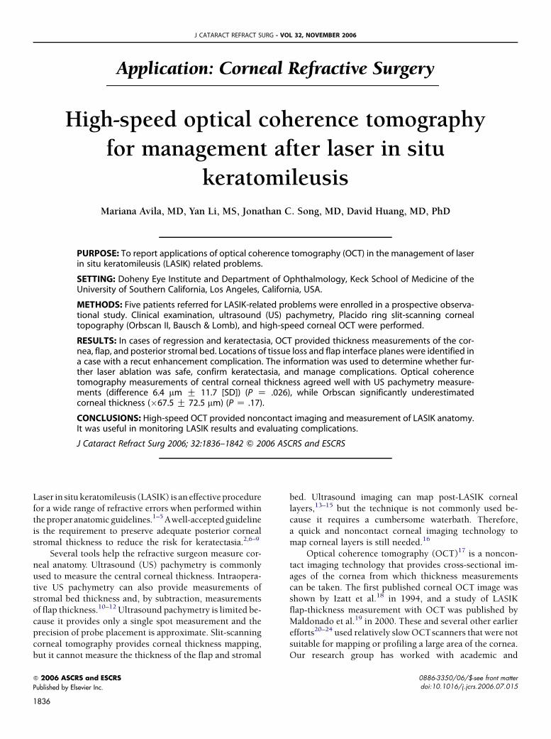
tectasia (Figure 1, A). The map showed an elevated spot in-
ferocentrally that was 56 mm above the best-fit sphere.There was no risk factor for keratectasia before the LASIK
surgery, and the preoperative corneal topography was nor-
mal. Pre-LASIK US pachymetry was 573 mm in the right eye
and 581 mm in the left eye. The Hansatome was used with
a 180 mm depth setting. Ablation depth was 88 mm in the
right eye and 69 mm in the left eye.
The uncorrected visual acuity (UCVA) was 20/40 in the
right eye and 20/60 in the left eye. The manifest refractionwas –1.00 C0.25 � 30 (20/20) and –1.25 C0.25 � 105
(20/15), respectively. The slitlamp examination was nor-
mal. Ultrasound central pachymetry was 532 mm in the
right eye and 550 mm in the left eye. Orbscan anterior to-
pography was normal (Figure 1, A).
The OCT profile scans of the right eye (Figure 1, B) and
left eye showed normal central thickness on the corneal,
flap, and stromal bed scans (Table 1).In the absence of any abnormality on the anterior to-
pography, it was not believed that the increased posterior
elevation on Orbscan indicated keratectasia. The OCT
- VOL 32, NOVEMBER 2006 1837
ASI: OCT IN LASIK MANAGEMENT
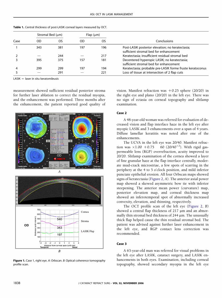
Table 1. Central thickness of post-LASIK corneal layers measured by OCT.
Stromal Bed (mm) Flap (mm)
Case OD OS OD OS Conclusions
1 343 381 197 196 Post-LASIK posterior elevation; no keratectasia;sufficient stromal bed for enhancement
2 d 244 d 217 Keratectasia; insufficient residual stromal bed3 395 375 157 181 Decentered hyperopic LASIK; no keratectasia;
sufficient stromal bed for enhancement4 299 299 197 194 Keratectasia; probable pre-LASIK forme fruste keratoconus5 d 291 d 221 Loss of tissue at intersection of 2 flap cuts
LASIK Z laser in situ keratomileusis
measurement showed sufficient residual posterior stroma
for further laser ablation to correct the residual myopia,
and the enhancement was performed. Three months afterthe enhancement, the patient reported good quality of
Figure 1. Case 1, right eye. A: Orbscan. B: Optical coherence tomography
profile scan.
J CATARACT REFRACT SURG1838
vision. Manifest refraction was C0.25 sphere (20/20) in
the right eye and plano (20/20) in the left eye. There was
no sign of ectasia on corneal topography and slitlampexamination.
Case 2
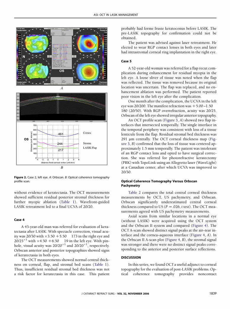
A 48-year-old woman was referred for evaluation of de-
creased vision and flap interface haze in the left eye after
myopic LASIK and 3 enhancements over a span of 4 years.
Diffuse lamellar keratitis was noted after one of the
enhancements.
The UCVA in the left eye was 20/40. Manifest refrac-
tion was �1.00 C0.75 � 60 (20/40�2). With rigid gas-
permeable lens (RGP) overrefraction, acuity improved to20/20. Slitlamp examination of the cornea showed a layer
of fine granular haze at the flap interface centrally, moder-
ate mud-crack microstriae, a few spots of scarring in the
periphery at the 4 to 5 o’clock position, and mild inferior
punctate epithelial erosion. All four Orbscan maps showed
signs of keratectasia (Figure 2, A). The anterior axial power
map showed a skewed asymmetric bow tie with inferior
steepening. The anterior mean power (curvature) map,posterior elevation map, and corneal thickness map
showed an inferotemporal spot of abnormally increased
convexity, elevation, and thinning, respectively.
The OCT profile scan of the left eye (Figure 2, B)
showed a central flap thickness of 217 mm and an abnor-
mally thin stromal bed thickness of 244 mm. The unusually
thick flap helped cause the thin residual stromal bed. The
patient was advised against further laser enhancement inthe left eye, and RGP contact lens correction was
recommended.
Case 3
A 63-year-old man was referred for visual problems inthe left eye after LASIK, cataract surgery, and LASIK en-
hancements in both eyes. Examination, including corneal
topography, showed secondary myopia in the left eye
- VOL 32, NOVEMBER 2006
ASI: OCT IN LASIK MANAGEMENT
without evidence of keratectasia. The OCT measurementsshowed sufficient residual posterior stromal thickness for
further myopic ablation (Table 1). Wavefront-guided
LASIK retreatment led to a final UCVA of 20/20.
Case 4
A 43-year-old man was referred for evaluation of kera-
tectasia after LASIK. With spectacle correction, visual acu-
ity was 20/30 with�3.50 C5.50� 173 in the right eye and
20/25�1 with �4.50 C6.50 � 19 in the left eye. With pin-
hole, visual acuity was 20/20�1 and 20/20�1, respectively.
Orbscan anterior and posterior topographies showed signs
of keratectasia in both eyes.
The OCT measurements showed normal central thick-ness on corneal, flap, and stromal bed scans (Table 1).
Thus, insufficient residual stromal bed thickness was not
a risk factor for keratectasia in this case. This patient
Figure 2. Case 2, left eye. A: Orbscan. B: Optical coherence tomography
profile scan.
J CATARACT REFRACT SUR
probably had forme fruste keratoconus before LASIK. The
pre-LASIK topography for confirmation could not be
obtained.
The patient was advised against laser retreatment. He
elected to wear RGP contact lenses in both eyes and later
had intrastromal corneal ring implantation in the right eye.
Case 5
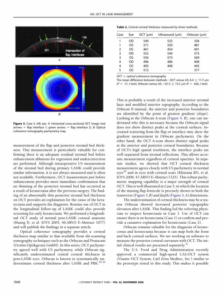
A 52-year-old woman was referred for a flap recut com-
plication during enhancement for residual myopia in the
left eye. A loose sliver of tissue was noted when the flap
was reflected. The tissue was removed because its original
location was uncertain. The flap was replaced, and no en-
hancement ablation was performed. The patient reportedpoor vision in the left eye after the complication.
One month after the complication, the UCVA in the left
eye was 20/200. The manifest refraction was C5.00 –1.50�180 (20/50). With RGP overrefraction, acuity was 20/25.
Orbscan of the left eye showed irregular anterior topography.
An OCT profile scan (Figure 3, A) showed two flap in-
terfaces that intersected temporally. The single interface in
the temporal periphery was consistent with loss of a tissuelenticule from the flap. Residual stromal bed thickness was
291 mm centrally. The OCT corneal thickness map (Fig-
ure 3, B) confirmed that the loss of tissue was centered ap-
proximately 1.5 mm temporally. The patient was intolerant
of an RGP contact lens and opted to have surgical correc-
tion. She was referred for photorefractive keratectomy
(PRK) with TopoLink using an Allegretto laser (WaveLight)
at a Canadian center, after which UCVA was improved to20/30.
Optical Coherence Tomography Versus OrbscanPachymetry
Table 2 compares the total central corneal thickness
measurements by OCT, US pachymetry, and Orbscan.
Orbscan significantly underestimated central corneal
thickness compared to US (P Z.026, t test). The OCT mea-
surements agreed with US pachymetry measurements.
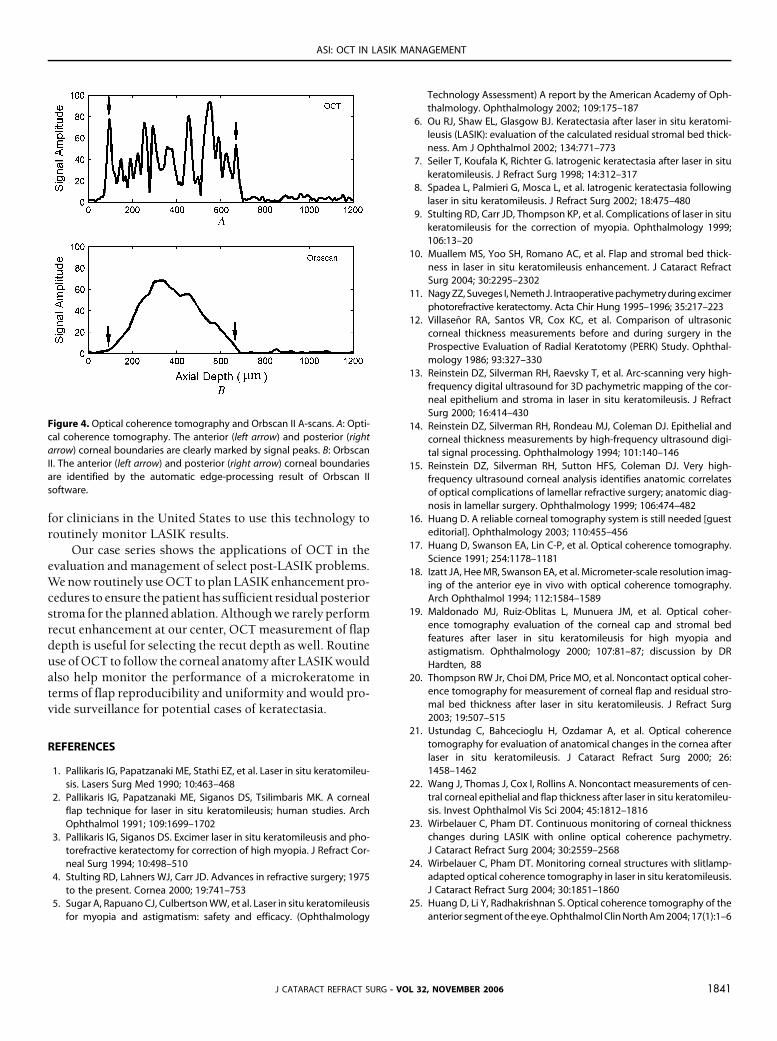
Axial scans from similar locations in a normal eye
(without LASIK) were acquired using the OCT system
and the Orbscan II system and compared (Figure 4). TheOCT A-scan showed distinct signal peaks at the air–tear in-
terface and the cornea–aqueous interface (Figure 4, A). In
the Orbscan II A-scan plot (Figure 4, B), the stromal signal
was stronger and there were no distinct signal peaks corre-
sponding to the anterior and posterior surface reflections.
DISCUSSION
In this series, we found OCTa useful adjunct to corneal
topography for the evaluation of post-LASIK problems. Op-
tical coherence tomography provides noncontact
G - VOL 32, NOVEMBER 2006 1839
ASI: OCT IN LASIK MANAGEMENT
measurement of the flap and posterior stromal bed thick-
ness. This measurement is particularly valuable for con-
firming there is an adequate residual stromal bed before
enhancement ablations for regression and undercorrection
are performed. Although intraoperative US measurement
of the stromal bed during primary LASIK could provide
similar information, it is not always measured and is often
not available. Furthermore, OCT measurement just beforeenhancement provides more immediate confirmation that
no thinning of the posterior stromal bed has occurred as
a result of keratectasia after the previous surgery. The find-
ing of an abnormally thin posterior stromal bed thickness
on OCT provides an explanation for the cause of the kera-
tectasia and supports the diagnosis. Routine use of OCT in
the longitudinal follow-up of LASIK could also provide
screening for early keratectasia. We performed a longitudi-nal OCT study of normal post-LASIK corneal anatomy
(Huang D, et al. IOVS 2005; 46:ARVO E-Abstract 1077)
and will publish the findings in a separate article.
Optical coherence tomography provides a corneal
thickness map similar to that available from slit-scanning
tomography techniques such as the Orbscan and Pentacam
(Oculus Optikgerate GmbH). In this series, OCT pachyme-
try agreed well with US pachymetry, while Orbscan sig-nificantly underestimated central corneal thickness in
post-LASIK eyes. Orbscan is known to systematically un-
derestimate corneal thickness after LASIK and PRK.29,30
Figure 3. Case 5, left eye. A: Horizontal cross-sectional OCT image (red
arrows Z flap interface 1; green arrows Z flap interface 2). B: Optical
coherence tomography pachymetry map.
J CATARACT REFRACT SURG -1840
This is probably a result of the increased anterior stromal
haze and modified anterior topography. According to theOrbscan II manual, the anterior and posterior boundaries
are identified by the point of greatest gradient (slope).
Looking at the Orbscan A-scan (Figure 4, B), one can un-
derstand why this is necessary because the Orbscan signal
does not show distinct peaks at the corneal surfaces. In-
creased scattering from the flap or interface may skew the
gradient measurement in Orbscan pachymetry. On the
other hand, the OCT A-scan shows distinct signal peaksat the anterior and posterior corneal boundaries. Because
of OCT’s high spatial resolution, the interface peaks are
well separated from stromal reflections. This allows accu-
rate measurement regardless of corneal opacities. In sepa-
rate studies, we showed that OCT corneal thickness
measurement agrees closely with US pachymetry in normal
eyes28 and in eyes with corneal scars (Khurana RN, et al.
IOVS 2006; 47:ARVO E-Abstract 1325). This robust pachy-metric mapping capability is a major strength of corneal
OCT. This is well illustrated in Case 5, in which the location
of the missing flap lenticule is precisely shown in both the
transverse (Figure 3, B) and depth (Figure 3, A) dimensions.
The underestimation of corneal thickness may be a rea-
son Orbscan showed increased posterior topographic
elevation after LASIK. This finding led the referring physi-
cian to suspect keratectasia in Case 1. Use of OCT canensure there is no keratectasia (Case 1) or confirm and pro-
vide a causative explanation for keratectasia (Case 2).
Orbscan remains valuable for the diagnosis of kerato-
conus and keratectasia because it can map both the front
and back corneal surfaces. We are working on software to
measure the posterior corneal curvature with OCT. The ini-
tial clinical results are presented separately.31
The U.S. Food and Drug Administration recentlyapproved a commercial high-speed CAS-OCT system
(Visante OCT System, Carl Zeiss Meditec, Inc.) similar to
the prototype tested in this study. This makes it possible
Table 2. Central corneal thickness measured by three methods.
Case Eye OCT (mm) Ultrasound (mm) Orbscan (mm)
1 OD 540 532 3361 OS 571 550 4812 OS 461 454 4013 OD 552 540 5733 OS 556 573 5694 OD 496 486 4084 OS 493 408 4455 OS 512 d 377
OCT Z optical coherence tomography
The mean difference between methodsdOCT versus US: 6.4 G 11.7 mm
(P Z .17, t test); Orbscan versus US: �67.5 G 72.5 mm (P Z .026, t test)
VOL 32, NOVEMBER 2006
ASI: OCT IN LASIK MANAGEMENT
for clinicians in the United States to use this technology to
routinely monitor LASIK results.
Our case series shows the applications of OCT in the
evaluation and management of select post-LASIK problems.
We now routinely use OCT to plan LASIK enhancement pro-cedures to ensure the patient has sufficient residual posterior
stroma for the planned ablation. Although we rarely perform
recut enhancement at our center, OCT measurement of flap
depth is useful for selecting the recut depth as well. Routine
use of OCT to follow the corneal anatomy after LASIK would
also help monitor the performance of a microkeratome in
terms of flap reproducibility and uniformity and would pro-
vide surveillance for potential cases of keratectasia.
REFERENCES
1. Pallikaris IG, Papatzanaki ME, Stathi EZ, et al. Laser in situ keratomileu-
sis. Lasers Surg Med 1990; 10:463–468
2. Pallikaris IG, Papatzanaki ME, Siganos DS, Tsilimbaris MK. A corneal
flap technique for laser in situ keratomileusis; human studies. Arch
Ophthalmol 1991; 109:1699–1702
3. Pallikaris IG, Siganos DS. Excimer laser in situ keratomileusis and pho-
torefractive keratectomy for correction of high myopia. J Refract Cor-
neal Surg 1994; 10:498–510
4. Stulting RD, Lahners WJ, Carr JD. Advances in refractive surgery; 1975
to the present. Cornea 2000; 19:741–753
5. Sugar A, Rapuano CJ, Culbertson WW, et al. Laser in situ keratomileusis
for myopia and astigmatism: safety and efficacy. (Ophthalmology
Figure 4. Optical coherence tomography and Orbscan II A-scans. A: Opti-
cal coherence tomography. The anterior (left arrow) and posterior (right
arrow) corneal boundaries are clearly marked by signal peaks. B: Orbscan
II. The anterior (left arrow) and posterior (right arrow) corneal boundaries
are identified by the automatic edge-processing result of Orbscan II
software.
J CATARACT REFRACT SURG
Technology Assessment) A report by the American Academy of Oph-
thalmology. Ophthalmology 2002; 109:175–187
6. Ou RJ, Shaw EL, Glasgow BJ. Keratectasia after laser in situ keratomi-
leusis (LASIK): evaluation of the calculated residual stromal bed thick-
ness. Am J Ophthalmol 2002; 134:771–773
7. Seiler T, Koufala K, Richter G. Iatrogenic keratectasia after laser in situ
keratomileusis. J Refract Surg 1998; 14:312–317
8. Spadea L, Palmieri G, Mosca L, et al. Iatrogenic keratectasia following
laser in situ keratomileusis. J Refract Surg 2002; 18:475–480
9. Stulting RD, Carr JD, Thompson KP, et al. Complications of laser in situ
keratomileusis for the correction of myopia. Ophthalmology 1999;
106:13–20
10. Muallem MS, Yoo SH, Romano AC, et al. Flap and stromal bed thick-
ness in laser in situ keratomileusis enhancement. J Cataract Refract
Surg 2004; 30:2295–2302
11. Nagy ZZ, Suveges I, Nemeth J. Intraoperative pachymetry during excimer
photorefractive keratectomy. Acta Chir Hung 1995–1996; 35:217–223
12. Villasenor RA, Santos VR, Cox KC, et al. Comparison of ultrasonic
corneal thickness measurements before and during surgery in the
Prospective Evaluation of Radial Keratotomy (PERK) Study. Ophthal-
mology 1986; 93:327–330
13. Reinstein DZ, Silverman RH, Raevsky T, et al. Arc-scanning very high-
frequency digital ultrasound for 3D pachymetric mapping of the cor-
neal epithelium and stroma in laser in situ keratomileusis. J Refract
Surg 2000; 16:414–430
14. Reinstein DZ, Silverman RH, Rondeau MJ, Coleman DJ. Epithelial and
corneal thickness measurements by high-frequency ultrasound digi-
tal signal processing. Ophthalmology 1994; 101:140–146
15. Reinstein DZ, Silverman RH, Sutton HFS, Coleman DJ. Very high-
frequency ultrasound corneal analysis identifies anatomic correlates
of optical complications of lamellar refractive surgery; anatomic diag-
nosis in lamellar surgery. Ophthalmology 1999; 106:474–482
16. Huang D. A reliable corneal tomography system is still needed [guest
editorial]. Ophthalmology 2003; 110:455–456
17. Huang D, Swanson EA, Lin C-P, et al. Optical coherence tomography.
Science 1991; 254:1178–1181
18. Izatt JA, Hee MR, Swanson EA, et al. Micrometer-scale resolution imag-
ing of the anterior eye in vivo with optical coherence tomography.
Arch Ophthalmol 1994; 112:1584–1589
19. Maldonado MJ, Ruiz-Oblitas L, Munuera JM, et al. Optical coher-
ence tomography evaluation of the corneal cap and stromal bed
features after laser in situ keratomileusis for high myopia and
astigmatism. Ophthalmology 2000; 107:81–87; discussion by DR
Hardten, 88
20. Thompson RW Jr, Choi DM, Price MO, et al. Noncontact optical coher-
ence tomography for measurement of corneal flap and residual stro-
mal bed thickness after laser in situ keratomileusis. J Refract Surg
2003; 19:507–515
21. Ustundag C, Bahcecioglu H, Ozdamar A, et al. Optical coherence
tomography for evaluation of anatomical changes in the cornea after
laser in situ keratomileusis. J Cataract Refract Surg 2000; 26:
1458–1462
22. Wang J, Thomas J, Cox I, Rollins A. Noncontact measurements of cen-
tral corneal epithelial and flap thickness after laser in situ keratomileu-
sis. Invest Ophthalmol Vis Sci 2004; 45:1812–1816
23. Wirbelauer C, Pham DT. Continuous monitoring of corneal thickness
changes during LASIK with online optical coherence pachymetry.
J Cataract Refract Surg 2004; 30:2559–2568
24. Wirbelauer C, Pham DT. Monitoring corneal structures with slitlamp-
adapted optical coherence tomography in laser in situ keratomileusis.
J Cataract Refract Surg 2004; 30:1851–1860
25. Huang D, Li Y, Radhakrishnan S. Optical coherence tomography of the
anterior segment of the eye. Ophthalmol Clin North Am 2004; 17(1):1–6
- VOL 32, NOVEMBER 2006 1841
ASI: OCT IN LASIK MANAGEMENT
26. Radhakrishnan S, Rollins AM, Roth JE, et al. Real-time optical coher-
ence tomography of the anterior segment at 1310 nm. Arch Ophthal-
mol 2001; 119:1179–1185
27. Huang D, Li Y, Radhakrishnan S, Chalita MR. Corneal and anterior
segment optical coherence tomography. In: Schuman JS, Puliafito
CA, Fujimoto JG, eds, Optical Coherence Tomography of Ocular Dis-
eases, 2nd ed. Thorofare, NJ, Slack, 2004; 663–673
28. Li Y, Shekhar R, Huang D. Corneal pachymetry mapping with high-
speed optical coherence tomography. Ophthalmology 2006; 113:
792–799
J CATARACT REFRACT SURG1842
29. Chakrabarti HS, Craig JP, Brahma A, et al. Comparison of corneal thick-
ness measurements using ultrasound and Orbscan slit-scanning to-
pography in normal and post-LASIK eyes. J Cataract Refract Surg
2001; 27:1823–1828
30. Prisant O, Calderon N, Chastang P, et al. Reliability of pachymetric
measurements using Orbscan after excimer refractive surgery. Oph-
thalmology 2003; 110:511–515
31. Tang M, Li Y, Avila M, Huang D. Measurement of total corneal power
before and after LASIK with high-speed optical coherence tomogra-
phy. In press, J Cataract Refract Surg
- VOL 32, NOVEMBER 2006