Embed Size (px)
Citation preview
High Yield Production of Influenza Virus in Madin DarbyCanine Kidney (MDCK) Cells with Stable Knockdown ofIRF7Itsuki Hamamoto1, Hiroshi Takaku2, Masato Tashiro1, Norio Yamamoto1,3*
1 Laboratory of Cell-based Vaccine Development, Influenza Virus Research Center, National Institute of Infectious Diseases, Musashimurayama-shi, Tokyo, Japan,
2 Department of Life and Environmental Science, Chiba Institute of Technology, Narashino-shi, Chiba, Japan, 3 Department of General Medicine, Juntendo University
School of Medicine, Bunkyo-ku, Tokyo, Japan
Abstract
Influenza is a serious public health problem that causes a contagious respiratory disease. Vaccination is the most effectivestrategy to reduce transmission and prevent influenza. In recent years, cell-based vaccines have been developed withcontinuous cell lines such as Madin-Darby canine kidney (MDCK) and Vero. However, wild-type influenza and egg-basedvaccine seed viruses will not grow efficiently in these cell lines. Therefore, improvement of virus growth is strongly requiredfor development of vaccine seed viruses and cell-based influenza vaccine production. The aim of our research is to developnovel MDCK cells supporting highly efficient propagation of influenza virus in order to expand the capacity of vaccineproduction. In this study, we screened a human siRNA library that involves 78 target molecules relating to three major type Iinterferon (IFN) pathways to identify genes that when knocked down by siRNA lead to enhanced production of influenzavirus A/Puerto Rico/8/1934 in A549 cells. The siRNAs targeting 23 candidate genes were selected to undergo a secondscreening pass in MDCK cells. We examined the effects of knockdown of target genes on the viral production using newlydesigned siRNAs based on sequence analyses. Knockdown of the expression of a canine gene corresponding to human IRF7by siRNA increased the efficiency of viral production in MDCK cells through an unknown process that includes themechanisms other than inhibition of IFN-a/b induction. Furthermore, the viral yield greatly increased in MDCK cells stablytransduced with the lentiviral vector for expression of short hairpin RNA against IRF7 compared with that in control MDCKcells. Therefore, we propose that modified MDCK cells with lower expression level of IRF7 could be useful not only forincreasing the capacity of vaccine production but also facilitating the process of seed virus isolation from clinical specimensfor manufacturing of vaccines.
Citation: Hamamoto I, Takaku H, Tashiro M, Yamamoto N (2013) High Yield Production of Influenza Virus in Madin Darby Canine Kidney (MDCK) Cells with StableKnockdown of IRF7. PLoS ONE 8(3): e59892. doi:10.1371/journal.pone.0059892
Editor: Yi Guan, The University of Hong Kong, China
Received September 10, 2012; Accepted February 22, 2013; Published March 26, 2013
Copyright: � 2013 Hamamoto et al. This is an open-access article distributed under the terms of the Creative Commons Attribution License, which permitsunrestricted use, distribution, and reproduction in any medium, provided the original author and source are credited.
Funding: This work was supported by a Health Sciences Research Grant-in-Aid for Emerging and Re-emerging Infectious Diseases from the Ministry of Health,Labour and Welfare of Japan, and the Program for the Promotion of Fundamental Studies in Health Sciences of the National Institute of Biomedical Innovation ofJapan. This study was also supported in part by a Grant-in-Aid (S1201013) from MEXT (Ministry of Education, Culture, Sports, Science and Technology) - SupportedProgram for the Strategic Research Foundation at Private Universities, 2012–2017. The funders had no role in study design, data collection and analysis, decisionto publish, or preparation of the manuscript.
Competing Interests: The authors declare that they have a patent relating to material pertinent to this article. The technology of modified MDCK cellsdescribed in this manuscript was filed with the Japan Patent Office for a patent application (Composition for culturing cells, method for the preparation ofinfluenza virus, and influenza virus, Japan patent application No. 2012093016, 16 April 2012). This does not alter the authors’ adherence to all the PLOS ONEpolicies on sharing data and materials.
* E-mail: [email protected]
Introduction
Influenza is a global public health issue that causes a serious
illness with a high mortality rate. Vaccination is one of the most
effective medical strategies to prevent influenza virus infection.
The current egg-based technology for manufacturing influenza
vaccine has been used since 1950s, but cell-based technology has
been developed to produce more effective influenza vaccines in
sufficient quantities in a shorter period of time. In recent years, two
continuous cell lines have been approved by regulatory authorities
to be used for the production of influenza vaccines: Madin Darby
canine kidney (MDCK) cells and African green monkey kidney-
derived Vero cells [1–5]. Human retina-derived cell line PER.C6
has also been shown useful for propagation of influenza viruses [6].
Although these cell lines produce notable yields of a wide variety of
influenza viruses, attempts to develop novel cell lines with greater
potentials have been made for more rapid preparation of influenza
vaccines. A recent study demonstrated that the siat7e-expressing
MDCK cells produce much more HA antigen than the parental
MDCK cells [5]. The siat7e-expressing cells that proliferate in
suspension would facilitate influenza virus productions [5].
Influenza viruses have been shown to be recognized by pattern-
recognition receptors (PRRs), such as the Toll-like receptors 3
(TLR3) [7] and 7/8 (TLR7/8) [8], and retinoic acid-inducible
gene (RIG-I)-like receptors (RLRs) [9]. Influenza viruses have
evolved strategies to counteract cellular antiviral mechanisms,
especially to circumvent the type I interferon (IFN-a/b) system
which is a first line of defense against viral infections [10,11]. The
mechanisms that underlie the induction of type I IFN genes have
been extensively studied in the context of immunity to viruses.
PLOS ONE | www.plosone.org 1 March 2013 | Volume 8 | Issue 3 | e59892
Type I IFNs are known to be critical for inflammation [12] and
play central roles in the activation of host antiviral responses to
control virus infections [13,14]. Therefore, we hypothesized that it
would be possible to obtain novel MDCK cells with greater
potential to propagate influenza viruses by means of RNA-
interference to inhibit the function of genes relating to IFN
signaling.
In this study, we identified canine IRF7 as a target in order to
establish modified MDCK cells which would be capable of
producing higher yield of influenza A viruses. Knockdown of IRF7
with siRNA showed 3 to 4-fold enhancement of influenza A virus
production. We also confirmed that MDCK cells with stable
knockdown of IRF7 by short hairpin RNA (shRNA) showed 2 to
8-fold enhancement of influenza virus production. In conclusion,
the modified MDCK cells with lower level of IRF7 expression may
be useful for producing higher titers of influenza viruses. The novel
MDCK cells will be beneficial for large-scale production of
influenza vaccines in the manufacturing process. In addition, the
established MDCK cells will be useful for isolation of influenza
viruses from clinical specimens with a low viral load and efficient
preparation of vaccine seed viruses in a shorter period.
Materials and Methods
CellsA549 cells were purchased from Japanese Collection of
Research Bioresources (JCRB; Osaka Japan). A549 cells were
cultured in Dulbecco’s modified Eagle’s medium (DMEM;
GIBCO, Carlsbad, CA) containing 10% fetal bovine serum
(FBS; GIBCO, Carlsbad, CA) and 100 units/ml penicillin/
streptomycin (GIBCO, Carlsbad, CA). MDCK (CCL-34) cells
were purchased from American Type Culture Collection (ATCC;
Rockville, MD). MDCK cells were cultured in Opti-Pro serum
free medium (Opti-pro SFM; GIBCO, Carlsbad, CA) containing
4 mM L-Glutamine (GIBCO, Carlsbad, CA). All cells were
cultured in a 5% CO2 humidified incubator.
VirusesViruses used in this study were A/Puerto Rico/8/1934 (PR8;
A(H1N1)), A/Narita/1/2009 (NR1; A(H1N1)pdm09), A/Victo-
ria/361/2011 (VC361; A(H3N2)), and B/Florida/4/2006 (FL4;
type B, Yamagata lineage). These viruses were propagated in
MDCK cells. The virus titers were determined by 50% tissue
culture infectious dose (TCID50) assay or plaque assay.
siRNA Transfection, Virus Infection and RNA ExtractionThe siRNA duplexes for A549 cells were selected from Silencer
select siRNA library (Ambion, Austin, TX). The siRNA duplexes
for MDCK cells were synthesized (Sigma-Aldrich, St. Louis, MO).
The sequences of siRNA duplexes for MDCK cells were listed in
Table 1. A549 cells were seeded on type I collagen coated 96-well
plate. Three target-specific siRNAs (Silencer select siRNA library,
Ambion, Austin, TX) or non-targeting negative control siRNA
(Silencer Negative Control #1 siRNA, Ambion, Austin, TX) at a
final concentration of 10 nM were transfected using transfection
reagent (Lipofectamine RNAiMAX; Invitrogen, Carlsbad, CA)
and incubated at 37uC for 48 h. For MDCK cells, the cells were
seeded on 96-well plate and three target-specific siRNAs (Sigma-
Aldrich, Inc., St. Louis, MO) or non-targeting negative control
siRNA (Silencer Negative Control #1 siRNA, Ambion, Austin,
TX) at a final concentration of 50 nM were transfected by siRNA
transfection reagent (Lullaby; OZ Biosciences, France). Cells were
washed with PBS three times and infected with influenza A virus at
a MOI of 0.01 in Opti-Pro SFM in the presence of 2 mg/ml of
trypsin acetylated (Sigma, Chemical Co., St. Louis, MO) at 34uCfor 1 h. Then, cells were washed with PBS three times and
incubated in Opti-Pro SFM supplemented with 2 mg/ml of trypsin
acetylated at 37uC for 24 h. The viral RNA from tissue culture
supernatant was mixed with the lysis buffer containing carrier
RNA derived from uninfected A549 or MDCK cells and was
extracted using MagMAXTM-96 Blood RNA Isolation Kit
(Ambion, Austin, TX) on King Fisher purification systems
(Thermo Scientific, Cambridge, MA). The total RNA from
cultured cells was extracted using RNeasy Mini Kit (Qiagen,
Hilden, Germany) followed by DNase I (Qiagen, Hilden,
Germany) treatment.
PlasmidsThe pRetro-U6 vector was constructed by inserting a human
U6 promoter amplified by PCR from genomic DNA into
pMSCV-puro (Clontech, Mountain View, CA), from which
DNA sequence between Nhe I and Xba I in the 3’ LTR was
deleted for generation of a self-inactivating virus. DNA fragments
encoding the small hairpin RNAs were generated by PCR,
digested with Bpi I, and ligated into pRetro-U6 between Bpi I sites
downstream of the U6 promoter. The target sequences of shRNAs
were as follows: shIRF7, 5’-CTGGGCAAATGCAAGGTCT-3’;
shCtrl, 5’-GACTACACAAATCAGCGAT-3’ (shCtrl targets
LacZ). The shRNA expression cassettes were then transferred to
pCS-BS, carrying a blasticidin S resistance gene expressed under
the control of the elongation factor 1a promoter. The pCS-BS
vector was constructed by replacing EGFP of the pCS-CDF-EG-
Table 1. Canis lupus familiaris siRNA used in this study.
Target Gene Orientation Sequences (59 to 39)
IRF7_1 Sense CUGGGCAAAUGCAAGGUCUTT
IRF7_1 Anti-sense AGACCUUGCAUUUGCCCAGTT
IRF7_2 Sense GGCGCCUGGGCAAAUGCAATT
IRF7_2 Anti-sense UUGCAUUUGCCCAGGCGCCTT
IRF7_3 Sense CAGAGAAGCUGCUGCAGCATT
IRF7_3 Anti-sense UGCUGCAGCAGCUUCUCUGTT
IRF3_1 Sense GAUCUGAUUGCCUUCAUCATT
IRF3_1 Anti-sense UGAUGAAGGCAAUCAGAUCTT
IRF3_2 Sense GGCUCUUGGUGCCUGAUGATT
IRF3_2 Anti-sense UCAUCAGGCACCAAGAGCCTT
IRF3_3 Sense CAGACAGUCUCCUGCCCAATT
IRF3_3 Anti-sense UUGGGCAGGAGACUGUCUGTT
MyD88_1 Sense GGGCAAAUGCCUGAGCGUUTT
MyD88_1 Anti-sense AACGCUCAGGCAUUUGCCCTT
MyD88_2 Sense CAGACAAACUAUCGGCUGATT
MyD88_2 Anti-sense UCAGCCGAUAGUUUGUCUGTT
MyD88_3 Sense GCAUCACCAUGCUUGAUGATT
MyD88_3 Anti-sense UCAUCAAGCAUGGUGAUGCTT
DDX58_1 Sense CAAACUGUGUGCUUCUCUUTT
DDX58_1 Anti-sense AAGAGAAGCACACAGUUUGTT
DDX58_2 Sense GUGUUUCAGUUACCCAACATT
DDX58_2 Anti-sense UGUUGGGUAACUGAAACACTT
DDX58_3 Sense GAUCUGAUUGCCUUCAUCATT
DDX58_3 Anti-sense UGAUGCAUUUAAAUCUGUCTT
doi:10.1371/journal.pone.0059892.t001
The Novel MDCK for High Yield Viral Production
PLOS ONE | www.plosone.org 2 March 2013 | Volume 8 | Issue 3 | e59892
PRE vector (a kind gift from Dr. Hiroyuki Miyoshi, RIKEN,
Tsukuba) with blasticidin S resistance gene amplified by PCR
from pcDNA6/myc-His A (Life Technologies, Carlsbad, CA).
Transduction of MDCK Cells with Lentiviral VectorsFor production of lentiviruses, 293T cells were cotransfected
with pCS-BS-shCtrl, or pCS-BS-shIRF7 together with the pCAG-
HIVgp, pRSV-Rev (kind gifts from Dr. H. Miyoshi, RIKEN,
Tsukuba) and pVSV-G (Clontech, Mountain View, CA) using
FuGENE 6 (Roche Applied Science, Indianapolis, IN). Culture
supernatants were collected 48 h after transfection and filtered.
MDCK cells were transduced with these lentiviruses for 12 h in
the presence of 8 mg/mL polybrene and cultured with fresh media.
After 48 h of culture, the media were replaced with the selection
media containing 10 mg/mL blastcidin S.
Quantitative Real-time One-step RT-RCRReal-time RT-PCR reactions were carried out using TaqMan
One-step RT-PCR Master Mix Reagents Kit (Applied Biosystems,
Foster City, CA) according to the manufacture’s instructions with
a total volume of 25 ml. The primers and probes used for
quantification of target mRNA were shown in Table 2. The total
volume of 5 ml of sample RNA was added into 20 ml of reaction
mix. Thermal cycling was performed in a Light Cycler 480 Real-
time PCR system II (Roche Diagnostics, Germany) with condi-
tions at 48uC for 30 min, 95uC for 10 min followed by 40 cycles at
95uC for 5 sec and 60uC for 1 min. The amount of target RNA
was normalized with the amount of 18S rRNA from host cells or
carrier RNA. The knockdown effect on the targeted gene by the
specific siRNA was examined by real-time RT-PCR with power
SYBR green PCR master mix (Applied Biosystems, Warrington,
UK).
Reverse Transcription, Amplification, and SequenceAnalysis of mRNA Expressed in MDCK Cells
Reverse transcription was performed using ReverTra-Ace
(Toyobo, Osaka, Japan) with random hexamers and the thermal
profiles consisted of one cycle at 37uC for 30 min, 42uC for
20 min, 99uC for 5 min and 4uC for 5 min. For detecting a canis
lupus familiaris gene corresponding to human IRF7, the cDNA
was amplified by KOD Dash polymerase (Toyobo, Japan) using
primers shown in Table 2. Thermal cycling conditions were as
follows: 30 cycles of 98uC for 10 sec, 55uC for 2 sec and 74uC for
5 min. PCR products were separated by electrophoresis at 100 V
for 1 h in a 1.5% (wt/vol) agarose S (Nippon Gene, Tokyo, Japan)
gel with TBE buffer and visualized by GelRed Nucleic Acid Gel
Stain (Wako, Japan) under UV transillumination. The desired
PCR products were extracted and purified using QIAquick gel
extraction Kit (Qiagen, Hilden, Germany). The purified products
were used for the reaction with BigDye Terminator cycle
sequencing ready reaction kit version 3.0 (Applied Biosystems,
Foster City, CA). Sequencing alignment was analyzed by using the
software Sequencher 4.10.1 (Gene Codes, Ann Arbor, MI).
Network AnalysisThe molecular interaction networks were analyzed by Ingenuity
Pathway Analysis (IPA; Ingenuity Systems, Mountain View, CA).
Virus Infection and Hemagglutination (HA) AssayThe MDCK cells expressing shRNA for IRF7 or control were
seeded on a 6-well plate at a density of 26105 cells/well and
cultured until confluent layers were obtained in Opti-pro SFM
containing 10 mg/ml of blasticidin S. Cells were infected with
indicated amount of PR8 (A/H1N1), NR1 (A/H1N1pdm09),
VIC361 (A/H3N2), or FL4 (B/Yam) in Opti-Pro SFM at 34uC for
1 h. Then, cells were cultured with Opti-Pro SFM in the presence
of 2 mg/ml of trypsin acetylated and incubated at 37uC for 48–
96 h. The culture supernatants were collected for following HA
assay. Red blood cells were purchased from Nippon Bio-Test
Laboratories, Tokyo, Japan. Guinea pig red blood cells (GRBC)
were washed three times with PBS and suspended in PBS at a final
concentration of 1% as a working suspension. Turkey red blood
cells (TRBC) or chicken red blood cells (CRBC) were washed with
0.85% NaCl and suspended in PBS at a final concentration of
0.5% as a working suspension. For NR1, the virus samples were
diluted with PBS serially in U-bottom 96-well plates and then
50 ml of 0.5% TRBC suspension was added into each well and
allowed to stand for 45 min at room temperature. For PR8 or
VIC361, 50 ml of 1% GRBC suspension was added to serially
diluted virus samples and allowed to stand for 60 min at 4uC. For
FL4, 50 ml of 0.5% CRBC suspension was added to serially diluted
virus samples and allowed to stand for 45 min at room
temperature. The HA titer was determined as the highest dilution
of the sample showing complete agglutination pattern on the
bottom of the well.
Results
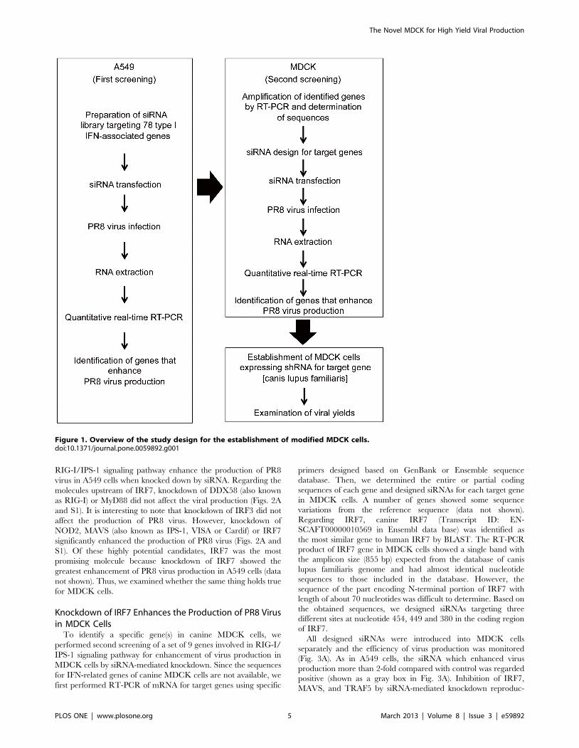
First siRNA Library ScreeningThe strategy to establish modified MDCK cells with higher
efficiency of influenza virus propagation was summarized in the
flowchart (Fig. 1). To identify key host factors in canine MDCK
cells, we used A549 human lung adenocarcinoma cell line for the
first screening because human genome database includes much
more information than canis lupus familiaris genome database and
related resources including pre-designed siRNA library are
available in human.
We screened 78 human genes associated with type I IFN using
siRNA library, for enhancement of the propagation of influenza A
virus in A549 cells. The siRNAs for each target gene were
individually transfected into A549 cells, and the cells were infected
with influenza A/Puerto Rico/34/8 (PR8) at a MOI of 0.01. We
used PR8 virus, because PR8 has suitable growth properties in
many cell lines and is widely used as a backbone virus for the
development of high growth reassortants for vaccine production
[15]. In the first screening, when knockdown of a cellular gene by
the corresponding siRNA showed more than 2-fold enhancement
of virus propagation, the gene was considered positive (Fig. S1). A
total of 23 out of 78 genes were chosen for further analysis to
validate the positive effect of siRNA in influenza virus multipli-
cation.
Knockdown of IRF7 Enhances the Production of PR8 Virusin A549 Cells
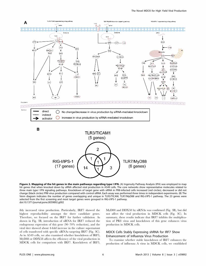
To understand which pathway is most important for improve-
ment of viral propagation, we mapped the genes and their
products identified in the type I IFN-related main pathways
(Fig. 2A) and categorized these molecules into 3 groups (Fig. 2B).
As the knockdown of NFKB1 enhanced the production of PR8
virus, we thought that NFKB1 might be one of the candidates.
NFKB1 serves in downstream signaling pathways leading to the
activation of type I IFN production and a common molecule in the
three signaling pathways (Fig. 2A and B). Also, knockdown of
TBK1, SMAD4 or IRF7 enhanced the production of PR8 virus
(Figs. 2A and S1). Seventeen out of 23 genes were shown to be
critically related to RIG-I/IPS-1-mediated type I IFN production
(Fig. 2B). These results indicate that the genes mainly involved in
The Novel MDCK for High Yield Viral Production
PLOS ONE | www.plosone.org 3 March 2013 | Volume 8 | Issue 3 | e59892
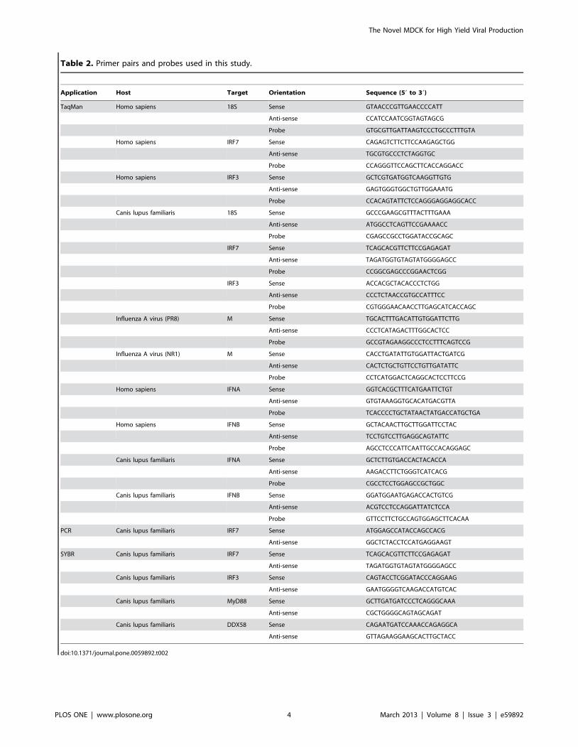
Table 2. Primer pairs and probes used in this study.
Application Host Target Orientation Sequence (59 to 39)
TaqMan Homo sapiens 18S Sense GTAACCCGTTGAACCCCATT
Anti-sense CCATCCAATCGGTAGTAGCG
Probe GTGCGTTGATTAAGTCCCTGCCCTTTGTA
Homo sapiens IRF7 Sense CAGAGTCTTCTTCCAAGAGCTGG
Anti-sense TGCGTGCCCTCTAGGTGC
Probe CCAGGGTTCCAGCTTCACCAGGACC
Homo sapiens IRF3 Sense GCTCGTGATGGTCAAGGTTGTG
Anti-sense GAGTGGGTGGCTGTTGGAAATG
Probe CCACAGTATTCTCCAGGGAGGAGGCACC
Canis lupus familiaris 18S Sense GCCCGAAGCGTTTACTTTGAAA
Anti-sense ATGGCCTCAGTTCCGAAAACC
Probe CGAGCCGCCTGGATACCGCAGC
IRF7 Sense TCAGCACGTTCTTCCGAGAGAT
Anti-sense TAGATGGTGTAGTATGGGGAGCC
Probe CCGGCGAGCCCGGAACTCGG
IRF3 Sense ACCACGCTACACCCTCTGG
Anti-sense CCCTCTAACCGTGCCATTTCC
Probe CGTGGGAACAACCTTGAGCATCACCAGC
Influenza A virus (PR8) M Sense TGCACTTTGACATTGTGGATTCTTG
Anti-sense CCCTCATAGACTTTGGCACTCC
Probe GCCGTAGAAGGCCCTCCTTTCAGTCCG
Influenza A virus (NR1) M Sense CACCTGATATTGTGGATTACTGATCG
Anti-sense CACTCTGCTGTTCCTGTTGATATTC
Probe CCTCATGGACTCAGGCACTCCTTCCG
Homo sapiens IFNA Sense GGTCACGCTTTCATGAATTCTGT
Anti-sense GTGTAAAGGTGCACATGACGTTA
Probe TCACCCCTGCTATAACTATGACCATGCTGA
Homo sapiens IFNB Sense GCTACAACTTGCTTGGATTCCTAC
Anti-sense TCCTGTCCTTGAGGCAGTATTC
Probe AGCCTCCCATTCAATTGCCACAGGAGC
Canis lupus familiaris IFNA Sense GCTCTTGTGACCACTACACCA
Anti-sense AAGACCTTCTGGGTCATCACG
Probe CGCCTCCTGGAGCCGCTGGC
Canis lupus familiaris IFNB Sense GGATGGAATGAGACCACTGTCG
Anti-sense ACGTCCTCCAGGATTATCTCCA
Probe GTTCCTTCTGCCAGTGGAGCTTCACAA
PCR Canis lupus familiaris IRF7 Sense ATGGAGCCATACCAGCCACG
Anti-sense GGCTCTACCTCCATGAGGAAGT
SYBR Canis lupus familiaris IRF7 Sense TCAGCACGTTCTTCCGAGAGAT
Anti-sense TAGATGGTGTAGTATGGGGAGCC
Canis lupus familiaris IRF3 Sense CAGTACCTCGGATACCCAGGAAG
Anti-sense GAATGGGGTCAAGACCATGTCAC
Canis lupus familiaris MyD88 Sense GCTTGATGATCCCTCAGGGCAAA
Anti-sense CGCTGGGGCAGTAGCAGAT
Canis lupus familiaris DDX58 Sense CAGAATGATCCAAACCAGAGGCA
Anti-sense GTTAGAAGGAAGCACTTGCTACC
doi:10.1371/journal.pone.0059892.t002
The Novel MDCK for High Yield Viral Production
PLOS ONE | www.plosone.org 4 March 2013 | Volume 8 | Issue 3 | e59892
RIG-I/IPS-1 signaling pathway enhance the production of PR8
virus in A549 cells when knocked down by siRNA. Regarding the
molecules upstream of IRF7, knockdown of DDX58 (also known
as RIG-I) or MyD88 did not affect the viral production (Figs. 2A
and S1). It is interesting to note that knockdown of IRF3 did not
affect the production of PR8 virus. However, knockdown of
NOD2, MAVS (also known as IPS-1, VISA or Cardif) or IRF7
significantly enhanced the production of PR8 virus (Figs. 2A and
S1). Of these highly potential candidates, IRF7 was the most
promising molecule because knockdown of IRF7 showed the
greatest enhancement of PR8 virus production in A549 cells (data
not shown). Thus, we examined whether the same thing holds true
for MDCK cells.
Knockdown of IRF7 Enhances the Production of PR8 Virusin MDCK Cells
To identify a specific gene(s) in canine MDCK cells, we
performed second screening of a set of 9 genes involved in RIG-I/
IPS-1 signaling pathway for enhancement of virus production in
MDCK cells by siRNA-mediated knockdown. Since the sequences
for IFN-related genes of canine MDCK cells are not available, we
first performed RT-PCR of mRNA for target genes using specific
primers designed based on GenBank or Ensemble sequence
database. Then, we determined the entire or partial coding
sequences of each gene and designed siRNAs for each target gene
in MDCK cells. A number of genes showed some sequence
variations from the reference sequence (data not shown).
Regarding IRF7, canine IRF7 (Transcript ID: EN-
SCAFT00000010569 in Ensembl data base) was identified as
the most similar gene to human IRF7 by BLAST. The RT-PCR
product of IRF7 gene in MDCK cells showed a single band with
the amplicon size (855 bp) expected from the database of canis
lupus familiaris genome and had almost identical nucleotide
sequences to those included in the database. However, the
sequence of the part encoding N-terminal portion of IRF7 with
length of about 70 nucleotides was difficult to determine. Based on
the obtained sequences, we designed siRNAs targeting three
different sites at nucleotide 454, 449 and 380 in the coding region
of IRF7.
All designed siRNAs were introduced into MDCK cells
separately and the efficiency of virus production was monitored
(Fig. 3A). As in A549 cells, the siRNA which enhanced virus
production more than 2-fold compared with control was regarded
positive (shown as a gray box in Fig. 3A). Inhibition of IRF7,
MAVS, and TRAF5 by siRNA-mediated knockdown reproduc-
Figure 1. Overview of the study design for the establishment of modified MDCK cells.doi:10.1371/journal.pone.0059892.g001
The Novel MDCK for High Yield Viral Production
PLOS ONE | www.plosone.org 5 March 2013 | Volume 8 | Issue 3 | e59892
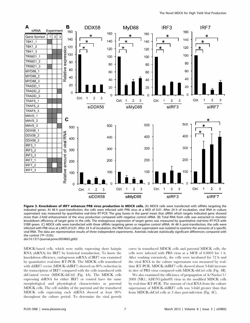
ibly increased virus production. Particularly, IRF7 showed the
highest reproducibility amongst the three candidate genes.
Therefore, we focused on the IRF7 for further validation. As
shown in Fig. 3B, introduction of siRNA for IRF7 reduced the
endogenous expression of this gene (90–70% reduction), and the
viral titer showed about 4-fold increase in the culture supernatant
of cells transfected with specific siRNAs targeting IRF7 (Fig. 3C).
As in A549 cells, we also examined whether knockdown of IRF3,
MyD88 or DDX58 affects the efficiency of the viral production in
MDCK cells for comparison with IRF7. Knockdown of IRF3,
MyD88 and DDX58 by siRNAs was confirmed (Fig. 3B), but did
not affect the viral production in MDCK cells (Fig. 3C). In
summary, these results indicate that IRF7 inhibits the multiplica-
tion of PR8 virus and knockdown of this gene enhances virus
production in MDCK cells.
MDCK Cells Stably Expressing shRNA for IRF7 ShowEnhancement of Influenza Virus Production
To examine whether stable knockdown of IRF7 enhances the
production of influenza A virus in MDCK cells, we established
Figure 2. Mapping of the hit genes in the main pathways regarding type I IFN. (A) Ingenuity Pathway Analysis (IPA) was employed to maphit genes that when knocked down by siRNA affected viral production in A549 cells. The core networks show representative molecules related tothree main type I IFN signaling pathways. Knockdown of target gene with siRNA in PR8-infected cells increased (red circles), decreased or did notchange (black circles) PR8 virus production compared with control siRNA. Each assay was performed three times in independent experiments. (B) TheVenn diagram indicates the number of genes overlapping and unique in TLR3/TICAM, TLR7/MyD88 and RIG-I/IPS-1 pathway. The 23 genes wereselected from the first screening and most target genes were grouped in RIG-I/IPS-1 pathway.doi:10.1371/journal.pone.0059892.g002
The Novel MDCK for High Yield Viral Production
PLOS ONE | www.plosone.org 6 March 2013 | Volume 8 | Issue 3 | e59892
MDCK-based cells, which were stably expressing short hairpin
RNA (shRNA) for IRF7 by lentiviral transduction. To know the
knockdown efficiency, endogenous mRNA of IRF7 was examined
by quantitative real-time RT-PCR. The MDCK cells transduced
with shIRF7 vector (MDCK-shIRF7) showed an 80% reduction in
the transcription of IRF7 compared with the cells transduced with
shControl vector (MDCK-shCtrl) (Fig. 4A). The MDCK cells
expressing shRNA for either IRF7 or control have the same
morphological and physiological characteristics as parental
MDCK cells. The cell viability of the parental and the transduced
MDCK cells expressing each shRNA showed at least 90%
throughout the culture period. To determine the viral growth
curve in transduced MDCK cells and parental MDCK cells, the
cells were infected with PR8 virus at a MOI of 0.0003 for 1 h.
After washing extensively, the cells were incubated for 72 h and
the viral RNA in the culture supernatant was measured by real-
time RT-PCR. MDCK-shIRF7 cells showed about 3-fold increase
in titer of PR8 virus compared with MDCK-shCtrl cells (Fig. 4B).
We also examined the efficiency of propagation of A/Narita/1/
2009 (NR1; A(H1N1)pdm09) virus in the modified MDCK cells
by real-time RT-PCR. The amount of viral RNA from the culture
supernatant of MDCK-shIRF7 cells was 5-fold greater than that
from MDCK-shCtrl cells at 3 days post-infection (Fig. 4C).
Figure 3. Knockdown of IRF7 enhances PR8 virus production in MDCK cells. (A) MDCK cells were transfected with siRNAs targeting theindicated genes. At 48 h post-transfection, the cells were infected with PR8 virus at a MOI of 0.01. After 24 h of incubation, viral RNA in culturesupernatant was measured by quantitative real-time RT-PCR. The gray boxes in the panel mean that siRNA which targets indicated gene showedmore than 2-fold enhancement of the virus production compared with negative control siRNA. (B) Total RNA from cells was extracted to monitorknockdown efficiency of target gene in the cells. The endogenous expression of target genes was measured by quantitative real-time RT-PCR withSYBR green. (C) MDCK cells were transfected with three siRNAs targeting genes or negative control siRNA. At 48 h post-transfection, the cells wereinfected with PR8 virus at a MOI of 0.01. After 24 h of incubation, the RNA from culture supernatant was isolated to examine the amounts of a specificviral RNA. The data are representative results of three independent experiments. Asterisks indicate statistically significant differences compared withthe control (*P,0.05).doi:10.1371/journal.pone.0059892.g003
The Novel MDCK for High Yield Viral Production
PLOS ONE | www.plosone.org 7 March 2013 | Volume 8 | Issue 3 | e59892
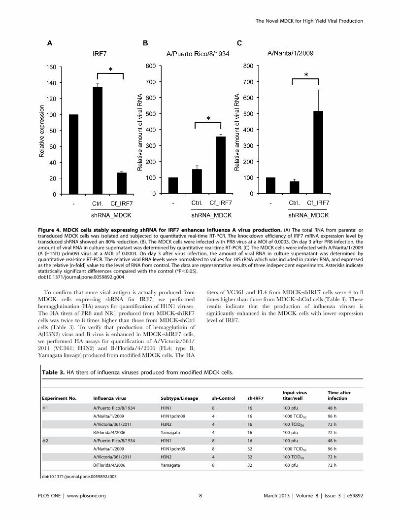
To confirm that more viral antigen is actually produced from
MDCK cells expressing shRNA for IRF7, we performed
hemagglutination (HA) assays for quantification of H1N1 viruses.
The HA titers of PR8 and NR1 produced from MDCK-shIRF7
cells was twice to 8 times higher than those from MDCK-shCtrl
cells (Table 3). To verify that production of hemagglutinin of
A(H3N2) virus and B virus is enhanced in MDCK-shIRF7 cells,
we performed HA assays for quantification of A/Victoria/361/
2011 (VC361; H3N2) and B/Florida/4/2006 (FL4; type B,
Yamagata lineage) produced from modified MDCK cells. The HA
titers of VC361 and FL4 from MDCK-shIRF7 cells were 4 to 8
times higher than those from MDCK-shCtrl cells (Table 3). These
results indicate that the production of influenza viruses is
significantly enhanced in the MDCK cells with lower expression
level of IRF7.
Figure 4. MDCK cells stably expressing shRNA for IRF7 enhances influenza A virus production. (A) The total RNA from parental ortransduced MDCK cells was isolated and subjected to quantitative real-time RT-PCR. The knockdown efficiency of IRF7 mRNA expression level bytransduced shRNA showed an 80% reduction. (B). The MDCK cells were infected with PR8 virus at a MOI of 0.0003. On day 3 after PR8 infection, theamount of viral RNA in culture supernatant was determined by quantitative real-time RT-PCR. (C) The MDCK cells were infected with A/Narita/1/2009(A (H1N1) pdm09) virus at a MOI of 0.0003. On day 3 after virus infection, the amount of viral RNA in culture supernatant was determined byquantitative real-time RT-PCR. The relative viral RNA levels were normalized to values for 18S rRNA which was included in carrier RNA, and expressedas the relative (n-fold) value to the level of RNA from control. The data are representative results of three independent experiments. Asterisks indicatestatistically significant differences compared with the control (*P,0.05).doi:10.1371/journal.pone.0059892.g004
Table 3. HA titers of influenza viruses produced from modified MDCK cells.
Experiment No. Influenza virus Subtype/Lineage sh-Control sh-IRF7Input virustiter/well
Time afterinfection
#1 A/Puerto Rico/8/1934 H1N1 8 16 100 pfu 48 h
A/Narita/1/2009 H1N1pdm09 4 16 1000 TCID50 96 h
A/Victoria/361/2011 H3N2 4 16 100 TCID50 72 h
B/Florida/4/2006 Yamagata 4 16 100 pfu 72 h
#2 A/Puerto Rico/8/1934 H1N1 8 16 100 pfu 48 h
A/Narita/1/2009 H1N1pdm09 8 32 1000 TCID50 96 h
A/Victoria/361/2011 H3N2 4 32 100 TCID50 72 h
B/Florida/4/2006 Yamagata 8 32 100 pfu 72 h
doi:10.1371/journal.pone.0059892.t003
The Novel MDCK for High Yield Viral Production
PLOS ONE | www.plosone.org 8 March 2013 | Volume 8 | Issue 3 | e59892
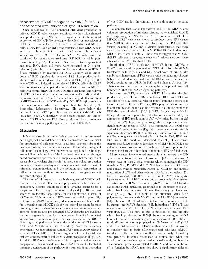
Enhancement of Viral Propagation by siRNA for IRF7 isnot Associated with Inhibition of Type I IFN Induction
Since knockdown of IRF7 enhanced PR8 virus production in
infected MDCK cells, we next examined whether this enhanced
viral production by siRNA for IRF7 might be due to the reduced
expression of IFN-a/b. To investigate the effects of knockdown of
IRF7 on expression levels of IFN-a/b in virus-infected MDCK
cells, siRNA for IRF3 or IRF7 was transfected into MDCK cells
and the cells were infected with PR8 virus. The efficient
knockdown of IRF3 or IRF7 mRNA (70% reduction) was
confirmed by quantitative real-time RT-PCR at 48 h post-
transfection (Fig. 5A). The viral RNA from culture supernatant
and total RNA from cell lysate were extracted at 24 h post-
infection (hpi). The relative expression level of endogenous IFN-a/
b was quantified by real-time RT-PCR. Notably, while knock-
down of IRF7 significantly increased PR8 virus production by
about 9-fold compared with the control at 24 hpi (Fig. 5B), the
level of IFN-a/b induced in the infected MDCK cells with siIRF7
was not significantly impaired compared with those in MDCK
cells with control siRNA (Fig. 5C). On the other hand, knockdown
of IRF3 did not affect the virus production (Fig. 5B) and the
expression levels of IFN-a/b remained unchanged after infection
of siIRF3-transfected MDCK cells (Fig. 5C). IFN-a/b proteins in
the supernatants, which were quantified by ELISA (PBL
Biomedical Laboratories, Piscataway, NJ), were below the
detectable level (,12.5 pg/ml) at 24 hpi in infected-A549 cells
(data not shown). Collectively, these results suggest that knock-
down of IRF7 enhances PR8 virus production by an unknown
mechanism including pathways independent of IFN-a/b.
Discussion
Influenza virus is currently being produced in embryonated
hen’s eggs, but a well-defined cell line is considered to have merit
for production of influenza virus to address concerns about the
limitations of egg-based influenza vaccines. Potential advantages of
cell-culture technology over conventional egg-based technology
are as follows: elimination of the long lead time required for egg-
based production systems, ease of supply of a substrate that is not
susceptible to virulent virus strains, a more controlled production
process involving closed-system bioreactors with reduced risk of
microbial contamination, and the isolation and replication of
influenza viruses without significant egg passage-dependent
antigenic changes [4].
The aim of this study is to establish engineered MDCK cells
that support efficient influenza virus propagation for better vaccine
production. Because inhibition of IFN signaling seems to be a
simple and efficient way to increase viral yield [16–18], we first
screened, to identify target genes for such inhibition, a series of
siRNAs for 78 human type I IFN-associated cellular genes (Fig.
S1). We used A549 human lung adenocarcinoma cell line for the
first screening and MDCK cells for the second screening because
human genome database has much more information than canine
genome database and pre-designed siRNA libraries are available
for human genes but not for canine genes. By siRNA-mediated
knockdown, a number of genes that are involved in the RIG-I/
IPS-1 signaling pathway enhanced the production of PR8 virus in
A549 and MDCK cells (Figs. 2 and 3). From the screening
experiments, we identified the human IRF7 gene in A549 cells and
a canine IRF7 in MDCK cells as a target gene for the knockdown-
induced enhancement of influenza A virus propagation (Figs. 2, 3,
4 and S1). IRF7 seems to be reasonable as a gene to enhance virus
propagation when knocked down by siRNA because it is located at
the most downstream of the pathways for transcriptional activation
of type I IFN and it is the common gene in three major signaling
pathways.
To confirm that stable knockdown of IRF7 in MDCK cells
enhances production of influenza viruses, we established MDCK
cells expressing shRNA for IRF7. By quantitative RT-PCR,
MDCK-shIRF7 cells were shown to produce more PR8 viruses
than MDCK-shCtrl cells (Fig. 4). HA assays for PR8 and other
viruses including H3N2 and B viruses demonstrated that more
viral antigens were produced from MDCK-shIRF7 cells than from
MDCK-shCtrl cells (Table 3). These results suggest that MDCK-
shIRF7 cells can propagate a variety of influenza viruses more
efficiently than MDCK-shCtrl cells.
In addition to IRF7, knockdown of MAVS, but not MyD88 or
DDX58, enhanced the production of PR8 virus from the siRNA-
transfected cells (Figs. 3A and S1). Knockdown of NOD2 also
exhibited enhancement of PR8 virus production (data not shown).
Sabbah et. al. demonstrated that NOD-like receptors such as
NOD2 could act as a PRR for RSV and influenza A virus [19].
Therefore, we speculate that there may be a reciprocal cross talk
between NOD2 and MAVS signaling pathways.
In contrast to IRF7, knockdown of IRF3 did not affect the viral
production (Figs. 3C and 5B) even though IRF3 and IRF7 are
considered to play essential roles in innate immune responses to
virus infections. Of the IRF family, IRF7 plays an important role
in anti-viral responses and can be activated in a similar manner to
IRF3 during viral infection [20,21]. IRF7 is largely responsible for
IFN production in response to viral infection, as evidenced by the
abrogation of IFN production in Irf7 2/2 mice, but not in Irf3
2/2 mice [22]. Importantly, although the virus production was
significantly enhanced in siIRF7 cells compared with siCtrl cells
and siIRF3 cells at 24 hpi (Fig. 5B), there was no statistically
significant difference (P.0.05) in the expression levels of IFN-a/bmRNA among cells transfected with siRNA for Ctrl, IRF3 and
IRF7 under the conditions described (Fig. 5C). These results
suggest that RNAi-mediated knockdown of IRF7 in MDCK cells
enhances virus propagation through an unknown process that
includes mechanisms other than inhibition of IFN-a/b induction.
Many viruses have evolved mechanisms to escape the IFN
system, an antiviral defense of host cells [23,24]. Influenza A
viruses have at least 3 viral proteins which counteract the IFN
signaling: NS1, PB1-F2 and PB2. NS1 binds directly to Cleavage
and Polyadenylation Specificity Factor 30 (CPSF30) and inhibits
maturation of IFN, and other cellular mRNAs in the nucleus [25].
NS1 can associate with RIG-I, as well as TRIM25, a ubiquitin
ligase required for RIG-I activation, to prevent its downstream
activation of the IFN-b promoter [9,26–28]. Both IRF3 translo-
cation and NFkB activation are impaired in the presence of NS1,
which blocks the induction of pro-inflammatory cytokines and
IFNs [29,30]. PB2, a subunit of the influenza virus RNA
polymerase, interacts with MAVS and inhibits IFN-b expression
[31]. The viral PB1-F2 inhibits RIG-I mediated induction of IFN
by suppressing MAVS function [32]. Induction of IFN-a/b was
not observed in MDCK cells by 24 h after infection with PR8
virus (Fig. 5C). This may be due to functions of viral proteins
which block production of IFN-b. In our screening of siRNA
library for human and canine genes, knockdown of RIG-I showed
no significant increase in propagation of influenza virus (Figs. 3A
and S1. RIG-I is shown as DDX-58 in these figures.). It is possible
to consider that in both siCtrl-transfected cells and siRIG-I-
transfected cells, the function of RIG-I was strongly blocked by
viral proteins. It seems reasonable to assume that when the
function of some cellular gene product is sufficiently inhibited by
virus-encoded protein(s) unrelated to siRNA, additional inhibition
of its function by siRNA may not show a significantly different
The Novel MDCK for High Yield Viral Production
PLOS ONE | www.plosone.org 9 March 2013 | Volume 8 | Issue 3 | e59892
phenotype. Inhibition of IFN signaling by viral proteins would
explain, at least in part, the reason why knockdown of some IFN-
related genes did not show enhancement of virus replication.
Furthermore, it could also be considered that the IFN-related
genes, that show enhancement of virus propagation triggered by
siRNA-mediated knockdown, may have another function different
from and independent of the IFN signaling so far described.
Considering the observation in the present paper, this could be
consistent with the idea that the enhancement of influenza virus
propagation by siRNA for IRF7 is caused by an unknown cellular
Figure 5. Enhancement of viral propagation with siIRF7 is not mediated by inhibition of type I IFN induction. (A) The total RNA fromsiRNA-transfected MDCK cells was isolated and subjected to quantitative real-time RT-PCR. (B) The siRNA transfected MDCK cells were infected withPR8 virus at a MOI of 0.03. The amount of viral RNA in culture supernatant was determined by quantitative real-time RT-PCR. Data are shown as foldchange in amounts of viral RNA from siRNA-transfected cells compared with that from control cells. (C) The expression of mRNA for each IFN-a/b wasmeasured at indicated time points after infection by quantitative real-time RT-PCR. Data are shown as fold change in expression of mRNA for eachIFN-a/b compared with that of control siRNA-transfected cells and normalized to the values for 18S rRNA. The data are representative results of threeindependent experiments. Asterisks indicate statistically significant differences compared with the control (*P,0.05).doi:10.1371/journal.pone.0059892.g005
The Novel MDCK for High Yield Viral Production
PLOS ONE | www.plosone.org 10 March 2013 | Volume 8 | Issue 3 | e59892
process that includes mechanisms other than inhibition of IFN-a/
b induction.
Cell-based manufacturing has many potential merits over egg-
based manufacturing, but the major drawback of cell-based
technique at present may be cost. The cost of cell-based vaccine
would be high due to the scale required to generate sufficient
products. If yield of viral antigen from cell substrate is improved, it
will contribute to reduction of the culture scale and the cost for
vaccine production. We consider that our modified MDCK cells
could be one of the potential remedies for the shortcoming of cell-
based manufacturing. Increase in virus yield by manipulation of
cell substrates would turn mammalian cell manufacturing into a
more viable approach.
Here, our results demonstrate that the novel MDCK cells
expressing shRNA for IRF7 can produce twice to 8 times more
influenza viruses than control cells and they will be useful for
larger and more rapid production of influenza vaccines. Further-
more, these cells will have an advantage for isolating influenza
viruses in a small amount in clinical specimens and thus preparing
seed viruses for vaccine production. The novel MDCK cells could
contribute to expanding availability of influenza vaccine to a large
number of people worldwide.
Supporting Information
Figure S1 Screening of siRNA library for human typeIinterferon-related genes. A549 cells were transfected with
siRNAs targeting 78 different genes at a final concentration of
10 nM. At 48 h post-transfection, A549 cells were infected with
PR8 virus at a MOI of 0.01. At 24 hpi, the viral RNA from culture
supernatant was extracted for quantitative real-time RT-PCR.
The relative amount of viral RNA was normalized to the values for
18S rRNA which was included in carrier RNA. The gray box
means that siRNA targeting the indicated gene increases the
amount of viral RNA more than 2-fold compared with control
siRNA in A549 cells. Each assay was performed three times in
independent experiments.
(TIF)
Acknowledgments
The authors thank Dr. H. Miyoshi (RIKEN Tsukuba Institute, Tsukuba,
Japan) for pCS-CDF-EG-PRE, pCAG-HIVgp, and pRSV-Rev plasmids.
Author Contributions
Conceived and designed the experiments: NY MT. Performed the
experiments: IH HT NY. Analyzed the data: IH NY. Contributed
reagents/materials/analysis tools: HT NY. Wrote the paper: IH HT MT
NY.
References
1. Kistner O, Barrett PN, Mundt W, Reiter M, Schober-Bendixen S, et al. (1998)
Development of a mammalian cell (Vero) derived candidate influenza virus
vaccine. Vaccine 16: 960–968.
2. Kistner O, Barrett PN, Mundt W, Reiter M, Schober-Bendixen S, et al. (1999) A
novel mammalian cell (Vero) derived influenza virus vaccine: development,
characterization and industrial scale production. Wiener klinische Wochens-
chrift 111: 207–214.
3. Halperin SA, Smith B, Mabrouk T, Germain M, Trepanier P, et al. (2002)
Safety and immunogenicity of a trivalent, inactivated, mammalian cell culture-
derived influenza vaccine in healthy adults, seniors, and children. Vaccine 20:
1240–1247.
4. Doroshenko A, Halperin SA (2009) Trivalent MDCK cell culture-derived
influenza vaccine Optaflu (Novartis Vaccines). Expert review of vaccines 8: 679–
688.
5. Chu C, Lugovtsev V, Golding H, Betenbaugh M, Shiloach J (2009) Conversion
of MDCK cell line to suspension culture by transfecting with human siat7e gene
and its application for influenza virus production. Proceedings of the National
Academy of Sciences of the United States of America 106: 14802–14807.
6. Pau MG, Ophorst C, Koldijk MH, Schouten G, Mehtali M, et al. (2001) The
human cell line PER.C6 provides a new manufacturing system for the
production of influenza vaccines. Vaccine 19: 2716–2721.
7. Le Goffic R, Pothlichet J, Vitour D, Fujita T, Meurs E, et al. (2007) Cutting
Edge: Influenza A virus activates TLR3-dependent inflammatory and RIG-I-
dependent antiviral responses in human lung epithelial cells. Journal of
immunology 178: 3368–3372.
8. Wang JP, Bowen GN, Padden C, Cerny A, Finberg RW, et al. (2008) Toll-like
receptor-mediated activation of neutrophils by influenza A virus. Blood 112:
2028–2034.
9. Opitz B, Rejaibi A, Dauber B, Eckhard J, Vinzing M, et al. (2007) IFNbeta
induction by influenza A virus is mediated by RIG-I which is regulated by the
viral NS1 protein. Cellular microbiology 9: 930–938.
10. Bonjardim CA, Ferreira PC, Kroon EG (2009) Interferons: signaling, antiviral
and viral evasion. Immunology letters 122: 1–11.
11. Garcia-Sastre A, Egorov A, Matassov D, Brandt S, Levy DE, et al. (1998)
Influenza A virus lacking the NS1 gene replicates in interferon-deficient systems.
Virology 252: 324–330.
12. Takeuchi O, Akira S (2010) Pattern recognition receptors and inflammation.
Cell 140: 805–820.
13. Stetson DB, Medzhitov R (2006) Type I interferons in host defense. Immunity
25: 373–381.
14. Theofilopoulos AN, Baccala R, Beutler B, Kono DH (2005) Type I interferons
(alpha/beta) in immunity and autoimmunity. Annual review of immunology 23:
307–336.
15. Wood JS, Robertson JS (2007) Reference viruses for seasonal and pandemic
influenza vaccine preparation. Influenza and other respiratory viruses 1: 5–9.
16. de Vries W, Haasnoot J, van der Velden J, van Montfort T, Zorgdrager F, et al.
(2008) Increased virus replication in mammalian cells by blocking intracellular
innate defense responses. Gene therapy 15: 545–552.
17. Sherwood V, Burgert HG, Chen YH, Sanghera S, Katafigiotis S, et al. (2007)
Improved growth of enteric adenovirus type 40 in a modified cell line that can
no longer respond to interferon stimulation. The Journal of general virology 88:
71–76.
18. Young DF, Andrejeva L, Livingstone A, Goodbourn S, Lamb RA, et al. (2003)
Virus replication in engineered human cells that do not respond to interferons.
Journal of virology 77: 2174–2181.
19. Sabbah A, Chang TH, Harnack R, Frohlich V, Tominaga K, et al. (2009)
Activation of innate immune antiviral responses by Nod2. Nature immunology
10: 1073–1080.
20. Honda K, Yanai H, Negishi H, Asagiri M, Sato M, et al. (2005) IRF-7 is the
master regulator of type-I interferon-dependent immune responses. Nature 434:
772–777.
21. Sato M, Suemori H, Hata N, Asagiri M, Ogasawara K, et al. (2000) Distinct and
essential roles of transcription factors IRF-3 and IRF-7 in response to viruses for
IFN-alpha/beta gene induction. Immunity 13: 539–548.
22. Honda K, Taniguchi T (2006) IRFs: master regulators of signalling by Toll-like
receptors and cytosolic pattern-recognition receptors. Nature reviews Immunol-
ogy 6: 644–658.
23. Garcia-Sastre A, Biron CA (2006) Type 1 interferons and the virus-host
relationship: a lesson in detente. Science 312: 879–882.
24. Haller O, Kochs G, Weber F (2006) The interferon response circuit: induction
and suppression by pathogenic viruses. Virology 344: 119–130.
25. Das K, Ma LC, Xiao R, Radvansky B, Aramini J, et al. (2008) Structural basis
for suppression of a host antiviral response by influenza A virus. Proceedings of
the National Academy of Sciences of the United States of America 105: 13093–
13098.
26. Pichlmair A, Schulz O, Tan CP, Naslund TI, Liljestrom P, et al. (2006) RIG-I-
mediated antiviral responses to single-stranded RNA bearing 5’-phosphates.
Science 314: 997–1001.
27. Mibayashi M, Martinez-Sobrido L, Loo YM, Cardenas WB, Gale M Jr, et al.
(2007) Inhibition of retinoic acid-inducible gene I-mediated induction of beta
interferon by the NS1 protein of influenza A virus. Journal of virology 81: 514–
524.
28. Gack MU, Albrecht RA, Urano T, Inn KS, Huang IC, et al. (2009) Influenza A
virus NS1 targets the ubiquitin ligase TRIM25 to evade recognition by the host
viral RNA sensor RIG-I. Cell host & microbe 5: 439–449.
29. Wang X, Li M, Zheng H, Muster T, Palese P, et al. (2000) Influenza A virus
NS1 protein prevents activation of NF-kappaB and induction of alpha/beta
interferon. Journal of virology 74: 11566–11573.
30. Donelan NR, Dauber B, Wang X, Basler CF, Wolff T, et al. (2004) The N- and
C-terminal domains of the NS1 protein of influenza B virus can independently
inhibit IRF-3 and beta interferon promoter activation. Journal of virology 78:
11574–11582.
The Novel MDCK for High Yield Viral Production
PLOS ONE | www.plosone.org 11 March 2013 | Volume 8 | Issue 3 | e59892
31. Graef KM, Vreede FT, Lau YF, McCall AW, Carr SM, et al. (2010) The PB2
subunit of the influenza virus RNA polymerase affects virulence by interactingwith the mitochondrial antiviral signaling protein and inhibiting expression of
beta interferon. Journal of virology 84: 8433–8445.
32. Varga ZT, Ramos I, Hai R, Schmolke M, Garcia-Sastre A, et al. (2011) The
influenza virus protein PB1-F2 inhibits the induction of type I interferon at the
level of the MAVS adaptor protein. PLoS pathogens 7: e1002067.
The Novel MDCK for High Yield Viral Production
PLOS ONE | www.plosone.org 12 March 2013 | Volume 8 | Issue 3 | e59892