Embed Size (px)
Citation preview

Highlights fromEHA 2015
Firenze18-19 Settembre
Globuli RossiMD Cappellini
A.Iolascon

Disclosures
Member of advisory board for Novartis, Celgene, Sanofi/Genzyme

Red Cells EHA 2015•
Global epidemiology of hemoglobinopathies:
New management challenges
Speaker: F Piel
Myths and Facts on the Management of Iron
Overload
Speaker: A Kattamis
Cure for thalassemia major – From allogeneic
hematopoietic stem cell transplantation to gene
therapy
Speaker: A Srivastava
Red Cells EHA 2015
Heme trafficking in iron metabolism: Notes from the
underground
Speaker: I Hamza
From Disease Models Of Abnormal Iron Metabolism
And Erythropoiesis To Novel Therapies
Speaker: S Rivella
Heme and erythropoiesis
Speaker: D Chiabrando
Working Group In Red Cell and
Iron
Iron a global Issue in Hematology
C. Camaschella
New Generation Sequencing In anemias
R Van Wiyck
Iron overload in rare anemias
M Muchenthaler
New perspective in treatment of Hbpathis
MD Cappellini

Focus on
• New Perspectives for hemoglobinophaties
treatment including gene therapy
• Interim results with Sotatercept
• New generation sequencing in anemias

New approaches to Thalassaemia treatment
• Gene Therapy
• Gene Therapy based on HbF induction
• Regulation of erythropoiesis
β-globin gene
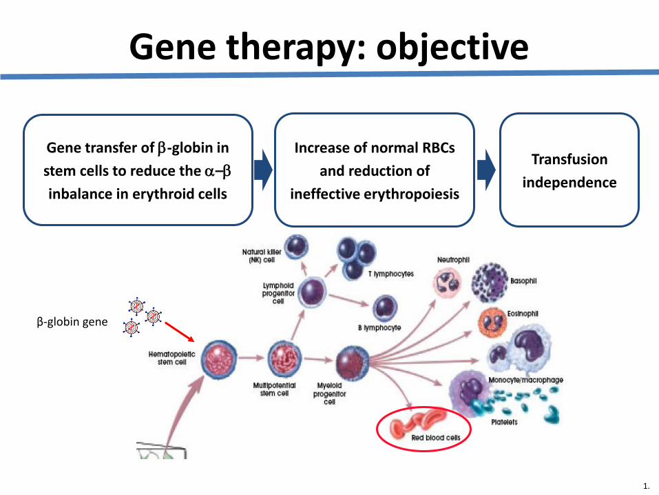
Increase of normal RBCs
and reduction of
ineffective erythropoiesis
Transfusion
independence
Gene transfer of -globin in
stem cells to reduce the –
inbalance in erythroid cells
Gene therapy: objective
1.

Source of stem cells
– mobilization is the preferred source of stem cells for gene therapy of thalassaemia
• G-CSF: safe and efficient mobilization of non-splenectomized patients
• plerixafor: safe and efficient mobilization of splenectomized patients
Yannaki E, et al. Mol Ther. 2012 ;20:230-8.

Ongoing and plannedthalassaemia gene therapy trials
Trial Patients
Paris Adults
Cincinnati Adults and children
Memphis Adults
New York Adults
Milan Adults and children
Thessaloniki Adults

12
Northstar (HGB-204) Study
• Non-randomized, open-label, international, multi-center, Phase 1/2 study in adults with β-thalassemia major– Age 18-35 years at the time of consent
– Transfused with ≥ 100 mL/kg/year of pRBCs or transfused ≥ 8 times in each of the preceding 2 years
– Sites: U.S. (4), Australia (1), Thailand (1)
• Centralized stem cell transduction with LentiGlobin BB305 lentiviral vector
• Primary objectives: Safety and efficacy of LentiGlobin BB305 Drug Product for the treatment of β-thalassemia major– Primary endpoint: ≥ 2g/dL of Hb AT87Q at 18-24 months post-infusion
– Safety endpoints: Clinical and laboratory adverse events, Replication competent lentivirus (RCL), insertion site analysis (ISA), oncogenesis
13
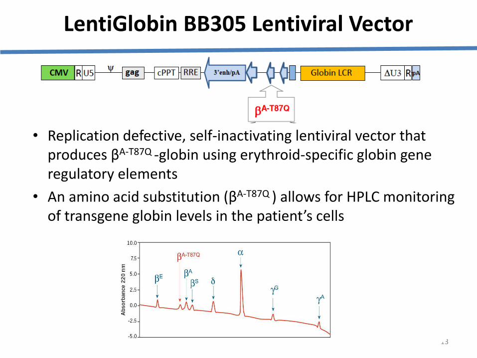
LentiGlobin BB305 Lentiviral Vector
• Replication defective, self-inactivating lentiviral vector that produces βA-T87Q -globin using erythroid-specific globin gene regulatory elements
• An amino acid substitution (βA-T87Q ) allows for HPLC monitoring of transgene globin levels in the patient’s cells

Advantages1
– no need for matched donor
– eliminates risks of GVHD and graft rejection
Challenges1,2
– optimal methods for bone-marrow conditioning
– safe and efficient gene transfer and engraftment
– lack of selection advantage for corrected cells
– consistent, safe, and therapeutic haemoglobin production in lineage-specific manner
Achievements
– correction of mouse haemoglobinopathies using ex vivo transduction of HSC with β-globin lentiviral vectors3–8
– 12 patients with severe -thalassaemia treated using lentiviral β-globin gene transfer9
GVHD, graft-versus-host disease.
1. Arumugam P, Mailk P. Hematology. 2010:445-50.2. Rothe M, et al. Curr Gene Ther. 2013;13:453-68. 3. May C, et al. Nature. 2000;406:82-6.
4. Pawliuk R, et al. Science. 2001;294:2368-71. 5. Hanawa H, et al. Blood. 2004;104:2281-90. 6. Imren S, et al. Proc Natl Acad Sci U S A. 2002;99:14380-85. 7. Levasseur DN, et al. Blood. 2003;102:4312-19.
8. Malik P, et al. Ann NY Acad Sci. 2005;1054:238-49. 9. Cavazzana-Calvo M, et al. Nature. 2010;467:318-22.
Summary Gene therapy: where are we now?

Domanda 1
• Quale è il vostro feeling nei confronti della terapia genica:
- rispetto al trapianto di midollo
- costo/beneficio

New approaches to Thalassaemia treatment
• Gene Therapy
• Gene Therapy based on HbF induction
• Regulation of erythropoiesis
• Iron chelation

Rivella S.
Looping model

Vettore pCL20cAnkyrinGG1DDiGFP
• Recruitment of LCR byLdb1 fused to a ZF thatbinds specificsequences on -gene
• Redirecting the developmental gene expression switch in favor to gamma globinshas therapeuticimplications
RivelleaS. et al EHA 2015

Baseline + ZF-Ldb10
20
40
60
80
βS/α-globins γ/α-globins γ/βS-globins
Baseline +ZF-Ldb10.0
0.2
0.4
0.6
0.8n
orm
alize
d b
y G
AP
DH
*
Baseline +ZF-Ldb10.0
0.5
1.0
1.5
no
rma
lize
d b
y G
AP
DH
** **
Baseline +ZF-Ldb10
2
4
6
8
no
rma
lize
d b
y a
-glo
bin
(le
ve
l o
f d
iffe
ren
tia
tio
n)
Hemoglobin F(α2γ2)
Hemoglobin S(α2βs
2)Hemoglobin A2
(α2δ2)
Shift of globin mRNA expression in vitro (coltures of Cd34 from SCD patients)
HbSHbF switch
Baseline + ZF-Ldb10
20
40
60
80
100
Baseline + ZF-Ldb10
5
10
15
20***
***
***
Hb
% Hb
%
Hb
%
Shift dell’espressione dell’mRNA delle globine
These results demonstrate the power of forced chromatin looping to reprogram developmental regulation of gene expression
It provide a novel proof of concept for activating the γ-globin gene for the benefit of patients with hemoglobinopathies
Conclusions

New approaches to Thalassaemia treatment
• Gene Therapy
• Gene Therapy based on HbF induction
• Regulation of erythropoiesis

Sotatecept (ACE-011)
Luspatercept (ACE-536)
23
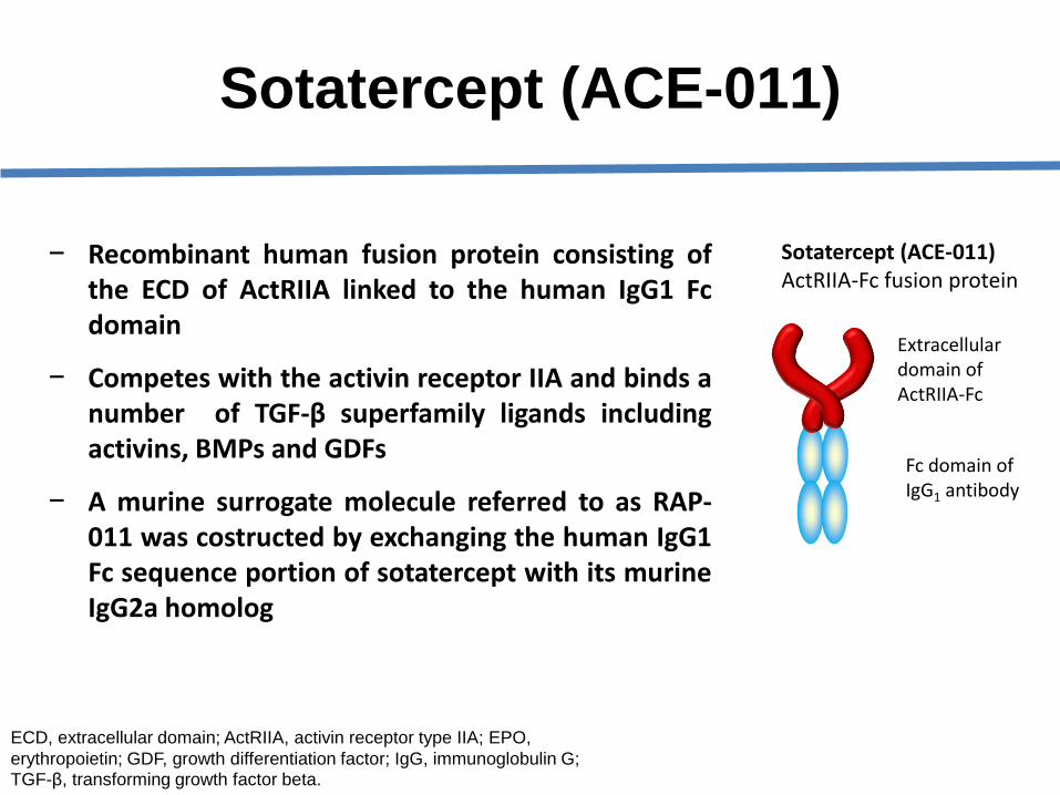
− Recombinant human fusion protein consisting ofthe ECD of ActRIIA linked to the human IgG1 Fcdomain
− Competes with the activin receptor IIA and binds anumber of TGF-β superfamily ligands includingactivins, BMPs and GDFs
− A murine surrogate molecule referred to as RAP-011 was costructed by exchanging the human IgG1Fc sequence portion of sotatercept with its murineIgG2a homolog
ECD, extracellular domain; ActRIIA, activin receptor type IIA; EPO,
erythropoietin; GDF, growth differentiation factor; IgG, immunoglobulin G; TGF-β, transforming growth factor beta.
Sotatercept (ACE-011)ActRIIA-Fc fusion protein
Extracellular domain of ActRIIA-Fc
Fc domain of IgG1 antibody
Sotatercept (ACE-011)

ACE-536• ACE-011, ACE-536 promote late-stage erythroid differentiation via a
mechanism distinct from ESAs (Suragani R et al., Nature Med 2014)
Modified ECD of ActRIIB
Fc Domain of human IgG1Antibody
ACE-536
ECD ofActRIIA
Fc Domain of human IgG1Antibody
Sotatercept
ESA, erythroid stimulating agent; ECD, extracellular domain;TGF-β, transforming growth factor β; GDF, growth differentiation factor
• ACE-536 and sotatercept bind to various ligands in the TGF-βsuperfamily with differing affinities
• Both bind to GDF11 and inhibit Smad 2,3 signaling

Key Findings From Prior Preclinical and Clinical Studies
• Sotatercept was initially evaluated as an anabolic bone agent1
• Pharmacodynamic effects include increased bone mass (anabolic and anti-resorptive2) and increased red cell parameters, as reported in multiple animal models and a phase I study of healthy postmenopausal women3
26
AEs=adverse events.
1. Pearsall RS, et al. PNAS. 2008;105:7082-7087. 2. Lotinun S, et al. Bone. 2010;46:1082-1088, 2. Ruckle J, et al. J Bone Miner Res.2009;24:744-752. 3. Data on file. Celgene Corporation.

Key Findings From Prior Preclinical and Clinical
Studies
• Administration of ACE-011 to monkeys or mice has resulted in the
reversal of bone loss and osteoporosis (Lotinun et al, Bone. 2010)
• In healthy postmenopausal women, sotatercept therapy was
associated with increased RBC parameters, including Hb level(Sherman ML, et al. J Clin Pharmacol. 2013)
• RAP-011 was effective in a β-thalassemia mouse model, supporting
clinical development of sotatercept (Dussiot M, et al. Nat Med. 2014;20:398-
407)
Model: Transgenic β-thalassemia intermedia mouse model (Hbbth1/th1)
Treatment : RAP-011 10mg/kg twice/week for 60 days
Outcome: Corrected anemia
Improved ineffective erythropoiesis
Decreased spleen weight and cellularity
Improved RBC morphology
Decreased iron overload
Wild-type Hbbth1/th1 +Vehicle Hbbth1/th1 + RAP-011

Interim Results From a Phase 2a, Open-Label, Dose-Finding Study to Determine the Safety, Efficacy, and Tolerability of Sotatercept (ACE-011) in Adults With β-Thalassemia
To determine a safe, tolerable, and effective dose of sotatercept in adult patients with β-thalassemia major who are transfusion dependent (TD), and adult patients with β-thalassemia intermedia who are TD or non-TD (NTD
Objectives

Results:NTD Patients
(S137) Interim results from phase 2A, open-label,
dose-findings study od Sotatercept (Ace 011) in
adult patients with Beta Thalassemia
MD Cappellini

Results: Dose-dependent Hb Increasein NTD Thalassemia Patients
Hb
ch
ange
fro
m b
asel
ine
(g/d
L)
3
2
1
0
−1
−2
−3
−41 43 85 127 169 211 253 295 337 379 421 463 505 547 589 631 673 715 757 799 841
Time (days)
0.1 mg/kg0.3 mg/kg0.5 mg/kg0.75 mg/kg1.0 mg/kg
0.1 mg/kg 6 6 6 5 5 3 3 3 3 3 3 3 3 3 3 2 2 2 2 10.3 mg/kg 6 6 6 6 6 6 6 6 6 6 6 6 6 6 6 6 5 5 4 4 3 3 2 2 2 2 2 2 2 2 2 2 2 2 2 2 2 2 2 10.5 mg/kg 6 6 6 6 5 5 5 5 5 4 4 4 4 4 4 4 4 3 3 2 2 2 2 2 2 2 2 2 2 2 2 1
0.75 mg/kg 7 7 7 7 6 6 6 6 6 6 5 5 3 3 3 3 3 3 3 2 2 2 2 21.0 mg/kg 5 5 5 5 5 4 4 3 2 2 2 1 1 1 1 1 1
Number of patients with sotatercept dose (mg/kg)
Interim data as of May 6, 2015.
Sotatercept dose
4 1 year 2 years

Results: NTD Thalassemia Patients With Hb Increase Sustained for ≥ 12 Weeks
33%
86%
67%
0%0%
Patients
(%
)
33%
20%
71%
0.1 mg/kg(n = 6)
0.3 mg/kg(n = 6)
0.5 mg/kg(n = 6)
0.75 mg/kg(n = 7)
0.1 mg/kg(n = 6)
67%
1.0 mg/kg(n = 5)
0.3 mg/kg(n = 6)
0.5 mg/kg(n = 6)
0.75 mg/kg(n = 7)
1.0 mg/kg(n = 5)
20%
≥ 1.0 g/dL Hb increase ≥ 1.5 g/dL Hb increase
Interim data as of May 6, 2015.Sotatercept dose
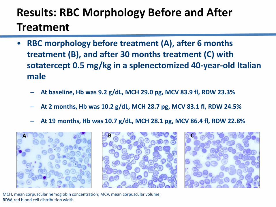
• RBC morphology before treatment (A), after 6 months treatment (B), and after 30 months treatment (C) with sotatercept 0.5 mg/kg in a splenectomized 40-year-old Italian male
‒ At baseline, Hb was 9.2 g/dL, MCH 29.0 pg, MCV 83.9 fl, RDW 23.3%
‒ At 2 months, Hb was 10.2 g/dL, MCH 28.7 pg, MCV 83.1 fl, RDW 24.5%
‒ At 19 months, Hb was 10.7 g/dL, MCH 28.1 pg, MCV 86.4 fl, RDW 22.8%
Results: RBC Morphology Before and After Treatment
A B C
MCH, mean corpuscular hemoglobin concentration; MCV, mean corpuscular volume; RDW, red blood cell distribution width.

• 30 year old male, non-transfusion dependent thalassemia intermedia• Baseline Hb 9.2 g/dL• History of lower limb ulcers since 2011
• Leg ulcer healing noticed 2 weeks after first dose of ACE-536 (0.4 mg/kg) • 2nd dose delayed due to unrelated bone marrow hypoplasia• Leg ulcer substantially resolved after 6 weeks on treatment• Patient received a total of 4 doses; maximum Hb on study 10.6 g/dL
Patient 0203 – Leg UlcerACE-536 -Thalassemia Phase 2 Clinical Trial
Pre-Treatment After 6 Weeks ACE-536
Study Day
-1
-0.5
0
0.5
1
1.5
2
2.5
3
1 22 43 64 85 113 141
A.Piga EHA 2014

Results:TD Patients
(S136) Luspatercept (Ace 356) increases hemoglobin and
decreases transfusion burden and liver iron concentration in
adult with Beta-thalassemia: preliminary results from phase 2
study
A. Piga

Results: Reduction in Transfusion Burden for TD β-Thalassemia Patients
Sotatercept dose
a Percentage change in transfusion burden (units/168 days) from baseline to on-treatment. Interim data as of May 6, 2015.
Change in tra
nsfu
sio
n b
urd
en (
%)a
20% reduction
0.1 mg/kg(n = 2)
0.3 mg/kg(n = 3)
0.5 mg/kg (n = 2)
0.75 mg/kg(n = 5)
1.0 mg/kg(n = 4)
• Mean transfusion burden reduction among patients treated with sotatercept ≥ 0.5 mg/kg was 32.25%

Results: Erythroid Response Correlates With Serum Sotatercept Exposure
• No apparent effects of weight, sex, age, or transfusion burden
on drug clearance were observed
Interim data as of April 1, 2015.
Average drug concentration (μg/mL)C
han
ge in
RB
C t
ran
sfu
sio
n b
urd
en
(un
its/
24
wee
ks)
0 2 4 6 8
−15
−10
−5
0
5
10 Slope = −6.3509 R = 0.71 P = 0.002
Transfusion change over 24 weeks (TD patients)
Average drug concentration (μg/mL)
Mea
n H
b c
han
ge (
g/d
L/2
4 w
eeks
)
0 2 4 6 8
−1
0
1
2
3Slope = 0.2923 R = 0.731 P = 0.0000
Mean Hb change over 24 weeks (NTD patients)
0.1 mg/kg0.3 mg/kg0.5 mg/kg0.75 mg/kg1.0 mg/kg
Sotatercept dose

38
Where Does Sotatercept Impact
Erythropoiesis?
EPO Dependent
Hemoglobin
SOTATERCEPT
Baso E ReticBFU-E CFU-E Pro E RBCPoly E Ortho E
38
EryA EryB EryC
How much EPO is required?

Conclusions
• These data suggest that long-term treatment with
sotatercept can:
– Increase Hb levels in NTD patients
– Reduce transfusion burden in TD patients
• Increase in Hb level and reduction in transfusion
burden correlated with increased drug exposure
• Sotatercept, or the related molecule luspatercept, may
provide a favorable benefit-risk profile for patients
with TD or NTD β-thalassemia, addressing a
significant unmet need

Domanda 2
• Quale è il meccanismo attraverso cui sotatercept riduce l’eritropoiesi inefficace?
• Pensate che l’efficacia terapeutica sia sufficiente per eliminare le trasfusioni?

WSG of EHA on RED CELLS AND IRON
Agenda:- Update on EHA Research Roadmap
( A. Iolascon)- Update on the preparation of the Guidelines:
. Splenectomy (A. Iolascon)
. HA and pregnancy (A. Taher)
. Microcytic atypic anemias (P. Aguilar-Martinez)
. HA iron overload :diagnosis and treatment (MD Cappellini)
- Collaborative Project on treatment of PK deficiency(Zanella A/Mizer K)
- New ENERCA projects: application and update(JV. Corrons, P. Aguilar-Martinez)
- MSc course in Inherited Haemoglobin Disorders (D. Loukopoulos)
- Proposal for future ad-hoc meetings

EHA RESEARCH ROADMAP . Sec. 4 : Anemias and related disorders
• Introduction
• Table: the best of research roadmap in anemias
• - epidemiology of anemias in Europe
• - common flow-charts for diagnosis
• - pathogenesis studies of rare inherited anemiasto have new therapeutic targets
• - enhance clinical trials for new drugs
• - use of new technologies for a personalized diagnosis and therapy

Applicazione della NGS allo
studio ed alla diagnosi delle
anemie rare
Achille Iolascon
Dipartimento Medicina Molecolare e Biotecnologie Mediche
Università degli Studi di Napoli “Federico II”
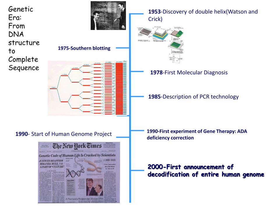
1953-Discovery of double helix(Watson and Crick)
1975-Southern blotting
1985-Description of PCR technology
1978-First Molecular Diagnosis
1990- Start of Human Genome Project 1990-First experiment of Gene Therapy: ADA deficiency correction
2000-First announcement of decodification of entire human genome
Genetic Era:From DNA structure to Complete Sequence
Era Pre-genomica Era Post-genomica

Whole genome sequencing vs Exome-sequencing:
diagnosis and discovery
Whole-genome sequencing (WGS):
characterize entire genomes of any
size and complexity
Exome sequencing: sequence
protein coding regions, as cost-
effective alternative to WGS
RNA-Seq: cDNA generated from
RNA allows for mutation analysis in
expressed genes
Discovery of “novel” disease genes: sequencing of
genomic DNA

is a rare inherited syndrome characterized by developmental defects, short
stature, bone marrow failure, and an increased risk to malignancies.
Fifteen genetic subtypes of FA have been identified so far
Cells derived from FA patients are hypersensitive to
chromosomal breakage induced by DNA inter strand
cross-linking agents (ICLs) such as mitomycin C
(MMC) or diepoxybutane (DEB) and assessment of
this cellular hypersensitivity is the classic diagnostic
test for FA.
However the chromosomal breakage test
is positive in only ~10% of patients and
occasionally the test gives false positive
results in other genetic disorders, such as
Nijmegen breakage syndrome (NBS; MIM
#251260) and Roberts syndrome (RBS,
MIM #268300).
Fanconi anemia (FA) in Pre-genomic era
Rosendorff and Bernstein, 1988; Gille et al., 2012

A male patient, aged 4 years and 11 months, was born to a healthy non-
consanguineous parents;
The patient had left hand preaxial polydactyly and an irregular hypo-
pigmentation spot on the left back trunk;
Surgery to correct the thumb deformity was performed when the patient was
6 months of age;
The patient's development was not officially tested, but was estimated to be
within normal range;
The patient's family history was unremarkable
Fanconi anemia (FA) in Post-genomic era


Targeted-NGS as diagnostic tool for genetic
disorders
We have not yet reached a point at which
routine sequencing of large numbers of
whole eukaryotic genomes is feasible
It is often necessary to select genomic
regions of interest and to enrich these
regions before sequencing
There are several enrichment approaches
(for example, HaloPlex)
Target re-sequencing facilitates the creation
of rapid, accurate and lower cost
diagnostic applications

Estimated prevalence of HHA: from 1/1.000 to 1/1.000.000
Differential diagnosis of HHA is often difficult and requires specialized analyses
A lot of non-specific and overlapping phenotypes between different conditions
Hereditary hemolytic anemias (HHA)
Exclusion of anemias due to:
• autoimmunity
• acquired condition
Hemolytic anemia
Normal or reduction
Reticulocyte
count
Hyporegenerative
anemias (HHA)
Increase
Hemolytic Anemias due
to red cell Membrane
Defects (HAMD)
Adapted from Andolfo I, Russo R, Iolascon A. Prosp Ped 2014;44:1-7

Congenital Dyserythropoietic Anemias (CDAs)
CDAs are mendelian diseases affecting the normal differentiation-proliferation pathway of
the erythroid lineage.
They belong to a subtype of bone marrow failure syndromes characterized by
monolineage involvement and morphological abnormalities in erythroid precursor cells
Williams Hematology, Nineth Edition, Chapter 39 by A. Iolascon - McGraw-Hill _ In press
Erythroid hyperplasia with
binuclearity or
multinuclearity involving
late erythroblasts
Mild hemoltyic anemia (9-10 g/dL)
Reduced reticulocyte count
Jaundice
Splenomegaly
Hemosiderosis
Gallstones
Transfusion dependence (≈ 20%)

Hemolytic Anemias due to red cell Membrane
Defects (HAMDs)
Adapted from Andolfo I, et al. Prosp Ped 2014;44:1-7
Mild hemoltyic anemia (9-10 g/dL)
Increased reticulocyte count
Jaundice
Splenomegaly
Gallstones

Differential diagnosis
CDA IIHS
Splenectomy
slightly increases
Hb level
Splenectomy is
the standard
treatment
↑ Unc. Bilirubin
↑ LDH
↓ Haptoglobin
Hepatosplenomegaly
RDWRDW
Reticulocyte
count
Reticulocyte
count
Autosomal
dominant (75%)
Autosomal
recessive
sTFRNormal or slightly
increased sTFR
Hemosiderosis
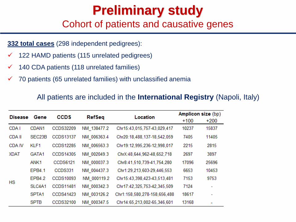
Preliminary studyCohort of patients and causative genes
332 total cases (298 independent pedigrees):
122 HAMD patients (115 unrelated pedigrees)
140 CDA patients (118 unrelated families)
70 patients (65 unrelated families) with unclassified anemia
All patients are included in the International Registry (Napoli, Italy)

Panel of 10 causative
genes of HS and CDAs (2012-2013)
Selection of 15 patients:
6 with known genotype;
6 with unknown genotype;
1 family
Pilot study
Coding regions, UTRs,
regulatory regions, 100 bp
flanking splice junctions
Inheritance pattern and
validation by Sanger
sequencing
Preliminary study design
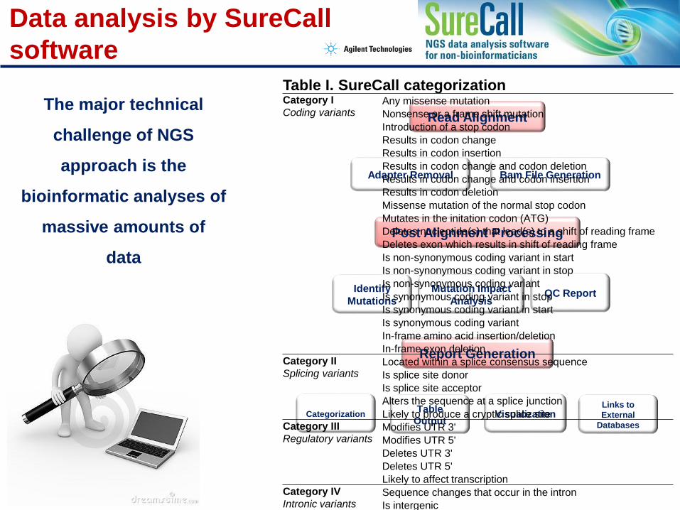
Read Alignment
Post Alignment Processing
Report Generation
Adapter Removal Bam File Generation
Identify
Mutations
Mutation Impact
AnalysisQC Report
CategorizationLinks to
External
Databases
VisualizationTable
Output
Table I. SureCall categorizationCategory I
Coding variantsAny missense mutation
Nonsense or a frame shift mutation
Introduction of a stop codon
Results in codon change
Results in codon insertion
Results in codon change and codon deletion
Results in codon change and codon insertion
Results in codon deletion
Missense mutation of the normal stop codon
Mutates in the initation codon (ATG)
Deletes nucleotide(s) that lead(s) to a shift of reading frame
Deletes exon which results in shift of reading frame
Is non-synonymous coding variant in start
Is non-synonymous coding variant in stop
Is non-synonymous coding variant
Is synonymous coding variant in stop
Is synonymous coding variant in start
Is synonymous coding variant
In-frame amino acid insertion/deletion
In-frame exon deletionCategory II
Splicing variantsLocated within a splice consensus sequence
Is splice site donor
Is splice site acceptor
Alters the sequence at a splice junction
Likely to produce a cryptic splice siteCategory III
Regulatory variantsModifies UTR 3'
Modifies UTR 5'
Deletes UTR 3'
Deletes UTR 5'
Likely to affect transcriptionCategory IV
Intronic variantsSequence changes that occur in the intron
Is intergenic
Is intronic variant
Data analysis by SureCall
software
The major technical
challenge of NGS
approach is the
bioinformatic analyses of
massive amounts of
data

Variants in clinical report of targeted-NGS-based
diagnosis for HHA patients
Total variants 62−122
Off-target gene variants 0−2
Target gene variants 55−105
Intronic and regulatory gene variants 48−92
Coding gene variants 5−13
Variants related to clincal phenotype
Variants modifying clinical phenotype
• SPTA1 α-LELY
Complete pedigree (12-father; 13-mother; 14-proband; 15-unaf. sister)
Patients with known genotype
Filtering variants (1):
MAF (1000 genomes and EVS)
Strend bias
Filtering variants (2):
PolyPhen
SIFT
HSF

Acknowledgements

Chairs: Patricia Aguilar-Martinez, Paola Bianchi, Achille Iolascon, Richard Van Wijk, Alberto Zanella
Topics: Disorders of Red Cell Production Disorders of Red Cell Survival – Hereditary Haemolytic Anaemias Disorders of Red Cell Survival – Acquired Haemolytic Anaemias Diagnosis and Treatment of Very Rare Anaemias
To register and for further information : www.esh.org
TRAINING COURSE ON
DIAGNOSIS AND MANAGEMENT
OF VERY RARE RED CELL AND IRON DISORDERS
Lisbon, Portugal
January 29-30, 2016

Paulson. Nature Medicine Editorial , 20 , 4 , 2014
After: Dussiot et al. Nat Med, 4, 398-407, 2014
Suragani et al. Nat Med, 4, 408-14 2014