Embed Size (px)
Citation preview

Research Article
HighlyOptimizedDNAVaccine TargetingHumanTelomeraseReverse Transcriptase Stimulates Potent AntitumorImmunity
Jian Yan1, Panyupa Pankhong2, Thomas H. Shin2, Nyamekye Obeng-Adjei2, Matthew P. Morrow1,Jewell N. Walters2, Amir S. Khan1, Niranjan Y. Sardesai1, and David B. Weiner2
AbstractHigh levels of human telomerase reverse transcriptase (hTERT) are detected in more than 85% of human
cancers. Immunologic analysis supports that hTERT is a widely applicable target recognized by T cells andcan be potentially studied as a broad cancer immunotherapeutic, or a unique line of defense against tumorrecurrence. There remains an urgent need to develop more potent hTERT vaccines. Here, a synthetic highlyoptimized full-length hTERT DNA vaccine (phTERT) was designed and the induced immunity was examinedin mice and non-human primates (NHP). When delivered by electroporation, phTERT elicited strong, broadhTERT-specific CD8 T-cell responses including induction of T cells expressing CD107a, IFN-g , and TNF-a inmice. The ability of phTERT to overcome tolerance was evaluated in an NHP model, whose TERT is 96%homologous to that of hTERT. Immunized monkeys exhibited robust [average 1,834 spot forming unit (SFU)/106 peripheral blood mononuclear cells (PBMC)], diverse (multiple immunodominant epitopes) IFN-gresponses and antigen-specific perforin release (average 332 SFU/106 PBMCs), suggesting that phTERTbreaks tolerance and induces potent cytotoxic responses in this human-relevant model. Moreover, in anHPV16-associated tumor model, vaccination of phTERT slows tumor growth and improves survival rate inboth prophylactic and therapeutic studies. Finally, in vivo cytotoxicity assay confirmed that phTERT-inducedCD8 T cells exhibited specific cytotoxic T lymphocyte (CTL) activity, capable of eliminating hTERT-pulsedtarget cells. These findings support that this synthetic electroporation-delivered DNA phTERT may have arole as a broad therapeutic cancer vaccine candidate. Cancer Immunol Res; 1(3); 179–89. �2013 AACR.
IntroductionImmunotherapy for cancer through induction of antitumor
cellular immunity has recently reemerged as an importantexperimental therapy for the treatment of nonresponsivecancers. However, most tumor-associated antigens (TAA) areexpressed in one or a few tumor types as tumors generallyexhibit tissue-specific features (1). In contrast, human telome-rase reverse transcriptase (hTERT), a catalytic subunit oftelomerase, is highly expressed in more than 85% of humantumors from diverse cancer phenotypes, with little or noexpression in normal somatic cells (2–6). Expression of hTERT
correlates with telomerase activity, which may be a require-ment for tumor survival (7). Telomerase activation/hTERTexpression is associated with little loss of telomere length andaccounts for the unlimited proliferative capacity of cancercells. As expression of hTERT is directly linked to tumor cellgrowth and contributes crucially to the long-term survival oftumor cells, loss of telomerase activity will lead to hTERT-positive tumor cell death by apoptosis (8). In addition, target-ing hTERT may have the potential to eliminate cancer stemcells as recent studies have suggested that cancer stemor stem-like cells express hTERT (9–11). These findings collectivelypoint to hTERT as an attractive TAA and provide the basis ofdeveloping hTERT-based universal vaccine for cancer immu-notherapy (12–14).
Therapeutic hTERT vaccines have been widely studiedbecause of their potential to stimulate the killing of tumorcells by enhancing the activity of telomerase-specific cytotoxicCD8 T cells (12). Many studies have been conducted to develophTERT peptide vaccines containing motifs that either bindto MHC class I (I540 and 572Y) or MHC class II molecules(GV1001; refs. 15–18). Moreover, multiple strategies are beingexplored to use full-length hTERT recombinant constructstargeting multiple CD8 and CD4 epitopes simultaneously. Asa result, the potency of autologous dendritic cells transducedwith hTERT mRNA (19, 20) and viral vector-based vaccines
Authors' Affiliations: 1Inovio Pharmaceuticals, Inc., Blue Bell; and2Department of Pathology and Laboratory Medicine, University of Penn-sylvania, Philadelphia, Pennsylvania
Note: Supplementary data for this article are available at Cancer Immu-nology Research Online (http://cancerimmunolres.aacrjournals.org/).
J. Yan and P. Pankhong contributed equally to this work.
Corresponding Author: David B. Weiner, Department of Pathology andLaboratory Medicine, University of Pennsylvania, 505 Stellar-Chance Lab-oratories, 422 Curie Boulevard, Philadelphia, PA 19104. Phone: 215-349-8365; Fax: 215-573-9436; E-mail: [email protected]
doi: 10.1158/2326-6066.CIR-13-0001
�2013 American Association for Cancer Research.
CancerImmunology
Research
www.aacrjournals.org 179
on June 23, 2018. © 2013 American Association for Cancer Research. cancerimmunolres.aacrjournals.org Downloaded from
Published OnlineFirst July 17, 2013; DOI: 10.1158/2326-6066.CIR-13-0001

(21, 22) has been reported with partial success in animalmodels. However, findings from initial clinical trials of hTERTvaccines in patients with cancer have shown that theseapproaches suffer from limited induction of CD8þ T cells andhad limited impact on overall survival (13). Therefore, thereremains an urgent need to develop more potent hTERTtherapeutic vaccines with a broader T-cell footprint.
Several clinical trials have evaluated the efficacy ofDNA vaccination against a variety of cancers (23). Althoughthere are some indications of limited immune responses invaccinated patients with melanoma or prostate cancer(24, 25), previous DNA vaccines generally seem to induceweak cellular immunity in humans. Recent synthetic DNAdesign strategies, such as codon/RNA optimization, theaddition of highly efficient immunoglobulin leader sequen-ces (26–28), use of more efficient DNA delivery methodsincluding in vivo electroporation (29), have been applied toimprove the immune responses induced by DNA vaccines inhumans, with recent significant success (30). However, thesenew approaches have not been combined to test a newhTERT DNA vaccine.
In this report, we attempt to extend this improved immunepotency to construct a synthetic DNA vaccine expressing afull-length hTERT with modifications using a combinationof approaches in gene optimization. The hTERT DNA wasdelivered by electroporation and its immunogenicity andantitumor effectwere evaluated in non-humanprimates (NHP)and mice. These data strongly support further study of thehTERT DNA vaccine in combination with electroporationdelivery as a potential immunotherapy platform against anarray of human and animal malignancies.
Materials and MethodsImmunogen design and expression
A synthetic hTERT DNA vaccine was generated using thehTERT sequence retrieved from GenBank (accession num-ber: AF018196) with several modifications (Fig. 1A). The full-length hTERT gene was 3,512 bp and subcloned into anexpression vector pGX0001.
In vitro hTERT expression was detected using TNTQuick Coupled Transcription/Translation System (Pro-mega). The gene product was immunoprecipitated usingan anti-HA (hemagglutinin) tag monoclonal antibody (Sigma-Aldrich) and analyzed by SDS-PAGE. The synthesized proteinwas detected by autoradiography.
An indirect immunofluorescent assay was conducted toconfirmhTERTexpression as previously described (31). Briefly,human rhabdomyosarcoma cells were transfected withphTERT and pGX0001 (1 mg/well) using TurboFectin8.0 Trans-fection Reagent (OriGene). Forty-eight hours later, the cellswere fixed and incubated with anti-hTERT (C-term) mono-clonal antibody (Millipore) overnight at 4�C. The slides werethen incubated with fluorescein isothiocyanate (FITC)–con-jugated secondary antibody (Cell Signaling Technology), andanalyzed by fluorescent microscopy (LeicaMicrosystems, Inc.)using the SPOT Advanced software (SPOT Diagnostic Instru-ments, Inc.).
Mice studiesMice and immunization. Female 8-week-old C57BL/6
mice were purchased from The Jackson Laboratory. Their carewas in accordance with the guidelines of NIH and Universityof Pennsylvania Institutional Animal Care and Use Com-mittee (IACUC). Mice were divided into two groups andimmunized with 50 mg of hTERT DNA by intramuscular injec-tion into the quadriceps followed by electroporation usingthe CELLECTRA adaptive constant current device (InovioPharmaceuticals, Inc.; ref. 32). The mice received four immu-nizations, two weeks apart. One week after the last immuni-zation, the mice were sacrificed and splenocytes were isolatedfor immunology studies.
ELISpot assay. Mouse IFN-g enzyme-linked immunosor-bent spot (ELISpot) assay was conducted as previouslydescribed (31). Peptides spanning the entire hTERT protein,each containing 15 amino acids overlapping by eight aminoacids, were synthesized by GenScript. The entire set of pep-tides was pooled at a concentration of 2 mg/mL/peptide intofour pools for stimulation of the IFN-g release.
IgELS
hTERT
HA tag
**
R589Y D1005Y
pG
X0
00
1
phT
ER
T
203 kD
116 kD
A
CBEcoRI
NotI
phTERT
6464 bp
hTERTpCMV
pUC ori
BGH pA
Kan
D
DAPI
FITC
Merge
phTERTMock
Figure 1. Design and expression of hTERT DNA vaccine. A, schematic ofhTERT antigen. The � denotes the incorporated point mutation. B,map ofphTERT. C, detection of phTERT expression by in vitro translation.The gene product was immunoprecipitated using an anti-HA(hemagglutinin) tag monoclonal antibody, visualized by SDS-PAGE andautoradiography. D, immunofluorescence assay of phTERT. Transfectedrhabdomyosarcoma cells expressing hTERT protein showed typicalFITC-fluorescence using a commercial hTERT (C-term) monoclonalantibody. DAPI, 40,6-diamidino-2-phenylindole.
Yan et al.
Cancer Immunol Res; 1(3) September 2013 Cancer Immunology Research180
on June 23, 2018. © 2013 American Association for Cancer Research. cancerimmunolres.aacrjournals.org Downloaded from
Published OnlineFirst July 17, 2013; DOI: 10.1158/2326-6066.CIR-13-0001

CD8þ T-cell depletion. CD8þ lymphocytes were depletedfrom splenocytes using Dynabeads mouse CD8 (Lyt2; LifeTechnologies). After CD8þ T cells depletion, IFN-g ELISpotwas conducted as described earlier.Intracellular cytokine staining. Intracellular cytokine
staining (ICS) was conducted as described previously (32).Briefly, splenocytes from vaccinated and na€�ve mice werestimulated with hTERT peptides, stained with FITC anti-mouse CD107a, and followed by ViViD Dye (Invitrogen). Cellswere then stained with the following extracellular antibodies:APC-Cy7 anti-mouse CD3e, PerCP-Cy5.5 anti-mouse CD4, andAPC anti-mouse CD8a (BD Biosciences). Intracellular cyto-kines were subsequently stained with the following antibodies:Alexa Fluor 700 anti-mouse IFN-g and PE-Cy7 anti-mouse TNF(BD Biosciences).Cell line. TC-1 cell line is a well-characterized lung epi-
thelial cell line immortalized with HPV16 E6/E7, and trans-formed with the c-Ha-ras oncogene (33). TC-1 cells werepurchased from American Type Culture Collection and main-tained in RPMI-1640 medium supplemented with 10% FBS,100 U/mL penicillin and 100 mg/mL streptomycin at 37�C.In vivo tumor challenge study. Ten female 8-week-old
C57BL/6 mice were immunized with 50 mg of phTERT fourtimes biweekly. One week after the last immunization, eachmouse was challenged with 5 � 104 TC-1 cells injected sub-cutaneously including 10 na€�ve mice that served as a control.Tumors were measured twice weekly with digital calipersspanning the shortest (width) and longest surface diameters(length; ref. 34). Tumor volumes were calculated accordingto the formula: V ¼ length � width2 � p/6 (35). Mice weresacrificed when tumor diameter reached 20mm in compliancewith our IACUC protocol.In vivo tumor treatment study. Female C57BL/6 mice
were separated into two groups of 10 mice: na€�ve and hTERTgroup. On day 0, all mice were injected subcutaneously with5 � 104 TC-1 cells in the right flank. All mice in the hTERTgroup were immunized with 50 mg of phTERT on days 3, 10, 17,and 24. Tumors measurement was conducted as describedearlier.In vivo cytotoxicity study. An in vivo cytotoxicity assay
was conducted as previously described (36, 37). Splenocytesfrom na€�ve mice were stained with carboxyfluorescein diace-tate succinimidyl ester (CFSE) at a concentration of 1 mmol/Lor 1 nmol/L. CFSEhi (1 mmol/L)–labeled cells were pulsed withthe relevant peptides (hTERT peptides), whereas CFSElo
(1 nmol/L)–labeled cells were pulsed with the irrelevant pep-tides (HPV6 E6/E7 peptides). Equal frequency of CFSEhi andCFSElo cells was combined and 107 cells were intravenouslyinjected into na€�ve or phTERT-immunized mice. Forty-eighthours later, splenocytes were isolated and analyzed by flowcytometry. The percentage killing was calculated as follows:100 � [(% relevant peptide pulsed in immunized/% irrelevantpeptide pulsed in immunized)/(% relevant peptide pulsed inna€�ve/% irrelevant peptide pulsed in na€�ve)] � 100.
Rhesus monkey studiesImmunization and PBMC isolation. Four rhesus maca-
queswere vaccinatedwith phTERT, four times intramuscularly
followed by electroporation using CELLECTRA adaptive con-stant current electroporation device, 6 weeks apart, at 2 mgDNA/each immunization. Blood was collected 2 weeks aftereach immunization and peripheral blood mononuclear cells(PBMC) were isolated by standard Ficoll-Hypaque densitygradient centrifugation.
IFN-g and perforin ELISpot assay. Monkey IFN-g andperforin ELISpot were conducted as previously described(38, 39). Antigen-specific responses were determined by sub-tracting the number of spots in the negative control wellsfrom the wells containing peptides. After subtracting thenegative control, the mean value in the wells with the PBMCscollected postvaccination had to exceed 50 SFU/106 PBMCsand be at least four times higher than prevaccination reactivityto be considered as a positive response.
Statistical analysis. Standard and paired Student t testswere applied to analyze statistical significance of all quanti-tative data produced in this study, and P < 0.05 was consideredstatistically significant.
ResultsDesign and construction of the full-length hTERT DNAvaccine
As indicated in Fig. 1A, the hTERT immunogen was devel-oped with several modifications, including codon/RNA opti-mization and the addition of a highly efficient leader sequence,to enhance the expression and immunogenicity of phTERT.Two mutations (R589Y and D1005Y) were incorporated intothe hTERT sequence to assist in breaking tolerance (40). Themodified gene was subcloned into pGX0001 and named asphTERT for further study (Fig. 1B)
Expression of hTERTIn vitro expression of hTERT was verified by T7 coupled
transcription and translation reaction. After immunoprecipi-tation with the anti-HA tag monoclonal antibody, hTERTexpression was analyzed by 12% SDS-PAGE. The hTERT pro-tein migrated to the corresponding molecular weight atapproximately 130 kDa (Fig. 1C). No protein bandwas detectedin the pGX0001 vector lane. An indirect immunofluorescenceassay was conducted to further confirm hTERT expression. Asshown in Fig. 1D, the cells expressing hTERT protein showedtypical FITC-fluorescence, supporting the expression of hTERTin a relatively native conformation. As a control, expressionwas not detected in pGX0001-transfected cells.
Vaccination with phTERT induces strong CD8-mediatedhTERT-specific responses in mice
IFN-g ELISpot was conducted to assess antigen-specificcellular immune responses induced by phTERT after fourimmunizations (Fig. 2A). The total response against fourpools of hTERT peptides in phTERT-immunized mice was1,817 � 211 SFU/106 splenocytes, which was significantlygreater than the immune responses in the na€�ve group(20� 6 SFU/106 splenocytes; P¼ 0.01; Fig. 2B). The C-terminusof the hTERT protein (peptide pool 4) was the most immu-nogenic region, accounting for about half the response elicitedby phTERT (970 � 113 SFU/106 splenocytes).
hTERT-DNA Vaccine Induces Strong Antitumor CTLs
www.aacrjournals.org Cancer Immunol Res; 1(3) September 2013 181
on June 23, 2018. © 2013 American Association for Cancer Research. cancerimmunolres.aacrjournals.org Downloaded from
Published OnlineFirst July 17, 2013; DOI: 10.1158/2326-6066.CIR-13-0001
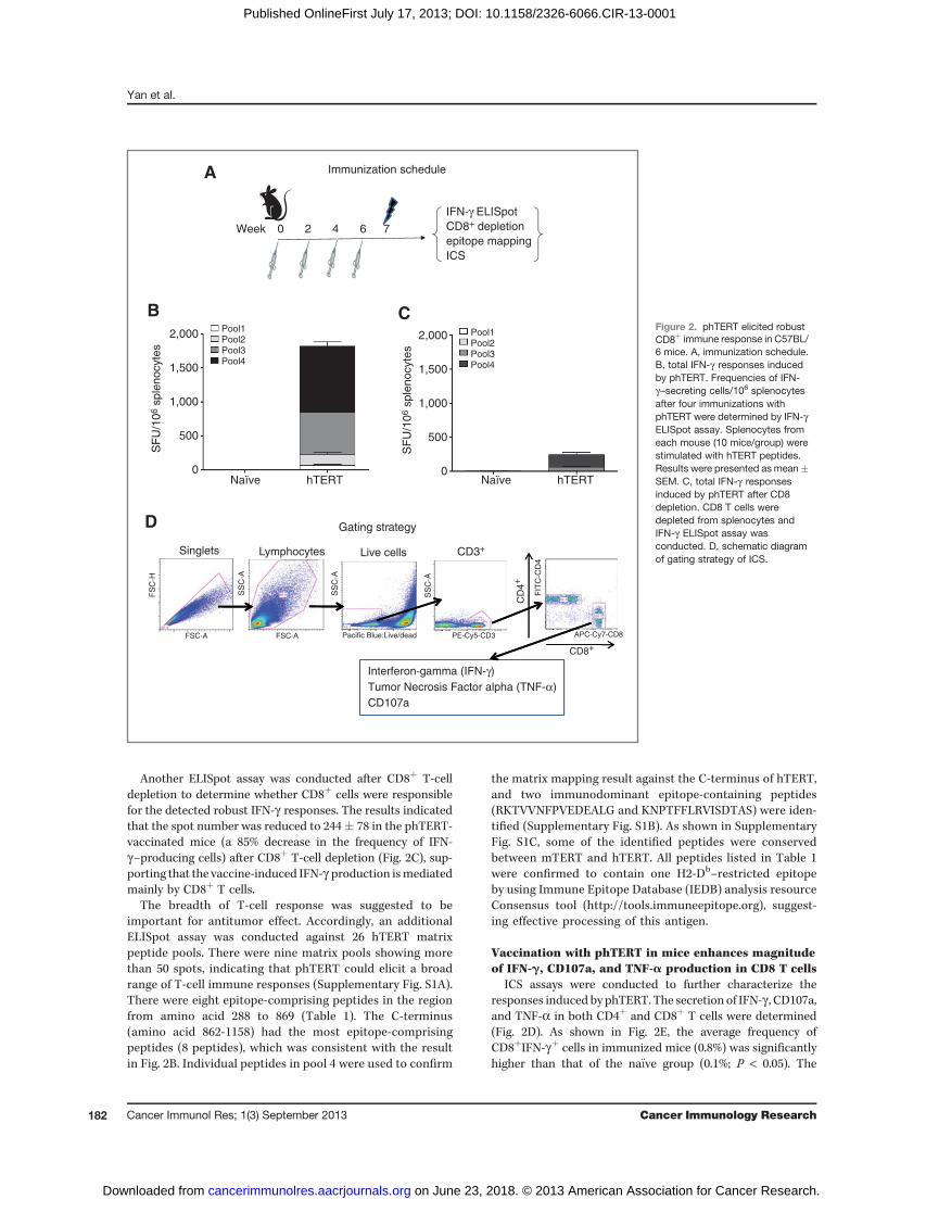
Another ELISpot assay was conducted after CD8þ T-celldepletion to determine whether CD8þ cells were responsiblefor the detected robust IFN-g responses. The results indicatedthat the spot number was reduced to 244� 78 in the phTERT-vaccinated mice (a 85% decrease in the frequency of IFN-g–producing cells) after CD8þ T-cell depletion (Fig. 2C), sup-porting that the vaccine-induced IFN-g production ismediatedmainly by CD8þ T cells.
The breadth of T-cell response was suggested to beimportant for antitumor effect. Accordingly, an additionalELISpot assay was conducted against 26 hTERT matrixpeptide pools. There were nine matrix pools showing morethan 50 spots, indicating that phTERT could elicit a broadrange of T-cell immune responses (Supplementary Fig. S1A).There were eight epitope-comprising peptides in the regionfrom amino acid 288 to 869 (Table 1). The C-terminus(amino acid 862-1158) had the most epitope-comprisingpeptides (8 peptides), which was consistent with the resultin Fig. 2B. Individual peptides in pool 4 were used to confirm
the matrix mapping result against the C-terminus of hTERT,and two immunodominant epitope-containing peptides(RKTVVNFPVEDEALG and KNPTFFLRVISDTAS) were iden-tified (Supplementary Fig. S1B). As shown in SupplementaryFig. S1C, some of the identified peptides were conservedbetween mTERT and hTERT. All peptides listed in Table 1were confirmed to contain one H2-Db–restricted epitopeby using Immune Epitope Database (IEDB) analysis resourceConsensus tool (http://tools.immuneepitope.org), suggest-ing effective processing of this antigen.
Vaccination with phTERT in mice enhances magnitudeof IFN-g, CD107a, and TNF-a production in CD8 T cells
ICS assays were conducted to further characterize theresponses induced by phTERT. The secretion of IFN-g , CD107a,and TNF-a in both CD4þ and CD8þ T cells were determined(Fig. 2D). As shown in Fig. 2E, the average frequency ofCD8þIFN-gþ cells in immunized mice (0.8%) was significantlyhigher than that of the na€�ve group (0.1%; P < 0.05). The
Immunization schedule
6 7 420Week
IFN-γ ELISpot
CD8+ depletion
epitope mapping
2,000
1,500
1,000
500
0
Pool1Pool2Pool3Pool4
Pool1Pool2Pool3Pool4
Naïve hTERT Naïve hTERT
SF
U/1
06 s
ple
nocyte
s
2,000
1,500
1,000
500
0
SF
U/1
06 s
ple
nocyte
s
ICS
B C
A
D Gating strategy
Interferon-gamma (IFN-γ)
Tumor Necrosis Factor alpha (TNF-α)
CD107a
Singlets Lymphocytes Live cells CD3+
CD8+
CD
4+
FS
C-H
FSC-A FSC-A
SS
C-A
SS
C-A
Pacific Blue:Live/dead
SS
C-A
PE-Cy5-CD3
FIT
C-C
D4
APC-Cy7-CD8
Figure 2. phTERT elicited robustCD8þ immune response in C57BL/6 mice. A, immunization schedule.B, total IFN-g responses inducedby phTERT. Frequencies of IFN-g–secreting cells/106 splenocytesafter four immunizations withphTERT were determined by IFN-gELISpot assay. Splenocytes fromeach mouse (10 mice/group) werestimulated with hTERT peptides.Results were presented as mean�SEM. C, total IFN-g responsesinduced by phTERT after CD8depletion. CD8 T cells weredepleted from splenocytes andIFN-g ELISpot assay wasconducted. D, schematic diagramof gating strategy of ICS.
Yan et al.
Cancer Immunol Res; 1(3) September 2013 Cancer Immunology Research182
on June 23, 2018. © 2013 American Association for Cancer Research. cancerimmunolres.aacrjournals.org Downloaded from
Published OnlineFirst July 17, 2013; DOI: 10.1158/2326-6066.CIR-13-0001

Table 1. Identified epitope-comprising peptides in mice immunized with phTERT
Number of epitope-comprisingpeptides
Sequence of epitope-comprisingpeptides
aa 1–296 (pool 1) None N/A
aa 288–582 (pool 2)TGARRLVETIFLGSR
4 ETIFLGSRPWMPGTPRPLFLELLGNHAQCPLGNHAQCPYGVLLKT
aa 575–869 (pool 3)DGLRPIVNMDYVVGA
4 NMDYVVGARTFRREKSVLNYERARRPGLLGARRPGLLGASVLGLD
RKTVVNFPVEDEALGPVEDEALGGTAFVQMDTRTLEVQSDYSSYA
aa 862–1158 (pool 4) 8 QSDYSSYARTSIRASNSLQTVCTNIYKILLTNIYKILLLQAYRFHKNPTFFLRVISDTASRVISDTASLCYSILK
NOTE: Two identified immuno-dominant epitope-comprising peptides are highlighted in bold.Abbreviation: aa, amino acid.
CD4+ CD8+E
P < 0.05
P < 0.05
P < 0.05
Naïve
1.5
1.0
0.5
0.0
% I
FN
-γ+ c
ells
of
tota
l C
D4
+ T
ce
lls
% T
NF
-α+ c
ells
of
tota
l C
D4
+ T
ce
lls
% C
D1
07
a+ c
ells
of
tota
l C
D4
+ T
ce
lls
% C
D1
07
a+ c
ells
of
tota
l C
D8
+ T
ce
lls%
TN
F-α
+ c
ells
of
tota
l C
D8
+ T
ce
lls
% I
FN
-γ+ c
ells
of
tota
l C
D8
+ T
ce
lls
1.5
1.0
0.5
0.0
3.5
3.0
2.5
1.5
1.0
0.5
0.0
4.5
3.0
1.5
0.0
4.5
3.0
1.5
0.0
4.5
3.0
1.5
0.0
hTERT Naïve hTERT
Naïve hTERT Naïve hTERT
Naïve hTERT Naïve hTERT
Figure 2. (Continued ) E, secretionof CD107a, IFN-g , and TNF-apostvaccination in both CD4þ andCD8þ cells. Splenocytes werestimulatedwith hTERT peptides for5 hours before surface andintracellular staining. Background-subtracted percentages of hTERT-specific CD4þ or CD8þ T cellsproducing CD107a, IFN-g , andTNF-a were calculated. Theexperiment shown isrepresentative of three differentexperiments using 5 mice pergroup.
hTERT-DNA Vaccine Induces Strong Antitumor CTLs
www.aacrjournals.org Cancer Immunol Res; 1(3) September 2013 183
on June 23, 2018. © 2013 American Association for Cancer Research. cancerimmunolres.aacrjournals.org Downloaded from
Published OnlineFirst July 17, 2013; DOI: 10.1158/2326-6066.CIR-13-0001
percentage of TNF-a–secreting cells from the total CD8þ
T-cell population in phTERT-immunized mice was approxi-mately 1.2% on average, whereas the na€�ve group producedonly 0.1% (P < 0.05). After showing that antigen-specific CD8þ
T cells had the ability to secrete IFN-g and TNF-a, we inves-tigated whether these CD8þ T cells exhibited a phenotypeof putative CTLs. CD107a, a marker of cytolytic degranula-tion on lymphocytes, such asCD8þTcells, was used to evaluatethe CTL potential of vaccine-induced T cells. Following stim-ulation with hTERT peptides, the percentage of CD107a-pos-itive CD8 cells was 1.8%, which was significantly higher thanthe percentage in the na€�ve group. There was a trend showingthe increased production of IFN-g , TNF-a, and CD107a inCD4þ T cells in the immunized mice. However, the differencesin average frequencies of CD4þIFN-gþ, CD4þ TNF-aþ, andCD4þ CD107aþ cells were not statistically significant com-pared with what were observed in the na€�vemice. The immuneresponses elicited by phTERT are heavily skewed towarddriving CD8þ lymphocytes with the potential to lyse hTERT-expressing tumor cells.
Vaccination with phTERT is capable of breakingtolerance and generating robust hTERT-specific CTLs inrhesus macaques
The induction of T-cell immunity against the tumor antigenhTERT could be controlled by mechanisms of central andperipheral tolerance. Sequence homology analysis indicatedthat hTERT shares 64% identity with mouse TERT, and 96%identity with rhesus macaque TERT. Consequently, immunetolerance is expected to play amajor role in testing the efficacyof an hTERT vaccine in NHPs. Furthermore, rhesus T-cellimmunity is much closer to human T-cell immunity servingas a highly relevant model for immunotherapeutic vaccinedevelopment. Therefore, we moved forward to determinewhether phTERT is able to break tolerance and induce cellularresponses in rhesus macaques. The prebleed blood sampleswere studied to establish the background level of immuneresponse of each individual animal in the study.
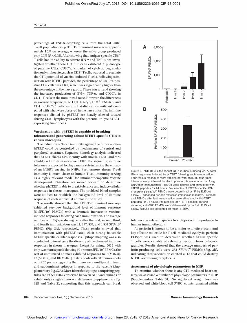
The results showed that the hTERT-immunized monkeysexhibited very low background level of immune response(5 SFU/106 PBMCs) with a dramatic increase in vaccine-induced responses following each immunization. The averagenumber of IFN-g–producing cells after the first, second, third,and fourth immunization was 11, 177, 834, and 1,834 SFU/106
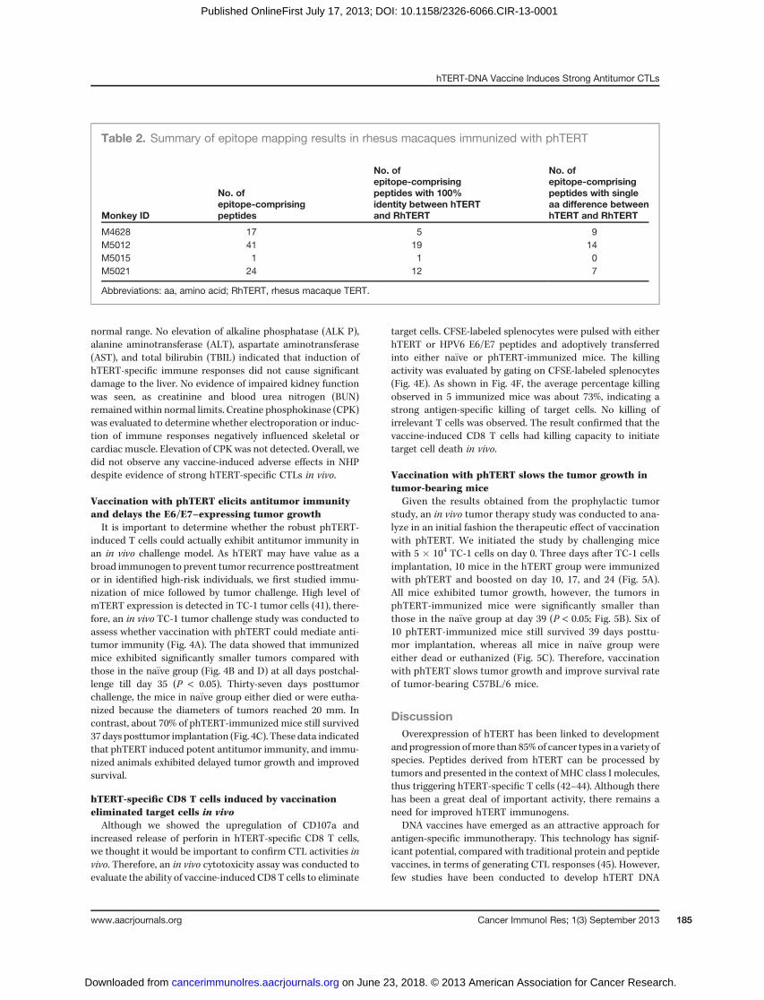
PBMCs (Fig. 3A), respectively. These results showed thatimmunization with phTERT could elicit strong boostablehTERT-specific cellular responses. Epitope mapping was alsoconducted to investigate the diversity of the observed immuneresponses in rhesus macaques. Except for animal 5015 withonly twomatrix pools showing 50 ormore SFU/106 PBMCs, therest of immunized animals exhibited responses to 9 (M4628),13 (M5012), and 10 (M5021)matrix pools with 50 ormore spotsout of 26 pools, suggesting that there were multiple dominantand subdominant epitopes in response to the vaccine (Sup-plementary Fig. S2A). Most identified epitope-comprising pep-tides are either 100% conserved between NHP and humans orexhibit only a single amino acid difference (Supplementary Fig.S2B and Table 2), supporting that this approach can break
tolerance in relevant species to epitopes with importance tohuman immunotherapy.
As perforin is known to be a major cytolytic protein andkey effector molecule for T cell–mediated cytolysis, perforinELISpot was used to determine whether hTERT-specificT cells were capable of releasing perforin from cytotoxicgranules. Results showed that the average numbers of per-forin-producing cells were 332 SFU/106 PBMCs (Fig. 3B),indicating that vaccination elicited CTLs that could destroyhTERT-expressing target cells.
Assessment of physiologic parameters in NHPTo examine whether there is any CTL-mediated host tox-
icity, we assessed a number of physiologic parameters in NHP(Supplementary Table S1). No significant weight loss wasobserved and white blood cell (WBC) counts remained within
A 3,000
2,000
1,000
Pre-vac Post-vac
SF
U/1
06 P
BM
Cs
Perf
orin S
FU
/10
6 P
BM
Cs
500
400
300
200
100
0
0
Pre
ble
ed
Imm
1
Imm
2
Imm
3
Imm
4
B
Figure 3. phTERT elicited robust CTLs in rhesus macaques. A, totalIFN-g responses induced by phTERT following each immunization.Four rhesus macaques were vaccinated with phTERT, four timesintramuscularly followed by electroporation, 6 weeks apart, at 2 mgDNA/each immunization. PBMCs were isolated and stimulated withhTERT peptides for 24 hours. Frequencies of hTERT-specific IFN-g–secreting cells/106 PBMCs were determined by IFN-g ELISpotassay. B, enhanced perforin release in immunized monkeys. Prebleedand PBMCs after last immunization were stimulated with hTERTpeptides for 24 hours. Frequencies of hTERT-specific perforin-secreting cells/106 PBMCs were determined by perforin ELISpotassay. Results are presented as mean � SEM.
Yan et al.
Cancer Immunol Res; 1(3) September 2013 Cancer Immunology Research184
on June 23, 2018. © 2013 American Association for Cancer Research. cancerimmunolres.aacrjournals.org Downloaded from
Published OnlineFirst July 17, 2013; DOI: 10.1158/2326-6066.CIR-13-0001
normal range. No elevation of alkaline phosphatase (ALK P),alanine aminotransferase (ALT), aspartate aminotransferase(AST), and total bilirubin (TBIL) indicated that induction ofhTERT-specific immune responses did not cause significantdamage to the liver. No evidence of impaired kidney functionwas seen, as creatinine and blood urea nitrogen (BUN)remained within normal limits. Creatine phosphokinase (CPK)was evaluated to determine whether electroporation or induc-tion of immune responses negatively influenced skeletal orcardiac muscle. Elevation of CPKwas not detected. Overall, wedid not observe any vaccine-induced adverse effects in NHPdespite evidence of strong hTERT-specific CTLs in vivo.
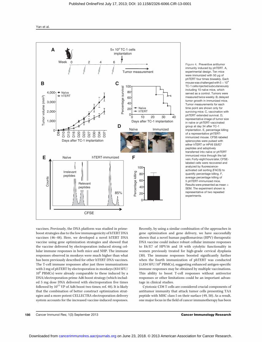
Vaccination with phTERT elicits antitumor immunityand delays the E6/E7–expressing tumor growthIt is important to determine whether the robust phTERT-
induced T cells could actually exhibit antitumor immunity inan in vivo challenge model. As hTERT may have value as abroad immunogen to prevent tumor recurrence posttreatmentor in identified high-risk individuals, we first studied immu-nization of mice followed by tumor challenge. High level ofmTERT expression is detected in TC-1 tumor cells (41), there-fore, an in vivo TC-1 tumor challenge study was conducted toassess whether vaccination with phTERT could mediate anti-tumor immunity (Fig. 4A). The data showed that immunizedmice exhibited significantly smaller tumors compared withthose in the na€�ve group (Fig. 4B and D) at all days postchal-lenge till day 35 (P < 0.05). Thirty-seven days posttumorchallenge, the mice in na€�ve group either died or were eutha-nized because the diameters of tumors reached 20 mm. Incontrast, about 70% of phTERT-immunized mice still survived37 days posttumor implantation (Fig. 4C). These data indicatedthat phTERT induced potent antitumor immunity, and immu-nized animals exhibited delayed tumor growth and improvedsurvival.
hTERT-specific CD8 T cells induced by vaccinationeliminated target cells in vivoAlthough we showed the upregulation of CD107a and
increased release of perforin in hTERT-specific CD8 T cells,we thought it would be important to confirm CTL activities invivo. Therefore, an in vivo cytotoxicity assay was conducted toevaluate the ability of vaccine-induced CD8 T cells to eliminate
target cells. CFSE-labeled splenocytes were pulsed with eitherhTERT or HPV6 E6/E7 peptides and adoptively transferredinto either na€�ve or phTERT-immunized mice. The killingactivity was evaluated by gating on CFSE-labeled splenocytes(Fig. 4E). As shown in Fig. 4F, the average percentage killingobserved in 5 immunized mice was about 73%, indicating astrong antigen-specific killing of target cells. No killing ofirrelevant T cells was observed. The result confirmed that thevaccine-induced CD8 T cells had killing capacity to initiatetarget cell death in vivo.
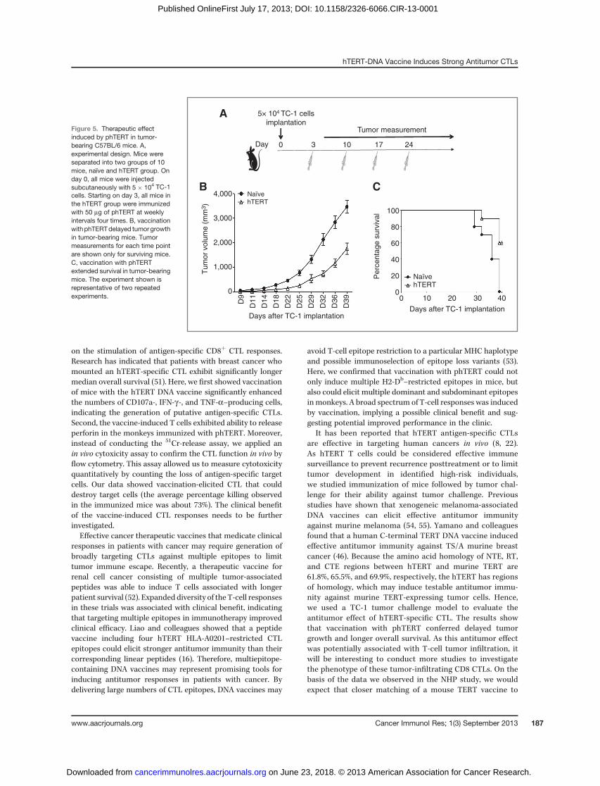
Vaccination with phTERT slows the tumor growth intumor-bearing mice
Given the results obtained from the prophylactic tumorstudy, an in vivo tumor therapy study was conducted to ana-lyze in an initial fashion the therapeutic effect of vaccinationwith phTERT. We initiated the study by challenging micewith 5 � 104 TC-1 cells on day 0. Three days after TC-1 cellsimplantation, 10 mice in the hTERT group were immunizedwith phTERT and boosted on day 10, 17, and 24 (Fig. 5A).All mice exhibited tumor growth, however, the tumors inphTERT-immunized mice were significantly smaller thanthose in the na€�ve group at day 39 (P < 0.05; Fig. 5B). Six of10 phTERT-immunized mice still survived 39 days posttu-mor implantation, whereas all mice in na€�ve group wereeither dead or euthanized (Fig. 5C). Therefore, vaccinationwith phTERT slows tumor growth and improve survival rateof tumor-bearing C57BL/6 mice.
DiscussionOverexpression of hTERT has been linked to development
and progression ofmore than 85%of cancer types in a variety ofspecies. Peptides derived from hTERT can be processed bytumors and presented in the context of MHC class I molecules,thus triggering hTERT-specific T cells (42–44). Although therehas been a great deal of important activity, there remains aneed for improved hTERT immunogens.
DNA vaccines have emerged as an attractive approach forantigen-specific immunotherapy. This technology has signif-icant potential, compared with traditional protein and peptidevaccines, in terms of generating CTL responses (45). However,few studies have been conducted to develop hTERT DNA
Table 2. Summary of epitope mapping results in rhesus macaques immunized with phTERT
Monkey ID
No. ofepitope-comprisingpeptides
No. ofepitope-comprisingpeptides with 100%identity between hTERTand RhTERT
No. ofepitope-comprisingpeptides with singleaa difference betweenhTERT and RhTERT
M4628 17 5 9M5012 41 19 14M5015 1 1 0M5021 24 12 7
Abbreviations: aa, amino acid; RhTERT, rhesus macaque TERT.
hTERT-DNA Vaccine Induces Strong Antitumor CTLs
www.aacrjournals.org Cancer Immunol Res; 1(3) September 2013 185
on June 23, 2018. © 2013 American Association for Cancer Research. cancerimmunolres.aacrjournals.org Downloaded from
Published OnlineFirst July 17, 2013; DOI: 10.1158/2326-6066.CIR-13-0001
vaccines. Previously, the DNA platform was studied in prime-boost strategies due to the low immunogenicity of hTERTDNAvaccines (46–48). Here, we developed a novel hTERT DNAvaccine using gene optimization strategies and showed thatthe vaccine delivered by electroporation induced strong cel-lular immune responses in both mice and NHP. The immuneresponses observed in monkeys were much higher than whathas been previously described for other hTERT DNA vaccines.The T-cell immune responses after just three immunizationswith 2mg of phTERT by electroporation inmonkeys (834 SFU/106 PBMCs) were already comparable to these induced by aDNA/electroporation prime Ad6 boost strategy (which includ-ed 5 mg dose DNA delivered with electroporation five timesfollowed by 1011 VP of Ad6 boost two times; ref. 48). It is likelythat the combination of better construct optimization strat-egies and a more potent CELLECTRA electroporation deliverysystem accounts for the increased vaccine-induced responses.
Recently, by using a similar combination of the approaches ingene optimization and gene delivery, we have successfullyshown that a novel human papillomavirus (HPV) therapeuticDNA vaccine could induce robust cellular immune responsesto E6/E7 of HPV16 and 18 with cytolytic functionality inwomen previously treated for high-grade cervical dysplasia(30). The immune responses boosted significantly furtherwhen the fourth immunization of phTERT was conducted(1,834 SFU/106 PBMCs), suggesting enhanced antigen-specificimmune responses may be obtained by multiple vaccinations.This ability to boost T-cell responses without antivectorresponses or other limitations could be an important advan-tage in clinical studies.
Cytotoxic CD8 T cells are considered crucial components ofantitumor immunity that attack tumor cells presenting TAApeptide with MHC class I on their surface (49, 50). As a result,onemajor focus in the field of cancer immunotherapy has been
0 2 7 64Week
5× 10⁴ TC-1 cells
implantation
Tumor measurement
100
80
60
40
20
0
4,000
3,000
2,000
1,000
0
100
80
60
40
20
0
0
D8
D10
D13
D16
D20
D23
D27
D30
D34
D37
10
Days after TC-1 implantation
Days after TC-1 implantation
20 30 40
Perc
enta
ge s
urv
ival
Tum
or
volu
me (
mm
3)
Perc
enta
ge k
illin
g
A
B C
DNaïve
Naïve hTERT
Naïve hTERT
Immunized
Naïve hTERT-immunized
Irrelevant
peptides
Relevant
peptides
CFSE
Counts
85%
E F
Figure 4. Preventive antitumorimmunity induced by phTERT. A,experimental design. Ten micewere immunized with 50 mg ofphTERT four times biweekly. Eachmousewaschallengedwith5�104
TC-1 cells injected subcutaneouslyincluding 10 naïve mice, whichserved as a control. Tumors weremeasured twiceweekly. B, delayedtumor growth in immunized mice.Tumor measurements for eachtime point are shown only forsurviving mice. C, vaccination withphTERT extended survival. D,representative image of tumor sizein naïve or phTERT-vaccinatedgroup at day 34 after TC-1implantation. E, percentage killingof a representative phTERT-immunized mouse. CFSE-labeledsplenocytes were pulsed witheither hTERT or HPV6 E6/E7peptides and adoptivelytransferred into naïve or phTERTimmunized mice through the tailvein. Forty-eight hours later, CFSE-labeled cells were recovered andanalyzed by fluorescence-activated cell sorting (FACS) toquantify percentage killing. F,average percentage killing of5 phTERT-immunized mice.Results were presented as mean�SEM. The experiment shown isrepresentative of two repeatedexperiments.
Yan et al.
Cancer Immunol Res; 1(3) September 2013 Cancer Immunology Research186
on June 23, 2018. © 2013 American Association for Cancer Research. cancerimmunolres.aacrjournals.org Downloaded from
Published OnlineFirst July 17, 2013; DOI: 10.1158/2326-6066.CIR-13-0001
on the stimulation of antigen-specific CD8þ CTL responses.Research has indicated that patients with breast cancer whomounted an hTERT-specific CTL exhibit significantly longermedian overall survival (51). Here, we first showed vaccinationof mice with the hTERT DNA vaccine significantly enhancedthe numbers of CD107a-, IFN-g-, and TNF-a–producing cells,indicating the generation of putative antigen-specific CTLs.Second, the vaccine-induced T cells exhibited ability to releaseperforin in the monkeys immunized with phTERT. Moreover,instead of conducting the 51Cr-release assay, we applied anin vivo cytoxicity assay to confirm the CTL function in vivo byflow cytometry. This assay allowed us to measure cytotoxicityquantitatively by counting the loss of antigen-specific targetcells. Our data showed vaccination-elicited CTL that coulddestroy target cells (the average percentage killing observedin the immunized mice was about 73%). The clinical benefitof the vaccine-induced CTL responses needs to be furtherinvestigated.Effective cancer therapeutic vaccines that medicate clinical
responses in patients with cancer may require generation ofbroadly targeting CTLs against multiple epitopes to limittumor immune escape. Recently, a therapeutic vaccine forrenal cell cancer consisting of multiple tumor-associatedpeptides was able to induce T cells associated with longerpatient survival (52). Expanded diversity of the T-cell responsesin these trials was associated with clinical benefit, indicatingthat targeting multiple epitopes in immunotherapy improvedclinical efficacy. Liao and colleagues showed that a peptidevaccine including four hTERT HLA-A0201–restricted CTLepitopes could elicit stronger antitumor immunity than theircorresponding linear peptides (16). Therefore, multiepitope-containing DNA vaccines may represent promising tools forinducing antitumor responses in patients with cancer. Bydelivering large numbers of CTL epitopes, DNA vaccines may
avoid T-cell epitope restriction to a particular MHC haplotypeand possible immunoselection of epitope loss variants (53).Here, we confirmed that vaccination with phTERT could notonly induce multiple H2-Db–restricted epitopes in mice, butalso could elicit multiple dominant and subdominant epitopesinmonkeys. A broad spectrum of T-cell responses was inducedby vaccination, implying a possible clinical benefit and sug-gesting potential improved performance in the clinic.
It has been reported that hTERT antigen-specific CTLsare effective in targeting human cancers in vivo (8, 22).As hTERT T cells could be considered effective immunesurveillance to prevent recurrence posttreatment or to limittumor development in identified high-risk individuals,we studied immunization of mice followed by tumor chal-lenge for their ability against tumor challenge. Previousstudies have shown that xenogeneic melanoma-associatedDNA vaccines can elicit effective antitumor immunityagainst murine melanoma (54, 55). Yamano and colleaguesfound that a human C-terminal TERT DNA vaccine inducedeffective antitumor immunity against TS/A murine breastcancer (46). Because the amino acid homology of NTE, RT,and CTE regions between hTERT and murine TERT are61.8%, 65.5%, and 69.9%, respectively, the hTERT has regionsof homology, which may induce testable antitumor immu-nity against murine TERT-expressing tumor cells. Hence,we used a TC-1 tumor challenge model to evaluate theantitumor effect of hTERT-specific CTL. The results showthat vaccination with phTERT conferred delayed tumorgrowth and longer overall survival. As this antitumor effectwas potentially associated with T-cell tumor infiltration, itwill be interesting to conduct more studies to investigatethe phenotype of these tumor-infiltrating CD8 CTLs. On thebasis of the data we observed in the NHP study, we wouldexpect that closer matching of a mouse TERT vaccine to
A
0 2417103 Day
5× 10⁴ TC-1 cellsimplantation
Tumor measurement
CB
100
80
60
40
20
0
4,000
3,000
2,000
1,000
00D
9
D11
D14
D18
D22
D25
D29
D32
D36
D39 10
Days after TC-1 implantationDays after TC-1 implantation
20 30 40
Perc
enta
ge s
urv
ival
Tum
or
volu
me (
mm
3)
Naïve hTERT
Naïve hTERT
Figure 5. Therapeutic effectinduced by phTERT in tumor-bearing C57BL/6 mice. A,experimental design. Mice wereseparated into two groups of 10mice, naïve and hTERT group. Onday 0, all mice were injectedsubcutaneously with 5 � 104 TC-1cells. Starting on day 3, all mice inthe hTERT group were immunizedwith 50 mg of phTERT at weeklyintervals four times. B, vaccinationwithphTERTdelayed tumorgrowthin tumor-bearing mice. Tumormeasurements for each time pointare shown only for surviving mice.C, vaccination with phTERTextended survival in tumor-bearingmice. The experiment shown isrepresentative of two repeatedexperiments.
hTERT-DNA Vaccine Induces Strong Antitumor CTLs
www.aacrjournals.org Cancer Immunol Res; 1(3) September 2013 187
on June 23, 2018. © 2013 American Association for Cancer Research. cancerimmunolres.aacrjournals.org Downloaded from
Published OnlineFirst July 17, 2013; DOI: 10.1158/2326-6066.CIR-13-0001

the native mouse sequence would further improve theeffectiveness.
A major challenge with regard to hTERT and other TAAimmunotherapy vaccines is to develop therapies capable ofgenerating a robust CTL against this self-antigen in a safemanner. Although hTERT is overexpressed in most tumorcells, its expression can also be detected in rare normal cellssuch as hematopoietic progenitor cells, spermatogonia in thetestis, activated lymphocytes, and certain epithelial cells (56).Consequently, the question of whether vaccine-inducedhTERT-specific CTLs carries the risk of inducing autoimmuneresponses with pathologic consequence was raised. Manystudies have shown the hTERT-specific CTLs have no detect-able effect on hTERT-positive CD34þ hematopoietic progen-itor cells or activated T cells (43, 44) and do not result inautoimmune responses that target normal hTERT-expressingcells (22, 57). In addition, clinical studies in patients withcancer using hTERT-based vaccines have not shown toxicity(8, 19, 58). These findings may reflect relatively low levels ofhTERT expression or ineffective processing of hTERT peptidesin normal cells. In the present study, several important phys-iologic parameters were evaluated and no vaccine-inducedadverse effects were detected in phTERT-immunizedmonkeysdespite evidence of strong hTERT-specific CTLs.
Taken together, we report that administration of a syn-thetic highly optimized hTERT DNA vaccine in combinationwith adaptive constant current electroporation delivery plat-form was capable of breaking immune tolerance and elicitingrobust and diverse CTLs in mouse and NHP models. Thesevaccine-induced CTLs seemed not to be associated with anymajor toxicities or organ damage, and were effective in
mounting a potent antitumor response. These data supportfurther study of phTERT in the setting of cancer immuno-therapy, to improve tumor immune surveillance in high-riskindividuals, and prevention of disease recurrence.
Disclosure of Potential Conflicts of InterestN.Y. Sardesai has ownership interest (including patents) in Inovio Pharma-
ceuticals, Inc. D.B. Weiner has commercial research grant from Inovio Pharma-ceuticals, Inc. and MVI, ownership interest (including patents) in Inovio Phar-maceuticals, Inc., and is a consultant/advisory board member of Inovio Phar-maceuticals, Inc., Novartis, VGXi, Pfizer, Merck, and Bristol-Myers Squibb.No potential conflicts of interest were disclosed by the other authors.
Authors' ContributionsConception and design: J. Yan, M.P. Morrow, N.Y. Sardesai, D.B. WeinerDevelopment of methodology: J. Yan, N. Obeng-Adjei, N.Y. Sardesai, D.B.WeinerAcquisition of data (provided animals, acquired and managed patients,provided facilities, etc.): J. Yan, T.H. Shin, M.P. Morrow, J.N. WaltersAnalysis and interpretation of data (e.g., statistical analysis, biostatistics,computational analysis): J. Yan, P. PankhongWriting, review, and/or revision of the manuscript: J. Yan, P. Pankhong,N. Obeng-Adjei, M.P. Morrow, A.S. Khan, N.Y. Sardesai, D.B. WeinerAdministrative, technical, or material support (i.e., reporting or orga-nizing data, constructing databases): J. Yan, N.Y. SardesaiStudy supervision: J. Yan, A.S. Khan, N.Y. Sardesai
Grant SupportThis work was supported in part by NIH grants and a grant from the Basser
Research Center for BRCA in the Abramson Cancer Center at the University ofPennsylvania awarded to D.B. Weiner. J.N. Walters notes funding from Ruth L.Kirschstein National Research Service Award (5T32CA115299-07).
The costs of publication of this article were defrayed in part by the paymentof page charges. This article must therefore be hereby marked advertisementin accordance with 18 U.S.C. Section 1734 solely to indicate this fact.
Received January 21, 2013; revised May 29, 2013; accepted July 11, 2013;published OnlineFirst July 17, 2013.
References1. Chiang CL, Benencia F, Coukos G. Whole tumor antigen vaccines.
Semin Immunol 2010;22:132–43.2. KimNW, PiatyszekMA, Prowse KR, Harley CB,West MD, Ho PL, et al.
Specific association of human telomerase activity with immortal cellsand cancer. Science 1994;266:2011–5.
3. Shay JW,Bacchetti S. A surveyof telomerase activity in humancancer.Eur J Cancer 1997;33:787–91.
4. Hiyama E, Hiyama K. Telomerase as tumor marker. Cancer Lett2003;194:221–33.
5. Greider CW, Blackburn EH. The telomere terminal transferase ofTetrahymena is a ribonucleoprotein enzyme with two kinds of primerspecificity. Cell 1987;51:887–98.
6. Hastie ND, Dempster M, Dunlop MG, Thompson AM, Green DK,Allshire RC. Telomere reduction in human colorectal carcinoma andwith ageing. Nature 1990;346:866–8.
7. Harrington L, Zhou W, McPhail T, Oulton R, Yeung DS, Mar V, et al.Human telomerase contains evolutionarily conserved catalytic andstructural subunits. Genes Dev 1997;11:3109–15.
8. Vonderheide RH, Domchek SM, Schultze JL, George DJ, Hoar KM,Chen DY, et al. Vaccination of cancer patients against telomeraseinduces functional antitumor CD8þ T lymphocytes. Clin Cancer Res2004;10:828–39.
9. HoMM,NgAV, LamS,Hung JY. Side population in human lung cancercell lines and tumors is enriched with stem-like cancer cells. CancerRes 2007;67:4827–33.
10. MarianCO, ChoSK,McEllin BM,Maher EA, Hatanpaa KJ,MaddenCJ,et al. The telomerase antagonist, imetelstat, efficiently targets glio-
blastoma tumor-initiating cells leading to decreased proliferation andtumor growth. Clin Cancer Res 2010;16:154–63.
11. Marian CO, Wright WE, Shay JW. The effects of telomerase inhibitionon prostate tumor-initiating cells. Int J Cancer 2010;127:321–31.
12. Harley CB. Telomerase and cancer therapeutics. Nat Rev Cancer2008;8:167–79.
13. Beatty GL, Vonderheide RH. Telomerase as a universal tumor antigenfor cancer vaccines. Expert Rev Vaccines 2008;7:881–7.
14. Shaw VE, Naisbitt DJ, Costello E, Greenhalf W, Park BK, NeoptolemosJP, et al. Current status of GV1001 and other telomerase vaccinationstrategies in the treatment of cancer. Expert Rev Vaccines 2010;9:1007–16.
15. Wenandy L, Sorensen RB, Sengelov L, Svane IM, thor Straten P,Andersen MH. The immunogenicity of the hTERT540-548 peptide incancer. Clin Cancer Res 2008;14:4–7.
16. Liao ZL, Tang XD, Lu MH, Wu YY, Cao YL, Fang DC, et al. Antitumoreffect of newmultiple antigen peptide based onHLA-A0201–restrictedCTL epitopes of human telomerase reverse transcriptase (hTERT).Cancer Sci 2012;103:1920–8.
17. Kokhaei P, PalmaM, Hansson L, Osterborg A,Mellstedt H, ChoudhuryA. Telomerase (hTERT 611-626) serves as a tumor antigen in B-cellchronic lymphocytic leukemia and generates spontaneously antileu-kemic, cytotoxic T cells. Exp Hematol 2007;35:297–304.
18. Inderberg-Suso EM, Trachsel S, Lislerud K, Rasmussen AM, Gauder-nack G. Widespread CD4þ T-cell reactivity to novel hTERT epitopesfollowing vaccination of cancer patients with a single hTERT peptideGV1001. Oncoimmunology 2012;1:670–86.
Yan et al.
Cancer Immunol Res; 1(3) September 2013 Cancer Immunology Research188
on June 23, 2018. © 2013 American Association for Cancer Research. cancerimmunolres.aacrjournals.org Downloaded from
Published OnlineFirst July 17, 2013; DOI: 10.1158/2326-6066.CIR-13-0001
19. Su Z, Dannull J, Yang BK, Dahm P, Coleman D, Yancey D, et al.Telomerase mRNA-transfected dendritic cells stimulate antigen-spe-cific CD8þ and CD4þ T cell responses in patients with metastaticprostate cancer. J Immunol 2005;174:3798–807.
20. Suso EM, Dueland S, Rasmussen AM, Vetrhus T, Aamdal S, KvalheimG, et al. hTERTmRNA dendritic cell vaccination: complete response ina pancreatic cancer patient associated with response against severalhTERT epitopes. Cancer Immunol Immunother 2011;60:809–18.
21. Rusakiewicz S, Dosset M, Mollier K, Souque P, Charneau P, Wain-Hobson S, et al. Immunogenicity of a recombinant lentiviral vectorcarrying human telomerase tumor antigen in HLA-B�0702 transgenicmice. Vaccine 2010;28:6374–81.
22. Adotevi O,Mollier K, Neuveut C, DossetM, Ravel P, FridmanWH, et al.Targeting human telomerase reverse transcriptase with recombinantlentivector is highly effective to stimulate antitumor CD8 T-cell immu-nity in vivo. Blood 2010;115:3025–32.
23. Rice J, Ottensmeier CH, Stevenson FK. DNA vaccines: precision toolsfor activating effective immunity against cancer. Nat RevCancer 2008;8:108–20.
24. Tagawa ST, Lee P, Snively J, Boswell W, Ounpraseuth S, Lee S, et al.Phase I study of intranodal delivery of a plasmid DNA vaccine forpatients with stage IV melanoma. Cancer 2003;98:144–54.
25. Todorova K, Ignatova I, Tchakarov S, Altankova I, Zoubak S, Kyurk-chiev S, et al. Humoral immune response in prostate cancer patientsafter immunizationwith gene-based vaccines that encode for a proteinthat is proteasomally degraded. Cancer Immun 2005;5:1.
26. Deml L, Bojak A, Steck S, Graf M, Wild J, Schirmbeck R, et al. Multipleeffects of codon usage optimization on expression and immunoge-nicity of DNA candidate vaccines encoding the human immunodefi-ciency virus type 1 Gag protein. J Virol 2001;75:10991–1001.
27. Muthumani K, Zhang D, Dayes NS, Hwang DS, Calarota SA, Choo AY,et al. Novel engineered HIV-1 East African Clade-A gp160 plasmidconstruct induces strong humoral and cell-mediated immuneresponses in vivo. Virology 2003;314:134–46.
28. Schneider R, Campbell M, Nasioulas G, Felber BK, Pavlakis GN.Inactivation of the human immunodeficiency virus type 1 inhibitoryelements allows Rev-independent expression of Gag and Gag/prote-ase and particle formation. J Virol 1997;71:4892–903.
29. Sardesai NY, Weiner DB. Electroporation delivery of DNA vaccines:prospects for success. Curr Opin Immunol 2011;23:421–9.
30. Bagarazzi ML, Yan J, Morrow MP, Shen X, Parker RL, Lee JC, et al.Immunotherapy against HPV16/18 generates potent TH1 and cyto-toxic cellular immune responses. Sci Transl Med 2012;4:155ra38.
31. Yan J, Yoon H, Kumar K, Ramanathan MP, Corbitt N, Kutzler M, et al.Enhanced diversity and magnitude of cellular immune responseselicited by a novel engineered HIV-1 subtype B consensus-basedenvelope DNA vaccine. Mol Ther 2007;15:411–21.
32. Shin TH, Pankhong P, Yan J, Khan AS, Sardesai NY, Weiner DB.Induction of robust cellular immunity against HPV6 and HPV11 in miceby DNA vaccine encoding for E6/E7 antigen. Hum Vaccin Immun-other 2012;8:470–8.
33. Lin KY, Guarnieri FG, Staveley-O'Carroll KF, Levitsky H, August T,Pardoll D, et al. Treatment of established tumors with a novel vaccinethat enhances major histocompatibility class II presentation of tumorantigen. Cancer Res 1996;56:21–16.
34. Yan J, Reichenbach DK, Corbitt N, Hokey DA, Ramanathan MP,McKinney KA, et al. Induction of antitumor immunity in vivo followingdelivery of a novel HPV-16 DNA vaccine encoding an E6/E7 fusionantigen. Vaccine 2009;27:431–40.
35. MenonC, Polin GM, Prabakaran I, Hsi A, CheungC,Culver JP, et al. Anintegrated approach to measuring tumor oxygen status using humanmelanoma xenografts as a model. Cancer Res 2003;63:7232–40.
36. Durward MA, Harms J, Magnani DM, Eskra L, Splitter GA. DiscordantBrucella melitensis antigens yield cognate CD8þ T cells in vivo. InfectImmun 2010;78:168–76.
37. Barber DL, Wherry EJ, Ahmed R. Cutting edge: rapid in vivo killing bymemory CD8 T cells. J Immunol 2003;171:27–31.
38. Boyer J, Robinson T, Kutzler M, Parkinson R, Calarota SA, Sidhu M,et al. SIV DNA vaccine co-administered with IL-12 expression plasmid
enhances CD8 SIV cellular immune responses in cynomolgus maca-ques. J Med Pramatol 2005;34:262–70.
39. Calarota SA, Hokey DA, Dai A, Jure-Kunkel MN, Balimane P, WeinerDB. Augmentation of SIV DNA vaccine-induced cellular immunityby targeting the 4-1BB costimulatory molecule. Vaccine 2008;26:3121–34.
40. Gross DA, Graff-Dubois S, Opolon P, Cornet S, Alves P, Bennaceur-Griscelli A, et al. High vaccination efficiency of low-affinity epitopes inantitumor immunotherapy. J Clin Invest 2004;113:425–33.
41. Zhang Z, Yang X, Zhang Y, Zeng B, Wang S, Zhu T, et al. Delivery oftelomerase reverse transcriptase small interferingRNA in complexwithpositively charged single-walled carbon nanotubes suppresses tumorgrowth. Clin Cancer Res 2006;12:4933–9.
42. Lev A, Denkberg G, Cohen CJ, TzukermanM, Skorecki KL, Chames P,et al. Isolation and characterization of human recombinant antibodiesendowed with the antigen-specific, major histocompatibility complex-restricted specificity of T cells directed toward the widely expressedtumor T-cell epitopes of the telomerase catalytic subunit. Cancer Res2002;62:3184–94.
43. Minev B, Hipp J, Firat H, Schmidt JD, Langlade-DemoyenP, Zanetti M.Cytotoxic T cell immunity against telomerase reverse transcriptase inhumans. Proc Natl Acad Sci U S A 2000;97:4796–801.
44. Vonderheide RH, Hahn WC, Schultze JL, Nadler LM. The telomerasecatalytic subunit is a widely expressed tumor-associated antigenrecognized by cytotoxic T lymphocytes. Immunity 1999;10:673–9.
45. Gurunathan S, Klinman DM, Seder RA. DNA vaccines: immunology,application, and optimization�. Annu Rev Immunol 2000;18:927–74.
46. Yamano T, Kaneda Y, Hiramatsu SH, Huang S, Tran AN, Giuliano AE,et al. Immunity against breast cancer by TERT DNA vaccine primedwith chemokine CCL21. Cancer Gene Ther 2007;14:451–9.
47. Conforti A, Cipriani B, Peruzzi D, Dharmapuri S, Kandimalla ER,Agrawal S, et al. A TLR9 agonist enhances therapeutic effects oftelomerase genetic vaccine. Vaccine 2010;28:3522–30.
48. Dharmapuri S,Peruzzi D,MennuniC,CalvarusoF,Giampaoli S,BarbatoG, et al. Coadministration of telomerase genetic vaccine and a novelTLR9 agonist in nonhuman primates. Mol Ther 2009;17:1804–13.
49. Yu P, Lee Y, Liu W, Chin RK, Wang J, Wang Y, et al. Priming of naive Tcells inside tumors leads to eradication of established tumors. NatImmunol 2004;5:141–9.
50. Ochsenbein AF, Sierro S, Odermatt B, Pericin M, Karrer U, Hermans J,et al. Roles of tumour localization, second signals and cross priming incytotoxic T-cell induction. Nature 2001;411:1058–64.
51. Domchek SM, Recio A, Mick R, Clark CE, Carpenter EL, Fox KR, et al.Telomerase-specific T-cell immunity in breast cancer: effect of vacci-nation on tumor immunosurveillance. Cancer Res 2007;67:10546–55.
52. Walter S, Weinschenk T, Stenzl A, Zdrojowy R, Pluzanska A, SzczylikC, et al.Multipeptide immune response to cancer vaccine IMA901 aftersingle-dose cyclophosphamide associates with longer patient surviv-al. Nat Med 2012;18:1254–61.
53. Bei R, Scardino A. TAA polyepitope DNA-based vaccines: a potentialtool for cancer therapy. J Biomed Biotechnol 2010;2010:102758.
54. TanakaM, Kaneda Y, Fujii S, Yamano T, Hashimoto K, HuangSK, et al.Induction of a systemic immune response by a polyvalent melanoma-associated antigen DNA vaccine for prevention and treatment ofmalignant melanoma. Mol Ther 2002;5:291–9.
55. Yamano T, Kaneda Y, Huang S, Hiramatsu SH, Hoon DS. Enhance-ment of immunity by a DNA melanoma vaccine against TRP2 withCCL21 as an adjuvant. Mol Ther 2006;13:194–202.
56. Kolquist KA, Ellisen LW, Counter CM, Meyerson M, Tan LK, WeinbergRA, et al. Expression of TERT in early premalignant lesions and asubset of cells in normal tissues. Nat Genet 1998;19:182–6.
57. Mennuni C, Ugel S, Mori F, Cipriani B, Iezzi M, Pannellini T, et al.Preventive vaccination with telomerase controls tumor growth ingenetically engineered and carcinogen-induced mouse models ofcancer. Cancer Res 2008;68:9865–74.
58. Parkhurst MR, Riley JP, Igarashi T, Li Y, Robbins PF, Rosenberg SA.Immunization of patients with the hTERT:540-548 peptide inducespeptide-reactive T lymphocytes that do not recognize tumors endog-enously expressing telomerase. Clin Cancer Res 2004;10:4688–98.
hTERT-DNA Vaccine Induces Strong Antitumor CTLs
www.aacrjournals.org Cancer Immunol Res; 1(3) September 2013 189
on June 23, 2018. © 2013 American Association for Cancer Research. cancerimmunolres.aacrjournals.org Downloaded from
Published OnlineFirst July 17, 2013; DOI: 10.1158/2326-6066.CIR-13-0001

2013;1:179-189. Published OnlineFirst July 17, 2013.Cancer Immunol Res Jian Yan, Panyupa Pankhong, Thomas H. Shin, et al. Reverse Transcriptase Stimulates Potent Antitumor ImmunityHighly Optimized DNA Vaccine Targeting Human Telomerase
Updated version
10.1158/2326-6066.CIR-13-0001doi:
Access the most recent version of this article at:
Cited articles
http://cancerimmunolres.aacrjournals.org/content/1/3/179.full#ref-list-1
This article cites 58 articles, 20 of which you can access for free at:
Citing articles
http://cancerimmunolres.aacrjournals.org/content/1/3/179.full#related-urls
This article has been cited by 3 HighWire-hosted articles. Access the articles at:
E-mail alerts related to this article or journal.Sign up to receive free email-alerts
Subscriptions
Reprints and
To order reprints of this article or to subscribe to the journal, contact the AACR Publications Department
Permissions
Rightslink site. Click on "Request Permissions" which will take you to the Copyright Clearance Center's (CCC)
.http://cancerimmunolres.aacrjournals.org/content/1/3/179To request permission to re-use all or part of this article, use this link
on June 23, 2018. © 2013 American Association for Cancer Research. cancerimmunolres.aacrjournals.org Downloaded from
Published OnlineFirst July 17, 2013; DOI: 10.1158/2326-6066.CIR-13-0001