Embed Size (px)
Citation preview

Histologic characteristics of small
hepatocellular carcinomas showing
atypical enhancement patterns on
4-phase multidetector CT examination
Injoong Kim
Department of Medicine
The Graduate School, Yonsei University

Histologic characteristics of small
hepatocellular carcinomas showing
atypical enhancement patterns on
4-phase multidetector CT examination
Directed by Professor Myeong-Jin Kim
The Master's Thesis
submitted to the Department of Medicine,
the Graduate School of Yonsei University
in partial fulfillment of the requirements for the degree
of Master of Medical Science
Injoong Kim
June 2012

This certifies that the Master's Thesis
Injoong Kim is approved.
------------------------------------ Thesis Supervisor : Myeong-Jin Kim
------------------------------------
Thesis Committee Member#1 : Jin Sub Choi
------------------------------------ Thesis Committee Member#2 : Sang Hoon Ahn
The Graduate School
Yonsei University
June 2012

ACKNOWLEDGEMENTS
I acknowledge my deep gratitude to Professor Myeong-Jin
Kim, who is my thesis director, for supporting my efforts with
total commitment and facilitating every step of the process.
My appreciation for his guidance and encouragement is
tremendous. I am also indebted to Professor Jin Sub Choi and
Sang Hoon Ahn, for their help for pertinent advice to assure
the superior quality of this paper.

<TABLE OF CONTENTS>
ABSTRACT ····································································· 1
I. INTRODUCTION ················································································ 3
II. MATERIALS AND METHODS ························································ 4
1. Patient ····························································································· 4
2. Pathologic analysis ·········································································· 5
3. CT Techniques ················································································ 6
4. Image analysis ··················································································· 6
5. Statistical analysis ·········································································· 8
III. RESULTS ························································································ 9
IV. DISCUSSION ················································································· 13
V. CONCLUSION ················································································ 15
REFERENCES ······················································································ 16
ABSTRACT(IN KOREAN) ································································· 19

LIST OF FIGURES
Figure 1. HCC with typical enhancement pattern ······················ 7
Figure 2. HCC with atypical enhancement pattern ···················· 8
LIST OF TABLES
Table 1. Histologic and clinical characteristics of atypical and
typical HCC ············································································· 10
1
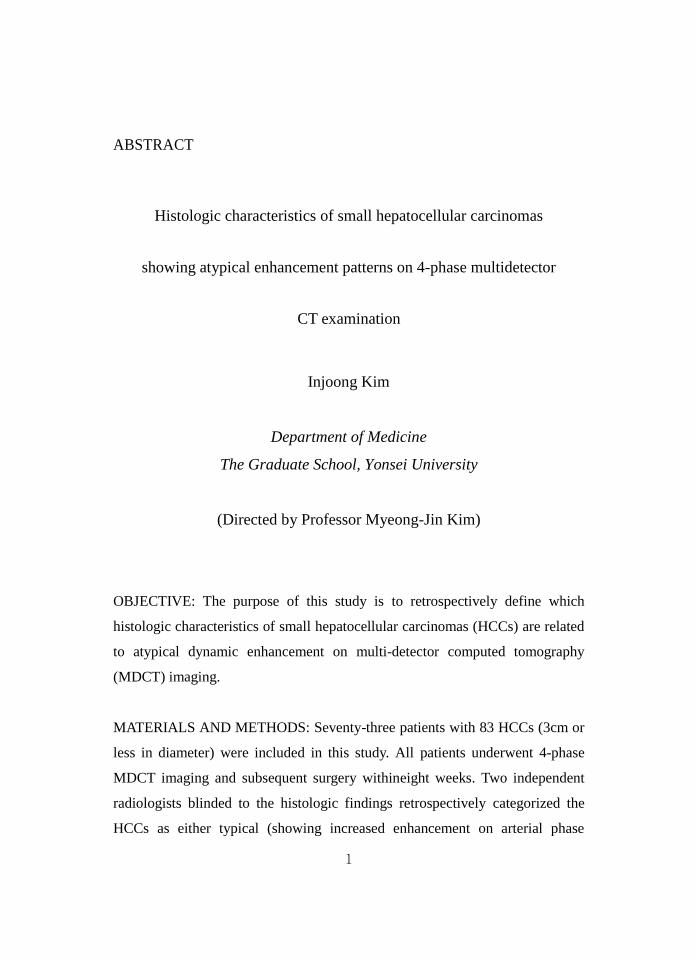
ABSTRACT
Histologic characteristics of small hepatocellular carcinomas
showing atypical enhancement patterns on 4-phase multidetector
CT examination
Injoong Kim
Department of Medicine
The Graduate School, Yonsei University
(Directed by Professor Myeong-Jin Kim)
OBJECTIVE: The purpose of this study is to retrospectively define which
histologic characteristics of small hepatocellular carcinomas (HCCs) are related
to atypical dynamic enhancement on multi-detector computed tomography
(MDCT) imaging.
MATERIALS AND METHODS: Seventy-three patients with 83 HCCs (3cm or
less in diameter) were included in this study. All patients underwent 4-phase
MDCT imaging and subsequent surgery withineight weeks. Two independent
radiologists blinded to the histologic findings retrospectively categorized the
HCCs as either typical (showing increased enhancement on arterial phase

2
images followed by washout in late phase images) or atypical lesions
demonstrating any other enhancement pattern. From the original pathologic
reports, various histologic characteristics including gross morphology, nuclear
histologic grades, presence of capsule formation, and capsule infiltration if a
capsule is present were compared between the two groups.
RESULTS: Atypical enhancement pattern was seen in 30 (36.2%) of the 83
HCCs. The mean size of atypical HCCs (1.71 ± 0.764) was significantly smaller
than that of typical HCCs (2.31 ± 0.598, p<0.001). Atypical HCCs were
frequently found to be vaguely nodular in gross morphology (n=13, 43.3%) and
to have grade I nuclear grades (n=17, 56.7%). Capsule formation was
significantly more common in typical HCCs (p<0.001). Capsular infiltration
was also more common in typical HCCs (p=0.001).
CONCLUSION: HCCs showing atypical dynamic enhancement on MDCT
imaging are usually smaller than typical HCCs, mostly of the vaguely nodular
type in gross morphology, well-differentiated in nuclear grades, and lack
capsule formation or capsular infiltration.
----------------------------------------------------------------------------------------
Key words : hepatocellular carcinoma, multi-detector computed tomography,
enhancement pattern, histology

3
Histologic characteristics of small hepatocellular carcinomas
showing atypical enhancement patterns on 4-phase multidetector
CT examination
Injoong Kim
Department of Medicine
The Graduate School, Yonsei University
(Directed by Professor Myeong-Jin Kim)
I. INTRODUCTION
The main workflow for the diagnosis of hepatocellular carcinoma
(HCC) has changed dramatically over the past few decades from invasive
procedures such as angiography or biopsy to noninvasive ones such as either
dynamic contrast-enhanced computed tomography (CT) or magnetic resonance
imaging (MRI) 1. The most recent guidelines issued by the American
Association for the Study of Liver Diseases (AASLD) state that if a lesion seen
in patients with a risk of HCC is more than 1cm in diameter and shows the
typical enhancement patterns on dynamic CT or MRI; which is arterial

4
hypervascularity and venous or delayed phase washout, can be treated under the
diagnosis of HCC 2. If the appearance is not typical for HCC, a second imaging
study (either CT or MRI) or biopsy is necessary 2.
Of the two modalities, multi-detector CT (MDCT) is more widely used
technique to diagnose HCCs, although gadolinium-enhanced dynamic MRI may
be superior to MDCT 3,4
. CT is more widespread and takes shorter examination
time. The typical appearance of HCC on dynamic CT or MRI is increased
enhancement on the arterial phase (arterial hypervascularity) followed by
decreased enhancement (washout) of the tumor in the portal venous or delayed
phases 5. However, some HCCs, especially less than 2–3 cm in diameter
6,7, and
well-differentiated ones lacking typical hemodynamic changes can make
diagnosing HCC a challenge 8. However, histologic differences are not well
known between the HCCs showing typical and atypical dynamic imaging
features. The purpose of this study was to retrospectively compare the histologic
characteristics of HCCs with typical and atypical dynamic enhancement
patterns on preoperative MDCT imaging in small HCCs of 3 cm or less in
diameter.
II. MATERIALS AND METHODS
1. Patients
Our institutional review board approved this retrospective study
and waived the informed consent requirement. Surgical resection
pathology records from June 2007 to February 2010 were reviewed to
identify the cases of patients with a pathologic diagnosis of HCC. Among
these patients, the study sample was selected on the basis of the
following inclusion criteria: pathologic diagnoses of HCC 3 cm or
smaller in diameter, available preoperative 4-phase MDCT scans
obtained according to the standard protocol for dynamic liver

5
CT, interval between pathologic diagnosis and CT of no longer than 8
weeks, and no history of previous adjuvant treatment, such as
transcatheter arterial chemoembolization, percutaneous ethanol injection,
or radiofrequency ablation. All the patients underwent surgery were
non-cirrhotic or have cirrhosis but still have well preserved liver
functions. In our institution, atypically enhancing but suspicious lesions
for HCC were closely followed up or treated with surgical resection
because of the malignant potential for development to HCC through
multistep progression of hepatocarcinogenesis9.
A total of 83 HCCs in 73 consecutive patients were included in
the study. Among the 73 patients, 64 patients had one HCC each, eight
had two HCCs, and one had three HCCs. Among the 83 HCCs, 12 HCCs
were 1cm or less in diameter. Three of these 12 HCCs were newly
appeared with typical enhancement during the surveillance. Remaing 9
HCCs showed atypical enhancement, but were resected together in the
surgery for another typical HCCs found in the preoperative MDCT.
2. Pathologic analysis
In all cases, pathologic reports including gross and histological
analyses were reviewed. Tumor size, gross morphology, tumor necrosis or
hemorrhage/peliosis, tumor grade, histology type, cell type, fatty change,
capsule formation (capsule infiltration if a capsule was present), portal vein
invasion, bile duct invasion and microvascular invasion were reviewed. Tumor
size grade was classified into three groups: group 1 was 1 cm or less in diameter,
group 2 was between 1 and 2 cm in diameter, and group 3 was between 2 and 3
cm in diameter. The tumor grade of the HCCs was classified as grade I(well
differentiated), grade II(moderately differentiated) and grade III (poorly
differentiated), or IV, according to the nuclear grading scheme by Edmondson

6
and Steiner 10
. If the histologic grade of tumor consisted of more than two
grades, the major component of the grade was recorded for the analysis. Gross
morphology was stratified into vaguely nodular, expanding, nodular and
perinodular extending, multinodular confluent or infiltrative type. Histologic
types were trabecular, pseudoglandular, scirrhous, compact and lymphoid. Cell
types were hepatic, clear or giant. Fibrous capsule formation was recorded as
either present (whether complete or partial) or absent.
3. CT Techniques
All CT scans were performed with multidetector scanners (Somatom
Sensation 16 or Sensation 64; Siemens Medical Solutions, Forchheim,
Germany). All patients received a 2 mL/kg dose (total volume <150 mL) of
nonionic contrast material (Iopromid [Ultravist]; Bayer Schering, Berlin,
Germany, or iohexol [Omnipaque 300]; Nycomed Amersham, GE Healthcare,
Milwaukee, Wis, NJ) intravenously with a power injector (EnVisionCT;
Medrad, Pittsburgh, Pa) with a 30-second fixed injection duration. A precontrast
scan was obtained before administration of contrast media. Using a bolus
tracking technique, arterial phase imaging was started after an 18-second delay
from the time of 100 Hounsfield units of aortic enhancement. A 30-second scan
delay after arterial phase imaging was used for portal venous phase imaging.
Equilibrium phase imaging was also obtained 150 seconds after the end of
portal venous phase imaging. The scanning parameters were as follows:
collimation, 16 rows×0.75mm or 64 rows×0.6mm; gantry rotation speed, 0.5
seconds; section thickness, 3 mm; image reconstruction increment, 1 mm;
120kV; and effective tube current-time charge, 200-250mA.
4. Image analysis
The attenuation of HCC was classified as hyperattenuated,
isoattenuated, and hypoattenuated, as compared with the surrounding liver

7
parenchyma on the unenhanced phase, arterial phase, portal venous phase, and
equilibrium phase images. Increased arterial enhancement was considered when
the tumor showed hyperattenuation compared to the surrounding liver
parenchyma during the arterial phase or the attenuation of tumor seen on
unenhanced images. On the portal venous phase and equilibrium phase images,
each lesion was subjectively evaluated for the presence of washout. Subjective
tumor washout was defined as present if the tumor hyper- or isoattenuating to
the liver on an arterial phase image subsequently appeared to be hypoattenuated
as compared to the surrounding liver parenchyma on the portal venous or
equilibrium phase images. Two independent radiologists blinded to the
histologic findings retrospectively stratified the HCCs into either typical or
atypical HCCs. Disagreements in interpretation were resolved by consensus.
Typical HCC was defined as a lesion that showed increased arterial
enhancement on arterial phase images followed by washout in late phase images
(Figure 1). Atypical HCC was defined as a lesion that did not show typical
enhancement pattern (Figure 2). After classification as typical and atypical HCC,
gross and histologic characteristics of the HCCs were compared between the
two groups.
Figure 1. A 43-year-old man with underlying B-viral hepatitis.
(A) Precontrast, (B) hepatic arterial, (C) portal venous, (D) equilibrium phase
images from triphasic MDCT scan. A hypoattenuating lesion is seen on the
precontrast phase image (A). The lesion shows increased arterial enhancement
on the arterial phase image (B) and washout of contrast enhancement on the
portal venous(C) and equilibrium phase image (D).
(E) Gross specimen of the lesion. Histologic examination demonstrated a poorly
differentiated (nuclear grade III) hepatocellular carcinoma of expanding type
gross morphology with partial capsule formation and infiltration.

8
(A) (B) (C) (D) (E)
Figure 2. A 49-year-old man with B-viral liver cirrhosis
(A)Precontrast, (B) hepatic arterial, (C) portal venous, (D) equilibrium phase
images from triphasic MDCT scan. On the precontrast (A) and arterial phase(B),
the lesion shows isoattenuation as compared to the surrounding parenchyma. On
the portal venous (C) and equilibrium phase (D), the lesion shows
hypoattenuation as compared to the surrounding parenchyma.
(E) Gross specimen of the lesion. Histologic examination showed a well
differentiated (nuclear grade I) hepatocellular carcinoma of vaguely nodular
type in gross morphology without capsule formation.
(A) (B) (C) (D) (E)
5. Statistical Analysis
An independent t-test was used to compare age and mean difference in
tumor size between the two groups. A chi-square test was used to compare sex
differences between two groups. The chi-square test or Fisher’s exact test was
used to compare categorical data according to the expected frequency in each
cell of the tables. P values of less than 0.05 were considered statistically

9
significant. All statistical analyses were done using statistical software (SPSS,
version 17.0.1, SPSS, Chicago, Ill).
Results
A total of 83 HCCs (2.09 ± 0.71 cm in diameter, 0.4cm to 3.0cm in
range) in 73 patients (54.7 ± 10.5 years old, 60 men and 13 women) were
included in this study. 69 (94%) out of 73 patients had liver cirrhosis. Most of
patients had cirrhosis caused by hepatitis B virus infection (n=60; 82.2%), and
the rest had cirrhosis caused by hepatitis C virus infection (n=6; 8.2%) or
alcohol (n=3; 4.1%).
Fifty–three (63.8%) HCCs were classified as typical and 30 (36.2%) as
atypical HCCs. (Table 1). Sixteen of 30 atypical HCCs show delayed phase
washout without arterial enhancement and 9 of 30 atypical HCCs show arterial
enhancement only without delayed phase washout. Five of 30 atypical HCCs
show neither arterial enhancement nor delayed phase washout.
The mean size of the atypical HCCs (1.7 ± 0.7) was significantly
smaller than that of the typical HCCs (2.3 ± 0.6, p<0.001). According to the
criteria of 2cm in diameter, 40(48.2%) HCCs were 2cm or less than 2cm in
diameter and 43(51.8%) HCCs were between 2cm and 3cm in diameter. Among
40 HCCs 2cm or less than 2cm in diameter, 19(47.5%) HCCs were typical HCC
and 21(52.5%) HCCs were atypical HCCs. But among 43 HCCs between 2cm
and 3cm in diameter, 34(79.1%) HCCs were typical HCC and 9(20.9%) HCCs
were atypical HCCs. Sex and age were not significantly different between the
typical and atypical HCCs.
Gross morphology of the vaguely nodular type was significantly more
common in atypical HCCs (p<0.001), but the expanding type was significantly
more common in typical HCCs (p=0.001). As for Edmondson-Steiner nuclear
histologic grades, well-differentiated (Grade I) HCCs were more common in

10
atypical HCC (p<0.001), but moderate (grade II) or poorly differentiated (grade
III) HCCs were significantly more common in typical HCCs (p<0.001). Capsule
formation was significantly more common in typical HCCs (p<0.001). Capsular
infiltration was more common in typical HCCs (p=0.001).
Other pathologic characteristics including tumor necrosis,
hemorrhage/peliosis, histologic types, cell types, fatty change, portal vein
invasion, bile duct invasion and microvascular invasion showed no significant
differences between the atypical and typical HCCs.
Table 1. Histologic and clinical characteristics of atypical and typical HCC
Atypical (n=30) Typical (n=53) p-value
N=83
Mean age = 54.72 ± 10.557 / M : F = 69 : 14
Sex (Male/Female) 25 / 5 44 / 9 0.971
Age 53.20 ±
10.162
55.58 ±
10.773
0.295
Size Grade
Mean 1.71 ± 0.764 2.31 ± 0.598 <0.001
1cm or less 9 (30.0%) 3 (5.7%) 0.001
<1cm – 2cm 12 (40.0%) 16 (30.2%)
<2cm – 3cm 9 (30.0%) 34 (64.2%)
Gross Type
Expanding
5 (16.7%) 28 (52.8%) 0.001
Multinodular
confluent
9 (30.0%) 19 (35.8%) 0.636

11
Nodular/perinodal
extension
1 (3.3%) 5 (9.4%) 0.411
Vagulely nodular 13 (43.3%) 1 (1.9%) <0.001
Infiltrative 2 (6.7%) 0 (0.0%) 0.128
Tumor necrosis (Available n = 82 )
No 25 (86.2%) 40 (75.5%) 0.252
Yes 4 (13.8%) 13 (24.5%)
Hemorrhage/peliosis (Available n = 82 )
No 27 (93.1%) 38 (71.7%) 0.022
Yes 2 (6.9%) 15 (28.3%)
Grade(major)
1.0 17 (56.7%) 6 (11.3%) <0.001
2.0 10 (33.3%) 40 (75.4%)
3.0 3 (10.0%) 7 (13.3%)
Grade(worst)
1.0 16 (53.3%) 1 (1.8 %) <0.001
2.0 8 (26.7%) 26 (49.1%)
3.0 6 (20.0%) 26 (49.1%)
Histology Type
Trabecular 30 (100.0%) 53 (100.0%)
Pseudoglandular 8 (26.7%) 18 (34.0%) 0.624
Scirrhous 1 (3.3%) 3 (5.7%) 1.000
Compact 3 (10.0%) 7 (13.2%) 0.741
Lymphoid 0 4 (7.5%) 0.291

12
Cell type
Hepatic 29 (96.7%) 53 (100.0%) 0.361
Clear 11 (36.7%) 20 (37.7%) 1.000
Giant 1 (3.3%) 4 (7.5%) 0.649
Fatty change (Available n = 82 )
No 14 (48.3%) 32 (60.4%) 0.291
Yes 15 (51.7%) 21 (39.6%)
Capsule formation (Available n = 82 )
No 19 (65.5%) 8 (15.1%) <0.001
Yes 10 (34.5%) 45 (84.9%)
Capsule infiltration (Available n = 82 )
No 22 (75.9%) 19 (35.8%) 0.001
Yes 7 (24.1%) 34 (64.2%)
Portal vein invasion(Available n = 78 )
No 24 (96.0%) 52 (98.1%) 0.541
Yes 1 (4.0%) 1 (1.9%)
Bile duct invasion (Available n = 78 )
No 24 (96.0%) 51 (96.2%) 1.000
Yes 1 (4.0%) 2 (3.8%)
Microvascular invasion (Available n = 78 )
No 17 (68.0%) 29 (54.7%) 0.266
Yes 8 (32.0%) 24 (45.3%)

13
IV. DISCUSSION
Our results showed that various histologic characteristics of HCCs are
related to atypical dynamic enhancement patterns on contrast-enhanced
dynamic CT.
In our study, 63.8% (50/83) of HCCs showed the typical enhancement
pattern of HCC of increased enhancement on arterial phase and washout on
portal venous or delayed phase images 11
. This typical enhancement pattern is
consistent with hepatocarcinogenesis, which causes vascular changes toward a
predominantly hepatic arterial supply with a lack of portal venous supply 12-14
.
However, the predominant enhancement patterns of HCC during the arterial and
portal venous phases differed significantly according to tumor size and cellular
differentiation of the tumor. In our study, the mean size of HCCs showing
typical enhancement pattern was larger than that of HCCs with atypical
enhancement pattern (p<0.001). And there was a significantly higher proportion
of typical HCCs among HCCs between 2cm and 3cm in diameter (p=0.0057).
These results are similar to those of previous studies 15-17
. Also, in our study,
well-differentiated HCCs were more common among atypical HCCs (p<0.001),
while moderate and poorly differentiated HCCs were significantly more
common among typical HCCs (p<0.001). A previous study that evaluated the
relationship of the vascularization of small HCC and the cellular differentiation
has shown that abnormal arterial supply within a nodule increases as the grade
of malignancy increases, while the normal hepatic arterial and portal venous
supply to the nodule gradually decreases 18
. Another studies reported that well
differentiated and small HCCs more often show various atypical CT
enhancement features 19,20
.
Early HCCs showed the typical dynamic enhancement pattern less
often than more advanced lesions in our study. Thirteen (43.3%) atypical
lesions were vaguely nodular in gross morphology, and 17 (56.7%) atypical
lesions were well-differentiated in tumor grade. These characteristics are

14
compatible with early HCC, defined as well-differentiated lesions that are
usually less than 2 cm in diameter, vaguely nodular in gross morphology, not
showing capsule formation, and usually hypovascular 21
.
Fibrous capsule that is known to be frequently observed around a
tumor during the growth of HCC was more frequently seen in typical HCCs in
our study. And our study also showed moderate and poorly differentiated HCCs
were significantly more common among typical HCCs (p<0.001). The capsule
is formed by host mesenchymal cells not by HCC cells. And the capsule
formation may result from interaction between tumor and host liver and
interfere the growth and invasion of HCC22
. This is supported by the clinical
evidence that the prognosis of patients with a HCC having capsule is better than
for those without 23-26
Therefore progressed HCC with typical enhancement
pattern in spite of its small size more often forms capsule than indolent early
HCC with atypical enhancement pattern in our study.
There are limitations to our study. First, our study is retrospective and
we included surgically confirmed HCC to compare the dynamic imaging
patterns with histologic characteristics. Therefore, small HCC that were
diagnosed based on the presence of a typical dynamic pattern and subsequently
treated with locoregional treatment were not included. This might have
increased the proportion of atypical lesions in our study. Second, the definitions
we used for increased arterial enhancement and presence of washout may differ
from those used by other investigators. We determined the presence of increased
arterial enhancement by comparing with precontrast images; some investigators
may determine the presence of increased arterial enhancement on the arterial
phase images alone. However, we believe that increased arterial enhancement
can be correctly assessed by referring to precontrast images because some
lesions showing hypoattenuation on precontrast images may show
isoattenuation on arterial phase images even though they have increased arterial
vascularity within the lesions. We considered washout to be present when a

15
lesion showed hypointensity relative to the surrounding liver on late phase
images. Some radiologists could argue that washout may not be present when a
lesion does not show increased arterial enhancement. However, we thought that
such comparison would cause greater interobserver variability. Third, atypical
HCCs are consisted of diverse subgroups according to the presence of the
arterial enhancement or delayed phase washout. But we considered atypical
HCCs as lesions that did not show typical enhancement pattern and did not
divided into subgroups in the analysis process. Further studies are needed to
define the characteristics of diverse subgroups of atypical HCCs. Lastly, we did
not analyze how many hypovascular lesions transform to hypervascular lesions.
A recent study revealed that hypoattenuating hepatic nodular lesions in chronic
liver disease depicted on dynamic CT have high malignant potential 27
. There
are chances that atypically enhancing HCCs progressed to typically enhancing
HCCs according to the hepatocarcinogenesis, but we analyzed only the
preoperative CT scan of just before the surgery, not serial CT scans.
V. CONCLUSION
We conclude that various histologic characteristics of HCC are
associated with atypical dynamic enhancement on contrast-enhanced dynamic
CT images. HCCs with atypical enhancement patterns tend to be smaller than
HCCs with typical enhancement pattern and are vaguely nodular type in gross
morphology and well-differentiated in histologic grades. Capsule formation and
capsular infiltration are significantly more common in typical HCCs. Awareness
of atypical enhancement patterns in small HCC and their histologic implications
may guide patient management.

16
REFERENCES
1. Choi BI, Lee JM. Advancement in HCC imaging: diagnosis, staging
and treatment efficacy assessments: imaging diagnosis and staging of
hepatocellular carcinoma. J Hepatobiliary Pancreat Sci 2010;17:369-73.
2. Bruix J, Sherman M. Management of hepatocellular carcinoma: An
update. Hepatology 2011;53:1020-2.
3. Colli A, Fraquelli M, Casazza G, Massironi S, Colucci A, Conte D, et al.
Accuracy of ultrasonography, spiral CT, magnetic resonance, and
alpha-fetoprotein in diagnosing hepatocellular carcinoma: a systematic
review. Am J Gastroenterol 2006;101:513-23.
4. Willatt JM, Hussain HK, Adusumilli S, Marrero JA. MR Imaging of
hepatocellular carcinoma in the cirrhotic liver: challenges and
controversies. Radiology 2008;247:311-30.
5. Marrero JA, Hussain HK, Nghiem HV, Umar R, Fontana RJ, Lok AS.
Improving the prediction of hepatocellular carcinoma in cirrhotic
patients with an arterially-enhancing liver mass. Liver Transpl
2005;11:281-9.
6. Parkin DM, Bray F, Ferlay J, Pisani P. Estimating the world cancer
burden: Globocan 2000. Int J Cancer 2001;94:153-6.
7. Mazzaferro V, Regalia E, Doci R, Andreola S, Pulvirenti A, Bozzetti F,
et al. Liver transplantation for the treatment of small hepatocellular
carcinomas in patients with cirrhosis. N Engl J Med 1996;334:693-9.
8. Bolondi L, Gaiani S, Celli N, Golfieri R, Grigioni WF, Leoni S, et al.
Characterization of small nodules in cirrhosis by assessment of
vascularity: the problem of hypovascular hepatocellular carcinoma.
Hepatology 2005;42:27-34.
9. Takayama T, Makuuchi M, Hirohashi S, Sakamoto M, Okazaki N,
Takayasu K, et al. Malignant transformation of adenomatous
hyperplasia to hepatocellular carcinoma. Lancet 1990;336:1150-3.

17
10. Edmondson HA, Steiner PE. Primary carcinoma of the liver: a study of
100 cases among 48,900 necropsies. Cancer 1954;7:462-503.
11. Forner A, Vilana R, Ayuso C, Bianchi L, Sole M, Ayuso JR, et al.
Diagnosis of hepatic nodules 20 mm or smaller in cirrhosis: Prospective
validation of the noninvasive diagnostic criteria for hepatocellular
carcinoma. Hepatology 2008;47:97-104.
12. Hwang SH, Yu JS, Kim KW, Kim JH, Chung JJ. Small hypervascular
enhancing lesions on arterial phase images of multiphase dynamic
computed tomography in cirrhotic liver: fate and implications. J
Comput Assist Tomogr 2008;32:39-45.
13. Lee J, Lee WJ, Lim HK, Lim JH, Choi N, Park MH, et al. Early
hepatocellular carcinoma: three-phase helical CT features of 16 patients.
Korean J Radiol 2008;9:325-32.
14. Shimizu A, Ito K, Koike S, Fujita T, Shimizu K, Matsunaga N.
Cirrhosis or chronic hepatitis: evaluation of small (<or=2-cm)
early-enhancing hepatic lesions with serial contrast-enhanced dynamic
MR imaging. Radiology 2003;226:550-5.
15. Hytiroglou P, Park YN, Krinsky G, Theise ND. Hepatic precancerous
lesions and small hepatocellular carcinoma. Gastroenterol Clin North
Am 2007;36:867-87, vii.
16. Monzawa S, Ichikawa T, Nakajima H, Kitanaka Y, Omata K, Araki T.
Dynamic CT for detecting small hepatocellular carcinoma: usefulness
of delayed phase imaging. AJR Am J Roentgenol 2007;188:147-53.
17. Monzawa S, Omata K, Shimazu N, Yagawa A, Hosoda K, Araki T.
Well-differentiated hepatocellular carcinoma: findings of US, CT, and
MR imaging. Abdom Imaging 1999;24:392-7.
18. Nakashima Y, Nakashima O, Hsia CC, Kojiro M, Tabor E.
Vascularization of small hepatocellular carcinomas: correlation with
differentiation. Liver 1999;19:12-8.

18
19. Yoon SH, Lee JM, So YH, Hong SH, Kim SJ, Han JK, et al.
Multiphasic MDCT enhancement pattern of hepatocellular carcinoma
smaller than 3 cm in diameter: tumor size and cellular differentiation.
AJR Am J Roentgenol 2009;193:W482-9.
20. Kim SH, Lee WJ, Lim HK, Park CK. SPIO-enhanced MRI findings of
well-differentiated hepatocellular carcinomas: correlation with MDCT
findings. Korean J Radiol 2009;10:112-20.
21. Pathologic diagnosis of early hepatocellular carcinoma: a report of the
international consensus group for hepatocellular neoplasia. Hepatology
2009;49:658-64.
22. Ishizaki M, Ashida K, Higashi T, Nakatsukasa H, Kaneyoshi T,
Fujiwara K, et al. The formation of capsule and septum in human
hepatocellular carcinoma. Virchows Arch 2001;438:574-80.
23. Okuda K, Musha H, Nakajima Y, Kubo Y, Shimokawa Y, Nagasaki Y,
et al. Clinicopathologic features of encapsulated hepatocellular
carcinoma: a study of 26 cases. Cancer 1977;40:1240-5.
24. Nagao T, Inoue S, Goto S, Mizuta T, Omori Y, Kawano N, et al.
Hepatic resection for hepatocellular carcinoma. Clinical features and
long-term prognosis. Ann Surg 1987;205:33-40.
25. Lai EC, Ng IO, Ng MM, Lok AS, Tam PC, Fan ST, et al. Long-term
results of resection for large hepatocellular carcinoma: a multivariate
analysis of clinicopathological features. Hepatology 1990;11:815-8.
26. Ng IO, Lai EC, Ng MM, Fan ST. Tumor encapsulation in
hepatocellular carcinoma. A pathologic study of 189 cases. Cancer
1992;70:45-9.
27. Takayasu K, Muramatsu Y, Mizuguchi Y, Okusaka T, Shimada K,
Takayama T, et al. CT Evaluation of the progression of hypoattenuating
nodular lesions in virus-related chronic liver disease. AJR Am J
Roentgenol 2006;187:454-63.

19
ABSTRACT(IN KOREAN)
MDCT에서 비전형적 조영증강양상을 보이는
소간세포암의 조직학적 특성
<지도교수 김명진>
연세대학교 대학원 의학과
김 인 중
목적: MDCT상 비전형적인 조영 증강 양상을 보이는
소간세포암의 조직학적 특성을 후향적으로 분석한다.
대상 및 방법: 2007년 6월부터 2010년 2월까지 본원에서 간암
수술을 시행한 환자 중에서 총 73명 환자의 83개의 소간세포암
(직경이 3cm 이하)이 연구에 포함되었다. 모든 환자들은 4-phase
MDCT를 시행한 후 8주 이내에 수술을 진행하였다. 두 명의
영상의학과 의사가 간암의 전형적인 조영 증강 양상(동맥기
조영 증강과 지연기의 조영 감소)을 보이는지에 따라 병변을
전형적 또는 비전형적 조영 증강 군으로 나누었다. 수술 후
병리 보고서를 통하여 육안적 분류, 조직학적 분화도, 피막 형성
유무, 피막이 있을 경우 피막 침습 유무 등에 대하여 두 군을

20
비교 분석하였다.
결과: 총 83개의 소간세포암중에서 30개(36.2%)의 병변이
비전형적 조영 증강을 보였다. 비전형적 조영 증강군의 평균
크기(1.71±0.764)가 전형적인 조영 증강군의 평균크기(2.31±
0.598)에 비하여 작았다(p<0.001). 비전형적 조영 증강을 보인
소간세포암의 경우에 육안적 분류상 경계불명료 결절형이 더
많았으며(13개, 43.3%), 조직학적 분화도는 등급 I 이 많이
관찰되었다(17개, 56.7%). 피막 형성은 전형적 조영 증강을
보인 소간세포암의 경우에 더 흔하게 관찰되었다(p<0.001).
피막 침습도 또한 전형적 조영 증강을 보인 소간세포암의
경우에 더 흔하게 관찰되었다(p=0.001).
결론: MDCT에서 비전형적 조영 증강을 보이는 소간세포암은
전형적 조영 증강을 보이는 소간세포암에 비하여 크기가 작고,
육안적 분류상 경계불명료 결절형이 더 많았으며, 조직학적
분화도상 고분화도를 보였고 피막 형성이나 피막 침습이
적었다.
----------------------------------------------------------------------------------------
핵심되는 말 : 간세포암, MDCT, 조영증강 양상, 조직학