Embed Size (px)
Citation preview
www.wjpls.org
112299
Alaezi CM et al. World Journal of Pharmaceutical and Life Sciences
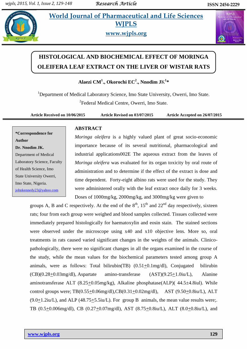
HISTOLOGICAL AND BIOCHEMICAL EFFECT OF MORINGA
OLEIFERA LEAF EXTRACT ON THE LIVER OF WISTAR RATS
Alaezi CM1., Okorochi EC
2., Nnodim JK
1*
1Department of Medical Laboratory Science, Imo State University, Owerri, Imo State.
2Federal Medical Centre, Owerri, Imo State.
Article Received on 10/06/2015 Article Revised on 03/07/2015 Article Accepted on 26/07/2015
ABSTRACT
Moringa oleifera is a highly valued plant of great socio-economic
importance because of its several nutritional, pharmacological and
industrial applications002E The aqueous extract from the leaves of
Moringa oleifera was evaluated for its organ toxicity by oral route of
administration and to determine if the effect of the extract is dose and
time dependent. Forty-eight albino rats were used for the study. They
were administered orally with the leaf extract once daily for 3 weeks.
Doses of 1000mg/kg, 2000mg/kg, and 3000mg/kg were given to
groups A, B and C respectively. At the end of the 8th
, 15th
and 22nd
day respectively, sixteen
rats; four from each group were weighed and blood samples collected. Tissues collected were
immediately prepared histologically for haematoxylin and eosin stain. The stained sections
were observed under the microscope using x40 and x10 objective lens. More so, oral
treatments in rats caused varied significant changes in the weights of the animals. Clinico-
pathologically, there were no significant changes in all the organs examined in the course of
the study, while the mean values for the biochemical parameters tested among group A
animals, were as follows: Total bilirubin(TB) (0.51+0.1mg/dl), Conjugated bilirubin
(CB)(0.28+0.03mg/dl), Aspartate amino-transferase (AST)(9.25+1.0iu/L), Alanine
aminotransferase ALT (8.25+0.05mg/kg), Alkaline phosphatase(ALP)( 44.5±4.8iul). While
control groups were; TB(0.55+0.06mg/dl),CB(0.31+0.02mg/dl), AST (9.50+0.8iu/L), ALT
(9.0+1.2iu/L), and ALP (48.75+5.5iu/L). For group B animals, the mean value results were;.
TB (0.5+0.006mg/dl), CB (0.27+0.07mg/dl), AST (8.75+0.8iu/L), ALT (8.0+0.8iu/L), and
wjpls, 2015, Vol. 1, Issue 2, 129-148 Research Article ISSN 2454-2229
World Journal of Pharmaceutical and Life Sciences WJPLS
www.wjpls.org
*Correspondence for
Author
Dr. Nnodim JK.
Department of Medical
Laboratory Science, Faculty
of Health Science, Imo
State University Owerri,
Imo State, Nigeria.
www.wjpls.org
113300
Alaezi CM et al. World Journal of Pharmaceutical and Life Sciences
ALP (45.0+5.6iu/L), whereas control groups were; TB(0.55+0.08mg/dl), CB
(0.29+0.03mg/dl), AST(9.75+0.8iu/L),ALT(9.25+1.2iu/L), and ALP (44.75+4.8iu/L). Also,
the group C animals had the following results: . TB (0.57+0.04mg/dl), CB (0.30+0.03mg/dl),
AST (10.25+0.4iu/L), ALT (8.5+0.5iu/L), and ALP (50.2+54.7iu/L). While control groups
had ., TB (0.58+0.04iu/L), CB (0.30+0.02mg/dl), AST (10.75+1.2iu/L), ALT (9.50+1.5iu/L),
and ALP (45.75+3.1). All the biochemical parameters tested above were within the reference
range as their values were not significant using the standard T-test hypothesis, having all
“parameters of liver, as p>0.05, all through the course of the study”. The study concluded that
Moringa oleifera leaf extract has no organ toxicity, it is time and dose dependent when within
a dose of 4000mg/kg and it is relatively safe for consumption.
KEYWORD: Moringa oleifera, leaf extract, liver, wistar rat.
INTRODUCTION
Moringa Oleifera a highly valued plant of socio economic importance owing to its several
nutritional, pharmacological (Caceres et al., 1991; Fuglie, 2000). Its industrial applications
has become of great significance in the society (Makkar and Becker, 1997; Foldl, 2001),.
Moringa oleifera is widely used in traditional and herbal medicine, and has gained popularity
so much that people have started probing the pharmacological importance in modern drug
and manufacture and therapy. Almost all parts of the plant is useful in traditional medicine
practice(Rathi et al.,2006).
Moringa Oleifera belongs to the family: Moringaceae. It is called drumstick tree or
horseradish and is a highly valued plant, distributed in many countries of tropics and
subtropics. It has been an ingredient of Indian diet since centuries. It is cultivated almost all
over the world and its leaves and fruits are used as vegetables. For centuries, the people of
Africa and Northern India have known of the many benefits to cultivate moringa plant, some
call it “Tree of life, the clarifier tree, wonder plant etc”. Looking at the unassuming plant,
one wonders why.In East Africa, the plant is known as “mother’s best friend”, the leaves of
the moringa plant prevent 300 diseases. The juice of the leaves mixed with honey is used for
the treatment of eye diseases (Sangeeta et al., 2003). In fact, no plant can claim all the
benefits moringa offers.
The phytochemical screening of the leaf revealed the presence of fatty acid, vitamin E,
carotenoid, selected mineral elements like Fe, Cu, Zn, Co and amino acid, two nitrile
www.wjpls.org
113311
Alaezi CM et al. World Journal of Pharmaceutical and Life Sciences
glycosides, Niazirin I and Niazirin II, three mustard oil glycosides, Niazinin, Niazimicin and
Niaziminin, Flavinoids, Tannin, Saponin, allcaloids, anthraquinone, triterpenoids and
reducing sugar. (Rathi et al., 2006). It has been already documented that the chemical
composition of plant extract is different in different locations (Gotep et al., 2010).
Animal and human studies have showcased that Moringa oleifera possesses wide spectrum of
pharmacological effects of antifertility, antitumor, antipyretic, antiepileptic, antispasmodic,
anti-inflammatory, diuretic, antiulcer, hypotensive, hypolipidemic, hypoglycemic,
hepatoprotective, antifungal and antibacterial activities (Sudha et al., 2010), antinephrotoxic
(Paliwal et al., 2011). In addition to its compelling water purifying powers and high
nutritional value. The various parts of this plant such as the leaves, roots, seed, bark, fruit,
flowers and immature pods act as cardiac and circulatory stimulants (Anwar et al., 2007).
The liver is the principal organ of the body deeply involved in the drug biotransformation,
excretion of products of body metabolism, drugs and chemicals.(Okolie, 2011). There could
be damage to the liver following administration of leaf extract of moringa oleifera especially
when there is no scientific data to evaluate the standard and suitability of the practice done by
the traditional medical practitioners.
Consequently, there is an urgent need to determine the effect of the leaf extract of moringa
oleifera on the liver in our local setting since the chemical composition of the leaf extract has
been known to vary according to geographical location. This can be assessed by using
histological and biochemical tools such as Bilirubin, Aspartate aminotransferase (AST),
Alanine amino-transferase (ALT) and Alkaline phosphate (ALP). Hence, this is done with the
view of educating and guiding the traditional herbal patronizers on the safety of the plant and
better scientific approach in using them.
Plant materials: The fresh leaves of Moringa Oleifera (5kg) were collected from the
botanical garden of Imo State University Owerri. It was identified and authenticated by
Professor S. E. Okeke, a plant taxonomist of the Department of Plant and Science
Biotechnology, Imo State University Owerri in the month of May 2013.
Plant Extraction Preparation
The leaves were collected, washed thoroughly with distilled water, they were subsequently
dried and comminuted using a grinding machine. Maximum extraction was achieved within
48 hours at temperature of 600c.The resulting extract was filtered using whatman No. 1 filter
www.wjpls.org
113322
Alaezi CM et al. World Journal of Pharmaceutical and Life Sciences
paper and concentrated in rota vapor of 500mmHg atmospheric pressure at 45oc to give a
semisolid residue. The extract concentrate was dried in an oven for further exploration.
Experimental Animals
48 healthy male and female albino rats (200-250g) were obtained from the Animal House of
College of Medicine and Health Sciences, Imo State University Owerri which were used for
the study. They were housed in plastic cages with steel netting in well ventilated house, of
temperature 27oc, for 12 hours under natural light, and 12 hours darkness. They were allowed
free access to sterilized tap water and dry rat pellet (purchased at Animal Friend Shop, No.
120 Royce Road, Owerri, Imo State, Nigeria.) They were allowed to acclimatize in two (2)
week before the experiment.
Experimental Design
Forty-eight healthy albino wister rat (male and female) acclimatized were weighed and
randomly distributed into four different groups, of twelve (12) in each. The groups were A-D.
Group A was treated with a dose of 1000mg/kg, Group B a dose of 2000mg/kg, and Group C
treated with a dose of 3000mg/kg while Group D was the control group not treated with any
extract but received only laboratory rat pellet and water. To identify the animal groups, a dye
was applied (Group A – blue dye, Group B – red dye, Group C – green dye and Control
group – black dye). The extracts dissolved in water were administered once daily, orally and
slowly to the healthy albino wister rat for three (3) weeks using cannula attached to a
graduated syringe and needle.
Sample Collection
Twenty four hours after the last doses were administered, the animals were anaesthetized
with chloroform vapour, quickly brought out of the jar and sacrificed. Whole blood was
collected by cardiac puncture from each animal into plain specimen bottles for biochemical
analysis. The animals were subsequently sacrificed, and the livers were dissected from each
group and control, fixed in neutral buffered formalin and processed by popular automatic
tissue processing method and all sections stained by haematoxylin and eosin staining
technique. While the liver enzymes were determined from the blood sample using the
following techniques: Total and Conjugated bilirubin by Powell method(1944), AST by
Reitman and Frankel Method(1957), ALT by Reitman and Frankel method(1957), and ALP
by modified King Armstrong method(1954).
www.wjpls.org
113333
Alaezi CM et al. World Journal of Pharmaceutical and Life Sciences
Statistical Analysis
Results were expressed on mean ± standard deviation. Analysis was carried out using student
T-test. The level of significance was considered at P<0.05.
RESULTS
Histological effects Moringer oligifera on wistar rats administered with different
concentration of extract and Control.
Histological sections of liver from control group showed normal histological features which
served as a basis for comparing with experiment groups.
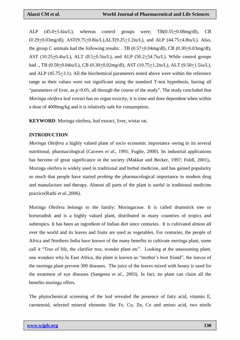
Plate 1: A photomicrograph of albino rat liver section from control group, showing the
hepatic vein, bile duct and hepatocytes. magnification x 400 H & E stain.
4.2 Albino Rats (Administered with 1000mg/kg body weight of Moringa oleifera leaf extract
at 8th
day of Administration).
Histological sections of the liver of albino rats treated with 1000mg/kg of extract at 8th
day of
administration did not differ from the control group (Normal histo-architecture).
www.wjpls.org
113344
Alaezi CM et al. World Journal of Pharmaceutical and Life Sciences
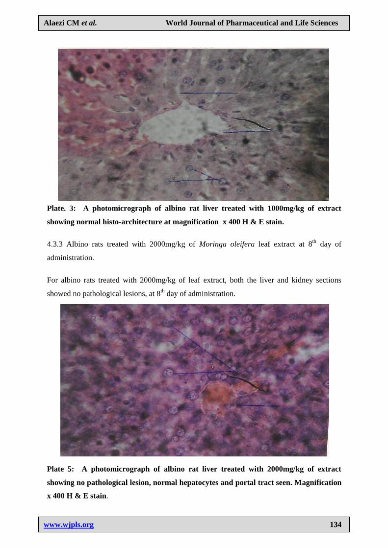
Plate. 3: A photomicrograph of albino rat liver treated with 1000mg/kg of extract
showing normal histo-architecture at magnification x 400 H & E stain.
4.3.3 Albino rats treated with 2000mg/kg of Moringa oleifera leaf extract at 8th
day of
administration.
For albino rats treated with 2000mg/kg of leaf extract, both the liver and kidney sections
showed no pathological lesions, at 8th
day of administration.
Plate 5: A photomicrograph of albino rat liver treated with 2000mg/kg of extract
showing no pathological lesion, normal hepatocytes and portal tract seen. Magnification
x 400 H & E stain.
www.wjpls.org
113355
Alaezi CM et al. World Journal of Pharmaceutical and Life Sciences
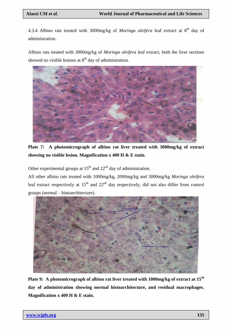
4.3.4 Albino rats treated with 3000mg/kg of Moringa oleifera leaf extract at 8th
day of
administration.
Albino rats treated with 3000mg/kg of Moringa oleifera leaf extract, both the liver sections
showed no visible lesions at 8th
day of administration.
Plate 7: A photomicrograph of albino rat liver treated with 3000mg/kg of extract
showing no visible lesion. Magnification x 400 H & E stain.
Other experimental groups at 15th
and 22nd
day of administration.
All other albino rats treated with 1000mg/kg, 2000mg/kg and 3000mg/kg Moringa oleifera
leaf extract respectively at 15th
and 22nd
day respectively, did not also differ from control
groups (normal – histoarchitecture).
Plate 9: A photomicrograph of albino rat liver treated with 1000mg/kg of extract at 15th
day of administration showing normal histoarchitecture, and residual macrophages.
Magnification x 400 H & E stain.
www.wjpls.org
113366
Alaezi CM et al. World Journal of Pharmaceutical and Life Sciences
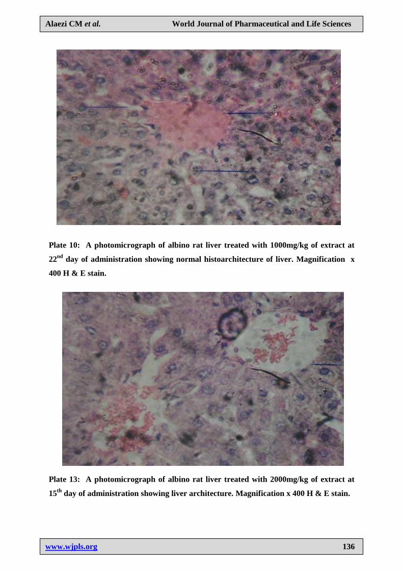
Plate 10: A photomicrograph of albino rat liver treated with 1000mg/kg of extract at
22nd
day of administration showing normal histoarchitecture of liver. Magnification x
400 H & E stain.
Plate 13: A photomicrograph of albino rat liver treated with 2000mg/kg of extract at
15th
day of administration showing liver architecture. Magnification x 400 H & E stain.
www.wjpls.org
113377
Alaezi CM et al. World Journal of Pharmaceutical and Life Sciences
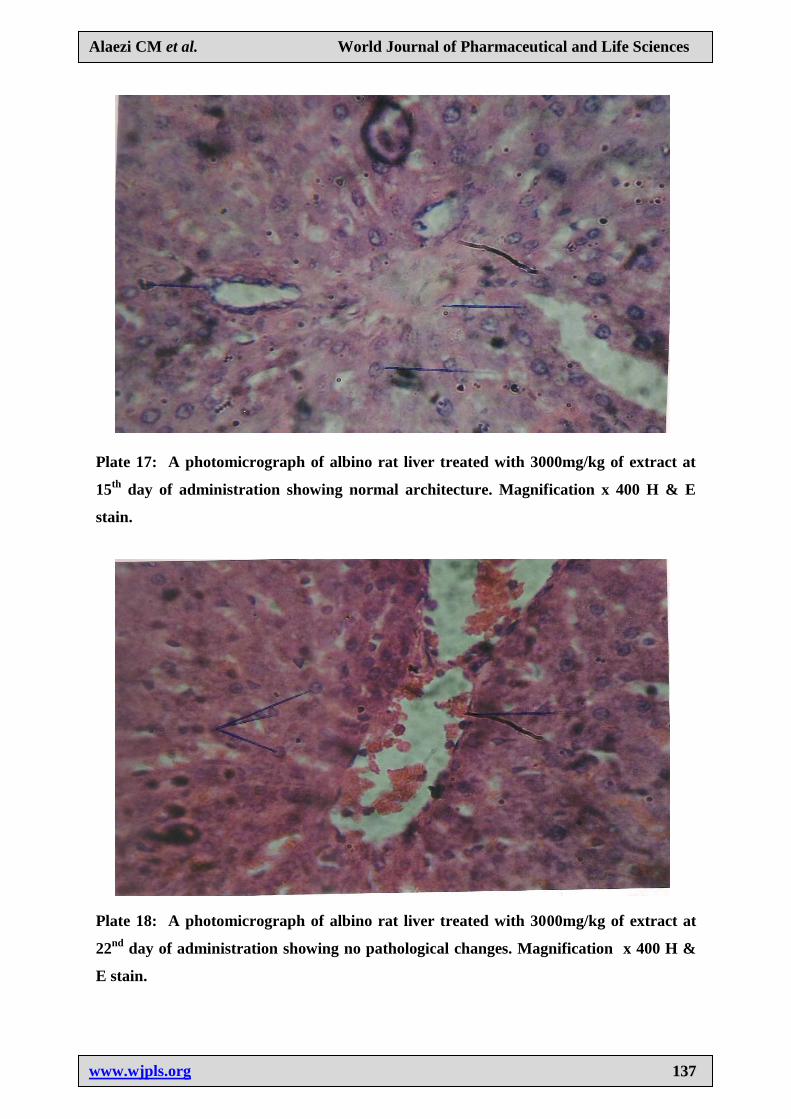
Plate 17: A photomicrograph of albino rat liver treated with 3000mg/kg of extract at
15th
day of administration showing normal architecture. Magnification x 400 H & E
stain.
Plate 18: A photomicrograph of albino rat liver treated with 3000mg/kg of extract at
22nd
day of administration showing no pathological changes. Magnification x 400 H &
E stain.
www.wjpls.org
113388
Alaezi CM et al. World Journal of Pharmaceutical and Life Sciences
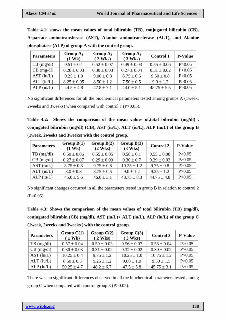
Table 4.1: shows the mean values of total bilirubin (TB), conjugated bilirubin (CB),
Aspartate aminotransferase (AST), Alanine aminotransferase (ALT), and Alanine
phosphatase (ALP) of group A with the control group.
Parameters Group A1
(1 Wk)
Group A2
( 2 Wks)
Group A3
( 3 Wks) Control 1 P-Value
TB (mg/dl) 0.51 0.1 0.52 0.07 0.49 0.03 0.55 0.06 P>0.05
CB (mg/dl) 0.28 0.03 0.30 0.03 0.27 0.04 0.31 0.02 P>0.05
AST (iu/L) 9.25 1.0 9.00 0.8 8.75 0.5 9.50 0.8 P>0.05
ALT (iu/L) 8.25 0.05 8.50 1.2 7.50 0.5 9.0 1.2 P>0.05
ALP (iu/L) 44.5 4.8 47.8 7.1 44.0 5.1 48.75 5.5 P>0.05
No significant differences for all the biochemical parameters tested among groups A (1week,
2weeks and 3weeks) when compared with control 1 (P>0.05).
Table 4.2: Shows the comparison of the mean values of,total bilirubin (mg/dl) ,
conjugated bilirubin (mg/dl) (CB), AST (iu/L), ALT (iu/L), ALP (iu/L) of the group B
(1week, 2weeks and 3weeks) with the control group.
Parameters Group B(1)
(1 Wk)
Group B(2)
(2 Wks)
Group B(3)
(3 Wks) Control 2 P-Value
TB (mg/dl) 0.50 0.06 0.55 0.05 0.58 0.1 0.55 0.08 P>0.05
CB (mg/dl) 0.27 0.07 0.29 0.03 0.30 0.7 0.29 0.03 P>0.05
AST (iu/L) 8.75 0.8 9.75 0.8 10.25 1.2 9.75 0.8 P>0.05
ALT (iu/L) 8.0 0.8 8.75 0.5 9.0 1.2 9.25 1.2 P>0.05
ALP (iu/L) 45.0 5.6 46.0 3.1 48.75 8.3 44.75 4.8 P>0.05
No significant changes occurred in all the parameters tested in group B in relation to control 2
(P>0.05).
Table 4.3: Shows the comparison of the mean values of total bilirubin (TB) (mg/dl),
conjugated bilirubin (CB) (mg/dl), AST (iu/L)< ALT (iu/L), ALP (iu/L) of the group C
(1week, 2weeks and 3weeks ) with the control group.
Parameters Group C(1)
( 1 Wk)
Group C(2)
( 2 Wks)
Group C(3)
( 3 Wks) Control 3 P-Value
TB (mg/dl) 0.57 0.04 0.59 0.03 0.56 0.07 0.58 0.04 P>0.05
CB (mg/dl) 0.30 0.03 0.31 0.02 0.32 0.02 0.30 0.02 P>0.05
AST (lu/L) 10.25 0.4 9.75 1.2 10.25 1.0 10.75 1.2 P>0.05
ALT (Iu/L) 8.50 0.5 9.25 1.2 9.00 1.0 9.50 1.5 P>0.05
ALP (Iu/L) 50.25 4.7 48.2 6.7 47.5 5.8 45.75 3.1 P>0.05
There was no significant differences observed in all the biochemical parameters tested among
group C when compared with control group 3 (P>0.05).
www.wjpls.org
113399
Alaezi CM et al. World Journal of Pharmaceutical and Life Sciences
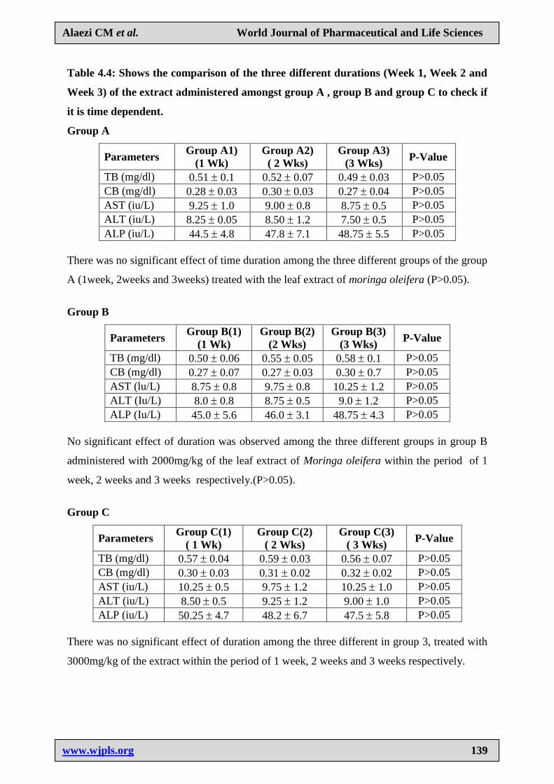
Table 4.4: Shows the comparison of the three different durations (Week 1, Week 2 and
Week 3) of the extract administered amongst group A , group B and group C to check if
it is time dependent.
Group A
Parameters Group A1)
(1 Wk)
Group A2)
( 2 Wks)
Group A3)
(3 Wks) P-Value
TB (mg/dl) 0.51 0.1 0.52 0.07 0.49 0.03 P>0.05
CB (mg/dl) 0.28 0.03 0.30 0.03 0.27 0.04 P>0.05
AST (iu/L) 9.25 1.0 9.00 0.8 8.75 0.5 P>0.05
ALT (iu/L) 8.25 0.05 8.50 1.2 7.50 0.5 P>0.05
ALP (iu/L) 44.5 4.8 47.8 7.1 48.75 5.5 P>0.05
There was no significant effect of time duration among the three different groups of the group
A (1week, 2weeks and 3weeks) treated with the leaf extract of moringa oleifera (P>0.05).
Group B
Parameters Group B(1)
(1 Wk)
Group B(2)
(2 Wks)
Group B(3)
(3 Wks) P-Value
TB (mg/dl) 0.50 0.06 0.55 0.05 0.58 0.1 P>0.05
CB (mg/dl) 0.27 0.07 0.27 0.03 0.30 0.7 P>0.05
AST (lu/L) 8.75 0.8 9.75 0.8 10.25 1.2 P>0.05
ALT (Iu/L) 8.0 0.8 8.75 0.5 9.0 1.2 P>0.05
ALP (Iu/L) 45.0 5.6 46.0 3.1 48.75 4.3 P>0.05
No significant effect of duration was observed among the three different groups in group B
administered with 2000mg/kg of the leaf extract of Moringa oleifera within the period of 1
week, 2 weeks and 3 weeks respectively.(P>0.05).
Group C
Parameters Group C(1)
( 1 Wk)
Group C(2)
( 2 Wks)
Group C(3)
( 3 Wks) P-Value
TB (mg/dl) 0.57 0.04 0.59 0.03 0.56 0.07 P>0.05
CB (mg/dl) 0.30 0.03 0.31 0.02 0.32 0.02 P>0.05
AST (iu/L) 10.25 0.5 9.75 1.2 10.25 1.0 P>0.05
ALT (iu/L) 8.50 0.5 9.25 1.2 9.00 1.0 P>0.05
ALP (iu/L) 50.25 4.7 48.2 6.7 47.5 5.8 P>0.05
There was no significant effect of duration among the three different in group 3, treated with
3000mg/kg of the extract within the period of 1 week, 2 weeks and 3 weeks respectively.
www.wjpls.org
114400
Alaezi CM et al. World Journal of Pharmaceutical and Life Sciences
DISCUSSION
In the present era, plant and herb resources are abundant, but these resources are dwindling
fast due to the onward march of civilization (Prakash et al., 1987). Although a significant
number of studies have been used to obtain purified plant chemical, very few screening
programmes have been initiated on crude plant materials. Plant has played a significant role
in maintaining human health and improving the quality of human life for many years. They
have also been a source of dyes, cosmetics, medicine, beverages and seasonings. Herbal
medicine is based on the fact that plants contain natural substances that can promote health
and alleviate sickness. The extract of moringa oleifera and the isolated compounds have
demonstrated spectrum of biological activities.
From table 4.1, it can be deduced that after the first week of administration of the leaf extract
of M.O, Groups A – C which were the experimental group had slight changes in their weight,
whereby there was an increase of weights in Groups A and C and control group respectively,
while Group B which was given a dose of 2000mg/kg of Moringa oleifera leaf extract had a
reduction in weight. However, in weights of animals at the 15th
and 22nd
day of
administration, there was no variation in weight, but as compared to the weight of animals at
8th
day of administration; there was a remarkable decrease in weight of the animals ranging
from Groups A to C. The control group (Group D) however had their weight increased
significantly.
Furthermore, this study which showed that all animals in Groups A, C and Control Group
(Table 4.1) after 8 days of administration of the extract gained weight but animals in Group B
showed a significant decrease in weight, collaborates with the research work by Awodele et
al. (2011) which states that there was significant difference in weight gain of the control
groups. It is also interesting to note that the weight of the animals at the 15th
and 22nd
day of
animals did not change but there was further decrease in weight of all the experimental
animals as the days of administering the leaf extract progressed, except for the control group
that had weight gain, which is not in agreement with Adedapo et al. (2009) which states that
all animals used in his study with Moringa oleifera leaf extract gained weight. Weight loss
for the experimental animals did not show any direct relationship with the graded doses of the
leaf extract, since at a low dose of 1000mg/kg, there was a significant increase in weight,
then at the medial dose of 2000mg/kg, there was a significant decrease in weight, while at the
high dose of 3000mg/kg, there was also a significant increase in weight, hence it can be
www.wjpls.org
114411
Alaezi CM et al. World Journal of Pharmaceutical and Life Sciences
deduced that at a moderate dose or recommended dose of 2000mg/kg, the weight of an
animal can be drastically reduced, and at a low dose of 1000mg/kg and high dose of
3000mg/kg respectively, the weight of animal can be increased. This may imply that at the
initial stage of administration of the extract, there might likely be an alteration of the body
system metabolism with respect to the different doses of the extract. However, as the time of
administration of extract progressed, there was weight loss in all the experimental animals at
the 15th
and 22nd
day of administration (Table 4.1). Therefore, it can be deduced that
Moringa oleifera leaf extract was time dependent at week 1 but was no longer time
dependent as the week progressed probable due to adjustment of the rat at the early stage.
Hence, it can be said that the extract is not time and dose extract dependent, since at whatever
dose, i.e. either the low dose, moderate dose, or high dose of the extract, all showed a loss in
weight of the animals at the 15th
and 22nd
day of animals respectively (Table 4.1). Could it be
that this findings has the ability of burning down fat in the rat i.e. has a hypo-cholesterolemic
effect or could there be other factors causing the reduction of weight in the rat.
Histological sections of rats kidneys treated with Moringa oleifera leaf extract Plates- 4, 6, 8,
11, 12, 15, 16, 19 and 20 were collected from rats treated with 1000mg/kg, 2000mg/kg,
3000mg/kg of extract at different time duration (8, 15 and 22 days respectively). Plates 4, 6
and 8 were collected early in the study, that is after 8 days of administration of extract and it
revealed no visible lesion or damage. Plates 11, 15 and 19 were collected midway in the
experiment, that is after 15 days of administration of extract and it revealed no visible lesion
or damage, while Plates 12, 16 and 20 were collected lastly, that is after 22 days of
administration of extract, which also showed no visible lesion but showed hyperplasia in the
glomerulus of the kidney, hence there was rapid regeneration of cells in the renal corpuscle.
These findings mean that the leaf extract of Moringa oleifera is not time and dose dependent
at within 4000mg/kg, with relation to organ toxicity of the extract since no visible
pathological lesion was associated with the administration of the extract. This findings tallies
with Adedapo et al.(2009), but disagrees with recent studies (Josephine et al., 2012).
Concerning the liver, Plates 3, 5 and 7 represent liver sections collected from rats treated with
1000mg/kg, 2000mg/kg and 3000mg/kg plant extract respectively at 8 days of administration
of Moringa oleifera leaf extract. The sections all revealed no pathological lesion.
www.wjpls.org
114422
Alaezi CM et al. World Journal of Pharmaceutical and Life Sciences
Furthermore, Plates 9, 13 and 17 represent liver sections collected from rats treated with
1000mg/kg, 2000mg/kg and 3000mg/kg at 15 days of administration of leaf extract. These
sections all revealed no visible lesion.
Lastly, Plates 10, 14 and 18 represent liver sections collected from rats treated with
1000mg/kg, 2000mg/kg and 3000mg/kg at 22nd
days of administration of leaf extract. These
sections also showed no visible lesion. All possessed normal histo-architecture of the liver.
These findings therefore implies that there is no organ toxicity associated with the oral
ingestion of Moringa oleifera leaf extract which tallies with (Joseph et al., 2011; Adedapo et
al., 2009) but disagrees with previous study (Josephine et al., 2012).
The liver function tests of the rats at 8th
, 15th
and 22nd
days of administration showed
insignificant changes in the levels of the ALP, ALT and AST, total bilirubin, conjugated
bilirubin. The values obtained for liver function parameters showed that the conjugated
ability of the liver was not compromised from the total and conjugated bilirubin levels
(Tables 4.3, 4.4, 4.5 respectively). There was also no hepatocellular damage as revealed by
ALT, ALP and AST values, (Tables 4.3, 4.4, 4.5 respectively). Aminotransferases (ALT and
AST) are produced in the liver and are good markers of damage to liver cells but not
necessarily the severity of the damage (Rej, 1989). They are normally present at low levels
in the blood so if the liver cells are damaged, it would be expected that some of the enzymes
leak into the blood and increases in levels. But since there was no significant increase or
decrease in the liver enzymes of the rat, it therefore impiles no hepatocellular damage. This
finding is in confirmation with Taofeeq et al. (2010) but contradicts previous studies (Paliwal
et al., 2011; Josephine et al., 2012).
CONCLUSION
In conclusion, Moringa oleifera leaf extract does not produce effect visible on time or dose
dependent in albino rats, and the leaf extract is relatively safe for consumption because there
is no organ toxicity as all liver including liver function test of rats show no evidence of
adverse effect.
Therefore, Moringa oleifera leaf extract should be recommended for consumption within
4000mg/kg dosage, since within this dose administered to rats in the study, there was no
adverse effect, rather regeneration of cells of renal corpuscle was seen, therefore this herbal
www.wjpls.org
114433
Alaezi CM et al. World Journal of Pharmaceutical and Life Sciences
remedy could be given to patients who have undergone kidney transplant, since it is capable
of regenerating renal cells.
REFERENCES
1. Adedapo, A.A., Mogbojuri, O. M. & Emikpe, B. O. Safety evaluations of the aqueous
extract of the leaves of Moringa oleifera in rats. Journal of Medicinal Plants Research,
2009; 3(8): 586-591.
2. Amaglah, F.K. and Benanng, A. Effectiveness of Moringa Oleifera seed as coagulant for
water purification. Afr. J. Agric. Res, 2009; 4(1): 119-123.
3. Anand, B. S. Cirrhosis of liver. The Western Journal of Medicine, 1999; 171: 110-115.
4. Anoop, V. R., Uma, P. & Kamath, R. In vivo radioprotective effect of Moringa oleifera.
Indian J. Exp. Biology, 2001; (39): 858.
5. Anwar, F., Latif, S., Ashraf, M., & Gilani, A. H. Moringa oleifera: a food plant with
multiple medicinal uses. Phytother Res, 2007; 21: 17-25.
6. Awodele, O., Oroagba, I.A., Odomas, da Silvia J.A., & Osunkalu, V.O. Toxicological
evaluation of the aqueous leaf extract of Moringa oleifera Lam (Moringaceae). J.
Ethnopharmacol, 2011; 139 (2): 330-6, doi 10:1016/jijep.
7. Ayotunde, E.O., Fagbenro, O.A., Adebayo, O.T., and Amoo, A.I. Toxicity of Aqueous
Extracts of Drumstick, Moringa Oleifera, seeds of Nile Tilapia, Oreochromis niloticus,
Fingerlings and Adults. Indian I. Exp. Biol. 2010; 11(2): 908-912.
8. Baker, F., Silverton, R.E., and Pallister, C.S. (2001). Liver Function tests and renal
function tests in Introduction to Medical Laboratory Technology 7th
Ed., Bountry Press
Limited Nigeria, 130-162.
9. Benneth, R., Mellon, F., Pratt J., Dupont, M., Pernins L.,& Kroon, P. Profiling
glucosinolates and phenolics in vegetative and reproductive tissues of multi-purpose trees
Moringa Oleifera (horseradish trees) and Moringa stenopetal L. J. Agric. Food Chem.,
2003; 51: 3546-5554.
10. Buraimoh, A.A., Bako, I.G. and Ibrahim, F.B. Hepatoprotective effect of ethanolic leaf
extract of Moringa Oleifera on the histology of paracetamol, induced liver damage in
wister rat. Int. J. Anim. Veter. Adv., 2010; 3(1): 10-13.
11. Caceres, A., Cabrera, O., Morales, O., Mollinedo, P., & Mendia, P. Pharmacological
properties of Moringa oleifera. I: Preliminary Screening for antimicrobial activity. J.
Ethnopharmacol. 1991; 33: 213-216.
www.wjpls.org
114444
Alaezi CM et al. World Journal of Pharmaceutical and Life Sciences
12. Caceres, A., Saravia, A., Zabala, L., & Leon, E. Pharmacologic properties of Moringa
oleifera. 2: screening for antispasmodic anti-inflammatory and diuretic activity. J.
Ethnopharmacol. 1992; 36: 233-7.
13. Cheesbrough, M. (2005). Clinical chemistry tests In: District Laboratory practice in
tropical countries, 2nd
Ed., Part 1, Cambridge Univ. Press, United Kingdom, 2005; pp:
239-255, 353-369.
14. Dahot, M.U. & Menon, A.R. Nutritive significance of oil extracted from Moringa oleifera
seeds. Journal of Pharmacy of the University of Karachi, 1985; 3(2): 75-80.
15. Dixon, W.J. The Up-and –Down method for small samples. J.Amer. Statist. Assoc., 1965;
60: 967-978.
16. Dixon, W.J. & Mood, A.M. A method for obtaining and analyzing sensitivity data. J.
Amer, Statist. Assoc., 1948; 43: 109-126.
17. Elaine, N. Marieb & Katja Hoehu. Kidney Functions and disease. In: Human Anatomy
and Physiology. 7th
Edition. Pearson Benjamin Cummings, San Francisco, France., 2007;
Pp: 998-1021.
18. Ezeamuzie, I. C., Ambakedermo, A.W. & Shode, F.O. Anti-inflammatory effect of
moringa oleifera root extract. Int. J. Pharmacol, 1996; 34: 207.
19. Fahey, J. A review of the medical evidence for its nutritional, therapeutic and
prophylactic properties. Trees of life Journal I., 2005; 7(2): 501.
20. Fahey, J.W., Zalcmann, A.T. & Talalay, P. The chemical diversity and distributin of
glucosinolates and isothiocyanates among plants. Phytochemistry, 2005; 56(1): 5-51.
21. Faizi, S., Siddiqui, B.S., & Saleem, R. Isolation and structure elucidation a new nitrile
mustard oil glycosides from moringa oleifera and effect on blood pressure, J. Nat. Prod.
1994; 57: 1251.
22. Faizi, S., Siddiqui, B.S., Saleem, R., Saddiqui, S., Aftab, K., & Gilani, A.H. Fully
acetylated carbonate and hypotensive thiocarbamate glycosides from Moringa oleifera.
Phytochemistry., 1995; 3: 957-963.
23. Faizi, S., Siddiqui, B.S., Saleem, R., Aftab, K., & Gilani, A.H. (1999). Hypotensive
constituent from the pods of moringa oleifera. Planta. Medica, 1999; 64: 225-228.
24. Foidl, N., Makker, H., & Becker, K. In the miracle tree: The multiple uses of moringa
(Ed, J.F.) Wageningen, The Netherlands, 2001; pp: 45-76.
25. Fuglie, L.J (2000). The miracle tree: The multiple attributes of moringa, Dakar.
www.wjpls.org
114455
Alaezi CM et al. World Journal of Pharmaceutical and Life Sciences
26. Fuglie, L.J. (1999). The miracle tree: Moringa Oleifera: Natural nutrition for the tropics.
Church World Service, Dakar pp. 68., revised in 2001 and published as The Miracle Tree:
The Multiple Attributes of Moringa, 1999; pp: 172.
27. Ghasi, S., Nwobodo, E., & Ofile, J.O. Hypocholesterolemic effects of crude extract of
leaf of moringa oleifera lam in high-fat diet fed wister rats. J. Ethnopharmacol., 2000;
23(4): 223-235
28. Gilani, A.H., Aftab, K. & Suria, A. (1994). Pharmacological studies on hypotensive and
spasmolytic activities of pure compounds from Moringa oleifera. Phytother Res, 1994; 8:
87.
29. Guevara, A.P., Vargas, C., Sakurai, H., Fujimara, Y., Hashimoto, K., Maoka, J., kozuka,
M., Ito, Y., Tokuda, H., & Nishino, H. (1999). An antitumor promoter from Moringa
oleifera lam. Mutation Research. 1999; 440: 181-188.
30. Gupta, M. & Mazumder, U.K. CNS activities of methanolic extract of Moringa oleifera
root in mice. Fitoterapia, 1999; 70(3): 244-250.
31. Joseph, O.A. & Dan L.B. Acute toxicity of aqueous extract of moringa oleifera leaf in
growing poultry. J. Anim. Sci., 2011; 89: 597.
32. Josephine, N.K., Gabriel, AS., Louz, O. & Jasper, W.O. (2012). Sub-acute toxicity
evaluation of Moringa oleifera leaves aqueous and ethanol extracts in Swiss Albino rats.
Journal of Medicinal Plant Research, 2012; 1(6): 075-081.
33. Koolman, J. & Roehm, K. Tissues and organs. In: Colour Atlas of Biochemistry, 4th
ed.
Oxford Press, U.S.A., 2005; pp: 306-307.
34. Limaye, O.A., Nimbkar, A.Y., Jain, R. & Ahmed, M. (1995). Cardiovascular effects of
the aqueous extract of Moringa pterygosperma. Physotherapy Research, 1995; 9: 37-40.
35. Limidi, J.K & Hyde, G.M. Evaluation of abnormal liver function tests. Post-graduate
Medical Journal, 2003; 79: 307-312.
36. Lippincott, W., & Wilkins, A. Stedman’s medical dictionary, 28th
ed. Wolters Wuwer
Company, London, 2006; pp: 305.
37. Lockelt, C.T., Calvert, C. & Grivetti, L. Energy and micronutrient composition of dietary
and medicinal wild plants consumed during drought. Study of rural Fulani, Northeastern
Nigeria. Int. J. Food Sci. Nutri., 2000; 51: 195-208.
38. Lote, C. J. (1994). Essential anatomy of the kidney. In: Principle of renal pathology, 3rd
edition, Champman and Hall, London., 1994; Pp: 21-29.
www.wjpls.org
114466
Alaezi CM et al. World Journal of Pharmaceutical and Life Sciences
39. Lorke, D.A. A new approach to practical acute toxicity testing. Arch. Toxicol, 1983; 54:
275-287.
40. Luna, L.G. Haematoxylin and Eosin staining protocol in: Manual of Histology Staining
Methods of the Armed Forces of Pathology, 3rd
edition. McGraw Hill, New York, 1998;
pp: 123-128.
41. Makkar, H. & Becker, K. Nutrients and anti-quality factors in different morphological
parts of the moringa oleifera tree. J. Agric. Sci., Cambridge, 1997; 128: 311-322.
42. Mauro, P., Renze, B., Wonter, W. (2006). Enzymes In: Tietz Textbook of Clinical
Chemistry and Molecular Diagnostics, 4th
Ed., Elsevier Press, 2006; pp: 604-616.
43. Mehta, L.K., Balaraman, R., Amin, A.H., Bafna, P.A. & Gulati, O.D. Effect of fruits of
Moringa oleifera on the lipid profile of normal and hypo-cholesteraemic rabbits. Journal
of Ethnopharmacology. 2003; 86: 191-195.
44. Nagashree, R., Latha, R., & Karthikeyan, V. Effect of leaves of Moringa Oleifera on
biochemical and physiological parameters in rats. J. Nat. Rem. 2011; 11(1): 12-15.
45. Natelson, S., Scoff. M. & Beffac, I. Laboratory method for creatinine estimation.
American Journal of Chemical pathology. 1951; 21: 275-276.
46. Nwanjo, H.U. (2006). Kidney functions and diagnostic tests for its disorders and liver
functions and diagnostic tests for its disorder In: Functional Tests of Organs
Humanitarian Center for research and Manpower Development Publishers, Owerri., 2006;
pp: 59.
47. Nwosu, M.O. & Okafor, J.I. Preliminary studies of the antifungal activities of some
medicinal plants against Bazidiobolus and some other pathogenic fungi, Mycoses, 1995;
38: 191-195.
48. Ochei, J. & Kolhatkar, A. (2000). Renal function tests and urine analysis In: Medical
Laboratory Science.
49. Okolie, N. J. Histological effects of artesunate administration on the kidney of wistar rats.
Orient Journal on Health Sciences, 2011; 1(1): 1-6.
50. Oyedepo, T.A., Babarinde, S.O., & Ajayeoba, T.A. Evaluation of anti-hyperlipidemic
effect of aqueous leaves extract of moringa oleifera in alloxan induced diabetic rats. Int.
J. Biochemistry Research and Review, 2013; 3(3): 11-18.
51. Oze Gabriel (2011). Acute toxicological profile of hydroalcoholic extract of alstonia
boonei on albino mice. Research of Journal of Health Sciences, 2(1); 2011.
www.wjpls.org
114477
Alaezi CM et al. World Journal of Pharmaceutical and Life Sciences
52. Pal, S.K., Mukherjee, P.K., & Saha, B.P. Studies on the antiulcer activity of Moringa
oleifera leaf extract on gastric ulcer models in rats. Phytotherapy Research., 1995; 9:463-
465.
53. Paliwal, R., Sharma, V., Pracheta, Sharma, S., Yadav. S., & Sharma, S. Anti-nephrotoxic
effect of administration of moringa oleifera lam in amelioration of DMBA-Induced renal
carcinogenesis in swiss albino mice. Biology and Medicine, 2011; 3(2): 27-35.
54. Pari, L. and Kumar Ashok, N. Hepatoprotective activity of moringa oleifera on anti-
tubercular drug-induced liver damage in rats. Journal of Medicinal Food., 2002; 5(3):
171-177 do: 10.1089/10966200260398206.
55. Paul, C.W. & Dibia, B.C. The effect of methanolic extract of moringa oleifera lam, roots
on the histology of kidney and liver of guinea pigs. Asian Journal of Medical Sciences,
2012; 4(1): 55-60.
56. Prakash, A.O., Pathak, S., Shukla, S., & Muthur, R. Uterine histoarchitecture during pre
and post implantation period of rats treated with aqueous extract of Moringa oleifera lam.
Acta Guropaea Fertihtatis, 1987; 18:129-135.
57. Rajanandh, M.G., Satishkumar, M.N., Elango, K., & Suresh, B. (2012). Moringa Oleifera
lam, a herbal medicine for hyperlipidemia. A pre-clinical report. Asian Pacific Journal of
Tropical Disease, S790 – S795.
58. Rathi, B.S, Bodhankar, S.H. & Baheti, A.M. Evaluation of aqueous leaves extract of
moringa oleifera linn for wound healing in albino rats. Indian Journal of Experimental
Biology., 2006; 44: 898-901.
59. Ray, K., Hazrai, R., & Guha, D. Central inhibitory effect of moringa oleifera root extract:
possible role of neurotransmitters. Indian J. Exp. Biol. 2003; 41(11): 1279-84.
60. Rej N. (1989) Hepato protective activity of moringa oleifera on antitubercular drug
induced liver damage in rats. Journals of Medicine and Food. 1989; 5: 171-177.
61. Ruckmani, K., Davimani, S., Jayakar, B. & Anandan, R. Anti-ulcer activity of the alkali
preparation of the root and fresh leaf juice of Moringa oleifera Lam. Ancient Sci. Life,
1998; 17(3): 220-223.
62. Sangeeta, C., Rahul, J., Saxeua, R., Chaurasia, R. and Rajeev S. Oleifera moringa plant.
Journal of Chemical and Pharmaceutical Research., 2003; 2(5): 458-460.
63. Singh, D., Aggarwal, A., Mathias, A. & Naik, S. Immunomodulatory activity of
semecarpus anacardum extract in mononuclear cells of normal individuals and
rheumatoid arthritis patients. J. Ethnopharmacol., 2006; 108: 398-406.
www.wjpls.org
114488
Alaezi CM et al. World Journal of Pharmaceutical and Life Sciences
64. Sudha, P., Syed, M.B., Sun, I.S., & Godwa, K. Immunomodulatory activity of methanolic
leaf extract of moringa oleifera in animals. Indian Journal of Physiological and
Pharmacology, 2010; 54(2): 133-140.
65. Taofeeq O., Ibrahim, B., Ganiyu, A., Abdulwaheed, A., Gassal, R., & Godwin A.
Hepatotoxicity and nephrotoxicity evaluation in wistar albino rats exposed to Morinda
Lucida leaf extract. N. Am. J. Med. Sci., 2010; 2(5): 230-233.
doi: 1o.4297/najms.20102230.
66. Thap, A.B. & Aniy, W. Liver function tests and their interpretation. Indian Journal of
Pediatric., 2007; 74: 663-671.
67. Tietz, N. Kidney disease In: Tietz Textbook of Clinical Chemistry and Molecular
Diagnostics, 4th
Edition (Edited by Carl A. Burtis, Edward R. Ashwood, David E. Bruno).
W.B Saunders Co. London, 2010; pp: 1671-1699.
68. Uzuegbu, U.E & Emeka, C.B. Changes in liver function biomarkers among malaria
patients at Ikeja, Lagos State, Nigeria. Current Research Journal of biological Sciences.,
2011; 3(3): 172-174.
69. Verma, K., Terra, G.J.A. Tropical vegetables, vegetable growing in the tropics and
subtropic. Pharmacological Research, 1976; 41(3); 319-323.
70. Victor P. Eroshenko. Functional correlations of liver. In: Difiore’s Atlas of Histology
with functional correlation, 11th
edition, Walters Kluwer India. 2008; pp: 316.
71. Vinay K., Abul, K. Abbas, Nelson F. and Richard N. Mitshell. Liver functions and
diseases. In: Robbins Basic Pathology. 8th
edition, Saunders Elsevier China. 2007; Pp:
633-665.