Embed Size (px)
Citation preview
1The Journal of Contemporary Dental Practice, Volume 8, No. 3, March 1, 2007
Histological Comparison of Bone to Implant Contact in Two Types of Dental Implant Surfaces:
A Single Case Study
Aim: The purpose of this single case study was to evaluate the influence of different implant surfaces onhuman bone and osseointegration.
Methods and Materials: A 47-year-old partially edentulous woman received two experimental implants along with conventional implant therapy. Experimental implants placed in the mandibular ramus consisted of machined and anodized surfaces, respectively. After three months of healing, the experimental implants were removed and prepared for ground sectioning and histological analysis.
Results: The data demonstrate anodized implant surfaces present a higher percentage of osseointegration when compared to a machined surface in cortical human bone after a healing period of three months.
Conclusion: This single case study suggests an anodized implant surface results in a higher percentage of bone to implant contact when compared to machined surfaced implants when placed in dense bone tissue.However, further investigations should be conducted.
Keywords: Dental implants, implant microstructure, implant surfaces, titanium dental implants,osseointegration, wound healing, human histology
Citation: Shibli JA, Feres M, de Figueiredo LC, Iezzi G, Piattelli A. Histological Comparison of Bone toImplant Contact in Two Types of Dental Implant Surfaces: A Single Case Study. J Contemp Dent Pract 2007 March;(8)3:029-036.
Abstract
© Seer Publishing
2The Journal of Contemporary Dental Practice, Volume 8, No. 3, March 1, 2007
IntroductionEarlier investigations have recognized that implant surface topography, also called dental implants microstructure, is one of the most important factors for the achievement of osseointegration.1-3 Consequently, severalstudies have focused their efforts in the searchof an implant surface modification that promotesmaximum bone-implant contact. Different dentalimplant microstructures can be achieved with either subtractive methods (sandblasting, acidetching, etc.) or additive methods (titaniumplasma spray, hydroxylapatite coating, etc.). Theanodic oxidation is a technique of dental implant surface modification that results in growth of an oxide layer to a thickness of 1 to 10 μm withnumerous pores of varying size.4-6
Few studies and case reports have beenpublished evaluating the peri-implant bone response in humans with differentmicrostructures.7-9 Therefore, the quality of thehuman bone-to-implant interface around anodized surfaces after a short period of healing is stillto be determined. The objective of this report was to evaluate the influence of different implant microstructures on bone-to-implant contact afteran unloaded healing period of three months in a human jaw.
Methods and MaterialsA 47-year-old partially edentulous woman wasadmitted to the Department of Periodontologyof Guarulhos University in Guarulhos, SP, Brazil for oral rehabilitation with dental implants. The patient was healthy and without a significantmedical history. The subject responded to an informed consent, which was approved by the local Ethics Committee for Human Research.
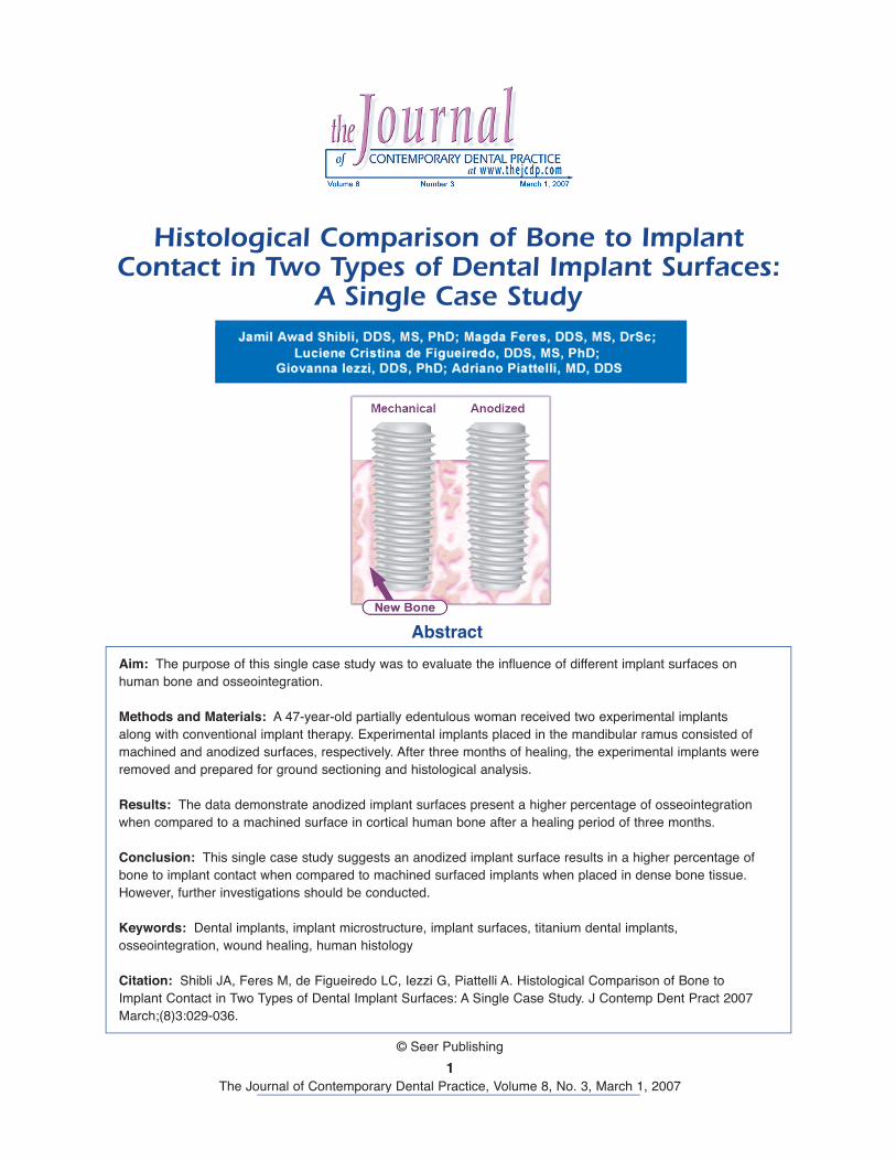
Implant Preparation (Anodic Oxidation) Two screw shaped implants made with Grade4 titanium (Conexão Sistemas de Prótese, São Paulo, SP, Brazil) were prepared with two surfacemorphologies: one machined and the other anodized (Figure 1). Each micro-implant was2.5 mm in diameter and 6.0 mm in length.
The anodic oxidation method used was the same as previously described by Zhu et al.10
The titanium screws were ultrasonically rinsedin acetone then pickled with a mixture of HF andHNO3 (the HF/HNO3 mole ratio was 1:3) andfinally rinsed with distilled water. Anodizing wasperformed using a regulated DC power supply in the constant current mode and an electrolyte consisting of calcium (Ca) glycerophosphate and Ca acetate. Both Ca glycerophosphate and Caacetate are used as food stabilizers and food
Figure 1. Scanning electron microphotograph showing the topography of the experimental implants (a) machined and (b) anodized surface (barr=10 μm).
3The Journal of Contemporary Dental Practice, Volume 8, No. 3, March 1, 2007
additives. They are nontoxic and contain calcium with almost no impurities. After being anodized, the experimental screws were rinsed with distilledwater several times and dried.
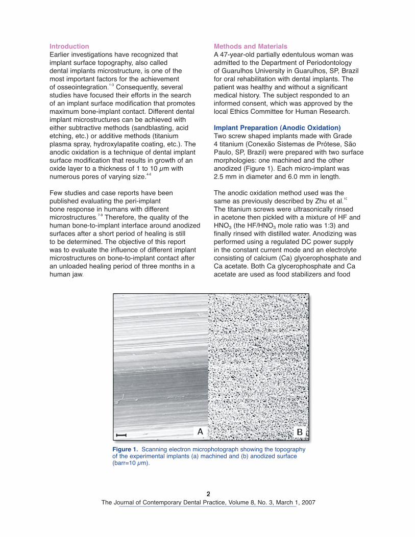
Surgical ProceduresThe two experimental implants (one with amachined surface and the other with an anodizedsurface) were surgically placed in the mandibularramus at the same time when two conventional implants were placed (3.75 x 13 mm – Porous - Conexão Dental Implants, São Paulo, SP, Brazil).Following the incision, mucoperiosteal flaps wereelevated and the conventional implants wereplaced. The recipient sites for the experimentalimplants were then prepared with a 2.0 mm diameter twist drill and inserted with a screwdriver(Figures 2 and 3).
All drilling procedures and implant placement were completed under profuse irrigation with sterile saline. Flaps were sutured with single
interrupted sutures, submerging all implants. Clindamicyn was given twice a day for a week, inorder to avoid post-surgical infection, while pain was controlled with paracetamol. The sutureswere removed after ten days.
The experimental implants were removed three months later using an internal 3.25 mm widetrephine. The experimental implants together withsurrounding bone tissue (Figure 4) were rinsed insterile saline solution and fixed by immersion in4% neutral formalin.
Histological Processing and EvaluationThe experimental implants and surrounding bone tissue were processed to obtain thin ground sections with the Precise 1 Automated System®
(Assing, Rome, Italy). The specimens were dehydrated in an ascending series of alcoholrinses and embedded in Technovit® glycolmethacrylate resin (7200 VLC, Kulzer, Wehrheim,Germany). After polymerization, the specimens were sectioned longitudinally along the major axis of the implant with a high-precision diamond diskat a thickness of approximately 150 μm and thenground down to about 30 μm.
One slide was obtained for each micro-implant.Each slide was stained with basic fuchsin and toluidine blue. Histomorphometry of bone-to-implant contact percentage as well as the bonearea within the limits of the implant threads werecompleted using a Laborlux S® light microscope (Leitz, Wetzlar, Germany) connected to a high-resolution JVC, 3CCD® video camera (JVC KY-F55B, Milan, Italy) and interfaced to a monitor
Figure 2. Clinical view of the experimental implant being placed in the surgical site behind the conventional implant.
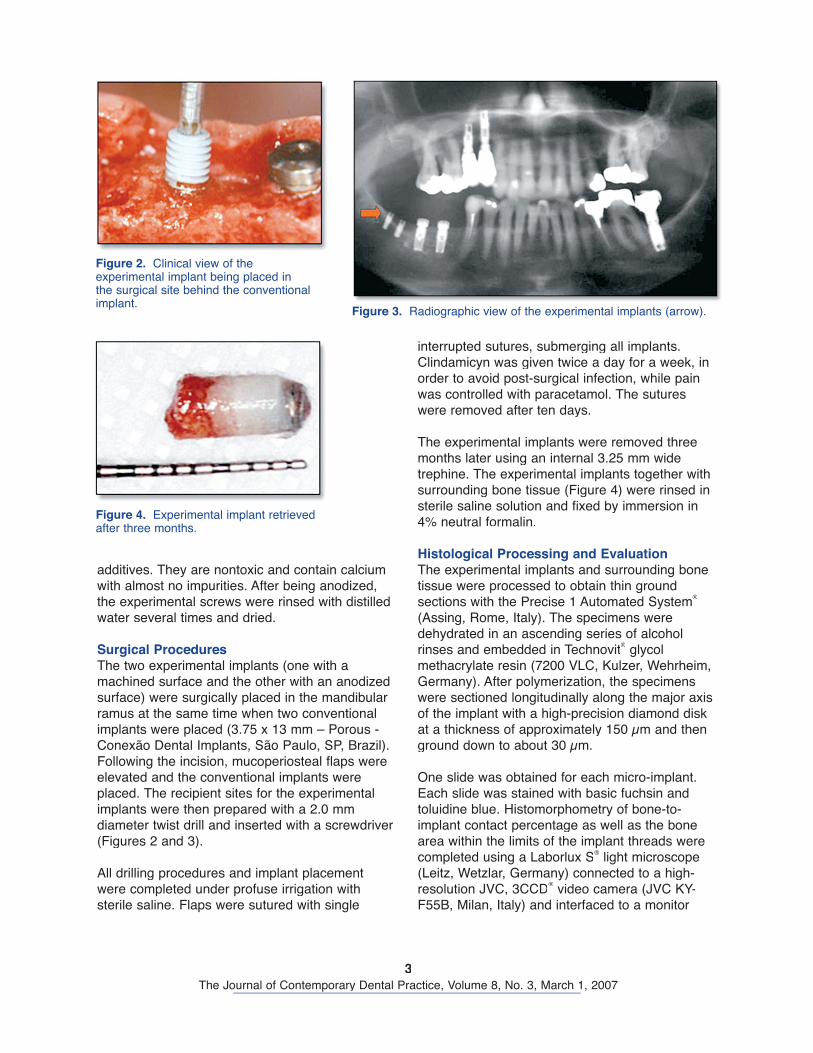
Figure 3. Radiographic view of the experimental implants (arrow).
Figure 4. Experimental implant retrieved after three months.
4The Journal of Contemporary Dental Practice, Volume 8, No. 3, March 1, 2007
and personal computer (Intel Pentium III 1200MMX). This optical system was associated with adigitizing pad (Matrix Vision GmbH, Milan, Italy) and Image-Pro Plus® 4.5 histometry software(Media Cybernetics Inc., Immagini & ComputerSnc, Milan, Italy) with image-capturing capabilities. The bone-to-implant contact and the amount of bone area within the threads (from the lowestpoint of the experimental implant head to the lastapical thread) were calculated and expressedas a percentage of bone-to-implant contact andpercentage of bone area, respectively.
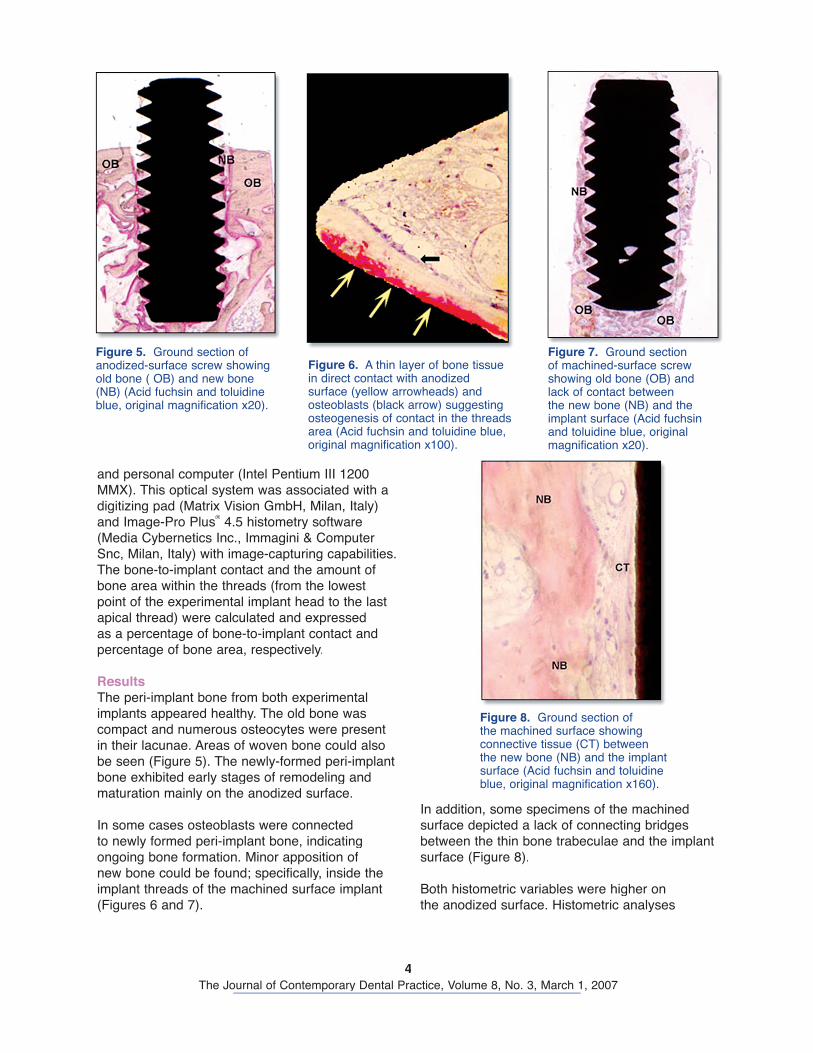
Results The peri-implant bone from both experimental implants appeared healthy. The old bone was compact and numerous osteocytes were present in their lacunae. Areas of woven bone could alsobe seen (Figure 5). The newly-formed peri-implantbone exhibited early stages of remodeling andmaturation mainly on the anodized surface.
In some cases osteoblasts were connected to newly formed peri-implant bone, indicatingongoing bone formation. Minor apposition ofnew bone could be found; specifically, inside theimplant threads of the machined surface implant (Figures 6 and 7).
In addition, some specimens of the machined surface depicted a lack of connecting bridgesbetween the thin bone trabeculae and the implant surface (Figure 8).
Both histometric variables were higher on the anodized surface. Histometric analyses
Figure 5. Ground section of anodized-surface screw showing old bone ( OB) and new bone (NB) (Acid fuchsin and toluidine blue, original magnification x20).
Figure 6. A thin layer of bone tissue in direct contact with anodized surface (yellow arrowheads) and osteoblasts (black arrow) suggesting osteogenesis of contact in the threads area (Acid fuchsin and toluidine blue, original magnification x100).
Figure 7. Ground section of machined-surface screw showing old bone (OB) and lack of contact between the new bone (NB) and the implant surface (Acid fuchsin and toluidine blue, original magnification x20).
Figure 8. Ground section of the machined surface showing connective tissue (CT) between the new bone (NB) and the implant surface (Acid fuchsin and toluidine blue, original magnification x160).
5The Journal of Contemporary Dental Practice, Volume 8, No. 3, March 1, 2007
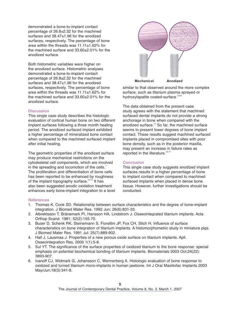
demonstrated a bone-to-implant contact percentage of 26.8±2.32 for the machinedsurfaces and 38.47±1.96 for the anodizedsurfaces, respectively. The percentage of bone area within the threads was 11.11±1.62% forthe machined surface and 33.60±2.01% for theanodized surface.
Both histometric variables were higher on the anodized surface. Histometric analyses demonstrated a bone-to-implant contact percentage of 26.8±2.32 for the machinedsurfaces and 38.47±1.96 for the anodizedsurfaces, respectively. The percentage of bone area within the threads was 11.11±1.62% forthe machined surface and 33.60±2.01% for theanodized surface.
DiscussionThis single case study describes the histologic evaluation of cortical human bone on two differentimplant surfaces following a three month healingperiod. The anodized surfaced implant exhibited a higher percentage of mineralized bone contactwhen compared to the machined surfaced implantafter initial healing.
The geometric properties of the anodized surfacemay produce mechanical restrictions on the cytoskeletal cell components, which are involvedin the spreading and locomotion of the cells.11
The proliferation and differentiation of bone cellshas been reported to be enhanced by roughness of the implant topography surface.12-13 It has also been suggested anodic oxidation treatment enhances early bone-implant integration to a level
similar to that observed around the more complex surface, such as titanium plasma sprayed orhydroxylapatite coated-surface.3,5-6
The data obtained from the present case study agrees with the statement that machined surfaced dental implants do not provide a strong anchorage in bone when compared with the anodized surface.14 So far, the machined surfaceseems to present lower degrees of bone implant contact. These results suggest machined surfaced implants placed in compromised sites with poor bone density, such as in the posterior maxilla,may present an increase in failure rates asreported in the literature.15-17
ConclusionThis single case study suggests anodized implantsurfaces results in a higher percentage of boneto implant contact when compared to machined surfaced implants when placed in dense bone tissue. However, further investigations should be conducted.
References1. Thomas K, Cook SD. Relationship between surface characteristics and the degree of bone-implant
integration. J Biomed Mater Res. 1992 Jun; 26(6):831-33.2. Albrektsson T, Bränemark PI, Hansson HA, Lindstrom J. Osseointegrated titanium implants. Acta
Orthop Scand. 1981; 52(2):155-70.3. Buser D, Schenk RK, Steinemann S, Fiorellini JP, Fox CH, Stich H. Influence of surface
characteristics on bone integration of titanium implants. A histomorphometric study in miniature pigs.J Biomed Mater Res. 1991 Jul; 25(7):889-902.
4. Hall J, Lausmaa J. Properties of a new porous oxide surface on titanium implants. Apll. Osseointegration Res. 2000 1(1):5-8.
5. Sul YT. The significance of the surface properties of oxidized titanium to the bone response: special emphasis on potential biochemical bonding of titanium implants. Biomaterials 2003 Oct;24(22):3893-907.
6. Ivanoff CJ, Widmark G, Johansson C, Wennerberg A. Histologic evaluation of bone response tooxidized and turned titanium micro-implants in human jawbone. Int J Oral Maxillofac Implants 2003 May/Jun;18(3):341-8.
6The Journal of Contemporary Dental Practice, Volume 8, No. 3, March 1, 2007
7. Degidi M, Petrone G, Iezzi G, Piattelli A. Bone contact around acid-etched implants: a histological andhistomorphometrical evaluation of two human-retrieved implants. J Oral Implantol. 2003;29(1):13-8.
8. Trisi P, Lazarra R, Rebaudi A, Rao W, Testori T, Porter SS. Bone-implant contact on machinedand dual acid-etched surfaces after 2 months of healing in the human maxilla. J Periodontol. 2003,Jul;74(7):945-56.
9. Ivanoff CJ, Hallgren C, Widmark G, Sennerby L, Wennerberg A. Histologic evaluation of the boneintegration of TiO2 blasted and turned titanium microimplants in human. Clin Oral Implants Res. 2001,Apr;12(2):128-34.
10. Zhu X, Kim KH, Jeong Y. Anodic oxide films containing Ca and P of titanium biomaterial. Biomaterials2001, Aug:22(16):2199-206.
11. Den Braber ET, De Ruijter JE, Smits HTJ, Ginsel LA, Von Recum AF, Jansen JA. Effect of parallel surface microgrooves and surface energy on cell growth. J Biomed Mater Res. 1995, Apr;29(4):511-8.
12. Martin JY, Schwartz Z, Hummert TW, Schraub DM, Simpson J, Cochran DL. Effect of titanium surface roughness on proliferation, differentiation and protein syntesis of human osteoblast-like cells. J Biomed Mater Res. 1995, Mar;29(3):389-401.
13. Schwartz Z, Lohmann CH, Oefinger J, Bonewald LF, Dean DD, Boyan BD. Implant surface characteristics modulate differentiation behavior of cells in the osteoblastic lineage. Adv Dent Res.1999 Jun;13:38-48.
14. Huang YH, Xiropaidis AV, Sorensen RG, Albandar JM, Hall J, Wikesjö UME. Bone formation attitanium porous oxide (TiUnite®) oral implants in type IV bone. Clin Oral Implants Res 2005; 16:105-111.
15. Buser D, Mericske-Stern R, Bernard JP, Behneke A, Behneke N, Hirt HP, Belser UC, Lang NP. Long-term evalution of non-submerged ITI implants. Part I. 8-year life table analysis of a prospective multi-center study with 2359 implants. Clin Oral Implants Res. 1997,Jun;8(3):161-72.
16. Friberg B, Jemt T, Lekholm U. Early failures in 4641 consecutively placed Bränemark dental implants.A study from stage I surgery to the connection of completed prostheses. Int J Oral Maxillofac Implants 1991, summer;6(2):142-6.
17. Jaffin RA, Berman CL. The excessive loss of Branemark fixtures in type IV bone. A 5-year analysis. J Periodontol. 1991, Jan;62(1):2-4.
18. Ivanoff CJ, Grondahl K, Sennerby L, Bergtrom C, Lekholm U. Influence of variations in implant diameters: a 3- to 5-year retrospective clinical report. Int J Oral Maxillofac Implants 1999;14:175-182.
19. Ivanoff CJ. On surgical and implant related factors influencing integration and function of titaniumimplants. Experimental and clinical aspects. [thesis]. Göteborg: Göteborg University, 1999.
20. Leib AM, Kowalsky CJ. Human histological research: is it necessary? Humane? Ethical?J Periodontol 2005; 76:1207-1210.
About the Authors
7The Journal of Contemporary Dental Practice, Volume 8, No. 3, March 1, 2007
AcknowledgementsThis study was supported by Dental Research Division, Guarulhos University, in Guarulhos, SP, Brazil. The authors would like to express their appreciation to Conexão Implants in São Paulo, SP, Brazil forproviding the micro-implants used in this study.