Embed Size (px)
Citation preview
“main” — 2010/4/30 — 21:31 — page 397 — #1
Anais da Academia Brasileira de Ciências (2010) 82(2): 397-404(Annals of the Brazilian Academy of Sciences)ISSN 0001-3765www.scielo.br/aabc
Histological modifications of the rat prostate following transectionof somatic and autonomic nerves
ROSAURA DIAZ, LUIS I. GARCIA, JOSE LOCIA, MILAGROS SILVA, SARA RODRIGUEZ,CESAR A. PEREZ, GONZALO E. ARANDA-ABREU, JORGE MANZO,
REBECA TOLEDO and MARIA ELENA HERNANDEZ
Programa de Neurobiologia, Universidad Veracruzana, Av. Luis Castelazo s/nCol. Industrial Animas, Xalapa, Ver., C.P. 91190, Mexico
Manuscript received on January 8, 2009; accepted for publication on January 25, 2010
ABSTRACT
It is known that hormones influence significantly the prostate tissue. However, we reported that mating induces an
increase in androgen receptors, revealing a neural influence on the gland. These data suggested that somatic afferents
(scrotal and genitofemoral nerves) and autonomic efferents (pelvic and hypogastric nerves) could regulate the structure
of the prostate. Here we assessed the role of these nerves in maintaining the histology of the gland. Hence, afferent or
efferent nerves of male rats were transected. Then, the ventral and dorsolateral regions of the prostate were processed
for histology. Results showed that afferent transection affects prostate histology. The alveoli area decreased and
increased in the ventral and dorsolateral prostate, respectively. The epithelial cell height increased in both regions.
Efferent denervation produced dramatic changes in the prostate gland. The tissue lost its configuration, and the epithe-
lium became scattered and almost vanished. Thus, afferent nerves are responsible for spinal processes pertaining to
the trophic control of the prostate, activating its autonomic innervation. Hence, our data imply that innervation seems
to be synergic with hormones for the healthy maintenance of the prostate. Thus, it is suggested that some prostate
pathologies could be due to the failure of the autonomic neural pathways regulating the gland.
Key words: pelvic nerve, hypogastric nerve, genitofemoral nerve, scrotal nerve.
INTRODUCTION
The prostate is the largest sexual gland in the male re-productive tract, and its structure and function have beenwidely investigated by a number of authors. Most re-ports, including ours (Hernandez et al. 2006, 2007), haveemphasized the importance of hormones in the growth,function, and pathological development of this gland.Thus, the leading therapy for prostate cancer is stillbased on endocrine manipulation. However, one issuethat has been less investigated is the role of the periph-eral nerves not only on the physiology of the gland, butalso on the development of cancer cells.
Previously, we argued that the execution of sex-
Correspondence to: Dr. Maria Elena HernandezE-mail: [email protected]
ual behavior induces an increase in androgen receptorsin the prostate tissue of male rats, which could be ex-plained as a neural influence on the gland (Hernandezet al. 2007). Also, we showed that the stimulation ofthe scrotal skin during copulation is important for theejaculation of a normal quantity of semen, in which theprostate plays a key role (Garcia et al. 2007). There-fore, we hypothesized that somatic afferent fibers (scro-tal and genitofemoral nerves) and the autonomic inner-vation of the prostate (pelvic and hypogastric nerves)seem to integrate into a spinal circuit that regulates thefunction, and perhaps contributes to the dysfunction, ofthe prostate gland.
The prostate is innervated by postganglionar fibersemerging from the major pelvic ganglion that, in turn,
An Acad Bras Cienc (2010) 82 (2)
“main” — 2010/4/30 — 21:31 — page 398 — #2
398 ROSAURA DIAZ et al.
is innervated by both the hypogastric nerve and the vis-cerocutaneous branch of the pelvic nerve (Hulseboschand Coggeshall 1982, Langworthy 1965, Pacheco et al.1989). This nerve complex regulates the function ofthe prostate via cholinergic, noradrenergic, and peptider-gic pathways (Kepper and Keast 1995, Nadelhaft 2003,Pennefather et al. 2000). Recently, it has been reportedthat these autonomic efferents are driven by a complexcircuit of central neurons stationed in different brain nu-clei, all of which are involved in the control of male ratsexual behavior (Huddleston et al. 2007). Spinal mech-anisms have also been proposed to influence the physi-ology of the prostate (Huang et al. 1997). However, theextent to which the spinal cord and peripheral nerves areinvolved in the autonomic regulation of the prostate is yetunknown. Thus, in this study we investigated the effectsproduced on the prostate tissue following the lesion ofafferent or efferent nerves in the pelvic area.
MATERIALS AND METHODS
SUBJECTS AND HOUSING
Sexually experienced Wistar male rats from Harlan Mex-ico were studied (250-300 g/bw). As copulation main-tains a continuous activity of the nerves and prostategland, the rats were submitted to regular copulation tri-als, as stated below. Ovariectomized sexually experi-enced females were used during the copulatory trials andtheir sexual receptivity was induced with subcutaneousinjections of estradiol benzoate (10μg; Sigma-AldrichMexico) followed by progesterone (2 mg; Sigma-Al-drich Mexico), both diluted in sesame oil and admin-istered subcutaneously 48 hr and 4 hr before mating, re-spectively. The rats were kept in Plexiglas cages (50 ×30 × 20 cm) with woodchip bedding (Harlan Mexico) ina temperature-controlled room (22 ± 2◦C), with free ac-cess to food (Harlan rodent chow) and water. The roomwas kept at a reversed 12:12 cycle (lights on at 8:00 pm).
ANIMAL USE AND CARE
Every surgical intervention and manipulation was exe-cuted according to the Policy on the Use of Animals inNeuroscience Research of the Society for Neuroscience.Every effort was made to minimize animal suffering andthe total number of animals used.
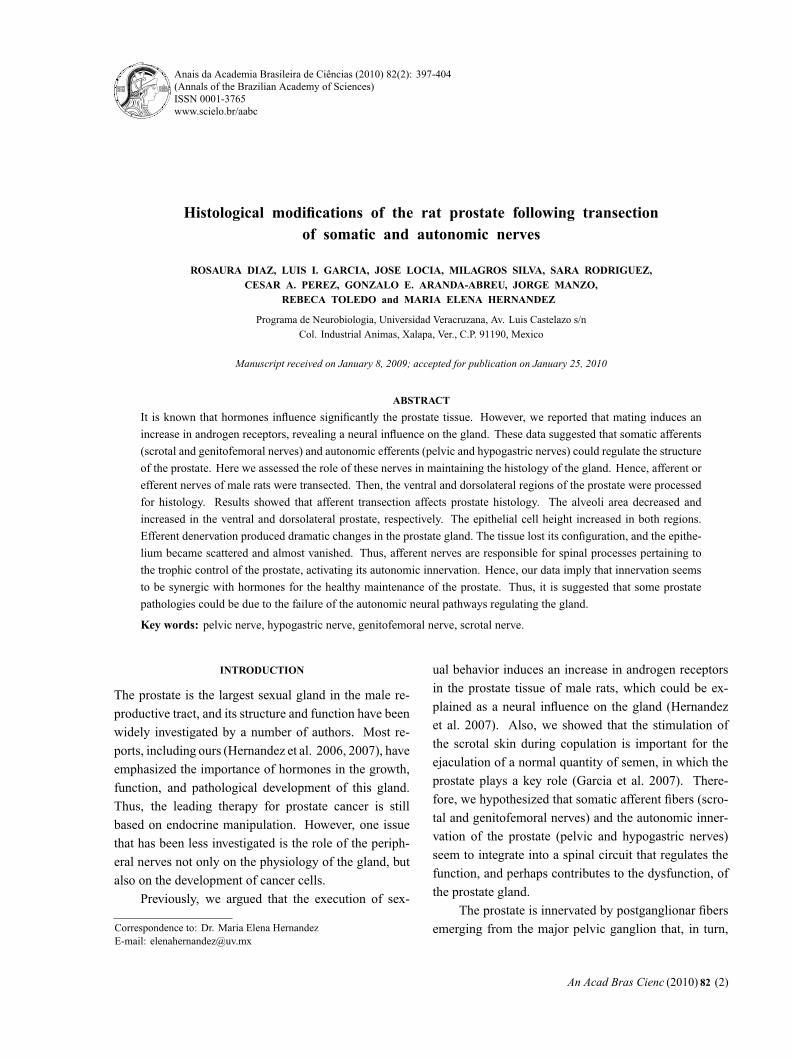
Fig. 1 – Somatic (afferent) and autonomic (efferent) nerves in the
pelvic area. The integration of the nerves occurs in the lumbar and
sacral segments of the spinal cord. The transected afferent nerves, scro-
tal and genitofemoral nerves, supply the scrotal and groin epithelium,
which are mechanically stimulated during copulation. The transected
efferent nerves, pelvic and hypogastric nerves, innervate the prostate
gland and other structures.
GROUPS AND EXPERIMENTAL RATIONALE
The areas that are mechanically stimulated during cop-ulation are the scrotum and groin. The scrotal area isinnervated by the scrotal (Sc) nerves, whereas the groinis innervated by the genitofemoral (Gf) nerves (Fig. 1).It is known that the transection of these nerves does notaffect the proper execution of sexual behavior (Garciaet al. 2007, Lucio et al. 2001). Nevertheless, we pre-viously hypothesized that scrotal afferents trigger spinalreflexes and activate autonomic efferents that supply sex-ual glands (Garcia et al. 2007). Thus, the experimen-tal objective of this study was to identify whether thesenerves influence the histological parameters of the pro-
An Acad Bras Cienc (2010) 82 (2)
“main” — 2010/4/30 — 21:31 — page 399 — #3
NEURAL BASIS OF PROSTATE HISTOLOGY 399
state. To do so, subjects were randomly assigned to threeexperimental groups: a group with bilateral lesions ofthe Sc nerve (n=8), a group with bilateral lesions of theGf nerve (n=8), and a group with bilateral lesions ofboth Sc+Gf nerves (n=8). Each group with its respectiveSham operated control (n=6).
The spinal integration of cutaneous afferents seemsto activate the autonomic nerves that supply the sexualglands, i.e., the pelvic (Pv) and hypogastric (Hg) nerves(Fig. 1). Previously, it was shown that the lesion ofthe Pv nerves modifies the quality of sexual behavior inmale rats (Lucio et al. 1994), whereas an Hg lesiondoes not modify this behavior at all (Cruz et al. 1999).Thus, following the cutaneous afferents paradigm, wecarried out similar experiments: a group with bilaterallesions of the Pv nerve (n=7), a group with bilateral le-sions of the Hg nerve (n=7), and a group with bilaterallesions of both Pv+Hg nerves (n=6). All of them withtheir respective Sham control (n=6).
NERVE SURGERY
Subjects were selected to participate in the experimentonce they proved to be sexually experienced (males withejaculation latencies between 5 and 15 min after fourtraining tests). Next, they were randomly assigned to agroup and were given the required nerve lesion carriedout under sodium pentobarbital anesthesia (30 mg/kg bw;Anestesal, Smith Kline, Mexico). A surgical microscopewas used to dissect and transect the nerves.
To reach the Sc nerves, a midline longitudinal inci-sion was made in the lower back. The muscles were sep-arated from the vertebrae to locate the pudendal artery,which was then bent to allow a proper view of the sacralplexus. The proximal and distal Sc nerves were dis-sected from their origin in the plexus up to their termi-nation in the periphery (Garcia et al. 2007, Pacheco etal. 1997), hooked, and then a 5 mm portion of the nerveswas extracted to avoid regeneration. These procedureswere done bilaterally.
The Gf nerves are the easiest to reach. After a mid-line abdominal longitudinal incision, the intestines werecarefully moved to one side to expose the descendingaorta. Left and right Gf nerves were located on eitherside of the descending aorta, hooked and cut, again a5 mm portion of the nerves was extracted.
A similar incision was done to observe the Hg andPv nerves. However, the major pelvic ganglion was firstlocated at its closest point of contact with the prostate,and then the nerves were followed in their path to thespinal cord. A Pv nerve lesion interferes with micturi-tion; hence, the bladder was manually compressed twicea day after surgery for the remainder of the experiment.This procedure kept animals in a healthy condition.
In all of the above-mentioned cases, muscle andskin were carefully sutured (simple interrupted suturewith Catgut 3-0) and a single dose of intramuscularantibiotic was injected after the surgical intervention(0.1 ml of Pengesod, 1,000,000 U, Lakeside Mexico).Sham-operated subjects underwent surgical procedureswhere the nerves were hooked, but not cut. At the end ofthe study, the animals were re-examined to confirm thatno regeneration had occurred.
BEHAVIORAL PROCEDURES AND PROSTATE SURGERY
In order to maintain a continuous activation of the pel-vic reproductive system, males were placed with sexu-ally receptive females three days following the surgeryto copulate and were removed after one ejaculation. Thisprocedure was held out at every two days for a total offive mating sessions. Following the last ejaculation (day15 after surgery), males were anesthetized with an in-traperitoneal injection of sodium pentobarbital (30 mg/kg bw; Anestesal, Smith Kline, Mexico). An abdominalmidline longitudinal incision was made to reach the pro-state, and tissue from the ventral (VP) and dorsolateral(DLP) regions of the gland were removed and processedfor histology using the hematoxilin-eosin method. Then,nerve transections were re-examined in each animal, andthey were subsequently sacrificed with a lethal dose ofsodium pentobarbital.
STAINING THE PROSTATE TISSUE
The hematoxilin-eosin staining procedure was perfor-med by immersing the prostate tissue for 24h in Hellyfixative (Mercuric Chloride 5%, Potassium Dichromate2.5%, Sodium Sulfate 1%, and Formalin 37%); then, thetissue was rinsed with tap water for 30 min. Dehydra-tion was carried out by immersing the prostate in ethanol70% (1 hr), 80% (1 hr), 96% (3 times, 2 hr each), abso-lute ethanol (overnight), and absolute ethanol (2 times,
An Acad Bras Cienc (2010) 82 (2)
“main” — 2010/4/30 — 21:31 — page 400 — #4
400 ROSAURA DIAZ et al.
1 hr each). The tissue was cleared by immersing theprostate in xylene (3 times, 1 hr each). Both dehydrationand clearing were carried out on a stirring plate. Next,the tissue was embedded in melted paraffin (2 times, 2hr each at 57◦C) and a block was carved using a micro-tome cassette. The paraffin block was sectioned on aLeica microtome (5μm). Sections were transferred to amounting bath of gelatinized water at 52◦C and mountedon slides that were warmed for 1 hr in an oven (∼58◦C).For staining, the sections were immersed in xylene (3times, 5 min each), absolute ethanol/xylene 1:1 (5 min),ethanol 96% (3 min), iodine alcohol (5 min), sodium thio-sulfate (10 min), tap water (2 min), Mayer’s Hematox-ilin (10 min), tap water (30 sec), acid ethanol (fast bath),tap water (10 sec), lithium carbonate (30 sec), tap water(10 sec), Eosin Y (4 fast baths), ethanol 96% (3 min),absolute ethanol (2 min), absolute ethanol/xylene 1:1(2 min), and xylene (5 min). Finally, sections were cov-erslipped using undiluted Permount. Each slide had sec-tions from one region of the prostate, either VP or DLP.The slides were observed under an Olympus Provis AX-70 microscope, and images were obtained and analyzedwith the help of Image Pro Plus software (in a Dell PCComputer with Windows XP Operating System). Mea-surements of the mean area of each of the alveoli andthe height of epithelial cells were compiled.
STATISTICS
Nonparametric statistics were used. Between-groupcomparisons were made using a Kruskal-Wallis analysisof variance, and paired contrasts were obtained usingthe Mann-Whitney U test. Significant differences wereinferred when p<0.05.
RESULTS
AFFERENT DENERVATION
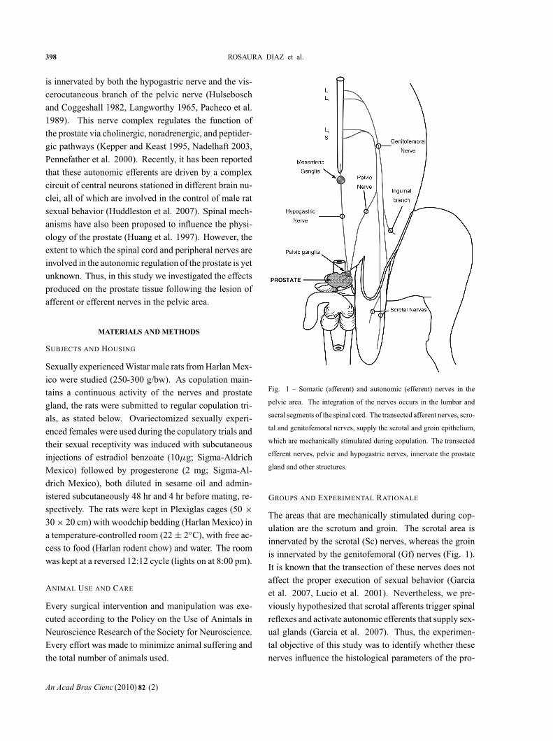
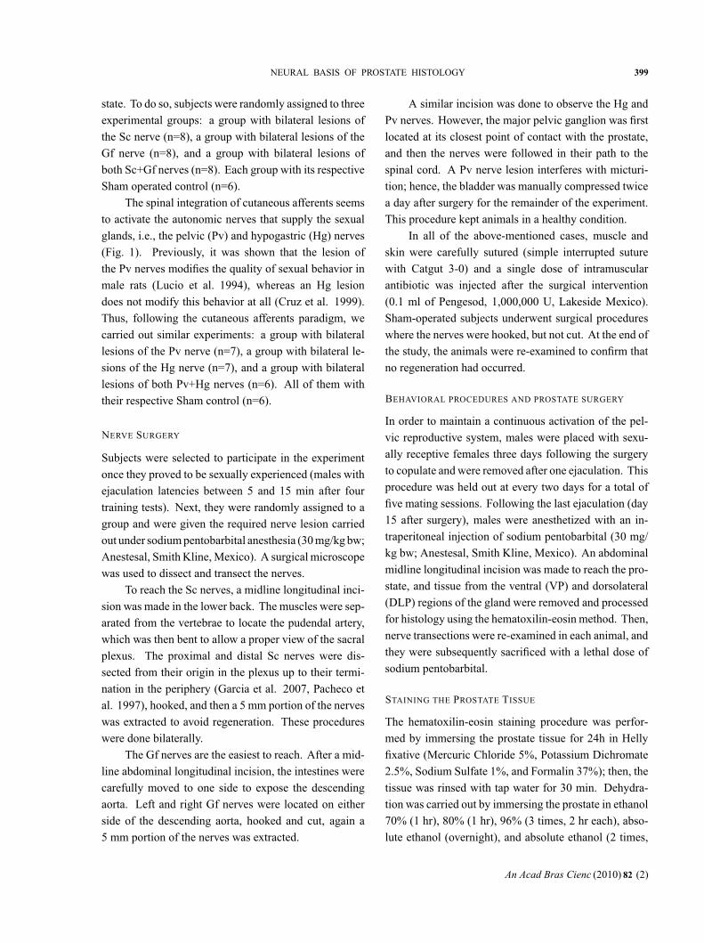
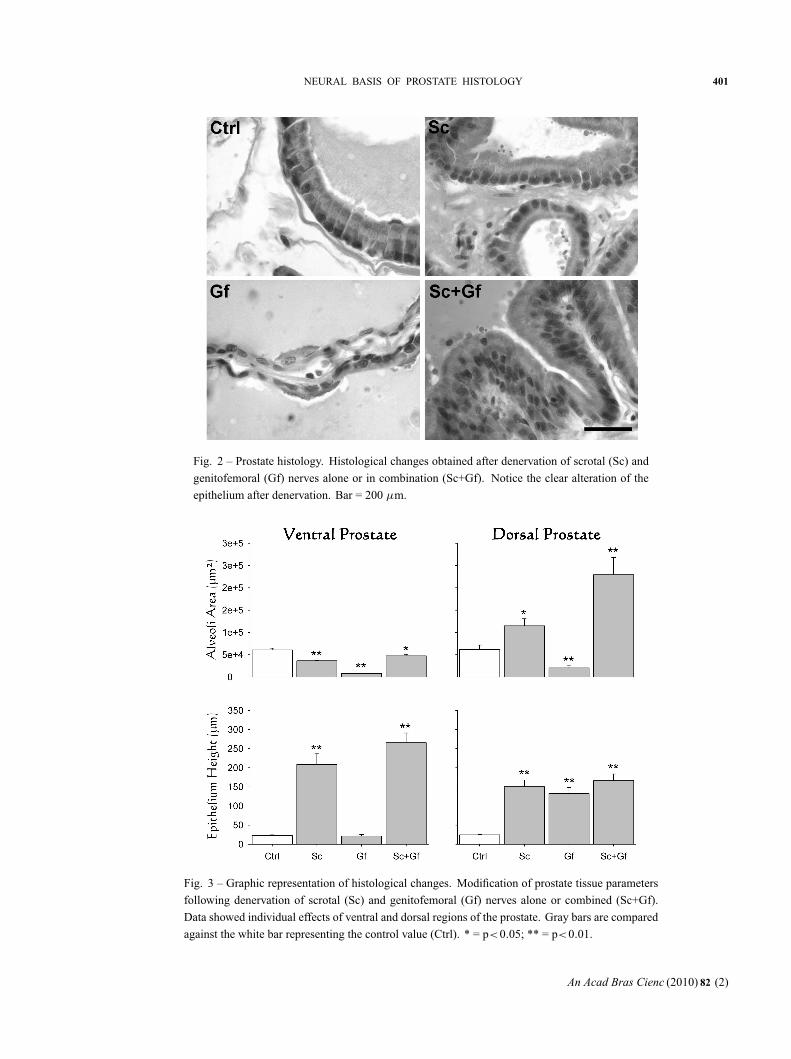
Transection of the Sc and/or Gf did not affect the properexecution of sexual behavior, as reported elsewhere(Garcia et al. 2007, Lucio et al. 2001). However, reli-able effects on prostate histology were observed (Fig. 2).The area of the alveoli in the VP significantly decreasedafter Sc, Gf or Sc+Gf lesions, although a Gf lesion pro-duced the most significant decrease (Fig. 3). The DLPshowed a different pattern. Sc transection, alone or com-bined with the Gf transection, yielded a significant in-
crease in the alveoli area, while a Gf transection aloneproduced a decrease (Fig. 3). On the other hand, theepithelial cell height significantly increased in both theVP and the DLP after Sc and/or Gf lesions. However,in the VP, a Gf transection alone did not produce anymodification in these cells (Fig. 3).
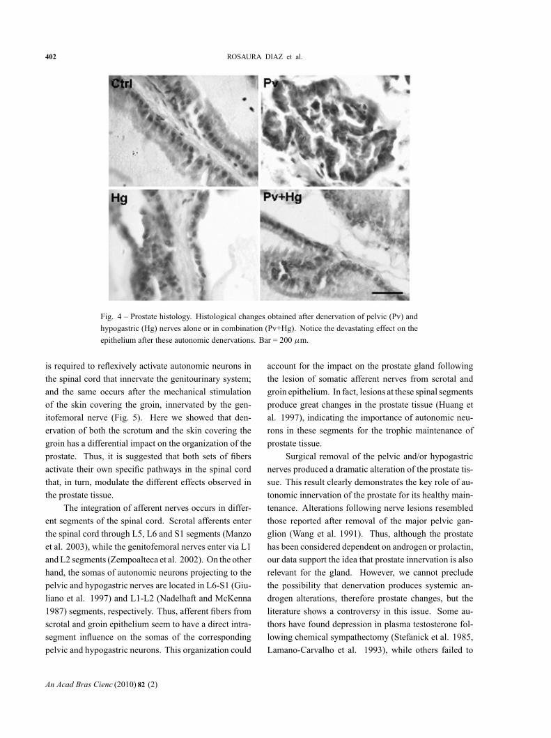
EFFERENT DENERVATION
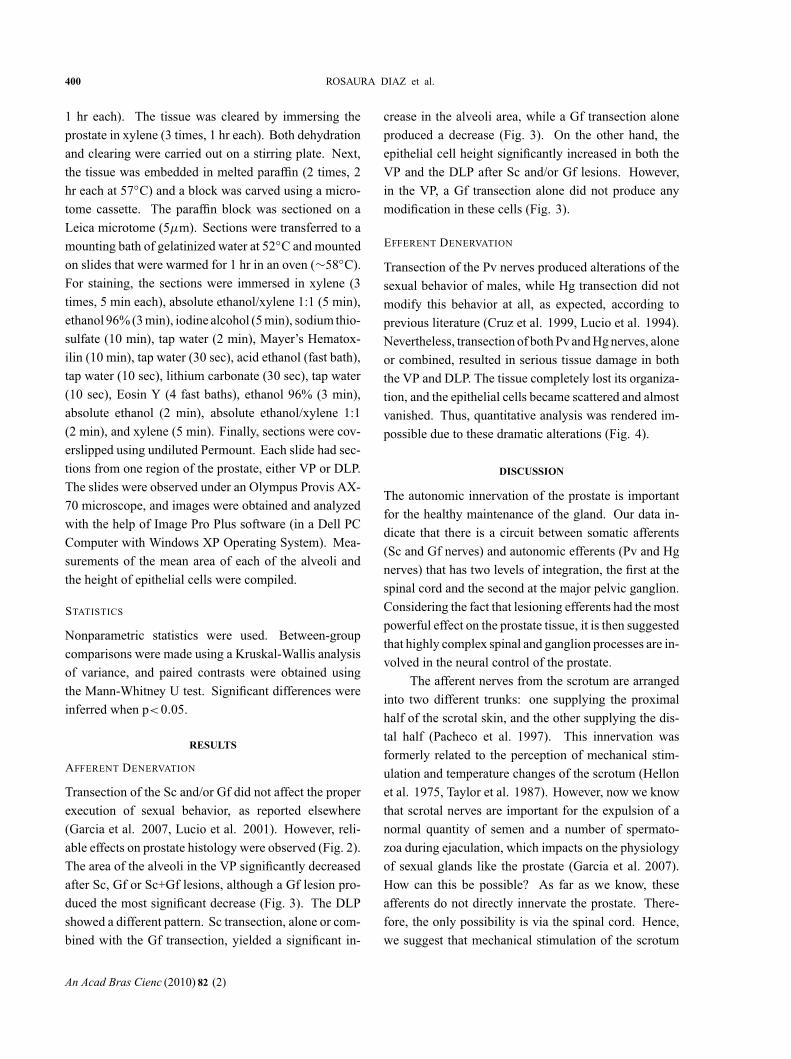
Transection of the Pv nerves produced alterations of thesexual behavior of males, while Hg transection did notmodify this behavior at all, as expected, according toprevious literature (Cruz et al. 1999, Lucio et al. 1994).Nevertheless, transection of both Pv and Hg nerves, aloneor combined, resulted in serious tissue damage in boththe VP and DLP. The tissue completely lost its organiza-tion, and the epithelial cells became scattered and almostvanished. Thus, quantitative analysis was rendered im-possible due to these dramatic alterations (Fig. 4).
DISCUSSION
The autonomic innervation of the prostate is importantfor the healthy maintenance of the gland. Our data in-dicate that there is a circuit between somatic afferents(Sc and Gf nerves) and autonomic efferents (Pv and Hgnerves) that has two levels of integration, the first at thespinal cord and the second at the major pelvic ganglion.Considering the fact that lesioning efferents had the mostpowerful effect on the prostate tissue, it is then suggestedthat highly complex spinal and ganglion processes are in-volved in the neural control of the prostate.
The afferent nerves from the scrotum are arrangedinto two different trunks: one supplying the proximalhalf of the scrotal skin, and the other supplying the dis-tal half (Pacheco et al. 1997). This innervation wasformerly related to the perception of mechanical stim-ulation and temperature changes of the scrotum (Hellonet al. 1975, Taylor et al. 1987). However, now we knowthat scrotal nerves are important for the expulsion of anormal quantity of semen and a number of spermato-zoa during ejaculation, which impacts on the physiologyof sexual glands like the prostate (Garcia et al. 2007).How can this be possible? As far as we know, theseafferents do not directly innervate the prostate. There-fore, the only possibility is via the spinal cord. Hence,we suggest that mechanical stimulation of the scrotum
An Acad Bras Cienc (2010) 82 (2)
“main” — 2010/4/30 — 21:31 — page 401 — #5
NEURAL BASIS OF PROSTATE HISTOLOGY 401
Fig. 2 – Prostate histology. Histological changes obtained after denervation of scrotal (Sc) and
genitofemoral (Gf) nerves alone or in combination (Sc+Gf). Notice the clear alteration of the
epithelium after denervation. Bar = 200 μm.
Fig. 3 – Graphic representation of histological changes. Modification of prostate tissue parameters
following denervation of scrotal (Sc) and genitofemoral (Gf) nerves alone or combined (Sc+Gf).
Data showed individual effects of ventral and dorsal regions of the prostate. Gray bars are compared
against the white bar representing the control value (Ctrl). * = p<0.05; ** = p<0.01.
An Acad Bras Cienc (2010) 82 (2)
“main” — 2010/4/30 — 21:31 — page 402 — #6
402 ROSAURA DIAZ et al.
Fig. 4 – Prostate histology. Histological changes obtained after denervation of pelvic (Pv) and
hypogastric (Hg) nerves alone or in combination (Pv+Hg). Notice the devastating effect on the
epithelium after these autonomic denervations. Bar = 200 μm.
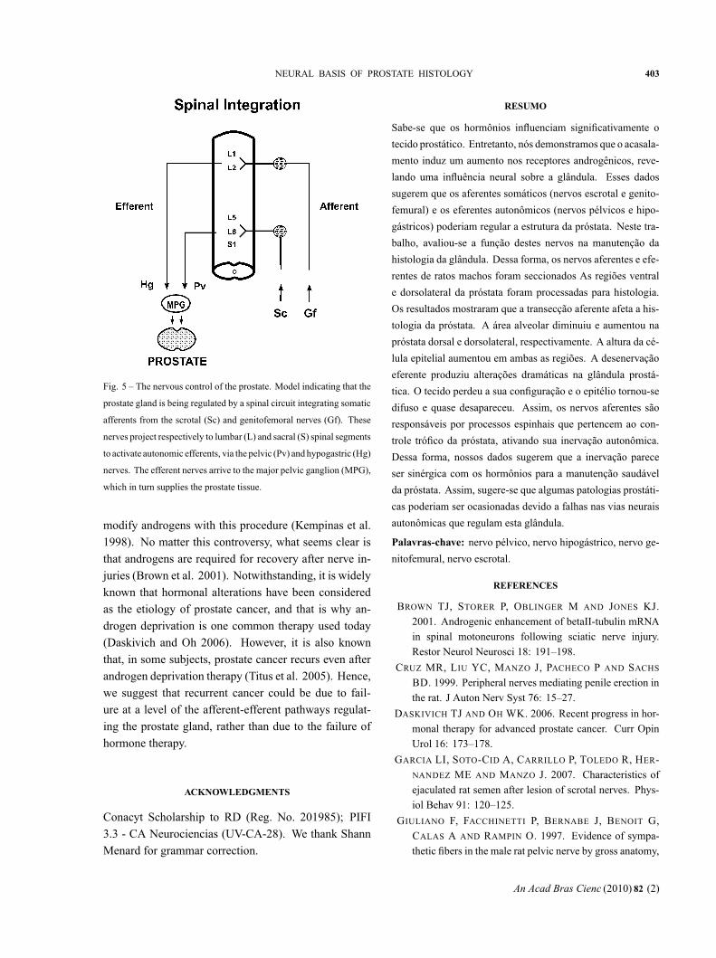
is required to reflexively activate autonomic neurons inthe spinal cord that innervate the genitourinary system;and the same occurs after the mechanical stimulationof the skin covering the groin, innervated by the gen-itofemoral nerve (Fig. 5). Here we showed that den-ervation of both the scrotum and the skin covering thegroin has a differential impact on the organization of theprostate. Thus, it is suggested that both sets of fibersactivate their own specific pathways in the spinal cordthat, in turn, modulate the different effects observed inthe prostate tissue.
The integration of afferent nerves occurs in differ-ent segments of the spinal cord. Scrotal afferents enterthe spinal cord through L5, L6 and S1 segments (Manzoet al. 2003), while the genitofemoral nerves enter via L1and L2 segments (Zempoalteca et al. 2002). On the otherhand, the somas of autonomic neurons projecting to thepelvic and hypogastric nerves are located in L6-S1 (Giu-liano et al. 1997) and L1-L2 (Nadelhaft and McKenna1987) segments, respectively. Thus, afferent fibers fromscrotal and groin epithelium seem to have a direct intra-segment influence on the somas of the correspondingpelvic and hypogastric neurons. This organization could
account for the impact on the prostate gland followingthe lesion of somatic afferent nerves from scrotal andgroin epithelium. In fact, lesions at these spinal segmentsproduce great changes in the prostate tissue (Huang etal. 1997), indicating the importance of autonomic neu-rons in these segments for the trophic maintenance ofprostate tissue.
Surgical removal of the pelvic and/or hypogastricnerves produced a dramatic alteration of the prostate tis-sue. This result clearly demonstrates the key role of au-tonomic innervation of the prostate for its healthy main-tenance. Alterations following nerve lesions resembledthose reported after removal of the major pelvic gan-glion (Wang et al. 1991). Thus, although the prostatehas been considered dependent on androgen or prolactin,our data support the idea that prostate innervation is alsorelevant for the gland. However, we cannot precludethe possibility that denervation produces systemic an-drogen alterations, therefore prostate changes, but theliterature shows a controversy in this issue. Some au-thors have found depression in plasma testosterone fol-lowing chemical sympathectomy (Stefanick et al. 1985,Lamano-Carvalho et al. 1993), while others failed to
An Acad Bras Cienc (2010) 82 (2)
“main” — 2010/4/30 — 21:31 — page 403 — #7
NEURAL BASIS OF PROSTATE HISTOLOGY 403
Fig. 5 – The nervous control of the prostate. Model indicating that the
prostate gland is being regulated by a spinal circuit integrating somatic
afferents from the scrotal (Sc) and genitofemoral nerves (Gf). These
nerves project respectively to lumbar (L) and sacral (S) spinal segments
to activate autonomic efferents, via the pelvic (Pv) and hypogastric (Hg)
nerves. The efferent nerves arrive to the major pelvic ganglion (MPG),
which in turn supplies the prostate tissue.
modify androgens with this procedure (Kempinas et al.1998). No matter this controversy, what seems clear isthat androgens are required for recovery after nerve in-juries (Brown et al. 2001). Notwithstanding, it is widelyknown that hormonal alterations have been consideredas the etiology of prostate cancer, and that is why an-drogen deprivation is one common therapy used today(Daskivich and Oh 2006). However, it is also knownthat, in some subjects, prostate cancer recurs even afterandrogen deprivation therapy (Titus et al. 2005). Hence,we suggest that recurrent cancer could be due to fail-ure at a level of the afferent-efferent pathways regulat-ing the prostate gland, rather than due to the failure ofhormone therapy.
ACKNOWLEDGMENTS
Conacyt Scholarship to RD (Reg. No. 201985); PIFI3.3 - CA Neurociencias (UV-CA-28). We thank ShannMenard for grammar correction.
RESUMO
Sabe-se que os hormônios influenciam significativamente o
tecido prostático. Entretanto, nós demonstramos que o acasala-
mento induz um aumento nos receptores androgênicos, reve-
lando uma influência neural sobre a glândula. Esses dados
sugerem que os aferentes somáticos (nervos escrotal e genito-
femural) e os eferentes autonômicos (nervos pélvicos e hipo-
gástricos) poderiam regular a estrutura da próstata. Neste tra-
balho, avaliou-se a função destes nervos na manutenção da
histologia da glândula. Dessa forma, os nervos aferentes e efe-
rentes de ratos machos foram seccionados As regiões ventral
e dorsolateral da próstata foram processadas para histologia.
Os resultados mostraram que a transecção aferente afeta a his-
tologia da próstata. A área alveolar diminuiu e aumentou na
próstata dorsal e dorsolateral, respectivamente. A altura da cé-
lula epitelial aumentou em ambas as regiões. A desenervação
eferente produziu alterações dramáticas na glândula prostá-
tica. O tecido perdeu a sua configuração e o epitélio tornou-se
difuso e quase desapareceu. Assim, os nervos aferentes são
responsáveis por processos espinhais que pertencem ao con-
trole trófico da próstata, ativando sua inervação autonômica.
Dessa forma, nossos dados sugerem que a inervação parece
ser sinérgica com os hormônios para a manutenção saudável
da próstata. Assim, sugere-se que algumas patologias prostáti-
cas poderiam ser ocasionadas devido a falhas nas vias neurais
autonômicas que regulam esta glândula.
Palavras-chave: nervo pélvico, nervo hipogástrico, nervo ge-
nitofemural, nervo escrotal.
REFERENCES
BROWN TJ, STORER P, OBLINGER M AND JONES KJ.2001. Androgenic enhancement of betaII-tubulin mRNAin spinal motoneurons following sciatic nerve injury.Restor Neurol Neurosci 18: 191–198.
CRUZ MR, LIU YC, MANZO J, PACHECO P AND SACHS
BD. 1999. Peripheral nerves mediating penile erection inthe rat. J Auton Nerv Syst 76: 15–27.
DASKIVICH TJ AND OH WK. 2006. Recent progress in hor-monal therapy for advanced prostate cancer. Curr OpinUrol 16: 173–178.
GARCIA LI, SOTO-CID A, CARRILLO P, TOLEDO R, HER-NANDEZ ME AND MANZO J. 2007. Characteristics ofejaculated rat semen after lesion of scrotal nerves. Phys-iol Behav 91: 120–125.
GIULIANO F, FACCHINETTI P, BERNABE J, BENOIT G,CALAS A AND RAMPIN O. 1997. Evidence of sympa-thetic fibers in the male rat pelvic nerve by gross anatomy,
An Acad Bras Cienc (2010) 82 (2)

“main” — 2010/4/30 — 21:31 — page 404 — #8
404 ROSAURA DIAZ et al.
retrograde labeling and high resolution autoradiographicstudy. Int J Impot Res 9: 179–185.
HELLON RF, HENSEL H AND SCHAFER K. 1975. Thermalreceptors in the scrotum of the rat. J Physiol 248: 349–357.
HERNANDEZ ME, SOTO-CID A, ROJAS F, PASCUAL LI,ARANDA-ABREU GE, TOLEDO R, GARCIA LI, QUIN-TANAR-STEPHANO A AND MANZO J. 2006. Prostateresponse to prolactin in sexually active male rats. ReprodBiol Endocrinol 4: 28.
HERNANDEZ ME, SOTO-CID A, ARANDA-ABREU GE,DIAZ R, ROJAS F, GARCIA LI, TOLEDO R AND MAN-ZO J. 2007. A study of the prostate, androgens and sex-ual activity of male rats. Reprod Biol Endocrinol 5: 11.
HUANG HF, LI MT, LINSENMEYER TA, OTTENWELLER
JE, POGACH LM AND IRWIN RJ. 1997. The effects ofspinal cord injury on the status of messenger ribonucleicacid for TRPM 2 and androgen receptor in the prostateof the rat. J Androl 18: 250–256.
HUDDLESTON GG, SONG CK, PAISLEY JC, BARTNESS TJAND CLANCY AN. 2007. Gonadal steroid receptors colo-calize with central nervous system neurons projecting tothe rat prostate gland. Am J Physiol Regul Integr CompPhysiol 292: R2196–R2205.
HULSEBOSCH CE AND COGGESHALL RE. 1982. An anal-ysis of the axon populations in the nerves to the pelvicviscera in the rat. J Comp Neurol 211: 1–10.
KEPPER M AND KEAST J. 1995. Immunohistochemical prop-erties and spinal connections of pelvic autonomic neuronsthat innervate the rat prostate gland. Cell Tissue Res 281:533–542.
KEMPINAS WD ET AL. 1998. Fertility of rat epididymalsperm after chemical and surgically induced sympathec-tomy. Biol Reprod 59: 897–904.
LAMANO-CARVALHO TL, FAVARETTO AL, PETENUSCI SOAND KEMPINAS WG. 1993. Prepubertal development ofrat prostate and seminal vesicle following chemical sym-pathectomy with guanethidine. Braz J Med Biol Res 26:639–646.
LANGWORTHY OR. 1965. Innervation of the pelvic organsof the rat. Invest Urol 2: 491–511.
LUCIO RA, MANZO J, MARTINEZ-GOMEZ M, SACHS BDAND PACHECO P. 1994. Participation of pelvic nervebranches in male rat copulatory behavior. Physiol Behav55: 241–246.
LUCIO RA, FLORES-ROJAS G, AGUILAR F, ZEMPOALTE-CA R, PACHECO P AND VELAZQUEZ-MOCTEZUMA J.2001. Effects of genitofemoral nerve transection on cop-
ulatory behavior and fertility in male rats. Physiol Behav73: 487–492.
MANZO J, GARCIA LI, CAMACHO MA, HERNANDEZ
ME AND PACHECO P. 2003. Influence of testosteroneon the electrical properties of scrotal nerves at the cuta-neous and spinal levels in the male rat. J Peripher NervSyst 8: 75–81.
NADELHAFT I. 2003. Cholinergic axons in the rat prostateand neurons in the pelvic ganglion. Brain Res 989: 52–57.
NADELHAFT I AND MCKENNA KE. 1987. Sexual dimor-phism in sympathetic preganglionic neurons of the rathypogastric nerve. J Comp Neurol 256: 308–315.
PACHECO P, MARTINEZ-GOMEZ M, WHIPPLE B, BEYER
C AND KOMISARUK BR. 1989. Somato-motor compo-nents of the pelvic and pudendal nerves of the female rat.Brain Res 490: 85–94.
PACHECO P, CAMACHO MA, GARCIA LI, HERNANDEZ
ME, CARRILLO P AND MANZO J. 1997. Electrophys-iological evidence for the nomenclature of the pudendalnerve and sacral plexus in the male rat. Brain Res 763:202–208.
PENNEFATHER JN, LAU WA, MITCHELSON F AND VEN-TURA S. 2000. The autonomic and sensory innervationof the smooth muscle of the prostate gland: a review ofpharmacological and histological studies. J Auton Phar-macol 20: 193–206.
STEFANICK ML, SMITH ER, SZUMOWSKI DA AND
DAVIDSON JM. 1985. Reproductive physiology and be-havior in the male rat following acute and chronic pe-ripheral adrenergic depletion by guanethidine. PharmacolBiochem Behav 23: 55–63.
TAYLOR DC, STEELE JE AND GAYTON RJ. 1987. An anal-ysis of the responses of rat striatal neurones to scrotal skintemperature. Brain Res 419: 352–356.
TITUS MA, SCHELL MJ, LIH FB, TOMER KB AND MOH-LER JL. 2005. Testosterone and dihydrotestosterone tis-sue levels in recurrent prostate cancer. Clin Cancer Res11: 4653–4657.
WANG JM, MCKENNA KE, MCVARY KT AND LEE C.1991. Requirement of innervation for maintenance ofstructural and functional integrity in the rat prostate. BiolReprod 44: 1171–1176.
ZEMPOALTECA R, MARTINEZ-GOMEZ M, HUDSON R,CRUZ Y AND LUCIO RA. 2002. An anatomical andelectrophysiological study of the genitofemoral nerve andsome of its targets in the male rat. J Anat 201: 493–505.
An Acad Bras Cienc (2010) 82 (2)