Embed Size (px)
Citation preview
Int J Clin Exp Pathol 2013;6(3):385-394www.ijcep.com /ISSN:1936-2625/IJCEP1209015
Original ArticleHistopathologic study of the rectum in 1,464 consecutive rectal specimens in a single Japanese hospital: II. malignant lesions
Tadashi Terada
Department of Pathology, Shizuoka City Shimizu Hospital, Shizuoka, Japan
Received September 20, 2012; Accepted December 17, 2012; Epub February 15, 2013; Published March 1, 2013
Abstract: The author investigated histopathology of 1,464 consecutive rectal specimens in of our pathology labo-ratory in Japan. A review of pathological reports was done by computer. Observation of histological slides was performed, when appropriate. The rectal specimens were composed of 1,041 benign lesions and 423 malignant lesions. The 423 malignant lesions were composed of 367 cases of primary rectal carcinoma, 41 cases of carci-noma in adenoma, 7 cases of neuroendocrine tumor, 3 cases of malignant lymphoma, 2 cases of gastrointestinal stromal tumors (GIST), and 3 cases of metastatic carcinoma. Of the 367 cases of primary rectal carcinoma, 37 cases were early carcinomas whose invasion was limited up to the submucosa (early rectal carcinoma). The remain-ing 330 cases were advanced carcinoma invading beyond the proper muscle layer. The histological types were well differentiated adenocarcinoma in 197 cases, moderately differentiated adenocarcinoma in 129 cases, poorly differentiated adenocarcinoma in 10 cases, mucinous adenocarcinoma in 24 cases, signet ring cell carcinoma in 6 cases, squamous cell carcinoma in 1 case In the 41 cases of carcinoma in adenoma, the carcinoma was well to moderately differentiated adenocarcinoma, and all cases were early carcinomas without invasion or with little inva-sions to subserosa. The size of carcinoma in adenoma was as follows: < 10 mm, 5 cases; 10-15 mm, 8 cases; 15-20 mm, 23 cases; > 20mm, 5 cases. The background adenoma was as follows: tubular adenoma (n=15), tubulo-villous adenoma (n=14), and villous adenoma (n=12). The 7 cases of neuroendocrine carcinoma consisted of 6 low grade neuroendocrine tumors (carcinoids) and 1 high grade neuroendocrine carcinoma (small cell carcinoma). All were submucosal lesions. Immunohistochemically, the tumor cells were positive for two or more of synaptophysin, chro-mogranin, neuron-specific enolase, CD56. In small cell carcinoma, KIT and PDGFRA were consistently positive. The 3 cases of malignant lymphoma were diffuse large B-cell lymphomas positive for CD20 and CD79a and negative for NK/T cell markers. The two cases of GIST was spindle cell type, and the risk was intermediate. Kit mutations were recognized in both GISTs. No PDGFRA mutations were seen. Of the 3 metastatic carcinomas, one was a metastasis from prostatic adenocarcinoma, and the remaining two was adenocarcinoma of unknown primary sites.
Keywords: Rectum, malignant lesions, histopathology
Introduction
Malignant lesions of the rectum include intraep-ithelial neoplasm (dysplasia), adenocarcinoma, mucinous adenocarcinoma, signet ring cell car-cinoma, small cell carcinoma, squamous cell carcinoma, adenosquamous cell carcinoma, medullary carcinoma, undifferentiated carcino-ma, neuroendocrine neoplasms (carcinoid), mixed carcinoid-adenocarcinoma, carcinoma in adenoma, gastrointestinal stromal tumor (GIST), leiomyosarcoma, angiosarcoma, Kaposi’s sarcoma, malignant melanoma, malig-
nant lymphomas, and secondary malignant tumors [1, 2]. In the present study, 423 malig-nant conditions of the rectum were described.
Materials and methods
The author investigated histopathology of 1,464 consecutive rectal specimens in the last 10 years of our pathology laboratory. The rectal specimens were composed of 1,041 benign lesions and 423 malignant lesions. Clinical records were also reviewed. The age ranged from 18 years to 95 years with a mean of 54
Rectal malignant conditions
386 Int J Clin Exp Pathol 2013;6(3):385-394
years. In appropriate cases, an immunohisto-chemical analysis had been performed with the use of Dako Envision method (Dako), as previ-ously described [3-7].
In GIST and neuroendocrine carcinoma cases, a molecular genetic analysis of KIT gene (exons 9, 11, 13, and 17) and PDGFRA (exons 12 and 18) gene was performed by the PCR direct sequencing method, as previously reported [6, 7]. The exons of both genes were selected
because they are frequent mutation sites [8-13].
Results
The author investigated histopathology of 1,464 consecutive rectal specimens in the last 10 years of our pathology laboratory. The review of the pathological diagnosis was done by a computer. Examination of histological slides was performed when appropriate. The rectal
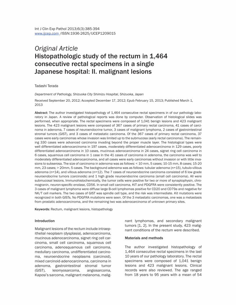
Figure 1. Early carcinoma of the rectum. A: Goss features of early rectal adenocarcinoma of polypoid type. B: Gross features early rectal adenocarcinoma of ulcerating type. C: Early squamous cell carcinoma of the rectum. Individual keratinization is seen. HE, x100. D: Early well differentiated adenocarcinoma of the rectum. HE, x100. E: Early moderately differenti-ated adenocarcinoma of the rectum. HE, x100.
Rectal malignant conditions
387 Int J Clin Exp Pathol 2013;6(3):385-394
specimens were composed of 1,041 benign lesions and 423 malignant lesions.
The 423 malignant lesions were composed of 367 cases of primary rectal carcinoma, 41 cases of carcinoma in adenoma, 7 cases of neuroendocrine tumor, 3 cases of malignant lymphoma, 2 cases of GIST, and 3 cases of metastatic carcinoma.
Of the 367 cases (frequency of all rectal malig-nancies, 86.8 %) of primary rectal carcinomas, 37 cases (8.7 %) were early carcinomas whose invasion was limited up to the submucosa. The remaining 330 cases (78%) were advanced car-cinomas invading beyond the proper muscle layer. The histological types were well differenti-ated adenocarcinoma in 197 cases, moderate-ly differentiated adenocarcinoma in 129 cases, poorly differentiated adenocarcinoma in 10
cases, mucinous adenocarcinoma in 24 cases, signet ring cell carcinoma in 6 cases, squa-mous cell carcinoma in 1 case.
In the 37 cases of early carcinoma, the gross features were classified into polypoid (n=14) (Figure 1A) and ulcerating (n=23) (Figure 1B). Histologically, 36 cases were adenocarcinoma and 1 case is squamous cell carcinoma (Figure 1C). Of the 36 adenocarcinomas, 21 were well differentiated adenocarcinomas (Figure 1D), and 15 were moderately differentiated adeno-carcinomas (Figure 1E). Lymphovascular per-meation was little or none in all cases, and lymph nodes dissected (n=18) showed no metastases. Immunohistochemically, p53 pro-tein was present in 8/10 cases, and Ki-67 labeling ranged from 32 to 64 % in the 10 cases examined.
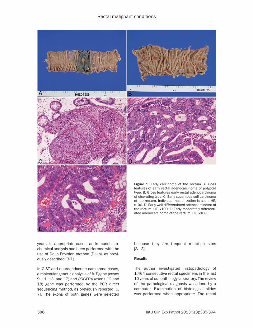
Figure 2. Advanced carcinoma of the rectum. A: Goss features of advanced rectal adenocarcinoma of polypoid type. B: Gross features advanced rectal adenocarcinoma of ulcerating type. C: Advanced well differentiated adenocarci-noma of the rectum. HE, x100. D: Advanced moderately differentiated adenocarcinoma of the rectum. HE, x100. E: Advanced poorly differentiated adenocarcinoma of the rectum. HE, x100. F: Advanced mucinous differentiated adenocarcinoma of the rectum. HE, x100. G: Advanced signet ring cell carcinoma of the rectum. HE, x100. H: Gross features of multiple rectal adenocarcinomas. I: Rectal carcinoma is associated with synchronous colon carcinoma.

Rectal malignant conditions
388 Int J Clin Exp Pathol 2013;6(3):385-394
In the 330 advanced carcinoma cases, the gross features were polypoid (Figure 2A) in 23 cases and ulcerated (Figure 2B) in 307 cases. Histologically, 179 cases were well differentiat-ed adenocarcinomas (Figure 2C), 114 cases were moderately differentiated adenocarcino-mas (Figure 2D), and 10 cases were poorly dif-ferentiated adenocarcinomas (Figure 2E), 24 cases were mucinous adenocarcinomas (Figure 2F), and 6 cases were signet ring cell carcinomas (Figure 2G). The depth of invasion was serosa in 25 cases, subserosa in 263 cases, and proper muscle in 42 cases. Lymphovascular invasions were recognized in 312 cases of the 330 cases, and lymph node metastases were present in 156 cases of the 298 cases dissected. Immunohistochemically, the poorly differentiated adenocarcinoma and signet ring cell carcinoma were positive for cytokeratins and negative for lymphocytic markers. Signet ring cell carcinoma and muci-nous adenocarcinoma contained neutral and acidic mucins as revealed by mucin stains. The p53 was present in 13 cases of the 15 cases examined, and Ki-67 labeling ranged from 26% to 87% in the 15 cases examined. Multiple rec-tal carcinoma was recognized in one case (Figure 2H), and 5 cases showed synchronous colon carcinomas other than the rectum (Figure 2I).
In the 41 cases (5.6 %) of carcinoma in adeno-ma (Figure 3A), the carcinoma is well to moder-ately differentiated adenocarcinoma (Figure 3B), and all cases were early carcinomas with little invasions. The size of carcinoma in adeno-ma was as follows: < 10 mm, 5 cases; 10-15
mm, 8 cases; 15-20 mm, 23 cases; > 20mm, 5 cases. Histologically, the background adenoma was as follows: tubular adenoma (n=15), tubu-lo-villous adenoma (n=14), and villous adeno-ma (n=12). The atypia of the background ade-noma is severe (high grade dysplasia) in all cases. Immunohistochemically, p53 protein was positive in carcinoma areas in 4 cases out of 5 cases examined. The Ki-67 labeling in car-cinoma areas ranged from 10% to 36%, while that of adenoma areas from 8% to 15%.
The 7 cases (1.7%) of neuroendocrine carcino-ma showed submucosal tumors in all cases (Figure 4A). Histologically, they consisted of 6 low grade neuroendocrine tumors (carcinoids) (Figure 4B) and 1 high grade neuroendocrine carcinoma (Figure 4C). The former showed a trabecular organoid pattern (Figure 4A and 4D) and rosette formations (Figure 4D) and ribbon-like features (Figure 4D), while the latter showed no apparent patterns (Figure 4B) equivalent to high grade neuroendocrine carci-noma (small cell carcinoma). Immunohisto-chemically, the tumor cells were positive for two or more of synaptophysin, chromogranin, neu-ron-specific enolase, CD56. KIT and PDGFRA were consistently positive (Figure 4E and 4F).
The 3 cases (0.7%) of malignant lymphoma were diffuse large B-cell lymphomas. Grossly, they assumed polypoid or ulcerated appear-ances (Figure 5A), similar to rectal carcinoma. Histologically, atypical large malignant lym-phoid cells were seen to diffusely proliferate (Figure 5B) in all cases. There were many mitot-ic figures (Figure 5B). Immunohistochemically, the tumor cells were negative for cytokeratins
Figure 3. Histology in carcinoma in adenoma of the rectum. A: Low power view. HE, x40. B: High power view of the carcinomatous area. HE, x200.

Rectal malignant conditions
389 Int J Clin Exp Pathol 2013;6(3):385-394
and positive for CD45, CD20, CD79α (Figure 5C). Light chain restriction was present in all cases. The tumor cells were negative for CD3,
TdT, and CD45RO. Marginal zone lymphoma, mantle cell lymphoma, and follicular lymphoma were denied by immunostaining for CD5, CD10,
Figure 4. Neuroendocrine tumor. A: Gross features of submucosal tumor natures are seen. B: The carcinoid show trabecular patterns. HE, x40. C: High power view of a carcinoid tumor showing ribbon and rosette formation, HE, x100. D: High grade neuroendocrine carcinoma (small cell neuroendocrine carcinoma). HE, x100. E: The carcinoid tumor shows immunoreactive synaptophysin. x 100. F: The high grade neuroendocrine carcinoma shows immuno-reactive CD56. x100.

Rectal malignant conditions
390 Int J Clin Exp Pathol 2013;6(3):385-394
CD23, and cyclin D1. CD15 and CD30 were negative. The lymphoma cells were positive focally for p53 protein, and KI-67 labeling ranged from 78% to 99%.
The two cases (0.47%) of GIST were serosal tumor (Figure 6A) in one case (45 mm in diam-eter) and submucosal tumor in another case (55 mm in diameter). Both cases consisted of spindle cell type (Figure 6B). Mitotic figures were 6 per 50 high power field (HPF) in one case and 8 per 50 HPF in another case. Ki-67 labeling was 15% and 24%, respectively. Immunohistochemically, tumor cells were posi-tive for KIT (Figure 6C), CD34 (Figure 6D), PDGFRA (Figure 6E), and negative for desmin, smooth muscle antigen, and S100 protein. The molecular analysis showed a point mutation of KIT gene exon 9 and a deletion of KIT gene exon 11. No mutations were found in PDGFRA genes.
Of the 3 metastatic carcinomas, one was a metastasis from prostatic adenocarcinoma positive for prostate-specific antigen (Figure 7A and 7B), and the remaining two were adenocar-cinomas of unknown primary sites.
Discussion
In the present study, the author reviewed 1,464 consecutive pathologic specimens of the rec-tum in our laboratory in the last 10 year.
The results of rectal adenocarcinoma were compatible with previous study. Early adeno-carcinoma of the rectum was found in 37 among 367 cases. The early carcinoma was free of lymphovascular invasions, suggesting that the good prognosis relative to advanced adenocarcinoma. In the present study, 24 cases of mucinous adenocarcinoma and 6 cases of signet ring cell carcinoma were pres-ent among the 367 rectal carcinomas. These two kinds of adenocarcinoma is relatively rare in the rectum, and different from ordinary rec-tal adenocarcinoma with respect to microsatel-lite instability [14] mismatch repair enzyme [15] BRAF, KRAS, LOH, [16], and poor prognosis [17], relative to ordinary rectal adenocarcino-ma. The present study presented a case of squamous cell carcinoma of the rectum. It was located only in the mucosa. The squamous cell carcinoma of the colorectum is extremely rare,
Figure 5. Malignant lymphoma of the rectum. A: Gross features. A large ulcerated tumor indistin-guishable from rectal carcinoma is seen. B: The tumor cells show diffuse large cell lymphoma. HE, x200. C: Tumor cells are positive for CD20. x200.

Rectal malignant conditions
391 Int J Clin Exp Pathol 2013;6(3):385-394
and only a few cases are reported in the colon but not in the rectum in the literatures [18, 19]. The histogenesis of squamous cell carcinoma of the rectum is unclear, but it may be possible that totipotential stem cells differentiated into squamous cell carcinoma of the rectum.
The malignant transformation of adenoma in the colorectum is well known. It is well estab-lished that colorectal carcinogenesis has two pathways; adenoma-carcinoma sequence and de novo carcinogenesis [2, 20]. In the former,
multistep carcinogenesis is considered; each step is accompanied by mutations of onco-genes such as RAS and of tumor suppresser genes such as p53 [2, 20, 21]. Therefore, colorectal adenomas are precancerous lesions [2, 20, 21]. In the present study, carcinoma in adenoma was largely observed in adenoma > 15 mm, suggesting a correlation between the malignant transformation and adenoma size. In the present study, carcinoma in adenoma was most frequent in tubular adenoma, followed in order by tubulo-villous adenoma and villous
Figure 6. GIST of the rectum. A: Gross features. A sol-id mass was seen in the serosa. B: Histology. Spindle cell proliferation is seen. HE, x200. C: Tumor cells are positive for KIT. X200. D: Tumor cells are positive for CD34, x200. E: Tumor cells are positive for PDGFRA. X200.

Rectal malignant conditions
392 Int J Clin Exp Pathol 2013;6(3):385-394
adenoma. According to WHO blue book [2], car-cinomatous transformation is most frequent in villous adenomas, followed in order by tubulo-villous adenomas and tubular adenomas. The WHO book also showed that adenomas > 2cm had malignant foci in 30-50%. In addition, the WHO book showed adenomas < 2cm had malig-nant foci in 1-10%. In the present study, p53 protein is frequently recognized in carcinoma in adenoma, suggesting the malignant transfor-mation is associated with p53 mutations. Much more studies of molecular alteration of onco-genes and antioncogens are required.
Carcinoids (neuroendocrine tumors) are well known to occur in the rectum [1, 2]. In the pres-ent study, 6 cases of typical carcinoids immu-noreactive for neuroendocrine antigens were found. In the present study, 1 case of high grade neuroendocrine carcinoma compatible with small cell carcinoma was recognized. This tumor was positive for neuroendocrine mark-ers, KIT and PDGFRA. No mutations of KIT and PDGFRA were noted. Rectal high grade neuro-endocrine tumors are extremely rare, only a few reports are seen in the literature [22-24]. Recently, high grade neuroendocrine carcino-ma is known to express KIT and PDGFRA [8, 10, 25-28]. However, no mutations of KIT and PDGFRA have been identified in small cell carci-nomas [25-28], as in the present study.
Malignant lymphoma of the rectum is relatively rare. In the present series, 3 cases of lympho-ma were identified. All the 3 cases were immu-nohistochemically-proven diffuse large B-cell
lymphoma. Wong and Eu [29] described that the frequency of colorectal lymphoma was 0.44% (14 of 3199 cases), and most of them were diffuse large B-cell lymphoma. Fan et al. [30] described that the frequency of colorectal lymphoma was 0.48 % (37/7,648) in colorectal malignancies, and that they consisted of 29 high grade lymphomas and 8 non-high grade lymphomas. The frequency in the present series was 0.71% (3 of 423 cases) of all rectal malignancies. Much more studies are needed in the rectal malignant lymphoma.
Rectal GIST is a rare tumor relative to gastroin-testinal GIST. MIettinen and Lasota [31] report-ed that the frequency of GIST was as follows: stomach (60%), jejunum and ileum (30%), duo-denum (4-5%), rectum (4%), and colon and appendix (1-2%). The frequency of rectal GIST of the present series was 0.47% (2 of 423 cases). There are a few case reports of rectal GIST [32, 33]. GIST is characterized by positive immunoreaction of KIT (CD117) and CD34, and by genetic mutations of KIT or PDGFRA genes [8-13], The KIT and PDGFRA mutations are mutually exclusive. The 2 cases of the present series were positive for KIT, CD34 and PDGFRA, and showed KIT gene mutations, one in exon 9 and another in exon 11. GIST is a potentially malignant tumor; the malignant potential depends on tumor type (spindle vs epithelioid), tumor size, mitotic figures, necrosis, and Ki-67 labeling. According to the consensus report of GIST by Fletcher et al. [34], the malignant potential of GIST depends on tumor size and mitotic counts. In very low malignant risk group,
Figure 7. Metastatic prostatic carcinoma of the rectum. A: Histology suggests prostatic origin. HE x100. B: The tumor cells are positive for prostate-specific antigen. X 100.

Rectal malignant conditions
393 Int J Clin Exp Pathol 2013;6(3):385-394
tumor size is less than 2 cm and mitotic counts are less than 5 per 50 high power field (HPF). In low malignant risk group, tumor size is 2cm < 5cm, and mitotic counts are < 5/50 HPF. In intermediate risk group, tumor size is 5cm < 10cm, and mitotic counts are < 5/50 HPF. In high risk group, tumor size is > 10 cm, and mitotic counts are > 10/50 HPF [34]. In the present study, according to mitotic counts and tumor size [34], the malignant risk was interme-diate or low risk.
Conflict of interest statement
The author has no conflict of interest.
Address correspondence to: Dr. Tadashi Terada, Department of Pathology, Shizuoka City Shimizu Hospital, Miyakami 1231 Shimizu-Ku, Shizuoka 424-8636, Japan. Tel: 81-54-336-1111; Fax: 81-54-336-1315; E-mail: [email protected]
References
[1] Rosai J. Large bowel. In Rosai and Ackerman’s Surgical Pathology. Ninth edition. Mosby, 2004. pp: 776-855.
[2] Hamilton SR, Rubio CA, Vogelstein B, Sobin LH, Kudo S, Fogt F, Riboli E, Winaswer SJ. Nakanu-ma S, Goldgar DE, Hainaut P, Jass JR. Tumours of the colon and rectum. In: Mamilton SR, Aal-tonen LA eds. World Health Organization Clas-sification of Tumours. Pathology and genetics in tumors of the digestive system. IARC Press, Lyon, 2000. pp: 103-155.
[3] Terada T, Kawaguchi M, Furukawa K, Sekido Y, Osamura Y. Minute mixed ductal-endocrine carcinoma of the pancreas with predominant intraductal growth. Pathol Int 2002; 52: 740-746.
[4] Terada T, Tanigichi M. Intraductal oncocytic papillary neoplasm of the liver. Pathol Int 2004; 54: 116-1237.
[5] Terada T. Large endocervical polyp with carti-laginous and osseous metaplasia: a hitherto unreported entity. Int J Gynecol Pathol 2009; 28: 98-100.
[6] Terada T. Primary multiple extragastrointesti-nal stromal tumors of the omentum with differ-ent mutations of c-kit gene. Would J Gastroen-terol 2008; 14: 7256-7259.
[7] Terada T. Gastrointestinal stromal tumor of the uterus: A case report with genetic analyses of c-kit and PDGFRA genes. Int J Gynecol Pathol 2009; 28: 29-34.
[8] Miettinen M, Lasota J. KIT (CD117): a review on expression in normal and neoplasmic tis-sue, and mutations and their clinicopathologic
correlation. Appl Immunohistochem Mol Mor-phol 2005; 13: 205-220.
[9] Hirota S, Isozaki K. Pathology of gastrointesti-nal stromal tumor. Pathol Int 2006; 56: 1-9.
[10] Losota J, Miettinen M. KIT and PDGFRA muta-tions in gastrointestinal stromal tumors (GISTs). Semin Diang Pathol 2006; 23: 91-101.
[11] Miettinen M, Lasota J. Gastrointestinal stromal tumor: review on morphology, molecular pa-thology, prognosis, and differential diagnosis. Arch Pathol Lab Med 2006; 130: 1466-1478.
[12] Hirota S, Isozaki K, Moriyama Y, Hashimoto K, Nishida T, Ishiguro S, Kawano K, Hanada M, Kurata A, Takeda M, Muhammad Tunio G, Mat-suzawa Y, Kanakura Y, Shimomura Y, Kitamura Y. Gain-of-function mutations of c-kit in human gastrointestinal stromal tumor. Science 1998; 279: 577-580.
[13] Hirota S, Ohashi A, Nishida T, Isozaki K, Kinoshita K, Shinomura Y, Kitamura Y. Gain-of-function mutations of platelet-derived growth factor receptor alpha gene in gastrointestinal stromal tumor. Gastroenterology 2003; 125: 660-667.
[14] Kakar S, Smyrk TC. Signet ring cell carcinoma of the colorectum: correlations between micro-satellite instability, clinicopathologic features and survival. Mod Pathol 2005; 18: 244-249.
[15] Kakar S, Aksoy S, Burgart JJ, Smyrk TC. Muci-nous carcinoma of the colon: correlation of loss of mismatch repair enzymes with clinico-pathologic features and survival. Mod Pathol 2004; 17: 696-70.
[16] Ogino S, Brahmandam M, Cantor M, Namgyal C, Kawasaki T, Kirkner G, Meyerhardt JA, Loda M, Fuchs CS. Distinct molecular features of colorectal carcinoma with signet ring cell com-ponent and colorectal carcinoma with muci-nous component. Mod Pathol 2006; 19: 59-68.
[17] King-Yin Lam A, Ong K, Ho YH. Colorectal muci-nous adenocarcinoma: the clinicopathologic features and significance of p16 and p53 ex-pression. Dis Colon Rectum 2006; 49: 1275-1283.
[18] Michelassi F, Mishlove LA, Stipa F, Block GE. Squamous cell carcinoma of the colon: experi-ence at the University of Chicago, review of the literature, report of two cases. Dis Colon Rec-tum 1988; 31: 228-235.
[19] Crismann JD. Adenosquamous and squamous cell carcinoma of the colon. Am J Surg Pathol 1978; 2: 47-54 .
[20] Vogelstein B, Fearon ER, Hamilton SR, Kern SE, Preisinger AC, Lappert M, Nakamura Y, White R, Smits AM, Bos JL. Genetic alterations during colorectal-tumor development. New Eng J Med 1988; 319: 525-532.

Rectal malignant conditions
394 Int J Clin Exp Pathol 2013;6(3):385-394
[21] Cho KR, Vogelstein B. Genetic alterations in the adenoma-carcinoma sequence. Cancer 1992; 70: 1727-1731.
[22] Cebrian J, Larach SW, Ferrara A, Williamson PR, Trevisani MF, Lujan HJ, Kassir A. Small cell carcinoma of the rectum: report of two cases. Dis Colon Rectum 1999; 42: 274-277.
[23] Shirouzu K, Morodomi T, Isomoto H, Yamauchi Y, Kakegawa T, Morimatsu M. Small cell carci-noma of the rectum, clinicopathologic study. Dis Colon Rectum 1985; 28: 434-439.
[24] Robidoux A, Monte M, Heppell J, Schurch W. Small cell carcinoma of the rectum. Dis Colon Rectum 1985; 28: 594-596.
[25] Terada T. Primary small cell carcinoma of the mediastinum: A case report with immunohisto-chemical and molecular genetic analyses of KIT and PDGFRA genes. Med Oncol 2009; 26: 247-250.
[26] Terada T. Primary small cell carcinoma of the ureter: A case report involving immunohisto-chemical and molecular genetic analyses of KIT and PDGFRA genes. Pathology 2010; 42: 101-102.
[27] Terada T. Autopsy case of primary small cell carcinoma of the urinary bladder: KIT and PDGFRA expression and mutations. Pathol Int 2009; 59: 247-250.
[28] Sihto H, Sarlomo-Rikara M, Tynnienen O, Tan-ner M, Andersson LC, Franssila K, Nupponen NN, Joensuu H. KIT and platelet-derived growth factor receptor alpha tyrosine kinase gene mu-tations and KIT amplifications in human solid tumors. J Clin Oncol 2005; 23: 49-57.
[29] Wong MT, Eu KW. Primary colorectal lympho-ma. Colorectal Dis 2006; 8: 586-591.
[30] Fan CW, Changchien CR, Wang JY, Chen JS, Hsu KC, Tang R, Chiang JM. Primary colorectal lymphoma. Dis Colon Rectum 2000; 43: 1277-1282.
[31] Miettinen M, Lasota J. Gastrointestinal stromal tumors: pathology and prognosis at different sites. Semin Diang Pathol 2006; 23: 70-83.
[32] Testroote M, Hoornweg M, Rhemrev S. Rectal GIST presenting as a submucosal calculus. Dig Dis Sci 2007; 52: 14047-1049.
[33] Sawai H, Okada Y, Funahashi H, Matsuo Y, Hay-akawa T, Tanaka M, Terayama H, Manabe T. Gastrointestinal stromal tumor of the rectum with liver metastasis: report of a case. Hepato-gastroenterology 2007; 54: 1113-1115.
[34] Fletcher CD, Berman JJ, Corless C, Gorstein F, Lasota J, Longley BJ, Miettinen M, O’Leary TJ, Remotti H, Rubin BP, Shmookler B, Sobin LH, Weiss SW. Diagnosis of gastrointestinal stro-mal tumors: a consensus approach. Hum Pathol 2002; 33: 459-465.