Embed Size (px)
Citation preview
HISTOPATHOLOGICAL EXAMINATION OF PLACENTA IN CASES OF REPRODUCTIVE FAILURE
ORIGINAL RESEARCH PAPER
DR. NIDHI BANSAL
ASSISTANT PROFESSOR, DEPT. OF IMMUNOHEMATOLOGY & BLOOD TRANSFUSION, GGS MEDICAL COLLEGE, FARIDKOT.
INTRODUCTIONThe placenta signifies the "second" or "embryonic" period of pregnancy (after the implantation period) and describes the establishment of a fully functional placenta. The placenta is an apposition of fetal and parental tissue for the purposes of physiological exchange.
The placenta is a unique organ with dual blood circulation, functioning throughout foetal development. Placental trophoblasts express and produce coagulation components, participating not only in haemostasis but also in placental vascular development and differentiation1. The earlier insight into this organ comes from drawing from illustrators such as Andreas Vesalius (1514-1564). The designation “placenta” was introduced by Gabriele de Falloppio (1523-1562). Others, however believe that this designation, placenta, stemmed from Realdus Columbus in 1559.
The placental membrane consists of chorion and amnion. The amnion which represents the innermost covering of the amniotic cavity is lined by single layer of flat epithelial cells resting on a basement membrane2,3. Squamous metaplasia is common in them, especially near the insertion of the cord. The chorion is composed of a connective tissue membrane that carries the fetal vasculature. The chorion associated with the membrane is referred to as chorion laeve and is distinguished from the chorion frondosum located in the placenta
4,5.proper
REPRODUCTIVE FAILURE:Reproductive failure comprises of spontaneous abortion, neonatal death and intra-uterine fetal death (IUFD).
Spontaneous abortion, or miscarriage, is defined as a clinically 6,7.recognized pregnancy loss before the 20th week of gestation
INTRAUTERINE FETAL DEATH:Intrauterine fetal death embraces all the fetal deaths weighing 500gm or more occurring both during pregnancy (antepartum death) and during labour (intrapartum). But death of a fetus weighing less than 500gm (before 22 weeks) has got a distinct etiology and is usually termed as abortion. Thus for practical purpose, antepartum death occurring beyond 28 weeks is termed as intrauterine death.
The fetal deaths are related to maternal, placental or fetal complications8.
MATERIALS & METHODSThe study will be conducted in pathology department (Histopathology Section) of Mahatma Gandhi Mission's Medical College & Hospital, Kamothe, Navi Mumbai Maharashtra. The study will be conducted over a period of two and half years between May 2011 - October 2013. Placental specimens will be received in our department by obtaining the consent from the patient/attendant.
Inclusion Criteria included all females of reproductive age group, all cases of intrauterine death before delivery.
Exclusion Criteria consisted of all cases of pregnancy with delivery of viable fetus.
The relevant clinical details of the mother were noted like age, parity, general medical history, pregnancy related history and fetal outcome (live birth, abortion, stillbirth, IUGR). Maternal, fetal and placental risk factors were analyzed and various gross and microscopic lesions encountered with these risk factors were studied. An attempt was made to correlate placental pathology with fetal outcome. Particular attention was paid to lesions seen in cases of intrauterine fetal deaths.The placentae were immersed in 10% formalin for overnight fixation sections were taken on next day and stained with Haematoxylin and Eosin Sections taken for histological examination:1)Two rolls of membranes (prepared in such a way that the margin of the site of rupture was in the centre of the roll) and two sections of umbilical cord(one from placental insertion and other from fetal side)2)Two sections of central part of placenta including the maternal surface were placed in the second cassette.
3)One section of central part of placenta including the fetal surface was placed in third cassette.
4)More sections were taken if any abnormality was detected on gross examination of the placenta. In twin placenta, sections of the T zone at the attachment of the septum to the fetal surface and a roll prepared from the septum was taken.
RESULTSThe study will be conducted in pathology department (Histopathology Section) of Mahatama Gandhi Mission's Medical College &Hospital, Kamothe, Navi Mumbai Maharashtra on 100 cases.
It shows the age wise distribution of cases. Majority of the placentas that we received were from mothers in the age group 21-25 years 44/100 (44%). None of the patients were more than 40 years of age or below 18 years of age.
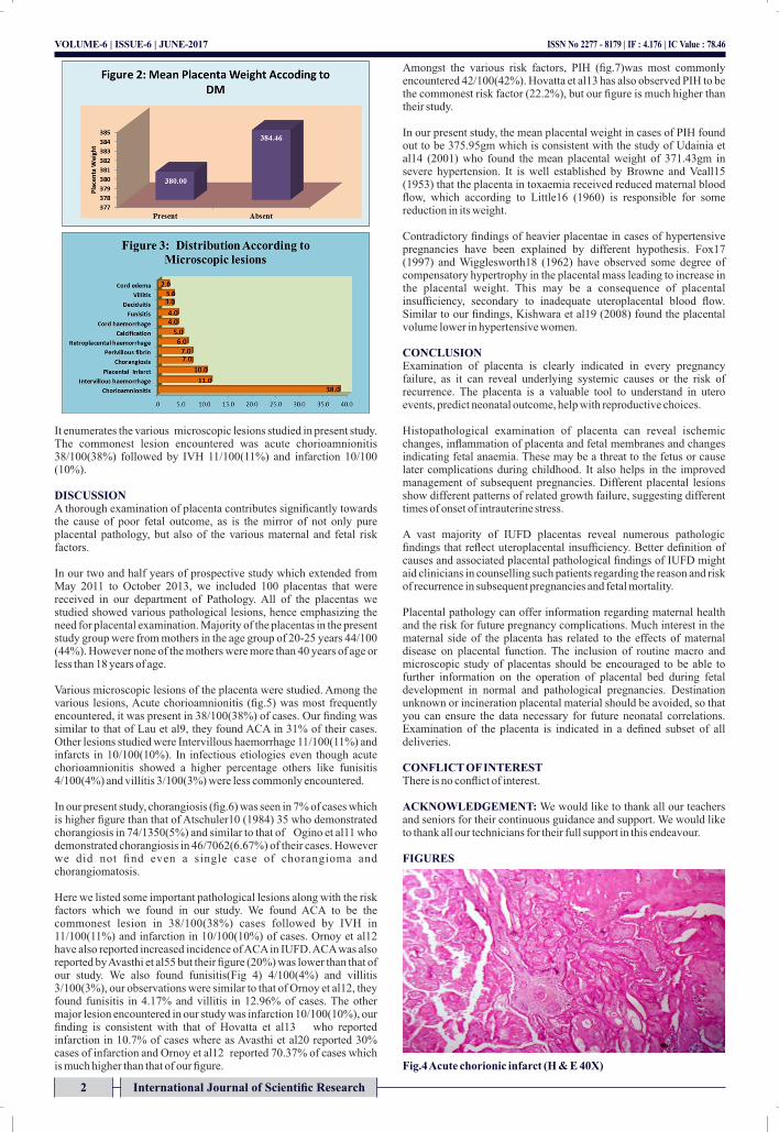
It enumerates the various maternal risk factors encountered. The commonest among the pregnancy related risk factor was PIH 42/100(42%) followed by fever 32/100(32%). The general maternal risk factors were headed non-infectious causes like DM was seen in 8/100(8%).
1International Journal of Scientific Research
INTERNATIONAL JOURNAL OF SCIENTIFIC RESEARCH
IMMUNOHEMATOLOGY
VOLUME-6 | ISSUE-6 | JUNE-2017 | ISSN No 2277 - 8179 | IF : 4.176 | IC Value : 78.46
KEYWORDS:Placenta, chorioamnionitis, Intrauterine
DR(MRS.) UJWALA MAHESHWARI
PROFESSOR, DEPT. OF PATHOLOGY, MGM MEDICAL COLLEGE & HOSPITAL, NAVI MUMBAI
DR. ARNAV KR. ROYCHOUDHURY
ASSISTANT PROFESSOR, DEPT. OF PATHOLOGY, ADESH INSTITUTE OF MEDICAL SCIENCES & RESEARCH, BATHINDA.
ISSN No 2277 - 8179 | IF : 4.176 | IC Value : 78.46VOLUME-6 | ISSUE-6 | JUNE-2017
2 International Journal of Scientific Research
It enumerates the various microscopic lesions studied in present study. The commonest lesion encountered was acute chorioamnionitis 38/100(38%) followed by IVH 11/100(11%) and infarction 10/100 (10%).
DISCUSSIONA thorough examination of placenta contributes significantly towards the cause of poor fetal outcome, as is the mirror of not only pure placental pathology, but also of the various maternal and fetal risk factors.
In our two and half years of prospective study which extended from May 2011 to October 2013, we included 100 placentas that were received in our department of Pathology. All of the placentas we studied showed various pathological lesions, hence emphasizing the need for placental examination. Majority of the placentas in the present study group were from mothers in the age group of 20-25 years 44/100 (44%). However none of the mothers were more than 40 years of age or less than 18 years of age.
Various microscopic lesions of the placenta were studied. Among the various lesions, Acute chorioamnionitis (fig.5) was most frequently encountered, it was present in 38/100(38%) of cases. Our finding was similar to that of Lau et al9, they found ACA in 31% of their cases. Other lesions studied were Intervillous haemorrhage 11/100(11%) and infarcts in 10/100(10%). In infectious etiologies even though acute chorioamnionitis showed a higher percentage others like funisitis 4/100(4%) and villitis 3/100(3%) were less commonly encountered.
In our present study, chorangiosis (fig.6) was seen in 7% of cases which is higher figure than that of Atschuler10 (1984) 35 who demonstrated chorangiosis in 74/1350(5%) and similar to that of Ogino et al11 who demonstrated chorangiosis in 46/7062(6.67%) of their cases. However we did not find even a single case of chorangioma and chorangiomatosis.
Here we listed some important pathological lesions along with the risk factors which we found in our study. We found ACA to be the commonest lesion in 38/100(38%) cases followed by IVH in 11/100(11%) and infarction in 10/100(10%) of cases. Ornoy et al12 have also reported increased incidence of ACA in IUFD. ACA was also reported by Avasthi et al55 but their figure (20%) was lower than that of our study. We also found funisitis(Fig 4) 4/100(4%) and villitis 3/100(3%), our observations were similar to that of Ornoy et al12, they found funisitis in 4.17% and villitis in 12.96% of cases. The other major lesion encountered in our study was infarction 10/100(10%), our finding is consistent with that of Hovatta et al13 who reported infarction in 10.7% of cases where as Avasthi et al20 reported 30% cases of infarction and Ornoy et al12 reported 70.37% of cases which is much higher than that of our figure.
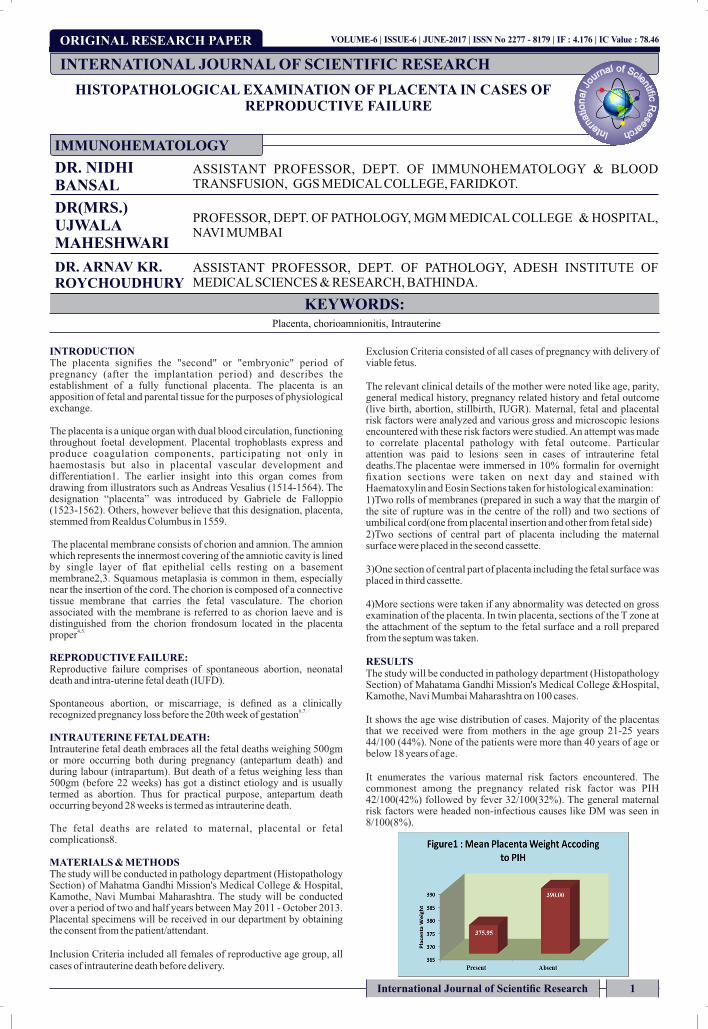
Amongst the various risk factors, PIH (fig.7)was most commonly encountered 42/100(42%). Hovatta et al13 has also observed PIH to be the commonest risk factor (22.2%), but our figure is much higher than their study.
In our present study, the mean placental weight in cases of PIH found out to be 375.95gm which is consistent with the study of Udainia et al14 (2001) who found the mean placental weight of 371.43gm in severe hypertension. It is well established by Browne and Veall15 (1953) that the placenta in toxaemia received reduced maternal blood flow, which according to Little16 (1960) is responsible for some reduction in its weight.
Contradictory findings of heavier placentae in cases of hypertensive pregnancies have been explained by different hypothesis. Fox17 (1997) and Wigglesworth18 (1962) have observed some degree of compensatory hypertrophy in the placental mass leading to increase in the placental weight. This may be a consequence of placental insufficiency, secondary to inadequate uteroplacental blood flow. Similar to our findings, Kishwara et al19 (2008) found the placental volume lower in hypertensive women.
CONCLUSIONExamination of placenta is clearly indicated in every pregnancy failure, as it can reveal underlying systemic causes or the risk of recurrence. The placenta is a valuable tool to understand in utero events, predict neonatal outcome, help with reproductive choices.
Histopathological examination of placenta can reveal ischemic changes, inflammation of placenta and fetal membranes and changes indicating fetal anaemia. These may be a threat to the fetus or cause later complications during childhood. It also helps in the improved management of subsequent pregnancies. Different placental lesions show different patterns of related growth failure, suggesting different times of onset of intrauterine stress. A vast majority of IUFD placentas reveal numerous pathologic findings that reflect uteroplacental insufficiency. Better definition of causes and associated placental pathological findings of IUFD might aid clinicians in counselling such patients regarding the reason and risk of recurrence in subsequent pregnancies and fetal mortality.
Placental pathology can offer information regarding maternal health and the risk for future pregnancy complications. Much interest in the maternal side of the placenta has related to the effects of maternal disease on placental function. The inclusion of routine macro and microscopic study of placentas should be encouraged to be able to further information on the operation of placental bed during fetal development in normal and pathological pregnancies. Destination unknown or incineration placental material should be avoided, so that you can ensure the data necessary for future neonatal correlations. Examination of the placenta is indicated in a defined subset of all deliveries.
CONFLICT OF INTERESTThere is no conflict of interest.
ACKNOWLEDGEMENT: We would like to thank all our teachers and seniors for their continuous guidance and support. We would like to thank all our technicians for their full support in this endeavour.
FIGURES
Fig.4 Acute chorionic infarct (H & E 40X)
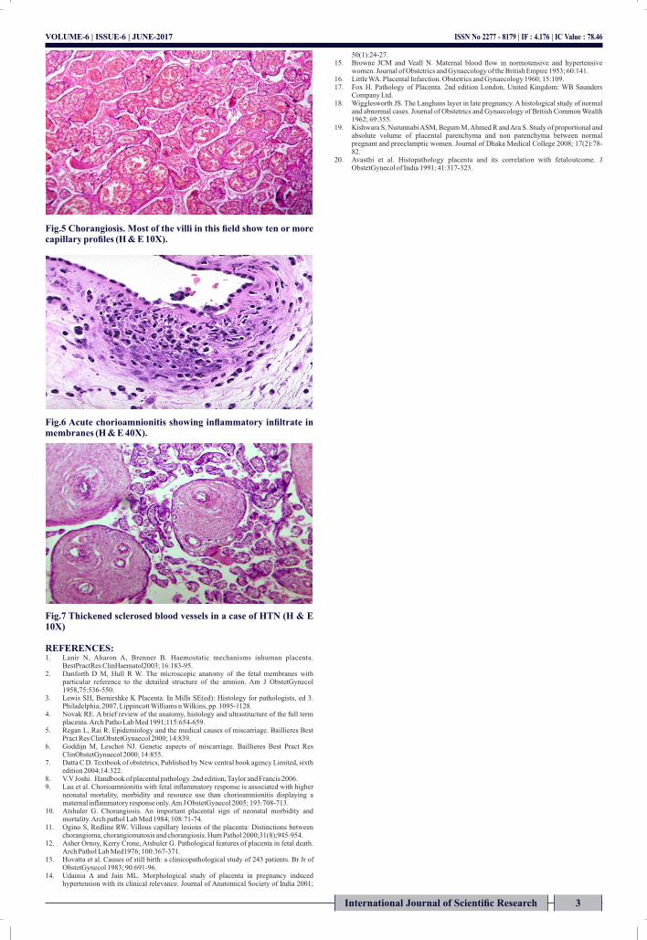
Fig.5 Chorangiosis. Most of the villi in this field show ten or more capillary profiles (H & E 10X).
Fig.6 Acute chorioamnionitis showing inflammatory infiltrate in membranes (H & E 40X).
Fig.7 Thickened sclerosed blood vessels in a case of HTN (H & E 10X)
REFERENCES:1. Lanir N, Aharon A, Brenner B. Haemostatic mechanisms inhuman placenta.
BestPractRes ClinHaematol2003; 16:183-95.2. Danforth D M, Hull R W. The microscopic anatomy of the fetal membranes with
particular reference to the detailed structure of the amnion. Am J ObstetGynecol 1958,75:536-550.
3. Lewis SH, Bernirshke K Placenta. In Mills SE(ed): Histology for pathologists, ed 3. Philadelphia, 2007, Lippincott Williams n Wilkins, pp. 1095-1128.
4. Novak RE. A brief review of the anatomy, histology and ultrastructure of the full term placenta. Arch Patho Lab Med 1991;115:654-659.
5. Regan L, Rai R. Epidemiology and the medical causes of miscarriage. Baillieres Best Pract Res ClinObstetGynaecol 2000; 14:839.
6. Goddijn M, Leschot NJ. Genetic aspects of miscarriage. Baillieres Best Pract Res ClinObstetGynaecol 2000; 14:855.
7. Datta C D. Textbook of obstetrics, Published by New central book agency Limited, sixth edition 2004;14:322.
8. V.V Joshi. Handbook of placental pathology. 2nd edition, Taylor and Francis 2006.9. Lau et al. Chorioamnionitis with fetal inflammatory response is associated with higher
neonatal mortality, morbidity and resource use than chorioamnionitis displaying a maternal inflammatory response only. Am J ObstetGynecol 2005; 193:708-713.
10. Atshuler G. Chorangiosis. An important placental sign of neonatal morbidity and mortality. Arch pathol Lab Med 1984; 108:71-74.
11. Ogino S, Redline RW. Villous capillary lesions of the placenta: Distinctions between chorangioma, chorangiomatosis and chorangiosis. Hum Pathol 2000;31(8);945-954.
12. Asher Ornoy, Kerry Crone, Atshuler G. Pathological features of placenta in fetal death. Arch Pathol Lab Med1976; 100:367-371.
13. Hovatta et al. Causes of still birth: a clinicopathological study of 243 patients. Br Jr of ObstetGynecol 1983; 90:691-96.
14. Udainia A and Jain ML. Morphological study of placenta in pregnancy induced hypertension with its clinical relevance. Journal of Anatomical Society of India 2001;
50(1):24-27.15. Browne JCM and Veall N. Maternal blood flow in normotensive and hypertensive
women. Journal of Obstetrics and Gynaecology of the British Empire 1953; 60:141.16. Little WA. Placental Infarction. Obstetrics and Gynaecology 1960; 15:109.17. Fox H. Pathology of Placenta. 2nd edition London, United Kingdom: WB Saunders
Company Ltd.18. Wigglesworth JS. The Langhans layer in late pregnancy. A histological study of normal
and abnormal cases. Journal of Obstetrics and Gynaecology of British Common Wealth 1962; 69:355.
19. Kishwara S, Nurunnabi ASM, Begum M, Ahmed R and Ara S. Study of proportional and absolute volume of placental parenchyma and non parenchyma between normal pregnant and preeclamptic women. Journal of Dhaka Medical College 2008; 17(2):78-82.
20. Avasthi et al. Histopathology placenta and its correlation with fetaloutcome. J ObstetGynecol of India 1991; 41:317-323.
ISSN No 2277 - 8179 | IF : 4.176 | IC Value : 78.46VOLUME-6 | ISSUE-6 | JUNE-2017
3International Journal of Scientific Research