Embed Size (px)
Citation preview

Molecular Vision 1999; 5:27 <http://www.molvis.org/molvis/v5/p27>Received 24 May 1999 | Accepted 2 Nov 1999 | Published 3 Nov 1999
Correspondence to: W. Richard Green, MD, Johns Hopkins Hospi-tal, 600 North Wolfe Street, Baltimore, MD, 21287; Phone: (410)955-3455; FAX: (410) 614-3457; email: [email protected]
Histopathologic and clinicopathologic correlative stud-ies have delineated most of the morphologic features of age-related macular degeneration (AMD) [1-6] and the interrela-tionship of the various morphologic forms [5]. Figure 1 is aflow diagram that shows the interrelationships of the variousmorphologic features of AMD [6]. The entire area centralismay be involved but the most marked changes often occur inthe central area inclusive of the parafoveal area.
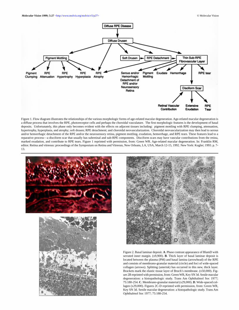
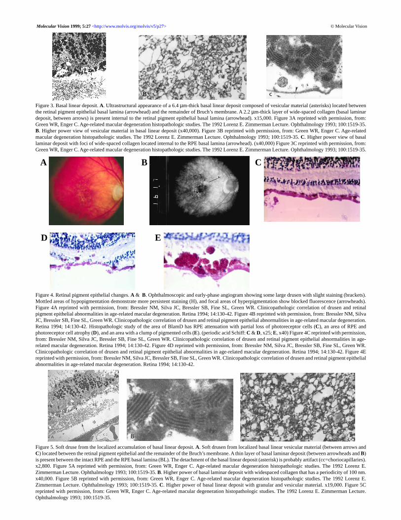
The earliest morphologic feature of AMD is the develop-ment of basal deposits external to the RPE, originally termed“diffuse drusen” [5]. It now seems clear that two distinct typesof deposits occur, basal laminar (BLamD) and basal linear(BLinD) [1]. This terminology was put forward in an attemptto reduce the confusion generated by previous conflictingterms. BLamD is composed of granular material with wide-spaced collagen located between the plasma membrane andthe basal lamina of the RPE [1] (Figure 2). BLinD is com-posed of material with coated and non-coated vesicles andsome membranous profiles that is located external to the basallamina of the RPE, that is, in the inner collagenous zone ofBruch’s membrane (Figure 3). This early stage of AMD maynot be evident by ophthalmoscopic examination but can beinferred by reduced retinal function and, in some cases, by avery faint, late fluorescein staining.
The presence of basal deposit becomes ophthalmoscopi-cally evident by secondary changes in the RPE, and by thedevelopment of soft drusen, choroidal neovascularization anddisciform scarring.
The RPE changes, often described as “pigment model-ing,” consist of RPE attenuation with depigmentation, hyper-trophy, hyperplasia and atrophy [1,7] (Figure 4). Accumula-tion of pigmented cells in the subretinal space contributes tothe appearance of clumping.
The second major feature is the development of soft (large)drusen [1,7]. These are usually larger than nodular drusen andhave a less discrete margin. Several types of soft drusen havebeen observed and include localized detachment of BLamD
with or without BLinD [1]. Localized accumulation of basallinear material is emerging as the most frequent form of softdrusen [7] (Figure 5).
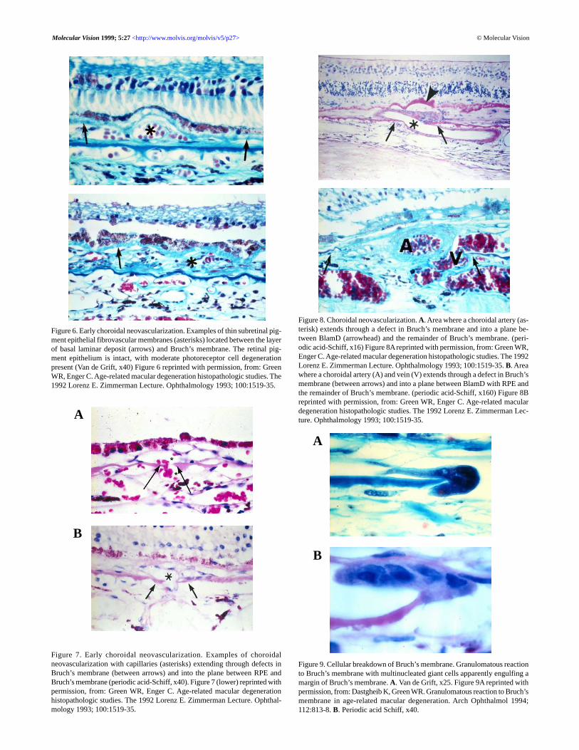
A third major feature is the development of choroidalneovascularization (CNV), which begins in the choroid andextends into a plane between BLamD along with RPE and theremainder of Bruch’s membrane [1] (Figure 6). Only rarelydo the vessels extend through the RPE and into the subretinalspace. In the early stages of AMD, these vessels are capillary-like [8] (Figure 7) and with time evolve into arteries and veins[1,5] (Figure 8). The number of points of origin of choroidalCNV varies from 1 to 12. Study of serial sections throughoutthe macular area of 63 eyes with CNV disclosed an average of2.2 sources per eye (Unpublished data, WR Green, July 1998).CNV may in turn lead to serous and/or hemorrhagic detach-ment of the RPE and/or retina, pigment modeling, exudationand RPE tears. CNV may be promoted by cellular breakdownof Bruch’s membrane (Figure 9) [9]. This early phase of CNVis observed in a small percentage of eyes. Most eyes with CNVare clinically occult as defined by fluorescein angiographicfeatures: fluorescein leakage of undetermined origin [10] (Fig-ure 10) and fibrovascular RPE detachment [11] (Figure 11).Findings of CNV by indocyanine green angiography includelate, plaque-like staining (Figure 12) and a “hot spot” [12,13](Figure 13).
The fourth major feature is the development of disciformscarring. In a study of a large series of disciform scars, Greenand Enger [1] found that the scar was non-vascularized in 25%,vascularized in 75%, and had a variable histologic pattern.The scar was thin, nonvascularized and located between theBLamD and the remainder of Bruch’s membrane in 6.5% ofeyes (Figure 14) and was a thin fibrovascular sub-RPE withBLamD in 13.2% of eyes (Figure 6). The scar had a singlesubretinal component in 32.2% of the cases (Figure 15) andtwo components (subretinal and sub-RPE with BLamD) in48.1% of cases (Figure 16). In those scars with two compo-nents, the subretinal portion was larger in 47.7% of eyes, thesub-RPE with BLamD was larger in 34.2% and the two com-ponents were about the same size in 18.1% of the cases. Ofthe 231 eyes with vascularized lesions, the new vessels werefrom the choroid only in 223 (96%), from the retina and chor-
© Molecular Vision
Histopathology of age-related macular degeneration
W. Richard Green
Departments of Ophthalmology and Pathology, Johns Hopkins Medical Institutions, Baltimore, MD
Age-related macular degeneration is a diffuse condition involving the retinal pigment epithelium, the photoreceptor celllayer, and perhaps the choriocapillaris. The early morphologic change is the development of basal deposits of two distincttypes. This phase is not ophthalmoscopically detectable but psychophysical testing may demonstrate reduced function.The process becomes detectable with the occurence of secondary changes in the pigment epithelium, soft drusen forma-tion, and choroidal neovascularization. A reparative response results in disciform scars. The various morphologic formsof age-related macular degeneration are interrelated.
© Molecular VisionMolecular Vision 1999; 5:27 <http://www.molvis.org/molvis/v5/p27>
Figure 1. Flow diagram illustrates the relationships of the various morphologic forms of age-related macular degeneration. Age-related macular degeneration isa diffuse process that involves the RPE, photoreceptor cells and perhaps the choroidal vasculature. The first morphologic features is the development of basaldeposits. Unfortunately, this phase only becomes evident with the effects on adjacent tissues including: pigment mottling with RPE clumping, attenuation,hypertrophy, hyperplasia, and atrophy; soft drusen; RPE detachment; and choroidal neovascularization. Choroidal neovascularization may then lead to serousand/or hemorrhagic detachment of the RPE and/or the neurosensory retina, pigment mottling, exudation, hemorrhage, and RPE tears. These features lead to areparative process—a disciform scar that usually has subretinal and sub-RPE components. Disciform scars may have vascular contributions from the retina,marked exudation, and contribute to RPE tears. Figure 1 reprinted with permission, from: Green WR. Age-related macular degeneration. In: Franklin RM,editor. Retina and vitreous: proceedings of the Symposium on Retina and Vitreous, New Orleans, LA, USA, March 12-15, 1992. New York: Kugler; 1993. p. 7-13.
Figure 2. Basal laminar deposit. A. Phase contrast appearance of BlamD withserrated inner margin. (x9,900). B. Thick layer of basal laminar deposit islocated between the plasma (PM) and basal lamina (arrowhead) of the RPEand consists of membrano-granular material (circle) and foci of wide-spacedcollagen (arrows). Splitting (asterisk) has occurred in this new, thick layer.Brackets mark the elastic tissue layer of Bruch’s membrane. (x50,000). Fig-ure 2B reprinted with permission, from: Green WR, Key SN 3d. Senile maculardegeneration: a histopathologic study. Trans Am Ophthalmol Soc 1977;75:180-254. C. Membrano-granular material (x29,000). D. Wide-spaced col-lagen (x29,000). Figures 2C-D reprinted with permission, from: Green WR,Key SN 3d. Senile macular degeneration: a histopathologic study. Trans AmOphthalmol Soc 1977; 75:180-254.
A
© Molecular VisionMolecular Vision 1999; 5:27 <http://www.molvis.org/molvis/v5/p27>
Figure 3. Basal linear deposit. A. Ultrastructural appearance of a 6.4 µm-thick basal linear deposit composed of vesicular material (asterisks) located betweenthe retinal pigment epithelial basal lamina (arrowhead) and the remainder of Bruch’s membrane. A 2.2 µm-thick layer of wide-spaced collagen (basal laminardeposit, between arrows) is present internal to the retinal pigment epithelial basal lamina (arrowhead). x15,000. Figure 3A reprinted with permission, from:Green WR, Enger C. Age-related macular degeneration histopathologic studies. The 1992 Lorenz E. Zimmerman Lecture. Ophthalmology 1993; 100:1519-35.B. Higher power view of vesicular material in basal linear deposit (x40,000). Figure 3B reprinted with permission, from: Green WR, Enger C. Age-relatedmacular degeneration histopathologic studies. The 1992 Lorenz E. Zimmerman Lecture. Ophthalmology 1993; 100:1519-35. C. Higher power view of basallaminar deposit with foci of wide-spaced collagen located internal to the RPE basal lamina (arrowhead). (x40,000) Figure 3C reprinted with permission, from:Green WR, Enger C. Age-related macular degeneration histopathologic studies. The 1992 Lorenz E. Zimmerman Lecture. Ophthalmology 1993; 100:1519-35.
Figure 4. Retinal pigment epithelial changes. A & B. Ophthalmoscopic and early-phase angiogram showing some large drusen with slight staining (brackets).Mottled areas of hypopigmentation demonstrate more persistent staining (H), and focal areas of hyperpigmentation show blocked fluorescence (arrowheads).Figure 4A reprinted with permission, from: Bressler NM, Silva JC, Bressler SB, Fine SL, Green WR. Clinicopathologic correlation of drusen and retinalpigment epithelial abnormalities in age-related macular degeneration. Retina 1994; 14:130-42. Figure 4B reprinted with permission, from: Bressler NM, SilvaJC, Bressler SB, Fine SL, Green WR. Clinicopathologic correlation of drusen and retinal pigment epithelial abnormalities in age-related macular degeneration.Retina 1994; 14:130-42. Histopathologic study of the area of BlamD has RPE attenuation with partial loss of photoreceptor cells (C), an area of RPE andphotoreceptor cell atrophy (D), and an area with a clump of pigmented cells (E). (periodic acid Schiff: C & D, x25; E, x40) Figure 4C reprinted with permission,from: Bressler NM, Silva JC, Bressler SB, Fine SL, Green WR. Clinicopathologic correlation of drusen and retinal pigment epithelial abnormalities in age-related macular degeneration. Retina 1994; 14:130-42. Figure 4D reprinted with permission, from: Bressler NM, Silva JC, Bressler SB, Fine SL, Green WR.Clinicopathologic correlation of drusen and retinal pigment epithelial abnormalities in age-related macular degeneration. Retina 1994; 14:130-42. Figure 4Ereprinted with permission, from: Bressler NM, Silva JC, Bressler SB, Fine SL, Green WR. Clinicopathologic correlation of drusen and retinal pigment epithelialabnormalities in age-related macular degeneration. Retina 1994; 14:130-42.
Figure 5. Soft druse from the localized accumulation of basal linear deposit. A. Soft drusen from localized basal linear vesicular material (between arrows andC) located between the retinal pigment epithelial and the remainder of the Bruch’s membrane. A thin layer of basal laminar deposit (between arrowheads and B)is present between the intact RPE and the RPE basal lamina (BL). The detachment of the basal linear deposit (asterisk) is probably artifact (cc=choriocapillaries).x2,800. Figure 5A reprinted with permission, from: Green WR, Enger C. Age-related macular degeneration histopathologic studies. The 1992 Lorenz E.Zimmerman Lecture. Ophthalmology 1993; 100:1519-35. B. Higher power of basal laminar deposit with widespaced collagen that has a periodicity of 100 nm.x40,000. Figure 5B reprinted with permission, from: Green WR, Enger C. Age-related macular degeneration histopathologic studies. The 1992 Lorenz E.Zimmerman Lecture. Ophthalmology 1993; 100:1519-35. C. Higher power of basal linear deposit with granular and vesicular material. x19,000. Figure 5Creprinted with permission, from: Green WR, Enger C. Age-related macular degeneration histopathologic studies. The 1992 Lorenz E. Zimmerman Lecture.Ophthalmology 1993; 100:1519-35.
A B C
D E
© Molecular VisionMolecular Vision 1999; 5:27 <http://www.molvis.org/molvis/v5/p27>
Figure 6. Early choroidal neovascularization. Examples of thin subretinal pig-ment epithelial fibrovascular membranes (asterisks) located between the layerof basal laminar deposit (arrows) and Bruch’s membrane. The retinal pig-ment epithelium is intact, with moderate photoreceptor cell degenerationpresent (Van de Grift, x40) Figure 6 reprinted with permission, from: GreenWR, Enger C. Age-related macular degeneration histopathologic studies. The1992 Lorenz E. Zimmerman Lecture. Ophthalmology 1993; 100:1519-35.
Figure 7. Early choroidal neovascularization. Examples of choroidalneovascularization with capillaries (asterisks) extending through defects inBruch’s membrane (between arrows) and into the plane between RPE andBruch’s membrane (periodic acid-Schiff, x40). Figure 7 (lower) reprinted withpermission, from: Green WR, Enger C. Age-related macular degenerationhistopathologic studies. The 1992 Lorenz E. Zimmerman Lecture. Ophthal-mology 1993; 100:1519-35.
Figure 8. Choroidal neovascularization. A. Area where a choroidal artery (as-terisk) extends through a defect in Bruch’s membrane and into a plane be-tween BlamD (arrowhead) and the remainder of Bruch’s membrane. (peri-odic acid-Schiff, x16) Figure 8A reprinted with permission, from: Green WR,Enger C. Age-related macular degeneration histopathologic studies. The 1992Lorenz E. Zimmerman Lecture. Ophthalmology 1993; 100:1519-35. B. Areawhere a choroidal artery (A) and vein (V) extends through a defect in Bruch’smembrane (between arrows) and into a plane between BlamD with RPE andthe remainder of Bruch’s membrane. (periodic acid-Schiff, x160) Figure 8Breprinted with permission, from: Green WR, Enger C. Age-related maculardegeneration histopathologic studies. The 1992 Lorenz E. Zimmerman Lec-ture. Ophthalmology 1993; 100:1519-35.
Figure 9. Cellular breakdown of Bruch’s membrane. Granulomatous reactionto Bruch’s membrane with multinucleated giant cells apparently engulfing amargin of Bruch’s membrane. A. Van de Grift, x25. Figure 9A reprinted withpermission, from: Dastgheib K, Green WR. Granulomatous reaction to Bruch’smembrane in age-related macular degeneration. Arch Ophthalmol 1994;112:813-8. B. Periodic acid Schiff, x40.
A
B
A
B

© Molecular VisionMolecular Vision 1999; 5:27 <http://www.molvis.org/molvis/v5/p27>
Figure 10. Occult choroidal neovascularization. A. Ophthalmoscopic appearance of area of pigment modeling contiguous with an inferior area of RPE detach-ment. B. Fluorescein angiographic appearance of lacy late staining superior and a serous RPE detachment inferiorly. Figures 10A-B reprinted with permission,from: Small ML, Green WR, Alpar JJ, Drewry RE. Senile macular degeneration. A clinicopathologic correlation of two cases with neovascularization beneaththe retinal pigment epithelium. Arch Ophthalmol 1976; 94:601-7. C. Area where a choroidal vessels (asterisks) extends through a defect in Bruch’s membraneand into a plane located between BlamD with RPE and the remainder of Bruch’s membrane (Verfoeff van Geisen, x40)Figures 10C reprinted with permission,from: Small ML, Green WR, Alpar JJ, Drewry RE. Senile macular degeneration. A clinicopathologic correlation of two cases with neovascularization beneaththe retinal pigment epithelium. Arch Ophthalmol 1976; 94:601-7.
Figure 11. Occult choroidal neovascularization. A. Red-free photograph of right macula showing subretinal fluid surrounded by lipid (arrows), overlyingapparent choroidal neovascularization. Figures 11A reprinted with permission, from: Bressler SB, Silva JC, Bressler NM, Alexander J, Green WR. Clinico-pathologic correlation of occult choroidal neovascularization in age-related macular degeneration. Arch Ophthalmol 1992; 110:827-32. B. Angiogram demon-strates occult choroidal neovascularization with staining of irregularly elevated area of retinal pigment epithelium and poorly demarcated leakage in the latephase in which the exact boundaries of the neovascularization cannot be determined with certainly. Figures 11B reprinted with permission, from: Bressler SB,Silva JC, Bressler NM, Alexander J, Green WR. Clinicopathologic correlation of occult choroidal neovascularization in age-related macular degeneration. ArchOphthalmol 1992; 110:827-32. C. Area of fluorescein staining corresponds to a 100 µm-thick scar with a 10 µm non-vascularized subretinal and 90 µmvascularized intraBruch’s membrane components. Periodic acid Schiff, x25. Figures 11C reprinted with permission, from: Bressler SB, Silva JC, Bressler NM,Alexander J, Green WR. Clinicopathologic correlation of occult choroidal neovascularization in age-related macular degeneration. Arch Ophthalmol 1992;110:827-32. D. Area shows a single source of neovascularization (asterisk) from the choroid through a defect in Bruch’s membrane (between arrows). Periodicacid Schiff, 40. Figures 11D reprinted with permission, from: Bressler SB, Silva JC, Bressler NM, Alexander J, Green WR. Clinicopathologic correlation ofoccult choroidal neovascularization in age-related macular degeneration. Arch Ophthalmol 1992; 110:827-32.
A B C
A B
C D

© Molecular VisionMolecular Vision 1999; 5:27 <http://www.molvis.org/molvis/v5/p27>
Figure 12. Occult choroidal neovascularization. A. Late-phaseindocyanine green angiogram demonstrating a well-delineated areaof hyperfluorescence consistent with a plaque of CNV. Figures 12Areprinted with permission, from: Chang TS, Freund KB, de la CruzZ, Yannuzzi LA, Green WR. Clinicopathologic correlation of chor-oidal neovascularization demonstrated by indocyanine green angiog-raphy in a patient with retention of good vision for almost four years.Retina 1994; 14:114-24. B. Late staining plaque corresponds to anarea of CNV located between BlamD with RPE and the remainder ofBruch’s membrane. C. One of 6 sources of neovascularization fromthe choroid. Figures 12C reprinted with permission, from: ChangTS, Freund KB, de la Cruz Z, Yannuzzi LA, Green WR. Clinico-pathologic correlation of choroidal neovascularization demonstratedby indocyanine green angiography in a patient with retention of goodvision for almost four years. Retina 1994; 14:114-24.
Figure 13. Indocyanine green hot spot. A. Preoperative clinical pho-tograph demonstrating subretinal hemorrhage in the macular area.The inferior portion of the extravasated blood has lost its hemoglo-bin, resulting in the color change. B. Late-phase indocyanine greenangiogram demonstrating blocked fluorescence from the thicker lay-ers of hemorrhage. A more extensive superior area ofhyperfluorescence corresponds to an area of occult CNV. In addi-tion, a linear area of more intense hyperfluorescence just nasal to theblocked fluorescence is consistent with a site of neovascularization(“hot spot”). C. Submacular membranectomy specimen with basallaminar deposit (arrows) and numerous blood vessels (asterisks).Paraphylenediamine, phase contrast, x544.
A
B
C

oid in 6(2.5%) and from the retina only in 2 (0.6%). The meandiameter and thickness of the scars was 3.73 mm and 0.44mm, respectively [1]. A tear of RPE and BLamD was presentin 6.8% of eyes with disciform scars [1-5] (Figure 17). RPEand photoreceptor cell degeneration was progressively greateras the diameter and thickness of the disciform scar increased.Scars of 200 µm or more in thickness had remaining photore-ceptor cells in only about 25% of the surface over the scar.
In two-component disciform scars, the intraBruch’s mem-brane component with blood vessels may extend into thesubretinal component through small defects in BLamD and
© Molecular VisionMolecular Vision 1999; 5:27 <http://www.molvis.org/molvis/v5/p27>
Figure 14. Sub-RPE disciform scar. Example of thin, non-vascularized,subretinal pigment epithelial fibrocellular disciform scars (asterisk) locatedbetween basal laminar deposit (arrow) and the remainder of Bruch’s mem-brane. The retinal pigment epithelium and photoreceptor cell layer are atro-phic over the scar. Periodic acid-Schiff, x25. Figure 14 reprinted with per-mission, from: Green WR, Enger C. Age-related macular degeneration histo-pathologic studies. The 1992 Lorenz E. Zimmerman Lecture. Ophthalmol-ogy 1993; 100:1519-35.
Figure 15. Subretinal disciform scar. Disciform scar with single, subretinalcomponent (asterisk). The scar is located internal to a layer of BlamD (ar-row). Periodic acid-Schiff, x25. Figure 15 reprinted with permission, from:Green WR, Enger C. Age-related macular degeneration histopathologic stud-ies. The 1992 Lorenz E. Zimmerman Lecture. Ophthalmology 1993; 100:1519-35.
Figure 16. Two-component disciform scar. Two-component disciform scarwith the thin subretinal component (asterisk) and thicker intra-Bruch’s mem-brane component (two asterisks) that are separated by a thick layer of BlamD(arrow) with some residual RPE. Periodic acid-Schiff, x25.
Figure 17. Disciform scar with tear of RPE and BlamD. A. A tear of RPE andBlamD (between arrows) is present at the nasal margin of a two-componentdisciform scar. Periodic acid-Schiff, x2.5. Figure 17A reprinted with permis-sion, from: Green WR, Enger C. Age-related macular degeneration histo-pathologic studies. The 1992 Lorenz E. Zimmerman Lecture. Ophthalmol-ogy 1993; 100:1519-35. B. Higher power view of the RPE/BLamD tear wherethe nasal margin of the tear has pulled under temporally (arrows) and the areaof the tear is filled in by fibrous tissue. Periodic acid-Schiff, x25. Figure 17Breprinted with permission, from: Green WR, Enger C. Age-related maculardegeneration histopathologic studies. The 1992 Lorenz E. Zimmerman Lec-ture. Ophthalmology 1993; 100:1519-35.
A
B

any residual RPE [1,14] (Figure 18). Larger defects in BLamD/RPE (RPE tears) [1,2] is an additional situation in which thetwo components become continuous [1,2] and blood vesselsmay extend into the subretinal component from theintraBruch’s membrane component. In 8 of 310 (2.6%) of eyeswith disciform scars, retinal vessels extended into the disci-form scar [1,15] (Figure 19).
Some form of detachment was observed in 79 (10.4%) of760 eyes with AMD [1]. The detachments were sub-RPE withBLamD in 13 of 79 (16.5%) eyes, serous neurosensory in 35(44.3%), hemorrhagic neurosensory in 3 (3.8%), serosanguin-eous RPE in 3 (3.8%) and serosanguineous neurosensory in 7(8.9%). Massive hemorrhage was present in 6 eyes (7.6%) [1]and was often associated with the use of aspirin or other anti-
coagulants [1,16].RPE atrophy was present in 282 (37.1%) of 760 eyes [1].
Of the 282 eyes, areolar atrophy was associated with disci-form scars in 95 (33.7%). Atrophy was unassociated with dis-ciform scars in 187 (24.6%) of 760 eyes. Twenty-one (11.2%)of the 187 eyes with areolar atrophy (2.8% 760 eyes) had nodisciform scarring or neovascularization (Figure 20). This formof AMD has been referred to as the “dry form” of AMD.BLamD, BLinD and soft drusen are often present in such eyes.
The various morphologic forms of AMD are a continuum.Points of therapeutic intervention include prevention withmicronutrients, antioxidants, and reduced light exposure [17];antiangiogenesis; destruction of CNV [8,18-23]; antiinflam-matory agents; surgical removal of membranes [24-27]; andmacular translocation [28]. The morphologic features of AMDgive little hope that surgery (submacular membranectomy) willbe of much benefit.
© Molecular VisionMolecular Vision 1999; 5:27 <http://www.molvis.org/molvis/v5/p27>
Figure 18. Choroidal neovascularization. A. A two-component, 100 µm maxi-mal thickness, disciform scar with a small subretinal component (single as-terisk) and a small area of serous detachment of the macula (arrowhead).There is moderate to total loss of the photoreceptor cell layer over the scar,which is most marked over the thicker intra-Bruch’s membrane component(double asterisk). (periodic acid-Schiff; x35). Figure 18A reprinted with per-mission, from: Green WR, Enger C. Age-related macular degeneration histo-pathologic studies. The 1992 Lorenz E. Zimmerman Lecture. Ophthalmol-ogy 1993; 100:1519-35. B. Higher power view of the vascularized intra-Bruch’s membrane (BM) component (double asterisk) where the basal lami-nar deposit has a typical brush-like inner surface and a defect (between ar-rows) where a capillary (arrowheads) extends from the intra-Bruch’s mem-brane component to the thin subretinal component (Van de Grift; x360). Fig-ure 18B reprinted with permission, from: Green WR, Enger C. Age-relatedmacular degeneration histopathologic studies. The 1992 Lorenz E. ZimmermanLecture. Ophthalmology 1993; 100:1519-35.
Figure 19. Retinal vascularization of disciform scar. Low (A) and higher powerview (B) of a two-component, 289 µm maximal thickness, disciform scarwith marked to total atrophy of the photoreceptor cell layer. The subretinalcomponent (single asterisk) is vascularized by a vessel (arrow) from the retina.A 7.1 µm-thick layer of basal laminar deposit (arrowhead) separates thesubretinal (single asterisk) and intra-Bruch’s membrane (double asterisk)components of the scar. The intra-Bruch’s membrane component is vascular-ized by an artery and vein from the choroid at a different level. Periodic acid-Schiff: A, x45; B, x155. Figure 19A reprinted with permission, from: GreenWR, Enger C. Age-related macular degeneration histopathologic studies. The1992 Lorenz E. Zimmerman Lecture. Ophthalmology 1993; 100:1519-35.Figure 19B reprinted with permission, from: Green WR, Enger C. Age-re-lated macular degeneration histopathologic studies. The 1992 Lorenz E.Zimmerman Lecture. Ophthalmology 1993; 100:1519-35.
A
B

© Molecular VisionMolecular Vision 1999; 5:27 <http://www.molvis.org/molvis/v5/p27>
Figure 20. Central areolar retinal pigment epithelial atrophy. A. There is total retinal pigment epithelial and photoreceptor cell atrophy in a central 2 mm area(between arrows). Basal laminar deposit has been lost in a 1 mm central area (asterisk and B). Basal laminar deposit remains in the peripheral zone of the areaof areolar RPE atrophy (between arrowheads and arrows and C). Outside the area of RPE areolar atrophy (to either side of arrows and D), the RPE andphotoreceptors are intact and basal laminar deposit (arrow in D) is present (periodic acid-Schiff: A, x10; B, C, and D, x40). Figure 20A reprinted with permis-sion, from: Green WR, Enger C. Age-related macular degeneration histopathologic studies. The 1992 Lorenz E. Zimmerman Lecture. Ophthalmology 1993;100:1519-35. Figure 20B reprinted with permission, from: Green WR, Enger C. Age-related macular degeneration histopathologic studies. The 1992 Lorenz E.Zimmerman Lecture. Ophthalmology 1993; 100:1519-35. Figure 20C reprinted with permission, from: Green WR, Enger C. Age-related macular degenerationhistopathologic studies. The 1992 Lorenz E. Zimmerman Lecture. Ophthalmology 1993; 100:1519-35. Figure 20D reprinted with permission, from: Green WR,Enger C. Age-related macular degeneration histopathologic studies. The 1992 Lorenz E. Zimmerman Lecture. Ophthalmology 1993; 100:1519-35.
ACKNOWLEDGEMENTSSupported in part by the Independent Order of Odd Fellows,Winston-Salem, NC and the Macula Foundation, New York,NY.
REFERENCES1. Green WR, Enger C. Age-related macular degeneration histopatho-
logic studies. The 1992 Lorenz E. Zimmerman Lecture. Oph-thalmology 1993; 100:1519-35.
2. Green WR, McDonnell PJ, Yeo JH. Pathologic features of senilemacular degeneration. Ophthalmology 1985; 92:615-27.
3. Green WR. Retina. In: Spencer WH, editor. Ophthalmic pathol-ogy: an atlas and textbook. 4th ed. Vol 3. Philadelphia: W. B.Saunders; 1996. p. 982-1050.
4. Green WR, Schwartz DM. Aspects histopathologiques. In: CoscasG, editor. Degenerescences maculaires acquises liees a l’age etneovaisseaux sous-retiniens. Paris: Masson; 1991. p. 90-119.
5. Green WR, Key SN 3d. Senile macular degeneration: a histopatho-logic study. Trans Am Ophthalmol Soc 1977; 75:180-254.
6. Green WR. Age-related macular degeneration. In: Franklin RM,editor. Retina and vitreous: proceedings of the Symposium onRetina and Vitreous, New Orleans, LA, USA, March 12-15,
1992. New York: Kugler; 1993. p. 7-13.7. Bressler NM, Silva JC, Bressler SB, Fine SL, Green WR. Clinico-
pathologic correlation of drusen and retinal pigment epithelialabnormalities in age-related macular degeneration. Retina 1994;14:130-42.
8. Schneider S, Greven CM, Green WR. Photocoagulation of well-defined choroidal neovascularization in age-related maculardegeneration: clinicopathologic correlation. Retina 1998;18:242-50.
9. Dastgheib K, Green WR. Granulomatous reaction to Bruch’s mem-brane in age-related macular degeneration. Arch Ophthalmol1994; 112:813-8.
10. Small ML, Green WR, Alpar JJ, Drewry RE. Senile macular de-generation. A clinicopathologic correlation of two cases withneovascularization beneath the retinal pigment epithelium. ArchOphthalmol 1976; 94:601-7.
11. Bressler SB, Silva JC, Bressler NM, Alexander J, Green WR.Clinicopathologic correlation of occult choroidalneovascularization in age-related macular degeneration. ArchOphthalmol 1992; 110:827-32.
12. Chang TS, Freund KB, de la Cruz Z, Yannuzzi LA, Green WR.Clinicopathologic correlation of choroidal neovascularizationdemonstrated by indocyanine green angiography in a patient
A B
C D

© Molecular VisionMolecular Vision 1999; 5:27 <http://www.molvis.org/molvis/v5/p27>
with retention of good vision for almost four years. Retina 1994;14:114-24.
13. Green WR, Küchle M. Histopathologic studies of choroidalneovascularization. In: Yannuzzi LA, Flower RW, Slakter JS,editors. Indocyanine green angiography. St. Louis: Mosby; 1997.p. 151-6.
14. Green WR, Harlan Jr JB. Histopathologic features. In: BergerJW, Fine SL, Maguire MG, editors. Age-related macular de-generation. St. Louis: Mosby; 1999. p. 81-154.
15. Green WR, Gass JD. Senile disciform degeneration of the macula.Retinal arterialization of the fibrous plaque demonstrated clini-cally and histopathologically. Arch Ophthalmol 1971; 86:487-94.
16. el Baba F, Jarrett WH 2d, Harbin TS Jr, Fine SL, Michels RG,Schachat AP, Green WR. Massive hemorrhage complicating age-related macular degeneration. Clinicopathologic correlation androle of anticoagulants. Ophthalmology 1986; 93:1581-92.
17. Harlan JB, Weidenthal DT, Green WR. Histologic study of ashielded macula. Retina 1997; 17:232-8.
18. Meyer D, Harris WP, Fine SL, Green WR. Clinicopathologiccorrelation of argon-laser photocoagulation of an idiopathicchoroidal neovascular membrane in the macula. Retina 1984;4:107-14.
19. Guyer DR, Fine SL, Murphy RP, Green WR. Clinicopathologiccorrelation of krypton and argon laser photocoagulation in apatient with a subfoveal choroidal neovascular membrane. Retina1986; 6:157-63.
20. Grossniklaus HE, Frank RE, Green WR. Subretinalneovascularization in a pseudophakic eye treated with kryptonlaser photocoagulation. A clinicopathologic case report. ArchOphthalmol 1988; 106:78-81.
21. Schwartz D, Green WR. Clinicopathologic correlation of treatedchoroidal neovascular membranes. In: Gitter KA, Schatz H,Yannuzzi LA, McDonald HR, editors. Laser photocoagulationof retinal disease. San Francisco: Pacific Medical Press; 1988.p. 143-67.
22. Green WR. Clinicopathologic studies of treated choroidalneovascular membranes. A review and report of two cases.Retina 1991; 11:328-56.
23. Dastgheib K, Bressler SB, Green WR. Clinicopathologic corre-lation of laser lesion expansion after treatment of choroidalneovascularization. Retina 1993; 13:345-52.
24. Bynoe LA, Chang TS, Funata M, Del Priore LV, Kaplan HJ, GreenWR. Histopathologic examination of vascular patterns insubfoveal neovascular membranes. Ophthalmology 1994;101:1112-7.
25. Hsu JK, Thomas MA, Ibanez H, Green WR. Clinicopathologicstudies of an eye after submacular membranectomy for choroi-dal neovascularization. Retina 1995; 15:43-52.
26. Rosa RH, Thomas MA, Green WR. Clinicopathologic correla-tion of submacular membranectomy with retention of good vi-sion in a patient with age-related macular degeneration. ArchOphthalmol 1996; 114:480-7.
27. Grossniklaus HE, Green WR. Histopathologic and ultrastructuralfindings of surgically excised choroidal neovascularization.Submacular Surgery Trials Research Group. Arch Ophthalmol1998; 116:745-9.
28. de Juan E Jr, Loewenstein A, Bressler NM, Alexander J. Translo-cation of the retina for management of subfoveal choroidalneovascularization II: a preliminary report in humans. Am JOphthalmol 1998; 125:635-46.