Embed Size (px)
Citation preview

INTERACTIVE CASE DISCUSSIONS
Medical ClerkshipDepartment of Radiological Sciences

CA
SE 1
HIS
TO
RY O
F PR
ESEN
T
ILLNESS
PH
YSIC
AL
EX
AM
INATIO
N
(+) cough (+) difficulty of breathing (-) fever
Persistence.
VS HR 120/80 HR 96 RR 28 T 36.4°C in respiratory distress, supraclavicular,
intercostal and subcostal retractions symmetric chest expansion, BS left lower
lung, (+) coarse crackles & rhonchi, L
1 wk PTC
Consult.
30/F CC: cough

CH
ES
T X
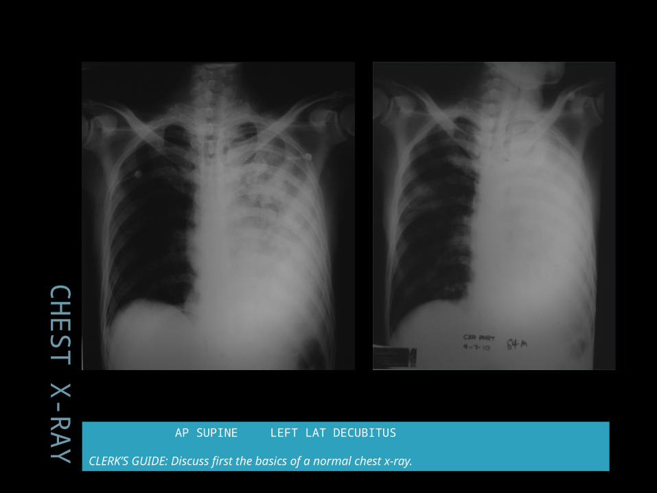
-RAY AP SUPINE LEFT LAT DECUBITUS
CLERK’S GUIDE: Discuss first the basics of a normal chest x-ray.
CC: Cough
action of the body takes to get rid of substances that are irritating the air passages
occurs when mechanical or chemical afferent nerves get irritated and trigger a chain of events
Air in lungs is forced out under high pressure.
Analysis
Cough › Acute < 3weeks› Persistent >3weeks› Chronic >8weeks
› Acute cough Infectious Non infectious

Acute Cough
Acute Cough – signs and symptoms
Infectious Non infectious
Fever, chills, body aches, sore throat vomiting, headache, sinus
pressure, runny nose, night sweats, and postnasal drip.
sputum, or phlegm,
exposure to certain chemicals or irritants in the environment,
coughs that may improve with inhalers or allergy medications

Indications for a Chest X-ray
For patient with acute cough Abnormal vital signs Chest examination suggestive of
pneumonia
Patient: RR 28 › in respiratory distress, supraclavicular,
intercostal and subcostal retractions› symmetric chest expansion, BS left lower
lung, (+) coarse crackles & rhonchi, L

AP supine
if px can’t assume upright position, though
can’t critically evaluate the size of the heart because of hypoventilation, diaphragmatic elevation pushing the base of the heart upwards.

Lateral Decubitus position
px lies on right or left side; the beam traverses the body in horizontal position
px w/pleural effusion, pneumothorax – presence of fluid gravitates to dependent portions
demonstrate fluid levels in cavities
Normal Chest X-ray
Chest anatomy: Evaluation
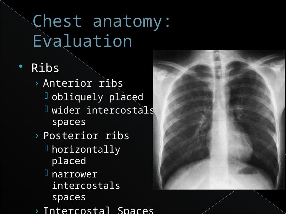
Ribs› Anterior ribs
obliquely placed wider intercostals
spaces› Posterior ribs
horizontally placed narrower intercostals
spaces› Intercostal Spaces
Chest anatomy: Evaluation
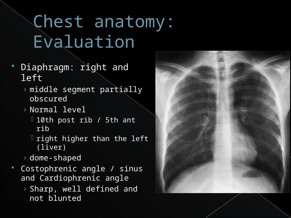
Diaphragm: right and left› middle segment partially
obscured› Normal level
10th post rib / 5th ant rib right higher than the left
(liver)› dome-shaped
Costophrenic angle / sinus and Cardiophrenic angle› Sharp, well defined and
not blunted
Chest anatomy: Evaluation
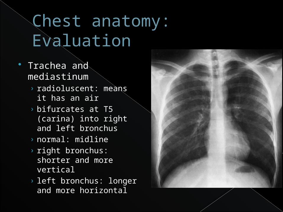
Trachea and mediastinum› radioluscent: means it
has an air› bifurcates at T5
(carina) into right and left bronchus
› normal: midline› right bronchus: shorter
and more vertical› left bronchus: longer
and more horizontal

Chest anatomy: Evaluation
Hila, bronchovascular markings› pulmonary artery and vein› bronchial artery and vein› bronchus› lymph nodes
Normal: › left hilum higher than the
right› pulmonary artery crosses
above the left and below the right bronchus
› size of hilum varies depending on pulmonary blood flow

Chest anatomy: Evaluation
Lungs› Radiolucent
Inner, middle, outer zones› Inner zone: from
sternoclavicular joint draw a vertical line following contour of the chest, big blood vessels are located
› middle zone: medium blood vessels are located
› outer zone: junction of the clavicle and 1st rib draw a vertical line; small blood vessels are located

Chest anatomy: Evaluation
Upper, middle, lower lung fields› landmarks: 2nd and
4th anterior ribs› upper lung field:
further subdivided by the clavicle into supraclavicular (apex) and infraclavicular
› significance: for localization of the lesions
Chest anatomy: Evaluation
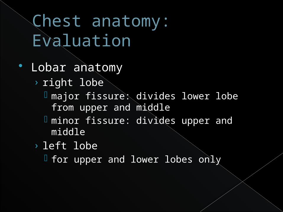
Lobar anatomy› right lobe
major fissure: divides lower lobe from upper and middle
minor fissure: divides upper and middle› left lobe
for upper and lower lobes only

Chest anatomy: Evaluation
Heart shadow› Superior mediastinum
draw a line from the sternal angle to the 4th vertebra
› Anterior mediastinum bounded by posterior
surface of the sternum and anterior surface of the heart
› Posterior mediastinum bounded by posterior part
of the heart and anterior spinal muscle
A
S
P
Chest anatomy: Evaluation
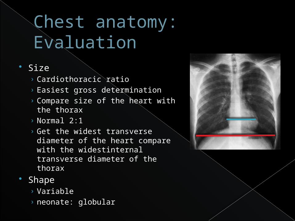
Size› Cardiothoracic ratio› Easiest gross determination› Compare size of the heart with the
thorax› Normal 2:1› Get the widest transverse
diameter of the heart compare with the widestinternal transverse diameter of the thorax
Shape› Variable› neonate: globular

Chest anatomy: Evaluation
Right border› Superior vena cava› Right atrium› Inferior vena cava

Chest anatomy: Evaluation
Right border› Superior vena cava› Right atrium› Inferior vena cava
Left border› Aortic knob› Main pulmonary trunk› Left ventricle

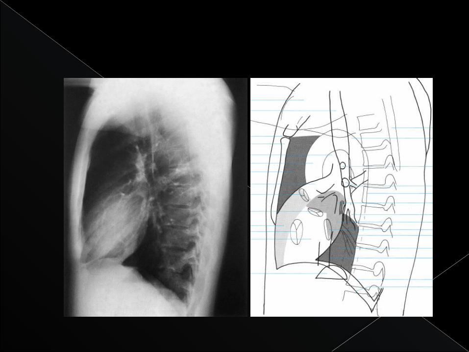
Lateral View
Left atriumLeft ventricleRight ventricle
AORTA
Main Pulmonary Artery
trachea
RC
RS

Patients Chest X-ray
CH
ES
T X
-RAY AP SUPINE LEFT LAT DECUBITUS
CLERK’S GUIDE: Discuss first the basics of a normal chest x-ray.
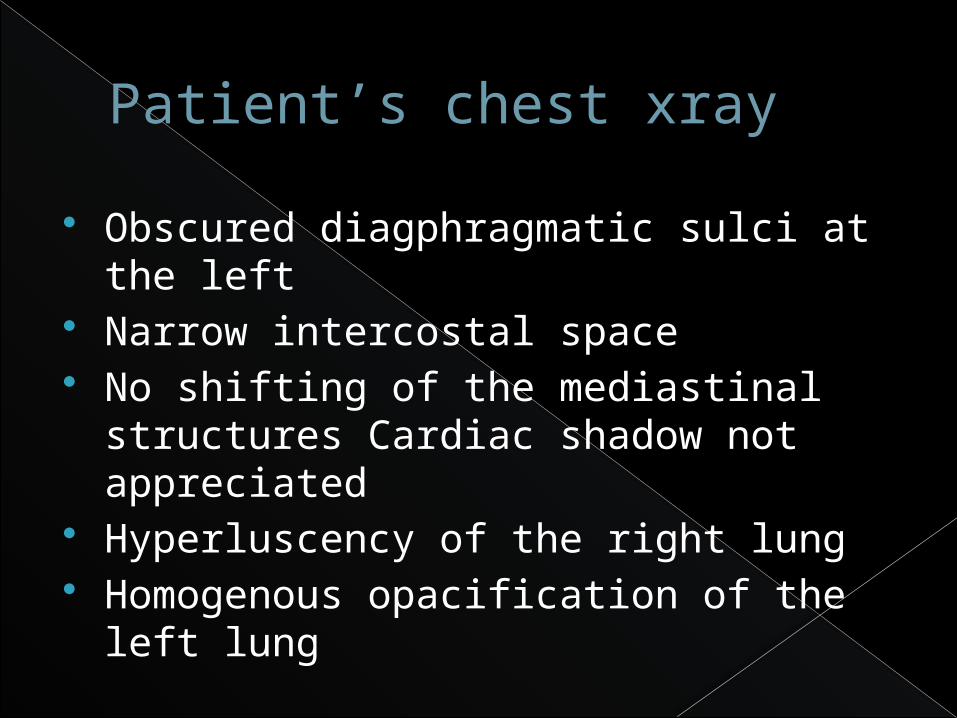
Patient’s chest xray
Obscured diagphragmatic sulci at the left
Narrow intercostal space No shifting of the mediastinal
structures Cardiac shadow not appreciated
Hyperluscency of the right lung Homogenous opacification of the left
lung
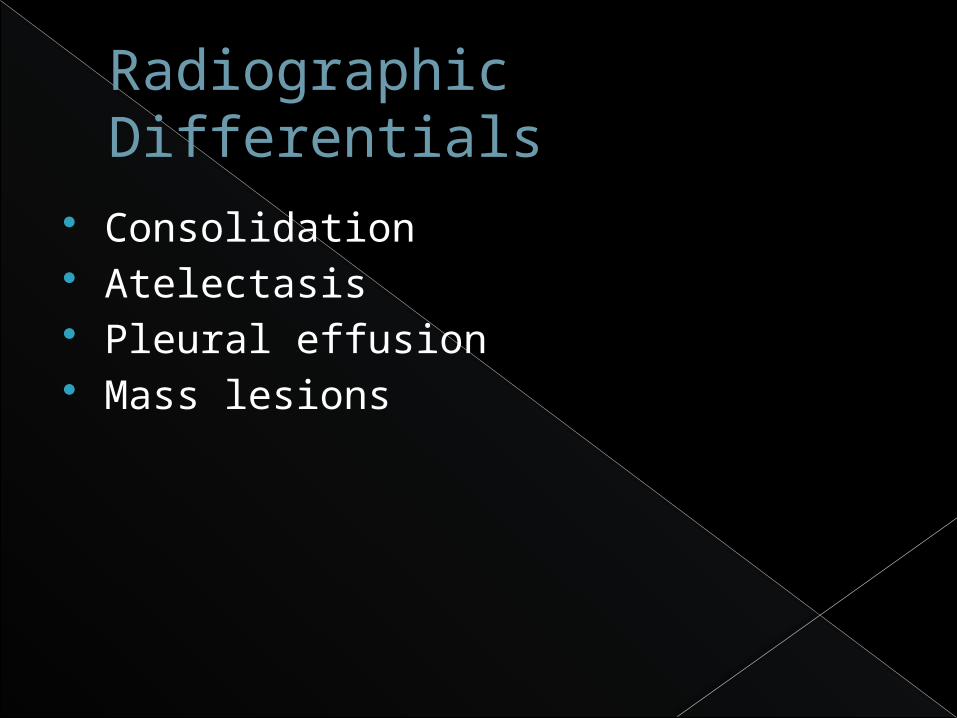
Radiographic Differentials
Consolidation Atelectasis Pleural effusion Mass lesions
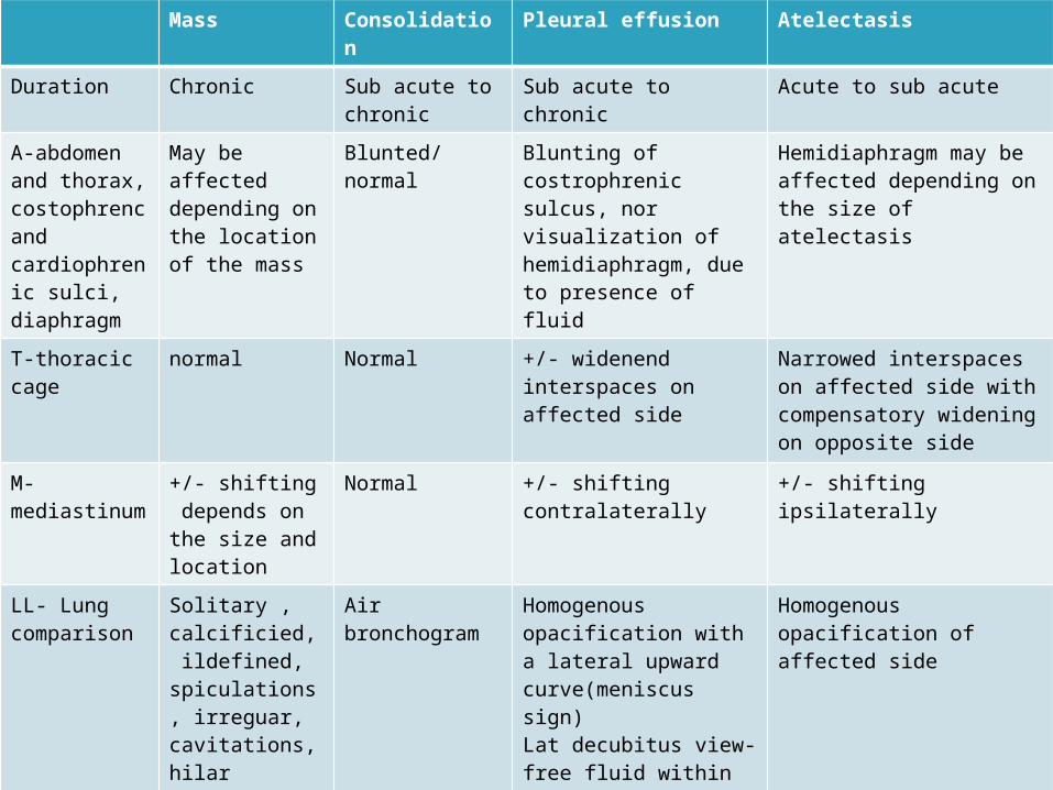
Mass Consolidation
Pleural effusion Atelectasis
Duration Chronic Sub acute to chronic
Sub acute to chronic Acute to sub acute
A-abdomen and thorax, costophrenc and cardiophrenic sulci, diaphragm
May be affected depending on the location of the mass
Blunted/ normal
Blunting of costrophrenic sulcus, nor visualization of hemidiaphragm, due to presence of fluid
Hemidiaphragm may be affected depending on the size of atelectasis
T-thoracic cage
normal Normal +/- widenend interspaces on affected side
Narrowed interspaces on affected side with compensatory widening on opposite side
M-mediastinum
+/- shifting depends on the size and location
Normal +/- shifting contralaterally
+/- shifting ipsilaterally
LL- Lung comparison
Solitary , calcificied, ildefined, spiculations, irreguar, cavitations, hilar enlargement, effusions
Air bronchogram
Homogenous opacification with a lateral upward curve(meniscus sign)Lat decubitus view- free fluid within the pleural space +layering oon the dependent part
Homogenous opacification of affected side
Atelectasis
means “lack of stretch” refers to collapse or loss of lung
volume 2 types: Obstructive or Non obstructive
Atelectasis
Obstructive› blockage of an airway › Air retained distal to the occlusion is then
resorbed from nonventilated alveoli› affected regions become totally airless
Non obstructive› caused by loss of contact between the
parietal and visceral pleurae, parenchymal compression, loss of surfactant, or replacement of lung tissue by scarring or infiltrative disease.
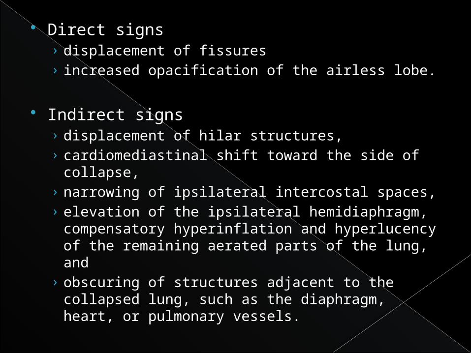
Direct signs › displacement of fissures › increased opacification of the airless lobe.
Indirect signs › displacement of hilar structures, › cardiomediastinal shift toward the side of
collapse, › narrowing of ipsilateral intercostal spaces, › elevation of the ipsilateral hemidiaphragm,
compensatory hyperinflation and hyperlucency of the remaining aerated parts of the lung, and
› obscuring of structures adjacent to the collapsed lung, such as the diaphragm, heart, or pulmonary vessels.

ROLE OF MAGNETIC RESONANCE IMAGING
can distinguish between obstructive and nonobstructive atelectasis
Obstructive atelectasis › displays high signal intensity on T2-weighted images due
to proton-rich mucus accumulation. Nonobstructive atelectasis
› low signal intensity on T1 and T2 weighted spin-echo images, since the residual alveolar gas has a low proton concentration, and magnetic susceptibility effects between alveolar walls lead to a decrease in signal.
The use of MRI in diagnosing atelectasis is still experimental, and more experience needs to be accrued
Treatment
Continuous positive airway pressure (CPAP)
Fiberoptic bronchoscopy for the extraction of secretions
Mucolytic therapy


NORMAL CHEST X RAY


![[Pharma] cough](https://img.pdfslide.net/doc/110x75/55ac456e1a28ab7f538b4570/pharma-cough.jpg)