Embed Size (px)
Citation preview

HMGCR Pathway Mediates Cerebral-Vascular Stability
and Angiogenesis in Developing Zebrafish
Shahram Eisa-Beygi
Thesis submitted to the
Faculty of Graduate and Postdoctoral Studies
in partial fulfillment of the requirements for the degree of
Doctor of Philosophy in Biology
Ottawa-Carleton Institute of Biology
Faculty of Science
University of Ottawa
© Shahram Eisa-Beygi, Ottawa, Canada, 2013

ii
“Look at the Perfect One at the Circle’s Center:
He Spins and whirls like a Golden Compass,
Beyond all that is rational,
To show this dear world that Everything,
Everything in Existence Does point to God. ”
---- Hafez of Persia
This PhD dissertation is humbly dedicated to the loving memory of my
grandparents.

iii
Acknowledgements
I would like to extend my gratitude to Dr. Thomas W. Moon and Dr. Marc Ekker,
two brilliant and accomplished scientists, for giving me the unique opportunity to study
under their guidance and for their constant support and inexorable patience. They both have
exerted a profound contribution to my scientific and personal growth and have compelled
me to pursue further studies in life-sciences. I would also like to thank my committee
members, Drs Carole Yauk, Marie-Andrée Akimenko, and Michael Jonz for their
continuous critiques and suggestions, which have indubitably helped me refine this project.
The personal friendship and kindness of Andrey Massarsky is appreciated beyond words.
This project could not have progressed to this extent if it were not for the constant technical
assistance and friendship of Vishal Saxena, Sandra Noble, Bill Fletcher, Gary Hatch, Jamie
Holden, Khaled Eid, Rafael Godoy and Raymond W.M. Kwong. I would also like to extend
my heartfelt gratitude and love to my parents and my siblings for enriching my life and
giving me a sense of purpose and fulfillment. Last but not least, I would like to express my
sincere appreciation of my dear friends, Ali Al-Rewashdy and Saud Ayed, with whom I
enjoyed our intense but friendly philosophical and existential discourses during our frequent
tea breaks. I would also like to acknowledge that this work was supported by grants
primarily from NSERC to TM and ME, as well as a grant from Pfizer Pharmaceuticals to
TM.
iv
Abstract
Intracerebral hemorrhage (ICH) is a severe form of stroke, with a high mortality
rate and often resulting in irreversible neurological deterioration. Although animal studies
have provided insight into the etiology of the disease, many of the causative genes and
mechanisms implicated in cerebral-vascular malformations are unknown. Treatment options
remain ineffective. With the present models, the pathophysiological consequences of ICH
can only be assessed in situ and after histological analysis. Furthermore, common
deficiencies of the current models include the heterogeneity, low expression and low
reproducibility of the desired phenotype. Hence, there is a requirement for novel approaches
to model ICH pathogenesis. Zebrafish (Danio rerio) has gained recognition as a vertebrate
model for stroke research.
Through a combination of pharmacological blockers, metabolite rescue, genetic
approaches, and confocal imaging analysis, I demonstrate a requirement for the 3-hydroxy-
3-methylglutaryl-CoA reductase (HMGCR) pathway in regulating developmental cerebral-
vascular stabilization. A transient loss in HMGCR function induces ICH, characterised by
progressive dilation of blood vessels, vascular permeability and vessel rupture. These effects
are likely due to reduced prenylation of Rho GTPases, evidenced by morpholino-mediated
blocking of the prenylation pathway and in vivo assessment of endothelial-specific
localization of cdc42, a Rho GTPase family protein. These results are in conformity with
recent clinical and experimental evidence.
I have further shown that this model consistently replicates common
pathoghysiological processes associated with ICH. The hemorrhages are associated with the
disruption of the blood-brain barrier, vessel disintegration, hematoma expansion and edema
into the adjacent brain regions. Also, enhanced apoptosis, activation of inflammatory
mediators in the periphery of the hematoma, enriched heme oxygenase 1 (HO-1) expression
and localised thrombosis were observed in these embryos. I show that the patterning and
distribution of catecholaminergic neurons, response to sensory stimulus and swimming
speed were impaired as a consequence of ICH.

v
These results suggest that HMGCR contributes to cerebral-vascular stabilisation
through Rho GTPase mediated-signalling and that zebrafish can serve as a powerful
paradigm for the systemic analysis of the etiological and pathophysiological underpinnings
of ICH and can help establish the basis for future studies into screening for putative
therapeutics and elucidating mechanisms aiding functional recovery.
vi
Résumé
L’hémorragie intracérébrale (HIC) est une forme grave d’accident cérébro-vasculaire
associé à un taux élevé de mortalité et qui cause une détérioration neurologique sévère.
Quoique des études sur des modèles animaux nous ont permis de mieux comprendre
l’étiologie de cette maladie, plusieurs gènes responsables et les mécanismes sous-jacents aux
malformations cérébro-vasculaires sont encore inconnus. Les options thérapeutiques
demeurent inefficaces. En utilisant les modèles courants, les conséquences patho-
physiologiques des HIC peuvent seulement être examinées in situ à l’aide d’analyses
histologiques. De plus, un désavantage des modèles courants est le faible taux d’expression
de de reproductibilité des phénotypes. Il nous faut donc de nouveaux modèles pour la
pathogénèse des HIC. Le poisson-zèbre, Danio rerio, a acquis une renommée comme
modèle pour l’étude des accidents cérébro-vasculaires.
En utilisant une combinaison d’agents pharmacologiques, de manipulations
métaboliques, d’approches génétiques, et d’imagerie confocale, j’ai montré le rôle essentiel
joué par la voie métabolique impliquant la 3-hydroxy-3-méthylglutaryl-CoA réductase
(HMGCR) dans le contrôle et la stabilisation du système cérébro-vasculaire. Une perte
transitoire de la fonction de HMGC induit une HIC caractérisée par la dilatation progressive
des vaisseaux sanguins, une perméabilité vasculaire et la rupture des vaisseaux. Ces effets
sont attribuables à une prénylation réduite des Rho GTPases, tel que démontré par l’effet de
morpholinos dur la voie de prénylation et un examen in vivo de la localisation spécifique de
cdc42, un membre de la famille des Rho GTPases. Ces résultats sont en accord avec de
récentes données cliniques et expérimentales.
J’ai de plus montré que ce modèle reflète fidèlement les processus patho-
physiologiques les plus couramment associés à l’HIC. Les hémorragies coïncident avec un
dérangement de la barrière hémato-encéphalique, une désintégration des vaisseaux, une
expansion des hématomes et de l’oedème dans les régions avoisinantes du cerveau. De plus,
une apoptose accrue, l’activation des médiateurs inflammatoires dans la périphérie de
l’hématome, une hausse localisée de l’hème oxygénase et une thrombose localisée sont

vii
observées chez ces embryons. J’ai montré que le profil des neurones catécholaminergiques,
la réponse à une stimulation tactile et la vitesse natatoire étaient affectées suite à l’HIC.
Ces résultats suggèrent que l’HMGCR contribue à la stabilisation cérébro-vasculaire
via la voie de signalisation des Rho GTPases et que le poisson-zèbre constitue un paradigme
pertinent pour l’analyse systématique des causes étiologiques et patho-physiologiques de
l’HIC . Ce modèle peut contribuer à établir une base pour des études ultérieures visant le
criblage de thérapies potentielles ainsi que la découverte de mécanismes facilitant la
guérison.
viii
TABLE OF CONTENTS
Acknowledgements ____________________________________________________ iii
Abstract _____________________________________________________________ iv
Table of Contents _____________________________________________________ viii
List of Figures _______________________________________________________ xiii
List of Abbreviations __________________________________________________ xvi
Chapter 1: General introduction __________________________________________ 2
1.1 Rationale for the study ______________________________________________ 3
1.2 HMG CoA reductase _______________________________________________ 3
1.2.1 Regulation of HMGCR activity _____________________________________ 4
1.2.2 HMGCR pathway and pharmacology of statins ________________________ 9
1.2.3 HMGCR pathway and embryonic development _______________________ 16
1.2.4 HMGCR pathway and angiogenesis ________________________________ 18
1.2.5 HMGCR pathway and stroke ______________________________________ 20
1.3 Cerebral-vascular stabilisation in zebrafish ___________________________ 23
1.4 Utility of zebrafish as a model for the etiology and pathophysiology of ICH _ 31
1.5 Research Objectives _______________________________________________ 32
ix
Chapter 2: The 3-hydroxy-3-methylglutaryl-CoA reductase (HMGCR) pathway
regulates developmental cerebral-vascular stabilisation via a prenylation-dependent
signalling pathway _____________________________________________________________________ 36
2.1 Abstract _________________________________________________________ 38
2.2 Introduction _____________________________________________________ 39
2.3 Materials and Methods ____________________________________________ 43
2.3.1 Zebrafish husbandry and transgenic lines ____________________________ 43
2.3.2 Drug treatment and metabolite rescue experiments _____________________ 43
2.3.3 Whole-mount o-Dianisidine (OD) staining and cryosectioning ___________ 44
2.3.4 Confocal microscopy ____________________________________________ 45
2.3.5 Morpholino design, resuspension and microinjection ___________________ 45
2.3.6 Microangiography ______________________________________________ 46
2.3.7 DAF-2DA Staining _____________________________________________ 46
2.3.8 RNA extraction, RT-PCR and sequencing ___________________________ 46
2.4 Results __________________________________________________________ 48
2.4.1 Pharmacological inhibition of the HMGCR enzyme leads to cerebral-vascular
defects in developing zebrafish _________________________________________ 48
2.4.2 Morpholino-mediated depletion of hmgcrb mRNA levels leads to cerebral-
vascular defects in developing zebrafish _________________________________ 61
2.4.3 A defficiency in GGPP biosynthesis and impaired GGTase I-facilitated
prenylation of Rho GTPases leads to cerebral-vascular defects in developing zebrafish
__________________________________________________________________ 65
x
2.5 Discussion _______________________________________________________ 70
Chapter 3: Molecular and pathophysiological processes underlying cerebral
hemorrhage in developing zebrafish Danio reriro ___________________________ 81
3.1 Abstract _________________________________________________________ 83
3.2 Introduction _____________________________________________________ 84
3.3 Materials and Methods ____________________________________________ 86
3.3.1 Zebrafish husbandry and transgenic lines ____________________________ 86
3.3.2 Drug treatment _________________________________________________ 87
3.3.3 Detection of apoptotic cells _______________________________________ 87
3.3.4 Neutral-red staining of macrophages ________________________________ 88
3.3.5 Sudan-black staining of neutorphils _________________________________ 88
3.3.6 Whole-mount o-dianisidine (OD) staining of erythrocytes _______________ 88
3.3.7 CM-H2DCFDA staining __________________________________________ 89
3.3.8 RNA extraction, cDNA synthesis and real-time RT-PCR _______________ 89
3.3.9 Confocal microscopy ____________________________________________ 90
3.3.10 Western blot analysis ___________________________________________ 90
3.4 Results and Discussion _____________________________________________ 91
3.4.1 Vessel disintegration, hematoma expansion and cerebral edema mark the early
phases of ICH ______________________________________________________ 91
3.4.2 Apoptosis is enhanced in response to ICH ___________________________ 92
xi
3.4.3 ICH triggers an immune response __________________________________ 95
3.4.4 Increased heme oxygenase (HO-1) expression and ROS generation following ICH
__________________________________________________________________ 98
3.4.5 Evidence for thrombosis after ICH _________________________________ 99
3.5 Conclusions _____________________________________________________ 104
Chapter 4: Disruption of dopaminergic neuron development and impaired locomotor
function in zebrafish (Danio rerio) with intracerebral hemorrhage ____________________________________________________________________ 108
4.1 Abstract ________________________________________________________ 110
4.2 Introduction ____________________________________________________ 111
4.3 Materials and Methods ___________________________________________ 115
4.3.1 Zebrafish husbandry and transgenic lines ___________________________ 115
4.3.2 Drug treatment ________________________________________________ 115
4.3.3 Confocal microscopy ___________________________________________ 115
4.3.4 Whole-mount anti-tyrosine hydroxylase (TH) immunohistochemistry _____ 116
4.3.5 Testing for locomotor activity ____________________________________ 116
4.4 Results and Discussion ____________________________________________ 117
4.4.1 Pharmacologically induced ICH disrupts catecholaminergic neuron development
and locomotor function in zebrafish ___________________________________ 117

xii
Chapter 5: General discussion and future directions ____________________________________________________________________ 128
References ___________________________________________________________ 135
xiii
LIST OF FIGURES
Figure 1.1 The HMGCR-dependent mevalonate pathway _____________________ 5
Figure 1.2 Zebrafish possess two paralogous genes encoding a functional HMGCR
protein with conserved inhibitor-binding residues __________________________ 11
Figure 1.3 Chemical structure of the HMG-CoA moiety (dihydroxy heptanoic acid) in
statins. ____________________________________________________________ 14
Figure 1.4 Basic properties of atorvastatin (ATV) and cerivastatin (CTV) _______ 15
Figure 1.5 The optical transparency of zebrafish facilitates easy evaluation of ICH
during development _________________________________________________ 29
Figure 1.6 HMGCR-mediated regulation of Rho GTPase activity _____________ 34
Figure 2.1 Pharmacological inhibition of the HMGCR pathway induces cerebral-
vascular defects in developing zebrafish _________________________________ 50
Figure 2.2 Atorvastatin exposure results in clusters of abnormally dilated cerebral
vessels in the forebrain and hindbrain____________________________________ 52
Figure 2.3 Embryos with CNS hemorrhaging do not have defects in trunk angiogenesis
and patterning ______________________________________________________ 54
Figure 2.4 Pharmacological inhibition of the HMGCR pathway induces cerebral
hemorrhage in developing zebrafish _____________________________________ 56
Figure 2.5 Pharmacological inhibition of the HMGCR results in loss of vascular
stability and induces vessel rupture _____________________________________ 59
xiv
Figure 2.6 MO-mediated depletion of the HMGCR expression phenocopies ATV-
treatment __________________________________________________________ 63
Figure 2.7 MO-mediated ablation of pggt1b mimics statin and hmgcrb MO-induced
cerebral hemorrhages and effectively reduces the wild-type pggt1b mRNA ______ 67
Figure 2.8 Inhibition of the HMGCR pathway abolishes the intra-endothelial expression
of cdc42 __________________________________________________________ 71
Figure 2.9 At high doses (5-10 mg/L), ATV-exposure induces defective angiogenesis
__________________________________________________________________ 75
Figure 2.10 Embryos resolve cerebral-vascular defects and resume normal development
__________________________________________________________________ 79
Figure 3.1 Exposure to Atorvastatin (ATV) induces vascular dilation, followed by
hemorrhage and vascular disintegration __________________________________ 93
Figure 3.2 Exposure to Atorvastatin (ATV) induces hematoma expansion and edema
formation in the brain. ________________________________________________ 96
Figure 3.3 ICH-induced pathophysiological processes observed at 48 hpf ______ 100
Figure 3.4 Increased heme oxygenase HO-1 expression and ROS generation following
ICH at 48 hpf _____________________________________________________ 102
Figure 3.5 Evidence for thrombosis after ICH ____________________________ 105
Figure 4.1 The Tg(dat:EGFP);(gata-1:DsRed) facilitates real-time analysis of ICH-
induced DA-neuron neurotoxicity _____________________________________ 119

xv
Figure 4.2 Disruption of catecholaminergic-neuron development in zebrafish with ICH
_________________________________________________________________ 124
Figure 4.3 Impaired locomotor function in zebrafish with ICH ______________ 126
xvi
LIST OF ABBREVIATIONS
AMPK: AMP-activated protein kinase
ATV: Atorvastatin
bp: Base pairs
CNS: Central nervous system
CCM: Cerebral cavernous malformation
CVT: Cerivastatin
cDNA: Complementary DNA
dpf: days post fertilisation
DAB: Diaminobenzidine
DA: Dopaminergic
DLAV: Dorsal longitudinal anastomotic vessel
ER: Endoplasmic reticulu
EGFP: Enhanced green fluorescent protein
GGTase I: Geranylgeranyltransferase type 1
GFP: Green fluorescent protein
hpf: hours post fertilisation
HRP: Horseradish peroxidase
HMGCR: 3-hydroxy-3-methylglutaryl-CoA reductase
ICH: Intracerebral hemorrhage
ISV: Intersegmental vessel
LDL: low density lipoprotein
MO: Morpholino oligonucleotides
NO: Nitric oxide
MCeV: Middle cerebral vein
OD: o-Dianisidine
PFA: Paraformaldehyde
PBST: 1X Phosphate Buffered Saline Tween-20
PPrA: Primitive prosencephalic artery
PMBC: Primordial midbrain channel
PrA: Prosencephalic artery
PTU: 1-pheny1-2-thiourea
pak: p21-activated kinase
SCAP: SREBP-cleavage activating protein
SREBP: sterol-regulatory element binding protein
SPARCL: Stroke Prevention by Aggressive Reduction in Cholesterol Levels
ROS: Reactive oxygen species
RT-PCR: Reverse transcriptase polymerase chain reaction
TH: Tyrosine hydroxylase

TH: Tyrosine hydroxylase
VEGF: Vascular endothelial growth factor
VE-cadherin: Vascular endothelial cadherin

2
Chapter 1
General Introduction
3
1. Introduction
1.1. Rationale for the study
The 3-hydroxy-3-methylglutaryl-CoA reductase (HMGCR) is a metabolic
enzyme, regulating the rate-limiting step in the biosynthesis of cholesterol and
isoprenoids. Through an initial small-scale toxicity assay of various pharmaceuticals, I
have isolated two structurally distinct pharmacological inhibitors of HMGCR (statins)
that induce an intracerebral hemorrhage (ICH) phenotype in zebrafish embryos and
larvae. These observations emerged in parallel with recent experimental and clinical
studies suggesting a link between inhibition of HMGCR function and ICH pathogenesis.
These findings compelled me to elucidate the precise mechanisms and signalling
pathways under the control of HMGCR that mediate cerebral-vascular stabilisation.
1.2. The 3-hydroxy-3-methylglutaryl-CoA reductase (HMGCR) pathway
The enzyme, 3-hydroxy-3-methylglutaryl coenzyme A reductase (HMGCR; E.C.
1.1.1.88), is an endoplasmic reticulum (ER)-bound, polytopic glycoprotein with eight
trans-membrane helices located at the N-terminus, and a catalytic domain at the C-
terminal, which projects into the cytosol (Gil et al., 1985). Although HMGCR expression
is highly enriched in liver cells (hepatocytes) (Hwa et al., 1992), it is also expressed in
extra-hepatic tissues, including the heart (Laufs et al., 2002), and the brain (Thelen et al.,
2005; Michalak and Wender, 1996).
The trans-membrane domain of HMGCR anchors the protein to the ER and the
trans-membrane domain contains the residues required for the binding of regulatory
proteins to the enzyme (DeBose-Boyd et al., 2008). The soluble C-terminal domain
4
catalyzes the rate-limiting step in the de novo synthesis of mevalonate, the precursor for
the biosynthesis of cholesterol and bile acids, as well as several non-sterol isoprenoid
metabolites, including Farnesyl-pyrophosphate (FPP) and Geranyl-geranyl
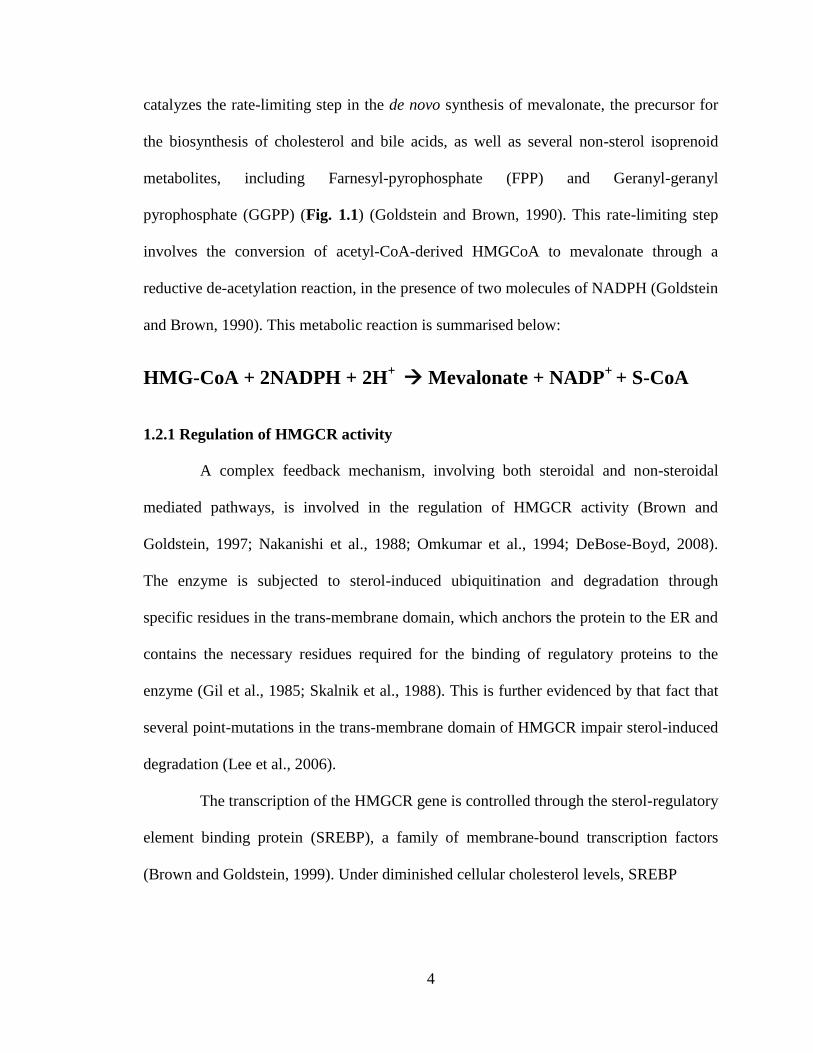
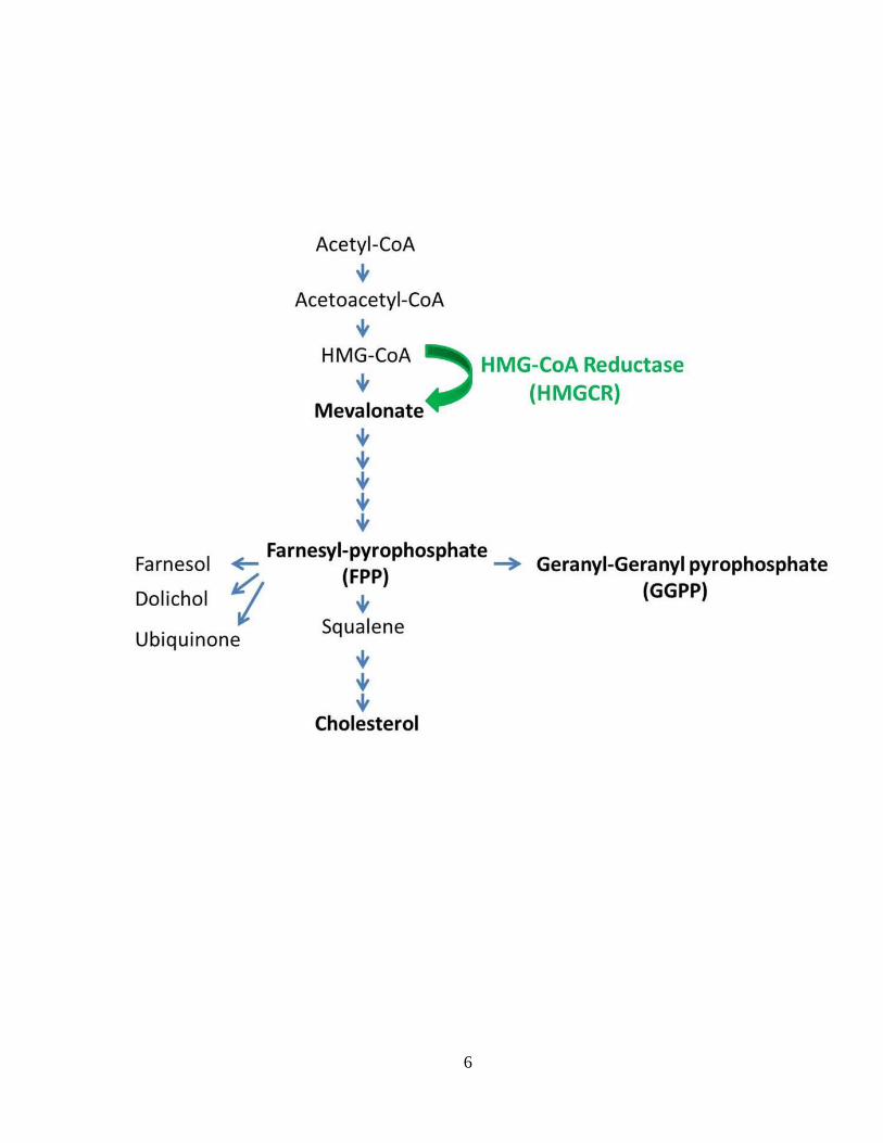
pyrophosphate (GGPP) (Fig. 1.1) (Goldstein and Brown, 1990). This rate-limiting step
involves the conversion of acetyl-CoA-derived HMGCoA to mevalonate through a
reductive de-acetylation reaction, in the presence of two molecules of NADPH (Goldstein
and Brown, 1990). This metabolic reaction is summarised below:
HMG-CoA + 2NADPH + 2H+
Mevalonate + NADP+
+ S-CoA
1.2.1 Regulation of HMGCR activity
A complex feedback mechanism, involving both steroidal and non-steroidal
mediated pathways, is involved in the regulation of HMGCR activity (Brown and
Goldstein, 1997; Nakanishi et al., 1988; Omkumar et al., 1994; DeBose-Boyd, 2008).
The enzyme is subjected to sterol-induced ubiquitination and degradation through
specific residues in the trans-membrane domain, which anchors the protein to the ER and
contains the necessary residues required for the binding of regulatory proteins to the
enzyme (Gil et al., 1985; Skalnik et al., 1988). This is further evidenced by that fact that
several point-mutations in the trans-membrane domain of HMGCR impair sterol-induced
degradation (Lee et al., 2006).
The transcription of the HMGCR gene is controlled through the sterol-regulatory
element binding protein (SREBP), a family of membrane-bound transcription factors
(Brown and Goldstein, 1999). Under diminished cellular cholesterol levels, SREBP
5
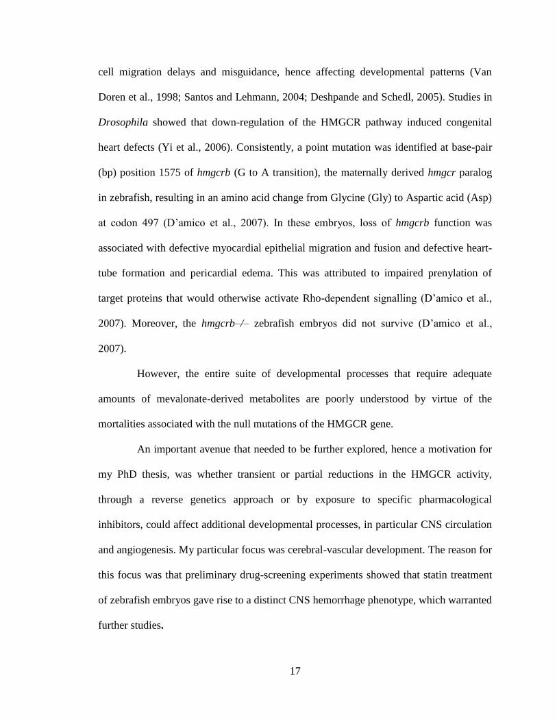
Figure 1.1: The HMGCR-dependent mevalonate pathway. HMGCR-mediated lipid
biosynthesis, showing the rate-limiting step of mevalonate generation (HMGCR), along
with the synthesis of farnesyl-pyrophosphate (FPP) and geranylgeranyl-pyrophosphate
(GGPP), all of which are derivatives of mevalonate.
6
7
interacts with the SREBP-cleavage activating protein (SCAP), which facilitates
sequential cleavage of SREBP by S1P, a serine protease belonging to the subtilisin/kexin
family, and S2P, a zinc metalloprotease. After this sequential processing in the Golgi
apparatus, the water-soluble and transcriptionally active N-terminal fragment of SREBP,
of approximately 480 amino acids, is translocated to the nucleus (Ye et al., 2000).
Upon translocation to the nucleus, this SREBP fragment binds to the ‘Sterol
regulatory element’, which flanks the low density lipoprotein (LDL) receptor gene and
other genes involved in the HMGCR pathway. This enhances the uptake and synthesis of
extracellular cholesterol by activating the transcription of target genes encoding HMGCR
and other cholesterol biosynthetic enzymes, including the LDL-receptor gene (Brown and
Goldstein, 1999). As such, pharmacological inhibition of cholesterol biosynthesis triggers
SREBP-mediated up-regulation of HMGCR gene expression, evidenced by both in vitro
and in vivo studies (Brown and Goldstein, 1999; Krycer et al., 2009; Mammen et al.,
2011).
Early kinetic experiments performed on microsomes isolated from the rat liver
showed that HMGCR kinetics could also be regulated at the level of phosphorylation,
where ATP would serve as a phosphate donor (Gibson, 1985). It was shown that the
catalytic activity of HMGCR could be inhibited, in a dose-dependent fashion, when
microsomes were incubated in a solution of ATP, Mg2+
, and a cytosolic fraction isolated
from rat liver (Clarke and Hardie, 1990). In subsequent experiments, it was demonstrated
that HMGCR is phosphorylated at Ser-871, near the C-terminus of the protein, by AMP-
activated protein kinase (AMPK) (Hardie, 1992). It was further reported that this
phosphorylation event reduced HMGCR activities by approximately 80% (Hardie, 1992).
8
Over-expression of a cDNA construct encoding a mutant form of hamster
HMGCR, where Alanine is substituted for Serine at residue 871, further verified that
phosphorylation of the Serine-871 residue is a critical regulator of HMGCR enzyme
activity and that this process is mediated by AMPK (Sato et al., 1993). Furthermore, the
SerineAlanine substitution, through site-directed mutagenesis, failed to prevent the
post-transcriptional regulation of HMGCR activity by mevalonate, cholesterol or LDL
(Sato et al., 1993). It was further established that incubation of rat hepatocytes with
fructose resulted in a surge in cellular AMP levels and that this was followed by
activation of the AMPK, which resulted in reduced HMGCR activities (Gillespie and
Hardie, 1992). Similarly, phosphorylation of Serine-871 was shown to decrease the
activity of HMGCR in humans (Clarke and Hardie, 1990). In terms of the specific
mechanism, it is predicted that phosphorylation of Ser-817 reduced the activities of the
enzyme by reducing its affinity for NADPH, thereby preventing the synthesis of
mevalonate (Sato et al., 1993; Omkumar et al., 1994).
The HMGCR-mediated lipid biosynthesis is also regulated through ubiquitin-
mediated proteoloysis of the enzyme HMGCR. This involves the attachment of ubiquitin,
thus marking HMGCR for degradation and recycling. This process is catalyzed by Ufd1,
an adaptor protein residing in the ER. As such, cells expressing higher levels of Ufd1
tend to exhibit reduced HMGCR activity. Hence, de-novo synthesis of cholesterol is
significantly reduced due to the ubiquitination (Cao et al., 2007).
Recent evidence points towards alternative splicing of HMGCR pre-mRNA as
contributing to inter-individual variations in enzyme activity in response to treatments
with pharmacological inhibitors (Medina et al., 2008; Medina and Krauss, 2009). This
9
splicing event gives rise to a variant transcript that is shorter in length when compared
with the wild-type isoform. Although it does not disrupt the open-reading-frame, this
alternative splicing event generates an isoform in which exon-13 is deleted, which
otherwise codes for a part of the catalytic domain of HMGCR (exons 11-20) (Medina and
Krauss, 2009). In addition, it was speculated that deletion of exon-13 would reduce
enzyme activity due to loss of critical residues and truncation of the L-domain, which
contains part of the substrate-binding region (Medina et al., 2010).
1.2.2 HMGCR pathway and pharmacology of statins
As a result of the direct effects the endogenous levels of cholesterol, HMGCR-
mediated lipid biosynthesis and its mechanism of regulation are exploited by human
pharmaceuticals that selectively bind, in a reversible manner, to the active site of
HMGCR and render it less active. As such, statins (both naturally-derived and synthetic
statins) are known as agonists of the natural substrate, HMG-CoA, since they compete for
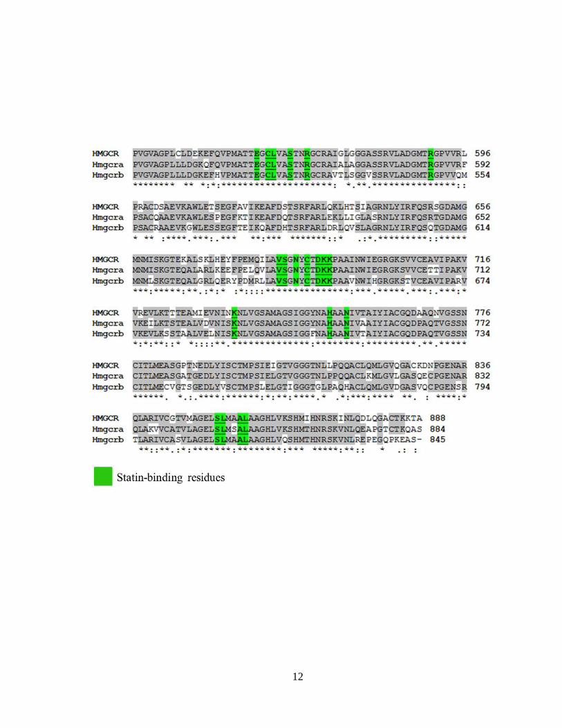
the active site on the enzyme (Istvan and Deisenhofer, 2001). Sequence alignment of the
partial catalytic sites of the HMGCR proteins indicates that the statin-binding residues
(Istvan and Deisenhofer, 2001) on the C-terminus site of the enzyme are identical in both
of the HMGCR paralogs identified in zebrafish, Danio rerio (Fig. 1.2) (Please refer to
Chapter 2 for more information).
In addition to effectively lowering the rate at which HMGCR can produce
cholesterol, statins also curtail the biosynthesis of other metabolites downstream of
mevalonate, namely isoprenoid intermediates, which serve as essential lipid attachments
for heterotrimeric G proteins and the Rho/Ras GTPase family of proteins (Liao, 2002).
10
This post-translational modification ensures that the prenylated complexes are localised
to the cell membrane, which facilitates their activation and mediation of their signalling
(Berzat et al., 2006).
Statins are primarily prescribed to inhibit cholesterol biosynthesis in the liver,
since liver is the major organ where de novo cholesterol synthesis takes place. In
addition, liver is where cholesterol is converted into bile salts, and where lipoproteins
involved in transporting cholesterol are synthesised and exported (Lennernas and Fager,
1997). The high efficacy of statins in the treatment of hypercholesterolemia has been
extensively demonstrated through the use of animal models and through clinical studies
(Johnston et al., 2001; Dubuc et al., 2004; Wu et al., 2005; Versmissen et al., 2008).
Statin-induced depletion of intracellular cholesterol biosynthesis stimulates the up-
regulation of the HMGCR pathway, through transcription, by the SREBP-mediated
feedback mechanisms noted above (Brown and Goldstein, 2004). This, in turn, increases
the rate of lipoprotein uptake from the circulation through the cytoplasmic membrane of
hepatocytes, by increasing the transcription of LDL-receptor gene. Once delivered to the
interior of the cell by the newly synthesised LDL-receptors, the LDL molecules are then
digested and the cholesterol is used for metabolic processes or storage (Goldstein et al.,
1975). Therefore, statin treatment decreases both de novo cholesterol synthesis and serum
cholesterol levels, while maintaining a steady level of cholesterol in the liver (Brown and
Goldstein, 2004).
The statin family of pharmaceuticals can be classified on the basis of whether
they are naturally-derived or synthetic (Weitz-Schmidt, 2002). All statins share the
common HMG-CoA-like moiety (dihydroxy heptanoic acid), in an either closed (lactone)

11
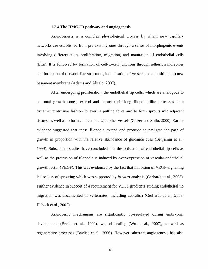
Figure 1.2: Zebrafish possess two paralogous genes encoding functional HMGCR
proteins with conserved inhibitor-binding residues. Amino acid sequence alignment of
the partial catalytic (C-terminus) sites of human HMGCR and zebrafish orthologs is
shown. Numbers denote amino acid positions. Identical amino acids are highlighted in
grey. Amino acid residues thought to be critical for statin interaction are highlighted in
green.
12
13
or opened (acid) ring confirmation (Fig. 1.3). Once delivered to the serum, the lactone
form of the statin is activated (hydrolyzed) by a family of serine esterases known as
carboxylesterases. This modification allows statins to bind as an intermediate analogue to
the active site of HMGCR (Fukami et al., 2010). Statins have a higher affinity for the
active site of HMGCR than the natural substrate HMG-CoA (Istvan and Deisenhofer,
2001). However, despite their identical mode of function, statins display variation with
respect to their chemical structure, hepato-selectivity, molecular weight, pharmacokinetic
profiles, biochemical metabolism, and affinity for the active site of HMGCR, half-life,
and excretory pathways (Igel et al., 2001). All of these parameters can affect the
efficiency with which statins can reduce serum LDL and total cholesterol levels.
Furthermore, both animal and human studies have attested to the efficacy of statins to
reduce serum cholesterol levels through inhibition of HMGCR pathway (Johnston et al.,
2001; Dubuc et al., 2004; Wu et al., 2005; Versmissen et al., 2008).
Lipophilic statins are less hepato-selective, as they can passively diffuse through
cell membranes, whereas hydrophilic statins display minimal perfusion to other tissues,
since they require specific membrane interactive transport mechanism, making them
more hepato-selective (Sirtori, 1993). Hence, hydrophilic statins, due to lower distal
tissue absorption, are more tolerated in patients and with less reported side-effects or non-
specific toxicity (Sirtori, 1993, Hemant et al., 1999; Liao and Laufs, 2005). The two
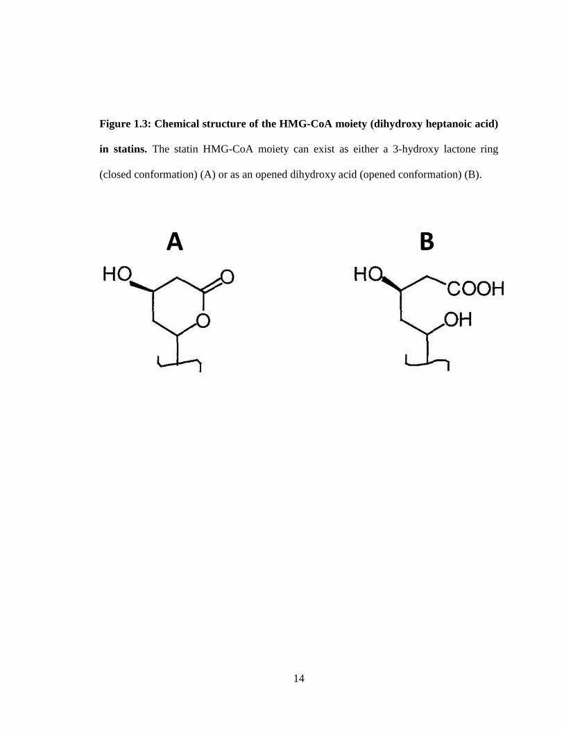
synthetic and lipophilic statins I have used in this project are atorvastatin (ATV) and
cerivastatin (CVT) (Fig. 1.4).
14
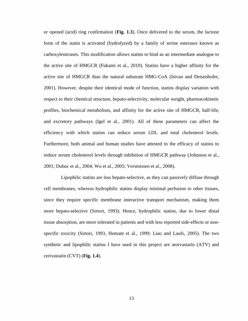
Figure 1.3: Chemical structure of the HMG-CoA moiety (dihydroxy heptanoic acid)
in statins. The statin HMG-CoA moiety can exist as either a 3-hydroxy lactone ring
(closed conformation) (A) or as an opened dihydroxy acid (opened conformation) (B).
15
Figure 1.4: Basic properties of atorvastatin (ATV) and cerivastatin (CTV) are
presented.
16
1.2.3 HMGCR pathway and embryonic development
Gene-expression analysis in chick tissues, Drosophila, zebrafish and murine
models demonstrate that HMGCR transcripts are enriched during embryonic
development (Alejandre et al., 1981; Gertler et al., 1988; Thisse and Thisse, 2004;
Brewer et al., 1993) reflecting a developmental requirement for products of HMGCR-
mediated metabolism. However, the specific developmental processes that require
mevalonate-derived products have only been explored recently. Toxicology experiments
to assess the possible teratogenicity of statins, using rats and rabbits, have reported that at
high doses, statins could induce developmental and maternal toxicity, affecting
parameters such as body weight and levels of food consumption (Dostal et al., 1994).
Preliminary experiments in pregnant rats showed that exposure to high dose mevinolin (a
fungal-derived/natural inhibitor of HMGCR), resulted in fetuses with malformed
vertebrae and ribs, along with failure of the abdominal wall to close resulting in
protrusion of the stomach and intestine to the outside (gastroschisis) (Minsker et al.,
1983).
In subsequent studies, targeted disruption of the HMGCR pathway in mice was
achieved through sequence replacement gene-targeting technology in order to prevent
translation of the entire carboxyl-terminus of the enzyme (Ohashi et al., 2003). The
Hmgcr–/– mice experienced embryonic lethality prior to reaching the implantation stage
(Ohashi et al., 2003). These results highlight the crucial function that HMGCR-mediated
metabolism and downstream signalling events play during mammalian development.
More recent work in Drosophila and zebrafish suggested that deficiencies in
HMGCR activity during development were implicated in prenylation-dependent germ
17
cell migration delays and misguidance, hence affecting developmental patterns (Van
Doren et al., 1998; Santos and Lehmann, 2004; Deshpande and Schedl, 2005). Studies in
Drosophila showed that down-regulation of the HMGCR pathway induced congenital
heart defects (Yi et al., 2006). Consistently, a point mutation was identified at base-pair
(bp) position 1575 of hmgcrb (G to A transition), the maternally derived hmgcr paralog
in zebrafish, resulting in an amino acid change from Glycine (Gly) to Aspartic acid (Asp)
at codon 497 (D’amico et al., 2007). In these embryos, loss of hmgcrb function was
associated with defective myocardial epithelial migration and fusion and defective heart-
tube formation and pericardial edema. This was attributed to impaired prenylation of
target proteins that would otherwise activate Rho-dependent signalling (D’amico et al.,
2007). Moreover, the hmgcrb–/– zebrafish embryos did not survive (D’amico et al.,
2007).
However, the entire suite of developmental processes that require adequate
amounts of mevalonate-derived metabolites are poorly understood by virtue of the
mortalities associated with the null mutations of the HMGCR gene.
An important avenue that needed to be further explored, hence a motivation for
my PhD thesis, was whether transient or partial reductions in the HMGCR activity,
through a reverse genetics approach or by exposure to specific pharmacological
inhibitors, could affect additional developmental processes, in particular CNS circulation
and angiogenesis. My particular focus was cerebral-vascular development. The reason for
this focus was that preliminary drug-screening experiments showed that statin treatment
of zebrafish embryos gave rise to a distinct CNS hemorrhage phenotype, which warranted
further studies.
18
1.2.4 The HMGCR pathway and angiogenesis
Angiogenesis is a complex physiological process by which new capillary
networks are established from pre-existing ones through a series of morphogenic events
involving differentiation, proliferation, migration, and maturation of endothelial cells
(ECs). It is followed by formation of cell-to-cell junctions through adhesion molecules
and formation of network-like structures, lumenisation of vessels and deposition of a new
basement membrane (Adams and Alitalo, 2007).
After undergoing proliferation, the endothelial tip cells, which are analogous to
neuronal growth cones, extend and retract their long filopodia-like processes in a
dynamic protrusive fashion to exert a pulling force and to form sprouts into adjacent
tissues, as well as to form connections with other vessels (Zelzer and Shilo, 2000). Earlier
evidence suggested that these filopodia extend and protrude to navigate the path of
growth in proportion with the relative abundance of guidance cues (Benjamin et al.,
1999). Subsequent studies have concluded that the activation of endothelial tip cells as
well as the protrusion of filopodia is induced by over-expression of vascular-endothelial
growth factor (VEGF). This was evidenced by the fact that inhibition of VEGF-signalling
led to loss of sprouting which was supported by in vitro analysis (Gerhardt et al., 2003).
Further evidence in support of a requirement for VEGF gradients guiding endothelial tip
migration was documented in vertebrates, including zebrafish (Gerhardt et al., 2003;
Habeck et al., 2002).
Angiogenic mechanisms are significantly up-regulated during embryonic
development (Breier et al., 1992), wound healing (Wu et al., 2007), as well as
regenerative processes (Bayliss et al., 2006). However, aberrant angiogenesis has also
19
been implicated in many diverse pathological conditions including tumor metastasis
(Strieter et al., 2006), atherosclerosis (Sluimer and Daemen, 2009), diabetic retinopathy
(Crawford et al., 2009), chronic inflammation (Costa et al., 2007), chronic kidney disease
(Futrakul et al., 2008), and rheumatoid arthritis (Paleolog, 2002).
Variations in HMGCR activity are shown to exert effects on angiogenesis, as
suggested by early in vitro and in vivo studies. Evidence obtained in vitro demonstrated
that high dose statin exposure induced anti-angiogenic effects by provoking EC
apoptosis, delaying EC migration, reducing endothelial VEGF2 release and decreasing
the total VEGF2-receptor levels in primary human adult dermal microvascular ECs
(HMVECs) (Weiss et al., 2002). These anti-angiogenic effects were attributed to
curtailment in prenyl biosynthesis, which are metabolites required for post-translational
modification of Rho GTPases (Weiss et al., 2002). Similarly, hydrophobic statins were
shown to induce apoptosis in rat pulmonary vein ECs (PVECs) due, in part, to mis-
localisation of RhoA to the plasma membrane (Kaneta et al., 2003). Likewise, the
inhibition of geranylgeranylation and RhoA-mediated pathways is linked with apoptosis
in human-derived ECs (Hippenstiel et al., 2002). More recently, it was shown that statin
treatment of human liver sinusoidal ECs (HLSECs) resulted in apoptosis through
inhibition of isoprenoid-dependent signalling pathways (Acquevella et al., 2010).
HMGCR inhibitors were also shown to induce apoptosis in vascular smooth muscle cells
(mural cells) in culture, which otherwise contribute to the elasticity and physical integrity
of blood-vessels and regulation of blood flow (Guijarro et al., 1999). This suggests that
HMGCR inhibitors could affect vascular smooth muscle cell interactions with ECs, hence
raising the possibility that statins may interfere with vascular stabilisation in vivo.
20
The anti-angiogenic effects of statins were further substantiated through in vivo
observations. At high doses, statin treatment inhibited capillary growth in chick
chorioallantoic membranes and mouse corneas (Park et al., 2002). More substantial in
vivo evidence was reported in studies using zebrafish, where a high dose statin exposure
(10 μM) resulted in incomplete or defective anterior to posterior sprouting/migration of
intersegmental vessels (ISV) from the dorsal aorta (DA), thereby severely restricting
circulation in the developing organism (Choi et al., 2011). Overall, the endothelial-
specific consequences of statin exposure are shown to be due to their cholesterol-
independent or pleiotropic effects, through reduction of prenyl biosynthesis. Collectively,
these studies support an anti-angiogenic effect associated with the inhibition of HMGCR
function, with potential therapeutic implications. Of clinical relevance is the fact that
preliminary studies exploiting the anti-angiogenic properties of statins have provided
encouraging data regarding their efficacy to decrease tumor vascularisation, volume, and
mass (Hindler et al., 2006; Wang et al., 2010).
However, an outstanding question that will be addressed in this thesis was
whether products of HMGCR-mediated metabolism are required for cerebral-vascular
development and stabilisation, and, if so, through what mechanisms? Another question
that necessitated inquiry was the pathophysiological consequences of statin-induced
interference with angiogenesis and vascular stabilisation during embryonic development.
1.2.5 The HMGCR pathway and stroke
By virtue of the association between high serum cholesterol levels and the
pathogenesis of coronary artery disease (Hutter et al., 2004), statins were considered ideal
21
to ameliorate the risk of stroke. Nevertheless, more recent evidence does not support a
strong relationship between the risk of stroke and serum cholesterol levels. Yet, studies
have attested to the effectiveness of statins in reducing the risk of acute ischemic
complications and atherosclerosis, through their effectiveness to reduce the serum
cholesterol levels (Borghi et al., 2000; Elahi et al., 2008). The efficacy of statins has also
been attributed to their non-cholesterol dependent or pleiotropic effects that lead to
plaque stabilisation, reduction of reactive oxygen species (ROS), and enhanced
endothelial function by increased nitric oxide (NO) synthesis (Vaughan et al., 1999).
Furthermore, a recent study uncovered a GT single nucleotide polymorphism
(SNP) in the HMGCR gene, which, based upon pyrosequencing data derived from more
than 23,000 participants, was found to be associated with an elevated risk for stroke,
which was independent of high blood pressure (Freitas et al., 2010).
However, of direct relevance and importance to my thesis are the conclusions of
a large-scale, randomised clinical study conducted by The Stroke Prevention by
Aggressive Reduction in Cholesterol Levels (SPARCL) Investigators. The results
suggested that while statin treatment significantly attenuated the risk of ischemic stroke
in patients, there was a slight, but significant risk for intracerebral hemorrhage (ICH) in
these same individuals (Amarenco et al., 2006). Although the study was aimed at
addressing whether statins could reduce the risk of stroke in individuals with prior history
of stroke or transient ischemic attack, the unexpected result that statins are associated
with ICH has raised tremendous interest as it has paved the way for follow-up clinical
studies most of which were based on meta-analysis of randomised controlled trials.
22
Subsequent studies further evaluated the association between statin use and the
risk for ICH, and led to contentious results with some reporting no observable risk for
ICH (Hackam et al., 2012; Spence, 2012; McKinney and Kostis, 2012) to other studies
suggesting complete avoidance of statins in order to mitigate the risk for ICH (Arboix et
al., 2010; Westover et al., 2011). Of clinical importance is whether this unanticipated
adverse outcome of ICH, as suggested by at least some of the aforementioned studies,
could be outweighed by the potential cardiovascular benefits of statins. There has been no
reproducible etiological or angio-structural analysis in the context of animal models to
further investigate this possibility or the mechanism(s) of statin-induced ICH. However,
some of these clinical studies led us to the hypothesis that statins may interfere with the
integrity of vascular endothelium, thus resulting in hemorrhage. This could be especially
relevant in individuals with a weaker vascular endothelium, such as those with high blood
pressure, in a developing organism or in individuals with genetic vascular dysplasia
resulting from mutations in cerebral cavernous malformation (CCM) genes and those
with previous history of stroke.
In sharp contrast with the mounting clinical evidence, it has been reported, in at
least one study that in mice with a genetic predisposition for cerebral-vascular
permeability defect resulting from a heterozygous mutation of CCM2, treatment with
statins effectively restores the endothelial barrier function by inhibiting Rho GTPase
activation (Whitehead et al., 2009). These findings raised the possibility for statin
treatment as a therapeutic for CCM-like pathologies and to prevent cerebral hemorrhage
(Whitehead et al., 2009).
23
Coincidently, these clinical and animal studies, which highlighted a link between
the HMGCR pathway and cerebral-vascular integrity, emerged in parallel with the
progress of my PhD project, which was geared towards dissecting the mechanisms of
statin-induced ICH in zebrafish. Hence, I reasoned that the development of a robust
animal model (zebrafish) for statin-induced cerebral-vascular disorders may assist to
address the discrepancies highlighted by clinical and animal studies. There exists
considerable overlap between the genes and mechanisms implicated in vascular
morphogenesis in zebrafish and mammals (Gore et al., 2012). Therefore, it is my
prediction that the findings of this project can contribute to our understanding of some of
the molecular and cellular mechanisms leading to stroke in humans, as it highlights a
metabolic contribution to cerebral-vascular stabilisation.
1.3. Cerebral-vascular stabilisation during development
Vascular stabilisation is a crucial process required to establish and maintain a
barrier function so as to ensure the integrity of nascent vessels that are formed through
either angiogenesis or vasculogenesis (Mizuguchi et al., 2004; Loeys et al., 2005). Once
the newly formed vessels stop the process of re-modelling, attain their adequate size and
form vascular tubes, they must be stabilised to prevent rupture and subsequent
hemorrhage into the interstitial spaces. The establishment of a functional vasculature
requires cessation of angiogenesis, followed by ECs entering a quiescent state.
Timely and rapid stabilisation of new vessels, through establishment of
endothelial cell-to-cell contacts and interactions of the endothelium with support (mural)
cells, is particularly crucial during development, since these vessels are particularly
24
fragile and prone to hemorrhage (Jain, 2003). Such is also the case during cancer
metastasis, as the nascent vasculature supplying oxygen and nutrients to the newly
formed tumors tends be highly permeable, since these vessels lack adequate investment
by mural cells, making them fragile and disorganised (Abramsson et al., 2002).
Moreover, several genetic defects underlying disruption of cell-cell junctions and
detachment of ECs from the vessel wall have been identified and characterised in mice
and, more recently, in zebrafish (Clatterbuck et al., 2001; Bergers and Song, 2005; Butler
et al., 2011).
Although the mechanisms and signalling pathways regulating angiogenesis and
vasculogenesis have been examined in great detail through both in vitro and in vivo
experiments, the endogenous signalling pathways involved in developmental vascular
stabilisation remain underexplored (Adams and Alitalo, 2007). Endothelial cell-cell
contacts are established and maintained by trans-membrane adhesion molecules that are
anchored to cytoskeletal elements. The intra-endothelial cell communication is mediated
through gap junctions, whereas the adhesion molecules that confer structural support and
contribute to vascular stability include adherens junctions (AJ) and tight junctions (TJ)
(Dejana et al., 1995; Gumbiner et al., 2000; Tsukita et al., 2001). Nonetheless, the genetic
and mechanistic bases that regulate junction assembly for a functional cerebral-vascular
system, particularly in the context of zebrafish development, are not yet elucidated.
Gene expression and functional analysis have led to the identification and
characterisation of several EC-specific and trans-membrane adhesion molecules involved
in the maintenance of endothelial barrier integrity. These include vascular endothelial
(VE)-cadherin, claudin-5, filamin-A, and β-catenin (Feng et al., 2006; Gavard and
25
Gutkind, 2008; Das et al., 2011; Glading and Ginsburg, 2010). Targeted inactivation of
VE-cadherin and truncation of the β-catenin-binding cytosolic domain of VE-cadherin
induces EC-specific apoptosis, along with defective remodelling and maturation of the
vasculature and early lethality(Carmeliet et al., 1999). Endothelial-specific conditional
deletion of β-catenin, a protein interacting with the cytoplasmic tail of VE-cadherin, is
associated with altered vascular patterning (Caveda et al., 1996), defective lumenisation
and frequent hemorrhage in vivo, as well as decreased intricacy of endothelial cell-cell
junctions in vitro, based on studies on mice(Cattelino et al., 2003). Similarly, a vascular-
specific defect associated with morpholino-induced partial and transient loss of VE-
cadherin was developed in the zebrafish (Montero-Balaguer et al., 2009). Morpholino
oligonucleotide-induced depletion of VE-cadherin in zebrafish resulted in vascular
instability, defective lumenisation, as well as frequent cerebral hemorrhage,
predominantly at 52 hpf (hour post-fertilisation) (Montero-Balaguer et al., 2009). More
complete depletion of VE-cadherin, through higher doses of morpholinos, resulted in
more profound defects including total inhibition of EC sprouting activity and mortality
(Montero-Balaguer et al., 2009). Functional analysis of claudin-5, another trans-
membrane tight junction protein, in both murine and zebrafish models, has highlighted a
functional role for this protein in maintaining the integrity of the endothelial blood–brain
barrier function, thus assigning for it a role in brain morphogenesis (Xie et al., 2010).
In addition to endothelial cell-cell contacts, vascular integrity is also maintained
through ECs interacting with perivascular mural/support cells. These mural cells include
smooth muscle cells, astrocytes, neuroepithelial cells, and pericytes (McCarty et al.,
2002). Whereas ECs establish a monolayer and line the inner surface of a vessel, mural
26
cells wrap around the outside of the vascular tube and assist in regulating blood flow. The
components of the signalling pathways involved in recruitment of mural cells to the
vascular endothelium have been identified and characterised (Kumar and Owens, 2003;
Larsson et al., 2001; Ballabh, 2009). Such close interactions between the endothelium
and mural cells are essential for the development and maintenance of the blood-brain
barrier and regulation of blood flow by controlling the contractility/elasticity of the
vessels. At the ultra-structural level, disruption of the focal contact between mural cells
and the endothelium is associated with cerebral hemorrhage (McCarty et al., 2002;
Ballabh, 2009).
Several loss-of-function mutations associated with the disruption of endothelial-
mural cell junction proteins have been reported in humans. These vascular-specific
disorders, which follow an autosomal-dominant pattern of inheritance, are collectively
referred to as cerebral cavernous malformations (CCMs). The CCM phenotype is
characterised by dilated, leaky central nervous system (CNS) blood-vessels lacking
adequate coverage by smooth muscle cells, and frequent hemorrhage into the brain
parenchyma (Clatterbuck et al., 2001). Thus far, three genes, which participate in a
signalling pathway involving Rho GTPase signalling, namely CCM1 (KRIT1), CCM2
(MGC4607) and CCM3 (PDCD10), have been identified (Sahoo et al., 1999; Denier et
al., 2004; Bergametti et al., 2005).
However the mechanism by which CCM gene products interact with Rho
GTPase signalling and how reductions in the expression of these structurally diverse gene
products can contribute to the pathogenesis of the CCM phenotype is still a subject of
intense research. Many studies have attempted to functionally characterise these genes
27
using murine models (Plummer et al., 2004; Whitehead et al., 2009; Louvi et al., 2011)
and, more recently, developing zebrafish (Gore et al., 2008; Yoruk et al., 2012). These
studies have greatly contributed to our understanding of the etiology and angio-structural
basis underpinning CCM pathogenesis.
One unique advantage of the zebrafish, which is particularly manifest during
their development, is their optical transparency which facilitates easy visualisation of
cerebral hemorrhage through the use of bright-field microscopy (Fig. 1.5). Another
advantage of the zebrafish is that it is highly prolific. As a result, the amenability of this
fish to large-scale chemical mutagenesis screening has allowed for the isolation of
numerous mutations that impact on the cardiovascular system and, as it relates to my
project, identification of mutations affecting developmental cerebral-vascular
stabilisation (Stainier et al., 1996). In this large-scale mutagenesis screening of the
zebrafish genome, the mutant bubble-head (bbh) was identified, which exhibited cranial
hemorrhage and ventricular edema, hence illustrative of weakened cerebral-vessel walls
(Stainier et al., 1996). Other mutations affecting cerebral-vascular stabilisation, the
viability of ECs, and caudal circulation were also identified in this study. These
mutations were referred to as gridlock (gdl), m413 and m521, and migraine (mig). All of
these genetic defects were associated with frequent hemorrhage and various degrees of
vascular disintegration at the same developmental stage in zebrafish (Stainier et al.,
1996). Of these lines, the gridlock (gdl) mutation was later characterised (Weinstein et
al., 1995; Zhong et al., 2000; Peterson et al., 2004). However, until recently, neither
genetic nor functional characterisations of the other mutations associated with ICH have
been reported.
28
Characterisation of the vascular-specific bubblehead (bbh) mutation was carried
out through positional cloning, in situ hybridisation, reverse genetics and high resolution
imaging (Liu et al., 2007). Positional cloning of bbh revealed a hypomorphic mutation in
the βPix gene, which otherwise encodes a p21-activated kinase (Pak)-interacting
exchange factor (Pix). βPix is a guanine exchange factor (GEF) which functions to
stimulate the exchange of Rho GTPase-bound GDP with cytosolic GTP thus activating
the Rho GTPases, Rac and CDC42 (Bagrodia et al., 1998; Manser et al., 1998). A single-
nucleotide mutation (G A) results in alternative splicing and exclusion of exon 14,
thus introducing a premature stop codon that gives rise to a truncated βPix protein. The
mutation results in an ICH phenotype, accompanied by hydrocephalus in zebrafish
embryos at 48 hpf. At the ultra-structural level, the hemorrhage arises due to defective
association between mural cells and ECs. Since βPix encodes a GEF that activates Rho
GTPase proteins by stimulating the exchange of GDP for GTP, it was speculated that a
mutation in the βPix would be associated with reduced levels of activated Rho GTPases
(Etienne-Manneville and Hall, 2002, Liu et al., 2007). Given that the Rho GTPase
signalling regulates a wide array of biological processes including mediation of
cytoskeletal organisation, cell motility, wound healing, apoptosis and immune function, it
would be expected that down-regulation of βPix expression would affect these
parameters.
29
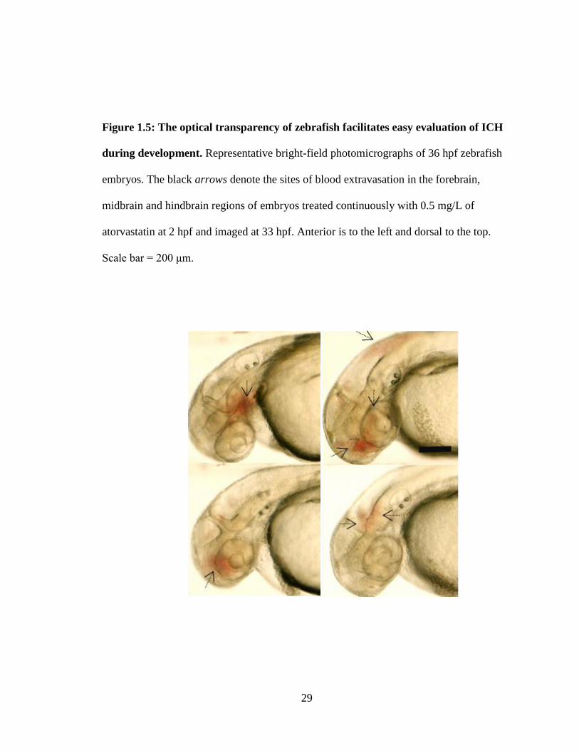
Figure 1.5: The optical transparency of zebrafish facilitates easy evaluation of ICH
during development. Representative bright-field photomicrographs of 36 hpf zebrafish
embryos. The black arrows denote the sites of blood extravasation in the forebrain,
midbrain and hindbrain regions of embryos treated continuously with 0.5 mg/L of
atorvastatin at 2 hpf and imaged at 33 hpf. Anterior is to the left and dorsal to the top.
Scale bar = 200 μm.
30
In parallel with the characterisation of the bbh mutation, a separate study
characterised a recessive zebrafish mutant termed redhead, which displayed a similar
ICH phenotype at the same developmental stage (Buchner et al., 2007). Interestingly, this
vascular defect was mapped to a mutation in p21-activated kinase 2a (pak2a), a member
of the p21-activated kinase gene family and a binding partner for βPix. Pak proteins are
serine/threonine kinases that act downstream of Rho GTPase signalling and are involved
in the transduction of this pathway (Hofmann et al., 2004).
In both redhead and bubblehead zebrafish embryos, these mutations were
mapped to genes that are involved in the activation and transduction of Rho GTPase-
mediated signalling pathways, which synergistically participate in the development of
vascular networks (Gore et al., 2008). In both mutant strains, the structural basis for the
hemorrhage phenotype stems from defective stabilisation and maturation of nascent
vessels (Buchner et al., 2007; Liu et al., 2007).
Of noteworthy relevance to my thesis project is that the activation of Rho
GTPase signalling is mediated, in part by a mevalonate-derived metabolite,
geranylgeranyl-pyrophosphate (GGPP), a 20-carbon lipid molecule (Bishop and Hall,
2000). GGPP serves as an end-product for the lipidation and subcellular localisation of
Rho GTPases to the plasma membrane. This process is critical for the activation and
function of CAAX-proteins (Bishop and Hall, 2000) (Fig. 1.6). This is evidenced by the
fact that depleted GGPP levels reduce the GTP-binding capacity of Rho GTPases,
rendering them cytosolic and inactive, which would target them for degradation in the
cytoplasm (Hirai et al., 1997). On this premise, one can deduce that inhibition of
HMGCR activity would reduce Rho GTPase signalling, thus leading to an ICH
31
phenotype. This assertion is contrary to evidence from murine models where down-
regulation of Rho GTPase signalling, by statin treatment, is shown to reverse/prevent the
ICH phenotype in mice. Evidently, a clear discrepancy exists between the two models
(zebrafish and mice) in terms of requirements for Rho signalling in cerebral-vascular
stabilisation.
1.4. Utility of zebrafish as a model for the etiology and pathophysiology of ICH
The zebrafish continues to gain recognition as an excellent vertebrate model for
the study of vascular development. The mechanisms and gene signalling pathways
mediating the formation and patterning of vessels were found to be similar to those
documented in other vertebrates (Gore et al., 2012). The optical transparency and external
development of embryos enables easy visualisation of active circulation and organ
development and makes them amenable to chemical screening and genetic manipulation.
Blood-vessel morphogenesis, immune response and thrombosis can be assayed
in real-time using stable transgenic embryos expressing fluorescently labeled blood-
vessels, macrophages, leukocytes, erythrocytes, and thrombocytes. Vascular permeability
can be assessed using high-resolution confocal microscopy and micro-angiography.
Protein function can be determined rapidly and efficiently through microinjection of
morpholino oligonucleotides which reduce gene expression in a dose-dependent fashion.
All of these features make the zebrafish an ideal system to study the morphogenesis of
cerebral-vascular system.
32
1.5. Research Objectives
My PhD thesis focuses on expanding our present knowledge regarding the
signalling pathways contributing to developmental cerebral-vascular stabilisation in
vertebrates. Cerebral-vascular disorders including ICH are associated with high mortality
and morbidity (Counsell et al., 1995; Qureshi et al., 2005). Even though considerable
research has been conducted in the field of vascular development, the genetic and
mechanistic underpinnings behind ICH pathogenesis are still not well characterised. The
results of several randomised clinical studies have suggested a significant risk for ICH in
patients taking statin drugs. However, these studies are limited in that no follow-up
animal studies have to this point been conducted to independently verify and dissect the
mechanism(s) involved. Furthermore, a number of convincing studies in zebrafish have
suggested that inhibition of Rho GTPase signalling can potentiate ICH in developing
vertebrates. This information, coupled with in vitro evidence that HMGCR activity is
required for both endothelial and mural cell viability, prompted me to characterise the
function of HMGCR in zebrafish with a particular interest in cerebral-vascular
development. I hypothesise that HMGCR regulates cerebral-vascular stabilisation
through GGPP-dependent signalling. As such my results will have clinical relevance in
terms of addressing part of the etiology and pathophysiology of human vascular
instability disorders, particularly in the case of spontaneous ICH. My research objectives
are:
Objective 1:
Establish whether HMGCR activity is required for developmental cerebral-vascular
stabilisation.

33
Objective 2:
Determine the mechanism(s) whereby HMGCR activity regulates cerebral-vascular
stabilisation.
Objective 3:
Characterise the pathophysiological and neurological processes underlying ICH
pathogenesis.
34
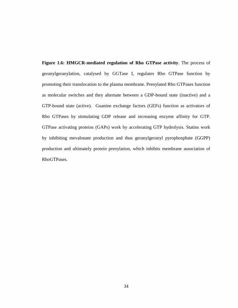
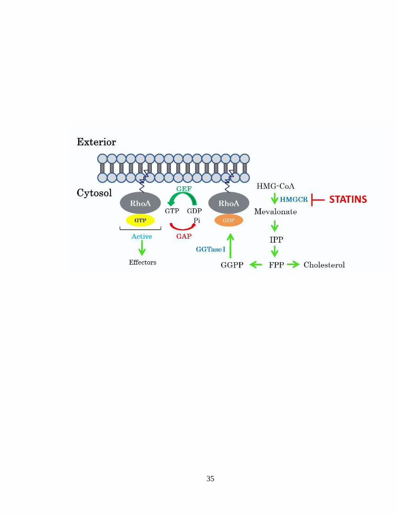
Figure 1.6: HMGCR-mediated regulation of Rho GTPase activity. The process of
geranylgeranylation, catalysed by GGTase I, regulates Rho GTPase function by
promoting their translocation to the plasma membrane. Prenylated Rho GTPases function
as molecular switches and they alternate between a GDP-bound state (inactive) and a
GTP-bound state (active). Guanine exchange factors (GEFs) function as activators of
Rho GTPases by stimulating GDP release and increasing enzyme affinity for GTP.
GTPase activating proteins (GAPs) work by accelerating GTP hydrolysis. Statins work
by inhibiting mevalonate production and thus geranylgeranyl pyrophosphate (GGPP)
production and ultimately protein prenylation, which inhibits membrane association of
RhoGTPases.
35
36
Chapter 2
The 3-hydroxy-3-methylglutaryl-CoA
reductase (HMGCR) pathway regulates
developmental cerebral-vascular stabilisation
by a prenylation-dependent signalling
pathway
The data presented in this chapter have been published in the following manuscript.
However, supplementary figures and textual alterations and additional information have
been added to this chapter:
Eisa-Beygi, S., Hatch, G., Noble, S., Ekker, M., Moon, T.W. (2013). The 3-
hydroxy-3-methylglutaryl-CoA reductase (HMGCR) pathway regulates
developmental cerebral-vascular stability via prenylation-dependent signalling
pathway. Dev. Biol. 373, 258-266.
37
Author contributions:
Mr. Gary Hatch replicated the drug-exposure and rescue experiments to
ensure consistency of results. Ms. Sandra Noble was instrumental in training
me for the use of confocal microscopy and helping me prepare and process
embryos for imaging. Dr. Thomas W. Moon and Dr. Marc Ekker helped me
design the experiments and assisted me in writing and revising this chapter
to ensure that the content is scientifically sound and the style is in
accordance with the FGPS requirements. I am profoundly grateful for the
assistance of the aforementioned individuals, without whom this chapter
would not have been completed.
Shahram Eisa-Beygi
April 20, 2013
38
2.1 Abstract
Intracerebral hemorrhage (ICH) is a debilitating form of stroke, with the highest
mortality rate of all stroke subtypes and, often, precipitating irreversible neurological
deterioration. Although recent studies have gained insight into the etiology of the disease,
many of the causative genes and mechanisms implicated in developmental cerebral-
vascular malformations underlying ICH pathogenesis are unknown. Recent evidence
derived from in vitro and in vivo studies in murine models have shown inhibition of the
3-hydroxy-3-methylglutaryl-CoA reductase (HMGCR) pathway to be effective in
stabilizing cranial vessels. Through a combination of pharmacological and genetic
approaches to specifically inhibit the HMGCR pathway in zebrafish (Danio rerio), we
demonstrate contrary to the work in murine models, a requirement for the HMGCR-
mediated metabolic pathway in developmental vascular stabilisation within the head.
Here we report that inhibition of HMGCR function in embryonic zebrafish disrupts
cerebral-vascular stability, resulting in progressive dilation of blood vessels, followed by
vessel rupture and hemorrhage into the brain parenchyma, mimicking cerebral cavernous
malformation (CCM)-like lesions in humans and murine models. These vascular defects
are rescued by prior exogenous supplementation with geranylgeranyl pyrophosphate
(GGPP), a 20-carbon metabolite of the HMGCR pathway, required for the membrane
localisation and activation of Rho GTPases. Consistent with this observation,
morpholino-induced depletion of the β-subunit of geranylgeranyltransferase I (GGTase
I), an enzyme that facilitates the post-translational transfer of the GGPP moiety to the C-
terminus of Rho family of GTPases, mimics the cerebral hemorrhage-induced by the
39
pharmacological and genetic ablation of HMGCR. In embryos with cerebral hemorrhage,
the endothelial-specific expression of cdc42, a highly-conserved Rho GTPase involved in
the regulation of vascular permeability, was significantly attenuated. Taken together, our
data reveal a metabolic contribution to the stabilisation of nascent cranial vessels during
embryogenesis, requiring protein geranylgeranylation acting downstream of the HMGCR
enzyme within the mevalonate pathway.
2.2 Introduction
3-Hydroxy-3-methylglutaryl-CoA reductase (HMGCR) is an endoplasmic
reticulum (ER)-bound enzyme that catalyzes the rate-limiting step within the mevalonate
pathway that converts acetyl-CoA derived HMGCoA to mevalonate, giving rise to
mevalonate-derived molecules, including cholesterol and isoprenoids (Goldstein and
Brown, 1990; see Chapter 1). A complex feedback mechanism, involving both steroidal
and non-steroidal mediated pathways, is involved in the regulation of HMGCR activity
through transcription, translation, phosphorylation, and degradation of the enzyme
(Brown and Goldstein, 1997; Nakanishi et al., 1988, Omkumar et al, 1994; DeBose-
Boyd, 2008). Statins, competitive inhibitors of HMGCR, are pharmaceuticals that bind
part of the HMG-binding residues of the enzyme and inhibit its activity through
competitive inhibition (Istvan and Deisenhofer, 2001). In addition to reducing de novo
cholesterol synthesis and total plasma LDL cholesterol levels, statins also curtail the
biosynthesis of other lipids, namely isoprenoids, which otherwise serve as essential lipid
attachments for the Rho family of GTPases (Bishop and Hall, 2000).
40
As such, an efficient way to elucidate HMGCR gene function during
development is achieved through the use of statin molecules for which the biochemical
mode of action is well characterised. Reduced HMGCR activities during development are
implicated in prenylation-dependent germ cell migration delay and misguidance in both
Drosophila and zebrafish (Danio rerio) models (Santos and Lehmann, 2004; Thorpe et
al., 2004). A point mutation identified at position 1575 of hmgcrb, the maternally-
expressed hmgcr paralog in zebrafish, results in defective heart-tube formation due to
impaired post-translational prenylation of small GTPases (D’amico et al., 2007). In
contrast, targeted disruption of hmgcr in mice results in early embryonic lethality which
is suggestive of the crucial function that HMGCR-mediated metabolism and downstream
signalling events play during mammalian development (Ohashi et al., 2003). Presently,
the developmental processes that are dependent upon HMGCR-derived products have not
been thoroughly identified.
Pharmacological inhibition of the HMGCR pathway has been shown to exert
both angiostatic and angiogenic effects in vitro and in vivo (Lu et al., 2004; Khaidakov,
2009; Weis, 2002; Kaneta, 2003; Li et al., 2002; Acquavella, 2009; Wang et al., 2010;
Choi et al., 2011). An outstanding question that needed to be addressed was whether
perturbations of the HMGCR pathway would disrupt the stabilisation of nascent vessels
and overall vascular morphogenesis in vivo, and, if so, what would be the
pathophysiological consequences of interference with these processes during a period of
rapid angiogenesis.
Zebrafish have recently gained recognition as a suitable model to dissect the
etiology of cerebral cavernous malformation (CCM) (Kleaveland, 2009; Hogan et al.,
41
2008; Yoruk et al., 2012). Moreover, recent work in zebrafish demonstrated that proteins
involved in the activation and transduction of Rho GTPase-mediated signalling pathways
synergistically participate in the development of the vascular network in zebrafish.
Deficiencies within this pathway impair the stabilisation and maturation of nascent
vessels by preventing the association of mural cells with endothelial cells, giving rise to
multiple hemorrhages in the brain (Buchner et al., 2007; Liu et al., 2007; Gore et al.,
2008), along with more pronounced defects in vascular morphogenesis (Epting et al.,
2010). It is known that the mevalonate-derived metabolite, geranylgeranyl-pyrophosphate
(GGPP) serves as an end-product for the lipidation and subcellular localisation of Rho
GTPases to the plasma membrane, which is critical for the activation and function of
these Rho GTPases (Bishop and Hall, 2000). Hence, depleted GGPP levels reduce the
GTP-binding capacity of Rho GTPases, rendering them cytosolic and inactive. Although
the Rho GTPase pathway has been implicated in CCM pathology, there remains a major
discrepancy between the mechanisms in mouse and zebrafish. More specifically, recent
studies in mice show that global inhibition of Rho GTPase prenylation through statin-
mediated inhibition of HMGCR within the mevalonate pathway restores endothelial
barrier integrity, thus reversing the vascular permeability dysfunction in mice that are
genetically predisposed to CCM (Whitehead et al., 2009). Consistent with this
observation, transcript levels of the Rho GTPases, RhoA, Rac and cdc42, were
significantly elevated in lesions of a mouse model of CCM (Louvi et al., 2011).
Hence, in mice, statin-mediated inhibition of Rho GTPase signalling, through
decreased GGPP biosynthesis, is reported to be an effective treatment to attenuate
vascular permeability (Li and Whitehead, 2010), whereas in zebrafish, mutation and
42
morpholino-induced loss of function of proteins that participate in the activation and
transduction of Rho GTPase signalling heightens vascular permeability (Buchner et al.,
2007; Liu et al., 2007). To what shall we attribute these divergent outcomes even though
the mechanism of statin inhibition is the same? One of the major motives behind this
project was to address this inconsistency.
Therefore, to investigate this discrepancy, we subjected developing zebrafish to
two structurally different statin molecules (atorvastatin or cerivastatin) and two
morpholino oligonucleotides (splice MO or ATG MO) with non-overlapping sequences
to inhibit HMGCR function. Our pharmacological and genetic approach consistently
resulted in grossly dilated vessels, followed by progressive loss of vascular stability in the
brain at specific developmental stages during which nascent cranial vessels are more
prone to rupture. Prior supplementation with exogenous mevalonate or GGPP, two of the
metabolites downstream of the HMGCR enzyme, are sufficient to rescue the cerebral
hemorrhage phenotype. Furthermore, morpholino-mediated specific disruption of the
GGTase I-mediated prenylation pathway mimics the cerebral hemorrhages attributed to
the inhibition of the HMGCR enzyme. Interestingly, in embryos with cerebral
hemorrhage, the vascular-specific expression of cdc42, a Rho GTPase implicated in the
mediation of endothelial barrier function (Kouklis et al., 2003; Broman et al., 2006;
Ramchandran et al., 2008; Spindler et al., 2010) and highly enriched on the vacuole
membranes (Eitzen et al., 2001; Isgandarova et al., 2007), was noticeably reduced in the
head and the trunk vasculature, as early as 24 hpf. Our results highlight a requirement for
HMGCR activities, through direct effects on GGPP biosynthesis, on the establishment of
cerebral-vascular stability during zebrafish development, a process likely mediated by
43
CAAX proteins that require geranylgeranylation for their subcellular localisation and
activation.
2.3 Materials and Methods
2.3.1 Zebrafish husbandry and transgenic lines
Adult wild-type and transgenic zebrafish were maintained under a constant
temperature of 28°C and a 14 h light:10 h dark photoperiod in the University of Ottawa
Aquatic Care Facility. Embryos were obtained through natural breeding of adult zebrafish
and were kept at 28.5°C in embryo medium (5 mmol/L NaCl, 0.17 mmol/L KCl, 0.33
mmol/L CaCl2, 0.33 mmol/L MgSO4). The double transgenic Tg(fli1:EGFP);(gata-
1:DsRed) zebrafish line was kindly provided by Dr. Beth Roman (University of
Pittsburgh, Pennsylvania). The Tg(fli1:EGFP-cdc42wt)y48
was kindly provided by Dr.
Brant Weinstein (National Institute of Child Health and Human Development,
Maryland). All experiments were carried out in accordance with a protocol approved by
the University of Ottawa Protocol Review Committee and conform to the published
guidelines of the Canadian Council on Animal Care for the use of animals in research and
teaching.
2.3.2 Drug treatment and metabolite rescue experiments
For pharmacological inhibition of HMGCR, zebrafish embryos (1-2 hours post
fertilisation; hpf) were treated with Atorvastatin (ATV; Pfizer Inc., Connecticut USA) or
Cerivastatin (CVT; Sequoia Research Products Ltd. Pangbourne, UK) in a single-
exposure manner in embryo medium. ATV was dissolved in DMSO, whereas CVT was
dissolved in sterile/distilled water. All solutions were aliquoted and kept at -20°C until
44
used. Embryos for the dose-response experiments were placed in final concentrations of
0.3, 0.5 or 1 mg/L ATV; CVT was administered at 0.15 mg/L. For mevalonate rescue
experiments, similar numbers of embryos were placed in embryo medium containing 0.5
mg/L ATV and 1, 1.6, 3.2 or 4.4 μmol/L mevalonic acid (Sigma-Aldrich) or DMSO as
vehicle control. Each treatment group consisted of more than 70 embryos and all
exposure experiments were repeated at least 8 times. GGPP rescue experiments were
performed by administering 0.5 mg/L ATV and 0.5, 2 or 4 mg/L GGPP (Sigma-Aldrich)
dissolved in DMSO/methanol or DMSO/methanol, as vehicle control.
2.3.3 Whole-mount o-Dianisidine (OD) staining and cryosectioning
Zebrafish embryos at various developmental stages were fixed in 4% PFA/1X
Phosphate Buffered Saline Tween-20 (PBST) overnight and subsequently stained for
hemoglobin using o-Dianisidine (OD), a sensitive marker of hemoglobin. Briefly,
unfixed embryos were dechorionated and stained with a solution of OD (0.6 mg/mL)
(Sigma-Aldrich) containing 0.01 M sodium acetate (pH 4.5), 0.65% hydrogen peroxide,
and 40% (v/v) ethanol in the dark for 15 min. Treated embryos were visualised using a
Nikon NBZ 1500 dissecting microscope and photographed with a Nikon DXM 1200 C
digital camera, and stored in 50% glycerol at 4°C. For cryosectioning, whole-mounted
embryos previously stained with OD were equilibrated to 30% sucrose/1X PBST
overnight. The next day, the samples were mounted in tissue freezing medium (Triangle
Biomedical Sciences). Transverse sections of ~10 μm thickness were cut on a Leica
CM1850 standard cryostat (Leica Microsystems) at -20 °C.
45
2.3.4 Confocal microscopy
For confocal microscopy, 48-52 hpf Tg(flk1:EGFP;gata1:dsRED) embryos
previously treated with ATV or DMSO were embedded in 1% low melting agarose in
embryo medium and tricaine mesylate (ethyl 3-aminobenzoate methanesulfonate; Sigma-
Aldrich). A rendered Z-stack, at ~80 micron thickness each, was taken with a Zeiss LSM
510 AxioImager.M1 confocal microscope using an Achroplan 40x/0.8 W objective with
an argon laser (488 nm) and a helium-neon laser (543 nm).
2.3.5 Morpholino design, resuspension and microinjection
A splice-modifying antisense morpholino oligonucleotide (MO) against hmgcrb
pre-mRNA (hmgcrb MO), a translation blocking MO targeting the AUG initiation site of
hmgcrb mRNA (AUG MO), a splice-modifying MO against pggt1b pre-mRNA (pggt1b
MO), and a standard control MO against a splice targeting the human β-globin pre-
mRNA (control MO), were obtained from GeneTools (Philomath, OR, USA). The
sequences for the morpholinos used were:
(5'-AACTGCATTCATAAACTCACCCAGT-3') (hmgcrb-splice MO);
(5'-GCCTGAAGAGACGCGACAGCATCAT 3') (hmgcrb-ATG MO);
(5'-CACGCGGTGTGTGGACTCACGGTCA-3') (pggt1b-splice MO); and
(5'-CCTCTTACCTCAGTTACAATTTATA-3') (control MO).
All MO solutions were diluted in Danieau buffer (58 mM NaCl, 0.7 mM KCl,
0.4 mM MgSO4, 0.6 mM Ca(NO3)2, 5.0 mM HEPES, pH 7.6) to a final concentration of
0.2 mM. The embryos were positioned in individual wells placed on a plate containing
46
1.0% agarose. The morpholino solution was then injected at quantities ranging from 0.5-
10 ng into 1 to 2 cell-stage embryos through the cell-yolk boundary.
2.3.6 Microangiography
Briefly, 50-56 hpf embryos were anesthetised in tricaine mesylate and injected
intra-vascularly with red fluorescent microspheres of 0.4 μm diameter (Thermo
Scientific) through the sinus venosus and/or common cardinal vein. Fluorescence
micrographs were acquired during a series of observations under the Nikon NBZ 1500
dissecting microscope, which was equipped with a standard dsRED filter set using a
Nikon DXM 1200C digital camera.
2.3.7 DAF-2DA Staining
DAF-2DA (4-amino-5-methylamino-2′-7′-difluoro-fluorescein diacetate;
Calbiochem) staining was performed as previously described (Lou et al., 2011), with
reduced incubation time. In brief, live embryos were incubated in 10 µM DAF-2DA (in
embryo medium, pH 7.0) and incubated for 5 h in the dark at 28.5°C, after which
fluorescence micrographs were acquired under the Nikon NBZ 1500 dissecting
microscope equipped with a standard GFP filter set, and photographed using a Nikon
DXM 1200C digital camera.
2.3.8 RNA extraction, RT-PCR and sequencing
Total RNA was extracted from a pool of embryos using TRIzol reagent
(Invitrogen). The RNA was DNase-treated using the Deoxyribonuclease kit (Invitrogen)
to minimise genomic DNA contamination. cDNA was synthesised from equal
concentrations of RNA using a 1st Strand cDNA Synthesis Kit and M-MLV RT
47
(Invitrogen) and used as a template in RT-PCR. PCR products were run on 1.75%
agarose gels. Band intensities were quantified using ImageJ software. The primers used
for PCR analysis were:
(5'-CAAACATGGGCTGGTTCAAG-3') (Elf1a forward) and
(5'-AGTGGTTACATTGGCAGGG-3') (Elf1a reverse).
The efficiency of the hmgcrb splice MO was assessed using primers flanking exon 3:
(5'-CCGTGACAATGTGCCTAATG-3') (hmgcrb forward) and
(5'-CAGATCAATCAGCAGCAGGA-3') (hmgcrb reverse).
The efficiency of the pggt1b splice MO was assessed using primers flanking exon 2:
(5'-TCTTCTGCCGGTCCATCC-3') (pggt1b forward) and
(5'-GCGGTAGTGAGCGGTGAT-3') (pggt1b reverse).
The PCR products were first separated by gel electrophoresis then excised from
the gel and purified using the Qiagen gel purification kit (Qiagen), according to the
manufacturer's instructions. Sequencing was performed to verify hmgcrb MO-induced
splicing using the CEQ-8000 Genetic Analysis System and the aforementioned set of
primers.
48
2.4 Results
2.4.1 Pharmacological inhibition of the HMGCR enzyme leads to cerebral-vascular
defects in developing zebrafish
Sequence alignment of the partial catalytic sites of the HMGCR proteins
revealed that the statin-binding residues (Istvan and Deisenhofer, 2001) are conserved in
both zebrafish HMGCR paralogs, which might suggest functional conservation between
mammals and fish. We first used the Tg(fli1:EGFP;Gata-1:dsRED) line for the
simultaneous visualisation of the vascular network and circulating erythrocytes during
development. Treatment of these embryos beginning at 1-2 hpf with 0.5 mg/L ATV in
embryo medium, in a single-exposure manner, did not affect the vascular patterning or
morphology at the 15-25 somite stage (19 hpf) (data not shown). However, by 28 hpf,
ATV treatment resulted in grossly dilated and diffusely shaped primitive cerebral vessels
in the forebrain and midbrain (Fig. 2.1A-E and Fig. 2.2A-D), in addition to the abnormal
confinement of stagnant dsRED-positive erythrocytes and an attenuated circulation in the
cerebral region (Fig. 2.1F and G). These observations are indicative of impaired vessel
wall function as the primary cause for the extravasation of erythrocytes at subsequent
stages of development. At the doses used, ATV-exposure did not induce defects in
angiogenesis, as the trunk vasculature and the formation of the segmental arteries were
unaffected in these embryos, reflecting the cerebral-vascular-specific nature of these
effects at the doses of statins used (Fig. 2.3A-D).
By 33 hpf, cerebral hemorrhage was evident in 46% of embryos (137/298). The
locality and severity of the phenotype were variable in the brain, reflecting the multifocal
nature of the hemorrhages (Fig. 2.4A-D). At 48 hpf, the extent of hematoma was
49
evaluated with o-Dianisidine (OD), a sensitive marker of hemoglobin, which revealed
extensive hemorrhaging in the forebrain, midbrain and the ventricular zone, along with
appreciable reductions in the abundance of circulating erythrocytes in the pericardium,
the sinus venosus and the tail region (Fig. 2.4E and F). Cross sections of embryos
stained with OD at 48 hpf revealed hematomas predominantly in the telencephalon and
tectum (Fig. 2.4G and H). The incidence of hemorrhage increased in a dose-dependent
manner with ATV treatment (Fig. 2.4I).
To confirm the vascular specificity of these defects, we performed confocal
microscopy at 48-52 hpf on Tg(fli1:EGFP);(gata-1:DsRed) embryos. This analysis
revealed progressive leakage of erythrocytes from the prosencephalic artery (PrA) in
ATV-treated embryos, and when compared with the fully functional PrA observed in
DMSO-treated embryos, the PrA in ATV-treated embryos supported only markedly
attnuated circulation (Fig. 2.5A-D). Taken together, these data confirm that the blood-
filled and vastly enlarged nascent cranial vessels observed at 26-28 hpf (Fig. 2.1A-G) are
prone to leakiness and rupture at subsequent stages of development.
Loss of vascular integrity in the brain was further substantiated using
fluorescence microangiography. This revealed that unlike the fully lumenised brain
vasculature in DMSO-treated embryos (judged by the retention of the fluorescent
microspheres in these vessels), ATV-treated embryos appeared to have less lumenisation
and also exhibited a general tendency to extravasate the injected microspheres at the sites
of hemorrhage (Fig. 2.5E and F), hence further attesting to the loss of vascular stability
in the brain. We next sought to assess the smooth muscle cell coverage in these embryos
using DAF-2 DA, a membrane-permeable compound that reacts with nitric oxide (NO) to
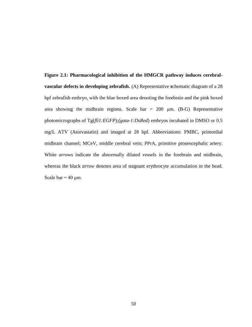
50
Figure 2.1: Pharmacological inhibition of the HMGCR pathway induces cerebral-
vascular defects in developing zebrafish. (A) Representative schematic diagram of a 28
hpf zebrafish embryo, with the blue boxed area denoting the forebrain and the pink boxed
area showing the midbrain regions. Scale bar = 200 μm. (B-G) Representative
photomicrographs of Tg(fli1:EGFP);(gata-1:DsRed) embryos incubated in DMSO or 0.5
mg/L ATV (Atorvastatin) and imaged at 28 hpf. Abbreviations: PMBC, primordial
midbrain channel; MCeV, middle cerebral vein; PPrA, primitive prosencephalic artery.
White arrows indicate the abnormally dilated vessels in the forebrain and midbrain,
whereas the black arrow denotes area of stagnant erythrocyte accumulation in the head.
Scale bar = 40 μm.
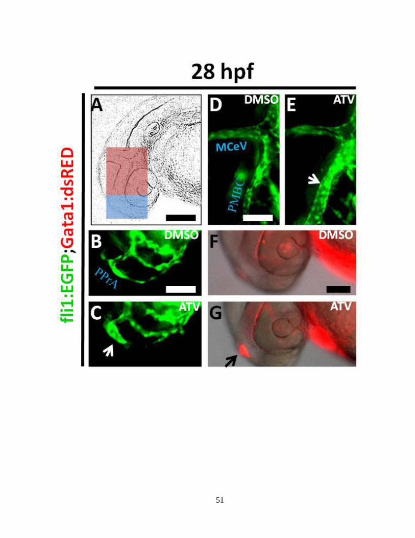
51
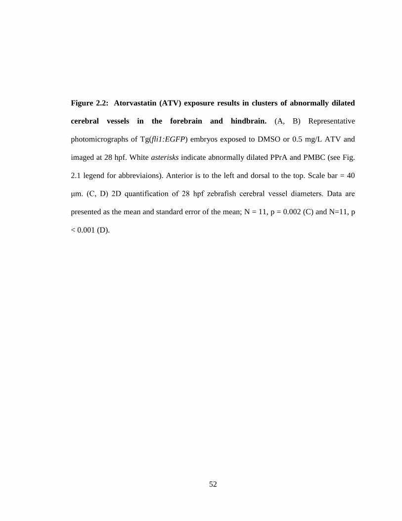
52
Figure 2.2: Atorvastatin (ATV) exposure results in clusters of abnormally dilated
cerebral vessels in the forebrain and hindbrain. (A, B) Representative
photomicrographs of Tg(fli1:EGFP) embryos exposed to DMSO or 0.5 mg/L ATV and
imaged at 28 hpf. White asterisks indicate abnormally dilated PPrA and PMBC (see Fig.
2.1 legend for abbreviaions). Anterior is to the left and dorsal to the top. Scale bar = 40
μm. (C, D) 2D quantification of 28 hpf zebrafish cerebral vessel diameters. Data are
presented as the mean and standard error of the mean; N = 11, p = 0.002 (C) and N=11, p
< 0.001 (D).
53
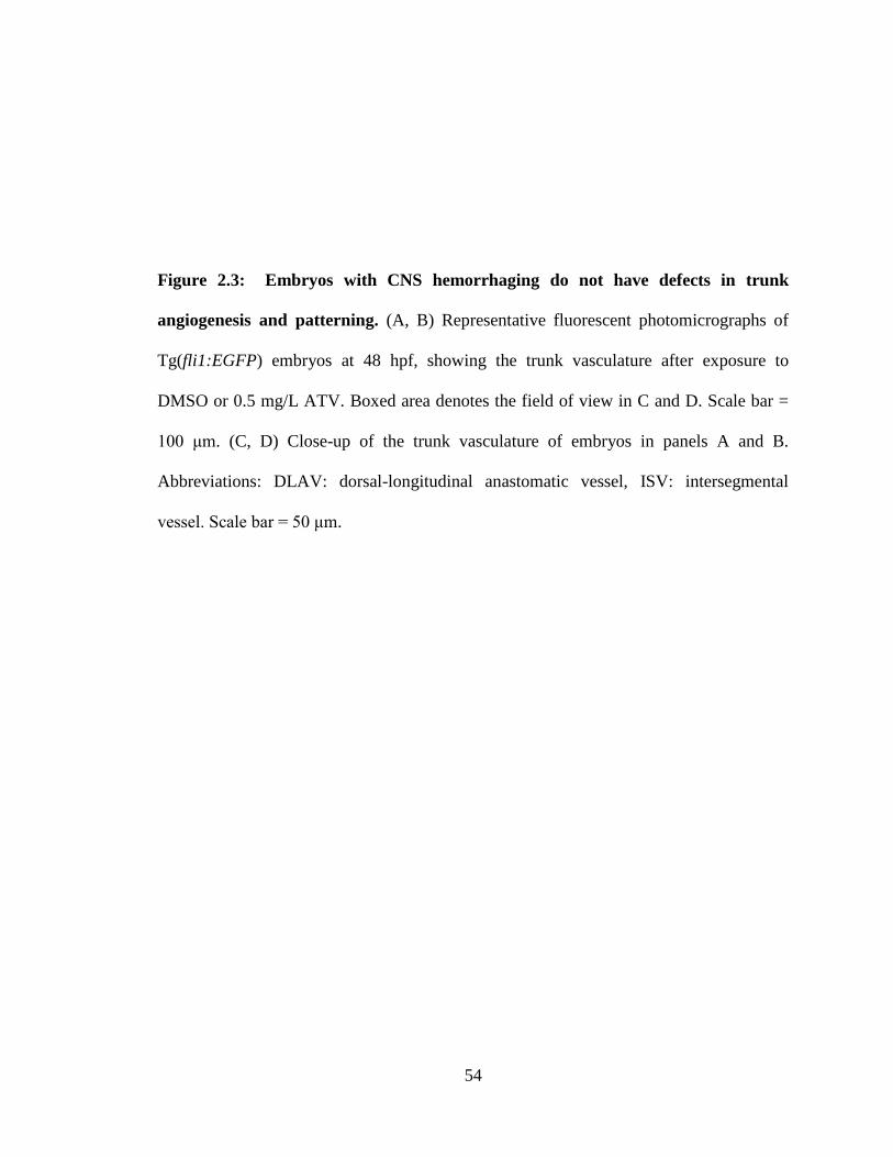
54
Figure 2.3: Embryos with CNS hemorrhaging do not have defects in trunk
angiogenesis and patterning. (A, B) Representative fluorescent photomicrographs of
Tg(fli1:EGFP) embryos at 48 hpf, showing the trunk vasculature after exposure to
DMSO or 0.5 mg/L ATV. Boxed area denotes the field of view in C and D. Scale bar =
100 μm. (C, D) Close-up of the trunk vasculature of embryos in panels A and B.
Abbreviations: DLAV: dorsal-longitudinal anastomatic vessel, ISV: intersegmental
vessel. Scale bar = 50 μm.
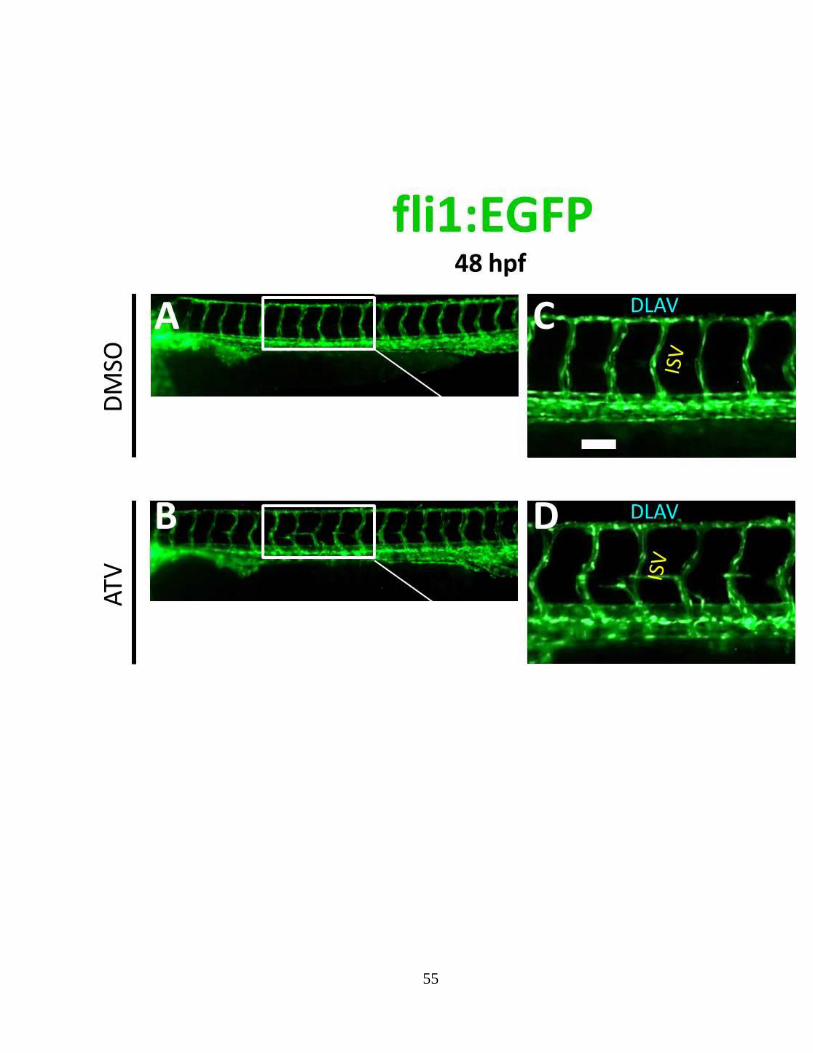
55
56
Figure 2.4: Pharmacological inhibition of the HMGCR pathway induces cerebral
hemorrhage in developing zebrafish. (A-D) Photomicrographs of embryos incubated in
0.5 mg/L ATV and imaged at 33 hpf. Black arrows denote areas of hemorrhage. Scale
bar = 200 μm. (E, F) Bright-field images of zebrafish embryos incubated in DMSO or 0.5
mg/L ATV, stained with o-Dianisidine (OD) and imaged at 48 hpf. Black arrows indicate
sites where abnormal accumulation of hemoglobin-containing blood was detected in the
brain. Blue arrow denotes OD-positive cells in the pericardium, green arrow shows the
sinus venosus and red arrow designates the tail region. Scale bar = 200 μm. (G, H)
Transverse sections through the embryo head at 48 hpf stained with OD. Abbreviations:
te, tectum; tel, telencephalon. Scale bar = 200 μm. (I) Percentage of embryos treated with
various doses of ATV and scored for the presence of cerebral hemorrhage at 48 hpf.
Numbers above the bars represent the ratios used to calculate the percentages.
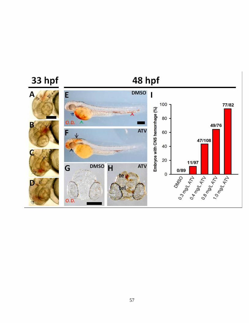
57
58
form the fluorescent DAF-2T which is a marker for functional smooth muscle cell
coverage in zebrafish (Zeng et al., 2009; Lou et al., 2011).
However, no vascular smooth muscle cell coverage was detected in the brain or
trunk during the early stages of normal zebrafish development (data not shown), which
suggests that nascent vessels are more prone to statin-induced hemorrhage, at least partly
by virtue of inadequate coverage with smooth muscle support. This observation is
consistent with molecular and genetic evidence suggesting that the expression of early
vascular smooth muscle cell markers in zebrafish are not detectable before the larval
stage (before hpf) (Santoro et al., 2009; Seiler et al., 2010).
We also exposed embryos at 1-2 hpf to Cerivastatin (CVT), a structurally
different statin molecule with a comparable affinity for the active site of HMGCR (Istvan
and Deisenhofer, 2001). Incubation of 2 hpf embryos in CVT (0.15 mg/L), in a single
exposure manner, resulted in 58% of embryos demonstrating cerebral hemorrhage by 33
hpf (108/174), which was a phenocopy of the ATV-treatment.
To further test the specific nature of the pharmacological treatments, a
metabolite rescue experiment was conducted using mevalonate, the immediate product of
the HMGCR-catalysed reaction and a precursor of both cholesterol and isoprenoids.
Treatment of 1-cell stage embryos with ATV (0.5 mg/L) in the presence of mevalonate
(3.2 μmol/L) was sufficient to rescue the hemorrhage phenotype, as none of these
embryos exhibited apparent hemorrhages (0/121). In contrast, 53% of the embryos treated
with ATV (0.5 mg/L) in the presence of DMSO showed cerebral hemorrhage by 48 hpf
(59/112). Treatment with mevalonate alone did not induce any overt developmental
defects. The ability of mevalonate to rescue the phenotype suggests that deficiencies in
59
Figure 2.5: Pharmacological inhibition of the HMGCR results in loss of vascular
stability and induces vessel rupture. (A, B) Representative bright-field micrographs of
48-52 hpf Tg(fli1:EGFP);(gata-1:DsRed) embryos treated with DMSO or 0.5 mg/L
ATV. The boxed region shows where hemorrhage is observed. Scale bar = 200 μm. (C,
D) Representative composite confocal z-stack projections of the boxed regions in the
same Tg(fli1:EGFP);(gata-1:DsRed) embryos. Abbreviations: PrA, prosencephalic
artery. Scale bar = 40 μm. (E, F) Representative photomicrographs of 48-52 hpf embryos
treated with DMSO or 0.5 mg/L ATV and subjected to fluorescent microangiography by
intravascular injection of red-fluorescent microspheres. White arrow shows the site of
microsphere extravasation. Scale bar = 200 μm.
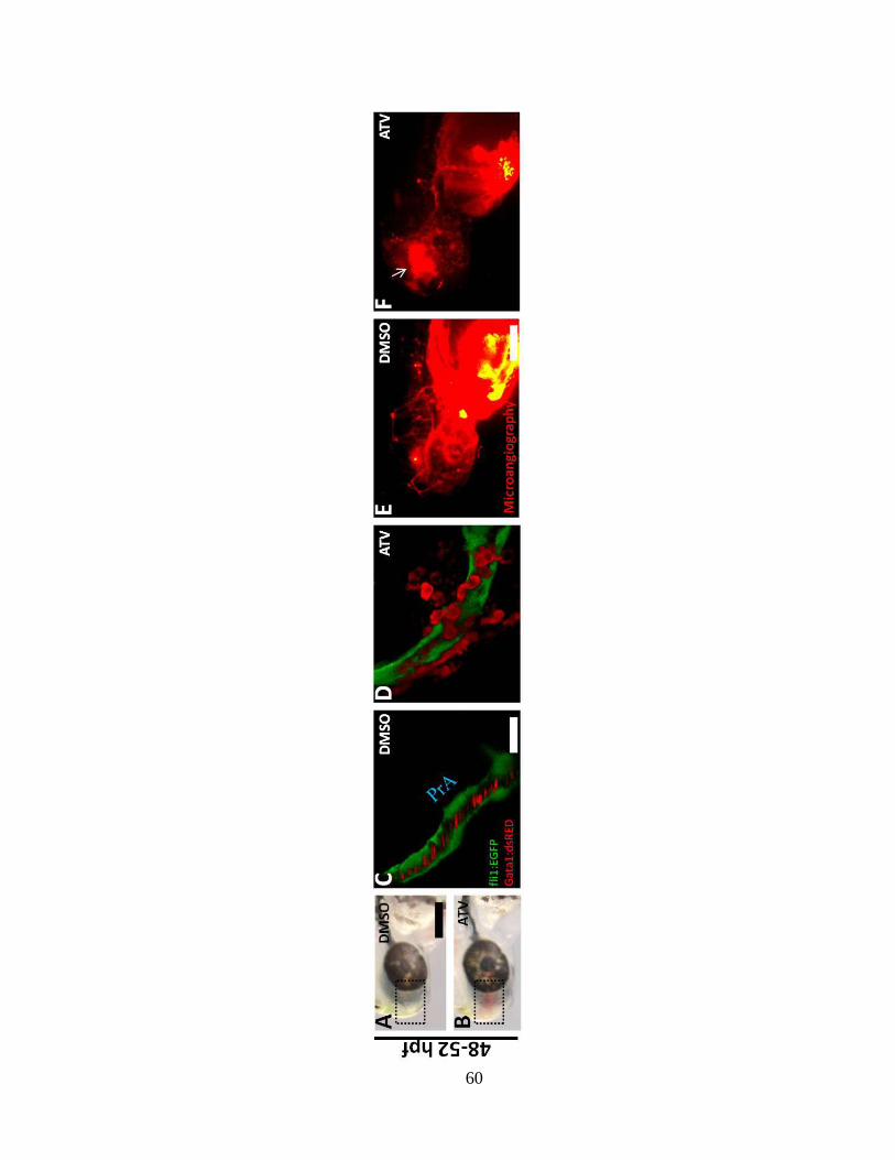
60
61
one or more of the metabolites downstream of mevalonate within the HMGCR pathway
may be implicated in the emergence of the hemorrhage phenotype.
2.4.2 Morpholino-mediated depletion of hmgcrb mRNA levels leads to cerebral-
vascular defects in developing zebrafish
Zebrafish possess two paralogous genes encoding HMGCR proteins. It is
speculated that teleosts, including zebrafish, underwent a complete genome duplication
event 250 million years ago (Van de Peer et al., 2009), resulting in the retention of many
genes in duplicate. Of the genes, The hmgcrb transcripts are maternally-enriched and
exhibit a ubiquitous expression pattern throughout early embryogenesis, whereas hmgcra
mRNA is only detected at 5 dpf and is confined to the liver and the anterior intestine
(Thorpe et al., 2004). A similar temporal expression pattern for these genes was observed
using RT-PCR on total RNA harvested from a pool of embryos at different
developmental stages (data not shown).
To substantiate the phenotypic specificity of the statin-induced cranial vascular
defects, we induced a transient loss of hmgcrb gene function with a morpholino
oligonucleotide (MO) designed to hybridise with the third exon-intron boundary of the
hmgcrb pre-mRNA (Fig. 2.6A). The injection of 2 ng of this hmgcrb MO resulted in
approximately 45% (91/203) of the embryos displaying cerebral hemorrhage at 48 hpf,
along with reduced abundance of erythrocytes in the pericardium and the sinus venosus
as detected by whole-mount OD staining (Fig. 2.6B and C).
The morphants demonstrated no other overt developmental defects and
eventually recovered from the initial hemorrhage, further indicating that the impaired
62
HMGCR function only affected cerebral-vascular development during a critical period
when nascent vessels in the brain are more prone to rupture. This suggests that hmgcrb
expression is dispensable in more mature vessels. Injection of higher quantities of the
hmgcrb MO (>8 ng) resulted in developmental arrest, defective angiogenesis in the trunk,
lack of circulation and occasional pericardial edema, and most of these embryos lacked
the cranial hemorrhage phenotype (data not shown). The efficiency of the genetic
ablation of hmgcrb expression was tested at 48 hpf by RT-PCR (Fig. 2.6D) and
sequencing (Fig. 2.6E and F) using primers spanning the third exon-intron boundary.
As with the statin-treatment, the exogenous supplementation of these embryos
with mevalonate nearly completely rescued the splice MO-induced vascular defects at 48
hpf, with only 2.2% of the MO-injected embryos (2 ng) incubated in mevalonate (4.4
μmol/L) showing cerebral vascular defects (7/318) at 48 hpf. A second MO directed
against the AUG translation-start site also mirrored the statin-induced hemorrhage
phenotype (data not shown). Overall, these observations further support an essential role
for HMGCR-mediated lipid metabolism in cerebral-vascular morphogenesis.
63
Figure 2.6: MO-mediated depletion of the HMGCR levels phenocopies ATV-
treatment. (A) Partial hmgcrb pre-mRNA structure (not to scale) showing the exons
(boxes), introns (black lines), the target of hmgcrb MO (red vertical arrow) and the
positions of forward and reverse primers (black arrows) used to test the efficiency of the
hmgcrb MO. (B, C) Representative bright-field micrographs of 48 hpf embryos injected
with hmgcrb MO and control MO, stained with OD. Black arrows indicate sites of
hemorrhage and blue arrow denotes the sinus venosus. Anterior is to the left and dorsal to
the top. Scale bar = 200 μm. (D) RT-PCR analysis of hmgcrb mRNA in hmgcrb
morphant and control MO-injected embryos at 48 hpf. MO injection results in the dose-
dependent appearance of a band corresponding to a transcript lacking exon-3. Doses of
ATV are indicated above each lane. (E-F) Sequence analysis of cDNA from splice MO-
injected embryos confirms the deletion of Exon-3.
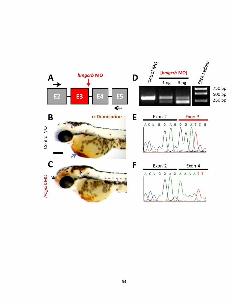
64
65
2.4.3 A deficiency in GGPP biosynthesis and impaired GGTase I-facilitated
prenylation of Rho GTPases leads to cerebral-vascular defects in developing
zebrafish
We next tested the hypothesis that statin-induced defects in vascular stability
were attributable to perturbations in GGPP biosynthesis. To test this hypothesis, 1-cell
stage embryos (1-2 hpf) were treated with ATV (0.5 mg/L) in the presence of
geranylgeranyl-pyrophosphate (GGPP, 4 mg/L) or the vehicle (DMSO:methanol; 50:50,
v/v). The addition of GGPP effectively rescued the hemorrhage phenotype, and by 48 hpf
approximately 7% of the embryos co-administered with ATV and GGPP demonstrated
cerebral hemorrhages (9/131), compared with 56% of control embryos receiving ATV
and the vehicle (80/143). The near-complete rescue of the vascular phenotype with GGPP
supplementation suggested a requirement for prenylation in the establishment of
developmental cerebral-vascular stability. To further demonstrate that decreased GGPP
bioavailability underlies these cerebral-vascular defects, we selectively targeted the
prenylation pathway. Prenylation of small GTPases, such as Rac1, RhoA and cdc42, is
catalyzed by the cytosolic isoprenyl transferase, geranylgeranyl pyrophosphate I
(GGTase I) (Peterson et al., 2006; Roberts et al., 2008).
This enzyme, GGTase I, catalyzes the transfer and binding of the 20-carbon
geranylgeranyl isoprenoid to the cysteine-residue of the CAAX motif at the C-terminus
of target Rho GTPases (Fig. 2.7A). This coupling constitutes a critical step for the
translocation of the Rho GTPases to the plasma membrane and interaction with other
lipophilic proteins. To specifically impair this pathway, we injected a splice-blocking
morpholino targeting the second exon-intron boundary of the pggt1b pre-mRNA, which
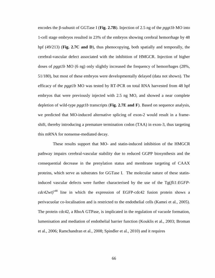
66
encodes the β-subunit of GGTase I (Fig. 2.7B). Injection of 2.5 ng of the pggt1b MO into
1-cell stage embryos resulted in 23% of the embryos showing cerebral hemorrhage by 48
hpf (49/213) (Fig. 2.7C and D), thus phenocopying, both spatially and temporally, the
cerebral-vascular defect associated with the inhibition of HMGCR. Injection of higher
doses of pggt1b MO (6 ng) only slightly increased the frequency of hemorrhages (28%,
51/180), but most of these embryos were developmentally delayed (data not shown). The
efficacy of the pggt1b MO was tested by RT-PCR on total RNA harvested from 48 hpf
embryos that were previously injected with 2.5 ng MO, and showed a near complete
depletion of wild-type pggt1b transcripts (Fig. 2.7E and F). Based on sequence analysis,
we predicted that MO-induced alternative splicing of exon-2 would result in a frame-
shift, thereby introducing a premature termination codon (TAA) in exon-3, thus targeting
this mRNA for nonsense-mediated decay.
These results support that MO- and statin-induced inhibition of the HMGCR
pathway impairs cerebral-vascular stability due to reduced GGPP biosynthesis and the
consequential decrease in the prenylation status and membrane targeting of CAAX
proteins, which serve as substrates for GGTase I. The molecular nature of these statin-
induced vascular defects were further characterised by the use of the Tg(fli1:EGFP-
cdc42wt)y48
line in which the expression of EGFP-cdc42 fusion protein shows a
perivacuolar co-localisation and is restricted to the endothelial cells (Kamei et al., 2005).
The protein cdc42, a RhoA GTPase, is implicated in the regulation of vacuole formation,
lumenisation and mediation of endothelial barrier function (Kouklis et al., 2003; Broman
et al., 2006; Ramchandran et al., 2008; Spindler et al., 2010) and it requires
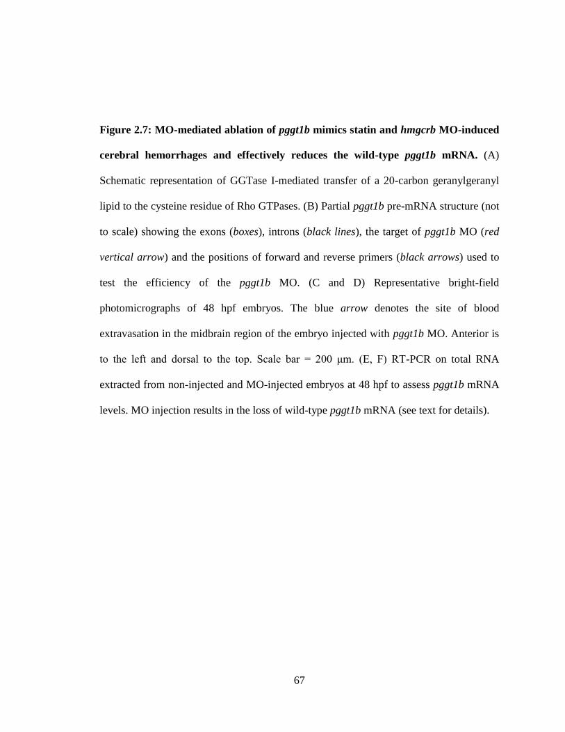
67
Figure 2.7: MO-mediated ablation of pggt1b mimics statin and hmgcrb MO-induced
cerebral hemorrhages and effectively reduces the wild-type pggt1b mRNA. (A)
Schematic representation of GGTase I-mediated transfer of a 20-carbon geranylgeranyl
lipid to the cysteine residue of Rho GTPases. (B) Partial pggt1b pre-mRNA structure (not
to scale) showing the exons (boxes), introns (black lines), the target of pggt1b MO (red
vertical arrow) and the positions of forward and reverse primers (black arrows) used to
test the efficiency of the pggt1b MO. (C and D) Representative bright-field
photomicrographs of 48 hpf embryos. The blue arrow denotes the site of blood
extravasation in the midbrain region of the embryo injected with pggt1b MO. Anterior is
to the left and dorsal to the top. Scale bar = 200 μm. (E, F) RT-PCR on total RNA
extracted from non-injected and MO-injected embryos at 48 hpf to assess pggt1b mRNA
levels. MO injection results in the loss of wild-type pggt1b mRNA (see text for details).
68
69
geranylgeranylation, downstream of HMGCR pathway, for its activation (Peterson et al.,
2006; Roberts et al., 2008).
Treatment of Tg(fli1:EGFFP-cdc42wt)y48
embryos with ATV at the 1-cell stage
led to a noticeable reduction in intra-endothelial expression of EGFP-cdc42 in the brain
and the trunk as early as 24 hpf (data not shown). The decline in vascular-specific EGFP-
cdc42 expression was also clearly evident in the head and trunk vasculature of these
embryos at 48 hpf (Fig. 2.8A and B), the stage when cerebral hemorrhage was most
preponderant in statin-treated embryos. The attenuated EGFP-cdc42 expression was
further validated by measuring EGFP signal intensity using ImageJ software (Fig. 2.8E
and F).
We further established that the reduction in fluorescence intensity was not
attributable to global developmental defects, vascular regression or defective
angiogenesis resulting from a down-regulation of the fli promoter in Tg(fli1:EGFP-
cdc42wt)y48
, since the expression of fli1:EGFP was unaffected in response to statin
treatment, and the brain and trunk vessel morphology and segmental artery formation was
unaltered (Fig. 2.8C and D). Similarly, quantification of the EGFP signal intensity in
these fish confirmed unaltered expression of the fli1:EGFP protein in the head or trunk
vessels (Fig. 2.8G and H). Together, these data reflect a specific reduction in cdc42
levels as a consequence of statin treatment. To investigate the localisation of cdc42 in
response to statin exposure, we performed confocal microscopy on 48-52 hpf
Tg(fli1:EGFP-cdc42wt)y48
embryos. We focused our attention on intersegmental vessels
(ISVs) due to the ease of imaging. Whereas the EGFP-cdc42 expression in DMSO-
treated embryos exhibited perivacular localisation, outlining the boundaries of the
70
intercellular lumen in the ISVs, the ATV-treated embryos had reduced EGFP-cdc42
expression in the ISVs and any expression was concentrated primarily at the base of the
ISVs (Fig. 2.8I and J). Taken together, our evidence suggests that the endothelial-
specific expression of cdc42 is markedly reduced concomitant with the onset and
expansion of hematoma in the brain.
2.5 DISCUSSION
Through a combination of pharmacological blockers, metabolite rescue and
genetic approaches, this study demonstrates that a transient disruption in the HMGCR
pathway induces intracranial hemorrhaging in zebrafish at a predictable embryonic stage,
which highlights a metabolic contribution to developmental vessel stabilisation. This
cerebral-vascular-specific phenotype was not associated with any other developmental
abnormalities. Of particular clinical significance is that these embryos manifest certain
pathophysiological features that closely resemble CCM pathologies in humans and mice,
such as abnormally dilated and blood-filled cranial microvessels prior to the onset of
extravasation, intracranial hemorrhage and expansion of hematoma, along with evidence
of thrombosis in the vicinity of the lesions. It is likely that inhibition of the HMGCR
pathway perturbs signalling processes that mitigate the assemblage of angioblasts into
nascent vascular tubes, thus making them more prone to rupture and leakage.
Interestingly, the extravasation of blood components is only detected in the head,
implying that the vessels in this region are more fragile at this developmental period,
hence hence more easily ruptured.
71
Figure 2.8: Inhibition of the HMGCR pathway abolishes the intra-endothelial
expression of cdc42. (A, B) Representative photomicrographs of Tg(fli1:EGFP-
cdc42wt)y48
embryos treated with DMSO or 1.0 mg/L ATV and imaged at 48-52 hpf. (C,
D) Representative photomicrographs of Tg(fli1:EGFP) embryos treated with DMSO or
1.0 mg/L ATV and imaged at 48-52 hpf. Anterior is to the left and dorsal to the top. Scale
bar = 200 μm. (E-H) Graphs plotting relative EGFP signal intensities in the head and
trunk vasculature (as denoted by the dotted white boxes in A-D) of (fli1:EGFP-
cdc42wt)y48
and Tg(fli1:EGFP) embryos treated with DMSO or 0.5 mg/L ATV. Data are
presented as the mean and standard error of the mean. N = 15 embryos, t-test, p<0.001
(for E and F). (I, J) Representative composite confocal z-stack projections of the trunk
region of Tg(fli1:EGFP-cdc42wt)y48
embryos, showing the localisation of EGFP-cdc42 in
a single intersegmental artery (ISV). Green arrow denotes perivacuolar expression of
EGFP-cdc42. Scale bar = 40 μm. Anterior is to the left and dorsal to the top.
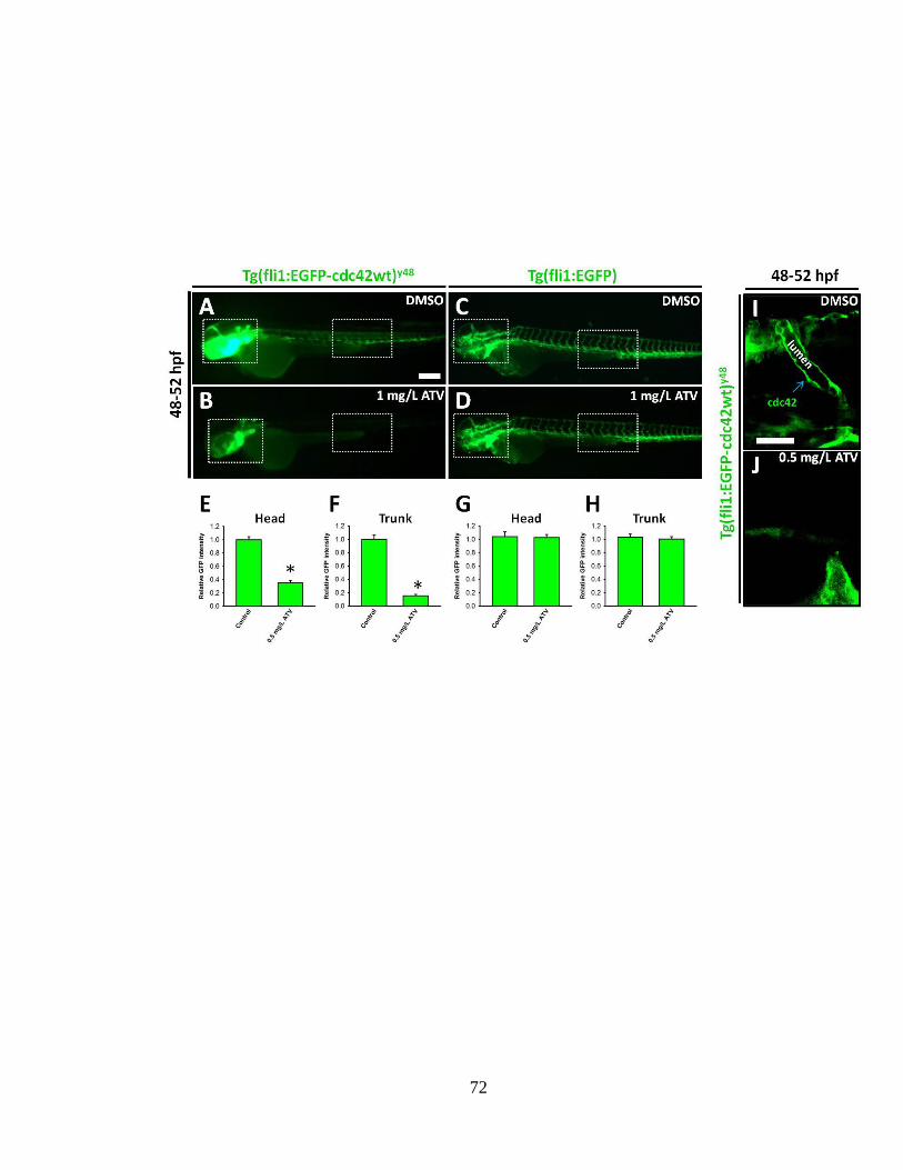
72
73
Since the onset of hemorrhage and the expansion of hematoma correspond to a
period of rapid angiogenesis during which numerous newly formed blood vessels invade
the brain (Isogai et al., 2001), it is plausible that these nascent vessels that carry blood for
the first time would lack adequate coverage with support cells, making them weak and
prone to rupture. Consistent with this, DAF-2DA staining showed no positive signal for
smooth muscle cells in the brain or trunk vasculature during early development,
consistent with previous studies (Santoto et al., 2009), which suggests that inadequate
endothelial contact with support cells can predispose these early vessels to statin-induced
hemorrhage. We have shown by fluorescence microangiography that defects in the
establishment of endothelial cell-to-cell associations contribute to the loss of vascular
stability in these embryos. Consistent with this assertion, zebrafish deficient in VE-
cadherin, an endothelial-specific adhesion protein located in cell-to-cell junctions and
important for vessel stabilisation, exhibit cerebral-vascular fragility and hemorrhage at 48
hpf in zebrafish (Montero-Balaguer et al., 2009).
At higher doses (5-10 mg/L), we observed that statin treatment significantly
inhibited angiogenic processes in the trunk (Fig. 2.9A-C), as evidenced by the
incomplete or defective anterior to posterior sprouting of intersegmental vessels (ISV)
emanating from the dorsal aorta (DA), thereby severely restricting circulation in the fish,
and often resulting in pericardial edema, with almost no cerebral hemorrhages observed.
This leads us to speculate that at higher doses, statin-treatment affects endothelial
survival and migration, whereas at lower doses, vascular stability in the brain is
compromised, to which the hemorrhage phenotype attests. This is also confirmed through
74
our reverse genetics approach, with high doses of hmgcrb MO inducing defective
angiogenesis in the trunk.
It is intriguing that the temporal and spatial distribution of the vascular defects
reported here are similar to those associated with the bbh (bubblehead) and rhd (redhead)
mutations which were previously characterised in zebrafish (Buchner et al., 2007; Liu et
al., 2007; see Chapter 1). These fish were reported to have hypomorphic mutations in the
pak2a and βpixA genes that otherwise encode for proteins involved in regulating the
activity and mediating the function of the Rho GTPases, Rac and cdc42 (Buchner et al.,
2007; Liu et al., 2007). The disruption of vascular permeability in these embryos is
consistent with the well-established roles of Rac and Cdc42 in the maintenance and
stabilisation of endothelial barrier function (Spindler et al., 2010). The similar nature of
the hemorrhages described in zebrafish that are deficient in pak2a and βPixA and those
we have reported here suggests a molecular interaction between the HMGCR pathway
and Rho GTPase signalling in the regulation of developmental vascular stability. This is
further supported by the fact that both Rac and cdc42 are natural substrates for the β-
subunit of GGTase I, which facilitates their post-translational modification by the
addition of a mevalonate-derived geranylgeranyl lipid, a critical step required for the
activation of these Rho GTPases (Peterson et al., 2006; Roberts et al., 2008).
Interestingly, using the Tg(fli1:EGFP-cdc42wt)y48
line in which cdc42 protein is over-
expressed in an endothelial-specific manner, we observed a decrease in the expression of
EGFP-cdc42 in the cerebral and trunk vessels upon treatment with statins. This decrease
was evident as early as 24 hpf and persisted to the onset of hemorrhage. We showed that
the reduction in fluorescence intensity was not attributable to global developmental
75
Figure 2.9: At high dose (5-10 mg/L) ATV-exposure induces defective angiogenesis.
(A-B) Representative fluorescent photomicrographs of the trunk region in embryos
incubated in DMSO or 6 mg/L ATV and imaged at 28 hpf. Anterior is to left. Blue
arrows show fully sprouted intersegmental vessels (ISV), red arrows show where
endothelial cell migration is completely stalled and the white asterisks denote partially
sprouted ISVs. (C) Percentage of embryos treated with various doses of ATV and scored
for the presence of ISV sprouting defects at 28 hpf. Numbers above the bars represent the
ratios used to calculate the percentages.
76
77
defects or down-regulation of fli1 promoter activity, since examination of Tg(fli1:eGFP)
embryos showed no apparent alterations in fluorescence intensity nor signs of defects in
cranial or segmental angiogenesis.
Collectively, this observation is suggestive of statin-induced degradation of cdc42
expression in the vasculature, and it further highlights in vivo how alterations of HMGCR
activity can exert effects on the cellular dynamics of cdc42 expression. On the whole, the
rescue of statin-induced hemorrhages by the addition of GGPP, along with the
hemorrhage phenotype elicited by the disruption of GGTase I-mediated prenylation in
zebrafish strongly supports a requirement for HMGCR activity for the stabilisation of
nascent vessels during early angiogenesis. These data contrast, however with previous
findings in mice. More specifically, in mice, the defective endothelial stability underlying
CCM pathogenesis, thought to be mediated by RhoA hyperactivation, is greatly
attenuated upon pharmacological ablation of the HMGCR pathway, possibly due to non-
cholesterol dependent processes (Whitehead et al., 2009). This raises the possibility that
modulation of HMGCR activity could be a therapeutic strategy to ameliorate CCM
pathobiology (Li and Whitehead, 2010). More recently using a separate murine model of
CCM, a more specific pharmacological inhibition of RhoA activity through direct
targeting of the Rho kinase signalling pathway, was reported to be similarly effective in
significantly reducing CCM genesis (McDonald et al., 2012). The hemorrhage phenotype
described here does not induce lethality, as these embryos eventually resolve the brief
disruption in vascular stability and resume normal development (Fig. 2.10A-H). This is
in contrast with the embryonic lethality observed in mouse models of intracranial
hemorrhage.
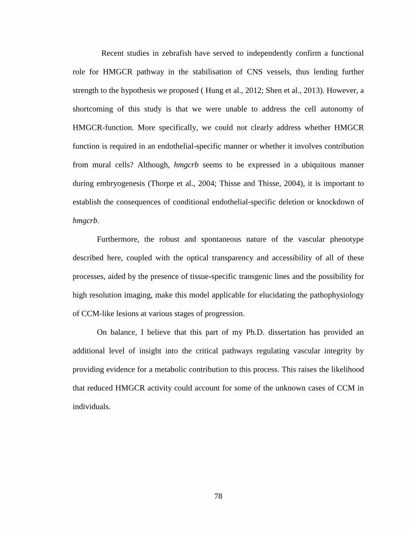
78
Recent studies in zebrafish have served to independently confirm a functional
role for HMGCR pathway in the stabilisation of CNS vessels, thus lending further
strength to the hypothesis we proposed ( Hung et al., 2012; Shen et al., 2013). However, a
shortcoming of this study is that we were unable to address the cell autonomy of
HMGCR-function. More specifically, we could not clearly address whether HMGCR
function is required in an endothelial-specific manner or whether it involves contribution
from mural cells? Although, hmgcrb seems to be expressed in a ubiquitous manner
during embryogenesis (Thorpe et al., 2004; Thisse and Thisse, 2004), it is important to
establish the consequences of conditional endothelial-specific deletion or knockdown of
hmgcrb.
Furthermore, the robust and spontaneous nature of the vascular phenotype
described here, coupled with the optical transparency and accessibility of all of these
processes, aided by the presence of tissue-specific transgenic lines and the possibility for
high resolution imaging, make this model applicable for elucidating the pathophysiology
of CCM-like lesions at various stages of progression.
On balance, I believe that this part of my Ph.D. dissertation has provided an
additional level of insight into the critical pathways regulating vascular integrity by
providing evidence for a metabolic contribution to this process. This raises the likelihood
that reduced HMGCR activity could account for some of the unknown cases of CCM in
individuals.
79
Figure 2.10: Embryos resolve cerebral-vascular defects and resume normal
development. (A-H) Representative bright-field photomicrographs of zebrafish embryos
incubated in DMSO or 0.5 mg/L ATV and stained with OD at various developmental
stages. Black arrows indicate areas of hemorrhage and blue arrows indicate sinus
venosus. Anterior is to the left and dorsal to the top. Scale bar = 200 μm.

80

81
Chapter 3
Molecular and pathophysiological processes
underlying intracerebral hemorrhage (ICH)
in developing zebrafish
82
Author contributions:
Dr. Raymond W.M. Kwong helped with the RT-PCR analysis and
assisted me with the western blot analysis. Ms. Sandra Noble and Mr. Rafael
Godoy trained me for use of confocal microscopy and helped me in
preparing and processing embryos for imaging. Dr. Steve F. Perry provided
reagents for real-time RT-PCR and western blot analysis. Dr. Thomas W.
Moon and Dr. Marc Ekker helped me design the experiments and assisted
me in writing and revising this chapter to ensure that the content is
scientifically sound and the style is in accordance with the FGPS
requirements. I am profoundly grateful for the assistance of the
aforementioned individuals, without whom this chapter would not have been
completed.
Shahram Eisa-Beygi
May 1, 2013
83
3.1 Abstract
In spite of the high mortality and morbidity associated with ICH, treatment
options remain ineffective. Several studies have used murine models as prognostic tools
in elucidating the pathophysiological processes induced by ICH. However, these studies
are limited by the heterogeneity, low expression and reproducibility of the desired
phenotype, as well as the high mortality observed in these models. Zebrafish are an
excellent vertebrate model to elucidate the etiology of cerebral-vascular disorders. We
previously reported that exposure to the statin drug Atorvastatin (ATV) resulted in ICH
in developing zebrafish. In this study, we expand upon these previous studies and draw
upon the unique advantages of the zebrafish system to evaluate several
pathophysiological processes associated with ICH and to use these variables to develop
an outcome model during vertebrate development. The pharmacologically-induced model
consistently replicates common underlying pathologies associated with ICH. We
demonstrate that hemorrhage is associated with the disruption of the blood-brain barrier
and cerebral vascular disintegration. Within hours of vascular rupture, rapid hematoma
expansion, accompanied by edema formation, is observed in the adjacent brain regions.
Enhanced TUNEL-positive staining is observed in and around the periphery of the
hematoma, which was also associated with the activation of an inflammatory response,
up-regulation of heme oxygenase 1 (HO-1) mRNA and protein levels, generation of
reactive oxygen species (ROS), and was followed by localised thrombosis. All of these
biomarkers serve as points of intervention for future studies geared towards large-scale
screening of zebrafish for potential therapeutics to mitigate symptoms of ICH. Hence, we
suggest a holistic approach towards modelling ICH pathophysiology, incorporating in
84
vitro studies using other vertebrate models including zebrafish, which can accelerate drug
discovery.
3.2 Introduction
Intracerebral hemorrhage (ICH), the pathological accumulation of blood within
the cranial vault, represents the second most common cause of stroke, as approximately
15% of all stroke subtypes are hemorrhagic in nature (Counsell et al., 1995; Qureshi et
al., 2005). Furthermore, ICH is associated with the greatest mortality rate of all stroke
subtypes, leaving most survivors with permanent neurological deficits and significant
disabilities (Broderick et al., 1993; Qureshi et al., 1999). Spontaneous hemorrhage can
arise as a result of cavernous malformations of the brain, typified by the accumulation of
dilated blood vessels that lack adequate investment by mural cells, which as a result
support only sluggish blood flow (Maeder et al., 1998; Eerola et al., 2003; Murakami et
al., 2012).
At the ultra-structural level, the disruption of endothelial cell-to-cell junctions
results in the detachment of endothelial cells from the vessel wall, making them fragile
and prone to rupture (Louvi et al., 2011). The timing for the onset of these vascular
malformations can be as early as the embryonic or postnatal stages of development
(Boulday et al., 2011). However, ICH can also arise in the absence of primary vascular
malformations and result from elevated blood pressure (Ebrahimi et al., 1999; Lang et al.,
2001; Sutherland and Aurer, 2006), bacterial infection (Babamahmoodi and
Babamahmoodi, 2011), trauma (Marion et al., 1997; Perel et al., 2009), or tumors (Lieu
et al., 1999).
85
Despite the relevant incidence and the high mortality and morbidity associated
with ICH, no surgical treatment or medical therapy has been effective in improving
functional outcomes (Yu et al., 1992; Morgenstern et al., 1998; Broderick et al., 1999;
Steiner et al., 2011). This is due in part to the variability in the timing of onset, the
location and severity of this condition, as well as incomplete penetrance of genetic
mutations pre-disposing the organism to the condition (Sahni and Weinberger, 2007).
Considerable progress has been made using murine models in dissecting the
pathophysiology and progression of ICH using a number of interventions, including
injection of bacterial collagenase into the basal ganglia (Rosenberg et al., 1990; Clark et
al., 1998), infusion of autologous blood into the brain parenchyma (Belayev et al., 2003),
and through genetic knockouts (Louvi et al., 2011). However, these studies are limited by
the low expression and reproducibility of the desired phenotype, heterogeneity of the
resulting hemorrhages, lack of feasibility for real-time/in vivo imaging of hematoma
expansion and edema formation, lack of optical clarity, high cost, and the high mortality
observed in the murine models utilised. Zebrafish continue to gain recognition as a
reliable model to complement in vitro and clinical studies to address vascular
malformations underlying human pathologies, such as ICH (Liu et al., 2007; Buchner et
al., 2007; Gore et al., 2008; Kwon et al., 2012; Eisa-Beygi et al., 2013; Hegarty et al.,
2013). However, to date no studies elaborating the pathophysiological features associated
with cerebral hemorrhage have been reported in this species.
We had previously reported that, in contrast with mammalian models, zebrafish
embryos and larvae with ICH exhibit functional recovery, as their development seems
unimpaired (Eisa-Beygi et al., 2013; Chapter 2). In addition, zebrafish embryos provide a
86
relatively rapid screening system with the genomic and functional complexity of a
vertebrate model organism (Lieschke and Currie, 2007). Hence, we reasoned that
improved understanding of the pathophysiological cascade of events and molecular
mechanisms ensuing ICH can have clinical relevance in terms of identifying potential
targets for new prognosis and therapeutic interventions to ameliorate the progression of
this disease in vivo. As a first step, we present here a highly reproducible and versatile
model of pharmacologically-induced ICH in a developing vertebrate, exhibiting
numerous primary and secondary pathological processes that closely mimic murine and
human ICH pathologies. In addition, the amenability of this model to small-molecule
screening makes it ideal for functional analysis of gene modulators or therapeutics that
can ameliorate ICH-induced secondary injury, thereby increasing the preclinical utility of
the model.
3.3 Materials and Methods
3.3.1 Zebrafish husbandry and transgenic lines
Adult zebrafish were maintained under a constant temperature of 28°C and a 14
h light: 10 h dark photoperiod in the University of Ottawa Aquatic Care Facility.
Embryos were acquired by natural breeding of adult zebrafish and were kept at 28.5°C in
E3 embryo medium (5 mmol/L NaCl, 0.17 mmol/L KCl, 0.33 mmol/L CaCl2, 0.33
mmol/L MgSO4). The Tg(CD41:GFP) line was a gift from Dr. Robert Handin (Harvard
University, Massachusetts); the Tg(mpx:GFP) line was kindly provided by Dr. Steve
Renshaw (University of Sheffield, UK); and, the Tg (gata-1:DsRed) line was provided
by Dr. Beth Roman (University of Pittsburgh, Pennsylvania). All experiments were
87
performed in compliance with a protocol approved by the University of Ottawa Protocol
Review Committee and conform to the published guidelines of the Canadian Council on
Animal Care for the use of animals in research and teaching.
3.3.2 Drug treatment
For pharmacological inhibition of HMGCR, zebrafish embryos (1-2 hours post
fertilisation; hpf) were incubated in Atorvastatin (ATV; Pfizer Inc., Connecticut USA) in
embryo medium at a final concentration of 0.5 mg/L as previously noted (Eisa-Beygi et
al., 2013; see Chapter 2).
3.3.3 Detection of apoptotic cells
Apoptotic cells were detected in 48 hpf embryos that were pre-treated with 1-
phenyl 2-thiourea (PTU) and fixed in 4% PFA/1X Phosphate Buffered Saline Tween-20
(PBST) overnight, then dehydrated and stored in 100% methanol at -20°C overnight.
After rehydration in PBST, embryos were permeabilised in acetone at -20°C for 10 min.
Apoptotic cells were detected using whole-mount terminal deoxynucleotidyltransferase-
mediated dUTP nick end labeling (TUNEL), according to the manufacturer’s instructions
(ApopTag In situ Apoptosis Detection Kit Plus-Millipore). TUNEL-positive cells were
visualised using diaminobenzidine (DAB) and the DAB-stained cells in the ventral head
region were quantified by manual counting. Treated embryos were visualised using a
Nikon NBZ 1500 dissecting microscope with a Nikon DXM 1200 C digital camera and
stored in 50% glycerol at 4°C. For acridine orange (AO) staining, embryos were treated
with a 5 µg/mL solution of AO (Sigma-Aldrich) for 1 h at room temperature, followed by
several rinses in embryo medium. The embryos were then anesthetised in 0.017%
tricaine, after which fluorescence micrographs of embryos were acquired under the Nikon
88
NBZ 1500 dissecting microscope, equipped with a standard GFP filter set, using a Nikon
DXM 1200C digital camera.
3.3.4 Neutral-red staining of macrophages
Macrophages in live embryos were visualised by incubating 48 hpf embryos in a
2.5 μg/mL neutral-red (Sigma-Aldrich) solution in E3 embryo medium at 25–30°C for 8
h, as previously described (Gray et al., 2007). These embryos were then visualised using
a Nikon NBZ 1500 dissecting microscope equipped with a Nikon DXM 1200 C digital
camera as above.
3.3.5 Sudan-black staining of neutrophils
Neutrophils in embryos were visualised by whole-mount Sudan black staining,
as previously described (Yoo and Huttenlocher, 2011). In brief, 48 hpf embryos were
fixed in 4% PFA/PBST for 2 h at RT, followed by incubation in 0.18% Sudan Black
solution (Sigma-Aldrich), followed by extensive washes in 70% ethanol. Treated
embryos were visualised using a Nikon NBZ 1500 dissecting microscope with a Nikon
DXM 1200 C digital camera as above and stored in 50% glycerol at 4°C.
3.3.6 Whole-mount o-Dianisidine (OD) staining of erythrocytes
Zebrafish embryos at 48 hpf were fixed in 4% PFA/PBST overnight, followed
by several washes in PBST. Erythrocytes were stained using o-Dianisidine, a sensitive
marker for hemoglobin, as previously described (Eisa-Beygi et al., 2013; Chapter 2). In
brief, embryos were incubated in OD (0.6 mg/mL) (Sigma-Aldrich), containing 0.01 M
sodium acetate (pH 4.5), 0.65% hydrogen peroxide, and 40% (v/v) ethanol in the dark for
89
15 min. Stained embryos were washed in PBST and visualised using Nikon DXM 1200 C
digital camera as above and stored in 50% glycerol at 4°C.
3.3.7 CM-H2DCFDA staining
Embryos at 48 hpf with or without cerebral hemorrhage were incubated in 5-
(and-6)-chloromethyl-2′,7′-dihydrodichlorofluorescein diacetate (CM-H2DCFDA; 1000
ng/mL; Molecular Probes) for 1 h at 25–30°C and imaged under the Nikon NBZ 1500
dissecting microscope, which was equipped with a standard GFP filter set, using a Nikon
DXM 1200C digital camera.
3.3.8 RNA extraction, cDNA synthesis and real-time RT-PCR
Total RNA was extracted from a pool of 20-30 embryos using TRIzol reagent
(Invitrogen). The RNA was DNase-treated using the Deoxyribonuclease kit (Invitrogen)
to minimise genomic DNA contamination. cDNA was synthesised from equal
concentrations of RNA using a 1st Strand cDNA Synthesis Kit and Super Script II RT
(Invitrogen) and used as a template in real-time RT-PCR (qPCR). The qPCR assays were
performed using a Bio-Rad CFX96 qPCR system with Brilliant III SYBR Green Master
Mix (Agilent Technologies, USA). All qPCR reactions were performed using the
following conditions: 95°C for 3 min, 40 cycles of 95°C for 20 s and 58°C for 20 s, with
a final extension for 5 min at 72°C. The data were normalised to the expression of 18S
rRNA gene, and were presented relative to the control group. Values are reported with
standard errors. The primers used for qPCR analysis were:
(5'-CTGTCTGAACAGATAAAAGCAGTC-3') (HO-1 forward) and
(5'-TGTGTTTGTGTGATCTGTCCTT-3') (HO-1 reverse).
(5'-CAAACATGGGCTGGTTCAAG-3') (18S forward) and
90
(5'-GGTGGTGCCCTTCCGTCAATTC-3') (18S reverse).
3.3.9 Confocal microscopy
For confocal microscopy, 48-52 hpf Tg(flk1:EGFP;gata1:dsRED) or
Tg(mpx:EGFP;gata1:dsRED) embryos that were previously treated with ATV or DMSO
were embedded in 1% low melting agarose in E3 embryo medium and tricaine mesylate
(Sigma-Aldrich). A rendered Z-stack, at ~80 m thickness, was taken with a Zeiss LSM
510 AxioImager.M1 confocal microscope using an Achroplan 40x/0.8 W objective with
an argon laser (488 nm) and a helium-neon laser (543 nm).
3.3.10 Western blot analysis
Fifteen larvae were pooled as one sample and homogenised in RIPA lysis buffer
(150 mM NaCl, 1% Triton X-100, 0.5% sodium deoxycholate, 0.1% SDS, 50 mM Tris-
HCl, 1 mM EDTA, 1 mM phenylmethanesulfonyl fluoride) containing a general protease
inhibitor cocktail (Roche, USA). Extracted protein samples was heated for 10 min at
70°C, loaded onto a 10% SDS-PAGE and transferred to a PVDF membrane (Bio-Rad,
USA). The membrane was blocked for 1 h with 5% skimmed milk, probed with anti-HO-
1 antibody at a 1:1000 dilution in 2% skimmed milk, and subsequently incubated at 4°C
overnight. Subsequently, the membrane was probed with 1:15,000 dilution of goat anti-
rabbit secondary antibody (Pierce, USA) for 2 h at room temperature, and immune-
reactive bands were detected using Luminata Western HRP Substrates (Millipore, USA).
To control for equal protein loading, the membrane was re-probed with β-actin antibodies
(1:4000; A2066, Sigma-Aldrich) for 2 h at room temperature after stripping with Re-Blot
Plus solution (Millipore, USA). Ban intensities were quantified using ImageJ software.
91
3.4 Results and Discussion
3.4.1 Vessel disintegration, hematoma expansion and cerebral edema mark the early
phases of ICH
We previously reported that pharmacological and genetic ablation of HMGCR
function perturbs vascular development in zebrafish as early as 28 hpf (Eisa-Beygi et al.,
2013; Chapter 2). This is characterised by prevalence of abnormally dilated, diffusely
shaped and blood-filled nascent vessels in the forebrain and midbrain which support
noticeably attenuated or sluggish blood circulation. This phenotype closely mimics the
cerebral cavernous malformation (CCM)-like lesions observed in humans and murine
models. Furthermore, these vascular-specific effects are heterogeneous in severity and
distribution (Fig. 3.1A-D), as observed using the Tg(fli1:EGFP) embryos in which
cranial blood vessels are marked by GFP expression (Lawson and Weinstein, 2002).
This reflects a requirement for products of the mevalonate pathway in the stabilisation of
nascent cranial vessels, particularly during development.
Using confocal microscopy of Tg(fli1:EGFP);(gata-1:DsRed) embryos, these
bulged vessels in statin-treated embryos (marked by GFP expression) are leaky and prone
to rupture at later stages of development. This is evidenced by extravasation of DsRed-
positive erythrocytes from a specific site into adjacent brain regions at 48 hpf, resulting in
the fragmentation and complete disintegration of the underlying vascular architecture
(Fig. 3.1E-J). In more than 70% of humans experiencing ICH, hematoma growth is
observed, from single or multiple vessels, which is an independent determinant of
mortality and functional outcome of this disease (Davis et al., 2006).
92
Consistently within hours after the onset of hemorrhage in the zebrafish brain, rapid
hematoma expansion is observed into the adjacent brain regions, along with significant
hydrocephalus or edema formation between 36-48 hpf in the forebrain and the ventricular
zones, due predictably to the resulting intracranial pressure exerted by the accumulation
of blood (Fig. 3.2A-C). Approximately 81% of the embryos treated with 0.5 mg/L ATV
showed a significant degree of hematoma expansion between 36-48 hpf (53/65).
Hematoma expansion could exacerbate brain injury, resulting in the herniation of
adjacent brain structures; as such pharmacological cessation of hematoma growth has
been shown to lead to improved clinical outcomes (Davis et al., 2006).
3.4.2 Apoptosis is enhanced in response to ICH
Evidence from murine models of ICH suggests that part of the cell-death in the
hematoma and surrounding tissues may involve apoptosis (Matsuchita et al., 2000;
Karwacki et al., 2005; Huang et al., 2012). We further reasoned that the rapid expansion
of the hematoma and the resulting mass effect, along with the activity of cytotoxic blood
components in the brain parenchyma shortly after the onset of hemorrhage, could trigger
apoptosis within the hematoma region and the surrounding tissues. Furthermore, this
could underlie the basis for effects such as degeneration of the vasculature and, possibly
neuronal injury and dysfunction (please refer to Chapter 4 for more on this aspect).
To test if statin-induced ICH would provoke apoptosis, we used a whole-mount
TUNEL assay to detect cells with DNA fragmentation in situ (Espín et al., 2013). In
93
Figure 3.1: Exposure to Atorvastatin (ATV) induces vascular dilation, followed by
hemorrhage and vascular disintegration. (A-D) Representative photomicrographs of
Tg(fli1:EGFP) embryos incubated in DMSO or 0.5 mg/L ATV from 1-2 hpf and imaged
at 33 hpf. Abbreviation: PMBC, primordial midbrain channel. White arrows indicate the
abnormally dilated vessels. Scale bar: 50 μm. (D-E) Representative bright-field
photomicrographs of Tg(fli1:EGFP);(gata-1:DsRed) embryos incubated as above and
imaged at 48 hpf. The asterisk denotes the hemorrhage and the black dotted area shows
the field of interest. Anterior is to the left and dorsal to the top. (E-J) Representative
composite confocal Z-stack projections of the black dotted area in the same
Tg(fli1:EGFP);(gata-1:DsRed) embryos. The white asterisk denotes DsRed-positive
erythrocytes and the white arrows show regions where vascular fragmentation is
observed. Anterior is to the top and scale bar: 200 μm.
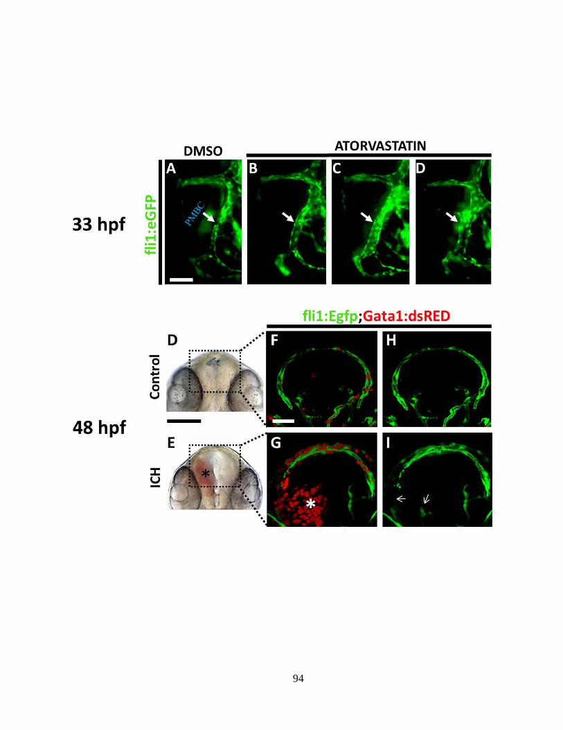
94
95
contrast with intact embryos, those with ICH showed enhanced apoptosis in the
hematoma area and the regions surrounding the hemorrhagic mass, particularly at 48 hpf
when the spatial extent of hemorrhage was maximal (Fig. 3.3A-D) (t-test, p<0.001,
N=14). Although the control group displayed some TUNEL-positive staining albeit to a
lesser degree than the ICH group, we speculate that this trend reflects programmed cell
death related to developmental and homeostatic mechanisms. Even though the triggers
responsible for initiating apoptosis in the ICH group are yet to be explored, we observed
a positive correlation between the number of apoptotic cells and the severity of
hemorrhage or the extent of hematoma expansion (data not shown). These observations
were also confirmed in vivo using acridine orange (AO), a cell-permeable nucleic acid
marker (Fig. 3.3E and F). It is plausible that some of these apoptotic cells include
neuronal populations, as dopaminergic (DA) neurons have been shown to be co-localised
in the pretectum and telencephalon regions at this stage (please refer to Chapter 4).
3.4.3 ICH triggers an immune response
In human patients and murine models of ICH, an acute inflammatory response
constitutes part of the normal physiological cascade of events triggered by the entry of
cytotoxic blood components into the brain parenchyma (Zhao et al., 2007; Loftspring et
al., 2009). Furthermore, this response is generally characterised by the infiltration of
activated leukocytes (neutrophils and macrophages) to the sites of injury for the removal
and neutralisation of the blood components and other cellular debris (Power et al., 2003;
Zhao et al., 2007; Lotfspring et al., 2009).
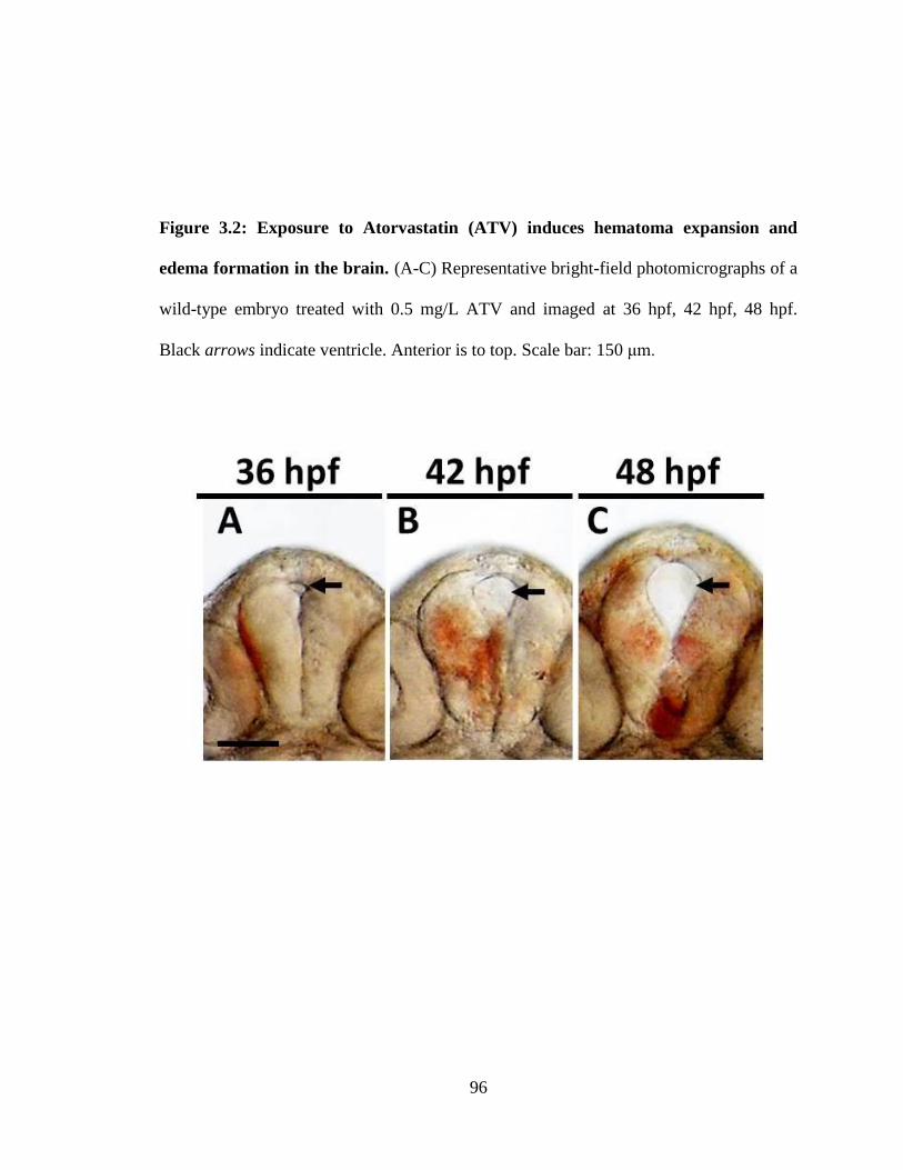
96
Figure 3.2: Exposure to Atorvastatin (ATV) induces hematoma expansion and
edema formation in the brain. (A-C) Representative bright-field photomicrographs of a
wild-type embryo treated with 0.5 mg/L ATV and imaged at 36 hpf, 42 hpf, 48 hpf.
Black arrows indicate ventricle. Anterior is to top. Scale bar: 150 μm.
97
To test whether statin-induced hemorrhage in the zebrafish brain would trigger
a similar inflammatory response during early phases of development, we used neutral red,
a histological dye that is readily taken up by early granulocytes, to localise active
macrophages in the brain (Gray et al., 2007). Compared with intact embryos in which a
small population of resident macrophages were noted, in embryos with ICH, there were
clearly more macrophages which also appeared enlarged in diameter, concentrated in and
around the periphery of hemorrhage in the head by 48-52 hpf (Fig. 3.3G and H),
supporting the recruitment and activation of these cells. In contrast, neutrophils
distribution and abundance as assessed by whole-mount sudan-black staining remained
unaltered when compared with untreated embryos (Fig. 3.3I and J).
To further verify that the neutrophil abundance in the brain is unaffected by
ICH, the Tg(mpx:eGFP) line was outcrossed with the erythrocyte marker Tg(gata-
1:DsRed) line. The resulting double-transgenic Tg(mpx:EGFP);(gata-1:DsRed) line
allowed for the simultaneous visualisation of neutrophils (marked by EGFP expression)
and the area occupied by the hematoma (marked by DsRED expression) in live embryos.
This transgenic model further confirmed that there was no recruitment or localised
proliferation of neutrophils (Fig. 3.3K and L), suggesting that the neutrophils observed
belong to a non-circulating, resident population of neutrophils. These analyses further
confirmed macrophage involvement in ICH pathogenesis, but there was, as expected, no
significant difference in the number of neutrophils (data not shown).
Overall, our data provides evidence that macrophage activation constitutes part
of the robust inflammatory response coincident with maximal hematoma size (48-52 hpf).
By contrast, the number of neutrophils in the vicinity of hemorrhage was unchanged,
98
suggesting that the primary immune response is characterised by the phagocytosis of
blood components through the activation macrophages.
3.4.4 Increased heme oxygenase (HO-1) expression and ROS generation following
ICH
Heme oxygenase-1 (HO-1) is the rate limiting enzyme facilitating the
metabolism of pro-oxidant heme into ferrous iron, carbon monoxide and biliverdin,
which is further oxidised to bilirubin (Prawan et al., 2005) (Fig. 3.4A). Since blood-
derived heme cannot be recycled, its timely metabolism is critical to alleviate the
symptoms of ICH and for the resolution of hemorrhage (Koeppen et al., 2004). Ferrous
iron, derived from the catabolism of heme, could potentiate the generation of reactive
oxygen species (ROS), which could induce secondary brain injury including endothelial
cell apoptosis or cerebral edema, as well as neuro-degeneration (Wagner et al., 2003;
Goldstein et al., 2003; Hua et al., 2007). HO-1 mRNA transcript levels in embryos at 48-
52 hpf were assessed using qPCR on total RNA harvested from a pool of embryos. In
embryos with ICH, the HO-1 transcript levels were significantly enriched (3.5-fold, n=4;
20 embryos per sample) (Fig. 3.4B).
Concomitant with the elevated HO-1 mRNA levels, the protein expression of
HO-1, assessed by western blot analysis in embryos experiencing ICH, also showed a
marked increased (n=3; 20 embryos per sample) (Fig. 3.4C). Using an intercellular
fluorescent ROS marker in live embryos (CM-H2DCFDA) (Anichtchik et al., 2008), we
noted that ICH induced significant ROS generation in the brain and ventricles of 48-52
hpf embryos (Fig. 3.4D and E). Taken together, these data highlight the functional
99
applicability of zebrafish embryos in replicating some of the pathophysiological details
associated with the degradation/lysis of blood components following ICH as early as 48
hpf.
3.4.5 Evidence for thrombosis after ICH
Hemostatis, the adhesion and aggregation of platelets to the sub-endothelium of ruptured
vessels, is a pivotal process to stop excessive bleeding (English et al., 2000). In patients
with ICH, hematoma expansion can elicit local activation of hemostatic mechanisms
(Fujii et al., 2001). In fact, pharmacological enhancement of hemostatis is suggested to
reduce hematoma growth, resulting in improved clinical outcomes (Mayer, 2005). In
zebrafish, thrombocytes are the functional equivalent of mammalian platelets and are
implicated in hemostasis in embryos and adults (Lin et al., 2005; Kim et al., 2010). To
test whether ICH in zebrafish initiate hemostasis and to analyze the temporal and spatial
distribution of thrombocytes in response to ICH in vivo, we used the Tg(CD41:GFP) line
in which nascent thrombocytes are tagged with a green fluorescent reporter protein (GFP)
(Lin et al., 2005). At earlier stages (24-48 hpf) and during the onset of vascular leakage,
GFP-positive cells were confined strictly to the cardiac sinus/yolk sac, along with
localisation in the caudal region (data not shown). This distribution pattern closely
mimicked that of wild-type embryos at the same developmental stages (data not shown).
However, by 3 dpf a large number of non-circulating GFP-positive cells had infiltrated
the sites of hemorrhage, which was visualised by o-Dianisidine (OD) staining (Fig. 3.5A-
D).
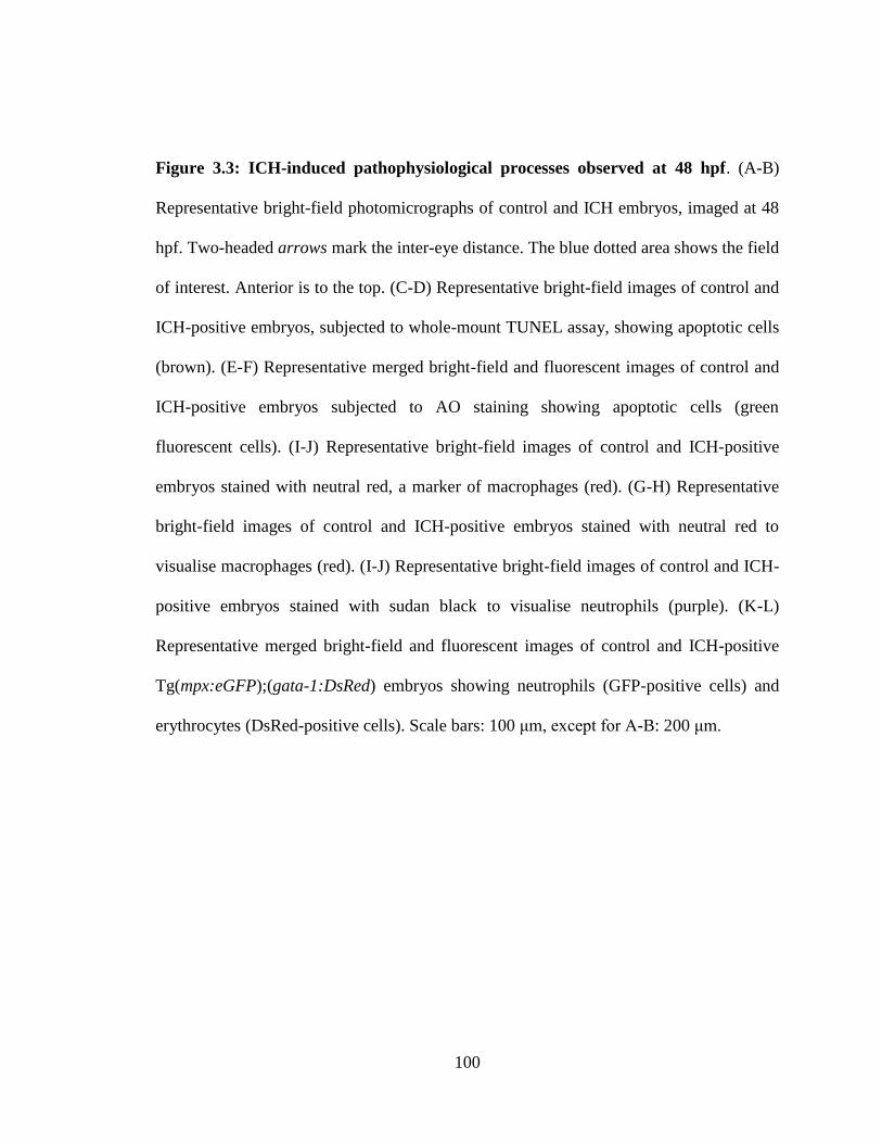
100
Figure 3.3: ICH-induced pathophysiological processes observed at 48 hpf. (A-B)
Representative bright-field photomicrographs of control and ICH embryos, imaged at 48
hpf. Two-headed arrows mark the inter-eye distance. The blue dotted area shows the field
of interest. Anterior is to the top. (C-D) Representative bright-field images of control and
ICH-positive embryos, subjected to whole-mount TUNEL assay, showing apoptotic cells
(brown). (E-F) Representative merged bright-field and fluorescent images of control and
ICH-positive embryos subjected to AO staining showing apoptotic cells (green
fluorescent cells). (I-J) Representative bright-field images of control and ICH-positive
embryos stained with neutral red, a marker of macrophages (red). (G-H) Representative
bright-field images of control and ICH-positive embryos stained with neutral red to
visualise macrophages (red). (I-J) Representative bright-field images of control and ICH-
positive embryos stained with sudan black to visualise neutrophils (purple). (K-L)
Representative merged bright-field and fluorescent images of control and ICH-positive
Tg(mpx:eGFP);(gata-1:DsRed) embryos showing neutrophils (GFP-positive cells) and
erythrocytes (DsRed-positive cells). Scale bars: 100 μm, except for A-B: 200 μm.
101
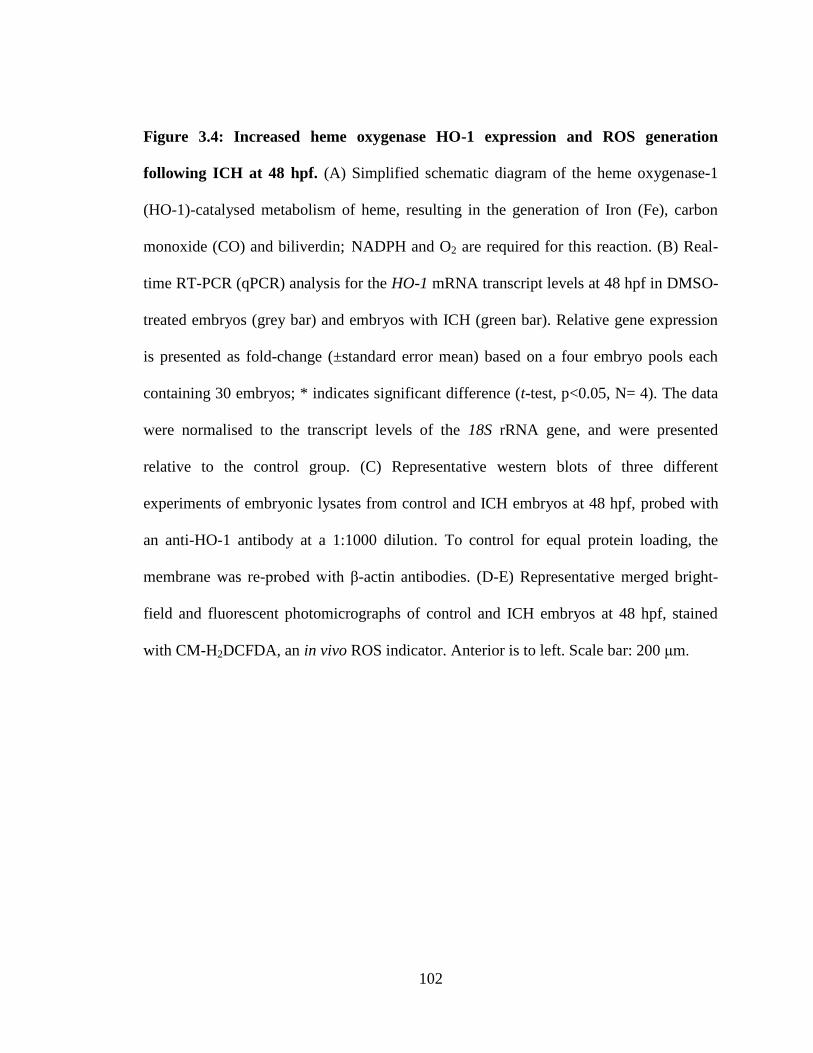
102
Figure 3.4: Increased heme oxygenase HO-1 expression and ROS generation
following ICH at 48 hpf. (A) Simplified schematic diagram of the heme oxygenase-1
(HO-1)-catalysed metabolism of heme, resulting in the generation of Iron (Fe), carbon
monoxide (CO) and biliverdin; NADPH and O2 are required for this reaction. (B) Real-
time RT-PCR (qPCR) analysis for the HO-1 mRNA transcript levels at 48 hpf in DMSO-
treated embryos (grey bar) and embryos with ICH (green bar). Relative gene expression
is presented as fold-change (±standard error mean) based on a four embryo pools each
containing 30 embryos; * indicates significant difference (t-test, p<0.05, N= 4). The data
were normalised to the transcript levels of the 18S rRNA gene, and were presented
relative to the control group. (C) Representative western blots of three different
experiments of embryonic lysates from control and ICH embryos at 48 hpf, probed with
an anti-HO-1 antibody at a 1:1000 dilution. To control for equal protein loading, the
membrane was re-probed with β-actin antibodies. (D-E) Representative merged bright-
field and fluorescent photomicrographs of control and ICH embryos at 48 hpf, stained
with CM-H2DCFDA, an in vivo ROS indicator. Anterior is to left. Scale bar: 200 μm.
103
104
This result suggests that hemostatic mechanisms are not impaired and that
enhanced hemostasis at the sites of vascular lesion constitutes part of the
pathophysiological cascade of events associated with ICH. We propose that hemostasis at
the sites of vascular lesion prevent excessive expansion of the hematoma, thereby
contributing to the resolution of hemorrhages in these embryos by 96 hpf.
3.5 Conclusions
ICH is the most debilitating type of all stroke subtypes, with the highest mortality
rate of all strokes (Broderick et al., 1993; Qureshi et al., 1999). Therapies geared towards
management of the disease have been ineffective; hence presently no consistently
effective surgical or pharmacological methods exist to reduce the mortality and morbidity
of ICH (Yu et al., 1992; Morgenstern et al., 1998; Broderick et al., 1999; Steiner et al.,
2011). This shortcoming is further compounded by the fact that there exists no high-
throughput chemical screening system for rapid assessment of pharmaceuticals that may
ameliorate the progression of ICH-associated pathological events. In addition,
pathophysiological studies using mammalian vertebrates tend to involve laborious
histological analysis that is often subject to bias. Here, we propose that zebrafish is a
reproducible model for the systematic assessment of the pathophysiological processes
resulting in ICH.
We have previously shown that embryonic and larval exposure to statin drugs
results in ICH in zebrafish (Eisa-Beygi et al., 2013; Chapter 2) which is consistent with
recent clinical evidence from human studies, suggesting a link between high dose statin
use and the risk of ICH (Amarenco et al., 2006; Westover et al., 2011). Of particular
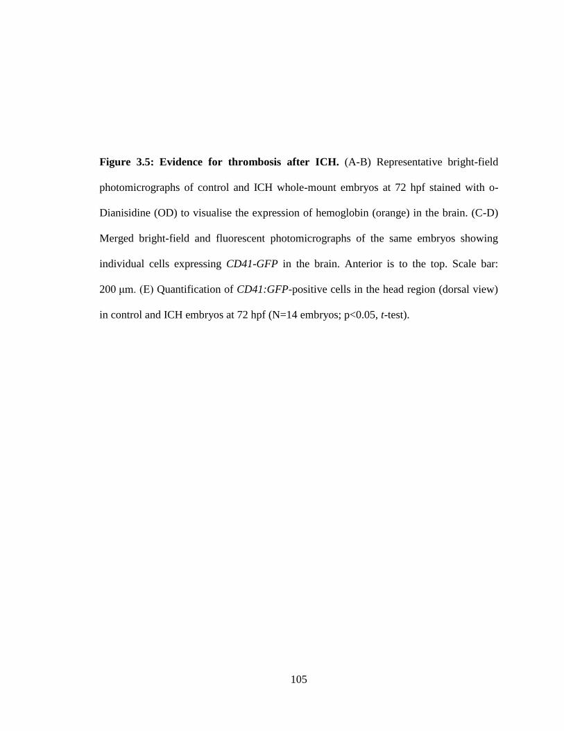
105
Figure 3.5: Evidence for thrombosis after ICH. (A-B) Representative bright-field
photomicrographs of control and ICH whole-mount embryos at 72 hpf stained with o-
Dianisidine (OD) to visualise the expression of hemoglobin (orange) in the brain. (C-D)
Merged bright-field and fluorescent photomicrographs of the same embryos showing
individual cells expressing CD41-GFP in the brain. Anterior is to the top. Scale bar:
200 μm. (E) Quantification of CD41:GFP-positive cells in the head region (dorsal view)
in control and ICH embryos at 72 hpf (N=14 embryos; p<0.05, t-test).
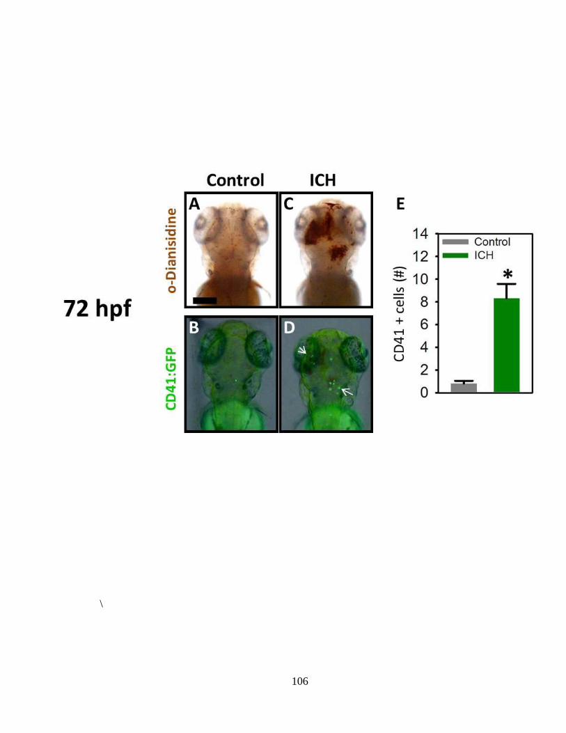
106
\
107
interest is that our model exhibits several of the clinical manifestations associated with
ICH pathophysiology, namely disruption of the blood-brain barrier and vascular
degeneration, hematoma expansion, apoptosis, inflammation, cerebral edema and
thrombosis. The optical transparency of the model, enhanced by the presence of tissue-
specific transgenic lines, and the reproducibility of the desired phenotypes make the
model applicable for high-throughput assessment of the efficacy and possible side-effects
of a broad range of small molecules that could promote hematoma resolution, mediate
inflammation, prevent apoptosis and neuro-degeneration and trigger vascular
stabilisation. Furthermore, the complete dissection of the entire suite of mechanisms
aiding recovery from ICH can yield important therapeutic potential.

108
Chapter 4
Disruption of catecholaminergic neuron
development and impaired locomotor
function in zebrafish with intracerebral
hemorrhage
109
Author contributions:
Ms. Sandra Noble and Mr. Rafael Godoy trained me for the use of
confocal microscopy and helped me in preparing and processing embryos
for imaging. Dr. Thomas W. Moon and Dr. Marc Ekker helped me design
the following experiments and assisted me in writing and revising this
chapter to ensure that the content is scientifically sound and the style is in
accordance with the FGPS requirements. I am profoundly grateful for the
assistance of the aforementioned individuals, without whom this chapter
would not have been completed.
Shahram Eisa-Beygi
May 14, 2013
110
4.1 Abstract
Hematoma expansion and perihematomal edema formation account for the
major causes of neurological deterioration following ICH. The extent of neurological
deterioration is generally exacerbated depending on the location and volume of
hematoma and the extent of edema. A recurrent problem with murine models is that the
degree of neuronal damage in tissues surrounding the hematoma is generally quantified
by means of laborious histological methods which can also be subject to bias. I have
previously shown that embryonic and larval exposure to statin drugs results in ICH in
zebrafish (Eisa-Beygi et al., 2013; Chapter 2). I have also documented some of the
conserved pathophysiological processes underlying ICH formation (Chapter 3), which
can potentiate neuronal damage or displacement.
The purpose of this aspect of my research is to evaluate some of the basic
neurological parameters in a zebrafish (Danio rerio) model of a pharmacologically-
induced ICH during development. To this end, the patterning and distribution of
dopaminergic (DA) neurons, response to sensory stimuli, and swimming speed were
assessed. Confocal microscopy using Tg(dat:EGFP);(Gata-1:DsRed) embryos revealed
that following ICH, blood components rapidly extravasate into the brain parenchyma
where DA-positive cells normally exhibit a spatially restricted distribution at 48 hpf.
Expansion of the hematoma is associated with loss and disorganisation of these DA
neurons, evidenced by marked reduction in dat:EGFP expression. Immunohistochemical
staining for tyrosine hydroxylase (TH), a marker for catecholaminergic neurons, showed
disruption of TH-positive cells in embryos demonstrating ICH. In addition, we observed
impaired swimming performance and response to tactile stimulation in these embryos, in
111
the absence of any apparent developmental defects. These analysis, will pave the way for
the modeling of ICH-induced neurodegeneration and, more importantly, establish the
basis for further studies into the possible mechanisms aiding functional recovery in
zebrafish and screening for putative neuroprotectants against ICH-induced neurotoxicity.
4.2 Introduction
There are presently no medical interventions or therapeutics to effectively
benefit in improving patient outcome after intracerebral hemorrhage or ICH (Broderick et
al., 2007). The rupture of cerebral vessels leads to the development of a space-occupying
hematoma in the brain parenchyma. Although ICH accounts for 10-15% of all stroke
cases (Counsell et al., 1995; Qureshi et al., 2005), it has a poorer prognosis than any other
form of stroke. The neurological deterioration in ICH patients is generally irreversible
and disabling (Hua et al., 2002). Several animal studies have been utilised to establish
prognostic tools in elucidating the neurological deficits induced by ICH and to test the
efficacy of drugs. Thanks to work on model organisms, there has been an upsurge, over
the last decade or so, in our understanding of the mechanisms underlying brain injury
induced by ICH.
The pathophysiological processes accompanying ICH (some of which are
reported in Chapter 3) can affect the viability of adjacent neurons. Several lines of
evidence have shown that the mechanical force exerted by the expanding hematoma and
edema can expand and displace neurons in the brain parenchyma (Hua et al., 2002;
Chang et al., 2011). In fact, the progressive deterioration of neurological condition in
patients with ICH is most often due to continuous bleeding and enlargement of the
112
hematoma (Kazui et al., 1996; Fujii et al., 1998). Similarly, as reported in Chapter 3,
zebrafish embryos demonstrating ICH display rapid hematoma expansion into the
adjacent brain regions and significant hydrocephalus in the forebrain and the ventricular
zones. These dynamic processes can account for the major causes for neuronal
disorganisation and neurotoxicity in the surrounding regions (Fujii et al., 1998). Other
secondary insults including macrophage activation, enhanced HO-1 expression and ROS
generation, could also potentiate further neurotoxicity in the form of loss and
disintegration of neurons. Each of these was also demonstrated in the zebrafish model
reported in Chapter 3.
In vitro studies have shown that activated macrophages can induce neuronal
toxicity through ROS generation and production of cytokines (Fordyce et al., 2005;
Kaushal et al., 2007). The extent of neuronal loss following ICH correlates, both
spatially and temporally, with the build-up of activated macrophages (Wasserman and
Schlichter, 2007a). Furthermore, iron overload due to break-up of blood components and
the lysis of the blood clot can induce neuronal apoptosis (Wagner et al., 2004; Hua et al.,
2006; Gu et al., 2009). In addition, the neurons in a developing brain are particularly
fragile (Anderson et al., 2000).
Following from Chapter 3 this study hypotheses that hemorrhages in the brain
can provoke neuronal damage. Therefore, as a first step in establishing a preclinical
transitional model for small molecule screening of neuroprotective agents, this study will
evaluate some of the basic and well-established neurological parameters in zebrafish
coinciding with ICH.
113
The general organisation of the central nervous system (CNS) between the
zebrafish and mammalian vertebrates share similar features, despite the apparent
differences in structural complexity and scale (Xi et al., 2011). Furthermore, the utility of
the zebrafish model for imaging and transgenesis has enabled the generation of lines
expressing fluorescent proteins in specific populations of neurons (Xi et al., 2011). For
example, ample information regarding the spatio-temporal expression and the function of
the dopaminergic (DA) neuron system has recently been reported in zebrafish thanks to
the generation of the dat:EGFP line where green fluorescent protein (GFP) is expressed
under the control of cis-regulatory elements of the dopamine transporter (dat) (Xi et al.,
2011). Analysis of these embryos has revealed that DA neurons can be detected as early
as 18 hpf as a collection of cells expressing GFP in the ventral diencephalon region (Xi et
al., 2011), thus making them ideal candidates to assess the possible neurotoxicity
associated with ICH.
DA neurons participate in various physiological functions including
neuroendocrine secretion, cognitive function, control of voluntary movement and
behavioural responses (Bjorklund and Lindvall, 1984). In addition, DA neurons are
shown to display similar function and expression patterns across vertebrates, including
zebrafish (Xi et al., 2011). The DA system was characterised in zebrafish development on
the basis of the expression patterns of tyrosine hydroxylase (TH) and the dopamine
transporter (DAT), both of which are specific markers of DA neurons (Holzschuh et al.,
2001; Rink and Wullimann, 2002; Zhao et al., 2012).
Furthermore, progressive degeneration of DA neurons is implicated in the
pathogenesis of Parkinson’s disease, which is characterised by reduced levels of
114
dopamine in the striatum and the accompanying movement and speech disorders
(Bjorklund et al., 2002). Similarly, selective ablation of DA neurons in developing
zebrafish results in locomotor defects, thus phenocopying some of the pathological and
behavioural aspects of the mammalian Parkinson’s disease (Zhao et al., 2012).
There is presently a need for development of an appropriate animal model that
can both facilitate the study of ICH and would be beneficial for evaluating novel
approaches to restore neurological function. Although several experimental models of
ICH have been developed (Kobari et al., 1988; Rosenberg et al., 1992; Wagner et al.,
1996; Del Bigio et al., 1996; Foerch et al., 2013), a major limitation of these models is
the fact that hematoma development, brain edema and neurological deterioration are
either evaluated by tedious histological analysis of different specimens or through
expensive magnetic resonance imaging (MRI). To bypass these challenges, there is a
need to use a model in which these pathophysiological and neurological consequences of
ICH can be evaluated in vivo, without sacrificing the animals. Such a model would allow
for the evaluation of the onset and progression of neurotoxicity following ICH, thus
allowing for the testing of the efficacy of potential therapeutics on these parameters. This
chapter will provide some preliminary evidence that zebrafish can provide a critical first
step in identifying novel and effective therapies for ICH-induced neurotoxicity. The
results outlined here can further strengthen the utility of zebrafish as an ideal model for
the study of ICH, at the level of etiology and pathophysiology.
115
4.3 Materials and Methods
4.3.1 Zebrafish husbandry and transgenic lines
Adult wild-type and transgenic zebrafish were maintained under a constant
temperature of 28°C and a 14 h light:10 h dark photoperiod in the University of Ottawa
Aquatic Care Facility. Embryos were obtained through natural breeding of adult zebrafish
and were kept at 28.5°C in E3 embryo medium (5 mmol/L NaCl, 0.17 mmol/L KCl, 0.33
mmol/L CaCl2, 0.33 mmol/L MgSO4). The double transgenic Tg(dat:EGFP);(gata-
1:DsRed) zebrafish line was obtained by crossing Tg(dat:EGFP) fish (Xi et al., 2011)
with Tg (gata-1:DsRed) fish (Traver et al., 2003). Pigmentation was inhibited by
incubating the embryos in 1-pheny1-2-thiourea (PTU) (Elsalini and Rohr, 2003). All
experiments were carried out in accordance with a protocol approved by the University of
Ottawa Protocol Review Committee and conform to the published guidelines of the
Canadian Council on Animal Care for the use of animals in research and teaching.
4.3.2 Drug treatment
For pharmacological inhibition of HMGCR, zebrafish embryos (1-2 hours post
fertilisation; hpf) were treated with Atorvastatin (ATV; Pfizer Inc., Connecticut USA) in
a single-exposure manner in embryo medium. ATV was dissolved in DMSO.Drug
solutions were aliquoted and stored at -20°C until used. Embryos were either incubated in
0.5 mg/L ATV in embryo medium or DMSO as vehicle control.
4.3.3 Confocal microscopy
For confocal microscopy, 48 hpf Tg(dat:EGFP);(gata-1:DsRed) embryos
previously treated with DMSO or 0.5 mg/L ATV were embedded in 1% low melting
116
agarose in E3-embryo medium and tricaine mesylate (Sigma-Aldrich). A rendered Z-
stack, at ~80 micron thickness each, from the ventral head position, was taken with a
Zeiss LSM 510 AxioImager.M1 confocal microscope using an Achroplan 40x/0.8 W
objective with an argon laser (488 nm) and a helium-neon laser (543 nm).
4.3.4 Whole-mount anti-tyrosine hydroxylase (TH) immunohistochemistry
Zebrafish embryos at 48 hpf were fixed in 4% paraformaldehyde (PFA)/1X
Phosphate Buffered Saline Tween-20 (PBST) overnight, rinsed and stored at -20°C in
100% EtOH. Upon re-hydration, the embryos were then permeabilised by incubation in
acetone at -20°C or 1% Triton/1X PBST for 30 min at room temperature, followed by
blocking in 10% calf-serum/1X PBST, 0.1% BSA for 1 h at room temperature. Embryos
were then incubated in a mouse monoclonal anti-tyrosine hydroxylase primary antibody
(EMD Millipore, AB152) (1:200 diluted in 10% calf-serum blocking solution) overnight
at 4°C. The next day, samples were washed with repeated washes in 1X PBST for 1 h.
This was followed by incubation with secondary antibody conjugated with horseradish
peroxidase (HRP). The signal was detected using DAB Peroxidase Subtrate Kit. After
staining, zebrafish were mounted with 3.5% methylcellulose and imaged, with the ventral
head region facing the field of view.
4.3.5 Testing for locomotor activity
The response of embryos to tactile stimulation at 48 hpf was evaluated using a
method previously described by Xi et al. (2010). In brief, the embryos were first
individually placed in a petri dish containing E3 medium and allowed to habituate for 10
min, after which a gentle tactile stimulus using forceps was applied to the tail of the
117
embryo and the response was recorded using a high-speed digital video camera (DXM
1200C). For each treatment condition, 25 embryos were used. The numbers of embryos
with an immediate or delayed response were manually counted. Embryos exhibiting no
tail movement or an absence of any movement were also noted. For statistical analysis, a
student's t-test was conducted and average values ± standard errors of the mean was
plotted. All data analysis was performed using SigmaPlot 11.0 software Systat Software
Inc, Chicago, IL).
For the assessment of swimming speed, we used a high-speed digital video
camera (FASTCAM) to measure the time it took for an embryo to disappear from the
microscopic field of view immediately following the application of the tactile stimulus.
4.4 Results and Discussion
4.4.1 Pharmacologically induced ICH disrupts catecholaminergic neuron
development and swimming behaviour in zebrafish
There are a plethora of animal studies highlighting the ICH-induced
neurotoxicity and behavioural defects. However, these studies are generally limited by
the labour-intensive histological analysis, unfeasibility of in vivo imaging, high cost,
external injection of collagenase or autologous blood into the brain parenchyma, and the
inability to control the heterogeneity in lesion size or neurological outcome. Nonetheless,
I have provided several lines of evidence in previous chapters that these aforementioned
limitations do not apply to zebrafish. In fact, zebrafish is gaining recognition as an
alternative model for ICH research (Gore et al., 2012; Butler et al., 2011). As mentioned
118
in these earlier chapters, an ICH phenotype can consistently be induced by incubating
embryos in a solution containing 0.5 mg/L ATV.
In order to simultaneously establish the progression of the hematoma and the
distribution of DA neuron networks in embryos with ICH, I out-crossed the
Tg(dat:EGFP) line (Xi et al., 2011) with the Tg(gata-1:DsRed) line (Traver et al., 2003)
and obtained a line expressing both transgenes. Using confocal microscopy on
Tg(dat:EGFP);(gata-1:DsRed) embryos, I observed at the onset of hemorrhage, the
aggregation of DsRed-positive erythrocytes into the pretectum and telencephalon regions
of the brain, where distinct populations of GFP-expressing DA neurons exhibit a
spatially-restricted distribution (Fig. 4.1B, D). By contrast, in embryos treated with
DMSO, the blood-brain barrier remains intact, and the DA neurons do not come into
contact with potentially cytotoxic blood components or are they subjected to the
mechanical stress of the hematoma (Fig. 4.1A, C). This non-invasive approach, which
takes advantage of the relatively translucent nature of the embryos and the prolific nature
of the zebrafish, enables us to assess the patterning and distribution of DA neurons in
response to hematoma expansion at various stages of development in vivo. As a further
measure of the neurological status in response to hemorrhage, individual neurons can also
be quantified, at higher magnification. Furthermore, it has been shown that DA neurons
can be partially replaced through proliferation of new DA neurons as a consequence of
selective ablation (Xi et al., 2011). Hence, the potential processes of neuroplasticity and
proliferation can also be evaluated in vivo without resorting to laborious histological
analysis.
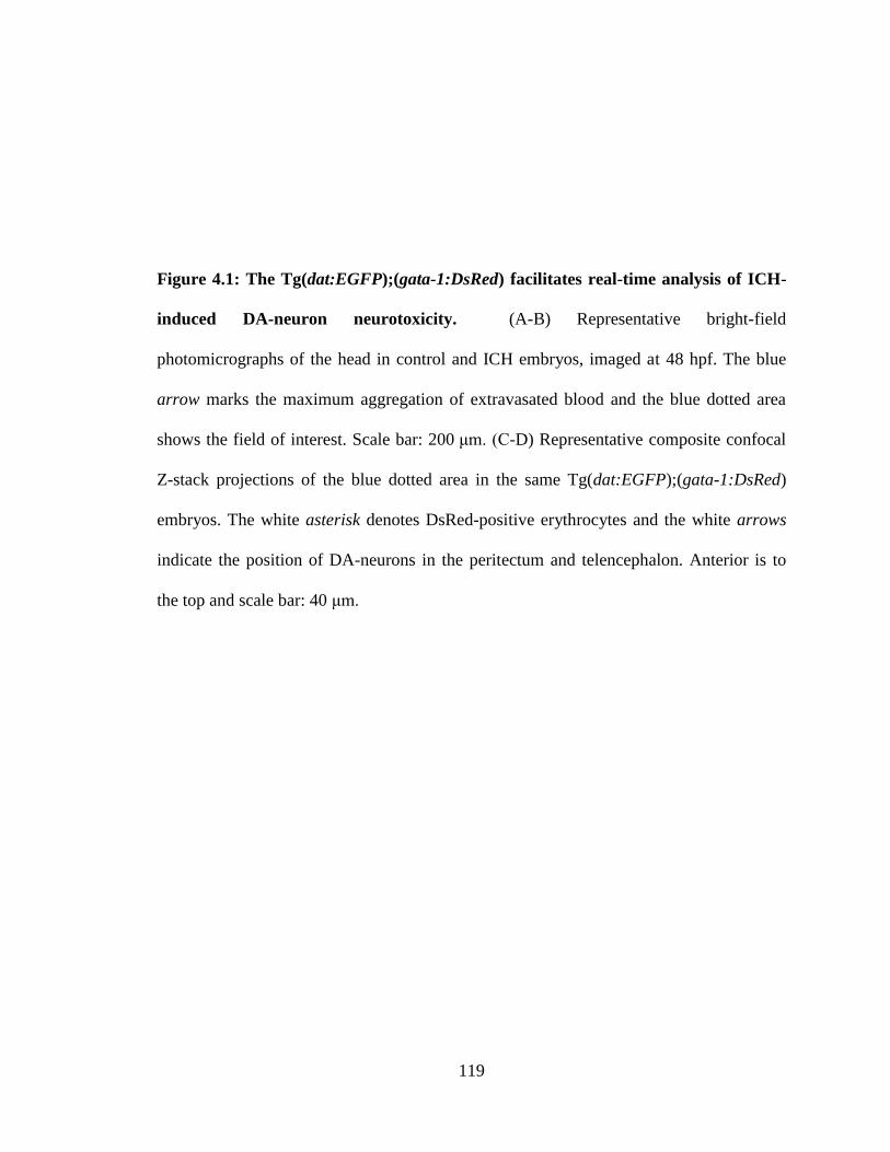
119
Figure 4.1: The Tg(dat:EGFP);(gata-1:DsRed) facilitates real-time analysis of ICH-
induced DA-neuron neurotoxicity. (A-B) Representative bright-field
photomicrographs of the head in control and ICH embryos, imaged at 48 hpf. The blue
arrow marks the maximum aggregation of extravasated blood and the blue dotted area
shows the field of interest. Scale bar: 200 μm. (C-D) Representative composite confocal
Z-stack projections of the blue dotted area in the same Tg(dat:EGFP);(gata-1:DsRed)
embryos. The white asterisk denotes DsRed-positive erythrocytes and the white arrows
indicate the position of DA-neurons in the peritectum and telencephalon. Anterior is to
the top and scale bar: 40 μm.
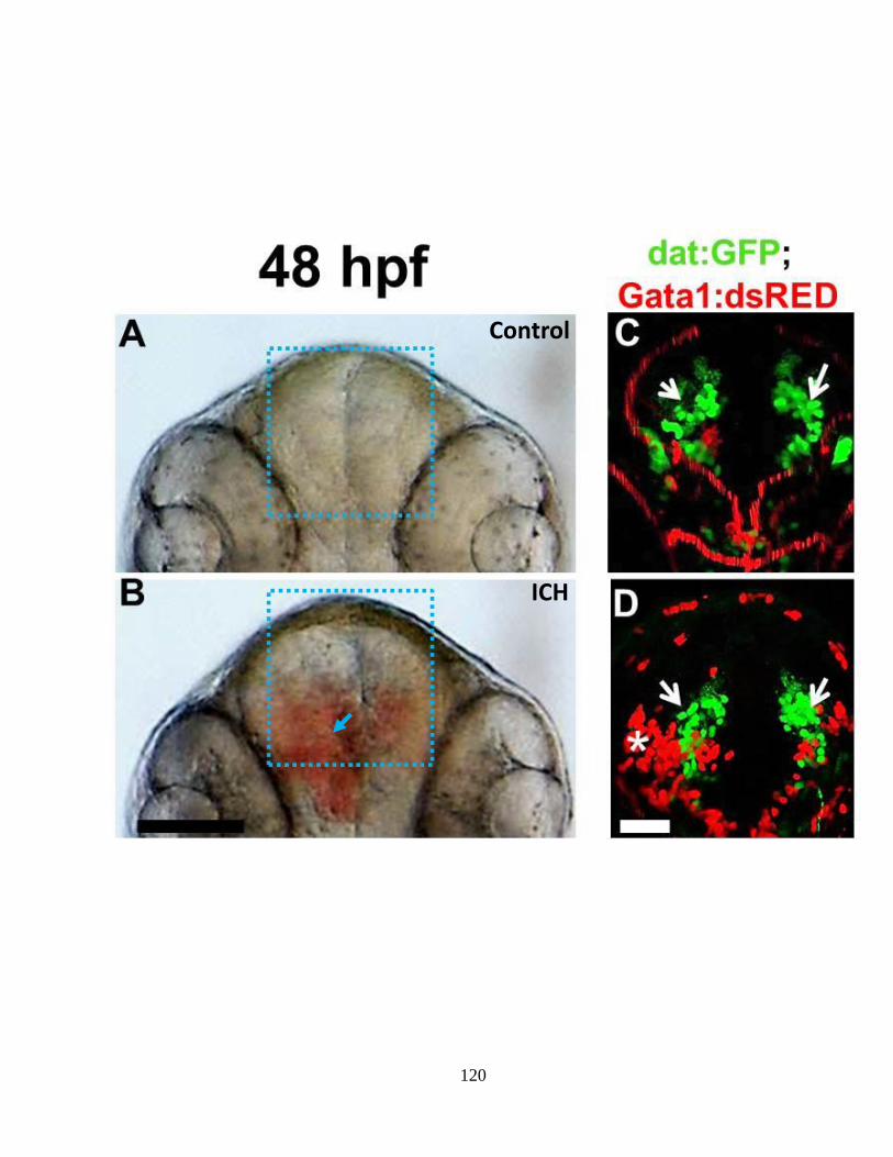
120
Control
ICH
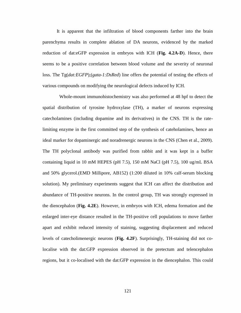
121
It is apparent that the infiltration of blood components farther into the brain
parenchyma results in complete ablation of DA neurons, evidenced by the marked
reduction of dat:eGFP expression in embryos with ICH (Fig. 4.2A-D). Hence, there
seems to be a positive correlation between blood volume and the severity of neuronal
loss. The Tg(dat:EGFP);(gata-1:DsRed) line offers the potential of testing the effects of
various compounds on modifying the neurological defects induced by ICH.
Whole-mount immunohistochemistry was also performed at 48 hpf to detect the
spatial distribution of tyrosine hydroxylase (TH), a marker of neurons expressing
catecholamines (including dopamine and its derivatives) in the CNS. TH is the rate-
limiting enzyme in the first committed step of the synthesis of cateholamines, hence an
ideal marker for dopaminergic and noradrenergic neurons in the CNS (Chen et al., 2009).
The TH polyclonal antibody was purified from rabbit and it was kept in a buffer
containing liquid in 10 mM HEPES (pH 7.5), 150 mM NaCl (pH 7.5), 100 ug/mL BSA
and 50% glycerol.(EMD Millipore, AB152) (1:200 diluted in 10% calf-serum blocking
solution). My preliminary experiments suggest that ICH can affect the distribution and
abundance of TH-positive neurons. In the control group, TH was strongly expressed in
the diencephalon (Fig. 4.2E). However, in embryos with ICH, edema formation and the
enlarged inter-eye distance resulted in the TH-positive cell populations to move farther
apart and exhibit reduced intensity of staining, suggesting displacement and reduced
levels of catecholimenergic neurons (Fig. 4.2F). Surprisingly, TH-staining did not co-
localise with the dat:GFP expression observed in the pretectum and telencephalon
regions, but it co-localised with the dat:GFP expression in the diencephalon. This could
122
be due to reduced TH expression in those cells at this stage or the low sensitivity of
colourimetric immunohistochemistry.
As a further measure of neurological outcome following ICH, I assessed
locomotor function (swimming speed, the acuteness of response to tactile stimulation in
these embryos). Zebrafish embryos have been shown to respond to tactile stimulation as
early as 24 hpf (Saint-Amant and Drapeau, 2000) and these behavioural responses have
been well-characterised at various stages of development (McLean and Fetcho, 2008). A
behavioural response is simply elicited by applying a tactile stimulation at the tail or head
regions. This response is typically expressed as a twitch at 1 dpf to a rapid escape by 2
dpf. Assessment of locomotor function is another method by which I can further
document ICH-induced neurological defects.
To accomplish this, embryos with or without ICH were individually subjected to
a gentle tactile stimulus (see Materials and Methods) at 48 hpf 10 min after individual
transfer to a petri dish. Individual responses were characterised as “immediate” (requiring
one touch), “delayed” (requiring at most three touches) or as “no-response”. Whereas
more than 80% of DMSO-treated embryos (n=25 embryos) displayed an immediate
response, of the embryos demonstrating ICH, only approximately 23% of this group
(n=25) showed rapid escape behaviour. In most embryos with ICH (more than 40%),
several repeated tactile stimuli were applied in order to provoke an escape response. In
addition, approximately 30% of embryos with ICH showed no response, even after three
repetitive tactile stimuli were applied. Differences in the mean proportion of responses to
tactile stimulation in the control and ICH groups were measured using a Student’s t-test
123
(N=25 embryos per treatment by only comparing the mean values for each of the three
response categories (Fig. 3A).
In addition, preliminary evidence regarding swimming speed was obtained upon
administration of a gentle tactile stimulus. Using a high-speed digital video camera and
ImageJ software, it took approximately 0.09 sec for control embryos to escape out of the
field of view, accompanied with high frequency tail-beat frequency. By contrast, embryos
with ICH showed lower frequency undulation of the tail, and, traveled at a much slower
speed, as judged by the slower time it takes them to disappear from the field of view (Fig.
3B).
Although it has been shown that reduced locomotion and weakened response to
sensory stimulus constitute part of the pathophysiology of DA neuron loss in zebrafish
(Xi et al., 2011), the observed phenotype could just as well be attributed to
musculoskeletal defects or loss of mechanosensory neurons. Further studies are warranted
to elaborately dissect the spatio-temporal fate of different subtypes of neurons, before and
after the induction of hemorrhage and to expand on the functional consequences of
neuronal loss as well as establish precisely the molecular basis for the observed
locomotor defects. In light of the detrimental neurological outcome of ICH in murine
models and patients, it is worth underscoring that zebrafish exhibit a tendency for full
functional recovery. Hence, there is merit in exploring how neurological function is
regained after stroke, which can yield potential benefits in the treatment of ICH.
124
Figure 4.2: Disruption of catecholaminergic-neuron development in zebrafish with
ICH. (A-B) Representative bright-field photomicrographs of the head in control and ICH
embryos, imaged at 48 hpf. The asterisk marks the aggregation of extravasated blood and
the black dotted area shows the field of interest. Anterior is to the top and scale bar:
200 μm. (C-D) Representative fluorescent photomicrographs of the black dotted area in
Tg(dat:EGFP) embryos. White arrows indicate the position of DA-neurons (E-F).
Representative bright-field photomicrographs of the black dotted area in wild-type
embryos immunostained for TH. Black arrows indicate the position of TH-positive
neurons.
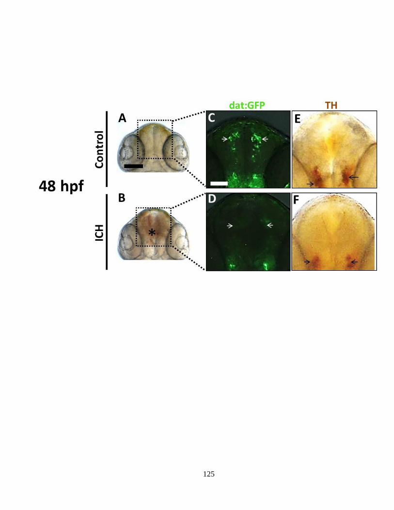
125
126
Figure 4.3: Impaired locomotor function in zebrafish with ICH. (A) Quantification
of the percentage of embryos showing immediate, delayed or no response in the wake of
gentle tactile stimulation. Differences in the mean proportion of responses to tactile
stimulation in the control and ICH groups were measured using Student’s t-test (25
embryos were used per each treatment. N=3 separate experiments) by only comparing the
mean values for immediate (p<0.001), delayed (p<0.05) and no-response categories. (B)
Representative photomicrographs of 3 separate experiments showing stimulus-evoked
escape response over different time frames, measured using a high-speed digital video
camera (FASTCAM).
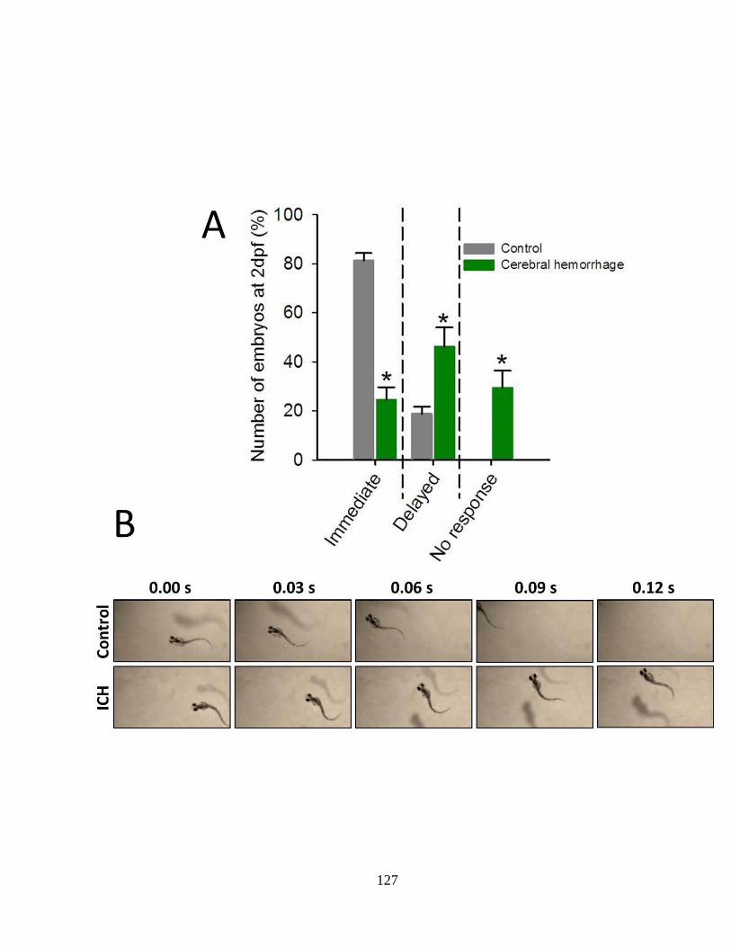
127

128
Chapter 5
General Discussion & Future Directions
129
Intracerebral hemorrhage (ICH) accounts for 10-15% of all stroke subtypes.
Furthermore, it has the highest mortality and morbidity rates of stroke cases (Counsell et
al., 1995; Qureshi et al., 2005). The hemorrhages in the CNS are triggered by various
factors and these also include cerebral cavernous malformations (CCMs), which can arise
sporadically or through a familial autosomal pattern of inheritance (Maeder et al., 1998;
Boulday et al., 2011, Eerola et al., 2003). Neither surgical evacuation nor
pharmacological interventions have been shown to confer clear post-stroke benefits to
individuals with ICH (Broderick et al., 1999; Badjatia and Rosand, 2005; Steiner et al.,
2011). This bleak outcome is due predominantly to rapid hematoma expansion, activation
of inflammatory mediators, disintegration of cranial vasculature, and focal neurological
deterioration (Davis et al., 2006; Zhao et al., 2007; Lotfspring et al., 2009; Huang et al.,
2012).
As a consequence, in addition to optimising and investigating novel treatment
options, more research needs to be directed towards gaining insights into the
development and maintenance of cerebral-vascular stabilisation. For this reason, it is
imperative to develop novel approaches in order to characterise the etiological and
pathophysiological processes underlying ICH genesis and progression, since treatment
options remain limited and ineffectual. This kind of an approach would pave the way for:
A) In vivo assessment of small molecules and pharmaceuticals to identify
modifiers of cerebral-vascular stabilisation;
B) Identification of additional genes and signalling pathways mediating
cerebral-vascular stabilisation; and,
130
C) Sequencing for mutations in these newly identified candidate genes in
human vascular disorders for early screening.
This can be accomplished through comparative analysis of different model
organisms, including zebrafish, along with functional work and pathway analysis.
Zebrafish, Danio rerio, continues to gain recognition as an ideal model for the study of
ICH given that the genes and molecular mechanisms contributing to vascular
morphogenesis are highly conserved across vertebrates (Gore et al., 2012) and that this
species is highly amenable to functional/expression analyses and imaging assays.
For my Ph.D. project I was primarily interested in defining the precise
mechanisms through which the HMGCR pathway contributes to cerebral-vascular
stabilisation in the context of a developing vertebrate. In its final format, this dissertation
further contributes to our understanding of the etiology of ICH and the ensuing
pathophsyiology. It is worth noting that this study was also motivated by the emerging
clinical evidence, which suggested that statin drug use confers an increased risk for ICH
in humans (Amarenco et al., 2006; Arboix et al., 2010; Westover et al., 2011) and that no
follow-up animal studies have, hitherto, been conducted to further explore and test this
clinical association. Could inhibition of HMGCR weaken CNS blood vessels and make
them prone to hemorrhage? There is already abundant evidence from in vitro studies that
statin drug use can induce apoptosis in endothelial cells (ECs) and smooth muscle cells
(Weiss et al., 2002; Hippenstiel et al., 2002; Acquevella et al., 2010; Guijarro et al.,
1999). Are the results strictly confined to zebrafish embryos and larvae? Does these
findings harbour any clinical relevance and, if so, what are the possible mechanistic
explanations?
131
To address these questions, it was important to first look at the processes that
regulate vascular stabilisation. Abnormal endothelial barrier function can be caused by
inadequate investment by mural cells and/or due to disruption of endothelial cell-cell
contacts. This is associated with aneurysms in micro-vascular channels, loss of vascular
stability and frequent hemorrhaging in the CNS (Maeder et al., 1998). Although these
defects in vascular stabilisation are equally likely to arise in vascular beds throughout the
body, the vessels in the CNS region seem to be particularly prone to hemorrhaging. The
severity of these vascular lesions can vary greatly and the onset can be as early as
embryonic or postnatal stages of development (Boulday et al., 2011). Mice and rats have
generally been utilised as models for elucidating the genetic basis and the signalling
pathways necessary for vascular stabilisation. As a result of these studies and based upon
sequencing of patients with familial cavernous malformations, several loss-of-function
mutations affecting endothelial barrier function have been identified. These mutations
result in the disruption of vascular stability and have been implicated in the pathogenesis
of ICH in both murine models and humans (Sahoo et al., 1999; Verlaan et al., 2004).
Studies in animal models have greatly contributed to our current knowledge
regarding the etiology, pathophysiology and neuro-pathology associated with ICH.
However, there is a need to identify novel genes and additional interacting signalling
molecules that contribute to the disease given that the prevalence of cerebral cavernous
malformations (CCMs) in the general population is relatively high (estimated to be 1 in
200) and the genes implicated in the pathogenesis of CCM (KRIT1, CCM2, and
PDCD10) account for only part of the disease phenotype (Cunningham et al., 2011).
However, despite the characterisation of these genes, relatively little is known about their
132
mode of function. In addition, almost nothing is known about the mechanisms leading to
the vascular lesions and hemorrhage in individuals harbouring these mutations. To further
complicate this issue, not all of the individuals exhibiting a CCM phenotype carry
mutations in any of three CCM genes, suggesting additional genetic modifiers that can
increase the susceptibility of individuals to the CCM pathology (Lucas et al., 2003).
The three genes implicated in CCM have similar expression patterns. Their
transcripts are particularly enriched in endothelial cell-cell junctions (Fischer et al.,
2013). The CCM proteins, all of which are non-homologous and lack catalytic domains,
interact to establish a protein complex (Serebriiskii et al., 1997; Glading et al., 2007;
Voss et al., 2007). These proteins have also been shown to interact with Rho GTPases to
maintain the functional integrity of ECs. However, models explaining this mechanism are
only speculative. Expression analyses show that loss-of-function mutations in CCM
genes are associated with hyper-activation and over-expression (protein and mRNA) of
Rho GTPases (Li and Whitehead, 2010). However, the precise nature of the signalling
cascades under the control of CCM genes remains to be explored. It is reasonable to
assert that blocking HMGCR function would invariably reduce Rho GTPase signalling
and, hence, can disrupt vascular stabilisation.
In sharp contrast with this assertion, a recent study in mice harbouring a genetic
predisposition for cerebral-vascular permeability defect resulting from heterozygous
mutation of CCM2, statin-treatment restores the endothelial barrier function by inhibiting
Rho GTPase activation (Whitehead et al., 2009). This result is conflicting and warrants
further studies.
133
As such, the identification and characterisation of genes and signalling pathways
contributing to cerebral-vascular stabilisation was one of the chief aims of my PhD
project. In this project I obtained numerous lines of evidence to support the hypothesis
that down-regulation of HMGCR activity can exert an effect on signalling pathways
under the control of CCM-interacting proteins, such as Rho GTPases.
More specifically, I demonstrated that geranylgeranylation of Rho GTPases is a
requirement for cerebral-vascular stabilisation in developing zebrafish, particularly at a
time when blood vessels lack adequate investment by smooth muscle cells. In these
embryos, inhibition of the HMGCR/mevalonate pathway results in bulged CNS blood
vessels, loss of vascular stability, vessel fragmentation and frequent cerebral
hemorrhages. Similar results were obtained when geranlygeranylation was blocked by
inhibition of GGTase I. These observations made in embryonic zebrafish resemble the
CCM lesions observed in murine models and human patients. Additionally, an identical
vascular-specific phenotype was reported in zebrafish in which components of Rho
GTPase signalling were down-regulated due to point-mutations in those genes (Buchner
et al., 2007; Liu et al., 2007). Lending further support to my findings is that at least two
recent studies independently confirmed a functional role for HMGCR in cerebral-
vascular stabilisation in zebrafish (Hung et al., 2012, Shen et al., 2013). An issue which I
could not address was the cell autonomy of HMGCR-function. Is HMGCR function
required in an endothelial-specific manner or does it also involve contribution from mural
cells?
On the basis of the data accumulated thus far, and based on the fact that
vascular stabilisation pathways in humans and zebrafish share common features (Gore et
134
al., 2012), it is reasonable to propose that sequencing of humans for mutations in the
HMGCR and GGTase I genes could be a valid diagnostic approach. Not only are these
gene sequences highly conserved across species, they also seem to functionally perform
identical enzyme functions.
In zebrafish embryos demonstrating ICH, the disruption of the blood-brain
barrier and the initiation of hemorrhage is coincident with vascular degeneration,
hematoma expansion, apoptosis, inflammation, cerebral edema and thrombosis.
Furthermore, I have shown that the extravasation of blood components into the brain
parenchyma can induce progressive loss of DA neurons, along with displacement of TH-
positive (catecholaminergic) neuronal populations. Concomitant with the apparent
neurotoxicity, I presented preliminary evidence for impaired locomotor function and
diminished response to tactile stimulus in these ICH embryos.
This is the first zebrafish study assessing the parameters that are hallmarks of
ICH pathophysiology and are points of intervention for future studies geared towards
large-scale screening of zebrafish for therapeutics to mitigate symptoms of ICH. The
optical transparency of the model, enhanced by the presence of tissue-specific transgenic
lines, as well as the reproducibility of the desired phenotypes make the model applicable
for high-throughput assessment of the efficacy and possible side-effects of a broad range
of small-molecules that could promote hematoma resolution, mediate inflammation and
minimise neuronal damage. Such a phenotype-based screening, facilitated by the
presence of stable transgenic lines marking unique clusters of neurons, also enables
assessment of neuroprotective agents.
135
References
Abramsson, A., Berlin, O., Papayan, H., Paulin, D., Shani, M. and Betsholtz, C.
(2002). Analysis of mural cell recruitment to tumor vessels. Circulation. 105, 112–
117.
Acquavella, N., Quiroga, M.F., Wittig, O. and Cardier, J.E., (2009). Effect of
simvastatin on endothelial cell apoptosis mediated by Fas and TNF-alpha.
Cytokine. 49, 45-50.
Adams, R.H. and Alitalo, K. (2007). Molecular regulation of angiogenesis and
lymphangiogenesis. Nat. Rev. Mol. Cell Biol. 8, 464–478.
Alejandre, M.J., Ramirez, H., Suarez, M.D. and Garcia-Peregrin, E. (1981). Different
developmental patterns of 3-hydroxy-3-methylglutaryl-CoA reductase in chick
tissues according to their role in cholesterogenesis. Biol. Neonate. 40, 232-236.
Amarenco, P., Bogousslavsky, J., Callahan, A., 3rd, Goldstein, L.B., Hennerici, M.,
Rudolph, A.E. et al. (2006). High-dose atorvastatin after stroke or transient
ischemic attack. N. Engl. J. Med. 355, 549–559.
Anderson, V., Catroppa, C., Morse, S., Haritou, F. and Rosenfeld, J. (2000). Recovery
of intellectual ability following traumatic brain injury in childhood: impact of
injury severity and age at injury. Pediatr. Neurosurg. 32, 282-290.
Anichtchik, O., Diekmann, H., Fleming, A., Roach, A., Goldsmith ,P., and
Rubinsztein, DC. (2008). Loss of PINK1 function affects development and results
in neurodegeneration in zebrafish. J. Neurosci. 28, 8199-8207.
Arboix, A., García-Eroles, L., Oliveres, M., Targa, C., Balcells, M. and Massons, J.
(2010). Pretreatment with statins improves early outcome in patients with first-
ever ischaemic stroke: a pleiotropic effect of statins or a beneficial effect of
hypercholesterolemia? BMC Neurol. 10, 47.
Babamahmoodi, F. and Babamahmoodi, A. (2011). Brucellosis, Presenting with
Guillain-Barré Syndrome. J. Glob. Infect. Dis. 3, 390-392.
Badjatia, N., and Rosand, J. (2005). Intracerebral hemorrhage. Neurologist. 11, 311-
324.
Bagrodia, S., Taylor, S. J., Jordon, K. A., van Aelst, L. and Cerione, R. A. (1998). A
novel regulator of p21-activated kinases. J. Biol. Chem. 273, 23633-23636.
Ballabh, P. Intraventricular hemorrhage in premature infants: mechanism of disease.
Pediatr Res. 67, 1-8.
Bayliss, P.E., Bellavance, K.L., Whitehead, G.G., Abrams, J.M., Aegerter, S.,
Robbins, H.S., Cowan, D.B., Keating, M.T., O'Reilly, T., Wood, J.M., Roberts,
136
T.M. and Chan, J. (2006). Chemical modulation of receptor signaling inhibits
regenerative angiogenesis in adult zebrafish. Nat. Chem. Biol. 2, 265-273.
Belayev, L., Saul, I., Curbelo, K., Busto, R., Belayev, A., Zhang, Y., Riyamongkol, P.,
Zhao, W. and Ginsberg, M.D. (2003). Experimental intracerebral hemorrhage in
the mouse: histological, behavioral, and hemodynamic characterization of a
double-injection model. Stroke. 34, 2221-2227.
Benjamin, L.E., D. Golijanin, A. Itin, D. Pode, and E. Keshet. (1999). Selective
ablation of immature blood vessels in established human tumors follows vascular
endothelial growth factor withdrawal. J. Clin. Invest. 103, 159–165.
Bergametti, F., Denier, C., Labauge, P., Arnoult, M., Boetto, S., Clanet, M., Coubes,
P., Echenne, B., Ibrahim, R., Irthum, B. et al. (2005). Mutations within the
programmed cell death 10 gene cause cerebral cavernous malformations. Am. J.
Hum. Genet. 76, 42–51.
Berzat, A.C., Brady, D.C., Fiordalisi, J.J. and Cox, A.D. (2006). Using inhibitors of
prenylation to block localization and transforming activity. Methods Enzymol.
407:575-597.
Bishop, A.L. and Hall, A. (2000). Rho GTPases and their effector proteins. Biochem.
J. 348, 241–255.
Bjorklund, L.M., Sánchez-Pernaute, R., Chung, S., Andersson, T., Chen, I.Y.,
McNaught, K.S., Brownell, A.L., Jenkins, B.G., Wahlestedt, C., Kim, K.S. and
Isacson, O. (2002). Embryonic stem cells develop into functional dopaminergic
neurons after transplantation in a Parkinson rat model. Proc. Natl. Acad. Sci. U S
A. 99, 2344-2349.
Bjorklund, A. and Lindvall, O. Dopamine-containing systmes in the CNS. In:
Handbook of Chemical Neuroanatomy: Classical Transmitter in the Rat, edited by
Bjorklund A, Hokfelt T. Amsterdam: Elsevier. 55–122.
Borghi, C, Prandin, M.G., Costa, F.V., Bacchelli, S., Degli Esposti, D. and
Ambrosioni, E. (2000). Use of statins and blood pressure control in treated
hypertensive patients with hypercholesterolemia. J. Cardiovasc. Pharmacol. 35,
549–555.
Boulday, G., Rudini, N., Maddaluno, L., Blécon, A., Arnould, M., Gaudric, A.,
Chapon, F., Adams, R.H., Dejana, E and Tournier-Lasserve, E. J. (2011).
Developmental timing of CCM2 loss influences cerebral cavernous malformations
in mice. Exp. Med. 208, 1835-1847.
Breier, G., Albrecht, U., Sterrer, S. and Risau, W. (1992). Expression ofvascular
endothelial growth factor during embryonic angiogenesis andendothelial cell
differentiation. Development. 114, 521-532.
137
Broderick, J.P., Brott, T., Tomsick. T., Miller, R. and Huster, G. (1993). Intracerebral
hemorrhage more than twice as common as subarachnoid hemorrhage. J.
Neurosurg. 78, 188–191.
Broderick, J.P., Adams, H.P.Jr., Barsan, W., Feinberg, W., Feldmann, E., Grotta, J.,
Kase, C., Krieger, D., Mayberg, M., Tilley, B., Zabramski, J.M. and Zuccarello,
M. (1999). Guidelines for the management of spontaneous intracerebral
hemorrhage: a statement for healthcare professionals from a special writing group
of the Stroke Council, American Heart Association. Stroke. 30, 905–915.
Broman, M.T., Kouklis, P., Gao, X., Ramchandran, R., Neamu, R.F., Minshall, R.D.
and Malik A.B.(2006). Cdc42 regulates adherens junction stability and endothelial
permeability by inducing alpha-catenin interaction with the vascular endothelial
cadherin complex. Circ. Res. 98, 73–80.
Brown, M.S. and Goldstein, J.L. (1997). The SREBP pathway: regulation of
cholesterol metabolism by proteolysis of a membrane-bound transcription factor.
Cell. 89, 331-340.
Brown, M.S. and Goldstein, J.L. (1999). A proteolytic pathway that controls the
cholesterol content of membranes, cells, and blood. Proc. Natl. Acad. Sci. U S A.
96, 11041-11048.
Buchner, D.A., Su, F., Yamaoka, J.S., Kamei, M., Shavit, J.A., Barthel, L.K., McGee,
B., Amigo, J.D., Kim, S., Hanosh, A.W., Jagadeeswaran, P., Goldman, D.,
Lawson, N.D., Raymond, P.A., Weinstein, B.M., Ginsburg, D. and Lyons, S.E.
(2007). pak2a mutations cause cerebral hemorrhage in redhead zebrafish. . Proc.
Natl. Acad. Sci. USA. 104, 13996–14001.
Butler, M.G., Gore, A.V. and Weinstein, B.M. (2011). Zebrafish as a model for
hemorrhagic stroke. Methods Cell Biol. 105:137-161
Cao, J., Wang, J., Qi, W., Miao, H.H., Wang, J., Ge, L., DeBose-Boyd, R.A., Tang,
J.J., Li, B.L. and Song, B.L. (2007). Ufd1 is a cofactor of gp78 and plays a key
role in cholesterol metabolism by regulating the stability of HMG-CoA reductase.
Cell Metab. 6, 115-128.
Cattelino, A., Liebner, S., Gallini, R., Zanetti, A., Balconi, G., Corsi, A., Bianco, P.,
Wolburg, H., Moore, R., Oreda, B., Kemler, R. and Dejana, E. (2003). The
conditional inactivation of the beta-catenin gene in endothelial cells causes a
defective vascular pattern and increased vascular fragility. J. Cell Biol. 2003. 162,
1111-1122.
Caveda, L., Martin-Padura, I., Navarro, P., Breviario, F., Corada, M.,Gulino, D.,
Lampugnani, M. G. and Dejana, E. (1996). Inhibition ofcultured cell growth by
vascular endothelial cadherin (cadherin-5/VE-cadherin). J. Clin. Invest. 98, 886-
893.
138
Chang, C.F., Chen, S.F., Lee, T.S., Lee, H.F., Chen, S.F. and Shyue, S.K. (2011).
Caveolin-1 deletion reduces early brain injury after experimental intracerebral
hemorrhage. Am. J. Pathol. 178, 1749-1761.
Chen, Y.C., Priyadarshini, M. and Panula, P. (2009). Complementary developmental
expression of the two tyrosine hydroxylase transcripts in zebrafish. Histochem.
Cell Biol. 132, 375-381.
Choi, J., Mouillesseaux, K., Wang, Z., Fiji, H.D., Kinderman, S.S., Otto, G.W.,
Geisler, R., Kwon, O. and Chen, J.N. (2011). Aplexone targets the HMG-CoA
reductase pathway and differentially regulates arteriovenous angiogenesis.
Development. 138, 1173-1181.
Clark, W., Gunion-Rinker, L., Lessov, N. and Hazel, K. (1998). Citicoline treatment
for experimental intracerebral hemorrhage in mice. Stroke. 29, 2136-2140.
Clarke, P.R. and Hardie, D.G. (1990). Regulation of HMG-CoA reductase:
identification of the site phosphorylated by the AMP-activated protein kinase in
vitro and in intact rat liver. EMBO J. 9, 2439–2446.
Costa, C., Incio, J. and Soares, R. (2007). Angiogenesis and chronic inflammation:
cause or consequence? Angiogenesis 10, 149–166.
Counsell, C., Boonyakarnkul, S., Dennis, M., Sandercock, P., Bamford, J., Burn, J.
and Warlow, C. (1995). Primary intracerebral hemorrhage in the Oxfordshire
Community Stroke Project. Cerebrovasc Dis. 5, 26–34.
Crawford, T.N., Alfaro, D.V., Kerrison, J.B. and Jablon, E.P. (2009). Diabetic
retinopathy and angiogenesis. Curr. Diabetes Rev. 5, 8–13.
Cunningham, K., Uchida, Y., O'Donnell, E., Claudio, E., Li, W., Soneji, K., Wang, H.,
Mukouyama, Y.S. and Siebenlist, U. (2011). Conditional deletion of Ccm2 causes
hemorrhage in the adult brain: a mouse model of human cerebral cavernous
malformations. Hum. Mol. Genet. 20, 3198-3206.
D’amico, L., Scott, I.C., Jungblut, B. and Stainier, D.Y. (2007). A mutation in
zebrafish hmgcr1b reveals a role for isoprenoids in vertebrate heart-tube formation.
Curr. Biol. 17, 252–259.
Das, M., Ithychanda, S.S., Qin, J. and Plow, E.F. (2011). Migfilin and filamin as
regulators of integrin activation in endothelial cells and neutrophils. PLoS One. 6,
e26355.
Davis, S.M., Broderick, J., Hennerici, M., Brun, N.C., Diringer, M.N., Mayer, S.A., et
al. (2006). Hematoma growth is a determinant of mortality and poor outcome after
intracerebral hemorrhage. Neurology. 66, 1175-1181.
DeBose-Boyd, R.A. (2008). Feedback regulation of cholesterol synthesis: sterol-
accelerated ubiquitination and degradation of HMG CoA reductase. Cell Res. 18,
609-621.
139
Dejana, E. and Del Maschio, A. (1995). Molecular organization andfunctional
regulation of cell to cell junctions in the endothelium. Thromb. Haemost. 74, 309-
312.
Del Bigio, M., Yan, H., Buist, R. and Peeling, J. (1996). Experimental intracerebral
hemorrhage in rats. Magnetic resonance imaging and histopathological correlates.
Stroke. 27, 2312–2319.
Denier, C., Goutagny, S., Labauge, P., Krivosic, V., Arnoult, M., Cousin, A., Benabid,
A. L., Comoy, J., Frerebeau, P., Gilbert, B., et al. (2004). Mutations within the
MGC4607 gene cause cerebral cavernous malformations. Am. J. Hum. Genet. 74,
326–337.
Deshpande, G. and Schedl, P. (2005). HMGCoA reductase potentiates hedgehog
signaling in Drosophila melanogaster. Dev. Cell. 9, 629-38.
Dostal, L.A., Schardein, J.L. and Anderson, J.A. (1994). Developmental toxicity of the
HMG-CoA reductase inhibitor, atorvastatin, in rats and rabbits. Teratology. 50,
387–394.
Dubuc, G., Chamberland, A., Wassef, H., Davignon, J., Seidah, N.G., Bernier, L. and
Prat, A. (2004). Statins upregulate PCSK9, the gene encoding the proprotein
convertase neural apoptosis-regulated convertase-1 implicated in familial
hypercholesterolemia. Arterioscler. Thromb. Vasc. Biol. 24, 1454–1459.
Ebrahimi, H.A., Shafa, M.A. and Saha, M.D. (1999). Non-traumatic brain hemorrhage
in Kerman, Iran. Kerman University of medical sciences. 10, 78.
Eerola, I., Boon, L.M., Mulliken, J.B., Burrows, P.E., Dompmartin, A., Watanabe, S.,
Vanwijck, R. and Vikkula, M. (2003). Capillary malformation-arteriovenous
malformation, a new clinical and genetic disorder caused by RASA1 mutations.
Am. J Hum. Genet.73, 1240-1249.
Eisa-Beygi, S., Hatch, G., Noble, S., Ekker, M. and Moon, T.W. (2013). The 3-
hydroxy-3-methylglutaryl-CoA reductase (HMGCR) pathway regulates
developmental cerebral-vascular stability via prenylation-dependent signalling
pathway. Dev. Bio. 373, 258-366.
Eitzen, G., Thorngren, N. and Wickner, W. (2001). Rho1p and Cdc42p act after Ypt7p
to regulate vacuole docking. EMBO J. 20, 5650-5656.
Elahi, M.M., Cagampang, F.R., Anthony, F.W., Curzen, N., Ohri, S.K. and Hanson,
M.A. (2008). Statin treatment in hypercholesterolemic pregnant mice reduces
cardiovascular risk factors in their offspring. Hypertension. 51, 939-944.
Elsalini, O.A. and Rohr, K.B. (2003). Phenylthiourea disrupts thyroid function in
developing zebrafish. Dev. Genes Evol. 212, 593-598.
English, D., Welch, Z., Kovala, A.T., Harvey, K., Volpert, O.V., Brindley, D.N. and
Garcia, J.G. (2000). Sphingosine 1-phosphate released from platelets during
140
clotting accounts for the potent endothelial cell chemotactic activity of blood
serum and provides a novel link between hemostasis and angiogenesis. FASEB J.
14, 2255-2265.
Epting, D., Wendig, B., Bennewitz, K., Dietz, C.T., Driever, W. and Kroll, J. (2010).
The Rac1 Regulator ELMO1 Controls Vascular Morphogenesis in Zebrafish. Circ.
Res. 107, 45-55.
Espín, R., Roca, F.J., Candel, S., Sepulcre, M.P., González-Rosa, J.M., Alcaraz-Pérez,
F., Meseguer, J., Cayuela, M.L., Mercader, N. and Mulero, V. (2013). TNF
receptors regulate vascular homeostasis in zebrafish through a caspase-8, caspase-
2 and P53 apoptotic program that bypasses caspase-3. Dis. Model. Mech. 6, 383-
396.
Etienne-Manneville, S. and Hall, A. (2002). Rho GTPases in cell biology. Nature. 420,
629-635.
Feng, Y., Chen, M. H., Moskowitz, I. P., Mendonza, A. M., Vidali, L., Nakamura, F.,
Kwiatkowski, D. J. and Walsh, C. A. (2006). Filamin A (FLNA) is required for
cell-cell contact in vascular development and cardiac morphogenesis. Proc. Natl.
Acad. Sci. USA. 103,19836–19841.
Fischer, A., Zalvide, J., Faurobert, E., Albiges-Rizo, C. and Tournier-Lasserve, E.
(2013). Cerebral cavernous malformations: from CCM genes to endothelial cell
homeostasis. Trends Mol. Med. 19, 302-308.
Foerch, C., Rosidi, N.L., Schlunk, F., Lauer, A., Cianchetti, F.A., Mandeville, E.,
Arai, K. Yigitkanli, K., Fan, X., Wang, X., van Leyen, K., Steinmetz, H., Schaffer,
C.B. and Lo, E.H. (2013). Intravenous tPA Therapy does not worsen acute
intracerebral hemorrhage in mice. PLoS ONE. 8, e54203.
Fordyce, C.B., Jagasia, R., Zhu, X. and Schlichter, L.C. (2005). Microglia
Kv1.3channels contribute to their ability to kill neurons. J. Neurosci. 25, 7139–
7149.
Freitas, R.N., Khaw, K.T., Wu, K., Bowman, R., Jeffery, H., Luben, R., Wareham,
N.J. and Rodwell, S. (2010). HMGCR gene polymorphism is associated with
stroke risk in the EPIC-Norfolk study. Eur. J. Cardiovasc. Prev. Rehabil. 17, 89-
93.
Fujii, Y., Takeuchi, S., Sasaki, O., Minakawa, T. and Tanaka, R. (1998). Multivariate
analysis of predictors of hematoma enlargement in spontaneous intracerebral
hemorrhage. Stroke. 29, 1160-1166.
Fujii, Y., Takeuchi, S., Harada, A., Abe, H., Sasaki, O. and Tanaka, R. (2001).
Hemostatic activation in spontaneous intracerebral hemorrhage. Stroke. 32, 883-
890.
141
Futrakul, N., Butthep, P., Laohareungpanya, N., Chaisuriya, P. and
Ratanabanangkoon, K. (2008). A defective angiogenesis in chronic kidney disease.
Ren. Fail. 2008, 30, 215-217.
Gavard, J. and Gutkind, J.S. (2008). E-cadherin and claudin-5: it takes two to tango.
Nat. Cell Biol. 10, 883-885.
Gerhardt, H., Golding, M., Fruttiger, M., Ruhrberg, C., Lundkvist, A., Abramsson, A.,
Jeltsch, M., Mitchell, C., Alitalo, K., Shima, D., et al. (2003). VEGF guides
angiogenic sprouting utilizing endothelial tip cell filopodia. J. Cell Biol. 16, 1163–
1177.
Gertler, F.B., Chiu, C.Y., Richter-Mann, L. and Chin, D.J. (1988). Developmental and
metabolic regulation of the Drosophila melanogaster 3-hydroxy-3-methylglutaryl
coenzyme A reductase. Mol. Cell. Biol. 8, 2713-2721.
Gil, G., Faust, J.R., Chin, D.J., Goldstein, J.L. and Brown, M.S. (1985). Membrane-
bound domain of HMG CoA reductase is required for sterol-enhanced degradation
of the enzyme. Cell. 41, 249-258.
Glading, A., Han, J., Stockton, R.A. and Ginsberg, M.H. (2007). KRIT-1/CCM1 is a
Rap1 effector that regulates endothelial cell–cell junctions. J. Cell Biol. 179,247–
254.
Glading, A.J. and Ginsberg, M.H. (2010). Rap1 and its effector KRIT1/CCM1
regulate beta-catenin signaling. Dis. Model Mech. 3, 73-83.
Goldstein, J. L., Dana, S. E., Brunschede, G. Y. and Brown, M. S. (1975). Genetic
heterogeneity in familial hypercholesterolemia: evidence for two different
mutations affecting functions of low-density lipoprotein receptor. Proc. Nat. Acad.
Sci. U S A.72, 1092-1096.
Goldstein, J.L. and Brown, M.S. (1990). Regulation of the mevalonate pathway.
Nature. 343, 425-430.
Goldstein, L., Teng, Z.P., Zeserson, E., Patel, M. and Regan, R.F. (2003). Hemin
induces an iron-dependent, oxidative injury to human neuron-like cells. J.
Neurosci. Res. 73, 113-121
Gore, A.V., Lampugnani, M.G., Dye, L., Dejana, E. and Weinstein, B.M. (2008).
Combinatorial interaction between CCM pathway genes precipitates hemorrhagic
stroke. Dis. Model. Mech. 1, 275-281.
Gore, A.V., Monzo, K., Cha, Y.R., Pan, W. and Weinstein, B.M. (2012). Vascular
development in the zebrafish. Cold Spring Harb. Perspect. Med. 2, a006684.
Gray, C., Packham, I.M., Wurmser, F., Eastley, N.C., Hellewell, P.G., Ingham, P.W.,
Crossman,D.C. and Chico, T.J. (2007). Ischemia is not required for arteriogenesis
in zebrafish embryos. Arterioscler. Thromb. Vasc . Biol. 27, 2135-2141.
142
Gu, Y., Hua, Y., Keep, R.F., Morgenstern, L.B. and Xi, G. (2009). Deferoxamine
reduces intracerebral hematoma-induced iron accumulation and neuronal death in
piglets. Stroke. 40, 2241–2243.
Guijarro, C., Blanco-Colio, L.M., Massy, Z.A., O’Donnell, M.P., Kasiske, B.L.,
Keane, W.F. and Egido, J. (1999). Lipophilic statins induce apoptosisof human
vascular smooth muscle cells. Kidney Int. Suppl. 71, 88–91.
Gumbiner, B. M. (2000). Regulation of cadherin adhesive activity. J. Cell Biol. 148,
399-404.
Habeck, H., Odenthal, J., Walderich, B., Maischein, H., Schulte-Merker, S. and
Tübingen 2000 screen consortium. (2002). Curr. Biol. 12, 1405-1412.
Hackam, D.G., Austin, P.C., Huang, A., Juurlink, D.N., Mamdani, M.M., Paterson,
J.M., Hachinski, V., Li, P. and Kapral, M.K. (2012). Statins and intracerebral
hemorrhage: a retrospective cohort study. Arch. Neurol. 69, 39-45.
Hardie, D.G. (1992). Regulation of fatty acid and cholesterol metabolism by the AMP-
activated protein kinase. Biochim. Biophys. Acta. 12, 231–238.
Hegarty, J.M., Yang, H. and Chi, N.C. (2013). UBIAD1-mediated vitamin K2
synthesis is required for vascular endothelial cell survival and development.
Development. 140, 1713-1719.
Hindler, K., Cleeland, C.S., Rivera, E. and Collard, C.D. (2006). The role of statins in
cancer therapy. Oncologist. 11, 306-315.
Hippenstiel, S., Schmeck, B., N'Guessan, P. D., Seybold, J., Krüll, M., Preissner, K.,
Von Eichel-Streiber, C. and Suttorp, N. (2002). Rho protein inactivation induced
apoptosis of cultured human endothelial cells. Am. J. Physiol. Lung Cell Mol.
Physiol. 283, L830–L838.
Hirai, A., Nakamura, S., Noguchi, Y., Yasuda, T., Kitagawa, M., Tatsuno, I., Oeda, T.,
Tahara, K., Terano, T., Narumiya, S., Kohn, L.D. and Saito, Y. (1997).
Geranylgeranylated rho small GTPase(s) are essential for the degradation of
p27Kip1 and facilitate the progression from G1 to S phase in growth-stimulated rat
FRTL-5 cells. J. Biol. Chem. 272, 13–16.
Hofmann, C., Shepelev, M. and Chernoff, J. (2004). The genetics of Pak. J. Cell Sci.
117, 4343–4354.
Hogan, B.M., Bussmann, J., Wolburg, H. and Schulte-Merker, S. (2008). ccm1 cell
autonomously regulates endothelial cellular morphogenesis and vascular
tubulogenesis in zebrafish. Hum. Mol. Genet. 17, 2424–2432.
Holzschuh, J., Ryu, S., Aberger, F. and Driever, W. (2001). Dopamine transporter
expression distinguishes dopaminergic neurons from other catecholaminergic
neurons in the developing zebrafish embryo. Mech. Dev. 101, 237-243.
143
Hua, Y., Nakamura, T., Keep, R.F., Wu, J., Schallert, T., Hoff, J.T. and Xi, G. (2006).
Long-term effects of experimental intracerebral hemorrhage: the role of iron. J.
Neurosurg. 104, 305–312.
Hua, Y., Keep, R.F., Hoff, J.T. and Xi, G. (2007). Brain injury after intracerebral
hemorrhage: the role of thrombin and iron. Stroke. 38, 759-762.
Huang, M., Hu, Y.Y., Dong, X.Q., Xu, Q.P., Yu, W.H. and Zhang ZY. (2012). The
protective role of oxymatrine on neuronal cell apoptosis in the hemorrhagic rat
brain. J. Ethnopharmacol. 143, 228-235.
Hung, M.W., Zhang, Z.J., Li, S., Lei, B., Yuan, S., Cui, G.Z., Man, Hoi, P., Chan, K.
and Lee, S.M. (2012). From omics to drug metabolism and high content screen of
natural product in zebrafish: a new model for discovery of neuroactive compound.
Evid. Based Complement. Alternat. Med. 2012, 605303.
Hutter, C.M., Austin, M.A. and Humphries, S.E. (2004). Familial
hypercholesterolemia, peripheral arterial disease, and stroke: a HuGE minireview.
Am. J. Epidemiol. 160, 430-435.
Hwa, J.J., Zollman, S., Warden, C.H., Taylor, B.A., Edwards, P.A., Fogelman, A.M.
and Lusis, A.J. (1992). Genetic and dietary interactions in the regulation of HMG-
CoA reductase gene expression. J. Lipid Res. 33, 711-725.
Igel, M., Sudhop, T. and von Bergmann, K. (2001). Metabolism and drug interactions
of 3-hydroxy-3-methylglutaryl coenzyme A-reductase inhibitors (statins). Eur. J.
Clin. Pharmacol. 57, 357-364.
Isgandarova, S., Jones, L., Forsberg, D., Loncar, A., Dawson, J., Tedrick, K. and
Eitzen, G. (2007). Stimulation of actin polymerization by vacuoles via Cdc42p-
dependent signalling. J. Biol. Chem. 282, 30466-30475.
Isogai, S., Horiguchi, M. and Weinstein, B.M. (2001). The vascular anatomy of the
developing zebrafish: an atlas of embryonic and early larval development. Dev.
Biol. 230, 278-301.
Istvan, E.S. and Deisenhofer, J. (2001). Structural mechanism for statin inhibition of
HMG-CoA reductase. Science. 292, 1160-1164.
Jain, R.K. (2003). Molecular regulation of vessel maturation. Nat. Med. 9, 685-693.
Johnston, T.P., Nguyen, L.B., Chu, W.A. and Shefer, S. (2001). Potency of select
statin drugs in a new mouse model of hyperlipidemia and atherosclerosis. Int. J.
Pharm. 229, 75-86.
Joshi, H.N., Fakes, M.G. and Serajuddin, A.T.M. (1999). Differentiation of 3-
hydroxy-3 methylglutaryl-coenzyme A (HMG-CoA) reductase inhibitors by their
relative lipophilicity. Pharm. Pharmacol. Commun. 5, 269-271.
144
Kamei, M., Saunders, W.B., Bayless, K.J., Dye, L., Davis, G.E. and Weinstein, B.M.
(2006). Endothelial tubes assemble from intracellular vacuoles in vivo. Nature.
442, 453–456.
Kaneta S., Satoh K., Kano S., Kanda M. and Ichihara K. (2003). All hydrophobic
HMG-CoA reductase inhibitors induce apoptotic death in rat pulmonary vein
endothelial cells. Atherosclerosis. 170, 237–243.
Karwacki, Z., Kowiański, P., Dziewiatkowski, J., Domaradzka-Pytel, B., Ludkiewicz,
B., Wójcik, S., Narkiewicz, O. and Moryś, J. (2005). The influence of sevoflurane
on the reactivity of astrocytes in the course of the experimental intracerebral
haemorrhage in rat. J. Physiol. Pharmacol. 56, 455-469.
Kaushal, V., Koeberle, P.D., Wang, Y. and Schlichter, L.C. (2007). The Ca2+
-activated
K+channel KCNN4/KCa3.1 contributes to microglia activation and nitric oxide-
dependent neurodegeneration. J. Neurosci. 27, 234–244.
Kazui, S., Naritomi, H., Yamamoto, H., Sawada, T. and Yamaguchi, T. (1996).
Enlargement of spontaneous intracerebral hemorrhage. Incidence and time course.
Stroke. 27, 1783–1787.
Khaidakov, M., Wang, W., Khan, J.A., J.A. Kang, B.Y., Hermonat, P.L. and Mehta,
J.L. (2009). Statins and angiogenesis: Is it about connections? Biochem. Biophys.
Res. Commun. 387, 543–547.
Kim, J., Wu, Q., Zhang, Y., Wiens, K.M., Huang, Y., Rubin, N., Shimada, H., Handin,
R.I., Chao, M.Y., Tuan, T.L., Starnes, V.A. and Lien, C.L. (2010). PDGF signaling
is required for epicardial function and blood vessel formation in regenerating
zebrafish hearts. Proc. Natl. Acad. Sci. U S A. 107, 17206-17210.
Kleaveland, B., Zheng, X., Liu, J.J., Blum, Y., Tung, J.J., Zou, Z., Chen, M., Guo, L.,
Zhou, D., Kitajewski, J., Affolter, M., Ginsberg, M.H. and Kahn, M.L. (2009).
Regulation of cardiovascular development and integrity by the heart of glass-
cerebral cavernous malformation protein pathway. Nat. Med. 15, 169–176.
Kobari, M., Gotoh, F., Tomita, M., Tanahashi, N., Shinohara, T., Terayama, Y. and
Mihara, B. (1988). Bilateral hemisphericreduction of cerebral blood volume and
blood flowimmediately after experimental cerebral hemorrhage in cats. Stroke. 19,
991–996.
Kouklis, P., Konstantoulaki, M. and Malik, A. B. (2003). VE-cadherin-induced Cdc42
signalling regulates formation of membrane protrusions in endothelial cells. J.
Biol. Chem. 278, 16230-16236.
Krycer, J.R., Kristiana, I. and Brown, A.J. (2009). Cholesterol homeostasis in two
commonly used human prostate cancer cell-lines, LNCaP and PC-3. PLoS ONE .
4, e8496.
145
Kumar, M.S. and Owens, G.K. (2003). Combinatorial control of smooth muscle-
specific gene expression. Arterioscler. Thromb. Vasc. Biol. 23, 737-747.
Kwon, H.B., Choi, Y.K., Lim, J.J., Kwon, S.H., Her, S., Kim, H.J., Lim, K.J., Ahn,
J.C., Kim, Y.M., Bae, M.K., Park, J.A., Jeong, C.H., Mochizuki, N. and Kim
,K.W. (2012). AKAP12 regulates vascular integrity in zebrafish. Exp. Mol. Med.
44, 225-235.
Lang, E.W., Ren Ya, Z., Preul, C., Hugo, H.H., Hempelmann, R.G., Buhl, R., Barth,
H., Klinge, H. and Mehdorn, H.M. (2001). Stroke pattern interpretation: the
variability of hypertensive versus amyloid angiopathy hemorrhage. Cerebrovasc.
Dis. 12, 121-130.
Larsson, J., Goumans, M. J., Sjostrand, L. J., van Rooijen, M. A., Ward, D.,Leveen,
P., Xu, X., ten Dijke, P., Mummery, C. L. and Karlsson, S. (2001). Abnormal
angiogenesis but intact hematopoietic potential in TGF-beta type Ireceptor-
deficient mice. EMBO J. 20, 1663-1673.
Laufs, U., Kilter, H., Konkol, C., Wassmann, S., Bohm, M. and Nickenig, G. (2002).
Impact of HMG CoA reductase inhibition on small GTPases in the heart.
Cardiovasc. Res. 53, 911-920.
Lawson, N.D. and Weinstein, B.M. (2002). In vivo imaging of embryonic vascular
development using transgenic zebrafish. Dev. Biol. 248, 307-318.
Lee, J.N., Song, B., DeBose-Boyd, R.A. and Ye, J. (2006). Sterol-regulated
degradation of Insig-1 mediated by the membrane-bound ubiquitin ligase gp78. J.
Biol. Chem. 281, 39308-39315.
Lennernas, H. and Fager, G. (1997). Pharmacodynamics and pharmacokinetics of the
HMG-CoA reductase inhibitors. Similarities and differences. Clin. Pharmacokinet.
32, 403-425.
Li, D.Y. and Whitehead, K.J. (2010). Evaluating strategies for the treatment of
cerebral cavernous malformations. Stroke. 41(suppl), S92-S94.
Li, X., Liu, L., Tupper, J.C., Bannerman, D.D., Winn, R.K., Sebti, S.M., Hamilton,
A.D. and Harlan, J.M. (2002). Inhibition of protein geranylgeranylation and
RhoA/RhoA kinase pathway induces apoptosis in human endothelial cells. J. Biol.
Chem. 277, 15309–15316.
Li, L., Yan, B., Shi, Y.Q., Zhang, W.Q. and Wen, Z.L. (2012). Live imaging reveals
differing roles of macrophages and neutrophils during zebrafish tail fin
regeneration. J. Biol. Chem. 287, 25353-25360.
Liao, J.K. and Laufs, U. (2005). Pleiotropic effects of statins. Annu. Rev. Pharmacol.
Toxicol. 45, 89-118.
Liao, J.K. (2002). Isoprenoids as mediators of the biological effect of statins. J. Clin
Invest. 110, 285–288.
146
Lien, C. L., Schebesta, M., Makino, S., Weber, G. J., and Keating, M. T. (2006). Gene
expression analysis of zebrafish heart regeneration. PLoS Biol. 4, e260.
Lieschke, G.J. and Currie, P.D. (2007). Animal models of human disease: zebrafish
swim into view. Nat. Rev. Genet. 8, 353-367.
Lieu, A.S., Hwang, S.L., Howng, S.L. and Chai, C.Y. (1999). Brain tumors with
hemorrhage. J. Formos. Med. Assoc. 98, 365-367.
Lin, H,F,, Traver, D,, Zhu, H., Dooley, K., Paw, B.H., Zon, L.I. and Handin, R.I.
(2005). Analysis of thrombocyte development in CD41-GFP transgenic zebrafish.
Blood. 106, 3803-3810.
Liu, J., Fraser, S.D., Faloon, P.W., Rollins, E.L., Vom Berg, J., Starovic-Subota, O.,
Laliberte, A.L., Chen, J.N., Serluca, F.C. and Childs, S.J. (2007). A betaPix Pak2a
signaling pathway regulates cerebral vascular stability in zebrafish. Proc. Natl.
Acad. Sci. USA. 104, 13990-13995.
Loeys, B. L., Chen, J., Neptune, E. R., Judge, D. P., Podowski, M., Holm, T.,
Meyers,J., Leitch, C. C., Katsanis, N., Sharifi, N., et al. (2005). A syndrome of
alteredcardiovascular, craniofacial, neurocognitive and skeletal development
caused bymutations in TGFBR1 or TGFBR2. Nat. Genet. 37, 275-281.
Loftspring, M.C., McDole, J., Lu, A., Clark, J.F. and Johnson, A.J. (2009).
Intracerebral hemorrhage leads to infiltration of several leukocyte populations with
concomitant pathophysiological changes. J. Cereb. Blood Flow Metab. 29, 137-
143.
Lou X., Deshwar A. R., Crump J. G. and Scott I. C., 2011. Smarcd3b and Gata5
promote a cardiac progenitor fate in the zebrafish embryo. Development. 138,
3113–3123.
Louvi, A., Chen, L., Two, A. M., Zhang, H., Min, W. and Gunel, M. (2011). Loss of
cerebral cavernous malformation 3 (Ccm3) in neuroglia leads to CCM and
vascular pathology. Proc. Natl. Acad. Sci. USA. 108, 3737-3742.
Lu, D., Goussev, A., Chen, J., Pannu, P., Li, Y., Mahmood, A. and Chopp, M., 2004.
Atorvastatin reduces neurological deficit and increases synaptogenesis,
angiogenesis, and neuronal survival in rats subjected to traumatic brain injury. J.
Neurotrauma. 21, 21–32.
Lucas, M., Costa, A. F., Garcia-Moreno, J. M., Solano, F., Gamero, M. A. and
Izquierdo, G. (2003). Variable expression of cerebral cavernous malformations in
carriers of a premature termination codon in exon 17 of the Krit1 gene. BMC
Neurol. 3, 5.
Maeder, P., Gudinchet, F., Meuli, R. and de Tribolet, N. (1998). Development of a
cavernous malformation of the brain. AJNR Am. J. Neuroradiol.19, 1141-1143.
147
Mammen, A.L., Chung, T., Christopher-Stine, L., Rosen, P., Rosen, A., Doering, K.R.
and Casciola-Rosen, L.A. (2011). Autoantibodies against 3-hydroxy-3-
methylglutaryl-coenzyme A reductase in patients with statin-associated
autoimmune myopathy. Arthritis Rheum. 63, 713-721.
Manser, E., Loo, T.H., Koh, C.G., Zhao, Z.S., Chen, X.Q., Tan, L., Tan, I., Leung, T.
and Lim, L. (1998). PAK kinases are directly coupled to the PIX family of
nucleotide exchange factors. Mol. Cell. 1, 183-192.
Marion, D.W., Penrod, L.E., Kelsey, S.F., Obrist, W.D., Kochanek, P.M., Palmer,
A.M., Wisniewski, S.R. and DeKosky, S.T. (1997). Treatment of traumatic brain
injury with moderate hypothermia. N. Engl. J. Med. 336, 540–546.
Matsushita, K., Meng, W., Wang, X., Asahi, M., Asahi, K., Moskowitz, M.A. and Lo,
E.H. (2000). Evidence for apoptosis after intercerebral hemorrhage in rat striatum.
J. Cereb. Blood Flow Metab. 20, 396–404.
Mayer, S,A. (2005). Ultra-early Hemoastatic therapy for primary intracerebral
hemmorhage: a review. Can. J. Neurol. Sci. S2, S31–37.
McCarty, J. H., Monahan-Earley, R. A., Brown, L. F., Keller, M.,Gerhardt, H., Rubin,
K., Shani, M., Dvorak, H. F., Wolburg, H., Bader,B. L., et al. (2002). Defective
associations between blood vessels and brainparenchyma lead to cerebral
hemorrhage in mice lacking alphav integrins. Mol. Cell. Biol. 22, 7667-7677.
McDonald, D.A., Shi, C., Shenkar, R., Stockton, R.A., Liu, F., Ginsberg, M.H.,
Marchuk, D.A. and Awad, I.A., 2012. Fasudil decreases lesion burden in a murine
model of cerebral cavernous malformation disease. Stroke. 43, 571–574.
McLean, D. L. and Fetcho, J. R. (2008). Using imaging and genetics in zebrafish to
study developing spinal circuits in vivo. Dev. Neurobiol. 68, 817–834.
McKinney, J.S. and Kostis, W.J. (2012). Statin therapy and the risk of intracerebral
hemorrhage: a meta-analysis of 31 randomized controlled trials. Stroke. 43, 2149-
2156.
Medina, M.W., Gao, F., Ruan, W., Rotter, J.I. and Krauss, R.M. (2008). Alternative
splicing of 3-hydroxy-3-methylglutaryl coenzyme A reductase is associated with
plasma low-density lipoprotein cholesterol response to simvastatin. Circulation.
118, 355-362.
Medina, M.W. and Krauss, R.M. (2009). The role of HMGCR alternative splicing in
statin efficacy. Trends Cardiovasc Med. 19, 173-177.
Medina, M.W. (2010). The relationship between HMGCR genetic variation,
alternative splicing, and statin efficacy. Discov. Med. 9, 495-499.
Michalak, S. and Wender, M. (1996). Cholesterol synthesis in the rat brain in course
of late development as determined by 3-hydroxy-3-methylglutaryl CoA reductase
activity. Folia Neuropathol. 34, 7-10.

148
Minsker, D.H., McDonald, J.S., Robertson, R.T. and Bokelman, D.L. (1983).
Mevalonate supplementation in pregnant rats suppresses the teratogenity of
mevalonic acid, an inhibitor of 3-hydroxy-3-methylglutaryl coenzyme A reductase.
Teratology. 28,449–456.
Mizuguchi, T., Collod-Beroud, G., Akiyama, T., Abifadel, M., Harada, N.,
Morisaki,T., Allard, D., Varret, M., Claustres, M., Morisaki, H., et al. (2004).
Heterozygous TGFBR2 mutation in Marfan syndrome. Nat. Genet. 36, 855-860.
Montero-Balaguer, M., Swirsding, K., Orsenigo, F., Cotelli, F., Mione, M. and Dejana,
E. (2009). Stable vascular connections and remodeling require full expression of
VE-cadherin in zebrafish embryos. PLoS One. 4, e5772.
Morgenstern, L.B., Frankowski, R.F., Shedden, P., Pasteur, W. and Grotta, J.C.
(1998). Surgical treatment for intracerebral hemorrhage (STICH): a single-center,
randomized clinical trial. Neurology. 51, 1359–1363.
Murakami, K., Endo, T. and Tominaga, T. (2012). An analysis of flow dynamics in
cerebral cavernous malformation and orbital cavernous angioma using indocyanine
green videoangiography. Acta. Neurochir. (Wien). 154, 1169-1175.
Nakanishi, M., Goldstein, J.L. and Brown, M.S. (1988). Multivalent control of 3-
hydroxy-3-methylglutaryl coenzyme A reductase. Mevalonate-derived product
inhibits translation of mRNA and accelerates degradation of enzyme. J. Biol.
Chem. 263, 8929-8937.
Ohashi, K., Osuga, J., Tozawa, R., Kitamine, T., Yagyu, H., Sekiya, M., Tomita, S.,
Okazaki, H., Tamura, Y., Yahagi, N., Iizuka, Y., Harada, K., Gotoda, T., Shimano,
H., Yamada, N. and Ishibashi, S. (2003). Early embryonic lethality caused by
targeted disruption of the HMG-CoA reductase gene. J. Biol. Chem. 278, 42936-
42941.
Omkumar, R.V., Darnay, B.G. and Rodwell, V.W. (1994). Modulation of Syrian
hamster 3-hydroxy-3-methylglutaryl-CoA reductase activity by phosphorylation.
Role of serine 871. J. Biol. Chem. 269, 6810-6814.
Paleolog, E.M. (2002). Angiogenesis in rheumatoid arthritis. Arthritis Res. 4, S81-S90.
Park, H,J., Kong, D., Iruela-Arispe, L., Begley, U., Tang, D. and Galper, J.B. (2002).
3-Hydroxy-3-methylglutaryl coenzyme A reductase inhibitors interfere with
angiogenesis by inhibiting the geranylgeranylation of RhoA. Circ. Res. 91,143-
150.
Perel, P., Roberts, I., Bouamra, O., Woodford, M., Mooney, J. and Lecky, F. (2009).
Intracranial bleeding in patients with traumatic brain injury: A prognostic study.
BMC Emerg Med. 9, 15.
Peterson, R.T., Shaw, S.Y., Peterson, T.A., Milan, D.J., Zhong, T.P., Schreiber, S.L.,
MacRae, C.A. and Fishman, M.C. (2004). Chemical suppression of a genetic
mutation in a zebrafish model of aortic coarctation. Nat. Biotechnol. 22, 595-599.
149
Peterson, Y. K., Kelly, P., Weinbaum, C. A. and Casey, P. J. (2006). A novel protein
geranylgeranyltransferase- I inhibitor with high potency, selectivity, and cellular
activity. J. Biol. Chem. 281, 12445–12450.
Plummer, N.W., Gallione, C.J., Srinivasan, S., Zawistowski, J.S., Louis, D.N. and
Marchuk, D.A. (2004). Loss of p53 sensitizes mice with a mutation in Ccm1
(KRIT1) to development of cerebral vascular malformations. Am. J. Pathol. 165,
1509–1518.
Prawan, A., Kundu, J.K. and Surh, Y.J. (2005). Molecular basis of heme oxygenase-1
induction: Implications for chemoprevention and chemoprotection. Antioxid.
Redox. Signal. 7, 1688-1703.
Qureshi, A.I., Bliwise, D.L., Bliwise, N.G., Akbar, M.S., Uzen, G. and Frankel, M.R.
(1999). Rate of 24-hour blood pressure decline and mortality after spontaneous
intracerebral hemorrhage: a retrospective analysis with a random effects regression
model. Crit. Care Med. 27, 480–485.
Qureshi, A.I., Mohammad, Y.M., Yahia, A.M., Suarez, J.I., Siddiqui, A.M., Kirmani,
J.F., Suri, M.F., Kolb, J. and Zaidat, O.O. (2005). A prospective multicenter study
to evaluate the feasibility and safety of aggressive antihypertensive treatment in
patients with acute intracerebral hemorrhage. J. Intensive Care Med. 20, 34-42.
Ramchandran, R., Mehta, D., Vogel, S.M., Mirza, M.K., Kouklis, P. and Malik, A.B.
(2008). Critical role of Cdc42 in mediating endothelial barrier protection in vivo.
Am. J. Physiol. Lung Cell Mol. Physiol. 295, 363–369.
Rink, E. and Wullimann, M. F. (2002). Development of the catecholaminergic system
in the early zebrafish brain: an immunohistochemical study. Brain Res. Dev. Brain
Res. 137, 89-100.
Roberts, P. J., Mitin, N., Keller, P. J., Chenette, E. J., Madigan, J. P., Currin, R. O.,
Cox, A. D., Wilson, O., Kirschmeier, P. and Der, C. J. (2008). Rho family GTPase
modification and dependence on CAAX motif-signaled posttranslational
modification. J. Biol. Chem. 283, 25150-25163.
Rosenberg, G.A., Mun-Bryce, S., Wesley, M. and Kornfeld, M. (1990). Collagenase-
induced intracerebral hemorrhage in rats. Stroke. 21, 801-807.
Rosenberg, G.A., Estrada, E.Y. and Dencoff, J.E. (1992). Matrix metalloproteinases
and TIMPs are associated with blood-brain barrier opening after reperfusion in rat
brain. Stroke. 29, 2189-2195.
Sahni, R. and Weinberger, J. (2007). Management of intracerebral hemorrhage. Vasc.
Health Risk. Manag. 3, 701-709.
Sahoo, T., Johnson, E.W., Thomas, J.W., Kuehl, P.M., Jones, T.L., Dokken, C.G.,
Touchman, J.W., Gallione, C.J., Lee-Lin, S.Q., Kosofsky, B., Kurth, J.H., Louis,
D.N., Mettler, G., Morrison, L., Gil-Nagel, A., Rich, S.S., Zabramski, J.M.,
150
Boguski, M.S., Green, E.D. and Marchuk, D.A. (1999). Mutations in the gene
encoding KRIT1, a Krev-1/rap1a binding protein, cause cerebral cavernous
malformations (CCM1). Hum. Mol. Genet. 8, 2325-2333.
Saint-Amant, L. and Drapeau, P. (2000). Motoneuron ac-tivity patterns related to the
earliest behavior ofthe zebrafish embryo. J. Neurosci. 20, 3964–3972.
Santoro, M.M., Pesce, G. and Stainier D.Y. (2009). Characterization of vascular mural
cells during zebrafish development. Mech. Dev. 126, 638-649.
Santos, A.C. and Lehmann, R., 2004. Isoprenoids control germ cell migration
downstream of HMGCoA reductase. Dev. Cell. 6, 283–293.
Sato, R., Goldstein, J.L. and Brown, M.S. (1993). Replacement of serine-871 of
hamster 3-hydroxy-3-methylglutaryl-CoA reductase prevents phosphorylation by
AMP-activated kinase and blocks inhibition of sterol synthesis induced by ATP
depletion. Proc. Natl. Acad. Sci. U S A. 90, 9261-9265.
Seiler, C., Abrams, J. and Pack, M. (2010). Characterization of zebrafish intestinal
smooth muscle development using a novel sm22α-b promoter. Dev. Dyn. 239,
2806-2812.
Serebriiskii, I., Estojak, J., Sonoda, G., Testa, J.R. and Golemis, E.A. (1997).
Association of Krev-1/rap1a with Krit1, a novel ankyrin repeat-containing protein
encoded by a gene mapping to 7q21-22. Oncogene. 15, 1043-1049.
Shen, M., Yu, H., Li, Y., Li, P., Pan, P., Zhou, S., Zhang, L., Li, S., Lee, S.M. and
Hou, T. (2013). Discovery of Rho-kinase inhibitors by docking-based virtual
screening. Mol. Biosyst. 9, 1511-1521.
Sirtori, C.R. (1993). Tissue selectivity of hydroxymethylglutaryl coenzyme A (HMG
CoA) reductase inhibitors. Pharmacol. Ther. 60, 431–459.
Skalnik, D.G., Narita, H., Kent, C. and Simoni, R.D. (1988). The membrane domain of
3-hydroxy-3-methylglutaryl-coenzyme A reductase confers endoplasmic reticulum
localization and sterol-regulated degradation onto beta-galactosidase. J. Biol.
Chem. 263, 6836-6841.
Sluimer, J.C. and Daemen, M.J. (2009). Novel concepts in atherogenesis: angiogenesis
and hypoxia in atherosclerosis. J. Pathol. 218, 7-29.
Spence, J.D. (2012). Statins do not cause intracerebral hemorrhage. Neurology. 79,
1076-1077.
Spindler, V., Schlegel, N. and Waschke, J.(2010). Role of GTPases in control of
microvascular permeability. Cardiovasc. Res. 87, 243–253.
Stainier, D.Y., Fouquet, B., Chen, J.N., Warren, K.S., Weinstein, B.M., Meiler, S.E.,
Mohideen, M.A., Neuhauss, S.C., Solnica-Krezel, L., Schier, A.F., Zwartkruis, F.,
Stemple, D.L., Malicki, J., Driever, W. and Fishman, M.C. (1996). Mutations
151
affecting the formation and function of the cardiovascular system in the zebrafish
embryo. Development. 123, 285-292.
Steiner, T., Vincent, C., Morris, S., Davis, S., Vallejo-Torres, L. and Christensen,
M.C. (2011). Neurosurgical outcomes after intracerebral hemorrhage: results of the
Factor Seven for Acute Hemorrhagic Stroke Trial (FAST). J. Stroke Cerebrovasc.
Dis. 20, 287-294.
Strieter, R.M., Burdick, M.D., Mestas, J., Gomperts, B., Keane, M.P. and Belperio,
J.A. (2006). Cancer CXC chemokine networks and tumour angiogenesis. Eur. J.
Cancer. 42, 768–778.
Sutherland, G.R. and Auer, R.N. (2006). Primary intracerebal hemorrhage. J. Clin.
Neuroscience. 13, 511–517.
Thelen, K.M., Rentsch, K.M., Gutteck, U., Heverin, M., Olin, M., Andersson, U., von
Eckardstein, A., Björkhem, I. and Lütjohann, D. (2005). Brain cholesterol
synthesis in mice is affected by high dose of simvastatin but not of pravastatin. J.
Pharmacol. Exp. Ther. 316, 1146-1152.
Thorpe, J.L., Doitsidou, M., Ho, S.Y., Raz, E. and Farber, S.A. (2004). Germ cell
migration in zebrafish is dependent on HMG-CoA reductase activity and
prenylation. Dev. Cell. 6, 295–302.
Traver, D., Paw, B. H., Poss, K. D., Penberthy, W. T., Lin, S. and Zon, L. I. (2003).
Transplantation and in vivo imaging of multilineage engraftment inzebrafish
bloodless mutants. Nat. Immunol. 4, 1238-1246.
Tsukita, S., Furuse, M. and Itoh, M. (2001). Multifunctional strands in tight junctions.
Nat. Rev. Mol. Cell Biol. 2, 285-293.
Van Doren, M., Broihier, H.T., Moore, L.A. and Lehmann, R. (1998). HMG-CoA
reductase guides migrating primordial germ cells. Nature. 396, 466-469.
Vaughan, C.J. and Delanty, N. (1999). Neuroprotective properties of statins in cerebral
ischemia and stroke. Stroke. 30, 1969-1973.
Verlaan, D.J., Laurent, S.B., Sure, U., Bertalanffy, H., Andermann, E., Andermann, F.,
Rouleau, G.A. and Siegel, A.M. (2004). CCM1 mutation screen of sporadic cases
with cerebral cavernous malformations. Neurology. 62, 1213-1215.
Versmissen, J., Oosterveer, D.M., Yazdanpanah, M., Defesche, J.C., Basart, D.C.,
Liem, A.H., Heeringa, J., Witteman, J.C., Lansberg, P.J., Kastelein, J.J. and
Sijbrands, E.J. (2008). Efficacy of statins in familial hypercholesterolaemia: a long
term cohort study. BMJ. 337, a2423.
Voss, K., Stahl, S., Schleider, E., Ullrich, S., Nickel, J., Mueller, T.D. and Felbor, U.
(2007). CCM3 interacts with CCM2 indicating common pathogenesis for cerebral
cavernous malformations. Neurogenetics. 8, 249-256.
152
Wagner, K.R., Xi, G., Hua, Y., Kleinholz, M., de Courten-Myers, G.M. and Myers,
R.E. (1998). Early metabolic alterations in edematous perihematomal brain regions
following experimental intracerebral hemorrhage. J. Neurosurg. 88, 1058–1065.
Wagner, K.R., Sharp, F.R., Ardizzone, T.D., Lu, A. and Clark, J.F. (2003). Heme and
iron metabolism: role in cerebral hemorrhage. J. Cereb. Blood Flow Metab. 23,
629-652.
Wagner, K.R., Xi, G., Hua, Y., Kleinholz, M., de Courten-Myers, G.M., Myers, R.E.,
Broderick, J.P. and Brott, T.G. (1996). Lobar intracerebral hemorrhage model in
pigs: rapid edema development in perihematomal white matter. Stroke. 27, 490–
497.
Wang, C., Tao, W., Wang, Y., Bikow, J., Lu, B., Keating, A., Verma, S., Parker, T.G.,
Han, R. and Wen, X.Y. (2010). Rosuvastatin, identified from a zebrafish chemical
genetic screen for antiangiogenic compounds, suppresses the growth of prostate
cancer. Eur. Urol. 58, 418-426.
Wasserman, J.K. and Schlichter, L.C. (2007a). Neuron death and inflammation ina rat
model of intracerebral hemorrhage: effects of delayed minocyclinetreatment. Brain
Res. 1136, 208–218.
Weinstein, B.M., Stemple, D.L., Driever, W. and Fishman, M.C. (2005). Gridlock, a
localized heritable vascular patterning defect in the zebrafish. Nat. Med. 1, 1143-
1147.
Weis, M., Heeschen, C., Glassford, A.J. and Cooke, J.P. (2002). Statins have biphasic
effects on angiogenesis. Circ. Res. 105,739–745.
Weitz-Schmidt, G. (2002). Statins as anti-inflammatory agents. Trends Pharmacol.
Sci. 23, 482–486.
Westover, M,B,, Bianchi, M,T., Eckman, M.H. and Greenberg, S.M. (2011). Statin use
following intracerebral hemorrhage: a decision analysis. Arch. Neurol. 68, 573-
579.
Whitehead, K.J., Chan, A.C., Navankasattusas, S., Koh, W., London, N.R., Ling, J.,
Mayo, A.H., Drakos, S.G., Marchuk, D.A., Davis, G.E. and Li, D.Y. (2009). The
cerebral cavernous malformation signalling pathway promotes vascular integrity
via Rho GTPases. Nat. Med. 15,177-184.
Wu, S.C., Shiang, J.C., Lin, S.L., Wu, T.L., Huang, W.C., Chiou, K.R. and Liu, C.P.
(2005). Efficacy and safety of statins in hypercholesterolemia with emphasis on
lipoproteins. Heart Vessels. 20, 217-223.
Wu, Y., Wang, J., Scott, P.G. and Tredget, E.E. (2007). Bone marrow-derived stem
cells in wound healing: a review. Wound Repair Regen. 15, S18-S26.
153
Xi, Y., Yu, M., Godoy, R., Hatch, G., Poitras, L. and Ekker, M. (2011). Transgenic
zebrafish expressing green fluorescent protein in dopaminergic neurons of the
ventral diencephalon. Dev Dyn. 240, 2539-2547.
Xie, J., Farage, E., Sugimoto, M. and Anand-Apte, B. (2010). A novel transgenic
zebrafish model for blood-brain and blood-retinal barrier development. BMC Dev.
Biol. 10, 76.
Ye, J., Rawson, R.B., Komuro, R., Chen, X., Davé, U.P., Prywes, R., Brown, M.S. and
Goldstein, J.L. (2000). ER stress induces cleavage of membrane-bound ATF6 by
the same proteases that process SREBPs. Mol. Cell. 6, 1355-1364.
Yi, P., Han, Z., Li, X. and Olson, E.N. (2006). The mevalonate pathway controls heart
formation in Drosophila by isoprenylation of Ggamma1. Science. 313, 1301-1303.
Yoo, S. K.,Deng, Q.,Cavnar, P. J.,Wu, Y. I., Hahn, K. M. and Huttenlocher, A. (2010).
Differential regulation of protrusion and polarity by PI3K during neutrophil
motility in live zebrafish. Dev. Cell. 18, 226–236.
Yoruk, B., Gillers, B.S., Chi, N.C. and Scott, I.C. (2012) Ccm3 functions in a manner
distinct from Ccm1 and Ccm2 in a zebrafish model of CCM vascular disease. Dev.
Biol. 362, 121-131.
Yu, Y.L., Kumana, C.R., Lauder, I.J., Cheung, Y.K., Chan, F.L., Kou, M., Chang,
C.M., Cheung, R.T. and Fong, K.Y. (1992). Treatment of acute cerebral
hemorrhage with intravenous glycerol: a double-blind, placebo-controlled,
randomized trial. Stroke. 23, 967–971.
Zelzer, E. and Shilo, B.Z. (2000). Cell fate choices in Drosophila tracheal
morphogenesis. Bioessays 22, 219–226.
Zeng, L., Carter, A.D. and Childs, S.J. (2009). miR-145 directs intestinal maturation in
zebrafish. Proc. Natl. Acad. Sci. USA. 106, 17793-17798.
Zhao, T., Zondervan-van der Linde, H., Severijnen, L.A., Oostra, B.A., Willemsen, R.
and Bonifati, V. (2012). Dopaminergic neuronal loss and dopamine-dependent
locomotor defects in Fbxo7-deficient zebrafish. PLoS One. 7, e48911.
Zhao, X., Sun, G., Zhang, J., Strong, R., Song, W., Gonzales, N., Grotta, J.C. and
Aronowski , J. (2007). Hematoma resolution as a target for intracerebral
hemorrhage treatment: role for peroxisome proliferator-activated receptor gamma
in microglia/macrophages. Ann. Neurol. 61, 352-362.
Zhong, T.P., Rosenberg, M., Mohideen, M.A., Weinstein, B. and Fishman, M.C.
(2010). gridlock, an HLH gene required for assembly of the aorta in zebrafish.
Science. 287, 1820-1824.