Embed Size (px)
Citation preview
Treatment challenges in childhoodand adolescents lymphoma :
role of FDG PET/CT
Laurence BrugièresInstitut Gustave Roussy,
Villejuif, France
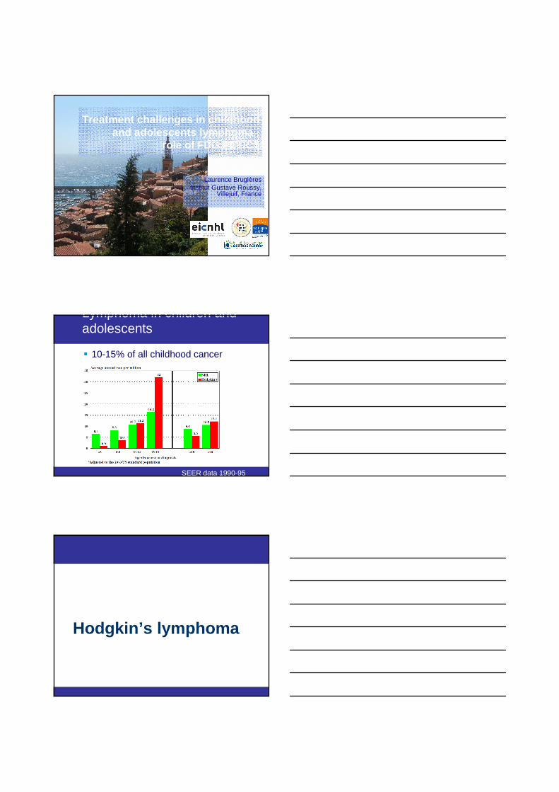
Lymphoma in children and adolescents
� 10-15% of all childhood cancer
SEER data 1990-95
Hodgkin’s lymphoma
Hodgkin’s lymphoma in children
� Very similar to adults LH� Subtypes distribution:
– Lymphocyte-predominant HL 5%
– Classic LH 95% • Nodular-sclerosing 70% • Mixed cellularity 20%• lymphocyte-rich 2-5% • Lymphocyte-depleted < 1%
EuroNet PHL-C1 EUDRACT-2006- 000995-33
� Aim : to limit the long term side effects :– Secondary malignancies (25% at 30 years)
– Male infertility
� Objectives:– To evaluate whether radiotherapy can be safely
omitted in patients with adequate PET response after 2 courses of OEPPA
– To evaluate whether procarbazine can be safely replaced by dacarbazine in therapy groups TG2 and TG3
Hodgkin lymphomaStaging in on-going pediatric trials
� FGD PET and conventional imagingmandatory
� 3 groups in the European protocolEuronetEuronet PHL C1 PHL C1 :– TG1 : I, IIA
– TG2 : IIB, IIIA
– TG3 :IIBIIBEE, IIIA, IIIAEE,, IIIB, IV, IIIB, IV,
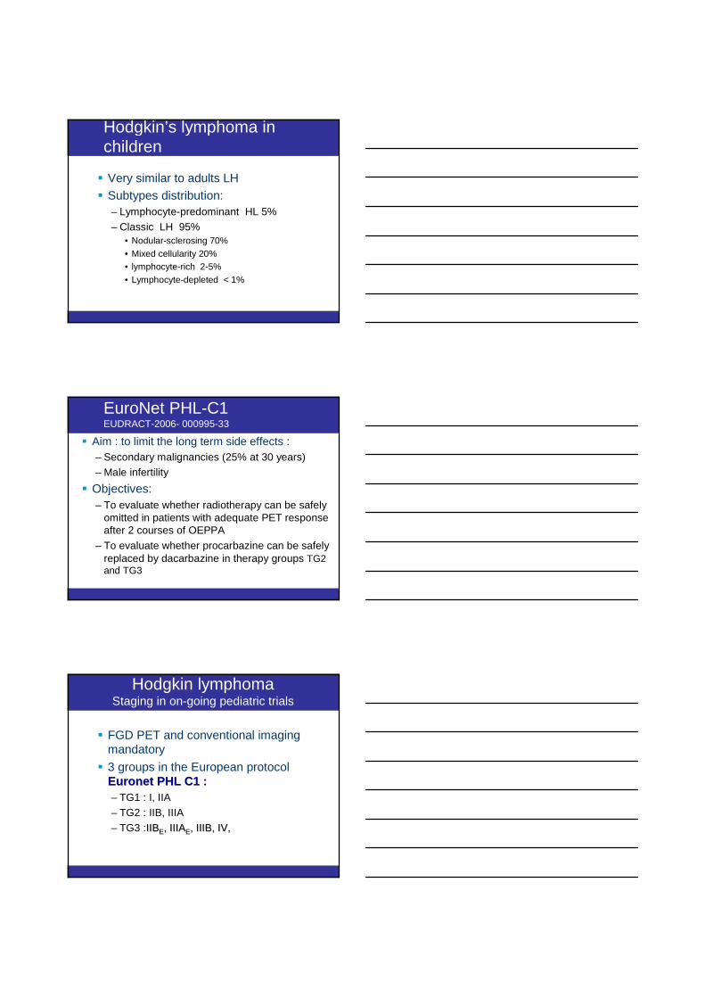
Euronet PHLC1 protocol
Non Hodgkin’s lymphoma
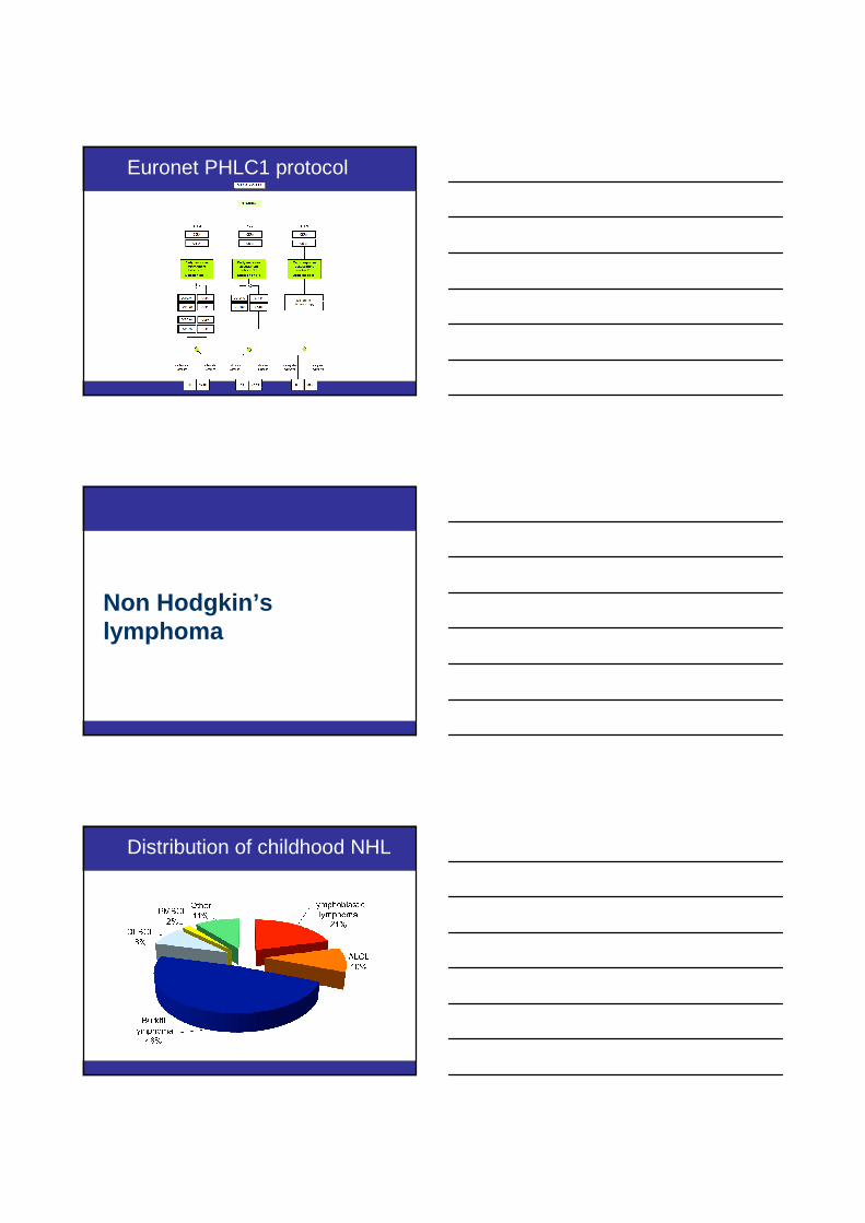
Distribution of childhood NHL
Childhood NHL
� Diffuse high grade lymphomas� Mostly extra-nodal localisations
– Abdomen : 37 %– ENT 17%– Mediastinal 28%– Lymph nodes 9%– Other 9%
� Specific staging system (St Jude classification)
� Rapid growth and risk of dissemination in bonemarrow and CSF requiring URGENT URGENT TREATMENTTREATMENT
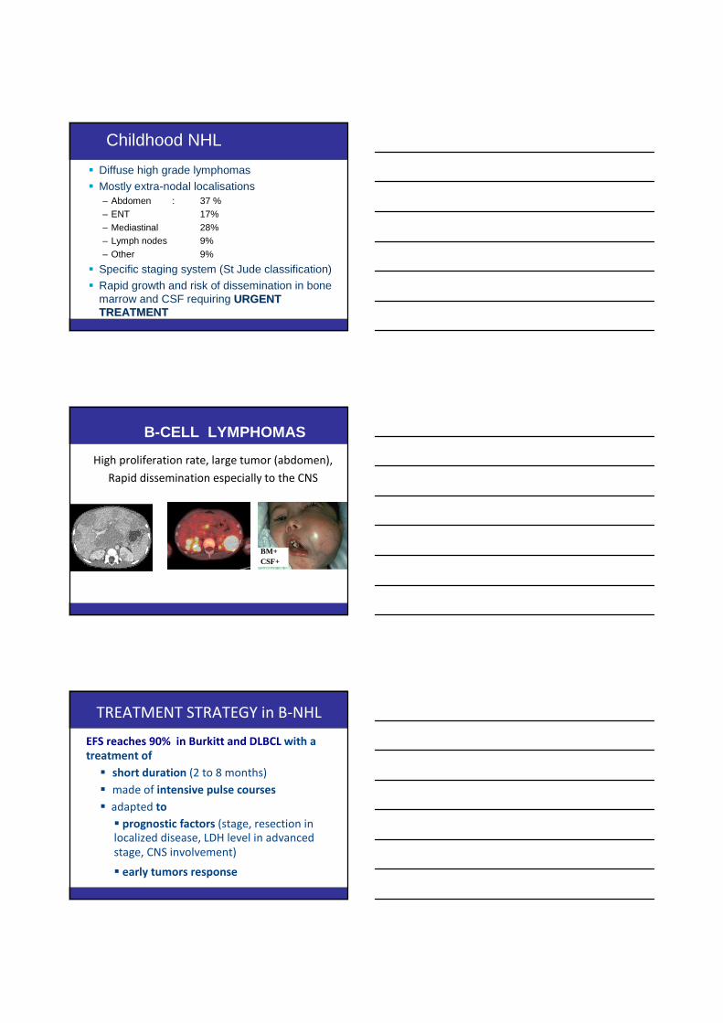
High proliferation rate, large tumor (abdomen),
Rapid dissemination especially to the CNS
B-CELL LYMPHOMAS
BM+ CSF+
EFS reaches 90% in Burkitt and DLBCL with a
treatment of
� short duration (2 to 8 months)
� made of intensive pulse courses
� adapted to
� prognostic factors (stage, resection in
localized disease, LDH level in advanced
stage, CNS involvement)
� early tumors response
TREATMENT STRATEGY in B-NHL
group prephase induction consolidation maintenancephase phase
A COPAD COPAD
B COP COPADM COPADM CYM CYM
C COP COPADM COPADM CYVE CYVE m1 m2 m3 m4n°1 n°2
FAB LMB protocol
On going trial to evaluate the impact of adding rituximab in high
risk patients : stage III and IV, high LDH
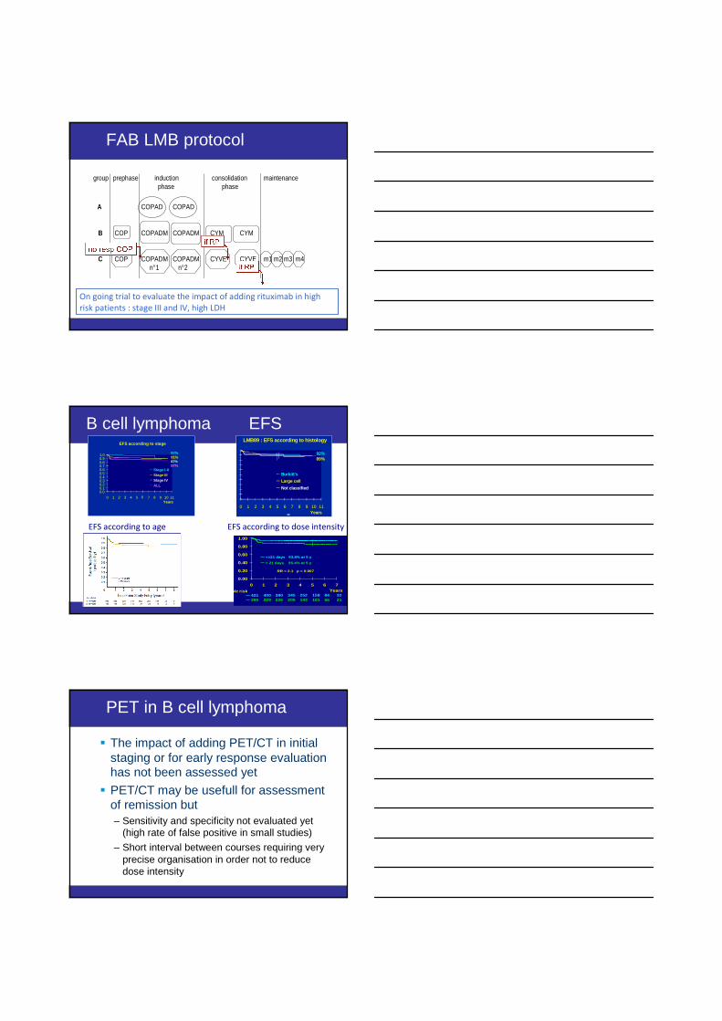
B cell lymphoma EFSLMB89 : EFS according to histology
0 1 2 3 4 5 6 7 8 9 10 11Years
Burkitt's
Large cell
Not classified
92%89%
interval between COPADM1 and COPADM2
32841582523453904004212155101143205226229265
0.00
0.20
0.40
0.60
0.80
1.00
0 1 2 3 4 5 6 7Years
<=21 days 93.8% at 5 y
> 21 days 85.4% at 5 y
At risk
RR = 2.1 p = 0.007
-EFS according to age EFS according to dose intensity
EFS according to stage
0.00.10.20.30.40.50.60.70.80.91.0
0 1 2 3 4 5 6 7 8 9 10 11Years
Stage I-IIStage IIIStage IVALL
97%91%87%87%
PET in B cell lymphoma
� The impact of adding PET/CT in initial staging or for early response evaluation has not been assessed yet
� PET/CT may be usefull for assessment of remission but– Sensitivity and specificity not evaluated yet
(high rate of false positive in small studies)
– Short interval between courses requiring very precise organisation in order not to reduce dose intensity
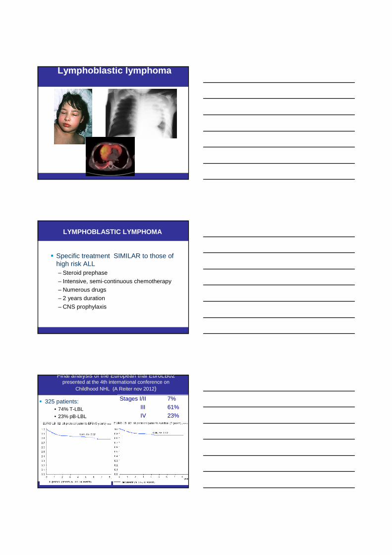
Lymphoblastic lymphoma
� Specific treatment SIMILAR to those of high risk ALL– Steroid prephase
– Intensive, semi-continuous chemotherapy
– Numerous drugs
– 2 years duration
– CNS prophylaxis
LYMPHOBLASTIC LYMPHOMA
Final analysis of the European trial EuroLB02 presented at the 4th international conference on
Childhood NHL (A Reiter nov 2012)
� 325 patients: • 74% T-LBL• 23% pB-LBL
Stages I/II 7%
III 61%
IV 23%
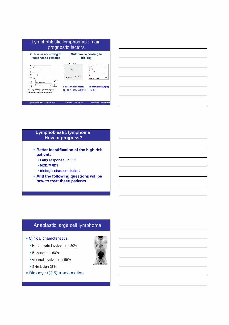
Lymphoblastic lymphomas : main prognostic factors
Outcome according to response to steroids
Outcome according to biology
French studies (45pts) BFM studies (230pts)
NOTCH/FBXW7 mutations 6qLOH
Uyttebroeck, Eur J Cancer 2008 C Callens, JCO, 2012B Burkhardt Leukemia2006
Lymphoblastic lymphoma How to progress?
� Better identification of the high risk patients• Early response: PET ?
• MDD/MRD?
• Biologic characteristics?
� And the following questions will be how to treat these patients
Anaplastic large cell lymphoma
� Clinical characteristics:
� lymph node involvement 80%
� B symptoms 60%
� visceral involvement 50%
� Skin lesion 25%
� Biology : t(2;5) translocation
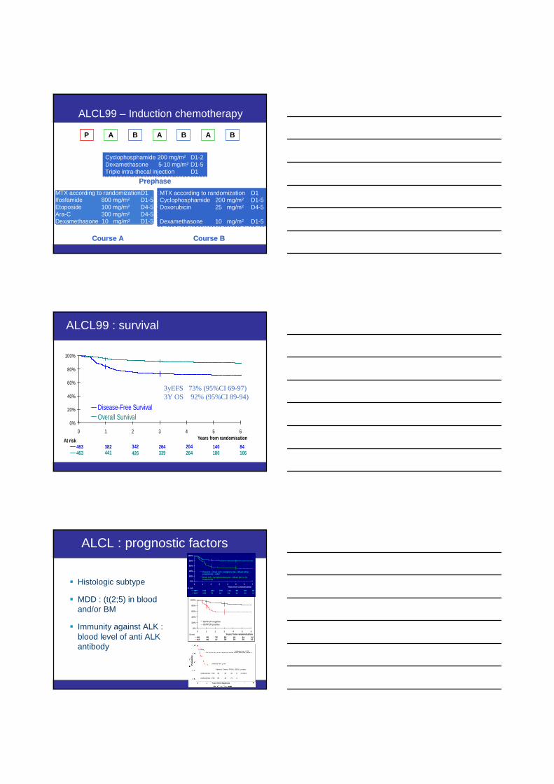
MTX according to randomizationD1Ifosfamide 800 mg/m² D1-5Etoposide 100 mg/m² D4-5Ara-C 300 mg/m² D4-5Dexamethasone 10 mg/m² D1-5
MTX according to randomization D1Cyclophosphamide 200 mg/m² D1-5Doxorubicin 25 mg/m² D4-5
Dexamethasone 10 mg/m² D1-5
MTX according to randomization D1Cyclophosphamide 200 mg/m² D1-5Doxorubicin 25 mg/m² D4-5
Dexamethasone 10 mg/m² D1-5
ALCL99 – Induction chemotherapy
Cyclophosphamide 200 mg/m² D1-2Dexamethasone 5-10 mg/m² D1-5Triple intra-thecal injection D1
Cyclophosphamide 200 mg/m² D1-2Dexamethasone 5-10 mg/m² D1-5Triple intra-thecal injection D1
P A B A B A B
Course ACourse A Course BCourse B
PrephasePrephase
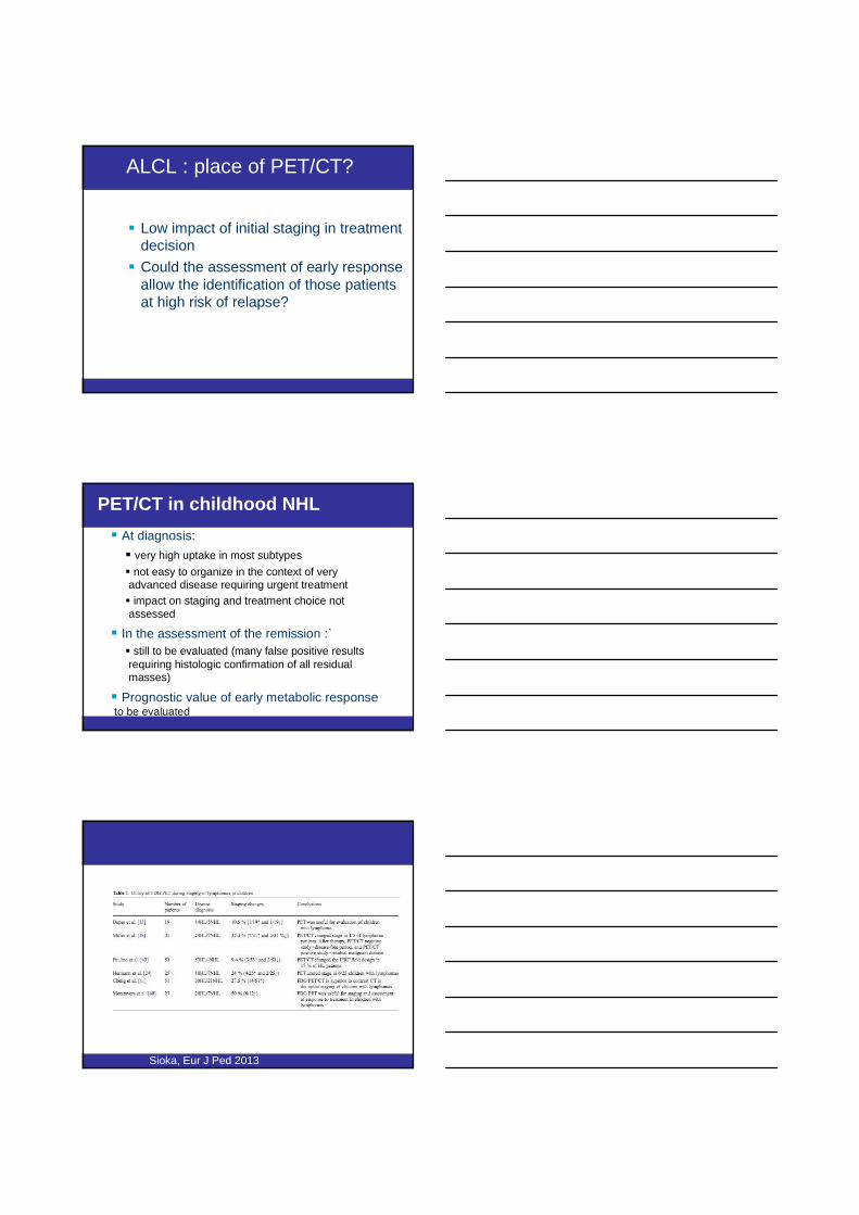
ALCL99 : survival
84140204264342382463106180264339426441463
0%
20%
40%
60%
80%
100%
0 1 2 3 4 5 6Years from randomisation
Disease-Free SurvivalOverall Survival
At risk
3yEFS 73% (95%CI 69-97)3Y OS 92% (95%CI 89-94)
Patients Events PFS% (SE%) p-value
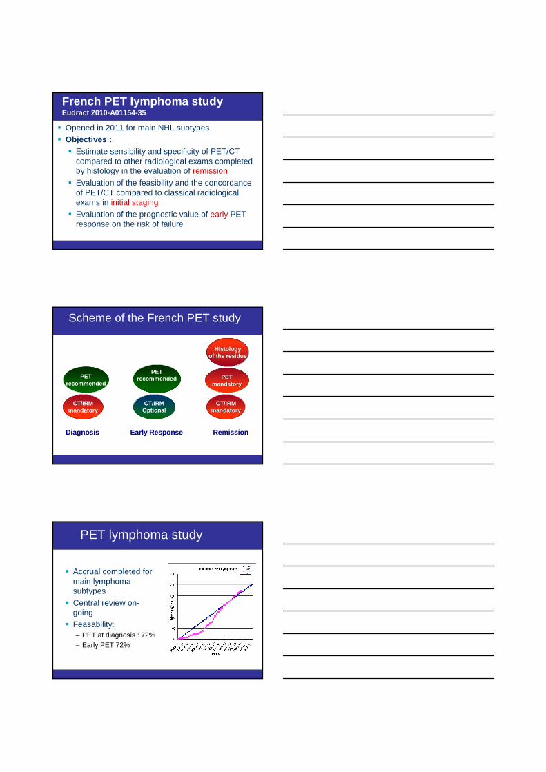
Antibody titer <750 39 22 40 8 <0.0001
Antibody titer >750 89 18 79 4
Antibody titer <750
Antibody titer >750
Patients Events PFS% (SE%) p-value
Antibody titer <750 39 22 40 8 <0.0001
Antibody titer >750 89 18 79 4
Antibody titer <750
Antibody titer >750
Years from diagnosis
Pro
babi
lity
ALCL : prognostic factors
� Histologic subtype
� MDD : (t(2;5) in blood and/or BM
� Immunity against ALK : blood level of anti ALK antibody
26599814418624325429582131506176103135
0%
20%
40%
60%
80%
100%
0 1 2 3 4 5 6 7
Years from randomization
Classical + Giant cell + Hodgkin's like + Mixed (ot hercomponent) + other
Small cell + Lymphohistiocystic + Mixed (SC or LHcomponent)
At risk
1224435575808610202939476682
0%
20%
40%
60%
80%
100%
0 1 2 3 4 5 6Years from randomization
BM PCR negativeBM PCR positive
At risk
ALCL : place of PET/CT?
� Low impact of initial staging in treatment decision
� Could the assessment of early response allow the identification of those patients at high risk of relapse?
PET/CT in childhood NHL
� At diagnosis:
� very high uptake in most subtypes
� not easy to organize in the context of very advanced disease requiring urgent treatment� impact on staging and treatment choice not assessed
� In the assessment of the remission :`� still to be evaluated (many false positive results requiring histologic confirmation of all residual masses)
� Prognostic value of early metabolic responseto be evaluated
Sioka, Eur J Ped 2013
French PET lymphoma studyEudract 2010-A01154-35
� Opened in 2011 for main NHL subtypes
� Objectives : � Estimate sensibility and specificity of PET/CT
compared to other radiological exams completed by histology in the evaluation of remission
� Evaluation of the feasibility and the concordance of PET/CT compared to classical radiological exams in initial staging
� Evaluation of the prognostic value of early PET response on the risk of failure
Scheme of the French PET study
Diagnosis Early Response Diagnosis Early Response RemissionRemission
PETrecommended
PETmandatorymandatory
PETrecommended
CT/IRMmandatory
CT/IRMOptional
CT/IRMmandatorymandatory
Histologyof the residue
PET lymphoma study
� Accrual completed for main lymphoma subtypes
� Central review on-going
� Feasability:– PET at diagnosis : 72%– Early PET 72%
CONCLUSIONS: Challenges for the future
� Most lymphoma in children are curable with a first line therapy
� Challenges are :– To identify those children with very good prognosis for
whom treatment intensity can be reduced– To identify very high risk patients for whom early
intensification of therapy is required
� Whether PET/CT will play a role in the identification of these patients is– obvious in HL– still questionable in NHL