Embed Size (px)
Citation preview
HODS - November 2006 1
Anatomy and PhysiologyHeart, Lungs, Pancreas, Liver, Kidneys and Skin
B. Paul White, MDHOD ID#: 2078
HODS - November 2006 2
HODS - November 2006 3
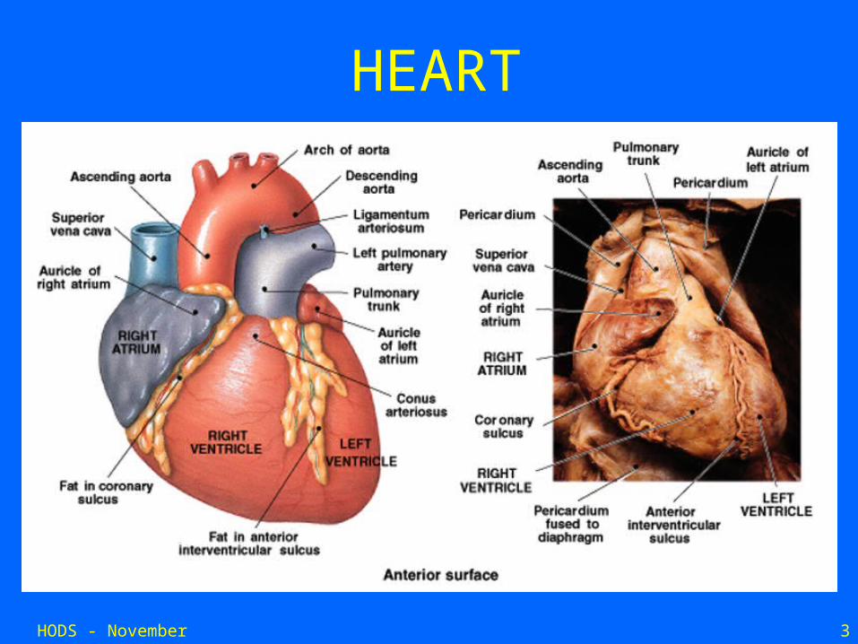
HEART
HODS - November 2006 4
HEART
• Hollow, muscular organ• 300 grams (size of a fist)• 4 chambers• found in chest between lungs • surrounded by membrane called
Pericardium• Pericardial space is fluid-filled to nourish
and protect the heart.
HODS - November 2006 5
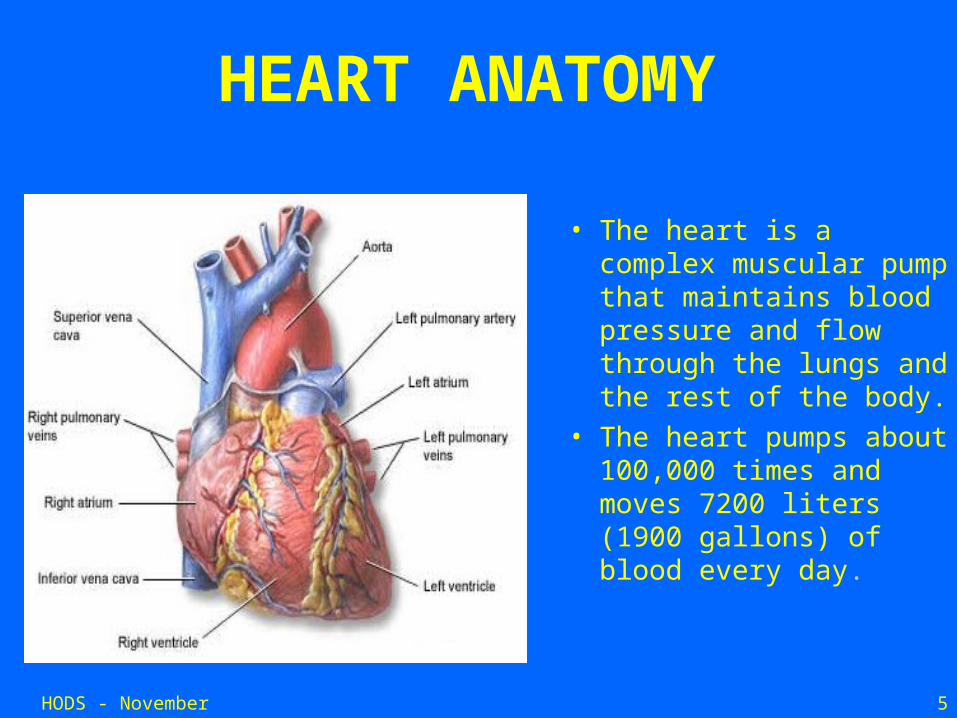
HEART ANATOMY
• The heart is a complex muscular pump that maintains blood pressure and flow through the lungs and the rest of the body.
• The heart pumps about 100,000 times and moves 7200 liters (1900 gallons) of blood every day.
HODS - November 2006 6
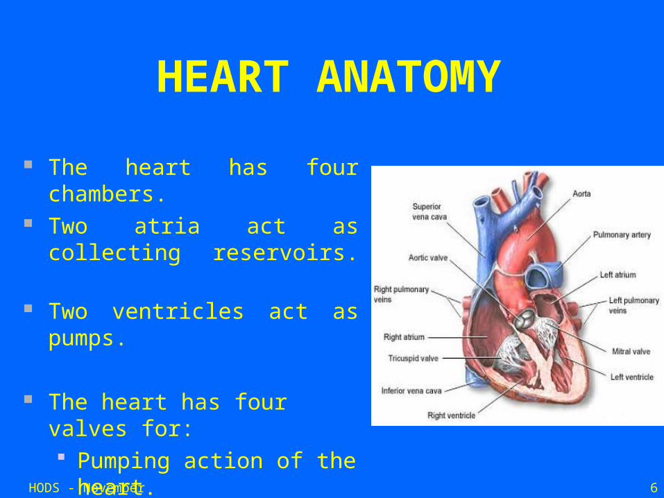
HEART ANATOMY
The heart has four chambers.
Two atria act as collecting reservoirs.
Two ventricles act as pumps.
The heart has four valves for: Pumping action of the
heart. Maintaining
unidirectional blood flow.
HODS - November 2006 7
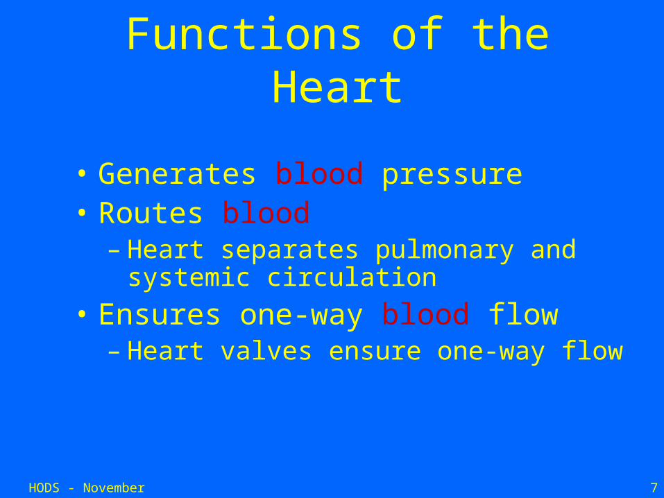
Functions of the Heart
• Generates blood pressure• Routes blood
– Heart separates pulmonary and systemic circulation
• Ensures one-way blood flow– Heart valves ensure one-way flow
HODS - November 2006 8
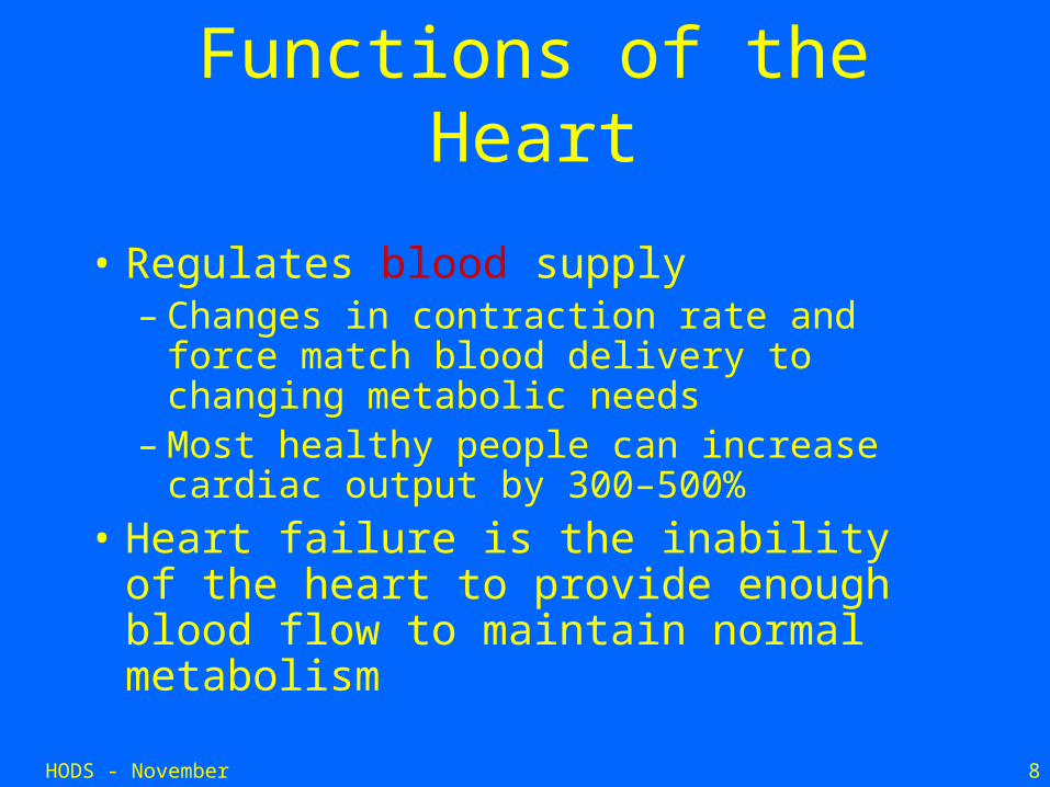
Functions of the Heart
• Regulates blood supply– Changes in contraction rate and force
match blood delivery to changing metabolic needs
– Most healthy people can increase cardiac output by 300–500%
• Heart failure is the inability of the heart to provide enough blood flow to maintain normal metabolism
HODS - November 2006 9
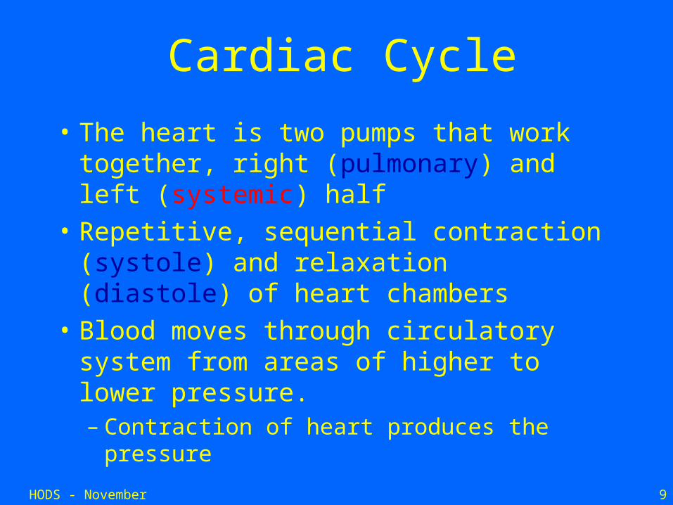
Cardiac Cycle
• The heart is two pumps that work together, right (pulmonary) and left (systemic) half
• Repetitive, sequential contraction (systole) and relaxation (diastole) of heart chambers
• Blood moves through circulatory system from areas of higher to lower pressure.– Contraction of heart produces the pressure
HODS - November 2006 10
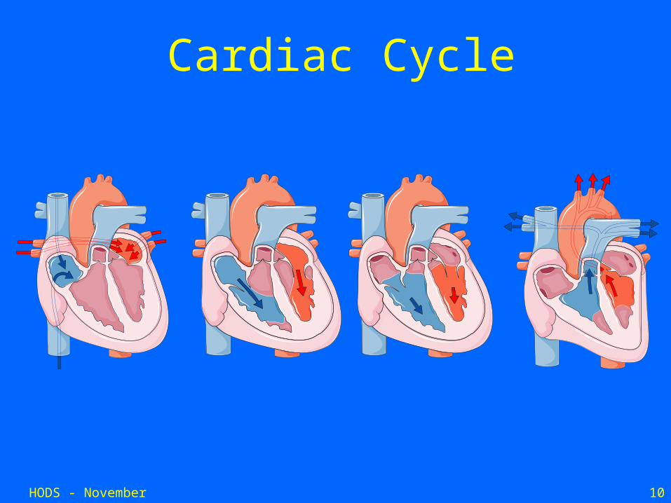
Cardiac Cycle
HODS - November 2006 11
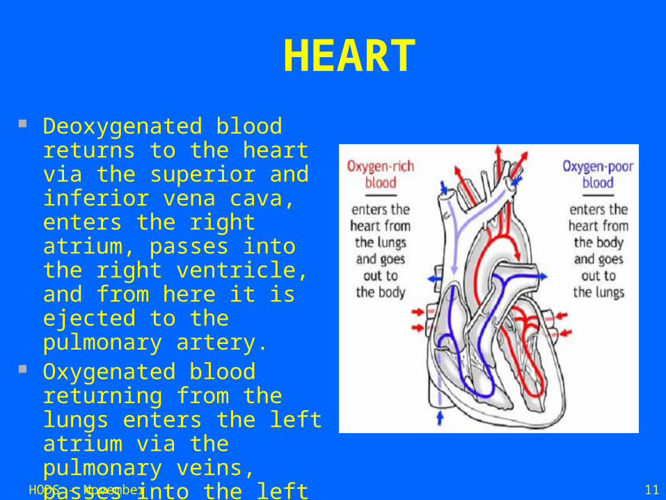
HEART Deoxygenated blood returns
to the heart via the superior and inferior vena cava, enters the right atrium, passes into the right ventricle, and from here it is ejected to the pulmonary artery.
Oxygenated blood returning from the lungs enters the left atrium via the pulmonary veins, passes into the left ventricle, and is then ejected to the aorta.
HODS - November 2006 12
Blood Vessels
• Blood vessels are divided into a pulmonary circuit and systemic circuit.
• Artery - vessel that carries blood away from the heart. Usually oxygenated
• Vein - vessel that carries blood towards the heart. Usually deoxygenated.
• Capillary - a small blood vessel that allow diffusion of gases, nutrients and wastes between plasma and interstitial fluid.
HODS - November 2006 13
Blood Vessels
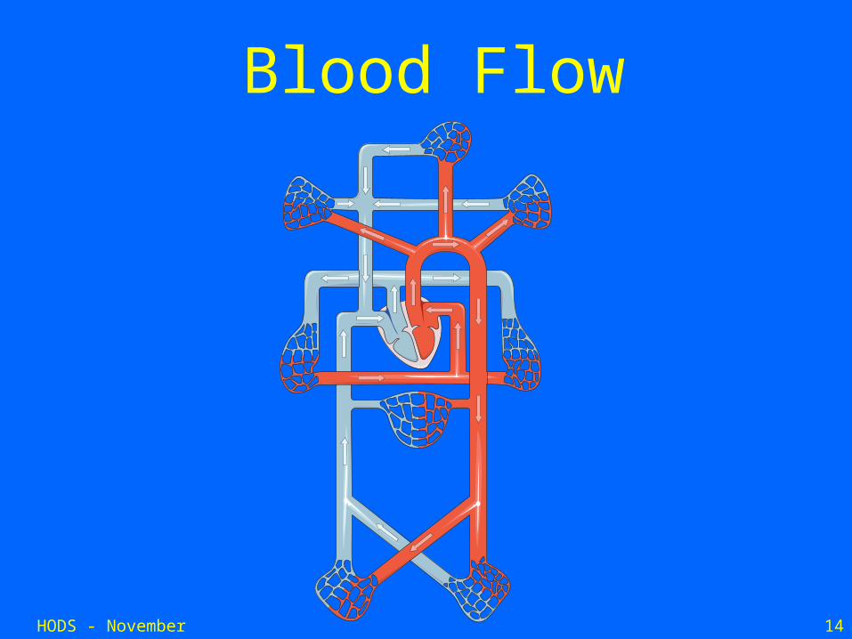
• Systemic vessels– Transport blood through the body part from
left ventricle and back to right atrium
• Pulmonary vessels– Transport blood from right ventricle through
lungs and back to left atrium• Blood vessels and heart are regulated to
ensure blood pressure is high enough for blood flow to meet metabolic needs of tissues
HODS - November 2006 14
Blood Flow
HODS - November 2006 15
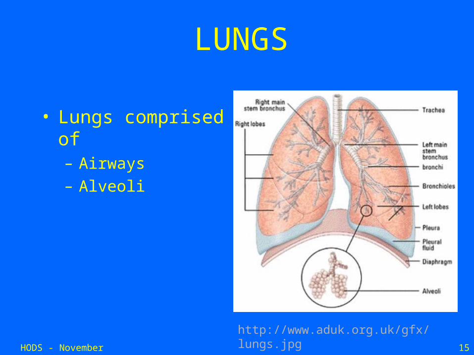
LUNGS
• Lungs comprised of – Airways– Alveoli
http://www.aduk.org.uk/gfx/lungs.jpg
HODS - November 2006 16
What do the lungs do?
• Primary function is gas exchange
• Let oxygen move in
• Let carbon dioxide move out
HODS - November 2006 17
How do the lungs do this?
• First, air has to move to the region where gas exchange occurs.
• For this, you need a normal ribcage and respiratory muscles that work properly (among other things).
HODS - November 2006 18
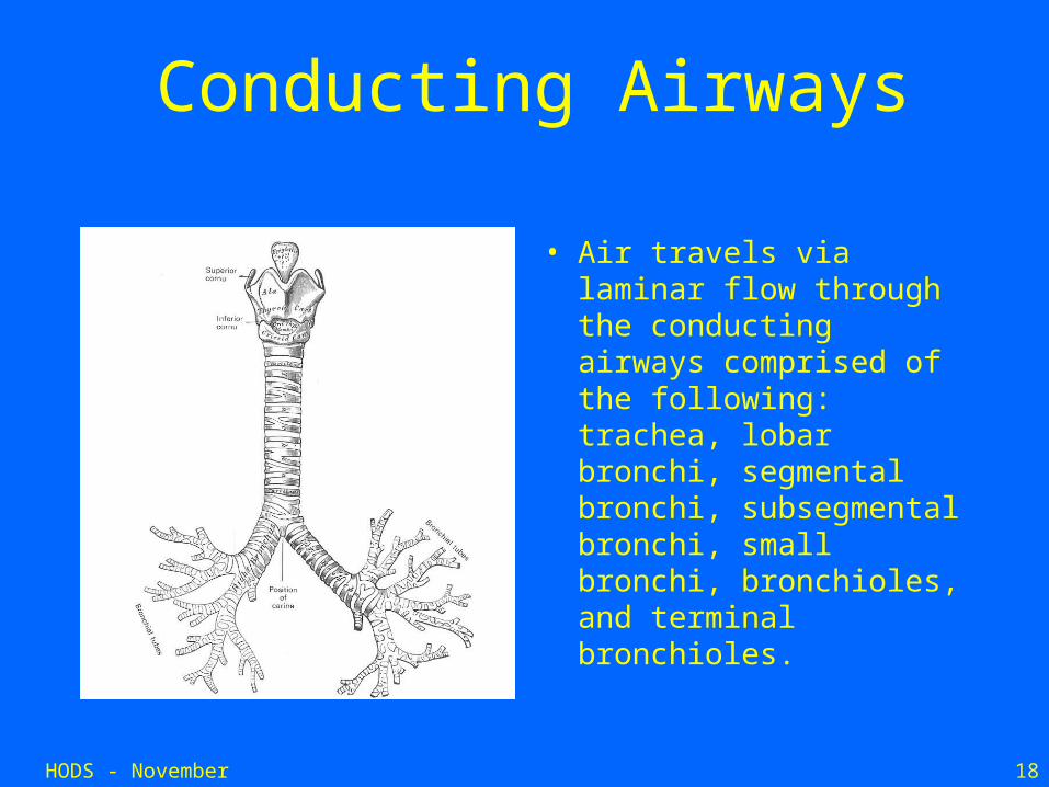
Conducting Airways
• Air travels via laminar flow through the conducting airways comprised of the following: trachea, lobar bronchi, segmental bronchi, subsegmental bronchi, small bronchi, bronchioles, and terminal bronchioles.
HODS - November 2006 19
How do the lungs do this?
• The airways then branch further to become transitional/respiratory bronchioles.
• The transitional/respiratory zones are made up of respiratory bronchioles, alveolar ducts, and alveoli.
HODS - November 2006 20
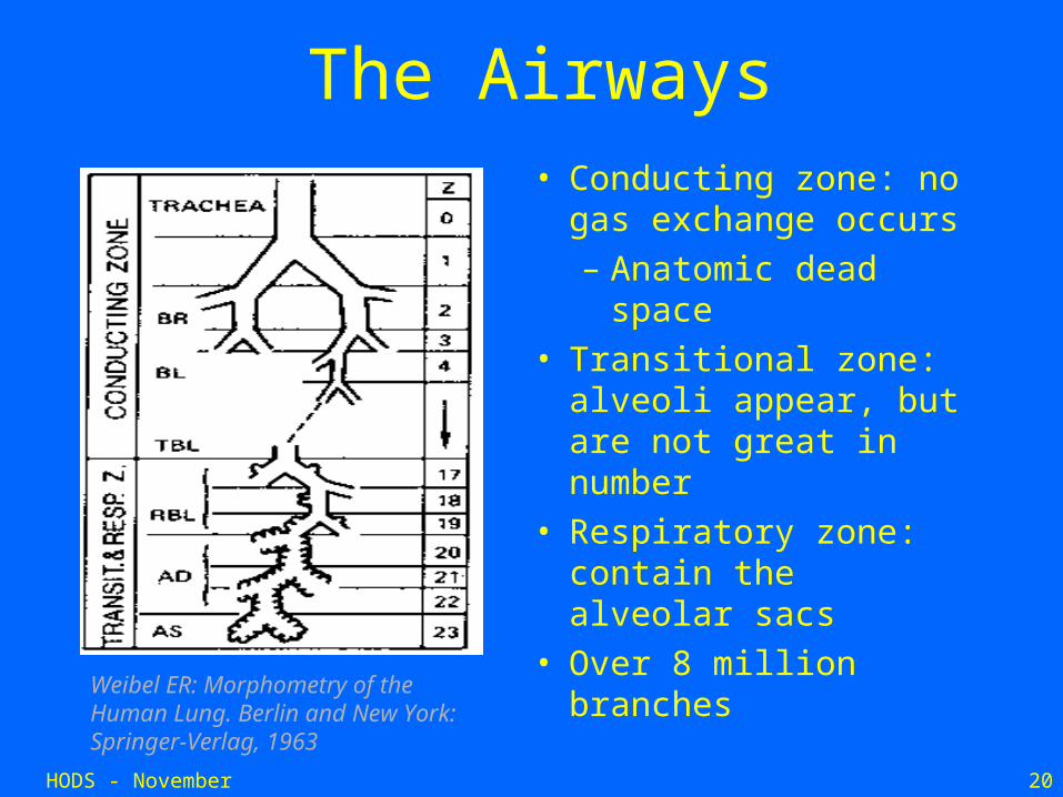
Weibel ER: Morphometry of the Human Lung. Berlin and New York: Springer-Verlag, 1963
The Airways• Conducting zone: no
gas exchange occurs
– Anatomic dead space
• Transitional zone: alveoli appear, but are not great in number
• Respiratory zone: contain the alveolar sacs
• Over 8 million branches
HODS - November 2006 21
How does gas exchange occur?
• Numerous capillaries are wrapped around alveoli.
• Gas diffuses across this alveolar-capillary barrier.
• This barrier is as thin as 0.3 μm in some places and has a surface area of 50-100 square meters!
HODS - November 2006 22
Gas Exchange
• Diffusion Barrier crossed by O2 moving from air to blood and CO2 from blood to air is made up of:
• 1. an aqueous surface film
• 2. epithelial cells of alveolus
• 3. interstitial layer
• 4. endothelial cells of capillaries
• 5. blood plasma
• 6. membrane of RBCs
HODS - November 2006 23
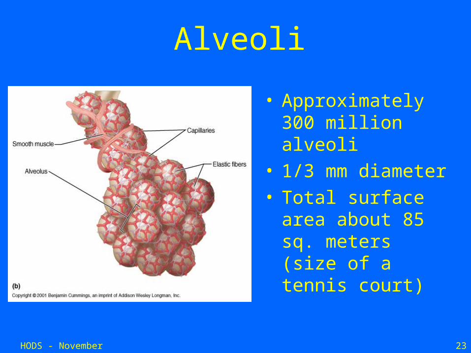
Alveoli
• Approximately 300 million alveoli
• 1/3 mm diameter• Total surface area
about 85 sq. meters (size of a tennis court)
HODS - November 2006 24
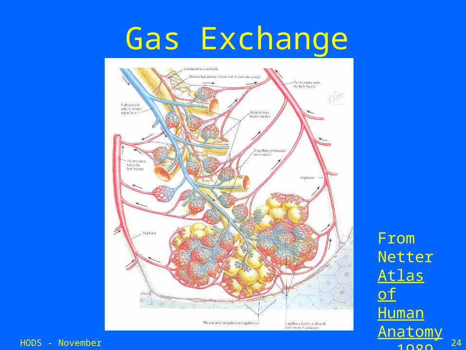
Gas Exchange
From Netter Atlas of Human Anatomy, 1989
HODS - November 2006 25
Control of Ventilation
• Arterial PO2– When PO2 is VERY low, ventilation increases
• Arterial PCO2– The most important regulator of ventilation, small
increases in PCO2, greatly increases ventilation
• Arterial pH– As hydrogen ions increase, alveolar ventilation
increases, but hydrogen ions cannot diffuse into CSF as well as CO2
PANCREAS
HODS - November 2006 27
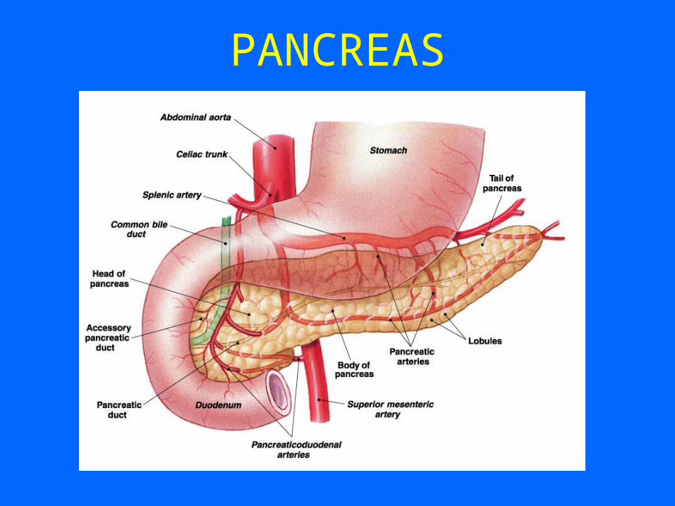
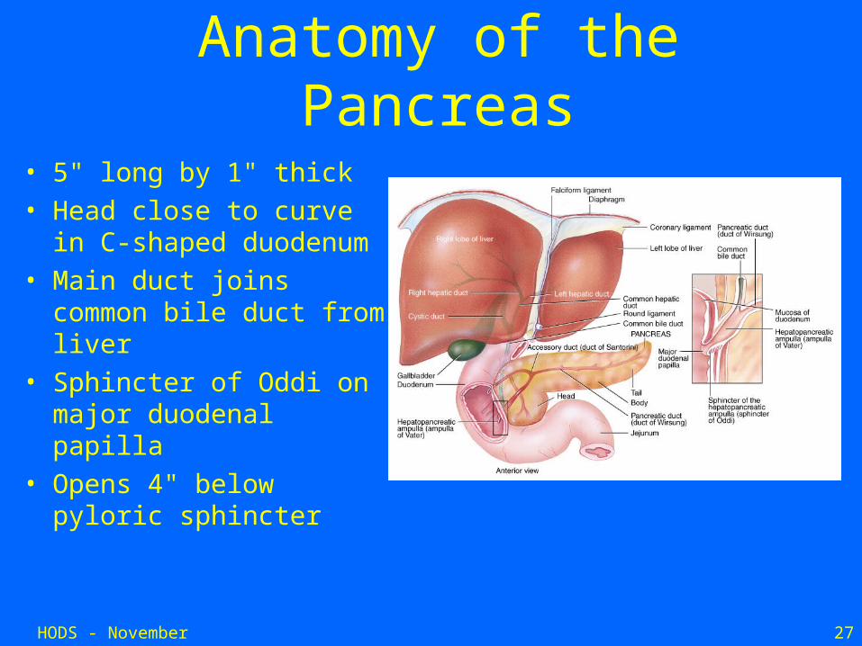
Anatomy of the Pancreas
• 5" long by 1" thick• Head close to curve in C-
shaped duodenum• Main duct joins common
bile duct from liver • Sphincter of Oddi on
major duodenal papilla• Opens 4" below pyloric
sphincter
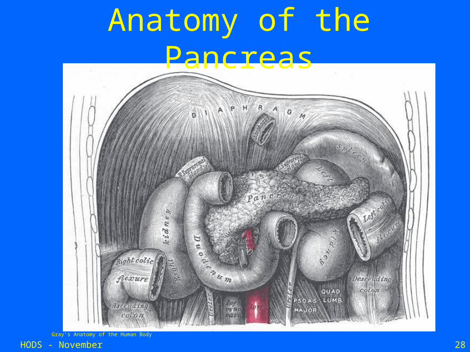
HODS - November 2006 28Gray’s Anatomy of the Human Body
Anatomy of the Pancreas
HODS - November 2006 29
Gray’s Anatomy of the Human Body Robbins Basic Pathologyhttp://faculty.clintoncc.suny.edu/faculty/Michael.Gregory/default.htm
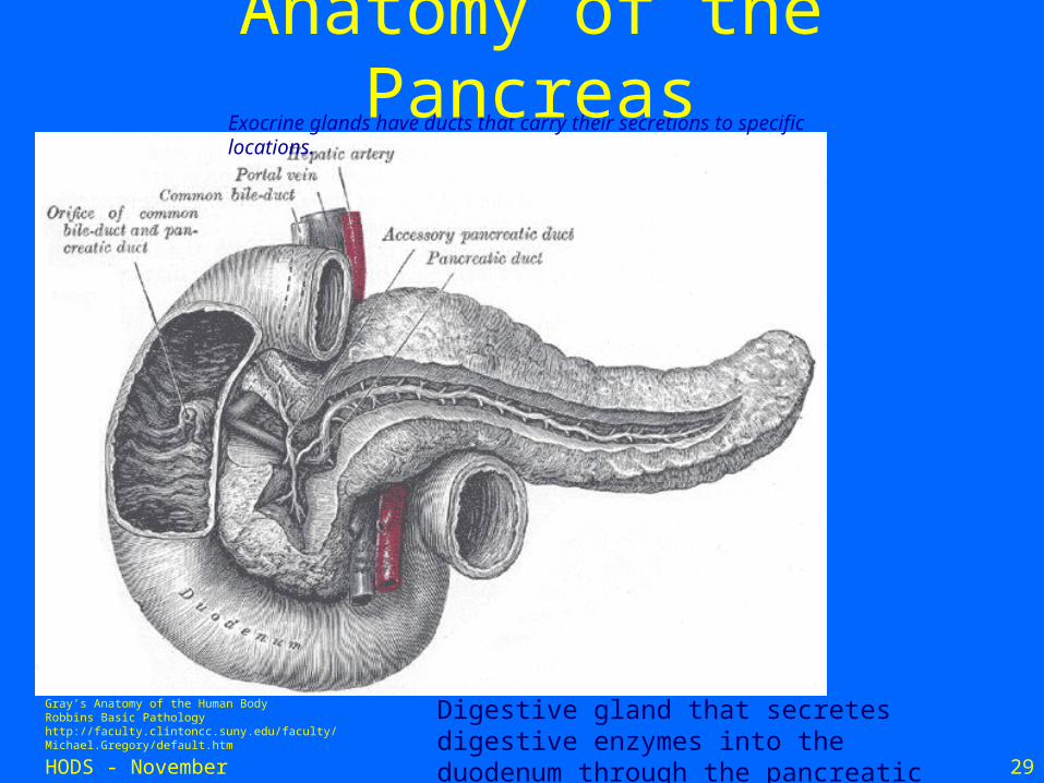
Digestive gland that secretes digestive enzymes into the duodenum through the pancreatic duct.
Exocrine glands have ducts that carry their secretions to specific locations.
Anatomy of the Pancreas
Histology of the Pancreas
HODS - November 2006 31
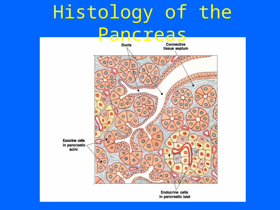
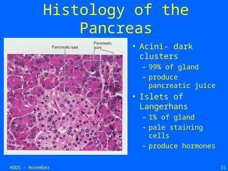
Histology of the Pancreas
• Acini- dark clusters – 99% of gland– produce pancreatic
juice
• Islets of Langerhans– 1% of gland– pale staining cells– produce hormones
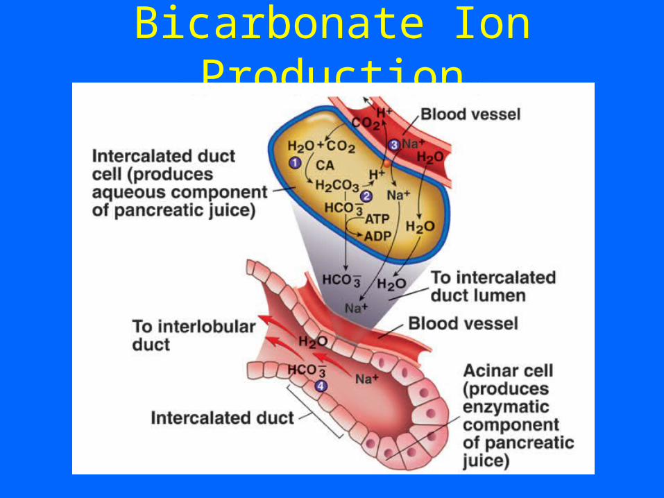
Bicarbonate Ion Production
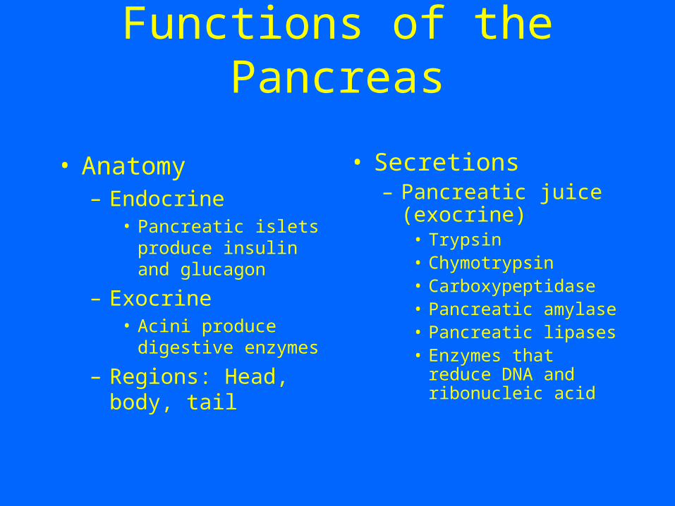
Functions of the Pancreas
• Anatomy– Endocrine
• Pancreatic islets produce insulin and glucagon
– Exocrine• Acini produce
digestive enzymes
– Regions: Head, body, tail
• Secretions– Pancreatic juice
(exocrine)• Trypsin• Chymotrypsin• Carboxypeptidase• Pancreatic amylase• Pancreatic lipases• Enzymes that reduce
DNA and ribonucleic acid
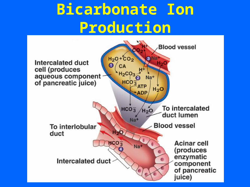
Bicarbonate Ion Production
LIVER
HODS - November 2006 36
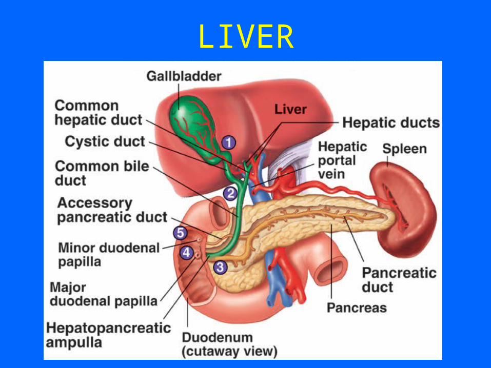
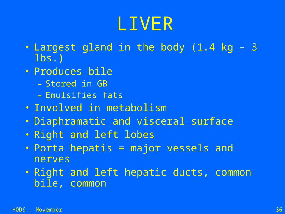
LIVER• Largest gland in the body (1.4 kg – 3 lbs.)• Produces bile
– Stored in GB– Emulsifies fats
• Involved in metabolism• Diaphramatic and visceral surface• Right and left lobes• Porta hepatis = major vessels and nerves• Right and left hepatic ducts, common bile,
common
HODS - November 2006 37
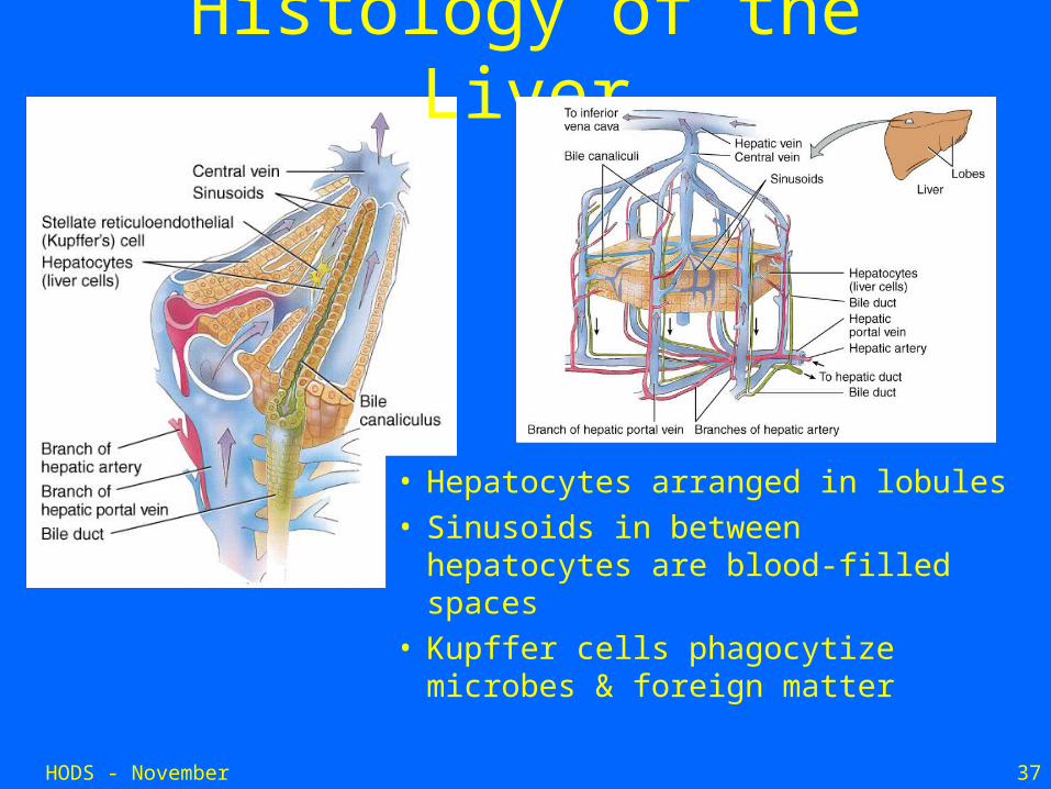
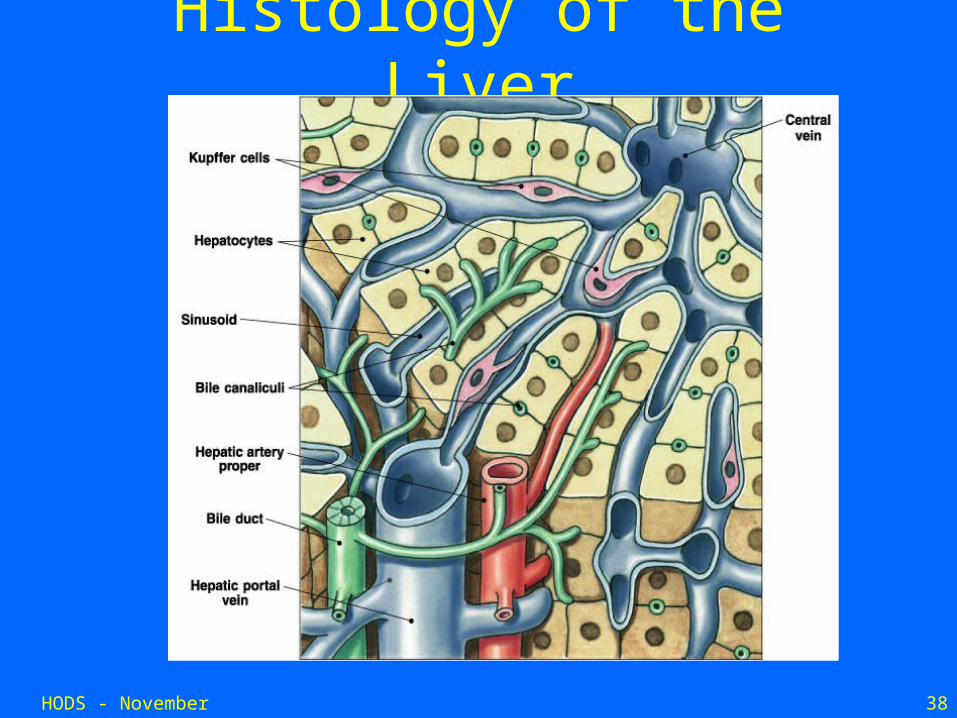
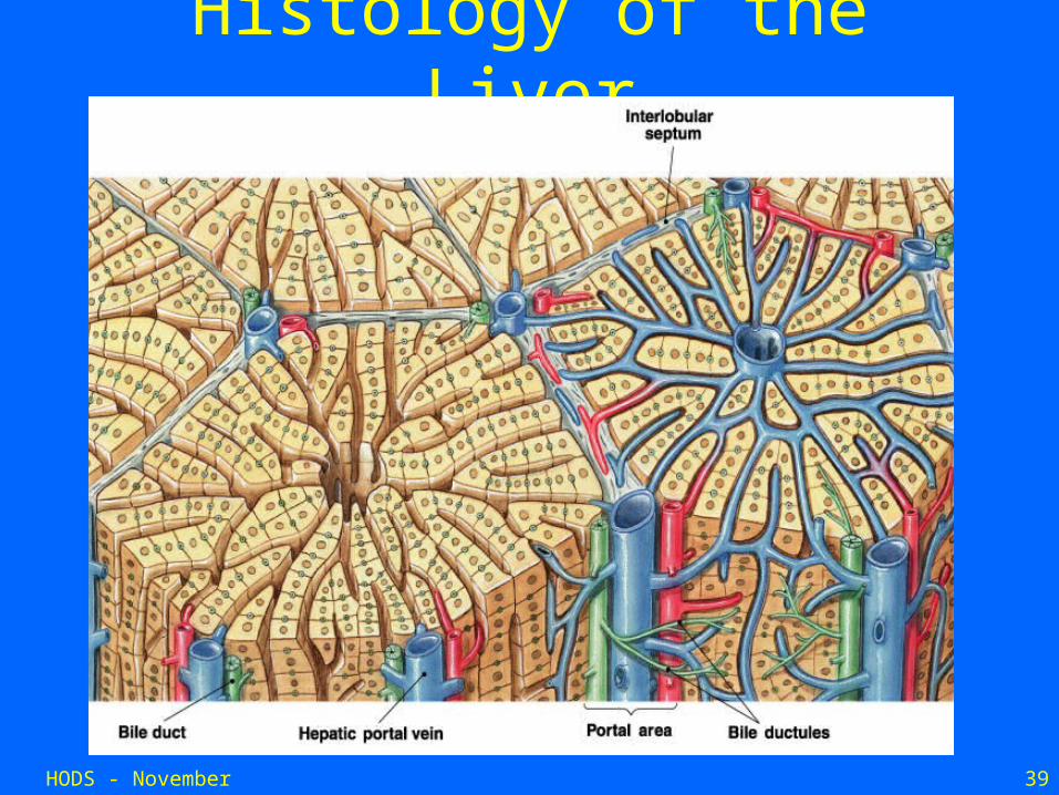
Histology of the Liver
• Hepatocytes arranged in lobules • Sinusoids in between hepatocytes
are blood-filled spaces • Kupffer cells phagocytize
microbes & foreign matter
HODS - November 2006 38
Histology of the Liver
HODS - November 2006 39
Histology of the Liver
HODS - November 2006 40
Functions of the Liver• Bile production
– Salts emulsify fats, contain pigments as bilirubin
• Storage– Glycogen, fat, vitamins, copper and iron
• Nutrient interconversion• Detoxification
– Hepatocytes remove ammonia and convert to urea
• Phagocytosis– Kupffer cells phagocytize worn-out and dying red and white blood cells,
some bacteria
• Synthesis– Albumins, fibrinogen, globulins, heparin, clotting factors
HODS - November 2006 41
Bile
• About 600 ml of bile is produced daily– Bile acid– Phospholipids– Cholesterol– Bilirubin– Waste products– Electrolytes– Mucin
HODS - November 2006 42
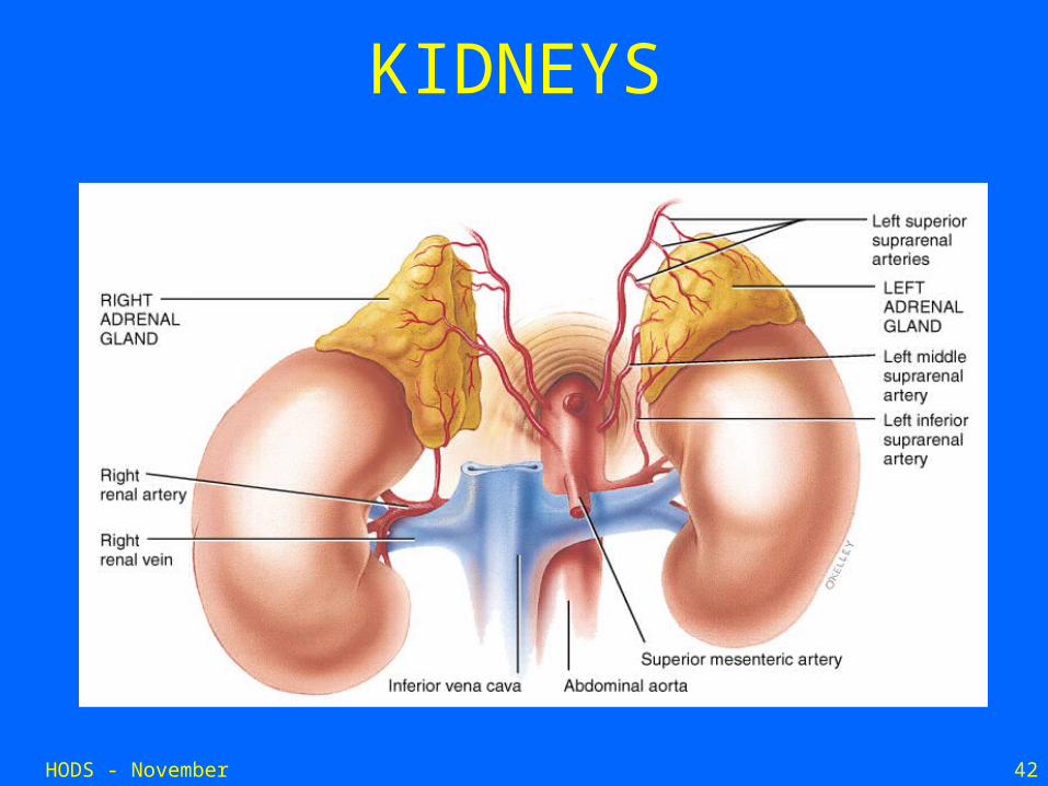
KIDNEYS
HODS - November 2006 43
KIDNEYS• Play a major role in maintaining homeostasis• Maintain water balance• Regulate the quantity and concentration of
ECF ions• Regulate the plasma volume• Regulate pH by controlling elimination of acid
and base in urine • Maintain osmolarity• Regulate the concentration of plasma
constituents (e.g. electrolytes and water)
HODS - November 2006 44
KIDNEYS
• Kidneys have excellent blood supply: 0.5% total body weight but ~20% of Cardiac Output.
• Kidneys process plasma portion of blood by removing substances from it, and in a few cases, by adding substances to it.
• Works with cardiovascular system (and others!) in integrated manner
HODS - November 2006 45
Functions of the kidneys
• Regulation of H2O and inorganic ion balance – most important function!
• Removal of metabolic waste products from blood and excretion in urine.
• Removal of foreign chemicals in the blood (e.g. drugs) and excretion in urine.
• Gluconeogenesis• Endocrine functions (e.g. renin, erythropoetin, 1,25-
dihydroxyvitamin D) • In kidney disease, build-up of waste serious, but not
a bad as ECF volume and composition disturbances.
HODS - November 2006 46
Functions of the kidneys
• Water balance
• Electrolyte balance
• Plasma volume
• Acid-base balance
• Osmolarity balance
• Excretion
• Hormone secretion
HODS - November 2006 47
Acid-Base Balance
• Kidneys VERY important for acid-base balance, along with respiratory system.
• Important because all biochemical processes must occur within an optimal pH window.
• Prevent ACIDOSIS or ALKALOSIS.• Although the lungs excrete a large amount of CO2, a
potential acid formed by metabolism, the kidneys are crucial for excreting non-volatile acids.
• To maintain acid-base balance, kidney must not only reabsorb virtually all filtered HCO3
-, but must also secrete into the urine the daily production of non-volatile acids.
HODS - November 2006 48
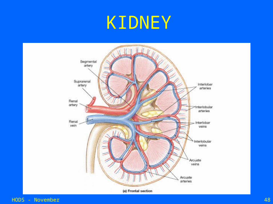
KIDNEY
HODS - November 2006 49
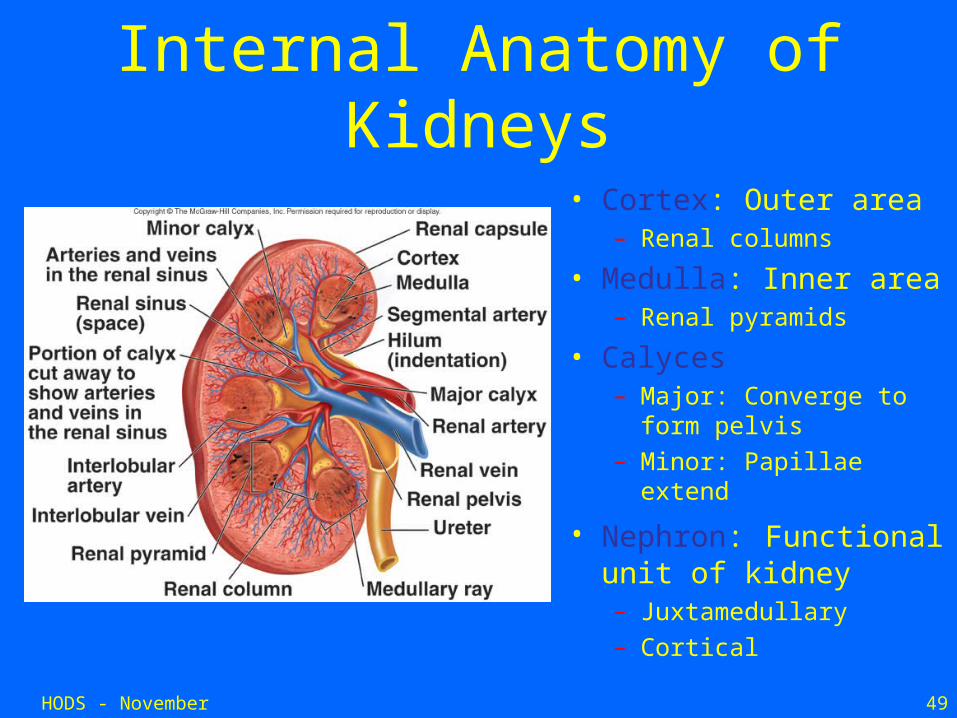
Internal Anatomy of Kidneys
• Cortex: Outer area– Renal columns
• Medulla: Inner area– Renal pyramids
• Calyces– Major: Converge to form
pelvis– Minor: Papillae extend
• Nephron: Functional unit of kidney– Juxtamedullary– Cortical

HODS - November 2006 50
Kidney Failure
• at age 49 years, the expected duration of life of a patient with end-stage renal disease on hemodialysis is 7 additional years compared with approximately 30 additional years for a person of the same age from the general population.
HODS - November 2006 51
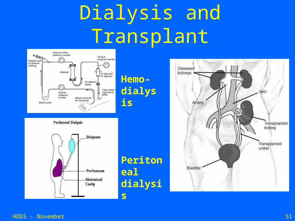
Dialysis and Transplant
Peritoneal dialysis
Hemo-dialysis
HODS - November 2006 52
SKIN
HODS - November 2006 53
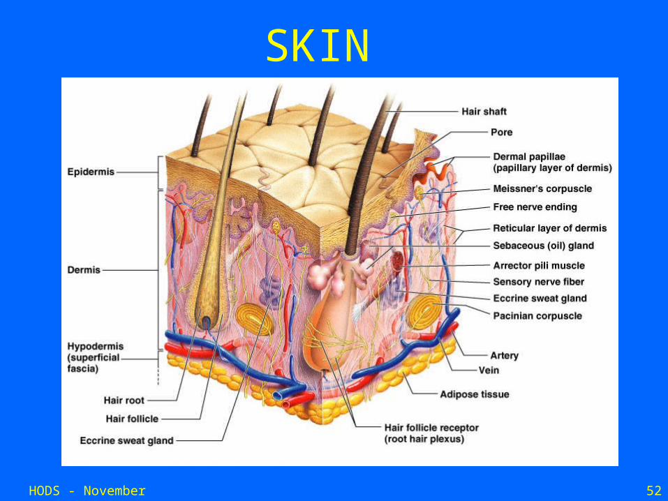
SKIN
• Largest organ of the body.
• Surface area 1.5 - 2 m2.
• Average adult weight 9 kg.
• Functions - protection, defence, sensation, thermoregulation, vit D synthesis, excretion, storage.
HODS - November 2006 54
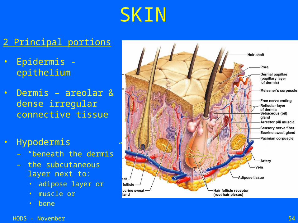
SKIN2 Principal portions
• Epidermis - epithelium
• Dermis – areolar & dense irregular connective tissue
• Hypodermis– “beneath the dermis”– the subcutaneous layer
next to:• adipose layer or
• muscle or
• bone
HODS - November 2006 55
Functions of the Skin• Protection
– Prevents invasion of environmental toxins and microorganisms
• Immunologic – Sebum has antibacterial properties which helps shed topical
bacteria
• Thermoregulation – Insulates from heat loss and controls loss of heat through
evaporation
HODS - November 2006 56
Functions of the Skin• Fluid and Electrolyte Balance
– Controls sodium excretion– Sebum retards fluid loss from skin
• Metabolism– Produces Vitamin D– Prevents excessive fluid loss
• Neurosensory – Nerve endings and receptors process environmental stimuli for pain,
touch, heat and cold
• Social and Interactive
– Provides body image and personal identity
HODS - November 2006 57
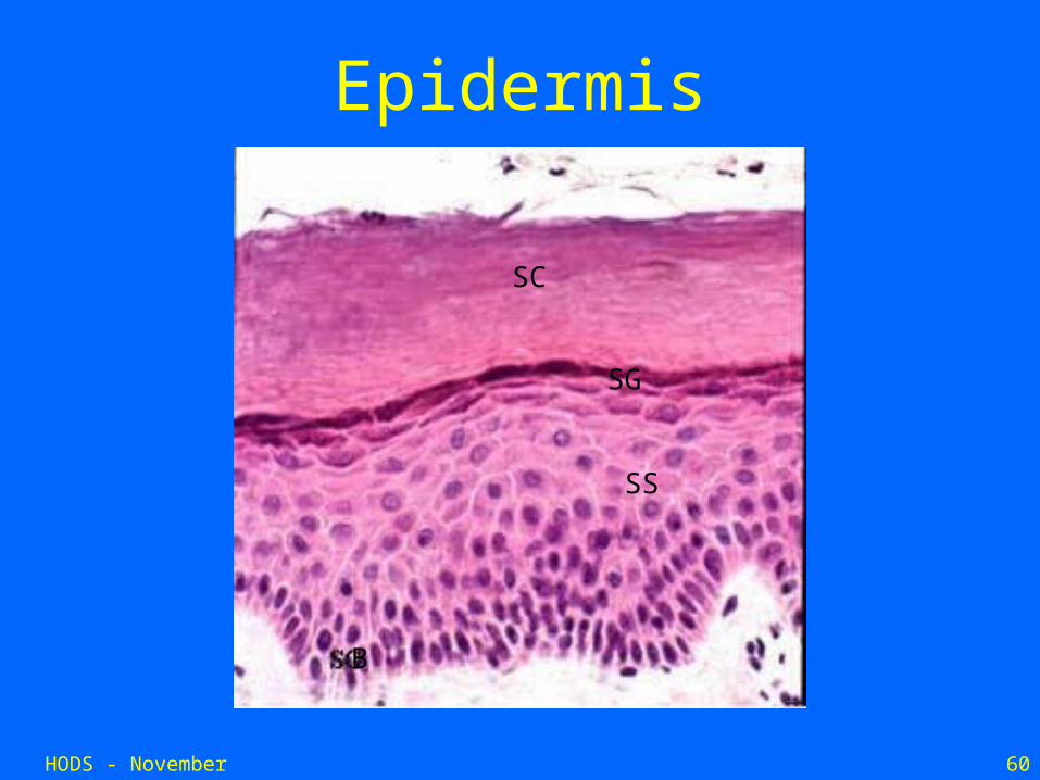
Epidermis• Provides barrier function.
• Multilayered structure, continually regenerating.
• Thickness dependent on exposure to friction.
• Stratified squamous epithelium, organised in five layers.– Stratum basale; Stratum spinosum, Stratum
granulosum, Stratum lucidum, Stratum corneum.
HODS - November 2006 58
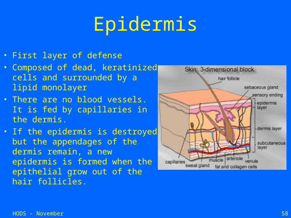
Epidermis
• First layer of defense• Composed of dead, keratinized
cells and surrounded by a lipid monolayer
• There are no blood vessels. It is fed by capillaries in the dermis.
• If the epidermis is destroyed but the appendages of the dermis remain, a new epidermis is formed when the epithelial grow out of the hair follicles.
HODS - November 2006 59
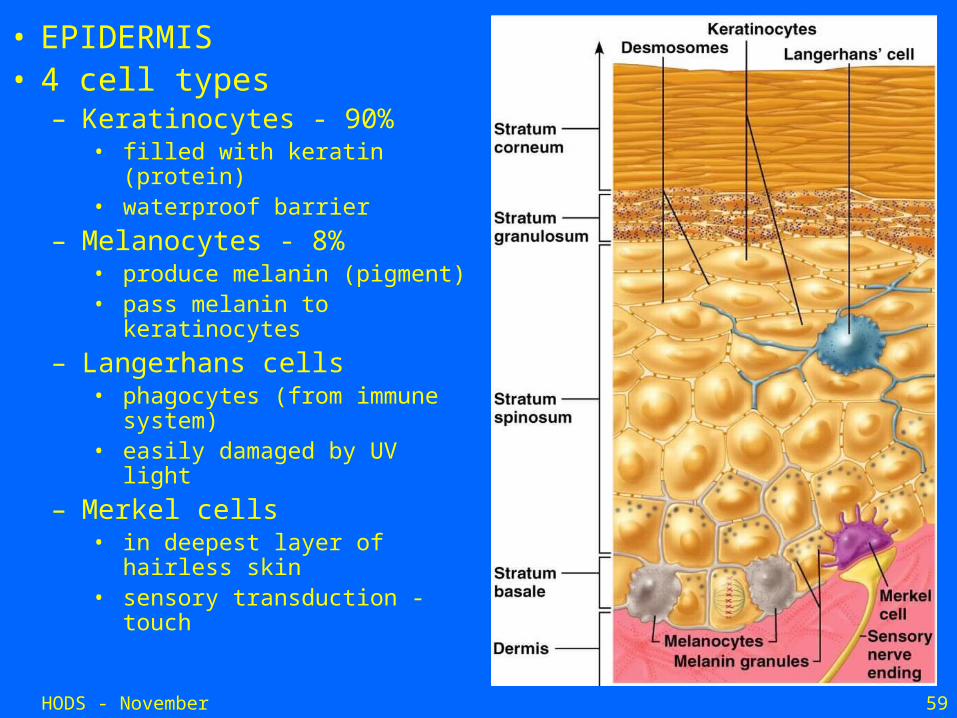
• EPIDERMIS• 4 cell types
– Keratinocytes - 90%• filled with keratin (protein)• waterproof barrier
– Melanocytes - 8%• produce melanin (pigment)• pass melanin to
keratinocytes
– Langerhans cells• phagocytes (from immune
system)• easily damaged by UV light
– Merkel cells• in deepest layer of hairless
skin• sensory transduction -
touch
HODS - November 2006 60
Epidermis
SS
SG
B
SC
HODS - November 2006 61
Dermis
• Varies in thickness across body.
• 1 mm on face , 4 mm on back.
• Responsible for most major functions of the skin.
• Two distinct layers:
Papillary dermis,
Reticular dermis.
HODS - November 2006 62
Dermis• Few cells present - fibroblasts,
macrophages, adipocytes
• Intracellular matrix thick with many protein fibers: collagen, elastin, reticular
• The location for blood vessels, nerves and sensory receptors, glands, hair follicles
HODS - November 2006 63
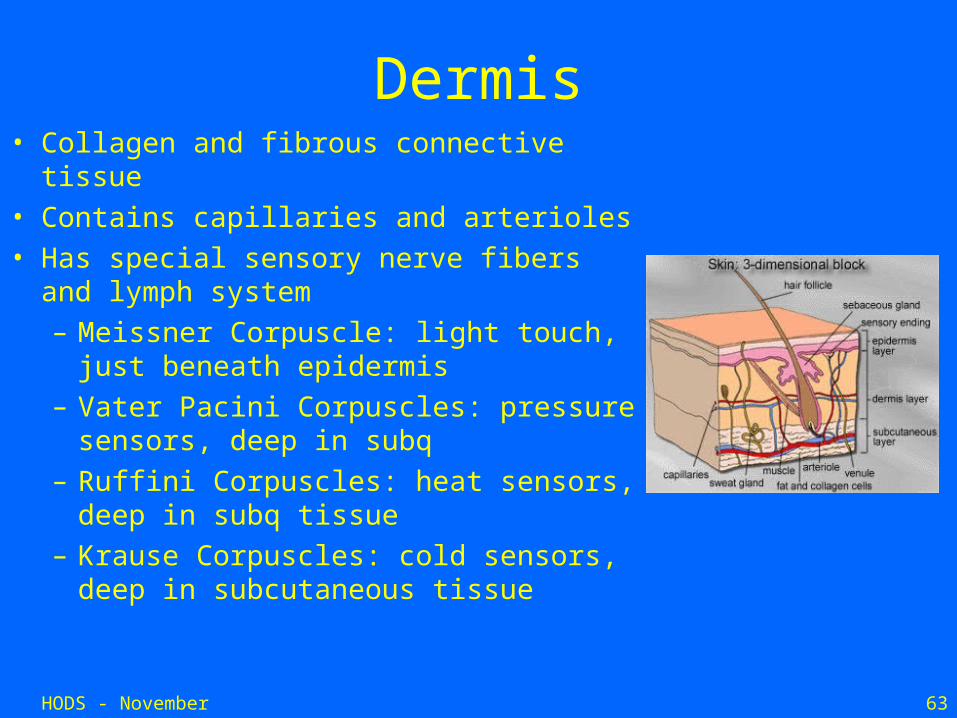
• Collagen and fibrous connective tissue• Contains capillaries and arterioles• Has special sensory nerve fibers and
lymph system– Meissner Corpuscle: light touch, just
beneath epidermis– Vater Pacini Corpuscles: pressure
sensors, deep in subq– Ruffini Corpuscles: heat sensors, deep
in subq tissue– Krause Corpuscles: cold sensors,
deep in subcutaneous tissue
Dermis
HODS - November 2006 64
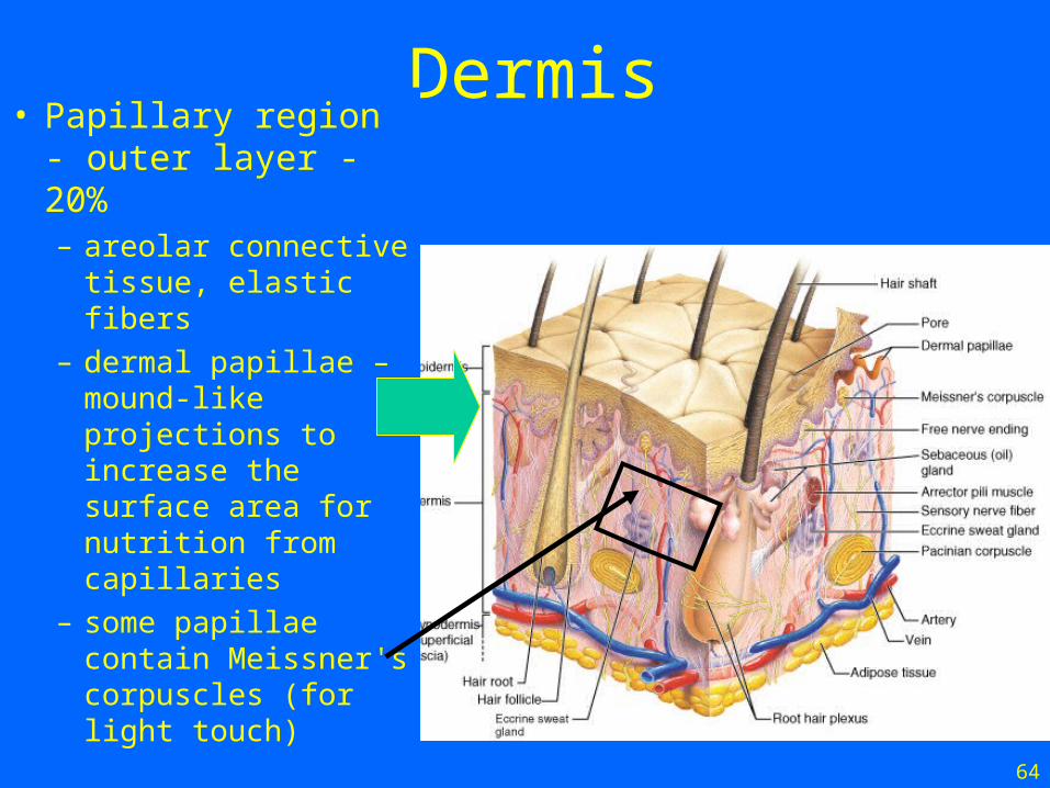
Dermis• Papillary region -
outer layer - 20%– areolar connective
tissue, elastic fibers– dermal papillae –
mound-like projections to increase the surface area for nutrition from capillaries
– some papillae contain Meissner's corpuscles (for light touch)
HODS - November 2006 65
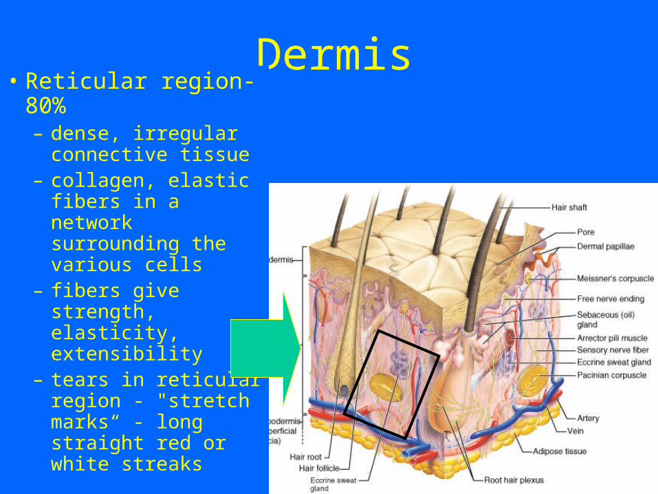
Dermis• Reticular region-
80%– dense, irregular
connective tissue – collagen, elastic
fibers in a network surrounding the various cells
– fibers give strength, elasticity, extensibility
– tears in reticular region - "stretch marks“ - long straight red or white streaks
HODS - November 2006 66
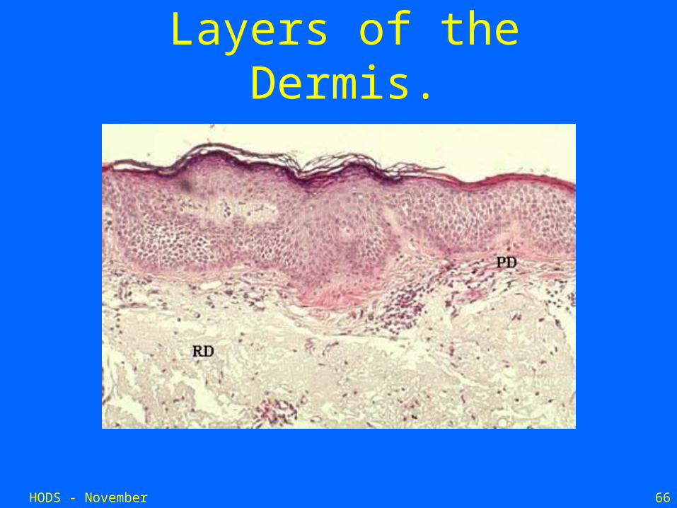
Layers of the Dermis.

HODS - November 2006 67
• Connective tissue• Fat cells in most areas• Blood vessels• Nerves• Base of hair follicles• Function:
• Insulation• Storage of nutrients
Subcutaneous Tissue
HODS - November 2006 68
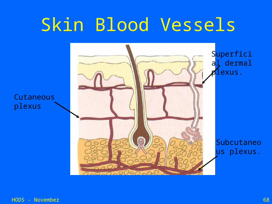
Skin Blood Vessels
Superficial dermal plexus.
Cutaneous plexus
Subcutaneous plexus.
HODS - November 2006 69
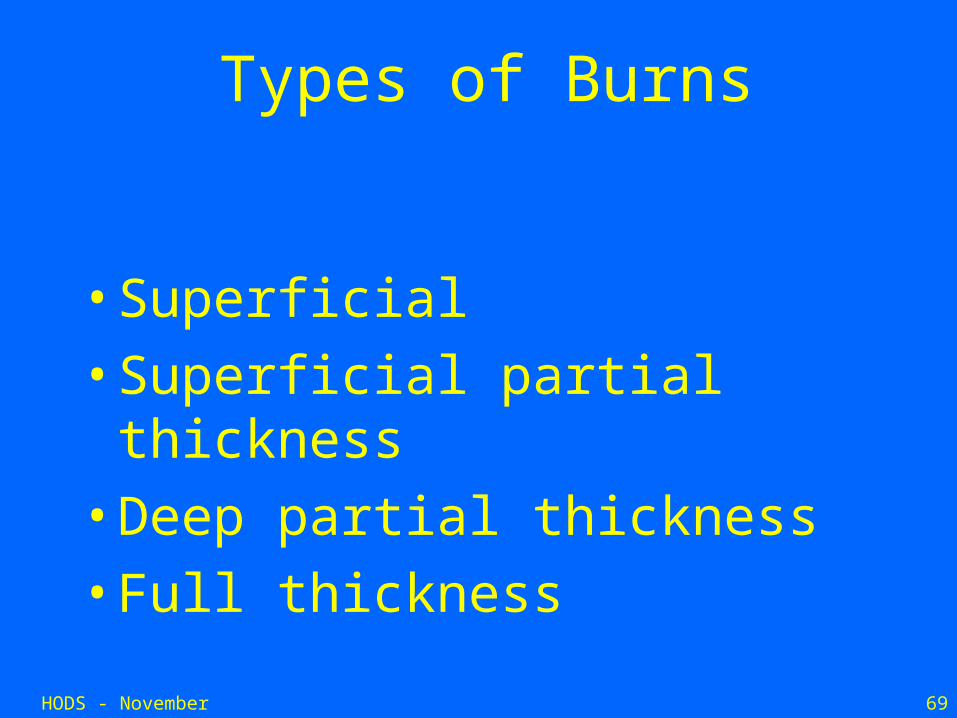
Types of Burns
• Superficial
• Superficial partial thickness
• Deep partial thickness
• Full thickness
HODS - November 2006 70
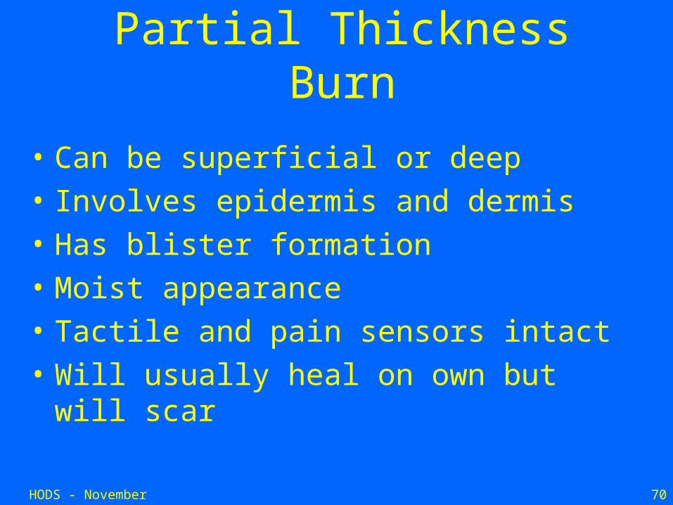
Partial Thickness Burn
• Can be superficial or deep
• Involves epidermis and dermis
• Has blister formation
• Moist appearance
• Tactile and pain sensors intact
• Will usually heal on own but will scar
HODS - November 2006 71
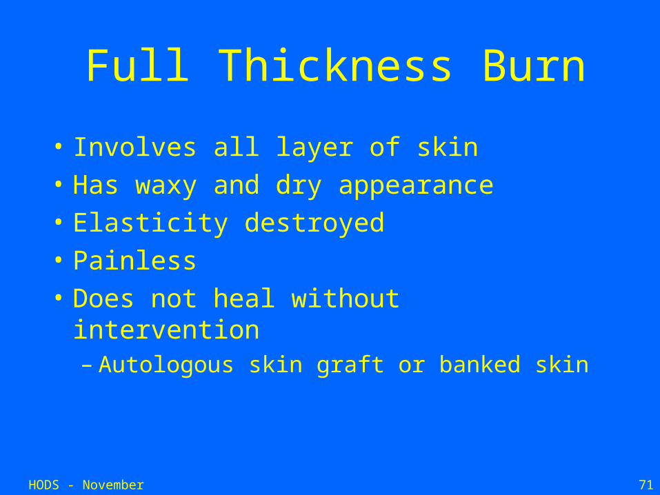
Full Thickness Burn
• Involves all layer of skin
• Has waxy and dry appearance
• Elasticity destroyed
• Painless
• Does not heal without intervention – Autologous skin graft or banked skin