Embed Size (px)
Citation preview

Hormones and Sex Steroid Receptors in the Female Pig
Effects of Mating, Artificial Insemination, Seminal Plasma and Genistein
Mattias Norrby Faculty of Veterinary Medicine and Animal Science
Department of Anatomy, Physiology and Biochemistry Uppsala
Doctoral Thesis Swedish University of Agricultural Sciences
Uppsala 2010

Acta Universitatis agriculturae Sueciae 2010:71
ISSN 1652-6880 ISBN 978-91-576-7516-3 © 2010 Mattias Norrby, Uppsala Print: SLU Service/Repro, Uppsala 2010
Cover: Drawing by Malin Carlson (www.malincarlson.se)

Hormones and Sex Steroid Receptors in the Female Pig – Effects of mating, artificial insemination, seminal plasma and genistein
Abstract Artificial insemination (AI) of sows and gilts is replacing mating in many countries. Another change over the last decades is that soya, rich in phytooestrogens, is routinely used in the diet to pigs. The aims of this thesis were to compare hormonal changes in blood plasma of the female pig during mating and insemination and to investigate if the phytooestrogen genistein influenced the hormonal responses and sex steroid receptor expression in the reproductive tract of inseminated gilts. Blood samples were taken via permanent catheters around mating or AI for analyses of hormones. Uterine and cervical cells were used in vitro to determine the role of seminal plasma. Three days after AI, gilts were euthanized and uterine and cervical tissues were prepared for morphological and immunohistochemical examination.
Plasma cortisol concentration increased more during mating than AI in sows, in contrast prostaglandin F2α metabolite (PGFM) increased only after AI. By incubating cells from porcine cervix and uterus with seminal plasma prostaglandin synthesis was inhibited in a dose dependent manner, providing a possible explanation for the difference in PGFM after mating. When a feed relevant dose of genistein was added to gilts from 3 days before oestrus until 3 days after AI, prostaglandin F2α exhibited more frequent pulses with higher amplitudes after AI, the PGE2 level was elevated and plasma oxytocin was almost doubled. PGFM, however, had a similar pattern in both groups. Nevertheless gilts administered genistein showed no significant difference in cell proliferation or the expression of oestrogen receptors (ER) α and β or progesterone receptor in uterus and cervix. However, intra-treatment evaluation and correlations indicated that genistein may modulate receptor patterns and induce subtle morphological changes. In view of the results presented in this thesis it is surprising that the difference in reproductive performance is not greater between AI and mating, especially with the addition of soya to the diet.
Keywords: Pig, oestrus, insemination, genistein, endocrinology, prostaglandin, oxytocin, oestrogen receptors, seminal plasma.
Author’s address: Mattias Norrby, SLU, Department of Anatomy, Physiology and Biochemistry, P.O. Box 7011, 750 07 Uppsala, Sweden E-mail: [email protected]

Dedication To all the sows and gilts in the world…
Om snöret inte håller, utan går av, är det bara att försöka med ett annat snöre.
Nalle Puh Det bästa är att veta vad man letar efter innan man börjar leta efter det.
Nalle Puh Instinct and intuition lend their guidance long before your head comprehends.
Dan Millan
Minds are like parachutes. They only function when they are open Sir James Dewar, Scientist (1877-1925)
To be absolutely certain about something, one must know everything or nothing about it.
Olin Miller
This report, by its very length, defends itself against the risk of being read. Winston Churchill (1874-1965)

Contents List of Publications 7
Abbreviations 9
1 Background 11 1.1 The female reproductive tract of the pig 12 1.2 Oestrus, gonadotropins, sex steroid hormones and their receptors 13 1.3 Mating and artificial insemination 15 1.4 Prostaglandins, oxytocin, and cortisol 16
1.4.1 Prostaglandins 16 1.4.2 Oxytocin 17 1.4.3 Cortisol 18
1.5 Seminal plasma 18 1.6 Phytooestrogens 19 1.7 Introduction to the thesis 20
Aims 21
2 Materials and Methods 23 2.1 Animals 23 2.2 Mating and insemination procedures 24 2.3 Venous cannulation and blood sampling (papers I and III) 24 2.4 Seminal plasma added to porcine and bovine cells (paper II) 25 2.5 Hormone analyses (papers I, II and III) 26 2.6 Reproductive tract (paper IV) 26
2.6.1 Tissue collection 26 2.6.2 Morphometric Analysis 27
2.7 Statistical analyses 27
3 Results 29 3.1 Experimental animals 29 3.2 Effects of mating or insemination (paper I and unpublished data) 29
3.2.1 Prostaglandin metabolite (PGFM) 29 3.2.2 Cortisol 29 3.2.3 Correlations 30 3.2.4 Oxytocin 30

3.3 Effects of seminal plasma on endometrial- and cervical cells (paper II) 30 3.3.1 Experiment 1 30 3.3.2 Experiment 2 30 3.3.3 Experiment 3 32
3.4 Effects of genistein on hormonal patterns (paper III) 32 3.4.1 Oxytocin 32 3.4.2 PGE2 32 3.4.3 PGF2α 32 3.4.4 PGFM 33 3.4.5 Cortisol 33 3.4.6 Correlations 33
3.5 Effects of genistein on sex steroid receptors and KI-67 (paper IV) 34 3.5.1 Morphology 34 3.5.2 ERα 34 3.5.3 ERβ 35 3.5.4 PR 35 3.5.5 Ki67 35
4 Discussion 37
5 Conclusions 45
6 Points of interest for future research 47
7 Populärvetenskaplig sammanfattning 49
8 References 53
9 Acknowledgements 67

7
List of Publications This thesis is based on the work contained in the following papers, referred to by Roman numerals in the text:
I Norrby M., Madsen MT., Borg Alexandersen C., Kindahl H., Madej A.
(2007). Plasma concentrations of cortisol and PGF2α metabolite in Danish sows during mating, and intrauterine and conventional insemination. Acta Vet Scand 49 (36).
II Madej M., Norrby M., Madsen MT., Johannisson A., Hansen C., Madej A. Effect of boar seminal plasma on production of prostaglandins and interleukin-6 from porcine endometrial and cervical cells and bovine endometrial cells (Manuscript).
III Norrby M., Madsen MT., Saravia F., Lundeheim N., Madej A. (2010) Genistein alters the release of oxytocin, prostaglandins, cortisol and LH during insemination in gilts. Reprod Dom Anim. doi: 10.1111/j.1439-0531.2010.01669.x
IV Norrby M., Madej A., Ekstedt E., Holm L. Effects of genistein on oestrogen and progesterone receptor, proliferative marker Ki-67 and carbonic anhydrase localization in the uterus and cervix of gilts after insemination (Manuscript).
Papers I and III are reproduced with the permission of the publishers.

8

9
Abbreviations AA ACTH
Arachidonic acid Adrenocorticotrophic hormone
AI Artificial insemination CRH Corticotrophin releasing hormone EDTA Ethylenediaminetetraacetic acid EIA Enzyme immunoassay ER Oestrogen receptor FSH Follicle-stimulating hormone GnRH Gonadotrophin releasing hormone IL Interleukin IUI Intrauterine insemination LH Luteinizing hormone OTR Oxytocin receptor pCE Porcine cervical epithelial cells pCS Porcine cervical stromal cells PGE2 Prostaglandin E2 PGF2α Prostaglandin F2α PGFM 15-keto-13,14-dihydro-prostaglandin PR Progesterone receptor pUE Porcine uterine epithelial cells pUS Porcine uterine stromal cells PVN Paraventricular nucleus RIA Radioimmunoassay SON Supraoptic nucleus TNF Tumor necrosis factor UTJ Utero tubular junction

10

11
1 Background The management of pigs has changed over the last decades. The increasing size of the herds has made it impractical to keep enough boars for mating, which has increased interest for artificial insemination as a consequence. Another advantage with artificial insemination is that the best boars can be more intensively used and the genetic improvements can be made faster.
However, there are differences between mating and artificial insemination in terms of the physiological response in female pigs. The major part of the seminal plasma is discarded when the insemination doses are prepared and other solutions are added to the small quantity of the sperm rich fraction which is needed. If these differences cause changes in the hormonal responses of the female has yet to be fully investigated, but smaller litter size after AI than mating are reported (Paredis, 1961; Tummaruk et al., 2000; Tanavots et al., 2002). In addition, gilts and sows respond differently to combinations of mating and AI in terms of reproductive performance (Flowers & Alhusen, 1992) and the presence of a boar during AI has a positive effect on litter size (Am-In et al., 2006). From both practical and theoretical aspects the discrepancies therefore seemed interesting and initiated this project.
Another aspect included in this thesis is the widespread use of soya as a protein source for pigs. Soya contains phytooestrogens e.g. genistein and daidzein, resembling the natural oestrogens necessary for normal reproduction. Gilts fed fodder containing soybean meal (11.6 %) consume approximately 725 mg total phytooestrogens daily (Ford et al., 2006). Genistein is rapidly absorbed into the blood plasma (Lundh, 1995). Comparison of hormonal and local effects in the reproductive organs of pigs given or not given phytooestrogens was therefore of interest.

12
1.1 The female reproductive tract of the pig
The female reproductive tract consists of ovaries, oviducts, uterus, cervix, vagina, vestibulum and vulva. The ovaries in the pig are approximately 5 cm long and irregular in shape, due to numerous follicles or corpora lutea protruding from the surface in cyclic animals. The ovaries have two principal functions: the storage and development of oocytes (egg cells), and the production of sex hormones. The oviducts, or uterine tubes, are about 20 cm in length and divided into three segments. Facing the ovary is the infundibulum, which capture the oocytes at ovulation. The oviducts then consist of the wider ampulla and the narrower isthmus, which connects to the uterus. The ampulla-isthmus junction is the site of fertilisation, i.e. where the spermatozoa meet the ova. The pig uterus consists of a short body and two long uterine horns (corpus and cornuae uteri). The cervix is up to 25 cm in length and has internal interdigitating mucosal prominences pulvini cervicales. The cervix lacks distinct ends; in particular the transition between cervix and vagina is continuous. The utero-cervical transition is somewhat more distinct. The vagina reaches from the cervix to the urethral orifice and the vestibulum reaches from the urethral orifice to the vulva. The vulva is composed of the labia and the clitoris.
In this thesis the uterus and cervix have been investigated at a light microscopy level. Therefore, the structure of these organs is described briefly. The uterine wall is composed of an endometrium towards the uterine lumen, a myometrium and the perimetrium towards the abdominal cavity. The endometrium consists of a surface epithelium, coiled, branched uterine glands and a loose connective tissue stroma with capillaries and small blood vessels. The myometrium is mainly composed of smooth muscle cells with two thin longitudinal layers and a thicker circular layer in between. The uterus is not considered to be fully morphologically mature until the second oestrus after puberty (Schnurrbusch & Erices, 1979). In the cervix of the sow main part of the surface epithelium is stratified and undergoes cyclic variations. The underlying stroma consists of dense connective tissue with capillaries and small blood vessels. The tunica muscularis, consists of smooth muscle cells in two layers, one circular and one longitudinal. Towards the abdominal cavity is a serosa of loose connective tissue (Priedkalns, 1993).
13
1.2 Oestrus, gonadotropins, sex steroid hormones and their receptors
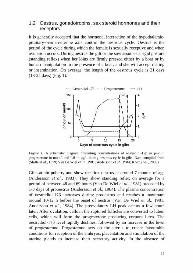
It is generally accepted that the hormonal interaction of the hypothalamic-pituitary-ovarian-uterine axis control the oestrous cycle. Oestrus is the period of the cycle during which the female is sexually receptive and when ovulation occurs. During oestrus the gilt or the sow assumes a rigid posture (standing reflex) when her loins are firmly pressed either by a boar or by human manipulation in the presence of a boar, and she will accept mating or insemination. On average, the length of the oestrous cycle is 21 days (18-24 days) (Fig. 1).
0 5 10 15 200
50
100
150
0
1
2
3
4
LHOestradiol-17β ProgesteronePGF2α
Days of oestrous cycle in gilts
pmol
/l or
nm
ol/l
µ g/l
Figure 1. A schematic diagram presenting concentrations of oestradiol-17β in pmol/l, progesterone in nmol/l and LH in µg/l, during oestrous cycle in gilts. Data compiled from (Shille et al., 1979; Van De Wiel et al., 1981; Andersson et al., 1984; Knox et al., 2003).
Gilts attain puberty and show the first oestrus at around 7 months of age (Andersson et al., 1983). They show standing reflex on average for a period of between 48 and 69 hours (Van De Wiel et al., 1981) preceded by 1-3 days of prooestrus (Andersson et al., 1984). The plasma concentration of oestradiol-17β increases during prooestrus and reaches a maximum around 10-12 h before the onset of oestrus (Van De Wiel et al., 1981; Andersson et al., 1984). The preovulatory LH peak occurs a few hours later. After ovulation, cells in the ruptured follicles are converted to lutein cells, which will form the progesterone producing corpora lutea. The oestradiol-17β level rapidly declines, followed by an increase in the level of progesterone. Progesterone acts on the uterus to create favourable conditions for reception of the embryos, placentation and stimulation of the uterine glands to increase their secretory activity. In the absence of

14
embryos, uterine cells start to produce prostaglandin F2α (PGF2α), which causes regression of the corpora lutea (luteolysis) on days 13 – 17 of the cycle and progesterone secretion declines (Shille et al., 1979).
If the gilts are pregnant the corpora lutea continue to produce progesterone and the farrowing takes place after 110-115 days. Usually, piglets are allowed to suckle for five weeks in Sweden. During lactation the ovarian activity is inhibited and the LH pulses from the hypophysis have a low frequency and low amplitude (De Rensis & Foxcroft, 1999). Immediately after weaning, available follicles are a target for increasing frequency of LH pulses. At the same time, mean plasma concentration of FSH rises followed by an increased concentration of oestradiol-17β. At a certain time FSH will start to decrease concomitant with a further increase of LH and oestradiol-17β. According to (Lucy et al., 2001) such hormonal changes indicate follicular development. Plasma concentrations of oestradiol-17β reach a maximum between 4 hours before and 18 after the onset of standing oestrus. When these hormonal changes are well coordinated, the weaning-to-oestrus interval in sows varies on average between 96 and 118 hours (Soede et al., 1994; Brandt et al., 2009). Oestradiol-17β causes the typical signs of oestrus such as a swollen and hyperaemic vulva, restlessness and riding behaviour as well as increased secretory activity, hypertrophy and oedema of the reproductive tract.
By acting on GnRH in hypothalamus oestradiol-17β has a positive feedback on the preovulatory LH peak, which reaches a maximum 5 – 20 h after oestradiol peak and ovulation occurs 26 – 34 h later (Brandt et al., 2007; Brandt et al., 2009). The length of oestrus can be as short as 32 h and as long as 72 h (Brandt et al., 2007; Brandt et al., 2009). The time from the onset of oestrus to ovulation ranges between 38 and 41 hours. The progesterone concentrations start to rise a short time after ovulation (Brandt et al., 2007).
The strict coordination between oestradiol-17β and progesterone during the oestrous cycle is necessary for adequate reactions in the uterus and cervix. The hormones are ligand regulating transcriptional factors through an interaction with target DNA sequences. Thereafter, gene expression modification, protein expression and hormonal effects emerge, which take several minutes to hours or days, reviewed in (Migliaccio et al., 2010). The biological effects of oestradiol-17β were found to be mediated through a specific receptor (oestrogen receptor = ER) in 1962 (Jensen & Jacobson, 1962). Since then the ER has been extensively studied and in 1995 (Kuiper et al., 1996) found a second ER, named ERβ. The first ER was cloned in 1986 (Walter et al., 1985; Green et al., 1986) and has been renamed ERα.

15
The expression of ERs is controlled by both progesterone and oestrogen (Mester et al., 1974). Oestradiol-17β up-regulates ERα and down-regulates ERβ as well as ERβ mRNA in the uterine stroma of mice (Weihua et al., 2000). However, studies on porcine myometrium described that one single dose of oestradiol-17β down-regulates ERs (Sahlin et al., 1990). Oestradiol-17β also greatly increases the expression of the progesterone receptor (PR) in the uterine tissues of guinea pigs and rats (Milgrom et al., 1973; Koseki et al., 1977). Later, it has been shown that oestradiol-17β induces PR in uterine stromal and glandular epithelial cells whereas it reduces PR in mice uterine surface epithelial cells (Weihua et al., 2000). It has been suggested that ERα activity controls the alternation of PR described by the latter authors, in contrast ERβ signalling decrease PR mRNA levels selectively in glandular cells (Sahlin et al., 2006).
1.3 Mating and artificial insemination
At mating, as well as during conventional artificial insemination (AI), semen is deposited into the cervical canal of the female. Semen transport along the uterine horns is elicited by myometrial contractions and a certain number of spermatozoa reach the uterotubal junction (UTJ) and the first part of isthmus. At the isthmus a sperm reservoir forms in order to (i) escape reflux and phagocytosis in the uterine lumen and (ii) ensure that sufficient numbers of potentially fertile spermatozoa are present at the site of fertilization when oocytes are released (Rodriguez-Martinez et al., 2005). The first successful AI was of a bitch, performed by Spallanzi in 1780, cited by Wishart (1944). AI in pigs has been used since the 1930s but the major development of AI in the pig industry took place in the 1980s, reviewed by Roca et al. (2006). In the pig, some changes to the methodology have occurred over the years, i.e. the semen was first deposited in the anterior end of the vagina (Wishart, 1944) instead of in the cervical canal, but in general the technique is the same. The quantity of the dose and the number of spermatozoa used during those first years of successful AI, are actually close to what is used today in conventional AI (Ito et al., 1949). However, new AI techniques have been introduced as well, such as intrauterine techniques when the semen is deposited in the body or the horn of the uterus (Vazquez et al., 2008). Nearly 50 % of gilts and sows were inseminated at the end of 1990ies worldwide and in some European countries the percentage is 80 %, reviewed by Roca et al. (2006).
The hormonal pattern during AI differs from that of mating. Stimulation by the boar during mating releases oxytocin, but does not alter the basal

16
concentration of prostaglandin F2α metabolite in sows. The release of oxytocin can be triggered by stimulation similar to that of the boar performed by an AI-technician in front of a boar (Madsen et al., 2002; Madej et al., 2005). Boar stimuli have been widely discussed and consist of olfactory, auditory, visual and tactile components which all have been found to affect specific processes around AI (Soede, 1993). Olfactory components, pheromones such as 5α-androstenone, were shown to affect both uterine activity and oxytocin release by Mattioli et al. (1986), but Langendijk et al. (2003) got contradictory results and no effects of 5α-androstenone. Gerritsen et al. (2005) showed that combinations of artificial boar stimuli do not mimic the real boar and it is unclear how boar stimuli should be imitated to elicit the optimal physiological response in the female pig.
1.4 Prostaglandins, oxytocin, and cortisol
1.4.1 Prostaglandins Prostaglandins were first discovered and isolated from human semen in 1935 (Goldblatt, 1935; von Euler, 1936). Von Euler named them prostaglandins assuming that they were synthesized in the prostate gland. The first successful isolation of a prostaglandin (PGF) was in 1957 (Bergstrom & Sjovall, 1957). Prostaglandins are synthesised in most cells in the body. They are hormones and they act as chemical messengers, but they are not produced in specific organs like the “classical hormones”.
Prostaglandins are carboxylic acids, consisting of a 20 carbon skeleton with a five member ring and two side chains of 7 and 8 carbons respectively. They are synthesized from the essential fatty acid; arachidonic acid (AA). The AA is membrane bound in cells and most cell types possess the enzyme system necessary for the conversion of AA to prostaglandin. The first step leads to the production of cyclic peroxides by cyclooxygenas or the COX-system. Further metabolism of the peroxides leads to prostaglandins. Stimuli that activate prostaglandin biosynthesis are many e.g.; hormonal, nervous, other chemical and mechanical stimuli (Samuelsson et al., 1975; Granstrom, 1981).
Prostaglandins participate in a wide range of body functions such as the contraction and relaxation of smooth muscles, the dilation and constriction of blood vessels, control of blood pressure, and modulation of inflammation. The prostaglandins are important in reproduction and those analyzed in this thesis are prostaglandin F2α (PGF2α), prostaglandin E2

17
(PGE2) and the prostaglandin metabolite 15-keto-13,14-dihydro-PGF2α (PGFM). In the pig, PGF2α is luteolytic (Gleeson et al., 1974) and PGE2 has a luteotrophic or anti-luteolytic effect (Akinlosotu et al., 1986; Akinlosotu et al., 1988). PGF2α and PGE2 both cause myometrial contractions (Carsten, 1974; Willenburg et al., 2004; Cao et al., 2005; Mueller et al., 2006).
PGFM is a semi-stable metabolite of PGF2α, which has a half-life of about 8 minutes (Kindahl et al., 1976). PGFM is often used to estimate PGF2α activity because PGF2α is rapidly metabolized during its passage through the lungs. The enzyme 15-hydroxyprostaglandin dehydrogenase (15-PDGH) has been demonstrated in porcine uterine surface epithelial and glandular cells (Franczak & Bogacki, 2009), which is of interest because in the pig only 18 % is metabolized during the first passage in the lungs, in contrast to 99 % of the PGF2α in the sheep (Davis et al., 1980).
1.4.2 Oxytocin Oxytocin is a peptide hormone, consisting of nine amino-acids produced by neurons in the paraventricular- and supraoptic nuclei of the hypothalamus (PVN and SON). By axonal transport oxytocin reaches the posterior pituitary gland where it can be stored or released into the blood. The prepubertal pig has fewer oxytocin neurons than the mature gilt or sow (Raymond et al., 2006).
Oxytocin was the first peptide hormone to be sequenced and synthesized (Vigneaud, 1956). Oxytocin is well known for its role during parturition. It stimulates smooth muscle cells in the uterine myometrium (Carsten, 1974). The peptide got its name from Greek and means ‘‘quick birth’’ after its uterine-contracting properties discovered by Dale in 1906 and thus was the first hormone to be linked directly to female reproduction, cited by Lee et al. (2009). Oxytocin also stimulates the myoepithelial cells surrounding the milk-secreting alveoli in the mammary glands causing milk let-down. Oxytocin mediates contractile response through the oxytocin receptor (OTR) which binds both oxytocin and vasopressin. In the myometrium OTRs are unevenly distributed in longitudinal and circular muscles and regions of the uterus (Kitazawa et al., 2001).
However, numerous other roles have been proposed for this peptide, e.g. its neuropeptide properties within the brain shown as effects on sexual and social behaviour (Lee et al., 2009). In this thesis it is the role of oxytocin during oestrus which is in focus. In pigs, the pattern of myometrial contractions differs depending on the stage in the oestrous cycle (Langendijk et al., 2002), which is important for the movement of

18
spermatozoa to the fertilization site at the ampullary-isthmus junction and later for the passage of the fertilized oocyte to the uterus (Rodriguez-Martinez et al., 1982). Oxytocin release has been shown in oestrus sows during boar contact (Langendijk et al., 2003) and during mating in the mare (Bader & Schams, 1987) and sow (Claus & Schams, 1990). The latter authors also showed that an infusion of oestradiol-17β to the porcine uterus causes oxytocin release. Several early findings suggest that adding oxytocin to AI doses enhances conception rates and litter size in pigs (Milovanov & Sergeev, 1961; Sergeev, 1963) and conception rates in cows (Hays et al., 1958).
1.4.3 Cortisol Corticotrophin releasing hormone (CRH) from the hypothalamus acts
on the anterior lobe of the hypophysis and causes release of ACTH, which is transported with the blood to the adrenal cortex where cortisol is released. Vasopressin and oxytocin also contribute to the release of adrenocorticotrophic hormone (ACTH), in synergy with CRH (McCann et al., 2000). Cortisol is a glucocorticoid (C21) which belongs to the steroid superfamily of hormones together with progestagens (C21), androgens (C19) and oestrogens (C18) that originates from cholesterol. Measurement of cortisol (or cortiscosterone in some species) in blood plasma is a traditional method for monitoring and evaluating stress in animals, even though the interpretation of the results can be difficult due to the wide definition of stress (Einarsson et al., 2008). Glucocorticoids are metabolic hormones with the primary function of preventing hypoglycaemia. They also have anti-inflammatory effects and are thought to alter gene expression by as much as 1 % in their target cells (Flower & Rothwell, 1994). An increase of cortisol in response to mating appears to be normal in the female pig (Barnett et al., 1982).
1.5 Seminal plasma
A better understanding of seminal plasma signalling in the female reproductive tract is important for optimal reproductive efficiency (Robertson, 2007). The boar ejaculate contains large amounts of oestrogens and testosterone and approximately half of the total oestrogen contents is bound to spermatozoa (Claus et al., 1985), but the roles of these hormones are not known. In humans, seminal plasma contains large amounts of PGE2 and PGF2α (Samuelsson, 1963) whereas only small amounts are found in boar seminal plasma (Cheng et al., 2003).

19
Recently, Kaczmarek et al. (2010) reported that seminal plasma stimulated production of PGE2 from porcine oviduct epithelial cells but did not influence secretion of PGF2α. The same study demonstrated that PGF2α synthase and prostaglandin 9-ketoreductase (CBR1), which converts PGE2 to PGF2α, were down-regulated one day after infusion of seminal plasma into the uterine horns (Kaczmarek et al., 2010). Furthermore, seminal plasma provokes ovulation in the pig. Waberski et al. (2006) suggested that semen-induced cytokines accelerate the final maturation of pre-ovulatory Graafian follicles.
In humans Sharkey et al. (2007) described a regulatory action of seminal plasma in expression of pro-inflammatory and immune-regulating cytokines in human cervical cells.
1.6 Phytooestrogens
Oestrogenic activity in plants inducing oestrus in animals was first reported by Loewe in 1926, who also wrote the first review on plant oestrogens in 1933, cited by Sharaf (1968). Bennetts et al. (1946) found reproductive disturbance in sheep grazing on clover pastures. The explanation was clear when it was found that clover contains phytooestrogens, and they are produced by many plants, including soya, which is a common protein source for pigs. Soya and their products are among the most significant sources of daidzein and genistein, two important isoflavones (Mazur & Adlercreutz, 1998). Genistein, as well as other isoflavones, resembles oestradiol-17β and activates ERα and ERβ (Kuiper et al., 1998). Interestingly, isoflavones seem to be oestrogen-antagonists in high oestrogen environments and oestrogen-agonists in low oestrogen environments (Hwang et al., 2006).
Genistein might more effectively induce ERα-mediated actions, despite having a higher affinity for ERβ (Whitten & Patisaul, 2001). Genistein is reported to have anti-oestrogenic effects in the central nervous system of rodents (Patisaul et al., 2002). In piglets born to sows administered daidzein ERβ, gene expression was down-regulated in the hypothalamus, but not in the hypophysis (Ren et al., 2001). It has also been shown that phytooestrogens, like genistein, are effective inhibitors of COX-2 (Hertrampf et al., 2005; Swami et al., 2007) but there are also reports showing that genistein increases the prostaglandin concentration in cattle (Woclawek-Potocka et al., 2005). In addition, genistein attenuates the release of cortisol in adrenal cells from humans (Mesiano et al., 1999) and

20
pigs (Kaminska et al., 2007), probably by suppressing the ACTH-induced activity of 21-hydroxylase (P450c21) (Mesiano et al., 1999).
1.7 Introduction to the thesis
In reproduction it is mainly the luteolytic effect of PGF2α which has been of interest during the oestrous cycle. It was therefore a new finding when (Madsen et al., 2002) reported that artificial insemination (AI) of sows resulted in a dramatic increase of PGFM, which was not seen in mated sows. This was confirmed after intrauterine insemination by Alexandersen et al. (2005). In later studies an interaction between cortisol and PGFM was related to housing systems during AI (Madej et al., 2005). In view of these studies it was decided to compare the effects on cortisol and PGFM in blood samples obtained from three groups of sows during AI by conventional technique (AI), intrauterine insemination (IUI) and boar mating. The results are presented in paper I.
What could be the reason for the increase of PGFM during AI? Could the removal of most of the seminal plasma in preparation of the AI dose be involved? This problem is raised in paper II, where endometrial and cervical cells were studied with or without the addition of seminal plasma and the effects on prostaglandin production. As mentioned in the background, pigs are receiving phytooestrogens via their feed. Systematic investigations of what this means in terms of hormonal patterns during AI (paper III) and if genistein alter the expression of sex steroid receptors after AI (paper IV) were performed.

21
Aims The overall aims of this thesis were to compare hormonal changes in blood plasma of the female pig during mating and insemination and to investigate to what extent the phytooestrogen genistein influenced the hormonal responses and sex steroid receptor expression in the reproductive tract of inseminated gilts. Specifically the studies aimed: to determine if patterns of PGFM, cortisol and oxytocin in blood
plasma differ during mating and insemination to investigate if seminal plasma inhibited the release of
prostaglandins in uterine and cervical cells to determine if genistein administration per os changed patterns of
PGF2α, PGE2, and PGFM, oxytocin, and cortisol concentrations in the blood plasma of gilts after AI, and
to study if genistein alters the expression of ERα, ERβ, PR, and proliferation marker Ki 67 in uterus and cervix of early pregnant gilts

22
23
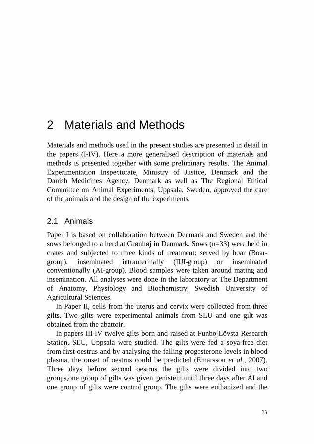
2 Materials and Methods Materials and methods used in the present studies are presented in detail in the papers (I-IV). Here a more generalised description of materials and methods is presented together with some preliminary results. The Animal Experimentation Inspectorate, Ministry of Justice, Denmark and the Danish Medicines Agency, Denmark as well as The Regional Ethical Committee on Animal Experiments, Uppsala, Sweden, approved the care of the animals and the design of the experiments.
2.1 Animals
Paper I is based on collaboration between Denmark and Sweden and the sows belonged to a herd at Grønhøj in Denmark. Sows (n=33) were held in crates and subjected to three kinds of treatment: served by boar (Boar-group), inseminated intrauterinally (IUI-group) or inseminated conventionally (AI-group). Blood samples were taken around mating and insemination. All analyses were done in the laboratory at The Department of Anatomy, Physiology and Biochemistry, Swedish University of Agricultural Sciences.
In Paper II, cells from the uterus and cervix were collected from three gilts. Two gilts were experimental animals from SLU and one gilt was obtained from the abattoir.
In papers III-IV twelve gilts born and raised at Funbo-Lövsta Research Station, SLU, Uppsala were studied. The gilts were fed a soya-free diet from first oestrus and by analysing the falling progesterone levels in blood plasma, the onset of oestrus could be predicted (Einarsson et al., 2007). Three days before second oestrus the gilts were divided into two groups,one group of gilts was given genistein until three days after AI and one group of gilts were control group. The gilts were euthanized and the

24
genital tract was removed, weighed and macroscopically examined. The number of corpora lutea (CL) was counted. From each of the uterine horns the tuba uterina and first part (20 cm) of the uterus including uterotubal junction (UTJ) were flushed with BSA-buffer for embryo collection and counting.
2.2 Mating and insemination procedures
The sows and gilts were checked for standing oestrus by means of the back pressure test and the riding test, in the presence of a boar. In paper (I) two Danish Duroc boars were evenly used, to serve the sows in the Boar-group. The artificially inseminated sows were assigned to either AI or IUI. All sows were served during the first oestrus after weaning. The sows in the Boar-group were moved to the boar-pen for mating, while AI- and IUI-groups were served in the crate (Paper I). The gilts in papers III and IV were served 24 hours after the first signs of standing oestrus behaviour (Almeida et al., 2000) during their second oestrus. The gilts and the sows, regardless of insemination method, were stimulated in front of a boar according to a 5-point stimulation plan (Madsen et al., 2002), i.e. manipulation of the abdominal area, the area under vulva, the inguinal area and the pelvic area followed by back pressure before AI. All inseminated animals received a dose of semen containing 2 × 10⁹ progressive motile spermatozoa. The semen was blended with EDTA-extender to a total volume of 80 ml.
2.3 Venous cannulation and blood sampling (papers I and III)
Blood samples were taken via an implanted catheter in the vena jugularis externa through vena auricularis. The pigs were sedated and restrained with a nose string during catheterization. In the first pigs (Paper I) it was difficult to penetrate the skin of the ear directly with the catheter (designed for use in humans) and in some cases the catheter tip was damaged by the procedure. We therefore modified the technique by making a small cut in the skin with a scalpel (Paper III). This made the procedure easier and the tip did not damage the vein. The hole was sealed immediately after insertion of the catheter, to minimize the risk for infection.
The blood samples had to be collected as calmly as possible to avoid stress affecting the results. The gilts got several days of adjustment to the new environment where the insemination was to occur. During that time they were trained to accept human contact and to be scratched gently. After

25
many hours all of the gilts were relaxed in the new environment. This gave the advantage that they allowed frequent blood sampling without any restraint and that it was easy to clean the ears and flush the catheter. All blood samples were collected using a 10 ml syringe with a minimum interval of 1 min before, during, and after service. The catheters were flushed at regular intervals with heparinised saline solution to avoid coagulation and that solution was discarded before the next blood sample was withdrawn. The syringe was emptied into pre-cooled tubes prepared with EDTA or heparin, and aprotinin, all to prevent enzymatic degradation of protein hormones. Blood plasma intended for analysis of prostaglandin was prepared with indomethacin to block the continued synthesis of prostaglandins in the sample.
2.4 Seminal plasma added to porcine and bovine cells (paper II)
Three experiments were performed. In Experiment 1, porcine endometrial stromal cells (pUS) and porcine cervical stromal cells (pCS) originating from two gilts after insemination. Preparation of the cells is described in detail in Paper II. The effects of adding seminal plasma at volumes from 0.0 to 1.0 ml to the culture medium on the production of PGFM by the cells were tested.
In Experiment 2, the investigations included a comparison between porcine and bovine cells. Bovine endometrial epithelial cells were purchased from Cell Applications, Inc., San Diego, USA, and cultured according to the recommendations of the manufacturer. Porcine endometrial epithelial (pUE), endometrial stromal (pUS), cervical epithelial (pCE) and cervical stromal (pCS) cells were collected from a slaughtered gilt and treated as in Experiment 1. In experiment 2, 0.2 ml of seminal plasma was added to the culture medium and the production of PGFM and PGF2α was analysed. In this experiments AA, (20 μg/ml) were added to some cells and compared with the controls.
In Experiment 3, porcine epithelial and stromal cells obtained from the endometrium as well as from the cervix during Experiment 2 were used. The cells were cultured for 8 days and harvested by enzymatic treatment. The cell suspension was frozen at -135oC, stored for 6 months and moved to -80oC. After several months of storage, cells were thawed and cultured as in Experiment 2 for 3 and 24 h with and without 20.0 µg/ml of AA. The effects of different volumes of seminal plasma on the production of PGFM, PGF2α and PGE2 were tested.

26
In all experiments, the medium was collected after 3 and 24 h incubation into tubes containing 10 µg/ml of indomethacin and stored at -20°C. The cells were lysed for the total cellular protein determination and also stored at -20°C.
2.5 Hormone analyses (papers I, II and III)
At the onset of this thesis work the previously used radioimmunoassay (RIA) for oxytocin in our department was no longer available. It was replaced with a commercial enzyme immunoassay (EIA) method. The first tests with the EIA kit for oxytocin revealed that it could not be used according to the manual from the supplier because the values were too high. This started a tedious step by step modification of the method as described in Paper III. These modifications resulted in an ED-50 value for the standard curve (expected concentration at 50 % bound) at 82.7 pmol/l compared with 123 pmol/l according to the manual. The recovery of oxytocin in spiked plasma samples was on average 97%.
PGE2 and PGF2α analyses were done using a commercial competitive EIA on indomethacin treated plasma and medium samples. PGE2 validation showed a mean recovery of 92%. The PGF2α analyses had, according to the manufacturer’s protocol, a 96.9% recovery in porcine plasma. PGFM in paper I, III and in Experiment 1 in paper II, was analysed by RIA (Kunavongkrit et al., 1983). In Experiment 2 and 3 of paper II the PGFM
was analysed by a commercial EIA. Cortisol analyses were done using a RIA kit, which was validated for
porcine plasma (Mwanza et al., 2000) and plasma progesterone was measured by RIA (Coat-A-Count Progesterone; Diagnostic Products Corporation), used according to the manufacturer’s recommendations and validated for porcine plasma (Epelu-Opio & Madej, 1988).
2.6 Reproductive tract (paper IV)
2.6.1 Tissue collection Approximately 20 – 30 cm from the uteri tubular junction (UTJ), a piece of the uterus was cut off; opened lengthwise and transverse slices were collected from the mesometrial side. The uterine part of cervixes was cut transversely and slices were divided into 4 pieces. The slices were fixed either in paraformaldehyde or in glutaraldehyde. Histological and immunohistochemical examinations were performed. A standard avidin–

27
biotin immunoperoxidase technique was applied to detect ERα, ERβ, PR and Ki-67 proteins. The morphometric measurements and evaluation of cells with positive immunostaining for ERα, ERβ, PR and Ki-67 was carried out on coded slides by the same person (M. Norrby).
2.6.2 Morphometric Analysis In the uterine endometrium the surface epithelium, the subepithelial area of the connective tissue stroma and the endometrial glands were evaluated. In the uterine myometrium, smooth muscle cells were evaluated. In the cervix, an evaluation of the surface epithelium and the connective tissue stroma of the subepithelial area were performed.
The height of the surface epithelium was measured in both the uterus and the cervix. The uterine glands were divided into superficial and deep glands and numbers of cross-sections were counted and the diameter was measured.
In cells positive for ERs and PR the staining intensity of the cell nuclei (average of all cells) was graded as weak, moderate, or strong. Because not all cells stained positive in all compartments, the proportion of positive to negative cells was estimated at four different levels (marked A–D): low proportion (30% positive cells, A); moderate proportion (30–60% positive cells, B); high proportion (60–90% positive cells, C) and almost all cells positive (more than 90%, D). In the subepithelial stroma of the uterine endometrium and cervix, the number of clearly ER- or PR- positive cells (moderate to strong staining) per mm2 was counted. The cells positive for Ki-67 in the connective tissue stroma was counted in the same manner as for the ERs and PR. In the surface epithelium and superficial glands, positive cells were counted as quantity per mm length of epithelium and per mm2 of tissue. The myometrial smooth muscle cells and deep glandular cells positive for KI-67 were counted, positively stained cells per mm length (surface epithelium) and per mm2 (glandular cells, stroma and myometrium).
2.7 Statistical analyses
In papers I and III the data were analysed using the SAS software version 9.2 (SAS Institute Inc., Cary, NC, USA). The repeated measurement analysis of variance (procedure MIXED) was applied to the variables (after log transformation of the data in paper III). The statistical model included the fixed effects of treatment, period, time nested within period (determined time intervals), the interaction between treatment and period,
28
and the random effect of animals within treatment. Least squares means were estimated and pair-wise tests of significance were performed. Data are presented as LSmeans ± SEM. In paper II; one-way ANOVA followed by Newman-Keuls Multiple Comparison Test for multiple groups were used to compare differences between treatment groups. Data are presented as a mean ± SEM. In paper IV, the mean of all primary variables was calculated for each animal and then analysed by Proc GLM (SAS software) giving one record per gilt. The statistical model included treatment (n=2), results are given as LSmean ± SEM unless otherwise stated. The differences within group were estimated with ANOVA (Tukey's Multiple Comparison Test) or student’s T-test in GraphPad Prism (ver. 5.02, GraphPad Software, Inc., CA, USA).
Correlations were calculated by partial correlation coefficients (paper I) and by Spearman rank correlations with data from the three first periods (paper III) or total data (paper IV). Probabilities less than 0.05 were considered significant.
29
3 Results
3.1 Experimental animals
Twenty-five out of the 33 sows used in Paper I became pregnant and farrowed at full term. The number of sows in each group was 8 in Boar-group, 9 in IUI-Group and 8 in AI-Group. There were no differences in the total number of piglets born per litter from the different groups. Successful insemination of the gilts was verified by counting the embryos which were in the 4-cell phase (paper III). There were no differences in the number of corpora lutea or numbers of embryos between groups.
3.2 Effects of mating or insemination (paper I and unpublished data)
3.2.1 Prostaglandin metabolite (PGFM) Mating did not significantly change PGFM levels in the females. However, PGFM started to increase 10 min after service, first in the IUI- followed by the AI-group. The concentrations of PGFM stayed elevated in both IUI and AI-group until 50 min after service, when the last sample was withdrawn.
3.2.2 Cortisol Before treatment (mating or insemination), concentrations of cortisol were higher in the IUI-group compared to the Boar-group. Plasma cortisol concentrations increased both during mating, AI- and IUI and stayed elevated for 7-8 min. From 10 min after service the concentrations of cortisol decreased gradually in AI- and IUI-group, whereas in the mated sows, the maximum cortisol concentration was reached 15 min after

30
mating. Within 10 to 20 min after service, cortisol concentrations were higher in the Boar-group than in sows from the AI-group.
3.2.3 Correlations There were no significant correlations between cortisol and PGFM concentrations in mated sows, but a significant positive correlation was noted in the AI group from -10 to + 10 min and 35 to 50 minutes after AI and in the IUI-group from 1 to 4 minutes and 35 to 50 minutes.
3.2.4 Oxytocin Due to technical problems, oxytocin was not reported at the time when Paper I was sent for publication. Once the analysis problems were solved it could be demonstrated that concentrations of oxytocin increased within 2-3 minutes after mating (Norrby et al., 2008).
3.3 Effects of seminal plasma on endometrial- and cervical cells (paper II)
3.3.1 Experiment 1 The seminal plasma inhibited spontaneous production of PGFM in the collected media from porcine uterine and cervical stromal cells in a dose dependant manner (Fig 2). Seminal plasma also triggered an oxytocin release from porcine cervical stromal cells (preliminary results, Fig 3).
3.3.2 Experiment 2
The basal concentrations of PGFM in the collected media from porcine endometrial and cervical cells did not differ after 3 and 24 h incubation, respectively. The prolongation of incubation time from 3 to 24 h resulted in an increase of PGF2α production in the control wells. In wells with AA supplemented, production of PGF2α increased in all cells after 3 h incubation. Addition of 0.2 ml seminal plasma inhibited production of PGF2α and PGFM in all cells, regardless of the time of incubation.

31
0 1.0 0.5 0.2 0.10.0
0.5
1.0
1.5
2.0
2.5
3.0
3.5
c
a ab b
Volume of seminal plasma (ml)
PGFM
pg/µg
pro
tein
Figure 2. Concentrations of PGFM in medium from uterine stromal cells after 3 hours incubation with different volumes of seminal plasma.
Effect of seminal plasma on oxytocin release
0 0.1 0.2 0.5 1.005
10152025303540
Volume of seminal plasma (ml)
OT
pg/µ
g pr
otei
n
Figure 3. Concentrations of oxytocin in medium from cervical stromal cells after 3 hours incubation with different volumes of seminal plasma.

32
3.3.3 Experiment 3 After storage in -80oC followed by thawing, the basal concentrations of PGFM, PGF2α and PGE2 in the collected media from porcine endometrial epithelial cells were low, but increased 5.4-fold, 35.7-fold and 11.6-fold, respectively, after addition of AA and 3 h incubation. Likewise, 3 h incubation with AA added to the medium of porcine cervical stromal cells resulted in a 1.7-fold, 31.1-fold and 38.5-fold increase of PGFM, PGF2α and PGE2 production, respectively. Also in the thawed cells, seminal plasma inhibited production of PGFM, PGF2α and PGE2 in the collected media from porcine uterine and cervical stromal cells in a dose dependant manner.
After storage in -80oC both porcine endometrial epithelial and porcine cervical stromal cells supplemented with AA produced almost equal amounts of PGF2α as fresh primary cells did during 3 h incubation.
3.4 Effects of genistein on hormonal patterns (paper III)
3.4.1 Oxytocin Plasma oxytocin concentrations increased immediately at the beginning of AI in both groups. In the control gilts, the oxytocin concentration reached the highest value one min after the start of AI (Fig. 4). In the genistein treated gilts, the oxytocin concentration reached its peak value 4 min after the AI commenced. The oxytocin concentrations were higher in genistein gilts than in control gilts from 1 to 20 min after AI. The plasma oxytocin values in both groups had returned to the same concentrations as before AI 25 min after AI.
3.4.2 PGE2 In the control gilts the concentrations of PGE2 did not change during the sampling period. In the genistein treated gilts, the concentrations of PGE2 were elevated already before AI, and increased further 15 min after AI. 60 min after AI the concentrations had return to the same values as before AI. PGE2 concentrations were higher in genistein treated gilts than in control gilts during the whole sampling period.
3.4.3 PGF2α In the genistein treated gilts PGF2α showed a pulsatile pattern with four peaks: at AI, 25 minutes, 35-40 minutes, and at 50 minutes after AI. This

33
pattern was not observed in the control gilts. In this group PGF2α concentrations increased showing one peak 15 min after AI.
3.4.4 PGFM The PGFM concentrations increased from AI until 20-25 min after AI in both groups. From 20 min after AI PGFM concentrations were higher compared with when AI commenced. No significant differences between groups were found except in samples taken at 70 min after AI, due to a faster decrease in control gilts.
3.4.5 Cortisol The plasma cortisol concentration in genistein treated gilts decreased during the last part of the sampling period compared to concentrations before AI, no significant increase was found. The cortisol concentration in control gilts were higher 4 to 10 min after AI then during AI (Fig. 4). No significant differences between treatment groups were found.
-10 0 10 20 30 40 50 60 70 80 900
255075
100125150175200225250
0
5
10
15
20
25
30
35
40
Control OTControl Cort
Time (min)
Cor
tisol
(nm
ol/l)
Oxytocin (pm
ol/l)
Figure 4. Concentrations of cortisol and oxytocin during AI in control gilts. Time 0 is when AI commenced.
3.4.6 Correlations In the genistein treated gilts, several significant correlations were found, PGFM correlated positively with both oxytocin and PGE2 and oxytocin had

34
a positive correlation with cortisol. In C-group, PGF2α had a significant positive correlation with both PGFM and PGE2 but no other significant correlations were determined. All correlations were estimated with data from all the samples from -10 to 20 min.
3.5 Effects of genistein on sex steroid receptors and KI-67 (paper IV)
There were no differences in the uterine or ovary weight between the two treatments. Neither the morphometric nor the immunohistochemical analyses detected significant differences between genistein treated gilts and control gilts. However, intra-treatment differences and correlations differed between groups.
3.5.1 Morphology A significant negative correlation was found in genistein gilts between the height of the uterine epithelium and the number of superficial glands. In control gilts negative significant correlations were found between the number of superficial glands and the height of the narrow cervical epithelium and between the diameter of deep glands and the height of the high cervical epithelium.
3.5.2 ERα The proportion of positively stained nuclei in the uterine surface epithelial cells was high, but the strength of the staining was low or moderate. In the uterine glands, almost all cells had stained nuclei. The staining was stronger in both superficial and deep glands when compared to the surface epithelium in both groups. The deep glands showed stronger staining than the superficial glands in control gilts but not in genistein gilts. In the nuclei of the cervical surface epithelium the proportion and strength of positively stained cells between individual gilts ranged from A to D and from weak to strong with no relation to treatment. Significant negative correlations between the intensity of stained cells in the uterine myometrium and the proportion of positively stained cells in the uterine myometrium were found in both groups. Furthermore, a significant positive correlation between the intensity of staining in cervical epithelial cells and the number of stained cervical stromal cells was found in genistein gilts.

35
3.5.3 ERβ The proportion of positively stained nuclei in the uterine surface epithelial and glandular cells was high and the strength of the staining was moderate to strong. In the nuclei of the cervical surface epithelium the proportion and strength of positively stained cells between individual gilts ranged from A to C and from weak to strong with no relation to treatment. In blood vessels of both the uterus and the cervix the endothelial cells were positive for ERβ. In genistein gilts the intensity of staining in the superficial glands was positively correlated to the intensity in deep glands. Furthermore, significant positive correlations were found between the proportion of stained cells in the uterine surface epithelium and the number of positively stained cells in the uterine subepithelial stroma and between the proportion of stained cells in the cervical surface epithelium and the number of positively stained cells in the cervical subepithelial stroma.
3.5.4 PR The proportion of positively stained nuclei in the uterine surface epithelial cells was high and the strength of the staining was moderate to strong. In the uterine glands, almost all cells had stained nuclei. The staining was stronger in the surface epithelium compared to deep glands in both groups of gilts. Moreover, genistein gilts had stronger staining in the surface epithelial cells than in the superficial glands, and the superficial glands were more strongly stained compared with the deep glands. In the nuclei of the cervical surface epithelium the proportion and strength of positively stained cells between individual gilts ranged from A to D and from weak to strong, with no relation to treatment. In genistein gilts the number of positively stained cells in uterine subepithelial stroma was positively correlated to the number of positively stained cells in cervical subepithelial stroma.
3.5.5 Ki67 The number of stained glandular cells / mm2 in control gilts was 2323 ± 355 for superficial glands and 1333 ± 236 for deep glands, with a tendency to have more stained cells in the superficial glands (P = 0.077). Genistein gilts had 1928 ± 324 and 1696 ± 215 positively stained cells / mm2 in the superficial glands and deep glands, respectively. Positive correlations were found in the genistein gilts between the number of positively stained cells in the superficial glands and the stained uterine epithelial cells, between uterine myometrium and cervical epithelial cells and between uterine myometrium and cervical subepithelial stromal cells. Furthermore,

36
significant positive correlations between the number of positively stained cells in cervical subepithelial stromal cells and cervical epithelial cells were found.

37
4 Discussion In this thesis it was confirmed that mating did not change PGFM concentrations as described earlier in sows (Madsen et al., 2002) and mares (Nikolakopoulos et al., 2000), whereas AI promoted markedly increased concentrations of PGFM (Madsen et al., 2002; Willenburg et al., 2004; Madej et al., 2005). It has been customary to draw conclusions on the concentrations and release patterns of PGF2α by measurements of PGFM, because PGF2α is rapidly metabolized in the lungs. Pigs, however, represent an exception, with much less metabolic activity in the lungs (Davis et al., 1980). When PGF2α and PGE2 were measured simultaneously with PGFM in the blood plasma from inseminated gilts interesting and unexpected patterns were revealed, which were more accentuated in gilts administered genistein.
The PGF2α concentration showed one early peak during AI followed by a pulsatile release pattern in the genistein treated gilts and in control gilts one high pulse of PGF2α was seen about 10 min after AI. Yet, PGFM concentrations increased in both groups with no difference between them, indicating that the first PGF2α peaks (reaching about 10 times the concentration compared to PGFM) were sufficient to increase PGFM concentrations after AI. However, a common oxidoreductase (9-ketoreductase) enzyme activity converts PGE2 to PGF2α as well as PGE2 metabolite to PGFM (Bremme et al., 1989; Madore et al., 2003). The PGE2 concentrations were therefore considered.
Basal levels of PGE2 in genistein gilts were elevated compared with control animals and increased further after AI. The changes in the plasma levels of all three substances in relation to time after AI support that PGE2
contributed to the increase in plasma PGFM concentration. The sustained elevation of PGE2 levels and the frequent peaks in PGF2α concentration in

38
genistein gilts could explain why PGFM tended to remain high longer in that group than in the control group.
Both PGF2α and PGE2 act as contractile agents on the myometrium (Carsten, 1974; Willenburg et al., 2004; Cao et al., 2005; Mueller et al., 2006) in addition to oxytocin. PGE2 causes dose-dependent contractions in both the longitudinal and circular smooth muscles of each region in the uterus. Russe (1972) compared oxytocin and PGE2 and reported that PGE2 produced contractions with longer duration than oxytocin. Mueller et al. (2006) administered PGE2, PGF2α and oxytocin to perfused whole porcine uteri and showed that PGE2 and oxytocin caused peristaltic waves mostly in the direction from the corpus uteri towards isthmus uteri, whereas administration of PGF2α resulted in contractions from isthmus to corpus. The results from the experiments involved in this thesis do not answer if PGE2 is synthesised during mating, but an increase of PGE2 was seen after AI in the genistein treated gilts.
PGFM did not increase during mating and therefore it appears unlikely that mating activates PGF2α synthesis. Rodriguez-Martinez & Kunavongkrit (1983) demonstrated that insertion of a catheter or manipulation of the vagina and cervix at surgery increased PGF2α production and release. Therefore, the elevation of PGFM seen in intrauterine and conventionally inseminated sows could be associated with insertion of the catheter. This was also suggested by Wongtawan et al. (2006) who reported that cervical stimulation during intrauterine insemination triggers the release of oxytocin, but also of prostaglandins. However, cervical insertion and dilatation in mares cause an increase in plasma concentrations of oxytocin without changes in plasma PGFM concentrations (Handler et al., 2003).
Plasma concentrations of oxytocin increase during mating (Claus & Schams, 1990; Madsen et al., 2002; Norrby et al., 2008). The concentrations of oxytocin increased to higher levels in gilts than in sows, as seen earlier (Kotwica et al., 1995). Also different kinds of vaginal manipulation give a larger increase of oxytocin concentration in heifers than in cows (Schams et al., 1982). This discrepancy between young and old animals could be due to stronger activation of the oxytocinergic neurons in the young animals (Kotwica et al., 1995). Oxytocin has long been considered an adequate way of stimulating uterine contractility (Langendijk et al., 2005). The pattern of the myometrial contractions differs depending on the stage of the oestrous cycle (Langendijk et al., 2002) and is important for the movement of sperm to the fertilisation site at the ampullary-isthmic junction and later for the passage of the fertilised oocyte to the uterus (Rodriguez-Martinez et al., 1982). Plasma oxytocin

39
levels do not increase during AI unless sows are intensively stimulated; and even then, plasma levels are not as high as during mating (Madsen et al., 2002; Madej et al., 2005; Norrby et al., 2008). Both genistein- and control gilts were given the same degree of human stimulation, and a boar was present during AI. Thus, the increased oxytocin concentration during AI in the control group was expected, but the higher level in the genistein group was a new finding. The pattern resembled that seen during mating (Madsen et al., 2002), and raised the question whether genistein can facilitate the release of oxytocin both from the hypothalamus – hypophysis as well as from cells in peripheral tissues by exerting its oestrogen-like effects.
Genistein can reach the hypothalamus and affect ERβ but probably not ERα dependent gene expression (Lephart et al., 2000; Patisaul et al., 2002) and in mice genistein increases oxytocin mRNA expression in the paraventricular nucleus in the hypothalamus (Patisaul et al., 2003). When ovariectomized Sprague Dawley rats are administered genistein subcutaneously, a two fold increase in oxytocin mRNA is observed in endothelial cells from the aorta, which is the same amplitude as when the rats are supplemented with estradiol-17b (Wang et al., 2003). Porcine granulosa cells are able to secrete significant amounts of oxytocin (Sirotkin & Makarevich, 2002), and granulosa as well as luteal cells from bovine ovaries secrete oxytocin after incubation with genistein (Mlynarczuk et al., 2009). Uterine myometrial cells have been shown to produce oxytocin (Hu et al., 2001) and our results indicate that cervical stromal cells start producing oxytocin when seminal plasma was added to the culture medium (Madej et al., 2008).
Oestrogens act synergistic with oxytocin to support uterine contractility in perfused porcine uteri (Maltaris et al., 2006) and oxytocin itself causes uterine contractions, and acts indirectly by stimulating prostaglandin release in bovine epithelial cells (Asselin et al., 1997). Genistein modifies uterine cell activity probably by its oestrogen-like effect, which could enhance the effects of oxytocin (Wrobel & Kotwica, 2007), or PGE2 and PGF2α (Cao et al., 2005). The accumulated evidence therefore supports the idea that the dietary administration of genistein to the gilts in all probability activated oestrogen-like effects both centrally and peripherally facilitating oxytocin-release during AI in the gilts studied in this thesis.
The high plasma concentrations of oxytocin in genistein gilts were followed by further elevation of the already increased plasma PGE2 levels and several high peaks in plasma PGF2α concentration. In bovine endometrial epithelial cells, both PGE2 and PGF2α increase with amplified

40
doses of oxytocin at the time of luteolysis, PGF2α increasing more rapidly than PGE2 with higher doses of oxytocin (Asselin et al., 1997) indicating dose-dependent effects of oxytocin in vitro. Similarly, oxytocin stimulates porcine endometrial cells as well as myometrial cells to produce PGF2α at 15-days post-oestrus (Whiteaker et al., 1994; Uzumcu et al., 1998; Franczak et al., 2004). This oxytocin-induced release of PGF2α appears to be primed by decreasing concentrations of progesterone and increasing concentrations of oestrogen (Edgerton et al., 2000). However, it is not known if the endometrial cells respond similarly during oestrus, but it would be interesting to investigate because progesterone levels are low and oestrogen levels are high also in that phase.
In bovine cervical cells, PGE2 has eight times higher basal concentration than PGF2α and is doubled when challenged with oxytocin during oestrus. The basal production of PGF2α in the cervical cells, on the other hand, is minimal during oestrus and post-oestrus, and oxytocin has no effect on PGF2α production (Shemesh et al., 1997). The high oxytocin levels in the genistein group were followed by increased plasma PGE2 concentration, but PGE2 was low and unchanged among controls. If there is a cause-effect relationship it could be suggested that plasma oxytocin concentration in the control group was not high enough to stimulate PGE2 secretion.
Cortisol concentrations became higher in mated sows than in sows conventionally inseminated. It is known that mating in gilts results in a rise in plasma cortisol levels with a duration of approximately 30 min (Kotwica et al., 2002). A transient increase in plasma concentrations of cortisol was also seen in oestrous gilts after the introduction of the boar but not in gilts during the back pressure test (Turner et al., 1998). Macaulay et al. (1986) reported that in heifers an increase in plasma cortisol after mating was higher than after AI, with peaks after 15 and 60 minutes respectively. The increased plasma cortisol levels could be due to excitement and mild stress and probably also to the increased concentrations of oxytocin during AI of gilts. Some support to the latter idea can be found in an article by Kotwica et al. (2002) showing that oxytocin administered i.v. to gilts results in increased cortisol concentrations. Furthermore, corticotrophin releasing hormone (CRH) mRNA has been located in porcine PVN (Vellucci & Parrott, 1998) which could indicate a simultaneous release pattern.
Cortisol metabolism and differential regulation can occur in different peripheral target tissues (Tomlinson & Stewart, 2001). Lee et al. (2007) showed that cortisol inhibits TNFα-stimulated PGE2 and PGF2α as well as basal PGF2α production in uterine stromal cells. Interestingly, cortisol does

41
not affect PGE2 production in stromal cells, nor oxytocin-stimulated prostaglandin production in epithelial cells. Since the endometrium consists of many more stromal than epithelial cells, cortisol-inhibited PGF2α production could be of physiological relevance.
Taken together, the results in this thesis support findings by others that the oestrogen-like activity of genistein facilitates the release of oxytocin and also prostaglandins. Boar seminal plasma holds huge amounts of oestrogens and infusions of oestrogens activate prostaglandin synthesis in the pig uterus (Claus et al., 1987; Claus et al., 1990). In view of the findings presented above, increased prostaglandin synthesis should be seen during mating. This did not happen, but experiments in conscious animals offered no explanation. The in vitro experiments, however, provided an indication. Thus, incubation with boar seminal plasma inhibited production of PGF2α, PGE2 and PGFM from porcine endometrial and cervical cells in a dose dependent pattern, whereas production of oxytocin from cervical stromal cells was increased (Madej et al., 2008). Furthermore, for the first time, to our knowledge, porcine primary endometrial and cervical cells were shown to produce PGFM, prostaglandins F2α and E2 after freezing, long-term storage at -80oC and thawing. The basal production of PGF2α and PGE2 from thawed cells were low, but the addition of AA to the medium increased production of these prostaglandins during 3 h incubation to almost the same level as that seen in fresh cells. The overall results from these in vitro studies show that some factor(s) in the seminal plasma inhibited prostaglandin synthesis. Whether such unknown factors belong to the spermadhesins family of proteins (Sanz et al., 1993; Caballero et al., 2009) needs further investigation.
The marked effect of genistein on hormonal responses during oestrus made it even more interesting to investigate if genistein affected the sex steroid receptors, because the steroid pattern is fundamental for normal reproduction. This was even more exciting because the presence of ERβ mRNA had been confirmed (Sukjumlong et al., 2009) but the protein not yet localized in the early pregnant pig.
The monoclonal ERβ specific antibody employed in our study was used to demonstrate ERβ localization in porcine skeletal muscle (Kalbe et al., 2007). These authors had uterine tissue as a positive control and reported nuclear staining in surface and glandular epithelial cells but no other details. Knapczyk et al. (2008) showed staining for ERβ in the stromal cells and luminal epithelium of aglandular uterus from foetal porcine reproductive tissues and ERβ mRNA has been detected in adult pig uterine tissue as well (Sukjumlong et al., 2009). Both ERα and ERβ were present

42
in all compartments of the uterus and cervix except in the endothelial cells of blood vessels where only ERβ was detected. In rats ERβ immunoreactivity were confined to the glandular epithelium according to Hiroi et al. (1999) but Wang et al. (1999) describes ERβ immunoreactivity in the same uterine compartments as in the present study. Several authors have localised ERα immunohistochemically in the adult porcine uterus and cervix (Sukjumlong et al., 2003; Sukjumlong et al., 2004; Srisuwatanasagul et al., 2010).
The semi-quantitative image analysis of PR, ERα, ERβ and KI-67 showed no significant differences between control and genistein gilts. However, intra-treatment evaluations and correlations indicate that genistein modulated the expression pattern of all three receptors and may have induced subtle morphological changes in both the uterus and cervix. The number of KI-67 positive cells in the cervical subepithelial stroma correlated with the number in the cervical epithelium. In a similar fashion, the number of positively stained cervical subepithelial stromal cells positive for ERα and ERβ correlated with the intensity of stained cervical surface epithelium positive for each receptor. For PR a positive correlation was found between the number of positively stained cervical and uterine subepithelial stromal cells. The involvement of stromal cells in many of the correlations is interesting since many events in the endometrium are controlled by the subepithelial stroma. Breeveld-Dwarkasing et al. (2002) hypothesised that the cervical epithelial function of the cow is controlled in a paracrine fashion via the action of steroid hormones on receptors located in stromal cells, as shown in the rat uterus (Kurita et al., 2000). It may therefore be plausible to suggest that genistein initiates cell activities which affect all compartments in both the uterus and the cervix, except the cervical myometrium which was not evaluated due to technical difficulties. If these subtle effects influence reproductive outcome in pigs remains to be investigated but, at least in this experiment, fertility was not affected.
The staining for PR in the surface epithelium confirms earlier studies in gilts (Geisert et al., 1994) and sows (Sukjumlong et al., 2005). In the genistein group, the staining was strongest in the surface epithelium, weaker in superficial glands and weakest in deep glands, which is in line with previous observations in early pregnant sows (Sukjumlong et al., 2005). Because the sows in that study were fed according to the Swedish standard it is likely that they had consumed about the same amount of genistein as the gilts in the present study.
The morphology of the uterus in both groups is in agreement with data collected from conventionally fed gilts during metoestrus (Schnurrbusch &

43
Erices, 1979). In earlier studies of ovariectomized gilts, genistein supplementation stimulated the development of the uterus resulting in both increased uterine wet weight and surface epithelial height (Ford et al., 2006). The dose was, however, 40 % higher than in our study and was given on top of a diet already containing soy and to animals lacking endogenous oestrogen.

44

45
5 Conclusions Natural mating of the female pig, when compared to artificial
insemination, resulted in hormonal responses with profound differences regarding PGFM, oxytocin and cortisol.
In vitro experiments provided new evidence suggesting that the difference in PGFM pattern seen after mating was due to seminal plasma inhibition of prostaglandin secretion by cells in the uterus and cervix.
Genistein per os elevated PGE2 levels and almost doubled plasma oxytocin in gilts after AI, when compared to animals on a phytooestrogen-free diet. High plasma oxytocin and PGE2 levels are considered to influence myometrial contractions in a favourable direction, in contrast to PGF2α. Since genistein also induced a pulsatile release pattern of PGF2α, compared to a transient short-lasting peak in control gilts, the overall effect on the myometrium is hard to predict.
Mating increased plasma oxytocin concentration immediately followed by cortisol elevations. This pattern is seen during AI as well and indicates that oxytocin and cortisol are linked and react to the activation of all senses in the female during the procedures. Corticotrophic releasing hormone has been localized in the same nuclei as oxytocin in the hypothalamus of pigs, which gives some support to that idea.
Although genistein administration resulted in profound differences in certain hormone patterns only subtle changes in steroid receptor expression and morphology were found in the uterus and cervix of exposed gilts.

46

47
6 Points of interest for future research Some intriguing observations were made in this thesis, which would be interesting to follow further. The role of the seminal plasma is touched upon, but there are many components of the seminal plasma which could affect fertilization. Introducing seminal plasma and spermatozoa into the female reproductive tract alerts the immune system.
The in vitro experiments revealed that the endometrial cells themselves produced cytokine IL-6 during a period of 3 hours of incubation with seminal plasma. This finding seemed paradoxical because seminal plasma has abundant amounts of oestrogen, which is considered to be an inhibitor of IL-6 (Girasole et al., 1999). After 24 h incubation with seminal plasma, IL-6 production was no longer elevated. The cells may therefore have responded to seminal plasma with an acute immune response, but after 24 h the oestrogens had inhibited IL-6 production. TNFα, is another factor of the immune system and that factor has been shown to increase the synthesis of PGF2α in endometrial cells between day 13 and 15 , (Blitek & Ziecik, 2006), i.e. the stage in the oestrous cycle when PGF2α causes luteolysis. When TNFα was added to the cells in our study, the PGF2α concentration in the culture medium took 24 h incubation to increase. Adding seminal plasma to the cells blocked the response, supporting the hypothesis that mating causes no increase in prostaglandin synthesis.
It was discovered early that mating shortens the time to ovulation (Signoret et al., 1972), probably through seminal plasma induced cytokines (Waberski et al., 2006). Transcervical insemination of seminal plasma shortens the oestrus-ovulation interval by up to 10 hours (Waberski et al., 1995) without influencing the LH profile (Waberski et al., 1997), indicating a local effect in the uterus. It appears that cytokines have specific effects at different phases of the oestrous cycle, which is an interesting and wide field to explore.

48
Although one fundamental difference between mating and AI has been clarified in this thesis, the question remains: why is prostaglandin synthesis stimulated during AI and not during mating, which is the “natural” procedure. What is the “boar effect”? Although the difference is small, there are indications that AI cannot fully replace the boar (Tummaruk et al., 2000; Gerritsen et al., 2005). During mating all senses of the female are alerted, but at present it has not been possible to discriminate between different senses, such as smelling, hearing, or seeing, the dilatation and massage of the cervical canal, and deposition of the ejaculate. Further studies along these lines would be interesting.
Genistein is only one of the phytooestrogens in soya. Are all substances acting similarly? Is the full potential of adding soya to the diet utilised? Further basic studies of interactions between phytooestrogens and the hormones and their receptors involved in reproduction seem desirable. In such experiments also the oxytocin and prostaglandin receptors should be included. The next step could be a full-scale experiment starting with gilts on soya-free vs. soya diet and follow them at the different stages during several parities with evaluation of reproductive performance.
However, in the near future my wish is to combine what I have learnt during my thesis and work with the biotechnology techniques (epigenetic and proteomic) I had the great fortune to touch upon during my stay in Canada. This would be to investigate the activity of the immune system and interleukins during the peri-implantation and implantation periods.

49
7 Populärvetenskaplig sammanfattning En förutsättning för goda reproduktionsresultat inom grisproduktionen är att hongrisen visar brunst och att hennes brunstcykel är normal. Under brunsten visar hongrisen s.k. ståreflex då hon tillåter galten att betäcka eller artificiell insemination. Brunstcykeln styrs av ett samspel mellan hypothalamus, hypofys, äggstockar och livmoder. Två hormoner från hypofysen, follikel stimulerande hormon (FSH) och luteiniserande hormon (LH) påverkar äggblåsor (folliklar) i äggstocken att producera östrogen. Höga koncentrationer av östrogen startar brunsten och hongrisen tillåter betäckning eller inseminering. Både vid betäckning och artificiell insemination deponeras sädesvätskan i livmoderhalsen och muskelkontraktioner i livmodern förflyttar spermierna till äggledaren. Muskelkontraktionerna i livmodern styrs bland annat av oxytocin som frisätts under betäckningen. Strax efter brunstens början sker en dramatisk central frisättning av LH till blodet som får follikeln att spricka och ägget fångas upp av äggledaren. Follikelcellerna omvandlas till gulkroppsceller som producerar dräktighetsbevarande hormon (progesteron). Brunstens första dag brukar räknas som brunstcykelns första dag och en brunstcykel hos suggor är ungefär 21 dagar.
Om äggen blir befruktade kommer progesteronkoncentrationen att bibehållas på en hög nivå och dräktigheten fortgår. Skulle hongrisen inte vara dräktig kommer livmodern att frisätta prostaglandin F2α som bryter ned gulkroppen. Därmed upphör progesteronproduktionen vilket gör att en ny cykel startar.
Grisproduktionen har i stora delar av världen gått från småskalig produktion till industriliknande storskaliga produktionscentra. Detta har bland annat inneburit att naturlig betäckning alltmer ersatts av artificiell insemination. Under de senaste 25 åren har antalet suggor per galt ökat med ~300 % (enl. SCB). Detta har underlättat aveln och resulterat i större

50
kullar och snabbare växande smågrisar. Det finns dock mycket lite information om fysiologiska skillnader mellan betäckning och artificiell inseminering hos hongrisen. Det finns tre större studier där kullstorlek jämförts efter artificiell insemination och naturlig betäckning. Studierna visar större kullar efter naturlig betäckning. En väsentlig skillnad mellan betäckning med galt och artificiell insemination är att seminalplasman är mycket utspädd i det senare fallet. Det har också rapporterats att det förekommer hormonella skillnader mellan betäckning och inseminering. Framförallt diskuteras hormonerna oxytocin, kortisol och familjen prostaglandiner.
För att kartlägga eventuella hormonella skillnader mellan betäckning och inseminering utfördes ett försök med suggor där en grupp inseminerades med galt närvarande och en grupp betäcktes. Blodprov togs med täta mellanrum under betäckningen respektive insemineringen och en knapp timme framåt. Koncentrationen av kortisol, oxytocin och prostaglandin F2α metabolit analyserades i blodplasman. Prostaglandin F2α metaboliseras mycket snabbt och därför analyserades dess mycket stabilare metabolit (PGFM). Kort tid (10-20 min) efter betäckning hade dessa suggor högre halt av kortisol i blodet jämfört med de artificiellt inseminerade djuren. Naturlig betäckning påverkade dock inte koncentrationen av PGFM, vilket artificiell inseminering gjorde. De inseminerade suggorna fick en 3-faldig ökning av PGFM 10 minuter efter inseminationen, en ökning som höll i sig under hela provtagningsperioden. Det betyder att de inseminerade suggorna hade en högre produktion av prostaglandin F2α än de suggor som betäckts. Prostaglandiner påverkar livmoderns muskelkontraktioner, som förflyttar spermierna mot ägget, vilket i sin tur kan inverka på dräktighetsresultatet. Vid utvärdering av vad som skiljer mellan insemination och betäckning, utom det rent mekaniska, är den kraftigt utspädda seminalplasman en central skillnad. Detta antagande ledde till ett nytt försök.
I denna studie användes odlade celler från livmoder och livmoderhals från könsmogna gyltor. Dessa celler producerade spontant prostaglandin F2α, dess metabolit samt prostaglandin E2 i mediet där de odlades. När seminalplasma tillsattes i ökande doser till cellodlingen minskade produktionen av prostaglandin F2α och E2 i ett dosberoende mönster, för att slutligen upphöra helt. Teoretiskt skulle alltså avsaknaden av seminalplasma kunna vara skälet till de förhöjda koncentrationerna av prostaglandin F2α- metabolit i de inseminerade suggorna i försök ett.
Soja är ett vanligt proteinfodermedel till grisar. I soja, och en rad andra växter, finns ämnen som kan härma hormonet östrogen. Dessa ämnen

51
kallas med ett gemensamt namn för växtöstrogener. Genistein, daidzein och formonetin är exempel på vanliga växtöstrogener och genistein är ett av de mest östrogenaktiva. Trots att soja rutinmässigt används i grisfoder vet man mycket lite om hur växtöstrogenerna påverkar reproduktionsorganen och de cykliska processer som är viktiga för god fruktsamhet. Det är dock sedan länge känt att vissa växtöstogener påverkar syntesen av prostaglandiner. Vi ville därför undersöka om ett av dem, genistein, påverkade prostaglandinerna i samband med artificiell insemination hos hongrisar. Dessutom ville vi undersöka om den östrogenliknande effekten av genistein påverkade uttrycket av östrogen- och progesteronreceptorerna samt vävnadens struktur i livmoder och livmoderhals hos samma grisar.
För detta försök rekryterades ett antal gyltor under sin första brunst. Redan då påbörjades utfodring med ett foder fritt från växtöstrogener. Ungefär 3 dagar innan andra brunst sattes en permanentkateter för blodprovstagning in i jugularvenen. Gyltorna delades in i två grupper och placerades i ensamboxar. Den ena gruppen fick genistein i samband med utfodringen, den andra gruppen fick samma foder men inget genistein. Dosen av genistein motsvarande den dos djuren kan få vid normal utfodring med ett kommersiellt grisfoder. Ett dygn efter första tecken på stående brunst inseminerades gyltorna (i direkt närhet till galt). Under inseminering och knappt en och en halv timme efter togs upprepade blodprov. I detta försök analyserades koncentrationen av prostaglandin F2α, prostaglandin E2 samt metaboliten av prostaglandin F2α. Dessutom analyserades kortisol och oxytocin.
Hos gyltor som fått genistein ökade frisättningen pulsartat av prostaglandin F2α under insemination. Dessutom var den basala koncentrationen av prostaglandin E2 förhöjd och oxytocin blev nästan dubbelt så hög. Både oxytocin och prostaglandin E2 anses påverka muskelkontraktionerna i livmodern på ett sätt som främjar befruktning, i motsats till prostaglandin F2α. Därigenom är det svårt att dra några slutsatser om de totala effekterna på livmodern av genistein.
För att kunna konstatera att gyltorna blivit befruktade avlivades de först efter tre dagar. Då togs reproduktionsorganen ut för undersökning och antalet embryon räknades. Alla grisarna var dräktiga och det var ingen skillnad mellan grupperna i antalet embryon. Vävnad från livmoder och livmoderhals preparerades därefter för mikroskopisk analys och för att möjliggöra detektion av de två olika östrogenreceptorerna ERα och ERβ samt progestronreceptor. Dessutom undersöktes distributionen av Ki-67, en proliferationsfaktor som finns närvarande i celler under celldelning. Alla

52
tre receptorerna fanns i de olika vävnadsstrukturer som undersöktes. Detta var första gången östrogenreceptor β lokaliserades i livmoder och livmoderhals på gyltor. Utfodring med genistein påverkade inte lokalisationen av receptorerna eller mängden celldelningar i någon större omfattning, men deras uttryck i olika cellager föreföll bättre korrelerade i gyltor som fått genistein.
Sammanfattningsvis visar denna avhandling att det finns skillnader i frisättningen av hormonerna prostaglandin, oxytocin och kortisol mellan suggor som antingen inseminerats artificiellt eller betäckts av en galt. En av orsakerna till dessa skillnader är troligen att den seminalplasma som galten levererar vid betäckning, men som till största del avlägsnats i semindosen, motverkar bildningen av åtminstone prostaglandiner. Avhandlingen visar också att en foderrelevant dos av växtöstrogenet genistein, som finns i sojabönor, både ökar och förändrar mönstret av prostaglandiner samt ökar bildningen av oxytocin i samband med artificiell insemination. Däremot påverkas inte livmodern eller livmoderhalsen nämnvärt av genistein och inte heller antalet befruktade embryon.

53
8 References Akinlosotu, B.A., Diehl, J.R. & Gimenez, T. (1986). Sparing effects of
intrauterine treatment with prostaglandin-E2 on luteal function in cycling gilts. Prostaglandins 32(2), 291-299.
Akinlosotu, B.A., Diehl, J.R. & Gimenez, T. (1988). Prostaglandin-E2 counteracts the effects of PGF2-alpha in indomethacin treated cycling gilts. Prostaglandins 35(1), 81-93.
Alexandersen, C.B., Norrby, M., Madej, A. & Madsen, M.T. (2005). Effect of intrauterine insemination on the hormone level of prostaglandin in danish sows. Dansk Veterinartidsskrift 88(23), 24-28.
Almeida, F.R.C.L., Novak, S. & Foxcroft, G.R. (2000). The time of ovulation in relation to estrus duration in gilts. Theriogenology 53(7), 1389-1396.
Am-In, N., Techakumphu, M. & Tantasuparak, W. (2006). Sow reproductive performance before and after implementation of an artificial insemination service in small-holder farms. In: Proceedings of the Annual Conference of the Faculty of Veterinary Science, Chulalongkorn University, Bangkok, Thailand, 27-28 April.2006. p. 67. Bangkok Thailand: Faculty of Veterinary Science, Chulalongkorn University. ISBN 0125-6491.
Andersson, A.M., Einarsson, S. & Edqvist, L.E. (1983). Prepubertal LH, oestradiol-17-beta and progesterone pattern in gilts. Zbl Vet Med A 30(6), 438-446.
Andersson, A.M., Einarsson, S., Edqvist, L.E. & Lundeheim, N. (1984). Endocrine pattern and external estrous symptoms at 2nd and 4th estrus in gilts. Anim Reprod Sci 6(4), 301-310.
Asselin, E., Drolet, P. & Fortier, M.A. (1997). Cellular mechanisms involved during oxytocin-induced prostaglandin F-2 alpha production in endometrial epithelial cells in vitro: Role of cyclooxygenase-2. Endocrinology 138(11), 4798-4805.
Bader, H. & Schams, D. (1987). Oxytocin release in the mare during natural mating and instrumental insemination. Reprod Domest Anim 22(3), 110-110.

54
Barnett, J.L., Hemsworth, P.H. & Cronin, G.M. (1982). The effect of mating on plasma corticosteroids in the female pig and the influence of individual and group penning on this response. Gen Comp Endocrinol 47(4), 516-521.
Bennetts, H.W., Underwood, E.J. & Shier, F.L. (1946). A specific breeding problem of sheep on subterranean clover pastures in Western Australia. Austr Vet J 22, 2-12.
Bergström, S. & Sjövall, J. (1957). The isolation of prostaglandin. Acta Chem Scand 11(6), 1086-1086.
Blitek, A. & Ziecik, A.J. (2006). Role of tumour necrosis factor alpha in stimulation of prostaglandins F-2 alpha and E-2 release by cultured porcine porcine endometrial cells. Reprod Domest Anim 41(6), 562-567.
Brandt, Y., Einarsson, S., Ljung, A., Lundeheim, N., Rodríguez-Martínez, H. & Madej, A. (2009). Effects of continuous elevated cortisol concentrations during oestrus on concentrations and patterns of progesterone, oestradiol and LH in the sow. Anim Reprod Sci 110(1-2), 172-185.
Brandt, Y., Lundeheim, N., Madej, A., Rodriguez-Martinez, H. & Einarsson, S. (2007). Effects of ACTH injections during estrus on concentrations and patterns of progesterone, estradiol, LH, and inhibin alpha and time of ovulation in the sow. Domest Anim Endocrinol 32, 122 - 137.
Breeveld-Dwarkasing, V.N.A., de Boer-Brouwer, M., Mostl, E., Soede, N.M., van der Weijden, G.C., Taverne, M.A.M. & van Dissel-Emiliani, F.M.F. (2002). Immunohistochemical distribution of oestrogen and progesterone receptors and tissue concentrations of oestrogens in the cervix of non-pregnant cows. Reprod Fertil Dev 14(8), 487-494.
Bremme, K., Eneroth, P., Gottlieb, C., Kindahl, H., Svanborg, K., Nilsson, B., Olsson, M. & Bygdeman, M. (1989). 15-keto-13,14-dihydroprostaglandin-E2-metabolite and F2-alpha metabolite levels in blood from men and women given prostaglandin-E2 orally. Prostaglandins Leukot Essent Fatty Acids 37(3), 169-176.
Caballero, I., Vazquez, J.M., Mayor, G.M., Alminana, C., Calvete, J.J., Sanz, L., Roca, J. & Martinez, E.A. (2009). Psp-I/psp-II spermadhesin exert a decapacitation effect on highly extended boar spermatozoa. Int J Androl 32(5), 505-513.
Cao, J.S., Yosida, M., Kitazawa, T. & Taneike, T. (2005). Uterine region-dependent differences in responsiveness to prostaglandins in the non-pregnant porcine myometrium. Prostaglandins Other Lipid Mediat 75(1-4), 105-122.
Carsten, M.E. (1974). Prostaglandins and oxytocin - their effects on uterine smooth-muscle. Prostaglandins 5(1), 33-40.

55
Cheng, H., Althouse, G.C. & Hsu, W.H. (2003). Concentrations of endogenous prostaglandin F-2 alpha in boar semen and effect of a 72-h incubation period on exogenous prostaglandin F-2 alpha concentration in extended boar semen. Prostaglandins Other Lipid Mediat 70(3-4), 285-290.
Claus, R., Hoangvu, C., Ellendorff, F., Meyer, H.D., Schopper, D. & Weiler, U. (1987). Seminal estrogens in the boar - origin and functions in the sow. J Steroid Biochem Mol Biol 27(1-3), 331-335.
Claus, R., Meyer, H.D., Gimenez, T., Hoangvu, C. & Munster, E. (1990). Effect of seminal estrogens of the boar on prostaglandin-F2-alpha release from the uterus of the sow. Anim Reprod Sci 23(2), 145-156.
Claus, R. & Schams, D. (1990). Influence of mating and intrauterine estradiol infusion on peripheral oxytocin concentrations in the sow. J Endocrinol 126(3), 361-365.
Claus, R., Schopper, D. & Hoangvu, C. (1985). Contribution of individual compartments of the genital-tract to estrogen and testosterone concentrations in ejaculates of the boar. Acta Endocrinol 109(2), 281-288.
Davis, A.J., Fleet, I.R., Harrison, F.A. & Walker, F.M.M. (1980). Pulmonary metabolism of prostaglandin-F2-alpha in the conscious non-pregnant ewe and sow. J Physiol (Lond.) 301(APR), P86-P86.
De Rensis, F. & Foxcroft, G.R. (1999). Correlation between LH response to challenges with GnRH and naloxone during lactation, and LH secretion and follicular development after weaning in the sows. Anim Reprod Sci 56(2), 143-152.
Edgerton, L.A., Kaminski, M.A. & Silvia, W.J. (2000). Effects of progesterone and estradiol on uterine secretion of prostaglandin F-2 alpha in response to oxytocin in ovariectomized sows. Biol Reprod 62(2), 365-369.
Einarsson, S., Brandt, Y., Lundeheim, N. & Madej, A. (2008). Stress and its influence on reproduction in pigs: A review. Acta Vet Scand 50(1), 48.
Einarsson, S., Ljung, A., Brandt, Y., Häger, M. & Madej, A. (2007). Impact of exogenous acth during pro-oestrus on endocrine profile and oestrous cycle characteristics in sows. Reprod Domest Anim 42(1), 100-104.
Epelu-Opio, J. & Madej, A. (1988). Direct measurement of progesterone in plasma and milk by a simple solid-phase radioimmunoassay. Vet Clin Pathol 17(1), 25-28.
Flower, R.J. & Rothwell, N.J. (1994). Lipocortin-1 - cellular mechanisms and clinical relevance. Trends Pharmacol Sci 15(3), 71-76.
Flowers, W.L. & Alhusen, H.D. (1992). Reproductive-performance and estimates of labor requirements associated with combinations of

56
artificial-insemination and natural service in swine. J Anim Sci 70(3), 615-621.
Ford, J.A., Jr., Clark, S.G., Walters, E.M., Wheeler, M.B. & Hurley, W.L. (2006). Estrogenic effects of genistein on reproductive tissues of ovariectomized gilts. J Anim Sci 84(4), 834-842.
Franczak, A. & Bogacki, M. (2009). Local and systemic effects of embryos on uterine tissues during early pregnancy in pigs. J Reprod Dev 55(3), 262-272.
Franczak, A., Woclawek-Potocka, I., Oponowicz, A., Kurowicka, B. & Kotwica, G. (2004). Oxytocin stimulates prostaglandin F2alpha secretion and prostaglandin F synthase protein expression in porcine myometrial tissue. Reprod Biol 4(2), 177-84.
Geisert, R.D., Pratt, T.N., Bazer, F.W., Mayes, J.S. & Watson, G.H. (1994). Immunocytochemical localization and changes in endometrial progestin receptor protein during the porcine estrous-cycle and early-pregnancy. Reprod Fertil Dev 6(6), 749-760.
Gerritsen, R., Langendijk, P., Soede, N.M. & Kemp, B. (2005). Effects of (artificial) boar stimuli on uterine activity in estrous sows. Theriogenology 64(7), 1518-1525.
Girasole, G., Giuliani, N., Modena, A.B., Passeri, G. & Pedrazzoni, M. (1999). Oestrogens prevent the increase of human serum soluble interleukin-6 receptor induced by ovariectomy in vivo and decrease its release in human osteoblastic cells in vitro. Clin Endocrinol 51(6), 801-807.
Gleeson, A.R., Thorburn, G.D. & Cox, R.I. (1974). Prostaglandin-F concentrations in utero-ovarian venous plasma of sow during late luteal phase of estrous-cycle. Prostaglandins 5(6), 521-529.
Goldblatt, M.W. (1935). Properties of human seminal plasma. J Physiol (Lond.) 84(2), 208-218.
Granström, E. (1981). Prostaglandin chemistry. Acta Vet Scand, 1-4. Green, S., Walter, P., Kumar, V., Krust, A., Bornert, J.M., Argos, P. &
Chambon, P. (1986). Human estrogen-receptor cDNA - sequence, expression and homology to v-erb-A. Nature 320(6058), 134-139.
Handler, J., Konigshofer, M., Kindahl, H., Schams, D. & Aurich, C. (2003). Secretion patterns of oxytocin and PGF2 alpha-metabolite in response to cervical dilatation in cyclic mares. Theriogenology 59(5-6), 1381-1391.
Hays, R.L., Vandemark, N.L. & Ormiston, E.E. (1958). Effect of oxytocin and epinephrine on the conception rate of cows. J Dairy Sci 41(10), 1376-1379.
Hertrampf, T., Schmidt, S., Laudenbach-Leschowsky, U., Seibel, J. & Diel, P. (2005). Tissue-specific modulation of cyclooxygenase-2 (COX-2) expression in the uterus and the v. cava by estrogens and phytoestrogens. Mol Cell Endocrinol 243(1-2), 51-57.

57
Hiroi, H., Inoue, S., Watanabe, T., Goto, W., Orimo, A., Momoeda, M., Tsutsumi, O., Taketani, Y. & Muramatsu, M. (1999). Differential immunolocalization of estrogen receptor alpha and beta in rat ovary and uterus. J Mol Endocrinol 22(1), 37-44.
Hu, J.B., Ludwig, T.E., Salli, U., Stormshak, F. & Mirando, M.A. (2001). Autocrine/paracrine action of oxytocin in pig endometrium. Biol Reprod 64(6), 1682-1688.
Hwang, C.S., Kwak, H.S., Lim, H.J., Lee, S.H., Kang, Y.S., Choe, T.B., Hur, H.G. & Han, K.O. (2006). Isoflavone metabolites and their in vitro dual functions: They can act as an estrogenic agonist or antagonist depending on the estrogen concentration. J Steroid Biochem Mol Biol 101(4-5), 246-253.
Ito, S., Niwa, T. & Kudo, A. (1949). On the quantity of semen and the number of spermatozoa to be introduced, litter size, etc., in the artificial insemination of swine. Japanese J Zootech Sci 19, 113.
Jensen, E.V. & Jacobson, H.I. (1962). Basic guides to the mechanism of estrogen action. Recent Prog Horm Res 18, 387-414.
Kaczmarek, M.M., Krawczynski, K., Blitek, A., Kiewisz, J., Schams, D. & Ziecik, A.J. (2010). Seminal plasma affects prostaglandin synthesis in the porcine oviduct. Theriogenology 74(7), 1207-1220.
Kalbe, C., Mau, M., Wollenhaupt, K. & Rehfeldt, C. (2007). Evidence for estrogen receptor alpha and beta expression in skeletal muscle of pigs. Histochem Cell Biol 127(1), 95-107.
Kaminska, B., Opalka, M. & Dusza, L. (2007). Phytoestrogens alter cortisol and androstenedione secretion by porcine adrenocortical cells. Acta Vet Hung 55(3), 359-367.
Kindahl, H., Edqvist, L.E., Bane, A. & Granström, E. (1976). Blood-levels of progesterone and 15-keto-13,14-dihydro-prostaglandin-F(alpha-2) during normal estrous-cycle and early-pregnancy in heifers. Acta Endocrinol 82(1), 134-149.
Kitazawa, T., Kajiwara, T., Kiuchi, A., Hatakeyama, H. & Taneike, T. (2001). Muscle layer- and region-dependent distributions of oxytocin receptors in the porcine myometrium. Peptides 22(6), 963-974.
Knapczyk, K., Duda, M., Szafranska, B., Wolsza, K., Panasiewicz, G., Koziorowsk, M. & Slomczynska, M. (2008). Immunolocalisation of oestrogen receptors alpha (ER alpha) and beta (ER beta) in porcine embryos and fetuses at different stages of gestation. Acta Vet Hung 56(2), 221-233.
Knox, R.V., Vatzias, G., Naber, C.H. & Zimmerman, D.R. (2003). Plasma gonadotropins and ovarian hormones during the estrous cycle in high compared to low ovulation rate gilts. J Anim Sci 81(1), 249-260.

58
Koseki, Y., Zava, D.T., Chamness, G.C. & McGuire, W.L. (1977). Progesterone interaction with estrogen and antiestrogen in rat uterus - receptor effects. Steroids 30(2), 169-177.
Kotwica, G., Franczak, A., Okrasa, S., Koziorowski, M. & Kurowicka, B. (2002). Effects of mating stimuli and oxytocin on plasma cortisol concentration in gilts. Reprod Biol 2(1), 25-37.
Kotwica, G., Sobczak, J. & Koziorowski, M. (1995). Effects' of opioid peptides, indomethacin and age on oxytocin and prolactin release during mating in sows. Reprod Domest Anim 30(5), 257-263.
Kuiper, G., Enmark, E., PeltoHuikko, M., Nilsson, S. & Gustafsson, J.Å. (1996). Cloning of a novel estrogen receptor expressed in rat prostate and ovary. Proc Natl Acad Sci U. S. A. 93(12), 5925-5930.
Kuiper, G., Lemmen, J.G., Carlsson, B., Corton, J.C., Safe, S.H., van der Saag, P.T., van der Burg, P. & Gustafsson, J.Å. (1998). Interaction of estrogenic chemicals and phytoestrogens with estrogen receptor beta. Endocrinology 139(10), 4252-4263.
Kunavongkrit, A., Kindahl, H. & Madej, A. (1983). Clinical and endocrinological studies in primiparous zero-weaned sows .2. Hormonal patterns of normal cycling sows after zero-weaning. Zbl Vet Med A 30(8), 616-624.
Kurita, T., Lee, K., Cooke, P.S., Taylor, J.A., Lubahn, D.B. & Cunha, G.R. (2000). Paracrine regulation of epithelial progesterone receptor by estradiol in the mouse female reproductive tract. Biol Reprod 62(4), 821-830.
Langendijk, P., Bouwman, E.G., Schams, D., Soede, N.M. & Kemp, B. (2003). Effects of different sexual stimuli on oxytocin release, uterine activity and receptive behavior in estrous sows. Theriogenology 59(3-4), 849-861.
Langendijk, P., Bouwman, E.G., Soede, N.M., Taverne, M.A.M. & Kemp, B. (2002). Myometrial activity around estrus in sows: Spontaneous activity and effects of estrogens, cloprostenol, seminal plasma and clenbuterol. Theriogenology 57(5), 1563-1577.
Langendijk, P., Soede, N.M. & Kemp, B. (2005). Uterine activity, sperm transport, and the role of boar stimuli around insemination in sows. Theriogenology 63(2), 500-513.
Lee, H.J., Macbeth, A.H., Pagani, J.H. & Young, W.S. (2009). Oxytocin: The great facilitator of life. Prog. Neurobiol 88(2), 127-151.
Lee, H.Y., Acosta, T.J., Tanikawa, M., Sakumoto, R., Komiyama, J., Tasaki, Y., Piskula, M., Skarzynski, D.J., Tetsuka, M. & Okuda, K. (2007). The role of glucocorticoid in the regulation of prostaglandin biosynthesis in non-pregnant bovine endometrium. J Endocrinol 193(1), 127-135.
Lephart, E.D., Thompson, J.M., Setchell, K.D.R., Adlercreutz, H. & Weber, K.S. (2000). Phytoestrogens decrease brain calcium-binding proteins but do not alter hypothalamic androgen

59
metabolizing enzymes in adult male rats. Brain Res 859(1), 123-131.
Lucy, M.C., Liu, J., Boyd, C.K. & Bracken, C.J. (2001). Ovarian follicular growth in sows. In: Proceedings of Control of pig reproduction VI: University of Missouri-Columbia, USA in June 2001.2001. pp. 31-45. Cambridge UK: Soc Reprod Fertil ISBN 0-906545-38-2.
Lundh, T. (1995). Metabolism of estrogenic isoflavones in domestic-animals. Proc Soc Experiment Biol Med 208(1), 33-39.
Macaulay, A.S., Roussel, J.D. & Seybt, S.H. (1986). Cortisol response in heifers to artificial-insemination, natural mating, and no mating at estrus. Theriogenology 26(1), 117-123.
Madej, A., Lang, A., Brandt, Y., Kindahl, H., Madsen, M.T. & Einarsson, S. (2005). Factors regulating ovarian function in pigs. Domest Anim Endocrinol 29(2), 347-361.
Madej, M., Madsen, M.T., Ekstedt, E., Norrby, M. & Madej, A. (2008). Effect of boar seminal plasma on production of oxytocin and prostaglandins from the porcine cervical stromal cells. Reprod Domest Anim 43, 120.
Madore, E., Harvey, N., Parent, J., Chapdelaine, P., Arosh, J.A. & Fortier, M.A. (2003). An aldose reductase with 20 alpha-hydroxysteroid dehydrogenase activity is most likely the enzyme responsible for the production of prostaglandin F2 alpha in the bovine endometrium. J Biol Chem 278(13), 11205-11212.
Madsen, M.T., Larsen, M., Mathiasen, J., Kindahl, H., Einarsson, S. & Madej, A. (2002). Plasma levels of oxytocin and PGF2α metabolite during AI and mating in multiparous sows. Reprod Domest Anim 37, 242-242.
Maltaris, T., Dittrich, R., Widjaja, W., Sindhuwinata, C., Hoffmann, I., Beckmann, M.W. & Mueller, A. (2006). The role of oestradiol in the uterine peristalsis in the perfused swine uterus. Reprod Domest Anim 41(6), 522-526.
Mattioli, M., Galeati, G., Conte, F. & Seren, E. (1986). Effect of 5-alpha-androst-16-en-3-one on oxytocin release in estrous sows. Theriogenology 25(3), 399-403.
Mazur, W. & Adlercreutz, H. (1998). Naturally occurring oestrogens in food. Pure Appl Chem 70(9), 1759-1776.
McCann, S.M., Antunes-Rodrigues, J., Franci, C.R., Anselmo-Franci, J.A., Karanth, S. & Rettori, V. (2000). Role of the hypothalamic pituitary adrenal axis in the control of the response to stress and infection. Brazilian J Med Biol Res 33(10), 1121-1131.
Mesiano, S., Katz, S.L., Lee, J.Y. & Jaffe, R.B. (1999). Phytoestrogens alter adrenocortical function: Genistein and daidzein suppress glucocorticoid and stimulate androgen production by cultured adrenal cortical cells. J Clin Endocrinol Metab 84(7), 2443-2448.

60
Mester, I., Martel, D., Psychoyo, A. & Baulieu, E.E. (1974). Hormonal-control of estrogen-receptor in uterus and receptivity for ovoimplantation in rat. Nature 250(5469), 776-778.
Migliaccio, A., Castoria, G. & Auricchio, F. (2010). Non-genomic action of sex steroid receptors. In: Bunce, C.M., et al. (Eds.) Nuclear receptors. pp. 365-380. London New York: Springer; 8). ISBN 978-90-481-3302-4.
Milgrom, E., Thi, L., Atger, M. & Baulieu, E.E. (1973). Mechanisms regulating concentration and conformation of progesterone receptor(s) in uterus. J Biol Chem 248(18), 6366-6374.
Milovanov, V.K. & Sergeev, N.I. (1961). The simultaneous use of oxytocin-a new method of increasing the effectiveness of artificial insemination of pigs. Zhivotnovodstvo 23(11), 70-73.
Mlynarczuk, J., Wrobel, M.H. & Kotwica, J. (2009). The adverse effect of phytoestrogens on the synthesis and secretion of ovarian oxytocin in cattle. Reprod Domest Anim doi: 10.1111/j.1439-0531.2009.01529.x.
Mueller, A., Maltaris, T., Siemer, J., Binder, H., Hoffmann, I., Beckmann, M.W. & Dittrich, R. (2006). Uterine contractility in response to different prostaglandins: Results from extracorporeally perfused non-pregnant swine uteri. Hum Reprod 21(8), 2000-2005.
Mwanza, A.M., Madej, A., Kindahl, H., Lundeheim, N. & Einarsson, S. (2000). Plasma levels of cortisol, progesterone, oestradiol-17beta and prostaglandin F2alpha metabolite after ACTH (synacthen depot®) administration in ovariectomized gilts. J Vet Med A 47, 193 - 200.
Nikolakopoulos, E., Kindahl, H., Gilbert, C.L., Goode, J. & Watson, E.D. (2000). Release of oxytocin and prostaglandin F-2 alpha around teasing, natural service and associated events in the mare. Anim Reprod Sci 63(1-2), 89-99.
Norrby, M., Madsen, M.T., Borg, A.C. & Madej, A. (2008). Changes in oxytocin secretion during intrauterine insemination and mating in sows. Reprod Domest Anim 43, 121-121.
Paredis, F. (1961). Investigations on fertility and artificial insemination in the pig: Ghent. (Thesis) vet Sci, state univ.
Patisaul, H.B., Melby, M., Whitten, P.L. & Young, L.J. (2002). Genistein affects ER{beta}- but not ER{alpha}-dependent gene expression in the hypothalamus. Endocrinology 143(6), 2189-2197.
Patisaul, H.B., Scordalakes, E.M., Young, L.J. & Rissman, E.F. (2003). Oxytocin, but not oxytocin receptor, is regulated by oestrogen receptor beta in the female mouse hypothalamus. J Neuroendocrinol 15(8), 787-793.
Priedkalns, J. (1993). Female reproductive system. (Dellmann, h.-d. Textbook of veterinary histology, fourth edition. IX+351p. Lea and febiger: Malvern, pennsylvania, USA. Illus. ISBN 0-8121-1553-8.

61
Raymond, A.D., Kucherepa, N.N.A., Fisher, K.R.S., Halina, W.G. & Partlow, G.D. (2006). Neurogenesis of oxytocin-containing neurons in the paraventricular nucleus (PVN) of the female pig in 3 reproductive states: Puberty gilts, adult gilts and lactating sows. Brain Res. 1102, 44-51.
Ren, M.Q., Kuhn, G., Wegner, J., Nurnberg, G., Chen, J. & Ender, K. (2001). Feeding daidzein to late pregnant sows influences the estrogen receptor beta and type 1 insulin-like growth factor receptor mRNA expression in newborn piglets. J Endocrinol 170(1), 129-135.
Robertson, S.A. (2007). Seminal fluid signaling in the female reproductive tract: Lessons from rodents and pigs. J Anim Sci 85, E36-E44.
Roca, J., Vazquez, J.M., Gil, M.A., Cuello, C., Parrilla, I. & Martinez, E.A. (2006). Challenges in pig artificial insemination. Reprod Domest Anim 41, 43-53.
Rodriguez-Martinez, H., Einarsson, S. & Larsson, B. (1982). Spontaneous motility of the oviduct in the anesthetized pig. J Reprod Fertil 66(2), 615-624.
Rodriguez-Martinez, H. & Kunavongkrit, A. (1983). Chronic venous catheterisation for frequent blood sampling in unrestrained pigs. Acta vet scand 42, 45 - 55.
Rodriguez-Martinez, H., Saravia, F., Wallgren, M., Tienthai, P., Johannisson, A., Vazquez, J.M., Martinez, E., Roca, J., Sanz, L. & Calvete, J.J. (2005). Boar spermatozoa in the oviduct. Theriogenology 63(2), 500-535.
Russe, M.W. (1972). Significance of prostaglandin E2 in the regulation of uterine contractions. I. In-vitro studies on the myometrium of the sow. Zuchthygiene 7(Heft 4), 162-169.
Sahlin, L., Masironi, B., Åkerberg, S. & Eriksson, H. (2006). Tissue- and hormone- dependent progesterone receptor distribution in the rat uterus. Reprod Biol Endocrinol 4.
Sahlin, L., Rodriguez-Martinez, H., Stanchev, P., Dalin, A.M., Norstedt, G. & Eriksson, H. (1990). Regulation of the uterine expression of messenger ribonucleic-acids encoding the estrogen-receptor and IGF-1 peptides in the pig uterus. J Vet Med A 37(10), 795-800.
Samuelsson, B. (1963). Isolation and identification of prostaglandins from human seminal plasma .18. Prostaglandins and related factors. J Biol Che 238(10), 3229.
Samuelsson, B., Granström, E., Green, K., Hamberg, M. & Hammarström, S. (1975). Prostaglandins. Annu Rev Biochem 44(1), 669-695.
Sanz, L., Calvete, J.J., Mann, K., Gabius, H.J. & Topferpetersen, E. (1993). Isolation and biochemical-characterization of heparin-binding proteins from boar seminal plasma - a dual role for spermadhesins in fertilization. Mol Reprod Dev 35(1), 37-43.

62
Schams, D., Baumann, G. & Leidl, W. (1982). Oxytocin determination by radioimmunoassay in cattle .2. Effect of mating and stimulation of the genital-tract in bulls, cows and heifers. Acta Endocrinol 99(2), 218-223.
Schnurrbusch, U. & Erices, J. (1979). Results obtained from histochemical and biochemical examinations of uterus of gilts during estric cycle. Arch Exp Vet Med 33(6), 909-932.
Sergeev, N.I. (1963). Results of artificially inseminating pigs when oxytocin and neuro-tropic substances were used. Zhivotnovodstvo 25(2), 76-79.
Sharaf, A. (1968). Estrogenicity of food plants and their possible effect on fertility in animals. Proc Int Cong Anim Reprod Artifi Insem, 805-807.
Sharkey, D.J., Macpherson, A.M., Tremellen, K.P. & Robertson, S.A. (2007). Seminal plasma differentially regulates inflammatory cytokine gene expression in human cervical and vaginal epithelial cells. Mol Hum Reprod 13(7), 491-501.
Shemesh, M., Dombrovski, L., Gurevich, M., Shore, L.S., Fuchs, A.R. & Fields, M.J. (1997). Regulation of bovine cervical secretion of prostaglandins and synthesis of cyclooxygenase by oxytocin. Reprod Fertil Dev 9(5), 525-530.
Shille, V.M., Karlbom, I., Einarsson, S., Larsson, K., Kindahl, H. & Edqvist, L.E. (1979). Concentrations of progesterone and 15-keto-13,14-dihydroprostaglandin-F2alpha in peripheral plasma during the estrous-cycle and early-pregnancy in gilts. Zbl Vet Med A 26(3), 169-181.
Sirotkin, A.V. & Makarevich, A.V. (2002). Growth hormone can regulate functions of porcine ovarian granulosa cells through the camp/protein kinase a system. Anim Reprod Sci 70(1-2), 111-126.
Soede, N.M. (1993). Boar stimuli around insemination affect reproductive processes in pigs - a review. Anim Reprod Sci 32(1-2), 107-125.
Soede, N.M., Helmond, F.A. & Kemp, B. (1994). Periovulatory profiles of estradiol, LH and progesterone in relation to estrus and embryo mortality in multiparous sows using transrectal ultrasonography to detect ovulation. J Reprod Fertil 101(3), 633-641.
Srisuwatanasagul, S., Tummaruk, P. & Kunavongkrit, A. (2010). Studies of oestrogen and progesterone receptors in reproductive organs of prepubertal gilts with reproductive disturbance. Thai J Vet Med 40(1), 15-24.
Sukjumlong, S., Dalin, A.M., Sahlin, L. & Persson, E. (2005). Immunohistochemical studies on the progesterone receptor (PR) in the sow uterus during the oestrous cycle and in inseminated sows at oestrus and early pregnancy. Reproduction 129(3), 349-359.
Sukjumlong, S., Kaeoket, K., Dalin, A.M. & Persson, E. (2003). Immunohistochemical studies on oestrogen receptor alpha (ER

63
alpha) and the proliferative marker Ki-67 in the sow uterus at different stages of the oestrous cycle. Reprod Domest Anim 38(1), 5-12.
Sukjumlong, S., Persson, E., Dalin, A.M., Janson, V. & Sahlin, L. (2009). Messenger RNA levels of estrogen receptors alpha and beta and progesterone receptors in the cyclic and inseminated/early pregnant sow uterus. Anim Reprod Sci 112(3-4), 215-228.
Sukjumlong, S., Persson, E., Kaeoket, K. & Dalin, A.M. (2004). Immunohistochemical studies on oestrogen receptor alpha (ER alpha) and the proliferative marker Ki-67 in the sow uterus at oestrus and early pregnancy. Reprod Domest Anim 39(5), 361-369.
Swami, S., Krishnan, A.V., Moreno, J., Bhattacharyya, R.B., Peehl, D.M. & Feldman, D. (2007). Calcitriol and genistein actions to inhibit the prostaglandin pathway: Potential combination therapy to treat prostate cancer. J Nutr 137(1), 205S-210.
Tanavots, A., Kaart, T. & Saveli, O. (2002). Artificial insemination in pig breeding in Estonia. Veterinarija ir Zootechnika (19), 109-111.
Tomlinson, J.W. & Stewart, P.M. (2001). Cortisol metabolism and the role of II beta-hydroxysteroid dehydrogenase. Best Pract Res Clin Endoc Metab 15(1), 61-78.
Tummaruk, P., Lundeheim, N., Einarsson, S. & Dalin, A.M. (2000). Reproductive performance of purebred Swedish landrace and Swedish Yorkshire sows: I. Seasonal variation and parity influence. Acta Agric Scand Sect A Anim Sci 50, 205 - 216.
Turner, A.I., Hemsworth, P.H., Hughes, P.E., Canny, B.J. & Tilbrook, A.J. (1998). The effect of repeated boar exposure on cortisol secretion and reproduction in gilts. Anim Reprod Sci 51(2), 143-154.
Uzumcu, M., Braileanu, G.T., Carnahan, K.G., Ludwig, T.E. & Mirando, M.A. (1998). Oxytocin-stimulated phosphoinositide hydrolysis and prostaglandin F secretion by luminal epithelial, glandular epithelial, and stromal cells from pig endometrium. I. Response of cyclic pigs on day 16 postestrus. Biol Reprod 59(5), 1259-1265.
Waberski, D., Dohring, A., Ardon, F., Ritter, N., Zerbe, H., Schuberth, H.J., Hewicker-Trautwein, M., Weitze, K.F. & Hunter, R.H.F. (2006). Physiological routes from intra-uterine seminal contents to advancement of ovulation. Acta Vet. Scand. 48, 48.13.
Walter, P., Green, S., Greene, G., Krust, A., Bornert, J.M., Jeltsch, J.M., Staub, A., Jensen, E., Scrace, G., Waterfield, M. & Chambon, P. (1985). Cloning of the human estrogen-receptor cDNA. Proc Natl Acad Sci U. S. A. 82(23), 7889-7893.
Van De Wiel, D.F.M., Erkens, J., Koops, W., Vos, E. & Van Landeghem, A.A.J. (1981). Periestrous and midluteal time courses of circulating LH, FSH, prolactin, estradiol-17beta and progesterone in the domestic pig. Biol Reprod 24(2), 223-233.

64
Wang, D., Gutkowska, J., Marcinkiewicz, M., Rachelska, G. & Jankowski, M. (2003). Genistein supplementation stimulates the oxytocin system in the aorta of ovariectomized rats. Cardiovasc Res 57(1), 186-194.
Wang, H., Masironi, B., Eriksson, H. & Sahlin, L. (1999). A comparative study of estrogen receptors alpha and beta in the rat uterus. Biol Reprod 61(4), 955-964.
Vazquez, J.M., Roca, J., Gil, M.A., Cuello, C., Parrilla, I., Vazquez, J.L. & Martinez, E.A. (2008). New developments in low-dose insemination technology. In: Proceedings of the VIth International Conference on Boar Semen Preservation, Alliston, Ontario, Canada, 12-15 August, 2007.2008. pp. 1216-1224. New York USA: Elsevier. ISBN 0093-691X.
Weihua, Z., Saji, S., Makinen, S., Cheng, G., Jensen, E.V., Warner, M. & Gustafsson, J.-Å. (2000). Estrogen receptor (ER) β , a modulator of ERα in the uterus. Proc Natl Acad Sci U. S. A. 97(11), 5936-5941.
Vellucci, S.V. & Parrott, R.F. (1998). Expression of mRNAs for vasopressin, oxytocin and corticotrophin releasing hormone in the hypothalamus, and of cyclooxygenases-1 and -2 in the cerebral vasculature, of endotoxin challenged pigs. Neuropeptides 32(5), 439-446.
Whiteaker, S.S., Mirando, M.A., Becker, W.C. & Hostetler, C.E. (1994). Detection of functional oxytocin receptors on endometrium of pigs. Biol Reprod 51(1), 92-98.
Whitten, P.L. & Patisaul, H.B. (2001). Cross-species and interassay comparisons of phytoestrogen action. Enviro Health Persp: 109(Suppl. 1),5-20.
Vigneaud, V.d. (1956). Trail of sulfur research: From insulin to oxytocin. Science 123(3205), 967-974.
Willenburg, K.L., Knox, R.V. & Kirkwood, R.N. (2004). Effect of estrogen formulation and its site of deposition on serum PGFM concentrations, uterine contractility, and time of ovulation in artificially inseminated weaned sows. Anim Reprod Sci 80(1-2), 147-156.
Wishart, D.S. (1944). Artificial insemination of livestock. Austr Vet J 20, 347-354.
Woclawek-Potocka, I., Okuda, K., Acosta, T.J., Korzekwa, A., Pilawski, W. & Skarzynski, D.J. (2005). Phytoestrogen metabolites are much more active than phytoestrogens themselves in increasing prostaglandin F2[alpha] synthesis via prostaglandin F2[alpha] synthase-like 2 stimulation in bovine endometrium. Prostaglandins Other Lipid Mediat 78(1-4), 202-217.
von Euler, U.S. (1936). On the specific vaso-dilating and plain muscle stimulating substances from accessory genital glands in man and

65
certain animals (prostaglandin and vesiglandin). J Physiol (Lond.) 88(2), 213-234.
Wongtawan, T., Saravia, F., Wallgren, M., Caballero, I. & Rodriguez-Martinez, H. (2006). Fertility after deep intra-uterine artificial insemination of concentrated low-volume boar semen doses. Theriogenology 65(4), 773-787.
Wrobel, M. & Kotwica, J. (2007). Phytoestrogens reduce the effect of polychlorinated biphenyls on the contractility of bovine myometrium in vitro. Vet Med 52(2), 55-62.

66

67
9 Acknowledgements These studies were conducted at the Department of Anatomy, Physiology and Biochemistry, Faculty of Veterinary Medicine and Animal Sciences, Swedish University of Agricultural Sciences (SLU), Uppsala. Financial support for these studies was partly provided by Frode S. Jacobsens Foundation, Denmark and Danish Agriculture and Food Council. I would, from the bottom of my heart, thank all of these for being a part of my work and life during my years as a graduate student. Professor Stig Drevemo, former Head of the Department of Anatomy, Physiology and Biochemistry, and Associate Professor Lena Holm, Head of Department, co-supervisor and co-author, for the laboratory facilities and technical assistance placed at my disposal and for general support and encouragement. To Department Manager Niels J. Kjeldsen at Danish Agriculture and Food Council, for believing in our new ideas and suggestions Professor Andrzej Madej, my main supervisor, for being my academic guide in the wonderful world of endocrinology, physiology and immunotechniques, sharing your vast knowledge and valuable time, for my sometimes crazy ideas, even though you were very busy. I will always remember our scientific discussions and our enjoyable conference trips together. My co-supervisors and co-authors, Professor Hans Kindahl for help with PGFM analyses and Associate Professor Nils Lundeheim, for your priceless help when I had statistical difficulties during data processing. My

68
thanks to both of you for being supportive and encouraging during all these years. To my dear physiology mentor Professor Emerita Kerstin Olsson, who has challenged my physiology knowledge to limits I never would have thought was possible, your never ending patience with my questions, long scientific discussions and for giving me “the right feeling” when things were rough... For all the help in the laboratory during hormonal and histochemical analyses, Gunilla “Gugge” Ericson-Forslund and Gunilla Drugge, without your knowledge and hands-on guidance, help with analyses and problem solving; my results would not exist without you. To Sören Johansson and Åsa Eriksson for all your help with practical issues, transporting animals, catheterisation, blood sampling etc. etc. Not to forget all the laughter and serious talks during late nights in the barn. I highly appreciate you both... To all other persons involved in my experiments during these years, Madeleine, Ellinor, Lisa, Carola, Marie and the staff at Lövsta. Thank you all for your hard work and valuable performance. To all the people at The Division of Reproduktion, Clinical Sciences, for valuable help during my work in your barn and the PhD-students for beer-night(s) with PhD-talks. To all my co-authors, Fernando Saravia, Elisabeth Ekstedt, Malgorzata Madej, Mads Thor Madsen, Charlotte Borg Alexandersen, Anders Johannisson and Claus Hansen. Without your valuable help and knowledge none of these studies would have been done. To Professor George Foxcroft for the opportunity to visit the Swine Reproduction and Development Program, Department of AFNS, University of Alberta, Canada. To Michael Dyck, Susan Novak, Tracy Gartner and all the staff and friends in the laboratory and my “rommies”. I really want to take the opportunity to thank all my friends that have supported me and not judged me too hard for not always having the time..., Anders and Nikki, Göran and Malin, Jonas and Sussann, Micke and

69
Siv, Pelle and Madeleine, Helene and Peter and of course Tobias and all my friends in Närsen Södra. I also want to acknowledge former and present PhD-students at the department AFB Anna W, Katja, Anna B, Anna By, Ellinor SN, Sayamon, Helena, Josefine, Marie, Mengistu and Pia. My dear “la familia” Alexandra and Tommy, Johanna and Joris, Daniel and Julia for lots and lots of tips and trix, nice dinners and barbecues and loooong movie nights... To all the members of Pharmen... To former and present PhD-students at HUV, thanks for joyful conversations during these years. To my family back home on Gotland, my brothers Mikael and Magnus and their families and my dear mother Gertie and father Erling for being supportive and having faith in me and all the wild ideas I put you through during my contradictory life. Well I really think I found my way this time… but you never know… I also want to acknowledge my daughter Alicia for not having me nearby during so many years. Although we do not live together I could have been a better father. To Mia for being supportive and for trying to understand why I wanted to sleep or walk the dog instead of socialize with you. And to Max for being my companion and not eating all my shoes during the long days and nights when I was too busy to walk, play and hunt with you.