Embed Size (px)
DESCRIPTION
What’s the Diagnosis? is a means for you to test your orthopaedic, rheumatologic and radiology/imaging knowledge. Monthly, new cases will be presented as unknowns. The answers will be available and indexed so that should you want to search on cases representative of a specific topic, you can do so. The cases are from the records of HSS and the teaching files of the Department of Radiology and Imaging. The cases are intended to be representative and informative demonstrating the comprehensive care of Orthopaedics, Rheumatology, Radiology and Imaging and related services at HSS. We know you like to be challenged and hope this section meets your expectations.
Citation preview

1What’s the Diagnosis – Case 61

2What’s the Diagnosis – Case 61

3What’s the Diagnosis – Case 61
4What’s the Diagnosis – Case 61

5What’s the Diagnosis – Case 61

6What’s the Diagnosis – Case 61

7What’s the Diagnosis – Case 61

8What’s the Diagnosis – Case 61

9What’s the Diagnosis – Case 61

10What’s the Diagnosis – Case 61
11What’s the Diagnosis – Case 61
Findings
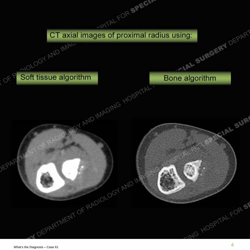
• Radiographs demonstrate irregularity and deformity of the proximal radius, periosteal reaction, and no identified radial head epiphyseal ossification center. CT images demonstrate punctate foci of ossification surrounded by soft tissue along the radial aspect of the proximal radius. MR imaging confirms these small foci of ossification with surrounding tissue which is the same signal intensity of the remainder of the unossified ends of the bones of the elbow.

12What’s the Diagnosis – Case 61

13What’s the Diagnosis – Case 61

14What’s the Diagnosis – Case 61

15What’s the Diagnosis – Case 61

16What’s the Diagnosis – Case 61

17What’s the Diagnosis – Case 61
Diagnosis: Displaced radial head/cartilage anlage fracture
• The elbow has a known time course for the appearance of its multiple epiphyses and apophyses (secondary growth centers). For the purposes of this case, the capitellum is followed by the radial head and then the medial epicondyle with the capitellum beginning to ossify at 1, radial head at 4, and then the medial epicondyle at 7. The cross sectional imaging shows clearly the fractured/displaced unossified radial head epiphysis as soft tissue attenuation on the CT and intermediate signal intensity on the MRI. It contains small areas of early ossification of the secondary growth center on both imaging modalities. Particularly on the coronal PD and MPGR sequences, the cartilage anlages all demonstrate intermediate signal.
• In this case the patient was a six year old boy who had sustained trauma several months prior and was seen at an outside institution and seen only recently at our institution. However, without a knowledge of prior trauma the periosteal reaction with a soft tissue component and ossification could be seen in the setting of a neoplasm. Periosteal reaction also may be seen in the setting of infection as may a soft tissue mass representing an abscess or phlegmon. This demonstrates the imperative nature of obtaining a good history particularly as relates to the interpretation of images.