Embed Size (px)
Citation preview

This thesis has been submitted in fulfilment of the requirements for a postgraduate degree
(e.g. PhD, MPhil, DClinPsychol) at the University of Edinburgh. Please note the following
terms and conditions of use:
This work is protected by copyright and other intellectual property rights, which are
retained by the thesis author, unless otherwise stated.
A copy can be downloaded for personal non-commercial research or study, without
prior permission or charge.
This thesis cannot be reproduced or quoted extensively from without first obtaining
permission in writing from the author.
The content must not be changed in any way or sold commercially in any format or
medium without the formal permission of the author.
When referring to this work, full bibliographic details including the author, title,
awarding institution and date of the thesis must be given.
Host Defence Peptides in Pregnancy; Influences
on the Microbiome and Preterm Labour
Tina Louise Baker
Thesis Submitted to the University of Edinburgh for the Degree of Doctor of Philosophy
September 2016

ii
This thesis has been submitted in fulfilment of the requirements for a
postgraduate degree (e.g. PhD, MPhil, DClinPsychol) at the University of
Edinburgh. Please note the following terms and conditions of use:
This work is protected by copyright and other intellectual property rights, which are retained by the thesis author, unless otherwise stated.
A copy can be downloaded for personal non-commercial research or study, without prior permission or charge.
This thesis cannot be reproduced or quoted extensively from, without first obtaining permission in writing from the author.
The content must not be changed in any way or sold commercially in any format or medium without the formal permission of the author.
When referring to this work, full bibliographic details including the author, title, awarding institution and date of the thesis must be given.
iii
Abstract
Although inflammation is a crucial mechanism in response to injury and pathogen
clearance, inappropriate or excessive induction of the inflammatory response in
pregnancy can cause initiation of the labour cascade and subsequent preterm
delivery.
Host Defence Peptides (HDPs) have important anti-microbial properties but are also
implicated as multifunctional modulators of immunity and infection. They are
predominantly secreted by mucosal epithelial cells and released by leukocytes. The
specific HDPs that are the focus of this thesis are Human beta-defensin 3 (hBD3) and
Human Cathelicidin (hCAP-18/LL-37). The immunomodulatory effect of HDPs in
reproductive tissues in response to infection/inflammation has not been well studied.
In a pregnant state, the hypothesis of this thesis is that HDPs have a dual role in
preventing ascending infection, but also preventing an exacerbated inflammatory
response that can cause preterm birth by initiation of the labour cascade. To explore
this I determine whether bacterial stimuli can regulate HDPs expression in pregnancy
tissues. I also explore what interactions HDPs have on the production/induction of
important cytokines that are vital to the inflammatory response. With the aid of HDP
knockout mice, the role of these peptides in infection/inflammation and continuation
of pregnancy is investigated in a mouse-model of induced preterm-labour. To
understand how ascending infection might be controlled by HDPs in pregnancy, I
explore how HDPs regulate commensal and pathogenic bacteria. This is achieved by
interrogating the maternal microbiome at mucosal sites in HDP knockout animals,
utilising the bacterial 16S rRNA gene and next generation sequencing.
Results
Placental explants respond to Lipopolysaccharide (LPS) challenge by increasing
production of pro-inflammatory cytokines. LL-37 but not hBD3 peptide was able to
modulate this inflammation by inhibiting the release of these pro-inflammatory
cytokines.
iv
To establish whether HDPs are critical in the continuation of pregnancy I use a LPS
induced mouse–model of preterm labour in animals lacking the genes for the HDPs,
Defb14 (Defb14-/-), or Camp (Camp-/-). Intrauterine injection of LPS induced
preterm labour in wildtype mice. However, the Defb14-/- and Camp-/- mice do not
have an increased rate of preterm labour. Key inflammatory mediators are increased
in response to LPS-induced PTL. Camp-/- animals have a similar inflammatory
response to wildtype mice when given LPS during pregnancy.
To understand how ascending infection might be controlled by HDPs, I interrogated
the maternal microbiome at mucosal sites in HDP knockout animals, utilising the
bacterial 16S rRNA gene. I established a workflow for 16S rRNA gene sequencing
on next-generation sequencing platforms and a bioinformatic pipeline for data
analysis. Using this approach I was able to show the mucosal microbiome of Camp-/-
animals were significantly different to that of wildtype controls, showing increased
diversity in the microbes present.
In murine pregnancy, there were very little global cumulative or progressive shifts in
bacteria, with the exception of Candidatus arthromitus, which significantly increases
with gestation compared to non-pregnancy
This thesis has demonstrated that Host Defence Peptides are expressed in pregnancy
tissues and have anti-inflammatory properties in response to bacterial stimuli. It is
not clear whether the HDPs, hBD3 and LL-37 are fundamental to the immune
defence in pregnancy by preventing excessive inflammation, Although, I have shown
LL-37 may have a role in modulation of the maternal microbiota.
v
Lay summary
Preterm birth occurs in 8% -12% of all pregnancies and is the main cause of death in
newborn infants. We still cannot predict which women will have a preterm birth,
although infection and inflammation in the area around the baby increases the risk.
There is increasing evidence suggesting that the lining of the female reproductive
tract is more than just a physical barrier protecting from invading pathogens. The
epithelial cells lining this area secrete proteins that kill invading pathogens and
regulate the traffic and activity of incoming immune cells to the area.
The aim of this thesis is to understand how a group of these proteins called, Host
Defence Peptides (HDPs), which are present in the reproductive tract, regulate both
the ‘good’ and pathogenic bacteria. This thesis also investigates what role these
proteins have on inflammation in response to any invading bacteria. This is of
importance in pregnancy as inappropriate or excessive induction of inflammation can
cause initiation of the labour cascade leading to preterm delivery.
A better understanding of these defence mechanisms within the female reproductive
tract will enlighten strategies to prevent and reduce adverse outcomes in pregnancy.
vi
Declaration
The studies undertaken in this thesis are the unaided work of the author, except
where due acknowledgment is made by reference. No part of this thesis has
previously been, or is currently being submitted in candidature for another degree.
Chapter 2 - I acknowledge the assistance of Ana Calarrao; Research Assistant,
University of Edinburgh, who collected primary tissues from women undergoing
elective caesarean section.
Chapter 7 – I acknowledge the assistance of Adrian Thomson; Edinburgh Preclinical
Imaging; University of Edinburgh, who assisted in the ultrasound in-vivo
experiments.
Chapter 5 & 7 – I acknowledge the assistance of Lisa Young and Dr Heather
MacPherson who gave me assistance in collecting tissue from in-vivo experiments.
Tina Baker
September 2016

vii
Acknowledgments
I would first like to thank my supervisors; Professor Julia Dorin, Dr Donald
Davidson and Dr Sarah Stock, for their guidance and support throughout my PhD.
My sincere thanks go to members of the Dorin lab; Dr Fiona Semple, Dr Heather
MacPherson, and Dr Sheila Webb for supporting me when times were tough. The
last 4 years would most definitely not have been as fun without their friendship.
I would like to thank members of the Tommy’s Research team, especially, Ana
Calarrao, Dr Sarah Rinaldi, and Dr Lorraine Frew for guidance on experiments.
Finally, I would like to thank my friends and family, for their constant support and
encouragement.
viii
Abbreviations
16S rRNA 16S ribosomal RNA
AMP Antimicrobial peptide
ANOVA Analysis of variance
AP-1 Activator protein 1
BMDM Bone-marrow-derived macrophage
BR Broad range
BV Bacterial vaginosis
CAL Calibrator
CCL Chemokine (C-C) motif ligand
CCR Chemokine (C-C) receptor
cDNA Complementary DNA
cffDNA Cell free fetal DNA
CNV Copy number variation
COX Cyclo-oxygenase
CRH Corticotrophin releasing hormone
CST Community state types
Ct Cycle threshold
CXCL Chemokine (C-X-C) motif ligand
dNTP Deoxyribonucleotide triphosphate
dsRNA Double-stranded RNA
DSS Dextran sulfate sodium
EGFR Epidermal growth factor receptor
ELISA Enzyme linked immunosorbent assay
ER Estrogen receptor
ERK Extracellular signal-regulated kinases
FISH Fluorescent in situ hybridization
FRP2 Formyl peptide receptor 2
FRT Female reproductive tract
GAPHD Glyceraldehyde-3-phosphate dehydrogenase
GAS group-A Streptococcus
GBS group-B-Streptococcus
G-CSF granulocyte colony-stimulating factor
GR Glucocorticoid receptor
GWAS Genome-wide association study
hBD Human beta defensin
HCL Hydrochloric acid
HDP Host defence peptide
hGC human chorionic gonadotropin
HIV Human immunodeficiency virus
HNP Human neutrophil peptides
HPLC High performance liquid chromatography
HRP Horseradish peroxidase
HS High sensitivity
HTS High throughput sequencing
ix
HUVEC Human umbilical vein endothelial cells
IL Interleukin
IL-1Ra Interleukin-1 receptor antagonist
IRAK Interleukin-1 receptor associated kinase
IRF Interferon regulatory factor
JNK c-Jun N-terminal kinase
LBP LPS binding protein
LDH Lactate dehydrogenase
LEfSe Linear discriminant analysis with effect size
LPS Lipopolysaccharide
LTA Lipoteichoic acid
MAPK Mitogen-activated protein kinase
MDA5 Melanoma differentiation-associated protein 5
MIC Minimum inhibitory concentrations
MMP Matrix metalloproteinase
MRSA Methicillin-resistant Staphylococcus aureus
MyD88 Myeloid differentiation primary response gene 88
NETs Neutrophil extracellular traps
NF-κB Nuclear factor kappa B
NK-T Natural killer T
NO Nitric oxide
NOD Nucleotide-binding oligomerization domain-like receptors
NS No significance
NTC No template control
NTxC No treatment control
OTR Oxytocin receptor
OTU Operational taxonomic units
PAMP Pathogen associated molecular patterns
PBS Phosphate buffered saline
PCOA Principle co-ordinate analysis
pDCs Plasmacytoid dendritic cells
PG Prostaglandin
PGM Personal genome machine
PMSG Pregnant mare's serum gonadotropin
polyI:C Polyinosinic:polycytidylic acid
PPROM Preterm premature rupture of membranes
PR Progesterone receptor
PTL Preterm labour
QIIME Quantitative insights in to microbial ecology
RA Rheumatoid arthritis
RANTES Regulated on activation, normal T cell expressed and secreted
RDP Ribosomal database project
RNA Ribonucleic acid
ROS Reactive oxygen species
RT Reverse transcriptase
RT-qPCR Quantitative real time polymerase chain reaction
SCFA Short chain fatty acids
x
SEM Standard error of the mean
SFB Segmented filamentous bacteria
SLE Systemic lupus erythematosus
SNP Single nucleotide polymorphism
SP-A Surfactant Protein-A
SPF Specific pathogen free
STAT Signal transducer and activator of transcription
STI Sexually transmitted infections
TBE Tris/Borate/EDTA
TLR Toll like receptor
TMB 3,3'5,5' Tetramethylbenzidine
TNF Tumour necrosis factor
TRAF TNF receptor associated factor
TRIF TIR domain containing adapter inducing interferon beta
UPGMA Unweighted pair group method with arithmetic means algorithm
UPL Universal probe library
USS Ultrasound sonography
UTR Untranslated region
VDRE Vitamin D response element
VRE Vancomycin-resistant Enterococci
xi
Contents
Abstract ........................................................................................................ iii
Lay summary .................................................................................................v
Declaration ................................................................................................... vi
Acknowledgments ...................................................................................... vii
Abbreviations ............................................................................................. viii
Contents ....................................................................................................... xi
List of Figures ............................................................................................ xvi
List of Tables .............................................................................................. xix
Chapter 1 Literature Review ........................................................................2
1.1 Labour and Preterm Labour ................................................................................ 2 1.1.1 Preterm Labour .................................................................................................. 2 1.1.2 Term and Preterm Labour Common Components ............................................. 3 1.1.3 Labour as an Inflammatory Event ...................................................................... 5
1.1.3.1 Inflammatory Mediators ............................................................................. 6 1.1.3.2 NF-κB ......................................................................................................... 6 1.1.3.3 Immune Cells ............................................................................................. 7 1.1.3.4 Toll Like Receptors .................................................................................. 10
1.1.4 Onset of Labour at Term .................................................................................. 13 1.1.5 Infection/Inflammation and Preterm Labour ..................................................... 13 1.1.6 Mechanisms for Inflammation/Infection ............................................................ 14 1.1.7 Therapeutics for Preterm Labour ..................................................................... 15
1.2 Host Defence Peptides ....................................................................................... 18 1.2.1 Defensins.......................................................................................................... 19
1.2.1.1 Defensins ................................................................................................. 19 1.2.1.2 Beta-defensins ......................................................................................... 20 1.2.1.3 Human beta-defensin-3 (hBD3) ............................................................... 22
1.2.1.3.1 Expression and Regulation ................................................................. 22 1.2.1.3.2 Antimicrobial and Immunomodulatory Properties ............................... 23
1.2.1.4 Murine Orthologue of hBD3; Mouse Defb14 ........................................... 25 1.2.2 Cathelicidin ....................................................................................................... 25
1.2.2.1 Cathelicidin .............................................................................................. 25 1.2.2.2 Expression and Regulation ...................................................................... 26 1.2.2.3 Antimicrobial Properties ........................................................................... 28 1.2.2.4 Modulation of Inflammation/Infection ....................................................... 29 1.2.2.5 Wound Healing and Tissue Remodelling ................................................ 32
1.2.3 HDPs and the Female Reproductive Tract ...................................................... 32 1.2.4 HDPs and Preterm Labour ............................................................................... 35
1.3 Mouse Models of Preterm Labour ..................................................................... 36
1.4 Microbiome .......................................................................................................... 39 1.4.1 The Vaginal Microbiome ................................................................................... 43
1.4.1.1 Vaginal Microbiome Development ........................................................... 43 1.4.1.2 Vaginal Microbiome Stability ................................................................... 43 1.4.1.3 Vaginal Microbiome Dysbiosis ................................................................. 45
1.4.2 Vaginal Microbiome in Pregnancy .................................................................... 46 1.4.2.1 Bacterial Vaginosis in Pregnancy ............................................................ 46 1.4.2.2 Vaginal Microbiome and Preterm Labour ................................................ 47
xii
1.4.3 Modulation of Vaginal Microbiome ................................................................... 47 1.4.4 Murine Vaginal Microbiome .............................................................................. 48 1.4.5 The Gut Microbiome ......................................................................................... 50 1.4.6 Gut Microbiome in Pregnancy .......................................................................... 53
Aims and Hypothesis ................................................................................. 54
Chapter 2 General Materials and Methods ............................................... 56
2.1 Animal Experiments ........................................................................................... 56 2.1.1 Fertility Experiments ......................................................................................... 56 2.1.2 Preterm Birth Model; Laparotomy Method ....................................................... 57 2.1.3 Preterm Birth Model; Ultrasound Method ......................................................... 59 2.1.4 Interval to Delivery and Number of Live-Born Pups ......................................... 60 2.1.5 Timed Collection ............................................................................................... 60 2.1.6 Microbiome Sample Collection ......................................................................... 61 2.1.7 HDP Expression in the Murine Reproductive Tract Experiments .................... 62
2.1.7.1 Estrus Cycling and Staging ..................................................................... 62 2.1.7.2 Vaginal Cell Cytology ............................................................................... 62
2.2 Molecular ............................................................................................................. 64 2.2.1 RNA Extraction and Reverse Transcription ..................................................... 64
2.2.1.1 TRI Reagent Method ............................................................................... 64 2.2.1.2 RNA Bee Method ..................................................................................... 64 2.2.1.3 Reverse Transcription .............................................................................. 65
2.2.2 RT-qPCR .......................................................................................................... 65 2.2.2.1 Taqman Assay (ABI PRISM) ................................................................... 66 2.2.2.2 Universal ProbeLibrary Assay (Roche LightCycler) ................................ 67 2.2.2.3 RT-qPCR analysis method ...................................................................... 68
2.2.3 ELISA ............................................................................................................... 68
2.3 HDPs in Placental Explants Experiment .......................................................... 70 2.3.1 LDH assay ........................................................................................................ 70
2.4 HDPs in Human Term Labour study ................................................................. 71
Chapter 3 Host Defence Peptides.............................................................. 73
3.1 Introduction ......................................................................................................... 73
3.2 Experimental Details .......................................................................................... 76 3.2.1 Patient Characteristics ..................................................................................... 76 3.2.2 Assays and Statistical Analysis ........................................................................ 77
3.3 Results ................................................................................................................. 80 3.3.1 HDPs in Human Term Labour .......................................................................... 80
3.3.1.1 Human beta-defensin 3 in Term-Labour.................................................. 80 3.3.1.2 Human Cathelicidin in Term-Labour ........................................................ 81
3.3.2 HDP Anti-inflammatory effect on Placental Explants ....................................... 82 3.3.2.1 LPS Dose and Time Response in Placental Explants ............................. 83 3.3.2.2 LL-37 Dose Response in Placental Explants .......................................... 85 3.3.2.3 HDPs in Placental Explants ..................................................................... 86
3.3.3 HDPs in the Murine Reproductive Tract ........................................................... 91 3.3.3.1 Murine Defb14 (hBD3 orthologue) in Pregnancy .................................... 93 3.3.3.2 Murine Camp (LL-37 orthologue) in Pregnancy ...................................... 94
3.4 Discussion ........................................................................................................... 95
3.5 Summary ............................................................................................................ 100
Chapter 4 Microbiome - 16S rRNA Gene Sequencing ........................... 102
4.1 Introduction ....................................................................................................... 102
xiii
4.2 Targeted 16S rRNA Gene Amplification ......................................................... 103
4.3 16S rRNA Copy Number qPCR ........................................................................ 106
4.4 16S rRNA Gene Sequencing Primer Design .................................................. 107 4.4.1 In-silico Variable Region Evaluation ............................................................... 107 4.4.2 In-silico Variable Region Primer Evaluation ................................................... 108
4.5 Contamination Considerations ....................................................................... 110
4.6 Sampling method .............................................................................................. 110
4.7 Bacterial DNA Extraction ................................................................................. 112
4.8 Library Preparation ........................................................................................... 113 4.8.1 Illumina MiSeq Primer Design and PCR Optimisation ................................... 113 4.8.2 Ion Torrent PGM Primer Design and PCR Optimisation ................................ 117 4.8.3 Gel Electrophoresis ........................................................................................ 122 4.8.4 Sample clean-up ............................................................................................ 122 4.8.5 DNA Quantification, Quality Control, and Equimolar Pool ............................. 122
4.9 Sequencing and Base Calling ......................................................................... 124
4.10 QIIME and Mothur ............................................................................................. 124
4.11 Downstream Quality Filtering and Data Analysis; MiSeq ............................. 125
4.12 Downstream Quality Filtering and Data Analysis; Ion Torrent .................... 127
4.13 R Package .......................................................................................................... 128
4.14 Rarefaction Curves ........................................................................................... 129
4.15 Alpha Diversity Analysis .................................................................................. 130 4.15.1 Statistical analysis of alpha diversity.......................................................... 130
4.16 Beta Diversity Analysis .................................................................................... 131 4.16.1 Statistical analysis of beta diversity ........................................................... 132 4.16.2 Jack-knife and UPGMA bootstrapped trees .............................................. 133
4.17 Significant Taxa (LEfSe) ................................................................................... 133
4.18 Mock Community .............................................................................................. 134
4.19 Discussion ......................................................................................................... 138
4.20 Summary ............................................................................................................ 141
Chapter 5 Modulation of the Microbiome ............................................... 143
5.1 Introduction ....................................................................................................... 143
5.2 Experimental Methods ..................................................................................... 145 5.2.1 Camp-/- Microbiome and Pregnancy Microbiome Study ............................... 145
5.3 Results ............................................................................................................... 147 5.3.1 Data Summary ............................................................................................... 147 5.3.2 Taxa Summary ............................................................................................... 149 5.3.3 Subsets ........................................................................................................... 150 5.3.4 Rarefaction Curves ......................................................................................... 151 5.3.5 Subsampling; Single Rarefaction ................................................................... 153 5.3.6 The Murine Fecal Microbiome in Relation to Vaginal Bacterial Communities (Subset 1) .................................................................................................................... 153
5.3.6.1 Alpha Diversity ....................................................................................... 153 5.3.6.2 Beta Diversity ......................................................................................... 155 5.3.6.3 Sample Cross Contamination ................................................................ 156
5.3.7 Fecal Microbiome of Camp-/- Animals (Subset 2) ......................................... 158 5.3.7.1 Phylum Level Classification ................................................................... 158
xiv
5.3.7.2 Genus Level Classification .................................................................... 161 5.3.7.3 Alpha Diversity ....................................................................................... 167 5.3.7.4 Beta Diversity ......................................................................................... 168 5.3.7.5 Jack-knife Support; UPMGA bootstrapping ........................................... 180 5.3.7.6 Significant Taxa (LEfSe) ........................................................................ 181
5.3.8 Pregnancy Modulation of the Microbiome (Subset 3) .................................... 184 5.3.8.1 The Stability of the Fecal Microbiome- Non Pregnant ........................... 184 5.3.8.2 Pregnancy Related Microbial Change; Alpha Diversity ......................... 189 5.3.8.3 Pregnancy Related Microbial Change; Genera Associated with Progressing Gestation............................................................................................. 191
5.4 Discussion ......................................................................................................... 198
5.5 Summary ............................................................................................................ 207
Chapter 6 Murine Vaginal Microbiome .................................................... 209
6.1 Introduction ....................................................................................................... 209
6.2 Results ............................................................................................................... 210 6.2.1 Murine Non-pregnant Vaginal Microbiome ..................................................... 210
6.2.1.1 Conclusion ............................................................................................. 215 6.2.2 Camp-/- Pregnant Vaginal Microbiome Study ................................................ 217
6.2.2.1 Conclusion ............................................................................................. 223
6.3 Discussion ......................................................................................................... 224
Chapter 7 Role of Host Defence Peptides in an Infection/ Inflammation Induced Mouse Model of Preterm Labour .............................................. 227
7.1 Introduction ....................................................................................................... 227
7.2 Experimental Details ........................................................................................ 229 7.2.1 Fertility in Host Defence Peptide Knockout Mice ........................................... 229 7.2.2 Timed Collection Experiment ......................................................................... 229 7.2.3 Laparotomy Model of PTL Experiment ........................................................... 230 7.2.4 Ultrasound Sonography guided Model of PTL ............................................... 231 7.2.5 Statistical Analysis .......................................................................................... 231
7.3 Results ............................................................................................................... 233 7.3.1 Fertility of Camp-/- and Defb14-/- Animals ..................................................... 233 7.3.2 HDPs and Inflammatory Gene Response to LPS-induced model of PTL in C57BL/6J mice - Timed Collection .............................................................................. 235
7.3.2.1 Defb14 mRNA Expression following PTL model ................................... 235 7.3.2.2 Camp mRNA Expression following PTL model ..................................... 236 7.3.2.3 Inflammatory and Immune Cell Mediators in Placenta .......................... 237 7.3.2.4 Inflammatory and Immune Cell Mediators in Uterus ............................. 239 7.3.2.5 Inflammatory and Immune Cell Mediators in Fetal Membrane.............. 241
7.3.3 Defb14 in LPS Induced Preterm Labour; Laparotomy Model ........................ 243 7.3.3.1 Interval to Delivery ................................................................................. 243 7.3.3.2 Pup Survival ........................................................................................... 245
7.3.4 Camp in LPS Induced Preterm Labour; Ultrasound Model ............................ 247 7.3.4.1 Interval to Delivery ................................................................................. 247 7.3.4.2 Pup Survival ........................................................................................... 249 7.3.4.3 Inflammatory and Immune Cell Mediators in Placenta, Uterus and Fetal Membrane in response to LPS induced PTL .......................................................... 250
7.3.4.3.1 Placenta ............................................................................................ 250 7.3.4.3.2 Uterus ............................................................................................... 255 7.3.4.3.3 Fetal Membrane ................................................................................ 259
7.3.4.4 Circulating Serum IL-6 and TNF levels in response to LPS induced PTL 263
7.4 Discussion ......................................................................................................... 265

xv
7.5 Summary ............................................................................................................ 271
Chapter 8 Discussion ............................................................................... 273
References ................................................................................................ 284
Appendices ............................................................................................... 314
Materials and Methods .................................................................................................. 314
Galaxy Workflow ............................................................................................................ 319
Full Taxonomic Classifications (LEfSe) ...................................................................... 324
xvi
List of Figures
Chapter 1 Literature Review
Figure 1.1 Common Components of Term and Preterm Labour ............................................. 3
Figure 1.2 Schematic of Types of Infection/Inflammation in Human Pregnancy ..................... 4
Figure 1.3 Schematic of Core Labour-associated Pathways ................................................... 7
Figure 1.4 Schematic of the TLR-4 Signalling Pathway ........................................................ 12
Figure 1.5 Overview of HDPs Modes of Action...................................................................... 18
Figure 1.6 Mechanisms for HDPs in Epithelial Cells of the Female Reproductive Tract ....... 34
Figure 1.7 Human Microbiome Diversity ................................................................................ 41
Figure 1.8 The Human Microbiome; Site Specific Distribution of Phyla ................................ 42
Chapter 2 General Materials and Methods
Figure 2.1 Mouse Model of Preterm Labour; Laparotomy Method ........................................ 58
Figure 2.2 Mouse Model of Preterm Labour; Ultrasound guided Injection Method ............... 59
Figure 2.3 Mouse Vaginal Cytology for Estrus Staging ......................................................... 63
Chapter 3 Host Defence Peptides
Figure 3.1 Expression of DEFB103 (hBD3) during Term Labour .......................................... 80
Figure 3.2 Expression of CAMP (LL-37) during Term Labour ............................................... 81
Figure 3.3 Placental Explants; TNF levels in LPS dose and Time Response ....................... 84
Figure 3.4 Placental Explants; TNF levels in LL-37 Dose Response .................................... 85
Figure 3.5 Effects of HDPs on Cytokine Secretion in Placenta Explants .............................. 87
Figure 3.6 Effects of HDPs on TNF Gene Expression in Placenta Explants ......................... 89
Figure 3.7 Endogenous DEFB103 and CAMP mRNA in Placental Explants ........................ 90
Figure 3.8 HDPs Gene Expression in the Mouse Reproductive Tract .................................. 92
Figure 3.9 Defb14 Gene Expression in Mouse Pregnancy Tissues ...................................... 93
Figure 3.10 Camp Gene Expression in Mouse Pregnancy Tissues ...................................... 94
Chapter 4 Microbiome - 16S rRNA Gene Sequencing
Figure 4.1 Schematic of the 16S rRNA Gene Variable Regions ......................................... 105
Figure 4.2 16S rRNA Copy Number; Murine Vaginal Sampling Optimisation ..................... 111
Figure 4.3 16S rRNA Copy Number; Extraction Kit Optimisation ........................................ 112
xvii
Figure 4.4 Gel Photo; MiSeq 16S rRNA Amplicon Library .................................................. 116
Figure 4.5 Gel Photo; 16S rRNA Amplicon Library .............................................................. 119
Figure 4.6 Overview of MiSeq Sequencing Quality Output; FASTQ 3D plots ..................... 125
Figure 4.7 Mock Community Relative Abundance ............................................................... 136
Figure 4.8 Percentage Bias in Sequenced Mock Community ............................................. 137
Chapter 5 Modulation of the Microbiome
Figure 5.1 Experimental Timeline for Camp-/- Microbiome Study ....................................... 145
Figure 5.2 MiSeq Processed Paired Reads and Filtered Contigs ....................................... 148
Figure 5.3 Taxonomic Classification Hierarchy.................................................................... 149
Figure 5.4 Rarefaction Curves - Fecal and Vaginal ............................................................. 152
Figure 5.5 Alpha Diversity Fecal vs. Vaginal ....................................................................... 154
Figure 5.6 UniFrac Distance 2D PCoA plots of Vagina and Fecal Community ................... 155
Figure 5.7 Venn diagram of Shared Taxa between Fecal and Vaginal Samples ................ 157
Figure 5.8 Phylum Level Classification in Camp-/- vs. C57BL/6J Fecal Samples ............... 159
Figure 5.9 Genus Level Classification in Camp-/- vs. C57BL/6J Fecal Samples ................ 165
Figure 5.10 Comparison of Alpha diversity for C57BL/6J vs. Camp-/- ................................ 167
Figure 5.11 Unweighted UniFrac Distance Boxplots comparing C57BL/6J vs. Camp-/- ..... 169
Figure 5.12 UniFrac Distance 2D PCoA plots - Coloured by Genotype .............................. 171
Figure 5.13 UniFrac Distance 2D PCoA plots - Coloured by Cage ..................................... 172
Figure 5.14 Unweighted UniFrac Distance Boxplots - Inter and Intra Animal Variation ...... 175
Figure 5.15 UniFrac Distance 2D PCoA plots - Coloured by Genotype; Day 0 Subset ...... 177
Figure 5.16 UniFrac Distance 2D PCoA plots - Coloured by Cage; Day 0 Subset ............. 178
Figure 5.17 Jack-knife Unweighted UniFrac UPGMA Bootstrapped Tree ........................... 180
Figure 5.18 Significant Differential Abundant Taxa in C57BL/6J vs. Camp-/- Fecal samples ............................................................................................................................................. 182
Figure 5.19 Top 6 Differential abundant OTUs .................................................................... 183
Figure 5.20 Stability of the non-pregnant Fecal Microbiome; Relative Abundance of Genera ............................................................................................................................................. 185
Figure 5.21 Stability of the non-pregnant Fecal Microbiome; Alpha Diversity Measures .... 186
Figure 5.22 Stability of the non-pregnant Fecal Microbiome; Beta Diversity ....................... 188
Figure 5.23 Unweighted UniFrac Distance Boxplots - Inter-animal and collection day ....... 188
Figure 5.24 Pregnancy Influence on Alpha Diversity Measures .......................................... 190
Figure 5.25 Linear Regression of Genera Abundance to Gestation .................................... 192
Figure 5.26 Linear Regression of Genera Abundance to Time-point .................................. 193
Figure 5.27 Linear Regression of Genera Abundance; Pregnant vs. Non-Pregnant .......... 194
Figure 5.28 Linear Regression of Candidatus arthromitus; Pregnant vs. Non-Pregnant .... 195
xviii
Figure 5.29 Boxplots of Significant Genera Associated with Pregnancy ............................. 197
Chapter 6 Murine Vaginal Microbiome
Figure 6.1 Bar Chart of Relative Proportions of Bacteria in Vagina; Phylum Level ............. 211
Figure 6.2 Bar Charts of Relative Proportions of Bacteria in Vagina; Genus Level ............ 212
Figure 6.3 Rarefaction Curves; Observed OTUs in Murine Vagina ..................................... 213
Figure 6.4 Rarefaction Curves; Shannon Index in Murine Vagina....................................... 214
Figure 6.5 Alpha Diversity of Camp-/- vs. Wildtype Vaginal Microbiome ............................ 218
Figure 6.6 Heat map of most Abundant OTUs showing their Genus Level Classification in Vaginal Samples from Camp-/- and C57BL/6J mice ........................................................... 219
Figure 6.7 UniFrac Distance 2D PCoA plots of Vaginal Community – Colour by Genotype 221
Figure 6.8 UniFrac Distance 2D PCoA plots of Vaginal Community – Colour by Pregnancy Status ................................................................................................................................... 222
Chapter 7 Role of Host Defence Peptides in an Infection/ Inflammation
Induced Mouse Model of Preterm Labour
Figure 7.1 Fertility of Camp-/- and Defb14-/- mice .............................................................. 233
Figure 7.2 Litter Size of Camp-/- and Defb14-/- mice .......................................................... 234
Figure 7.3 Expression of Defb14 in LPS-induced PTL Model ............................................. 235
Figure 7.4 Expression of Camp in LPS-induced PTL Model ............................................... 236
Figure 7.5 Placental Inflammatory and Immune Cell Mediator Response to LPS-induced PTL Model .................................................................................................................................... 238
Figure 7.6 Uterine Inflammatory and Immune Cell Mediator Response to LPS-induced PTL Model .................................................................................................................................... 240
Figure 7.7 Fetal Membrane Inflammatory and Immune Cell Mediator Response to LPS-induced PTL Model .............................................................................................................. 242
Figure 7.8 Interval to Delivery in Defb14-/- mice; LPS-induced PTL Model ........................ 244
Figure 7.9 Live Born Pup Ratio in Defb14-/- mice; LPS-induced PTL Model ...................... 246
Figure 7.10 Interval to Delivery in Camp-/- mice; LPS-induced PTL Model ........................ 248
Figure 7.11 Live Born Pup Ratio in Camp-/- mice; LPS-induced PTL Model ...................... 249
Figure 7.12 Placental Inflammatory and Immune Cell Mediator Response in Camp-/- LPS-induced PTL Model .............................................................................................................. 253
Figure 7.13 Placental Ptgs2 and Camp Response in Camp-/- LPS-induced PTL Model .... 254
Figure 7.14 Uterine Inflammatory and Immune Cell Mediator Response in Camp-/- LPS-induced PTL Model .............................................................................................................. 257
Figure 7.15 Uterine Ptgs2 and Camp Response in Camp-/- LPS-induced PTL Model ....... 258
Figure 7.16 Fetal Membrane Inflammatory and Immune Cell Mediator Response in Camp-/- LPS-induced PTL Model ...................................................................................................... 261
xix
Figure 7.17 Fetal Membrane Ptgs2 and Camp Response in Camp-/- LPS-induced PTL Model .................................................................................................................................... 262
Figure 7.18 IL-6 and TNF in Circulating Serum in Camp-/- LPS-induced PTL Model ......... 264
List of Tables
Chapter 1 Literature Review
Table 1.1 Mouse and Human Orthologous Defensin Genes ................................................. 21
Table 1.2 Chemokine/Cytokines and Receptor Nomenclature .............................................. 30
Table 1.3 Animal Models used to Study Infection/Inflammation-induced Preterm Labour .... 38
Table 1.4 Human Vaginal Community State Types (CST) .................................................... 44
Chapter 2 General Materials and Methods
Table 2.1 Taqman Assay Probes ........................................................................................... 66
Table 2.2 Primers and Probes Designed in Universal ProbeLibrary Assay Design Center .. 67
Table 2.3 R&D duosets ELISA ............................................................................................... 69
Chapter 3 Host Defence Peptides
Table 3.1 Clinical Details of Patients; HDPs in Labour study ................................................ 76
Table 3.2 Clinical Details of Patients; HDPs in Placental Explants study .............................. 77
Chapter 4 Microbiome - 16S rRNA Gene Sequencing
Table 4.1 qPCR Reaction Mix for 16S rRNA Copy Number Assay ..................................... 107
Table 4.2 qPCR Cycle Parameters for 16S rRNA Copy Number Assay ............................. 107
Table 4.3 PCR Reaction Mix; MiSeq 16S rRNA Amplicon Library ...................................... 114
Table 4.4 PCR Cycle Parameters; MiSeq 16S rRNA Amplicon Library .............................. 114
Table 4.5 PCR Reaction Mix; Ion Torrent 16S rRNA Amplicon Library ............................... 118
Table 4.6 PCR Cycle Parameters; Ion Torrent 16S rRNA Amplicon Library ....................... 118
Table 4.7 MiSeq Forward Primer Sequences ...................................................................... 120
Table 4.8 MiSeq Reverse Primer Sequences ...................................................................... 121
Table 4.9 Ion Torrent Forward and Reverse Primer Sequences ......................................... 121
Table 4.10 Mock Community HM-278D Species List .......................................................... 134

xx
Table 4.11 Sequenced Mock Community OTU Counts ....................................................... 135
Chapter 5 Modulation of the Microbiome
Table 5.1 Experimental Sample Number for Camp-/- Microbiome Study ............................ 146
Table 5.2 MiSeq Sequence Quality Summary ..................................................................... 148
Table 5.3 Phylum Level Classification in Camp-/- vs. C57BL/6J Fecal Samples ................ 160
Table 5.4 Genus Level Classification Camp-/- vs. C57BL/6J Fecal Samples ..................... 166
Table 5.5 Hypothesis testing comparing C57BL/6J vs. Camp-/- Samples .......................... 173
Table 5.6 Hypothesis testing comparing C57BL/6J vs. Camp-/- Animals; Day 0 subset .... 179
Chapter 6 Vagina Microbiome
Table 6.1 Murine Experimental Numbers - Camp-/- Vaginal Pregnancy Microbiome study 217
Chapter 7 Role of Host Defence Peptides in an Infection/ Inflammation
Induced Mouse Model of Preterm Labour
Table 7.1 Timed Collection Experiment - Animal Numbers and Treatment ........................ 229
Table 7.2 Laparotomy Model of PTL Experiment Numbers ................................................. 230
Table 7.3 USS Model of PTL Experiment Numbers ............................................................ 231

1
Chapter 1
Literature Review
Chapter 1
2
Chapter 1 Literature Review
1.1 Labour and Preterm Labour
1.1.1 Preterm Labour
The World Health Organisation estimates that 1 in 10 babies are born preterm (<37
weeks gestation) which is a global annual incidence of 15 million (World Health,
2013). Neonatal preterm labour is the main cause of morbidity and mortality in
newborn infants, resulting in a multitude of developmental, neurological and
cognitive problems (Galinsky et al., 2013). In addition to the huge burden on quality
of life, these long-term associated problems increase the need for medical care and
allied services.
In many developed countries antenatal care has greatly increased the survival of
extremely premature babies; however, the proportion of babies which survive with
major health complications has not changed (Martin et al., 2010). Research into
preterm labour has vastly increased in the last decade but this has not translated into
a reduction in the incidence of preterm labour and improved neonatal outcomes.
There are several low level risk factors for having a preterm labour; a prior history of
a preterm labour, multiple pregnancy, maternal stress, excessive physical work,
smoking, excessive alcohol consumption and infection. Gestational diabetes is also
associated with a 4-fold increase in the rate of Preterm Labour (Norman et al.,
2009b). In addition, pre-existing diabetes is the highest risk factor of any maternal
conditions associated with having a premature delivery.
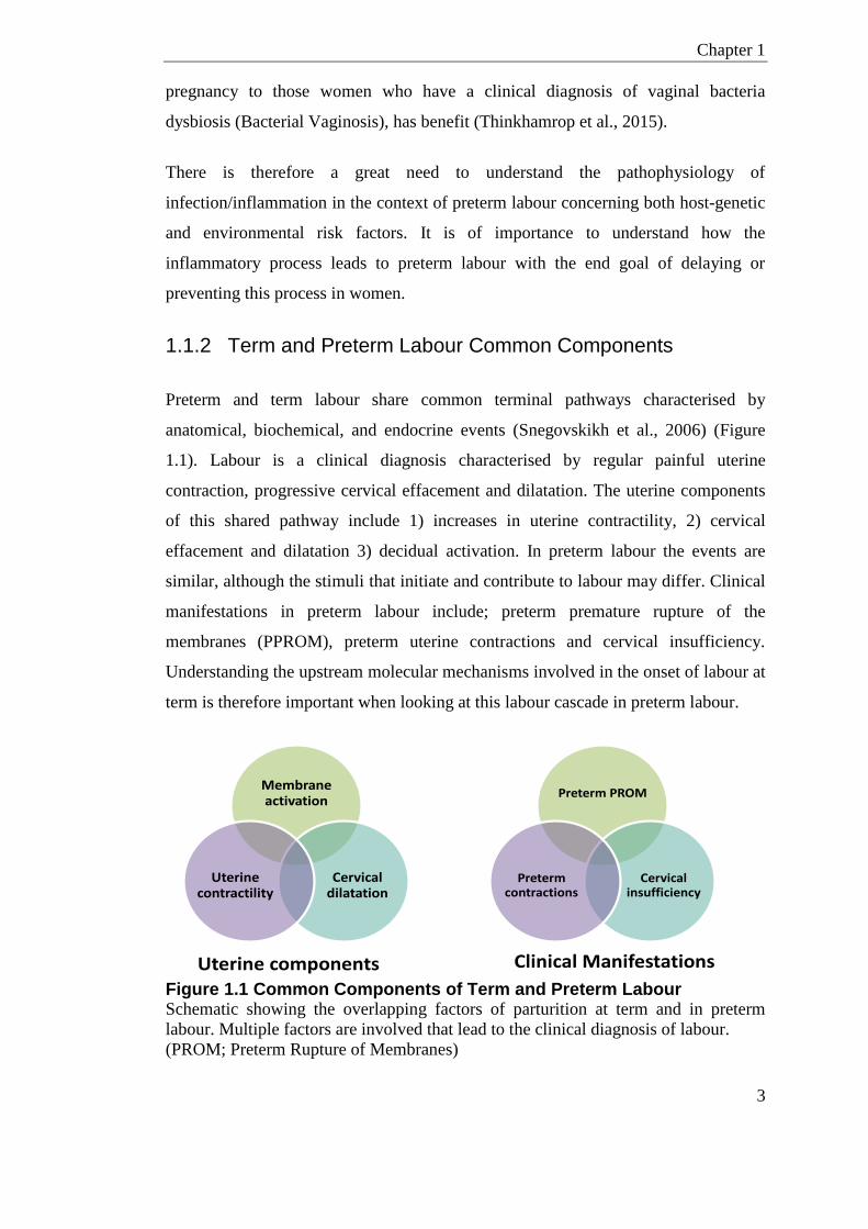
Nearly 40% of preterm deliveries are associated with intrauterine infection (Romero
et al., 2006) (Figure 1.2). There are conflicting reports whether antibiotic
intervention is beneficial in preventing preterm labour associated with intrauterine
infection (Lamont, 2015, King and Flenady, 2002). A review of meta-analysis
studies highlighted that the beneficial effects of antibiotics may have been masked by
some negative studies which were methodologically flawed (Lamont, 2015). There is
however, increasing support that certain prophylactic antibiotics given very early in
Chapter 1
3
pregnancy to those women who have a clinical diagnosis of vaginal bacteria
dysbiosis (Bacterial Vaginosis), has benefit (Thinkhamrop et al., 2015).
There is therefore a great need to understand the pathophysiology of
infection/inflammation in the context of preterm labour concerning both host-genetic
and environmental risk factors. It is of importance to understand how the
inflammatory process leads to preterm labour with the end goal of delaying or
preventing this process in women.
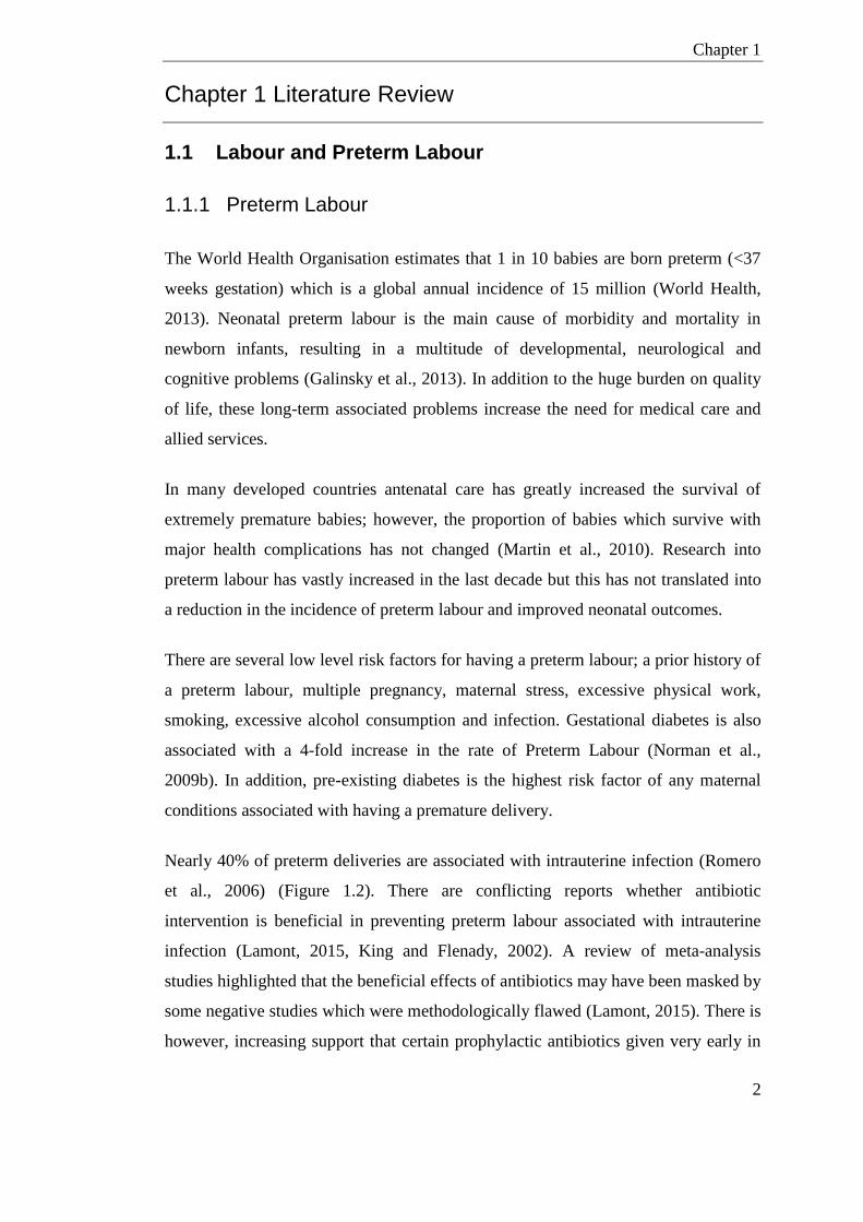
1.1.2 Term and Preterm Labour Common Components
Preterm and term labour share common terminal pathways characterised by
anatomical, biochemical, and endocrine events (Snegovskikh et al., 2006) (Figure
1.1). Labour is a clinical diagnosis characterised by regular painful uterine
contraction, progressive cervical effacement and dilatation. The uterine components
of this shared pathway include 1) increases in uterine contractility, 2) cervical
effacement and dilatation 3) decidual activation. In preterm labour the events are
similar, although the stimuli that initiate and contribute to labour may differ. Clinical
manifestations in preterm labour include; preterm premature rupture of the
membranes (PPROM), preterm uterine contractions and cervical insufficiency.
Understanding the upstream molecular mechanisms involved in the onset of labour at
term is therefore important when looking at this labour cascade in preterm labour.
Figure 1.1 Common Components of Term and Preterm Labour Schematic showing the overlapping factors of parturition at term and in preterm
labour. Multiple factors are involved that lead to the clinical diagnosis of labour.
(PROM; Preterm Rupture of Membranes)
Membrane activation
Cervical dilatation
Uterine contractility
Preterm PROM
Cervical insufficiency
Preterm contractions
Chapter 1
4
Figure 1.2 Schematic of Types of Infection/Inflammation in Human Pregnancy The types of inflammation and infection seen at the fetal-maternal interface.
Bacterial infections within the uterine compartment can occur between fetal and
maternal tissue (choriodecidual infection), within and between the two fetal
membranes (chorioamnionitis), within the placenta, in the amniotic cavity or within
the umbilical vessels (vasculitis) and cord substance (funisitis). Chorioamnionitis,
choriodecidua infection, and amniotic fluid infections are more prevalent in
association with ascending infectious agents through the cervix. Source: Goldenberg
et al., (2000).
Chapter 1
5
1.1.3 Labour as an Inflammatory Event
Labour is now generally accepted to be an inflammatory event where the emphasis
from an anti-inflammatory pregnancy state shifts pro-inflammatory preceding labour
(Youssef et al., 2009). At term, an infiltration of neutrophils and macrophages into
the cervix and maternal fetal membrane is observed (Osman et al., 2003, Gomez-
Lopez et al., 2010), in addition to increases in circulating leukocytes (Yuan et al.,
2009). Selective immune cell recruitment from the circulating blood involves the
production of chemotactic signals.
NF-𝜅B, a transcription factor family, is increased in the myometrium, the smooth
muscle tissue of the womb, and amnion in term labour (Pieber et al., 2001, Lappas
and Rice, 2009, Vora et al., 2010). This transcription factor has binding sites in key
pro-labour genes controlling their activity (Lindström and Bennett, 2005).
Increase in transcription factor NF-𝜅B, and also AP-1 and STAT (Stein et al., 1993)
result in the production of cytokines including IL-6, TNF, IL-1𝛼, IL-1𝛽, and IL-8
within the fetal membranes and uterine lining (Elliott et al., 2001, Lappas et al.,
2003). The relative contribution of these inflammatory transcription factors has been
shown to be spatial, regardless, alone their activity results in a similar myometrial
contractile pathways highlighting redundancy (Migale et al., 2016, MacIntyre et al.,
2014). These cytokines can stimulate prostaglandin production that in turn initiate
mobilisation of neutrophils (Lockwood et al., 2006, Arcuri et al., 2009). These
neutrophils infiltrate the tissue causing the production and release of
metalloproteases (MMPs). Prostaglandins directly stimulate myometrial contractions
in addition to indirect stimulation by the up-regulation of the oxytocin receptor
(COX-2) (Liggins, 1989, Crankshaw and Dyal, 1994). COX-2 is an inducible labour
associated gene, which is expressed at term labour in the human myometrium, while
release of MMPs can aid tissue remodelling in the cervix and fetal membrane (Heng
et al., 2012).
Chapter 1
6
1.1.3.1 Inflammatory Mediators
Microarray studies have highlighted labour is associated with a core inflammatory
response when comparing cervical, myometrial and fetal membrane tissue of women
before and after the onset of labour (Sharp et al., 2016, Bollapragada et al., 2009,
Nhan-Chang et al., 2010). Collectively these studies highlight that the genes up-
regulated during term labour are involved in inflammatory response, inflammatory
pathway signalling, cytokine-cytokine receptor interaction and extracellular matrix–
receptor interactions. Cytokines are cell-signalling molecules and were shown to be
the most up-regulated genes in normal term labour (Bollapragada et al., 2009).
1.1.3.2 NF-κB
NF-κB is a transcription factor family that has classic association to infection/
inflammation and is involved in innate and adaptive immune responses. NF-κB is
normally inactive in the cytoplasm, but in response to stimuli, such as LPS and pro-
inflammatory cytokines, its associated inhibitors, IκB proteins, are phosphorylated
and proteolytically degraded. This degradation releases NF-κB dimer that can
translocate to the nucleus (Ghosh and Baltimore, 1990, Hayden and Ghosh, 2004).
Within the nucleus, NF-κB binds to genes with receptive NF-κB biding sites where it
regulates the transcription of the associated genes.
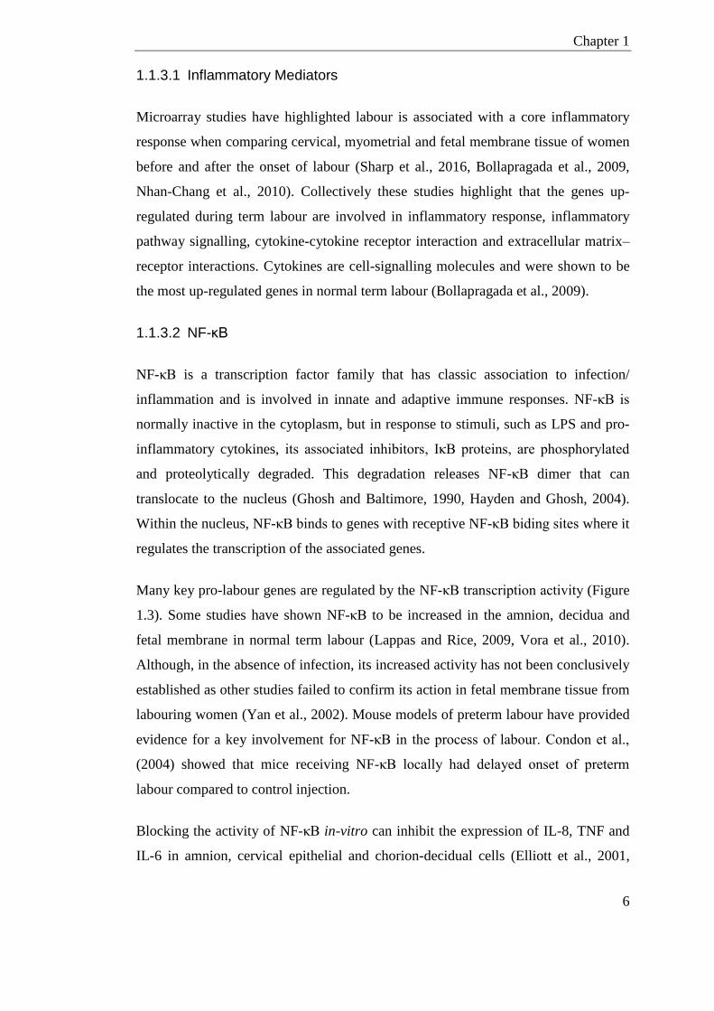
Many key pro-labour genes are regulated by the NF-κB transcription activity (Figure
1.3). Some studies have shown NF-κB to be increased in the amnion, decidua and
fetal membrane in normal term labour (Lappas and Rice, 2009, Vora et al., 2010).
Although, in the absence of infection, its increased activity has not been conclusively
established as other studies failed to confirm its action in fetal membrane tissue from
labouring women (Yan et al., 2002). Mouse models of preterm labour have provided
evidence for a key involvement for NF-κB in the process of labour. Condon et al.,
(2004) showed that mice receiving NF-κB locally had delayed onset of preterm
labour compared to control injection.
Blocking the activity of NF-κB in-vitro can inhibit the expression of IL-8, TNF and
IL-6 in amnion, cervical epithelial and chorion-decidual cells (Elliott et al., 2001,
Chapter 1
7
Lappas et al., 2003). It has also been shown to inhibit COX-2 expression and
production of prostaglandins in fetal membrane tissues and the release of MMPs
(Lindström and Bennett, 2005).
Figure 1.3 Schematic of Core Labour-associated Pathways Lipopolysaccharide (LPS); Corticotrophin Releasing Hormone (CRH); Surfactant
Protein-A (SP-A,); Progesterone Receptor (PR); Glucocorticoid Receptor (GR);
Phospholipase A2 (PLA2); Oxytocin Receptor (OTR). Source: Lindström and
Bennett (2005)
1.1.3.3 Immune Cells
Neutrophils are a mature type of white blood cell that have vital role in protecting
against diseases and infections. They can participate via direct actions such as;
phagocytosis, degranulation and generation of neutrophil extracellular traps (NETs).
Neutrophils also work by producing cytokines that can recruit and activate further
immune cells to the area of infection/inflammation.
Labouring-women at term and preterm have higher total numbers of circulating
neutrophils compared to before the initiation of labour. Circulating neutrophils in
Chapter 1
8
labouring women are also more primed than non-labouring neutrophils; they show
increased migration potential and have a higher production of reactive oxygen
species (Yuan et al., 2009).
The observation of leukocyte influx into reproductive and pregnancy tissues during
labour has been widely reported (Osman et al., 2003, Gomez-Lopez et al., 2010). It
has also been widely observed in combination with an increase in the neutrophil
chemokine IL-8 (CXCL8) mRNA gene transcript. Neutrophils in pregnancy tissues
release pro-inflammatory cytokines and MMPs, which degrade the extracellular
matrix of the fetal membranes and contribute to the labour cascade. However, it
remains to be established whether these leukocytes are a causative factor for the
initiation of labour, or predominantly involved in the subsequent tissue remodelling
following labour.
In animal studies, term-labour and preterm labour induced by inflammation, results
in an influx of neutrophils into the decidua and myometrium (Shynlova et al., 2013a,
Shynlova et al., 2013b, Rinaldi et al., 2014). This was not seen when preterm labour
was induced by blocking the hormone progesterone with mifepristone (Shynlova et
al., 2013a), suggesting a role for decidual neutrophils in labour. Similarly in humans,
the number of neutrophils in the decidua of women with infection-associated preterm
labour is greater than preterm/term labour not associated with infection (Hamilton et
al., 2012). Despite these findings, Rinaldi et al., (2014) showed that neutrophil influx
to the decidua is not necessary for initiation of preterm labour in an animal model of
PTL. The authors demonstrated that depleting neutrophils did however reduce the
amount of IL-1β produced in response to LPS.
Most macrophages are monocyte derived from the blood; however, tissues also have
their own type of tissue resident macrophages that are not always derived from the
bone marrow. Resident macrophages are mostly involved in homeostasis; patrolling
the microenvironment for damage signals from cell death and cells undergoing
phagocytosis.
Macrophages have dual functions in the response to inflammation. Macrophages are
involved in the pro-inflammatory process of sending signals to recruit more immune
Chapter 1
9
cells, but are also involved in resolution of inflammation and regulating the levels of
tissue repair to avoid tissue damage. In response to infection and other intrinsic cell
damage signals, they can phagocytose, releasing IL-1β and TNF as potent pro-
inflammatory cytokines. In resolving infection, macrophages can produce anti-
inflammatory cytokines including TGF-β and IL-10.
Macrophages are important in secreting cytokines, including IL-1β, IL-6, TNF but
also MMPs and nitric oxide (NO). Human and rodent studies have shown that uterine
macrophage numbers increase with gestation and there is an associated increase in
NO production (Tan et al., 2014), although there are conflicting reports of their role
in parturition. Some studies in rodents (Shynlova et al., 2013a, Buhimschi et al.,
1996, Mackler et al., 1999) have suggested that uterine macrophages are depleted
directly prior to the initiation of labour, which is in keeping with the report that NO
prevents uterine contractions in-vitro (Yallampalli et al., 1993).
Various studies have shown macrophage numbers in the cervix increase during
parturition. Elegant studies looking at macrophage surface markers have shown that
cervix macrophages are more favoured towards MMP activation and cell matrix
remodelling, and less favoured for adhesion and migration. This suggests a key role
in functions associated with ripening and dilation of the cervix, which is in keeping
with an animal study that showed depleting macrophages prevented cervical
remodelling (Gonzalez et al., 2011b). Depleting macrophages also protects mice
from LPS-induced PTL suggesting macrophages also have a key role in parturition
(Gonzalez et al., 2011b).
Although neutrophils and macrophages are the main infiltrating immune cells,
pregnancy tissues also have other resident and patrolling immune cells, including
granulocyte-derived mast cells, antigen-presenting dendritic cells and NK-T cells,
which are suggested to have a role in mediation of parturition. Adaptive immune
cells, such as T cells, B cells and regulatory T cells (T regs), are also resident in
pregnancy tissues and participate mostly in fetal-maternal tolerance during
pregnancy (reviewed in Gomez-Lopez et al., (2014).
Chapter 1
10
1.1.3.4 Toll Like Receptors
Many cells have pattern recognition receptors (PAMPs) at the cell surface, including
the transmembrane TLR proteins. Cells also have multiple intracellular receptors
such as NOD domains, which recognise bacterial and extracellular stimuli.
There are 11 transmembrane Toll like receptors described in humans and mice
collectively (TLR-1-11) (Akira and Takeda, 2004). These include TLR-4, where the
main ligand is LPS, and TLR-3, which detects dsRNA. TLR-2 is involved in the
detection of many Gram-positive bacteria products such as lipoteichoic acids (LTA).
TLR-9 is essential in the recognition of CpG motifs, which are abundant in many
bacteria. TLR-3 and TLR-7-9 are intercellular receptors, while TLR-1, TLR-2, TLR-
4-6 and TLR-11 are located at the cell surface.
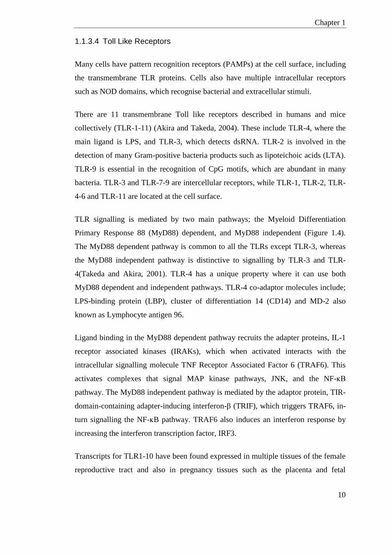
TLR signalling is mediated by two main pathways; the Myeloid Differentiation
Primary Response 88 (MyD88) dependent, and MyD88 independent (Figure 1.4).
The MyD88 dependent pathway is common to all the TLRs except TLR-3, whereas
the MyD88 independent pathway is distinctive to signalling by TLR-3 and TLR-
4(Takeda and Akira, 2001). TLR-4 has a unique property where it can use both
MyD88 dependent and independent pathways. TLR-4 co-adaptor molecules include;
LPS-binding protein (LBP), cluster of differentiation 14 (CD14) and MD-2 also
known as Lymphocyte antigen 96.
Ligand binding in the MyD88 dependent pathway recruits the adapter proteins, IL-1
receptor associated kinases (IRAKs), which when activated interacts with the
intracellular signalling molecule TNF Receptor Associated Factor 6 (TRAF6). This
activates complexes that signal MAP kinase pathways, JNK, and the NF-ĸB
pathway. The MyD88 independent pathway is mediated by the adaptor protein, TIR-
domain-containing adapter-inducing interferon-β (TRIF), which triggers TRAF6, in-
turn signalling the NF-ĸB pathway. TRAF6 also induces an interferon response by
increasing the interferon transcription factor, IRF3.
Transcripts for TLR1-10 have been found expressed in multiple tissues of the female
reproductive tract and also in pregnancy tissues such as the placenta and fetal
Chapter 1
11
membranes (reviewed in Nasu and Narahara (2010). TLR-2 and TLR-4 are stated to
be increased in the myometrium, cervix and fetal membrane of labouring women at
term (Kim et al., 2004, Bollapragada et al., 2009, Youssef et al., 2009). Interestingly
TLR-2 and TLR-4 are also increased in women with signs of infection (Kim et al.,
2004). Spontaneous preterm labour is associated with an increased expression of
TLR4 on maternal human blood monocytes (Pawelczyk et al., 2010).
There is also some evidence that low expression of TLR-4 may be protective against
preterm labour (Hirsch et al., 2006). TLR-4, has been shown to mediate
inflammation-induced preterm labour in animal models (Elovitz et al., 2003). Elovitz
et al., (2003) showed C3H/HeJ, TLR-4 mutant, mice had reduced levels of LPS-
induced preterm labour compared to the CD1 strain. In this study, TLR-4 did not
reduce the rate of fetal demise, whereby those animals that had no preterm labour
still displayed high rates of LPS-induced fetal death.
Interestingly, genetic variation in TLR-4 and TLR-9 have been associated with
incidences of dysbiosis in the vaginal microbiota, suggesting host-immune genotype
is important in maintaining bacteria homeostasis (Royse et al., 2012, Macones et al.,
2004).
Pioli et al., (2004) show TLRs have spatial expression patterns in the female
reproductive tract. Hirata et al. (2007) went on to show the expression of many of
these TLRs alters during the menstrual cycle (Hirata et al., 2007). TLR-1-9 are also
in the female reproductive tract of mice, where they similarly show temporal and
spatial expression patterns throughout estrus in the vagina and uterus (Soboll et al.,
2006, Hickey et al., 2013).
Chapter 1
12
Figure 1.4 Schematic of the TLR-4 Signalling Pathway TLR-4 singling through both the MyD88 (MyD88 dependent) and TRIF pathways
(MyD88 independent). Source: Mogensen (2009)

Chapter 1
13
1.1.4 Onset of Labour at Term
Humans first maintain pregnancy by the production of progesterone in the corpus
lutum; this is then superseded by placental progesterone production. In humans
labour is not associated with a systemic withdrawal of progesterone (Elovitz and
Mrinalini, 2004). However, emerging evidence suggests there may be local
functional progesterone withdrawal prior to the onset of labour (Pieber et al., 2001,
Mesiano et al., 2002).
In contrast in mice, placental progesterone is not thought to supersede that of the
corpus lutum and so removal of the ovaries during pregnancy induces labour
(Condon et al., 2004). The onset of labour in mice has been proposed to involve
Surfactant Protein A (SP-A) which is released from the pup lung. SP-A causes
mobilisation of macrophages from the amniotic fluid into the maternal tissues of the
uterus, this process activates pro-inflammatory cytokines such as IL-1β that in turn
stimulates NF-κB activation (Condon et al., 2004, Mendelson and Condon, 2005).
However, the causative factor that initiates the inflammatory cascade in Human term
labour still remains unclear.
1.1.5 Infection/Inflammation and Preterm Labour
There are many infectious agents that when contracted by the mother can transfer to
fetus and cause inflammation and fetal demise. Infectious agents include;
Toxoplasma gondi (toxoplasmosis), Cytomegalovirus, Herpes simplex virus, and
Lyme disease. However, these infections do not explain the infection/inflammation
seen in most preterm births. A local immune response in the decidua and chorion is
more commonly associated with preterm labour (Galinsky et al., 2013, Vrachnis et
al., 2010). These are frequently caused by ascending opportunistic commensal
bacteria from the vagina, through the cervix and into the fetal membrane (Allam et
al., 2013, DiGiulio et al., 2010). The upper genital tract is generally considered
sterile prior to the onset of labour (Jones et al., 2009) nonetheless nearly 40% of
preterm deliveries are associated with intrauterine infection and/or clinical
inflammation (Romero et al., 2006). Studies investigating bacterial DNA in placenta

Chapter 1
14
and fetal membranes discovered women who present with preterm labour have a
greater diversity of bacteria in these tissues (Prince et al., 2016, Aagaard et al.,
2014). It is therefore important to investigate the role of local infection when
considering premature labour.
1.1.6 Mechanisms for Inflammation/Infection
NF-𝜅B can also be activated by pro-inflammatory cytokines in response to pathogens
such as endotoxins from bacterial cell walls.
The surface of many immune cells including leukocytes, dendritic cells, epithelial
cells and trophoblasts, have pattern recognition receptors (e.g. TLRs) which when
stimulated by bacterial endotoxin or pro-inflammatory cytokines release transcription
factors including NF-𝜅B, STAT, and AP-1. This in turn results in the production of
cytokines and chemokines, such as IL-6, TNF, IL-1β, and IL-8, within the uterine
lining and the fetal membranes (Figure 1.3).
The involvement of cytokines/chemokines in maternal and fetal inflammation has
been well investigated in humans. Experimentally, animal models have been used
which allow for collection of gestational tissues at various time-points during
pregnancy.
Systemic LPS treatment in pregnant mice induces maternal inflammation and
preterm labour that is associated with rises in amniotic fluid cytokines (IL-1 and IL-
6) and maternal serum cytokines (IL-1, TNF, and IL-6) (Baumann et al., 1993, Fidel
et al., 1994).
A similar cytokine response was seen in a localised fetal inflammatory model, where
heat-killed E.coli was injected into ligated uterine horns (Hirsch et al., 2006). These
cytokines can stimulate the production of prostaglandins, which initiate mobilisation
of neutrophils. These neutrophils infiltrate tissue causing the production and release
of metalloproteases (MMPs) and induction of COX-2. COX-2 is expressed at term
labour in the human myometrium and also animal cervical fibroblasts (Sato et al.,
2001), there is also some evidence that low expression may be protective against
Chapter 1
15
preterm labour (Hirsch et al., 2006). As discussed previously, TLR-4 has been shown
to mediate inflammation-induced preterm labour in animal models (Elovitz et al.,
2003).
Prostaglandins produced in the amnion can stimulate uterine contractions when in
proximity to the uterine lining (Cheung et al., 1992). This activity is supressed during
pregnancy by the release of prostaglandin dehydrogenase in chronic tissue which acts
as a buffer for its activity (Cheung et al., 1992). In the context of an amniotic
infection, activity of prostaglandin dehydrogenase is reduced, which allows the
prostaglandins to reach the myometrium which in-turn can lead to premature
contractile activity (Van Meir et al., 1996). Infection can also increase MMPs, which
can weaken the chorion amniotic membrane causing it to prematurely rupture
(Catalano et al., 2010). Increased concentrations of iNO and prostaglandins in
amniotic fluid are also associated with intrauterine infection and continue to
propagate the labour cascade (Bethea et al., 1998).
LPS and exposure to some bacteria can cause hypoxia-inducing oxidative stress and
apoptosis in the placenta and fetal membranes (Garnier et al., 2008). Increases in cell
turn-over and apoptosis at the fetal-maternal interface could result in increased
apoptotic bodies and cell-free fetal DNA (cffDNA) from the placenta entering the
maternal circulation (Bischoff et al., 2005). High levels of cffDNA, which is
hypomethylated and a TLR-9 agonist, have previously been associated with preterm
labour, preterm rupture of the membrane and preeclampsia (Farina et al., 2005,
Jakobsen et al., 2012, Lim et al., 2013).
1.1.7 Therapeutics for Preterm Labour
Research into preterm labour has vastly increased in the last decade but this has not
translated into a reduction in the incidence of preterm labour. There are few
treatment options for preventing a preterm birth to improve neonatal outcomes.
A cervical cerclage is a suture inserted close to the cervix during pregnancy. It is
used in women with short cervical length with the aim of preventing early opening of

Chapter 1
16
the cervix. Kindinger et al., (2016) demonstrated that the type of material used for
the suture can affect the vaginal microbiome and inflammatory cytokine profile.
Drugs can be used in an attempt to supress the myometrial contractions in women
who are high risk of preterm labour. The main mechanisms for these tocolytic drugs
are to reduce the supply and transfer of intracellular calcium in the myometrium,
which is a key component in processes for myometrial contractions. Inhibitors for the
oxytocin receptor COX-2 are also used as tocolytic treatment; as COX-2 is the rate-
determining step for Prostaglandin synthesis and Prostaglandins directly stimulate
myometrial contractions.
Progesterone also acts on suppressing the availability and transport of intracellular
calcium ions and Prostaglandin synthesis in myometrial tissue. There have been
multiple randomised control-studies which show a reduction in PTL with weekly
injections of 17α-hydroxy progesterone caproate (Meis et al., (2003) reviewed in
Norwitz and Caughey (2011)). This protective effect was also shown in a study
which applied progesterone daily to the vagina (da Fonseca (2003) reviewed in
Norwitz and Caughey (2011)). Progesterone was not shown to be effective for
women presenting with PROM (Briery et al., 2011) or multiple pregnancies (Rouse
et al., (2007), Durnwald et al., (2010), Norman et al., (2009a) reviewed in Norwitz
and Caughey (2011)). Reviews have suggested that progesterone can also improve
neonate morbidity and mortality. Although recent published results from the
OPTIMUM randomised placebo-controlled trial, looking at progesterone as a
prophylaxis, showed no harm or benefit from vaginal progesterone on neonatal
outcome at 2 years of age, moreover no benefit to preterm labour (Norman et al.,
2016).
There are conflicting reports whether antibiotic intervention is beneficial in
preventing preterm labour associated with intrauterine infection (Lamont, 2015, King
and Flenady, 2002). There is however, increasing support that certain prophylactic
antibiotics given very early in pregnancy to those women who have a clinical
diagnosis of vaginal bacteria dysbiosis has benefit (Thinkhamrop et al., 2015).
Chapter 1
17
Novel drug development and clinical trials, especially in pregnancy is very restricted,
so looking towards currently available drugs already used for other conditions may
prove a quicker mode for translational medicine. An encouraging human study
looking into administration of chloroquine chemoprophylaxis, a known TLR-9
inhibitor and antimalarial agent, in pregnant mothers in West Africa reported lower
rates of preterm labour in those who had a ‘complete’ course of chloroquine (Sirima
et al., 2003).
Taken together, the development of preventative therapies for preterm labour
requires a mechanistic understanding of how key modifiers of the inflammatory
processes respond to bacterial infection during pregnancy.

Chapter 1
18
1.2 Host Defence Peptides
Antimicrobial peptides provide the first line of defence against infectious pathogens
in multicellular organisms. Peptides with antimicrobial functions have also been
defined in archaea, fungi, plants and bacteria. Over 1,972 animal host defence
peptides are currently described and they are evolutionarily conserved (Wang et al.,
2016). Structurally, antimicrobial peptide families are very diverse, but they are
categorised by their sequence composition and protein confirmation.
The most studied cationic antimicrobial peptides in humans are the Defensins and
Cathelicidin. Due to the discovery that these antimicrobial peptides have an array of
additional immunomodulatory properties (Figure 1.5), they are commonly adopting
the broader name of Host Defence Peptides (HDPs).
Figure 1.5 Overview of HDPs Modes of Action HDPs have potent microbicidal activity against a broad spectrum of bacteria, fungi,
and viral organisms; acting directly on the invading pathogens (blue). They can
supress the inflammatory process by binding and blocking receptors but also directly
binding to bacterial pathogens (yellow). They also influence immune response by
attracting inflammatory cells, inducing an inflammatory state, modulating apoptotic
cell death and cell degranulation (green). HDPs can play a role in tissue homeostasis;
including the induction of angiogenesis (pink). Source: Yarbrough et al., (2015)
Chapter 1
19
1.2.1 Defensins
1.2.1.1 Defensins
Human defensins are small cationic peptides, predominantly released by epithelial
cells at mucosal surfaces where their main role is in host innate immunity. In
addition to acting directly on invading pathogens, they also influence immune
response by attracting inflammatory cells and inducing an inflammatory state. There
is also evidence to suggest that certain defensins can function to modulate apoptotic
cell death (Yamaguchi et al., 2007) and some have immunosuppressive activity (Shi
et al., 2007, Semple et al., 2010)
Defensins are divided into three categories; alpha-defensins, beta-defensins, and
theta-defensins. Alpha and theta defensins arose from ancestral beta-defensin genes.
In other species such as snakes, beta-defensin derivatives have become specialised as
toxins (crotamines). Almost all defensins contain a canonical six-cysteine motif and
have a beta-sheet structure. They are distinguishable by their; genomic organisation,
the spacing of these cysteines, and their disulphide bond interactions. In-silico
analysis has predicted 28 defensin like sequences at 5 loci in the human genome
based on the six- cysteine motif (Schutte et al., 2002).
Alpha-defensins and beta-defensins are found in most vertebrate species. Leukocytes
and Paneth cells (specialised cells in the small intestine), are the main source of
alpha-defensins. Beta-defensins are expressed in a variety of tissues and cell types,
including epithelial cells, macrophages and dendritic cells.
In humans and many higher mammals, beta-defensin sequences have been found to
cluster together on chromosome arrangements. In humans, 10 beta-defensins are
clustered on an 8MB region of chromosome 8p23.1. Seven of the genes in this region
(DEFB103 – DEFB108 and DEFB4) are arranged in a copy number variable (CNV)
cluster, which ranges from 2-7 copies. CNV has been shown to some extent to be
correlated to protein level and CNV of this loci has been linked to the inflammatory
disease psoriasis (Hollox et al., 2008). A few beta-defensins are expressed
constitutively, for example Human beta-defensin 1 (hBD1). While other beta-
Chapter 1
20
defensins, such as Human beta-defensin 3 (hBD3) and Human beta-defensin 2
(hBD2), are inducible upon stimulation with bacteria or cell damage signals.
Interestingly many beta-defensins are expressed in the male and female reproductive
tract. The epididymis expression of beta-defensins is segmental and influences sperm
maturation (Zhou et al., 2013).
In mice, the genomic arrangement containing the alpha-defensins is flanked by two
beta-defensin regions. Mice have 27 beta-defensins, 26 cryptdin genes (defensin
related) and multiple defensin pseudogenes. Mouse defensins are highly repetitive in
nature with regard to their chromosomal position; this arrangement supports a
positive diversifying-selection model of evolution (Patil et al., 2004, Patil et al.,
2005). Unlike humans, mice have no neutrophil alpha defensins but they have
multiple cryptdin alpha-defensins in the small intestine.
There are several published beta-defensin mouse knockouts. Two studies suggest that
Defb1 homozygous null mice retain Haemophilus influenzae in the lung, and
Staphylococcus spp. in the bladder longer than wildtype mice. This implicates a role
of Defb1 in respiratory and urinary tract infection, resistance and pathogen clearance
(Morrison et al., 2002, Moser et al., 2002). There has been direct evidence linking
gut alpha-defensins to the modulation of microbiota in mice. Salzman et al., (2010)
demonstrated that Paneth cell alpha-defensins contributed to mucosal host defence
and regulate the composition of murine intestinal microbiota.
1.2.1.2 Beta-defensins
In humans, seven of the beta-defensin genes on chromosome 8 are copy number
variable and five of these, DEFB4 (encodes peptide hBD2), DEFB103 (encodes
hBD3), DEFB106, DEFB105 and DEFB107, have a mouse orthologue (Table 1.1).
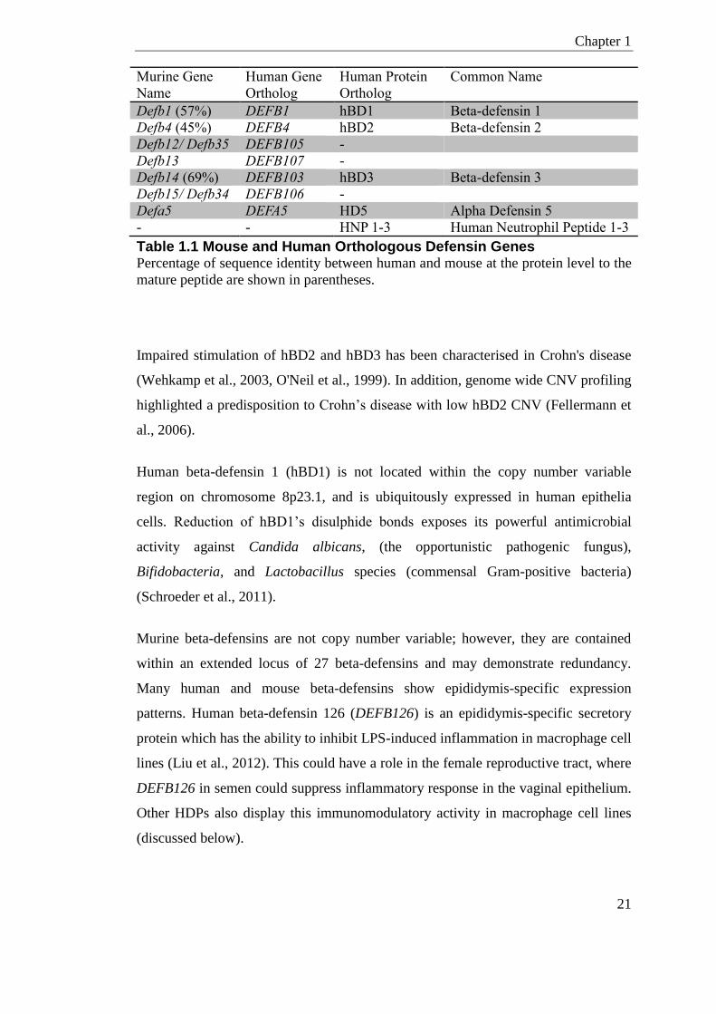
Chapter 1
21
Murine Gene
Name
Human Gene
Ortholog
Human Protein
Ortholog
Common Name
Defb1 (57%) DEFB1 hBD1 Beta-defensin 1
Defb4 (45%) DEFB4 hBD2 Beta-defensin 2
Defb12/ Defb35 DEFB105 -
Defb13 DEFB107 -
Defb14 (69%) DEFB103 hBD3 Beta-defensin 3
Defb15/ Defb34 DEFB106 -
Defa5 DEFA5 HD5 Alpha Defensin 5
- - HNP 1-3 Human Neutrophil Peptide 1-3
Table 1.1 Mouse and Human Orthologous Defensin Genes Percentage of sequence identity between human and mouse at the protein level to the
mature peptide are shown in parentheses.
Impaired stimulation of hBD2 and hBD3 has been characterised in Crohn's disease
(Wehkamp et al., 2003, O'Neil et al., 1999). In addition, genome wide CNV profiling
highlighted a predisposition to Crohn’s disease with low hBD2 CNV (Fellermann et
al., 2006).
Human beta-defensin 1 (hBD1) is not located within the copy number variable
region on chromosome 8p23.1, and is ubiquitously expressed in human epithelia
cells. Reduction of hBD1’s disulphide bonds exposes its powerful antimicrobial
activity against Candida albicans, (the opportunistic pathogenic fungus),
Bifidobacteria, and Lactobacillus species (commensal Gram-positive bacteria)
(Schroeder et al., 2011).
Murine beta-defensins are not copy number variable; however, they are contained
within an extended locus of 27 beta-defensins and may demonstrate redundancy.
Many human and mouse beta-defensins show epididymis-specific expression
patterns. Human beta-defensin 126 (DEFB126) is an epididymis-specific secretory
protein which has the ability to inhibit LPS-induced inflammation in macrophage cell
lines (Liu et al., 2012). This could have a role in the female reproductive tract, where
DEFB126 in semen could suppress inflammatory response in the vaginal epithelium.
Other HDPs also display this immunomodulatory activity in macrophage cell lines
(discussed below).
Chapter 1
22
A major site of beta-defensin expression is in the male reproductive tract of both
humans and mice. Zhou et al., (2013) has shown that homozygous deletion of 9 beta-
defensins on mouse chromosome 8 results in infertility in male mice. Infertile mice
displayed reduced sperm motility, increased sperm fragility and increased
spontaneous acrosome reaction. This is partly due to defects in sperm microtubule
formation and consequently results in increased intracellular calcium content.
Many human and mice beta-defensins have chemotactic properties. hBD1, hBD2 and
hBD3 are able to induce chemotaxis through CCR6 (Hoover et al., 2002, Oppenheim
and Yang, 2005). Similarly, the mouse Defb4 and Defb14 genes, orthologues of
genes encoding hBD2 and hBD3 respectively, also have chemotactic activity for
CCR6-expressing cells (Rohrl et al., 2008, Rohrl et al., 2010).
1.2.1.3 Human beta-defensin-3 (hBD3)
DEFB103 gene, which encodes hBD3, has two exons; the first exon contains
sequence for the 5’UTR and pre-protein, while the second exon contains the
sequence for the mature protein. The six-cysteine motif spacing, which forms the di-
sulphide bonds in hBD3, is different from the other defensins, namely hBD2 and
hBD1. However, the general shape of these defensins is similar despite lack of
identity between family members (Bauer et al., 2001). hBD3 is the most cationic and
when folded has the ability to form symmetrical amphipathic dimers (Dhople et al.,
2006); this is thought to be responsible for its salt in-sensitivity compared to other
HDPs.
1.2.1.3.1 Expression and Regulation
hBD3 is found highly expressed in psoriatic skin (Harder et al., 2001) (where it was
first isolated) but also in cells of the oral cavity, endometrium, placenta, and immune
cells. A broad range of bacteria (Gram-positive and Gram-negative) in addition to
pro-inflammatory cytokines such as IL-1β, TNF-α and INF-γ are able to
induce/increase the expression of hBD3.
Chapter 1
23
hBD3 in human keratinocytes is increased by bacterial challenge via stimulation of
the epidermal growth factor receptor (EGRF). In gingival fibroblasts, hBD3
expression increases in response to infection with Candida albicans (Gursoy and
Kononen, 2012). Oral squamous cells (Shuyi et al., 2011), gingival epithelial cells
and dendritic cells (Yin et al., 2010) all show a marked increase in hBD3 in response
to LPS treatment in-vitro.
In women, hBD3 and other defensins are known to show temporal expression
patterns in the reproductive tract during the menstrual cycle; where higher levels of
hBD3 are seen in the early and late secretory phase (King et al., 2003b, Das et al.,
2007).
DEFB103 at the transcript level is inhibited by corticosteroids (Duits et al., 2001);
these corticosteroids can also inhibit pro-inflammatory cytokines and cell surface
markers (Barnes, 2006). The activity of hBD3 can be impaired through the direct
inhibition and degradation of the protein. Saliva and serum can inhibit the
microbicidal activity of hBD3 to Gram-positive and Gram-negative bacteria
(Maisetta et al., 2005). hBD3 was also found to have reduced killing activity to
Gram-positive bacteria in low pH environments (Abou Alaiwa et al., 2014). Cysteine
proteases and streptococcal inhibitor complement produced by group-A
Streptococcus (GAS) can also directly degrade hBD3 peptide (Cogen et al., 2010).
1.2.1.3.2 Antimicrobial and Immunomodulatory Properties
hBD3 is known for its potent antimicrobial activity against a broad spectrum of
bacteria, fungi, and viral organisms. hBD3 can act on many Gram-negative and
Gram-positive bacterial species including methicillin-resistant Staphylococcus
aureus (MRSA) and vancomycin-resistant Enterococci (VRE) (Harder et al., 2001).
The exact mechanisms of how hBD3 kills bacteria are not fully understood, but are
thought to involve ionic interactions with the bacterial cell surface, blockage of
bacterial biosynthetic pathways, and bacteria cell wall lipid interaction (Sahl et al.,
2005). Staphylococcus aureus when treated with non-folded hBD3 shows rapid
perforation in the exterior cell wall (Midorikawa et al., 2003). Folded proteins of

Chapter 1
24
hBD3 also show a similar activity suggesting disulphide structure is not essential for
the antibacterial mode of action (Harder et al., 2001).
hBD3 can supress biofilm production in single organisms of Pseudomonas
aeruginosa and Staphylococcus aureus, but also biofilms of multiple-species (Zhu et
al., 2013, Lee et al., 2013). Studies have shown that hBD3 not only represses the
initiation and maturation of biofilms, but also represses those already established
(Zhu et al., 2013).
hBD3 and DEFB14 have potent anti-inflammatory effects on human and mouse
primary monocyte derived macrophages. This action effectively inhibits the LPS-
activated TLR4-induced TNF and IL-6 response, through both the MyD88 and TRIF
pathways. This effect has been shown both in-vitro and in-vivo (Semple et al., 2010,
Semple et al., 2011). hBD3 can act in synergy with other HDPs. For example, LL-37
and hBD3 have a combined effect reducing the LPS-induced secretion of cytokines
in gingival cells (Bedran et al., 2014).
Semple et al., (2015) showed that hBD3 exacerbates the production of INF-β in
response to polyI:C, a viral mimic and TLR-3 agonist, in both human and mouse
primary macrophages. Conversely, the authors showed reduction of CXCL10 in
response to this TLR-3 agonist in the presence of hBD3. This effect was also seen in-
vivo where co-treatment of mice with hBD3 peptide and polyI:C enhanced the
increase in INF-β, compared to polyI:C treatment alone. Using macrophages from
knockout mice, Semple et al., (2015) went on to show that hBD3 suppressive effects
were mediated by TICAM1 (TRIF in mouse), while the exacerbating effects were
mediated by MDA5 (IFIH1 in mouse).
In addition, hBD3 can bind to CpG and self-DNA to form net complexes; these
complexes are up-taken by pDCs and induce an INF-α response, acting as a TLR-9
agonist (Tewary et al., 2013). Such complexes are increased in patients with
autoimmune disease (Gilliet and Lande, 2008).
Chapter 1
25
1.2.1.4 Murine Orthologue of hBD3; Mouse Defb14
Mouse beta-defensin 14 (Defb14) has the highest sequence identity to hBD3 protein;
sharing 69% sequence identity at the protein level. Multiple studies have confirmed
similar activity of the human and mouse peptide suggesting they are functional
orthologues (Rohrl et al., 2008, Semple et al., 2010).
Like hBD3, DEFB14 exhibits broad-spectrum antimicrobial activity. Defb14
responds to INF-γ and TGF-α treatment in keratinocytes (Hinrichsen et al., 2008),
which were also identified as stimuli of hBD3 in human keratinocytes (Harder et al.,
2004). hBD3 and DEFB14 have the same potent anti-inflammatory effects on human
and mouse primary monocytes (Semple et al., 2010, Semple et al., 2011). Moreover,
hBD3 and Defb14 also have similar expression profiles in the female and male
reproductive tract (King et al., 2003b, Hickey et al., 2013, Zhou et al., 2013, Garcia-
Lopez et al., 2010)
1.2.2 Cathelicidin
1.2.2.1 Cathelicidin
Cathelicidins are cationic antimicrobial peptides, found mostly in neutrophils,
keratinocytes and epithelial cells, which have a vital role in protecting against
diseases and infections.
Multiple cathelicidins have been identified in mammals such as sheep, goat and pigs.
However, rodents and humans have only one gene for cathelicidin. The gene
encoding the sole human cathelicidin, CAMP is located on chromosome 3p21.3 and
is comprised of 4 exons. The N-terminal and the coding regions of the first three
exons, which include the cathelin and signal domain, are highly conserved. Exon 4
codes the antimicrobial peptide domain. The 16kDa preprotein, hCAP18, does not
exhibit antimicrobial activity and can either be immediately cleaved, or stored as a
proprotein in granules without the signal domain. Extracellular proteolytic cleavage
of the C-terminal of hCAP18 by proteinases 3, or kallikrein generates the active LL-
37 peptide (Sorensen et al., 2001, Yamasaki et al., 2006, Garcia-lopez et al., 2010).
Chapter 1
26
The LL-37 peptide has cationic and amphipathic properties forming an alpha- helical
structure.
Alternative microbicidal active forms of LL-37 have been reported in select
environments (Sorensen et al., 2001, Sorensen et al., 2003, Murakami et al., 2004).
These alternative fragments can be formed either by the additional degradation of
LL-37 by host/bacterial proteases, or by alternative cleavage of the pro-protein.
hCAP18 in seminal fluid can be processed by gastricsin derived from the prostatic
gland to form an antimicrobial peptide of 38 amino acids (Sorensen et al., 2003).
This peptide, ALL-38, has the same antimicrobial properties as LL-37 (Sorensen et
al., 2003). Gastricsin cleavage only occurs at low pH, akin to what is seen in the
vagina suggesting a mechanistic involvement preventing infection following sexual
intercourse. LL-37 is found to have reduced killing activity to Gram-positive bacteria
in low pH environments (Abou Alaiwa et al., 2014). Serine proteases in sweat can
also degrade LL-37 into reduced fragments; KR-20, RK-31, and KS-30 (Murakami
et al., 2004).
1.2.2.2 Expression and Regulation
CAMP gene expression is found at varying levels in many cell types; hCAP18/LL-37
is also present in many bodily fluids such as breast milk, sweat, saliva and vaginal
secretions (reviewed in Durr et al., (2006)). CAMP is commonly upregulated in many
immune and epithelial cells when challenged in infectious disease. Leukocytes and
epithelial cells can rapidly release hCAP18/LL-37 molecules extracellularly
following uncontrolled cell death, infection and degranulation (reviewed in Durr et
al., (2006)). TLR activation and various cytokines can also directly upregulate
CAMP gene expression (Mookherjee et al., 2006).
Upstream of the CAMP gene is a SINE repeat, which contains the putative Vitamin
D response element (VDRE). This is the primary element that is bound by Vitamin D
receptor (VDR) and responsible for the Vitamin D induced expression of CAMP in
many cell and tissue types. This element is not highly conserved, and is absent in all
other higher vertebrates including mouse.
Chapter 1
27
Bacteria produce short chain fatty acids such as butyrate from fermentation of dietary
fibres in the colon. SCFAs such as butyrate can also regulate the expression of
CAMP gene (Kida et al., 2006). In the gut, SCFA’s are mostly anti-inflammatory
involved in promoting gut barrier function and homeostasis. SCFA’s demonstrate
anti-inflammatory function by inhibiting NF-Kb signally by the suppression of pro-
inflammatory cytokines (Zeuthen et al., 2008, Segain et al., 2000). In contrast,
SCFA’s in the vagina show more of a pro-inflammatory response in those women
with bacterial vaginosis (Mirmonsef et al., 2012, Aldunate et al., 2015). The
difference in SCFA immune response could be inherent properties of the cell type,
pH, and TLR ligand combination in the different environments (Mirmonsef et al.,
2012).
Immune-Mediated Inflammatory Diseases
Chronic inflammatory disease has associations with dysregulation of the careful
cytokine/chemokine homeostatic balance. Several HDP, including LL-37 are
elevated in systemic lupus erythematosus (SLE) (Kreuter et al., 2011), and
rheumatoid arthritis (RA) (Paulsen et al., 2002).
Some conditions are associated with a downregulation of hCAP18/LL-37. Morbus
Kostmann is a condition where patients display reduced neutrophil counts and no
LL-37 in granulocytes, these patients also commonly display severe periodontal
disease (Putsep et al., 2002). Without treatment Morbus Kostmann patients die from
bacterial infection in infancy. Patients treated with recombinant granulocyte colony-
stimulating factor (G-CSF), retain reduced neutrophil defensins and absolute
deficiency in LL-37. Crohn’s, atopic dermatitis and chronic ulcers are all reported to
be associated with a decrease in CAMP gene expression or hCAP18/LL-37 levels
(Schauber et al., 2006, Sun et al., 2016, Reinholz et al., 2012).
Mice null for Camp, the murine orthologue of LL-37/hCAP18, have many inducible
phenotypes; showing increased vulnerability to bacterial infections of the
gastrointestinal tract (Chromek et al., 2012), urinary tract (Chromek et al., 2006),
skin (Nizet et al., 2001), lung (Kovach et al., 2012), and eye (Huang et al., 2007).
Chapter 1
28
The mechanisms for the anti-inflammatory and immunomodulatory activities of
human and mouse cathelicidin are very complex. Involving such processes as,
intracellular uptake, endocytic mobilisation and receptor interaction (reviewed in
Choi et al., (2012). These mechanisms result in signalling pathways being altered and
activation of various transcription factors.
Receptors
The most classic LL-37 receptor is formyl peptide receptor 2 (FRP2), but there are
also others such as CXR2, EGFR, and P2X7 (De et al., 2000). Major downstream
pathways of these receptors involve activation of protein kinases, the release of
intracellular calcium, and NF-kB activation. Activation of these receptors and
signalling pathways cause cell proliferation, mobilisation and cytokine release. These
processes are important for the immunomodulatory activity of LL-37 in chemotaxis,
angiogenesis and tissue repair (Carretero et al., 2008, Koczulla et al., 2003). The
ability for LL-37 to cross the cell membrane where it can bind proteins, such as
GAPDH intracellularly has also been shown to be an important factor in LL-37’s
immunomodulatory activity (Mookherjee et al., 2009). It has been shown that cells
such as vaginal epithelial and placenta cells express the classic LL-37 receptor FRP2
(Dong and Yin, 2014, Frew, 2013), but it is unclear if this is the main receptor for the
immunomodulatory properties of LL-37 in these cells (Frew, 2013).
1.2.2.3 Antimicrobial Properties
LL-37 like hBD3, is known for its broad-spectrum antimicrobial activity; acting on
many Gram-positive, Gram-negative bacteria, viruses, and also on the yeast Candida
albicans (reviewed in Vandamme et al., (2012)). LL-37 preferentially binds to
bacteria where its antibacterial action involves disrupting cell membrane integrity
using a toroidal pore carpet like mechanism (Oren et al., 1999, Henzler Wildman et
al., 2003, Lee et al., 2011). This process involves LL-37 binding parallel to the
membrane, where oligomers and monomers cause membrane strain and subsequent
translocation into the periplasm. This process of LL-37 molecules translocating into
the periplasm opens pores and causes leakage (Oren et al., 1999). Antifungal
properties of LL-37 are based on interaction with fungal cell wall and membrane
Chapter 1
29
ROS production (Rapala-Kozik et al., 2015). Antiviral properties of LL-37 have been
shown by its ability to directly interact with the viral envelope (Currie et al., 2016).
In addition, this peptide has shown strong inhibitory effects on the HIV-1 reverse
transcriptase where it can inhibit HIV-1 replication in PBMCs and T-cells (Bergman
et al., 2007).
Like hBD3, LL-37 has been reported to have anti-biofilm activity at sub-minimum
inhibitory concentrations (MIC) of a number of organisms, namely, Staphylococcus
epidermidis (Hell et al., 2010) Staphylococcus aureus (Mishra et al., 2016, Dean et
al., 2011), Pseudomonas aeruginosa (Overhage et al., 2008) and Escherichia coli
isolates (Kai-Larsen et al., 2010). This feature is not unique to all cathelicidins where
the mouse peptide, CAMP, did not show anti-biofilm activity on Pseudomonas
aeruginosa (Overhage et al., 2008). In addition to its activity against pre-establish
biofilms, LL-37 peptide can also inhibit biofilm formation of Staphylococcus aureus
(MRSA) (Dean et al., 2011). In animal in-vivo infection-models, LL-37 peptide
prevents Escherichia coli and Candida albicans from adhering to the urinary tract
cell wall, stopping subsequent biofilm formation (Chromek et al., 2006, Tsai et al.,
2011).
1.2.2.4 Modulation of Inflammation/Infection
Chemotactic Properties
LL-37 can act as a direct chemoattractant for cell types including; neutrophils, iDCs,
eosinophils, T lymphocytes and monocytes (Tjabringa et al., 2006, Soehnlein et al.,
2008, Chertov et al., 1997, Wang et al., 1996). Tjabringa et al., (2006) showed that
eosinophils and neutrophils are directly mobilised by LL-37 using a calcium chamber
assay. Soehnlein et al., (2008) went on to show a direct LL-37-induced mobilisation
of inflammatory-monocytes.
At and around the site of infection, low/modest physiological concentrations of LL-
37 induces the secretion of chemokines such as IL-8. This process also indirectly
recruits additional innate immune cells to the area. LL-37 induces secretion of these
cytokines in innate immune cells such as macrophages, but also in epithelial cells
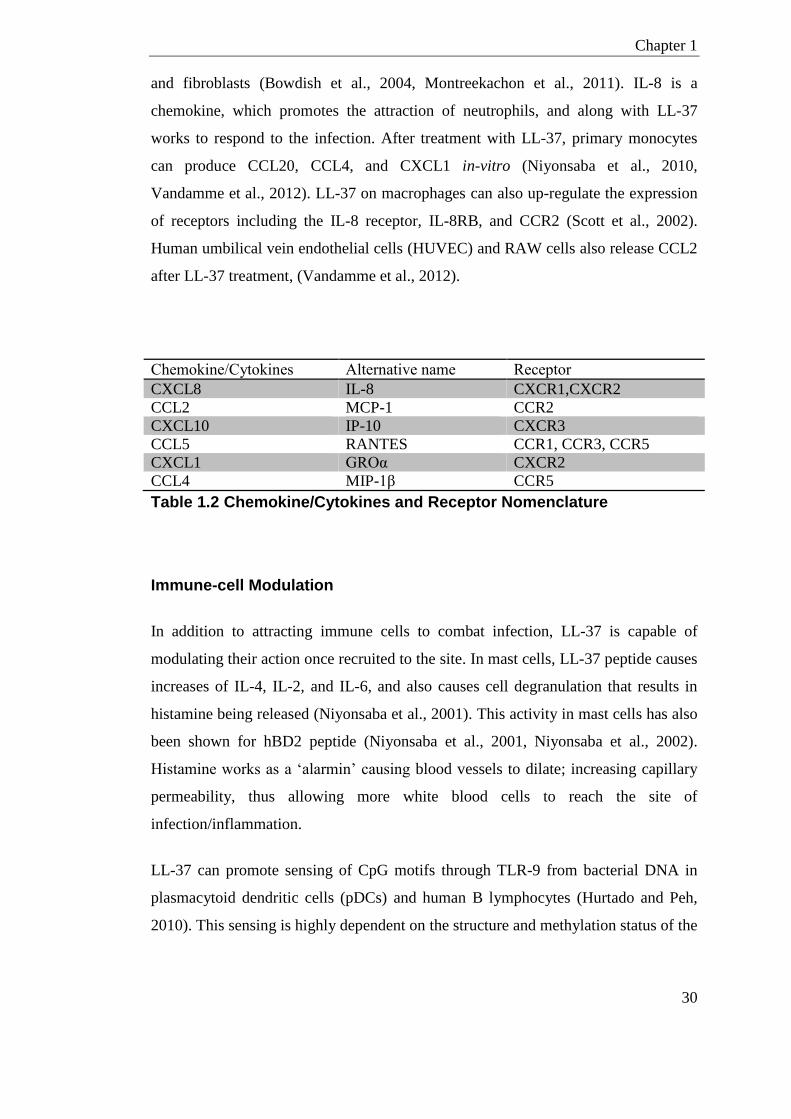
Chapter 1
30
and fibroblasts (Bowdish et al., 2004, Montreekachon et al., 2011). IL-8 is a
chemokine, which promotes the attraction of neutrophils, and along with LL-37
works to respond to the infection. After treatment with LL-37, primary monocytes
can produce CCL20, CCL4, and CXCL1 in-vitro (Niyonsaba et al., 2010,
Vandamme et al., 2012). LL-37 on macrophages can also up-regulate the expression
of receptors including the IL-8 receptor, IL-8RB, and CCR2 (Scott et al., 2002).
Human umbilical vein endothelial cells (HUVEC) and RAW cells also release CCL2
after LL-37 treatment, (Vandamme et al., 2012).
Chemokine/Cytokines Alternative name Receptor
CXCL8 IL-8 CXCR1,CXCR2
CCL2 MCP-1 CCR2
CXCL10 IP-10 CXCR3
CCL5 RANTES CCR1, CCR3, CCR5
CXCL1 GROα CXCR2
CCL4 MIP-1β CCR5
Table 1.2 Chemokine/Cytokines and Receptor Nomenclature
Immune-cell Modulation
In addition to attracting immune cells to combat infection, LL-37 is capable of
modulating their action once recruited to the site. In mast cells, LL-37 peptide causes
increases of IL-4, IL-2, and IL-6, and also causes cell degranulation that results in
histamine being released (Niyonsaba et al., 2001). This activity in mast cells has also
been shown for hBD2 peptide (Niyonsaba et al., 2001, Niyonsaba et al., 2002).
Histamine works as a ‘alarmin’ causing blood vessels to dilate; increasing capillary
permeability, thus allowing more white blood cells to reach the site of
infection/inflammation.
LL-37 can promote sensing of CpG motifs through TLR-9 from bacterial DNA in
plasmacytoid dendritic cells (pDCs) and human B lymphocytes (Hurtado and Peh,
2010). This sensing is highly dependent on the structure and methylation status of the
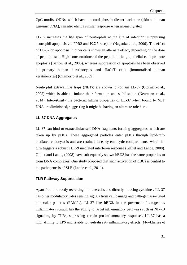
Chapter 1
31
CpG motifs. ODNs, which have a natural phosphodiester backbone (akin to human
genomic DNA), can also elicit a similar response when un-methylated.
LL-37 increases the life span of neutrophils at the site of infection; suppressing
neutrophil apoptosis via FPR2 and P2X7 receptor (Nagaoka et al., 2006). The effect
of LL-37 on apoptosis in other cells shows an alternate effect, depending on the dose
of peptide used. High concentrations of the peptide in lung epithelial cells promote
apoptosis (Barlow et al., 2006), whereas suppression of apoptosis has been observed
in primary human keratinocytes and HaCaT cells (immortalised human
keratinocytes) (Chamorro et al., 2009).
Neutrophil extracellular traps (NETs) are shown to contain LL-37 (Ciornei et al.,
2005) which is able to induce their formation and stabilisation (Neumann et al.,
2014). Interestingly the bacterial killing properties of LL-37 when bound to NET
DNA are diminished, suggesting it might be having an alternate role here.
LL-37 DNA Aggregates
LL-37 can bind to extracellular self-DNA fragments forming aggregates, which are
taken up by pDCs. These aggregated particles enter pDCs through lipid-raft-
mediated endocytosis and are retained in early endocytic compartments, which in-
turn triggers a robust TLR-9 mediated interferon response (Gilliet and Lande, 2008).
Gilliet and Lande, (2008) have subsequently shown hBD3 has the same properties to
form DNA complexes. One study proposed that such activation of pDCs is central to
the pathogenesis of SLE (Lande et al., 2011).
TLR Pathway Suppression
Apart from indirectly recruiting immune cells and directly inducing cytokines, LL-37
has other modulatory roles sensing signals from cell damage and pathogen associated
molecular patterns (PAMPs). LL-37 like hBD3, in the presence of exogenous
inflammatory stimuli has the ability to target inflammatory pathways such as NF-κB
signalling by TLRs, supressing certain pro-inflammatory responses. LL-37 has a
high affinity to LPS and is able to neutralise its inflammatory effects (Mookherjee et
Chapter 1
32
al., 2006). RAW cells treated with LPS, a TLR4 agonist, release less TNF and NO
when treated with LL-37, hence supressing inflammation (Scott et al., 2002).
Extracellular LL-37, and the mouse orthologue CAMP, can form complexes with
LPS by directly binding to it; these complexes prevent LPS interaction with TLR-4
signalling molecules (Scott et al., 2000, Scott et al., 2011). The formation of these
complexes is due to the physicochemical properties of LL-37/CAMP, such as its
charge, helicity, amphipathicity and hydrophobicity. It has also been suggested that
LL-37 can exhibit anti-endotoxic effects by binding the CD14 receptor directly, or by
dissociating LPS oligomers; both mechanisms prevent binding to the LPS binding
receptors (LBP) (Suzuki et al., 2011).
1.2.2.5 Wound Healing and Tissue Remodelling
As previously mentioned LL-37 can activate the FPR2 and P2X7 receptors on
epithelial cells and fibroblasts. It is proposed that this results in a similar proliferative
effect as EGFR activation. In endothelial cell lines, it has also been suggested that
LL-37 acts on the FPR2 receptor to induce angiogenesis (Salvado et al., 2013). One
study showed electroporation of a LL-37 plasmid in an ischemic hind limb model
upregulated antigenic chemokines, and also reduced muscular atrophy (Steinstraesser
et al., 2014).
1.2.3 HDPs and the Female Reproductive Tract
Cells in the female reproductive tract are a major source of multiple microbicides.
Reproductive tract epithelial cells, cervical glands, and neutrophils generate a wide-
range of HDPs that target viruses, bacteria and fungi, both directly and indirectly
(Figure 1.6).
LL-37 is found at high levels in fetal skin, amniotic fluid, vaginal fluid and the
vernix caseosa (reviewed in Yarbrough et al., (2015)). Steady state defensin
expression is cell specific and compartmentalised in the female reproductive tract in
Chapter 1
33
pregnant and non-pregnant women. hBD1-4 and human alpha defensin 5 (HD5) have
all been reported to be expressed with distinctive temporal profiles in the endometrial
epithelium, vagina and cervix (King et al., 2003a, Fleming et al., 2003, Quayle et al.,
1998). The kinetics of this differential expression most likely reflects the differing
immunological requirements along the length of the female reproductive tract.
In women, HDPs are known to show temporal expression patterns during the
menstrual cycle (King et al., 2003b). The mechanism of menses shares many
characteristics of acute inflammatory processes, involving localised increase in pro-
inflammatory cytokines, prostaglandins, and leukocyte migration. In women, hBD3
is higher in the early and late secretory phase of the menstrual cycle (Fleming et al.,
2003). Hickey et al., (2013) found that in non-pregnant mice Defb14 also had
specific estrus cycle mRNA expression profiles in the vagina and uterus. Like hBD3,
LL-37 is not uniformly expressed throughout the cycle. Mice null for Camp, the
murine orthologue of LL-37/hCAP18 have increased susceptibility to bacterial
urinary tract infections (Chromek et al., 2006). Defb1 homozygous knockout mice
also have increased susceptibility to urinary tract infections whereby they were
shown to retain Staphylococcus spp. in the bladder longer than wildtype mice
(Morrison et al., 2002).
Like LL-37, multiple alternative fragments of hBD1 with varying amino-acid termini
have been found in the human reproductive and urogenital system (Valore et al.,
1998, Zucht et al., 1998, Valore et al., 2002). These alternative forms have been
isolated in vaginal lavage fluids and urine, where they appear to have different levels
of antibacterial activity under specific conditions (Zucht et al., 1998).
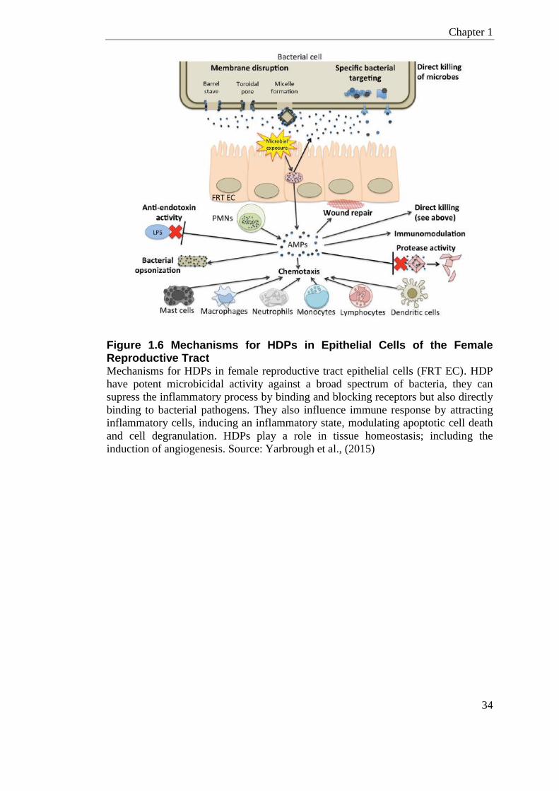
Chapter 1
34
Figure 1.6 Mechanisms for HDPs in Epithelial Cells of the Female Reproductive Tract Mechanisms for HDPs in female reproductive tract epithelial cells (FRT EC). HDP
have potent microbicidal activity against a broad spectrum of bacteria, they can
supress the inflammatory process by binding and blocking receptors but also directly
binding to bacterial pathogens. They also influence immune response by attracting
inflammatory cells, inducing an inflammatory state, modulating apoptotic cell death
and cell degranulation. HDPs play a role in tissue homeostasis; including the
induction of angiogenesis. Source: Yarbrough et al., (2015)
Chapter 1
35
1.2.4 HDPs and Preterm Labour
The development of preventative therapies for preterm labour requires a mechanistic
understanding of how the pro-inflammatory profile is induced. HDPs have been
shown to have multiple roles in this pro-inflammatory process.
Researchers have found that an immune genotype variation in human DEFB1 may
contribute clinically to women who deliver preterm (James et al., 2013). This
retrospective genotyping study highlighted rs1799946, a SNP in DEFB1, to be
associated with a doubled risk of preterm delivery and four-fold increase in the risk
of extreme preterm delivery (<28 weeks) (James et al., 2013). A second SNP,
rs1047031, was found to be protective. However, a larger GWAS study, which
included a different SNP in the DEFB1 gene, failed to highlight an association of this
locus to preterm labour (Uzun et al., 2013).
Women with elevated concentrations of cervicovaginal HNPs at 24-29 weeks of
gestation have a greater risk of delivering preterm (before 32 weeks) (Balu et al.,
2003). Further work on human extra-placental membrane in ex-vivo cultures has
shown increased hBD2 in the amnion when the choriodecidua were exposed to group
B streptococcus (GBS) (Boldenow et al., 2013).
An in-vitro study on human gingival fibroblasts found that hBD3 is pro-
inflammatory and increases both COX-2 expression and prostaglandin E2 (PGE2).
COX-2 is an inducible labour associated gene, which is expressed at term labour in
the human myometrium and also in rabbit cervical fibroblasts (Sato et al., 2001). The
importance of this effect in cervical and myometrial stromal fibroblasts in mouse and
human has yet to be determined.
As previously discussed, the active form of Vitamin D is a potent inducer of
CAMP/LL-37. LL-37 is upregulated by Vitamin D in placental explants and primary
human trophoblasts in a dose-dependent manner (Liu et al., 2009). This response is
similar to that observed in primary cultures of human macrophages (Martineau et al.,
2007). hBD2 but not hBD3 also shows a similar Vitamin D response in keratinocytes
(Wang et al., 2010).

Chapter 1
36
1.3 Mouse Models of Preterm Labour
Nearly 40% of preterm deliveries are associated with intrauterine infection (Romero
et al., 2006). Mouse preterm labour models have focused on stimulation with agents
to create an infection and/or inflammatory state; such as heat-killed E.coli, heat
killed bacteria, LPS, group B streptococci (GBS), Lipoteichoic acids (LTA), TNF
and IL-1β (Elovitz et al., 2003). Stimulation of the TLR4 pathways with LPS has
been the central method of invoking preterm labour in animal models (Table 1.3).
Live and heat-killed bacteria have also been used for preterm-labour models,
however the reproducibility has been reported to be low (Reznikov et al., 1999). In
humans, amniotic fluid cultures from most preterm births are negative for bacteria
(Romero et al., 2007), although amniotic inflammatory cytokines and LPS binding
proteins have been shown to be increased (Gardella et al., 2001). This lack of
bacterial detection could be due to the limitations of the culture-dependent detection
method. There is however, evidence to support signal transduction to the amnion
when the choriodecidua is stimulated with bacteria (Boldenow et al., 2013), this
could account for the increase in inflammatory response in the amnion without direct
bacterial invasion.
The mode of agent administration has a large impact on the immune response
mounted against the endogenous stimulating agent in animal models; intravaginal
(i.vag), intraperitoneal (i.p) intracervical (i.c) intrauterine (i.u) have all been used as
routes of administration.
The i.p model of preterm labour most likely mimics that associated with sepsis and
severe bacteraemia (Elovitz et al., 2003). As most women with preterm labour do not
present with elevated white blood cells or symptoms of illness, a localised model of
inflammation at the maternal-fetal interface would likely recapitulate what is seen in
women. In addition, systemic models of induced preterm-birth in mice are highly
variable, with high incidence of maternal morbidity (Aisemberg et al., 2013).
A more recent model for preterm labour involves intrauterine (i.u) administration of
an agent; this involves performing a mini-laparotomy in the late stage of pregnancy
Chapter 1
37
to expose the gestational uterine horn and inject between two fetal sacs.
Administering LPS using this method is reported to induce high preterm labour rates
while maintaining low maternal mortality. However, the biological relevance of
delivering a bolus of purified LPS into the uterus in the hope of mimicking human
ascending bacterial infection could be questioned.
There has been one study that used medical endoscope to navigate through the
murine cervix to administer infectious agents (Reznikov et al., 1999), however, due
to the dense mucus plug and small opening, results were highly variable and others
have failed to replicate this method.
Reznikov et al., (1999) developed a mouse model of infection/inflammation-induced
preterm labour by injecting E.coli intracervical (i.c) in pregnant mice. In this model,
the expression of fetal and/or maternal cytokines was demonstrated to up-regulate a
number of uterine factors such as prostaglandin hormones, prostaglandin receptors
and matrix metalloproteinases, which leads to premature initiation of parturition.
There has been some work on an intravaginal (i.vag) model of preterm labour, where
the agent is applied distal to the cervix and causes preterm labour by transcending the
cervix and/or through signal transduction (Gonzalez et al., 2011b, Gonzalez et al.,
2011a). The mucus plug during pregnancy is very dense and the quantity of agent
breeching this barrier is likely to be small, therefore higher loads of agent are needed.
There is variability across animal studies which use LPS as the inflammatory agent,
this could be due to several compounding factors, such as serotype of the LPS used
(Migale et al., 2015). The gestational age at which LPS is administered to induce
preterm labour is also a factor in animal models as early administration can lead to
resorption (Aisemberg et al., 2013).
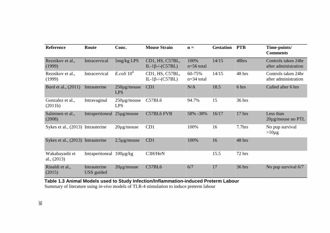
38
Reference Route Conc. Mouse Strain n = Gestation PTB Time-points/
Comments
Reznikov et al.,
(1999)
Intracervical 5mg/kg LPS CD1, HS, C57BL,
IL-1β-/-(C57BL)
100%
n=56 total
14/15 48hrs Controls taken 24hr
after administration
Reznikov et al.,
(1999)
Intracervical E.coli 104 CD1, HS, C57BL,
IL-1β-/-(C57BL)
60-75%
n=34 total
14/15 48 hrs Controls taken 24hr
after administration
Burd et al., (2011) Intrauterine 250µg/mouse
LPS
CD1 N/A 18.5 6 hrs Culled after 6 hrs
Gonzalez et al.,
(2011b)
Intravaginal 250µg/mouse
LPS
C57BL6 94.7% 15 36 hrs
Salminen et al.,
(2008)
Intraperitoneal 25µg/mouse C57BL6 FVB 58% -38% 16/17 17 hrs Less than
20µg/mouse no PTL
Sykes et al., (2013) Intrauterine 20µg/mouse CD1 100% 16 7.7hrs No pup survival
>10µg
Sykes et al., (2013) Intrauterine 2.5µg/mouse CD1 100% 16 48 hrs
Wakabayashi et
al., (2013)
Intraperitoneal 100µg/kg C3H/HeN 15.5 72 hrs
Rinaldi et al.,
(2015)
Intrauterine
USS guided
20μg/mouse C57BL6 6/7 17 36 hrs No pup survival 6/7
Table 1.3 Animal Models used to Study Infection/Inflammation-induced Preterm Labour Summary of literature using in-vivo models of TLR-4 stimulation to induce preterm labour
Chapter 1
39
1.4 Microbiome
Researchers have often quoted that the numbers of microbial cells in the human body
easily out-number the human cells 10:1. New data suggested this was a vast
overestimate and the number of human and bacterial cells in an average adult male is
actually about equal 1:1 (Sender and Milo, 2016). Nonetheless, bacterial cells offer a
plethora of microbial genes, providing important metabolic functions that have
specific selections based on co-adaptions.
The term ‘Microbiome’ refers to the full composition of genetic material of all the
microorganisms in a community. This includes the widely reported prokaryotic
entities but also eukaryotic, archaea and viral organisms, more frequently being
referred to as the ‘virome’. This thesis interrogates the prokaryotic organisms of the
microbiome using the general term bacteria.
Bacteria are an essential part of human health; they break down un-digestible food
fibres, and also provide a source of Vitamin K and Biotin. Many selections in the
bacterial gene complement can be induced by alterations in the host-environment,
such as antibiotic exposure (Langdon et al., 2016), dietary changes (Aron-
Wisnewsky and Clement, 2016) and hormone regulation (Sudo, 2014). Selective
metabolic functions also occur in other animal species. Rodents lacking exposure to
microorganisms (germ free) display severely stunted growth, reduced cardiac output,
reduced cell proliferation and other pathologies (Schwarzer et al., 2016, Blanton et
al., 2016).
The majority of the time the relationship between host and bacteria is synergistic,
existing in perfect homeostasis. However, disruptions or aberrations in this
relationship can sometimes cause pathologies and disease. Pathogenic
microorganisms are not the only causes of disease; disruption in commensal bacteria
that are typically held in check by the microbial network and host immune defence
factors can also be causative.
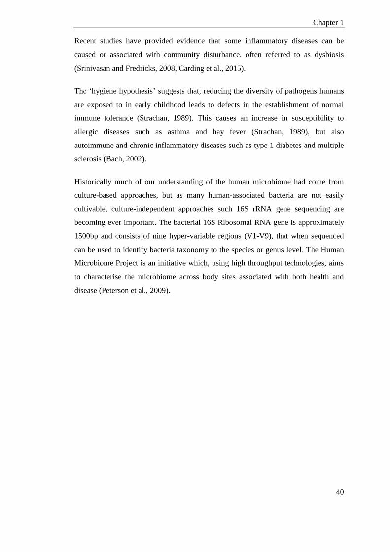
Chapter 1
40
Recent studies have provided evidence that some inflammatory diseases can be
caused or associated with community disturbance, often referred to as dysbiosis
(Srinivasan and Fredricks, 2008, Carding et al., 2015).
The ‘hygiene hypothesis’ suggests that, reducing the diversity of pathogens humans
are exposed to in early childhood leads to defects in the establishment of normal
immune tolerance (Strachan, 1989). This causes an increase in susceptibility to
allergic diseases such as asthma and hay fever (Strachan, 1989), but also
autoimmune and chronic inflammatory diseases such as type 1 diabetes and multiple
sclerosis (Bach, 2002).
Historically much of our understanding of the human microbiome had come from
culture-based approaches, but as many human-associated bacteria are not easily
cultivable, culture-independent approaches such 16S rRNA gene sequencing are
becoming ever important. The bacterial 16S Ribosomal RNA gene is approximately
1500bp and consists of nine hyper-variable regions (V1-V9), that when sequenced
can be used to identify bacteria taxonomy to the species or genus level. The Human
Microbiome Project is an initiative which, using high throughput technologies, aims
to characterise the microbiome across body sites associated with both health and
disease (Peterson et al., 2009).
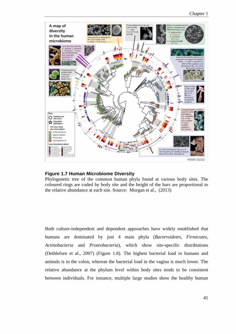
Chapter 1
41
Figure 1.7 Human Microbiome Diversity Phylogenetic tree of the common human phyla found at various body sites. The
coloured rings are coded by body site and the height of the bars are proportional to
the relative abundance at each site. Source: Morgan et al., (2013)
Both culture-independent and dependent approaches have widely established that
humans are dominated by just 4 main phyla (Bacteroidetes, Firmicutes,
Actinobacteria and Proteobacteria), which show site-specific distributions
(Dethlefsen et al., 2007) (Figure 1.8). The highest bacterial load in humans and
animals is in the colon, whereas the bacterial load in the vagina is much lower. The
relative abundance at the phylum level within body sites tends to be consistent
between individuals. For instance, multiple large studies show the healthy human
Chapter 1
42
colon is dominated by Bacteroidetes and Firmicutes (Dethlefsen et al., 2007, Lay et
al., 2005, Eckburg et al., 2005). The composition in the vagina at the phylum level
shows a little more variation, most women are dominated by just Firmicutes in the
vagina, but some women have a dominance of Actinobacteria phyla (Dethlefsen et
al., 2007).
Figure 1.8 The Human Microbiome; Site Specific Distribution of Phyla Coloured proportions represent the percentage of each 4 main phyla found in
different body-sites as determined by 16S rRNA gene sequencing (n=11 women).
The area of the pie chart somewhat represents the bacterial load, the number in
parentheses shows the mean number of phylotypes (approximated species level taxa).
Source: Dethlefsen et al., (2007)
Chapter 1
43
1.4.1 The Vaginal Microbiome
1.4.1.1 Vaginal Microbiome Development
The mode of delivery (caesarean or vaginal) has impact on the baby’s whole-body
biota, including the gastrointestinal tract, skin and first colonisation of the baby’s
reproductive tract (Dominguez-Bello et al., 2010).
Through childhood, the vaginal microbiota is sparsely populated with a variety of
anaerobic bacteria, with few or no Lactobacilli spp. (Hammerschlag et al., 1978, Hill
et al., 1995). In adolescence estrogen levels rise and Lactobacilli spp. become more
prevalent while anaerobic populations begin to diminish (Thoma et al., 2011,
Alvarez-Olmos et al., 2004). At reproductive age, estrogen cycling stimulates the
thickening of the vaginal epithelium and in turn glycogen production, which is when
Lactobacilli spp. become most abundant. Lactobacilli spp. help to maintain low pH
by metabolising glycogen to produce lactic acid (Linhares et al., 2011). There is
evidence that amylase is a key component in this process by breaking down glycogen
into smaller components for Lactobacillus spp to metabolise (Spear et al., 2014). At
menopause estrogen levels decline, the vaginal epithelium thins, Lactobacilli spp.
diminish and anaerobes become more prevalent again (Muhleisen and Herbst-
Kralovetz, 2016). This is akin to the adolescent microbial profile suggesting a strong
hormonal factor in driving microbial composition.
1.4.1.2 Vaginal Microbiome Stability
Most of our understanding of the vaginal microbiome has come from reproductive
age women; fewer studies have been done on the stability of the vaginal microbiome
in childhood or post-menopause.
In a large study Ravel et al., (2011) recruited 396 healthy women across 4 ethnic
groups, and sequenced their vaginal bacteria to assign taxonomic species. Bacterial
profiling in these 396 women confirmed variability of bacterial at the phyla level
between individuals. However, hierarchal clustering at the species level showed the
bacteria complement could be classified into so-called community state types (CST),

Chapter 1
44
of which there are 5. These community state types are defined by the dominance or
absence of certain Lactobacillus spp. (Table 1.4). Ravel et al., (2011) concluded at
95% confidence that women in North America will fit into one of these community
state types. Nonetheless, it still remains that one in every 20 women will not have a
bacteria profile which fits into one of these 5 groups.
Community State Type Group Dominant Bacteria/ Defining Feature
Group I Lactobacillus crispartus
Group II Lactobacillus gasseri
Group III Lactobacillus iners
Group IV Little/no Lactobacillus spp.,
Atopobium, Prevotella, Sneathia, Gardnerella
Group V Lactobacillus jenseni
Table 1.4 Human Vaginal Community State Types (CST) Vaginal community state type group of reproductive age women. Summary as
described by Ravel et al., (2011)
CST IV, which is a group defined as having very little Lactobacillus spp., is
interesting concerning vaginal homeostasis. Women with CST IV have a high
Nugent score (Gram stain scoring system from vaginal swabs) because of the lack of
Lactobacilli spp., however they are reported healthy and have not presented with
symptomatic Bacterial Vaginosis (BV), which is dysbiosis of the vaginal microbiota.
This is interesting as it highlights that the metabolic functions of bacteria in the
vagina are most likely conserved. Lactobacillus spp. are not the only bacteria to
produce lactic acid; Streptococcus spp. and Megaspherea spp. are also lactic acid
producing bacteria (LAB). Community sate types vary across ethnic groups (Ravel et
al., 2011); CST IV has a more frequent occurrence in black and Hispanic
populations, which interestingly is also the ethnic group which displays the highest
incidences of BV and PTL (Burris et al., 2011, Ness et al., 2003).
Longitudinal sampling (16 weeks) of women’s vaginal bacteria show that CST is not
a stable trait (Gajer et al., 2012). Women can transition between CST; some shifts are
temporal and consistent but others are more permanent. What factors are responsible
Chapter 1
45
for driving these dynamic shifts remains to be determined. It is likely that estrogen,
mechanism of menses, host genetics, microbicides and environmental factors all
might play a role. Knowledge of CST kinetics is important because each community
state type might differ in a risk to invasive diseases. A women’s relative risk
therefore might be a factor of the duration spent in the more vulnerable community
state types or transitioning between them.
1.4.1.3 Vaginal Microbiome Dysbiosis
Bacterial Vaginosis (BV) is a dysbiosis of the normal proportions of commensal
microbial species in the vagina, commonly shifting towards a depletion of
Lactobacilli spp., which are proposed to be the protective species in the healthy
vagina. BV affects 30% of women of reproductive age, has a high rate of
reoccurrence and is associated with a high risk of secondary infections (Lambert et
al., 2013).
Bacterial Vaginosis is a disease associated with increased inflammatory cytokines in
cervicovaginal secretions and mucosal epithelial cell damage (reviewed in Kumar et
al., (2011)). It has been proposed that this damage to the vaginal epithelial surface
may play a role in the increased susceptibility of women with BV to sexually
transmitted secondary infections/disease such as Neisseria gonorrhoeae, Chlamydia
trachomatis and HIV (Taha et al., 1998, Wiesenfeld et al., 2003). A recent study
suggests that the shedding of epithelial cells seen in patients with BV may be caused
by a single organism, Gardnerella vaginalis (Gilbert et al., 2013).
Genetic variation in immune-regulatory genes such as TLR-4, TLR-9, IL-8, IL-6 and
TNF are associated with incidences of BV, suggesting host-immune genotype is
important in the disease (Royse et al., 2012, Macones et al., 2004).
The standard treatment for BV in the UK is a course of oral/intravaginal antibiotics,
commonly metronidazole or in some cases clindamycin. As most women following
this treatment course will clinically present with recurrent BV, metronidazole is
partly ineffective as a treatment (Menard, 2011).
Chapter 1
46
1.4.2 Vaginal Microbiome in Pregnancy
Using bacterial 16S rRNA gene sequencing Aagaard et al., (2012) compared the
vaginal microbiome of pregnant women to those women not pregnant. Aagaard et al.,
(2012) showed that the pregnant bacterial complement is different, being less diverse
to those women not pregnant. Romero et al., (2014) sampled women through
pregnancy and classified their community state types as described by Ravel et al.,
(2011). The authors found the vaginal community of pregnant women differed to that
of non-pregnant women; having a higher abundance of Lactobacillus
vaginalis, Lactobacillus gasseri, Lactobacillus crispatus, and Lactobacillus jensenii.
CSTs in pregnant women were also more stable than non-pregnant women; showing
less CST shifts, which rarely included CST IV (group with little Lactobacillus spp.).
MacIntyre et al., (2015) included vaginal samples taken after delivery and showed
the postpartum community has a rapid decrease in Lactobacillus spp. The authors
displayed Prevotella and Anaerococcus spp. were increased in replacement of the
diminished Lactobacilli in a high proportion of postpartum women. It was proposed
the quick drop in estrogen levels postpartum is causing this alteration in community
structure.
1.4.2.1 Bacterial Vaginosis in Pregnancy
Although not generally a reportable disease, the occurrence of BV in pregnancy has
been estimated to be around 50% (Nelson and Macones, 2002). There have been
many studies that link dysbiosis of the vaginal microbiota, clinical BV and preterm
labour (Gravett et al., 1986, Svare et al., 2006). BV identified before 20 weeks
gestation has been indicated in preterm delivery and chorioamnionitis (Svare et al.,
2006). There is increasing support that certain prophylactic antibiotics given very
early in pregnancy to those women who have a clinical diagnosis of BV has benefit
(Thinkhamrop et al., 2015).
Bacterial vaginosis is associated with lower vaginal concentrations of hBD3, but not
hBD2 or HNP-3, in pregnant women (Balu et al., 2003, Mitchell et al., 2013).
Interestingly, maternal Vitamin D deficiency is associated with BV in the first
Chapter 1
47
trimester of pregnancy, and Vitamin D can induce hCAP18/LL-37 and hBD2 gene
expression in multiple cells (Wang et al., 2004b).
1.4.2.2 Vaginal Microbiome and Preterm Labour
Many bacterial species have been isolated from the amniotic fluid, placenta and
chorion/amnion from pregnancies that have delivered preterm. The most common
species observed are commensal bacteria from the lower genital tract and bacteria
associated with bacterial vaginosis. Other bacteria such as those associated with
STI’s and the gingival cavity have also been isolated from amniotic samples (Han et
al., 2004, Mitchell et al., 2013).
Hyman et al., (2014) studied the vaginal microbial diversity of women at high and
low risk of preterm labour. The authors concluded that there were differences in the
overall diversity between the two risk groups; showing a correlation between high
alpha diversity and preterm labour (Hyman et al., 2014). As previously mentioned
the type of material used in a cervical cerclage can have an impact on the vaginal
microbiome (Kindinger et al., 2016). Using braided sutures over monofilament
sutures increases the risk of women developing vaginal dysbiosis, inflammation, and
preterm labour (Kindinger et al., 2016). Taken together, this supports the hypothesis
that infection-induced preterm labour is associated with vaginal disruption of
commensal bacteria as well as invading pathogens.
1.4.3 Modulation of Vaginal Microbiome
Vaginal Lactobacilli dominance in reproductive age women could be a protective
mechanism to reduce pathogenies in the newborn infant. A baby’s first whole-body
microbiome is highly correlated with the organisms exposed to at the peri-partum
period. Those babies born by caesarean-section have microbiota resembling the
mothers’ skin, whereas those born by natural delivery have microbiotas resembling
the mother’s vagina. Multiple studies suggest that caesarean-section babies have a
higher incidence of allergic disease such as asthma, where the incidence was 20%
Chapter 1
48
higher in caesarean section children (Sevelsted et al., 2016, Sevelsted et al., 2015).
Caesarean section babies also pick-up more MRSA infections following birth than
vaginally delivered babies (CDC, 2006).
If the types of microbes in the mother’s vagina are pathogenic at the time of delivery,
these can also be passed on to baby. Lactobacillus dominance during pregnancy
could therefore have selective protective effects. Indeed as previously mentioned,
Romero et al., (2014) had shown that Community state type IV (group defined by
little/no Lactobacillus spp.) transition and occurrence was rarely seen in pregnant
women.
Probiotics have been considered as a therapy to colonise the vagina, by preventative
prophylaxis or to treat dysbiosis/pathogenic infection. Probiotics have the ability to,
selectively displace pathogenic organisms, provide metabolic compounds of different
kinetics, and directly compete with the resident bacteria. In-vitro studies have shown
that Lactobacillus spp. can disrupt biofilms of common BV associated bacteria and
inhibit the growth of urogenital pathogens including yeast. Clinical studies have
taken Lactobacillus spp. probiotics into women as a therapy for BV (Bradshaw and
Brotman, 2015). The most promising Lactobacillus spp. in these studies is
Lactobacillus rhamnosus GR-1, and Lactobacillus reuteri RC-14 (Martinez et al.,
2009). Studies have shown that orally administered Lactobacillus rhamnosus GR-1,
and Lactobacillus reuteri RC-14 can be recovered from the vagina (Reid and Burton,
2002), highlighting possible fecal-vaginal transfer of microorganisms. Combinations
of these bacteria are currently sold as oral live-cultures to promote urogenital
microbial health.
1.4.4 Murine Vaginal Microbiome
Animals offer researchers a vital tool by providing a model to test out host-
microbiome interactions under controlled environmental conditions. They also allow
for infectious models that help understand pathogenicity. Mice are used in many
Chapter 1
49
reproductive tract disease investigations, such as humanised mice as a model to study
vaginal transmission of HIV (Deruaz and Luster, 2013).
There has been very little work done on the commensal bacteria of the mouse
reproductive tract, presumably due to its extremely low bacterial load and
dependency on microbial burden in each animal facility.
Previous bacterial species identified to reside in the murine reproductive tract has
been through culture-dependent techniques (Noguchi et al., 2003). More work has
focused on colonisation and clearance of exogenous bacteria both in a probiotic
approach and in infectious models (Reid and Burton, 2002, Meysick and Garber,
1992).
There is very little known about the species level richness of bacteria that colonise
the murine vaginal mucosa. Publications that use techniques such as qPCR and
culture-dependent methods have shown that in contrast to humans, only a small
percentage of female mice harbour Lactobacillus species at the vaginal mucosa
(Noguchi et al., 2003, McGrory and Garber, 1992). Such culture-dependent studies
listed Streptococci, gram-negative rods, Bacteroidaceae and Staphylococci as found
in the non-pregnant vagina, however, these observations were not consistent and
were highly variable (Noguchi et al., 2003).
MacManes (2011) conducted a study looking at two different strains of wild mice,
one known to be promiscuous and the other monogamous. By using 16S rRNA gene
cloning and sanger sequencing MacManes (2011) showed that there was a greater
degree of diversity in the vaginal bacteria of the promiscuous strain of mice.
After review of the literature, only one study attempts to categorise the vaginal
microbiome of lab animals using high throughput16S rRNA gene sequencing
(Barfod et al., 2013). Barfod et al., (2013) study highlighted that the vaginal
community of BALB/c mice is highly variable. Although the authors only had n=8
samples they showed the vaginal community had two sub-clusters. One sub-cluster
appeared to be closely related to the lung microbiota, whereas the other sub-cluster
did not show similarity to either of the other two sample sites the authors
Chapter 1
50
investigated (lung and cecum). Streptococcus (Gram-positive) was attributed to the
separation of the two clusters; the authors also listed Acinetobacter, Sphinogmonas,
Enterococcus and Polaromonas as genera from the vaginal mucosa.
1.4.5 The Gut Microbiome
Microbes in the gut are essential for the break down of complex carbohydrates where
they can manipulate host metabolism by altering nutrient availability. The presence
and abundance of each bacterial group is fundamental since different species
differentially metabolise these nutrients.
The stomach has the lowest bacterial loads of the gastrointestinal tract, where
stomach acids maintain low bacteria numbers, the load then increases further from
the stomach as pH levels become less acidic and more favourable for anaerobic
growth. The mammalian gastrointestinal tract is dominated by two main phyla,
Firmicutes and Bacteroidetes. Shifts in the ratio of these two phyla have been
observed in many conditions such as obesity (Ley et al., 2006, Ley et al., 2005). Diet,
age, host genetics, and local immune defence also influence on the community
composition of these phyla (Mariat et al., 2009, Ussar et al., 2015, Salzman et al.,
2007). The Human Microbiome Project is an initiative which, using high throughput
technologies, aims to characterise the microbiome across body sites associated with
both health and disease (Peterson et al., 2009).
The gut epithelium, as the outermost single cell layer of the gut, secretes many
antimicrobial peptides including LL-37, defensins, but also S100 proteins, and C-
type leptins. Speciality cells such as Paneth cells also selectively secrete HDPs such
as the human alpha defensin 5 (HD5) and human alpha defensin 6 (HD6).
Interestingly HD6 has the ability to polymerize forming nanonets that are believed to
prevent bacteria translocating across the gut wall (Chu et al., 2012). These Paneth
cell defensins have also been shown to have anti-viral properties (Wang et al., 2013,
Hazrati et al., 2006).
Chapter 1
51
Gut epithelia cells have multiple pattern recognition receptors, including the
transmembrane TLRs, and also intracellular NOD receptors (Lavelle et al., 2010).
These pattern recognition receptors are not uniformly expressed along the
gastrointestinal tract. This is most probably a co-adaption strategy, to allow host
tolerances of commensal and mutualistic species. For example in the gut, in a steady
state, the numbers of TLR-4 and TLR-2 molecules are reduced, whereas TLR-3
(viral ligand) and TLR-5 (flagellin ligand) are more abundant. Another co-adaption
strategy to allow tolerances of commensals is the mucus barrier; this layer reduces
the ability of the effector molecules to sense bacteria. The colonic mucus barrier has
two constantly renewing layers; the innermost is predominantly kept sterile.
Germ-free mice have shown that bacteria are essential for the development of normal
gut epithelium. The major adaptor molecule MyD88 is essential for signalling via
TLRs. The gut epithelia of MyD88 knockout mice show abnormal development
(Frantz et al., 2012) and mice are consequently more susceptible to DSS colitis
(Araki et al., 2005). This was supported in a conditional MyD88 knockout which
displayed down regulation of mucus genes, antimicrobial peptides, and had
compromised antibacterial immunity (Frantz et al., 2012).
In the absence of infection and inflammation beta-defensins in humans and mice are
not prominently expressed in the small intestine, although in the colon beta-defensins
have been shown to be important (Meyer-Hoffert et al., 2008).
Crohn's and IBD are linked to key deficiencies in HD5 and HD6 Paneth cell function
(Wehkamp and Stange, 2010). Impaired induction of hBD2 and hBD3 have also
been characterised in Crohn's disease (Wehkamp et al., 2003, O'Neil et al., 1999).
Genomic variation in several defensin genes have been linked to the disease.
Genome wide CNV profiling highlighted that low hBD2 copy number predisposes to
Crohn’s disease (Fellermann et al., 2006), while a SNP in the DEFB1 gene (encodes
hBD1) was associated with an increased risk (Kocsis et al., 2008).
LL-37/Camp is upregulated during ileitis/colitis dependent on the level of
inflammation (Schauber et al., 2006), and mice null for Camp have more severe
phenotype in a DSS induced colitis model (Koon et al., 2011, Tai et al., 2013).
Chapter 1
52
Addition of exogenous CAMP could reverse some of the phenotypic severity of DSS
treatment in Camp-/- animals, such as cytokine production, apoptosis and mucus
production (Tai et al., 2013). Intracolonic CAMP peptide treatment in mice pre-
infected with Clostridium difficile also show large improvements in phenotypic
consequences of the pathogen (Hing et al., 2013). Mice showed better colonic
histology score, reduced apoptosis and supressed induction of myeloperoxidase
(MPO) and TNF-α. Although, endogenous cathelicidin was not shown to have
protective benefits in a similar model which delivered Clostridium difficile toxin A,
to Camp-/- and wildtype mice (Hing et al., 2013), suggesting LL-37 has more effect
on live bacteria in this model.
Graft versus host disease, a possible deadly side effect of bone marrow and stem cell
transplants, targets Paneth cells and causes subsequent impairment of defensin
secretion in mice; including Defa1, Defa4, and Defa5 (Eriguchi et al., 2012).
Interestingly, giving Blautia spp. to mice could protect them from graft versus host
disease (Jenq et al., 2015).
There has been direct evidence linking gut alpha-defensins to the modulation of
microbiota in mice. Salzman et al., (2010) demonstrated that Paneth cell alpha-
defensins contributed to mucosal host defence and regulate the composition of
murine intestinal microbiota. Mice deficient in defensins displayed increased
Firmicutes and decreased Bacteriodes in the gut. Whereas mice expressing the
human defensin (HD5) have Firmicutes decreased and increased Bacteriodes when
compared to wildtype littermates.
Chapter 1
53
1.4.6 Gut Microbiome in Pregnancy
There are vast metabolic changes that occur during pregnancy, especially in late
gestation, which could affect microbial communities. Different points in gestation
have different biochemical and physiological changes that could also have temporal
and dynamic impact on the microbiome. Indeed, alterations in the maternal
gastrointestinal microbiome have been implicated in metabolic adaptations to
pregnancy (Koren et al., 2012, Collado et al., 2008). Body weight is an obvious
contributing factor; pre-pregnancy weight has been linked to microbial change,
where obese mothers display higher bacterial diversity (Collado et al., 2008). Koren
et al., (2012) demonstrated that the gut microbiome of pregnant women changed
dramatically between the first and the third trimester, showing an increase in inter-
individual diversity. The authors also showed that the bacterial load of the gut
changed with gestation; increasing from the first to the third-trimester. Conflicting
reports whether pregnancy is truly connected with substantial shifts in gut bacteria,
has come from a larger longitudinal study that failed to show significant remodelling
of the gut microbiota in pregnant women (DiGiulio et al., 2015).
Aims and Hypothesis
54
Aims and Hypothesis
The work in this thesis is based on the hypothesis that Host Defence Peptides play a
fundamental role in mucosal immune defence. In a pregnant state, the hypothesis is
that HDPs have a dual role in preventing ascending infection, but also preventing an
exacerbated inflammatory response that can cause preterm birth by initiation of the
labour cascade.
To explore this I determine whether bacterial stimuli can regulate HDPs expression
in pregnancy tissues. I also explore what interactions HDPs have on the
production/induction of important cytokines, which are vital to the inflammatory
response.
As hBD3 and LL-37 suppress the inflammatory response of cells to LPS through
TLR-4, the hypothesis is that in-vivo they might offer protection from preterm birth
caused by inflammation. With the aid of HDP knockout mice, the role of these
peptides in infection/inflammation and continuation of pregnancy is investigated in a
mouse-model of induced preterm-labour.
Infection-induced preterm labour in women is associated with vaginal disruption of
commensal bacteria as well as invading pathogens. To understand how ascending
infection might be controlled by HDPs in pregnancy, I explore how HDPs regulate
commensal bacteria. This is achieved by interrogating the maternal microbiome at
mucosal sites in HDP knockout mice, utilising the bacterial 16S rRNA gene and next
generation sequencing.

55
Chapter 2
General Materials and Methods
Chapter 2
56
Chapter 2 General Materials and Methods
2.1 Animal Experiments
All mice were sourced ‘in-house’ and kept under specific pathogen free (SPF)
conditions at the University of Edinburgh. Mice were permitted food and water ad
libitum and maintained in 12-hour cycle of light and dark. Animal experiments were
conducted under a UK Home Office licence in line with the Animals Scientific
Procedure Act (1986). For full details of reagents and supplies, see Appendix on
page 314.
2.1.1 Fertility Experiments
Virgin female mice were set-up in mating pairs within genotype with a stud male.
During timed male exposure (96 hours), females were checked for the occurrence of
a vaginal plug each morning. Once a vaginal plug was found the female was
removed and monitored for subsequent confirmation of pregnancy. On day 17 of
gestation females were culled and the number of viable fetus in-utero counted. The
fertility of Camp-/- and Defb14-/- mice was determined by looking at several
pregnancy indices; plug rate, pregnancy rate and first litter size. The plug rate was
determined as the fraction of female mice where a copulatory plug was found,
compared to the total number set-up. Subsequent pregnancy rate is the fraction of the
plugged females who continued pregnancy to gestational day 17, compared to the
total number plugged. First litter sizes are described here as the number of viable
fetus found in-utero in pregnant dams on day 17 of gestation.
Chapter 2
57
2.1.2 Preterm Birth Model; Laparotomy Method
Virgin female mice were set-up in mating pairs with a Stud male of the same
genotype and checked for a copulatory plug each morning. The day the copulatory
plug was found is determined to be Day 0. On day 17 of murine gestation, pregnant
dams were anaesthetised for surgery using Isoflurane with oxygen as standard
(Isoflurane flow rate 5% for initial anaesthetisation, thereafter by 2.5% for
maintenance). Vetergesic analgesic [0.05mg/kg] and intra-operative fluid support
[25mls/kg] of sterile physiological saline [Sodium Chloride 0.9%] were both given
subcutaneously. A laparotomy was then performed which involves making a small
incision into the abdomen, where the uterine horns can be externalised from the
abdominal cavity (Figure 2.1). Treatment of either 25µl sterile physiological saline
[Sodium Chloride 0.9%] or 25µl of Ultrapure LPS (E.coli O111:B4) [0.8µg/µl] were
injected between the first and second fetal sac using a Hamilton 33-gauge needle.
The uterine horn selected for injection was the side with the greater number of
fetuses. Uterine horns were then returned to the cavity; the abdominal wall was
closed with rolling sutures and the skin closed with interrupted sutures. Dams
initially recovered in post-surgery heated chambers and then transferred to cages
with video recording equipment for observation. Labour was defined as the time
when a single pup was delivered or found in the lower vagina, either by video or by
observations.
Non-surgery and non-aesthetic control animals were also set-up in cages in parallel
on Day 17 (NTxC).
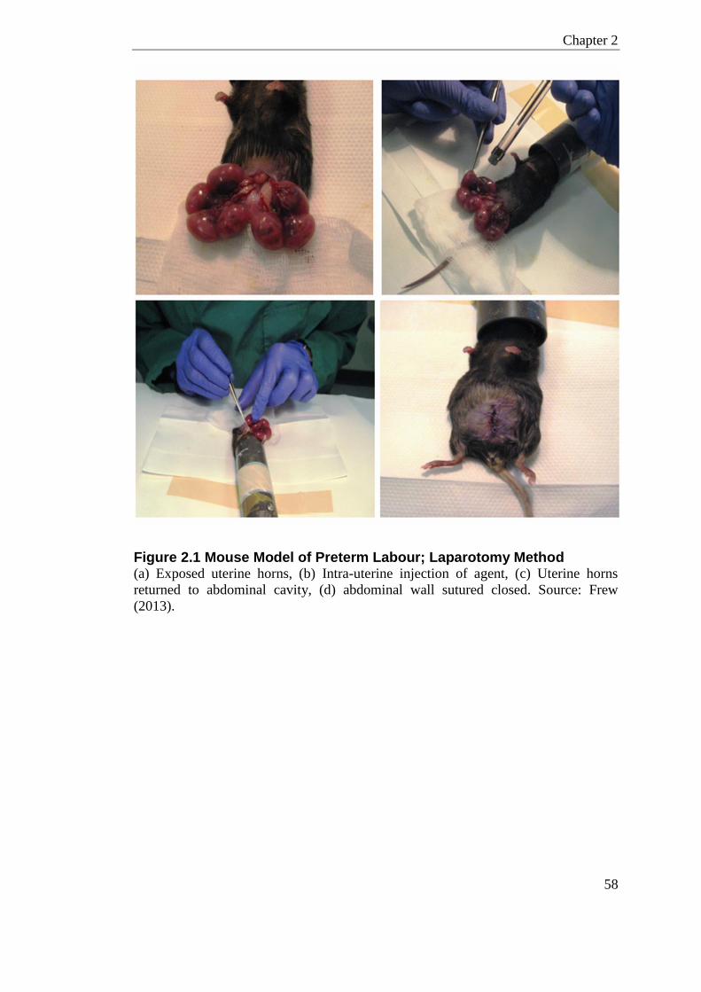
Chapter 2
58
Figure 2.1 Mouse Model of Preterm Labour; Laparotomy Method (a) Exposed uterine horns, (b) Intra-uterine injection of agent, (c) Uterine horns
returned to abdominal cavity, (d) abdominal wall sutured closed. Source: Frew
(2013).
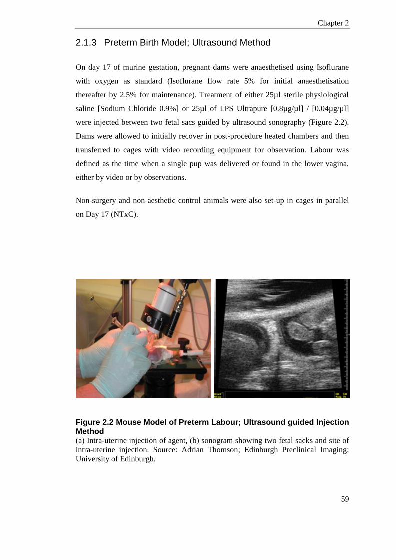
Chapter 2
59
2.1.3 Preterm Birth Model; Ultrasound Method
On day 17 of murine gestation, pregnant dams were anaesthetised using Isoflurane
with oxygen as standard (Isoflurane flow rate 5% for initial anaesthetisation
thereafter by 2.5% for maintenance). Treatment of either 25µl sterile physiological
saline [Sodium Chloride 0.9%] or 25µl of LPS Ultrapure [0.8µg/µl] / [0.04µg/µl]
were injected between two fetal sacs guided by ultrasound sonography (Figure 2.2).
Dams were allowed to initially recover in post-procedure heated chambers and then
transferred to cages with video recording equipment for observation. Labour was
defined as the time when a single pup was delivered or found in the lower vagina,
either by video or by observations.
Non-surgery and non-aesthetic control animals were also set-up in cages in parallel
on Day 17 (NTxC).
Figure 2.2 Mouse Model of Preterm Labour; Ultrasound guided Injection Method (a) Intra-uterine injection of agent, (b) sonogram showing two fetal sacks and site of
intra-uterine injection. Source: Adrian Thomson; Edinburgh Preclinical Imaging;
University of Edinburgh.
Chapter 2
60
2.1.4 Interval to Delivery and Number of Live-Born Pups
Labour was defined as the time when a single pup was delivered or found in the
lower vagina, either by video or by observation. Interval to delivery was recorded for
each animal as the number of hours between injection and labour. Preterm labour is
defined as an interval of <36 hours. The number of live-born pups was determined at
the first observation following labour; any pups found live in-utero were not counted
as ‘live-born’. The proportion of live-born pups was the number of viable fetus
counted at the time of injection divided by the number of live-born.
2.1.5 Timed Collection
Preterm labour model was conducted as detailed in section 2.1.3, but animals were
culled 6-hours post treatment by lethal CO2 exposure, where tissue (placenta, fetal
membrane, uterus, vagina, and cervix) and blood serum were collected for molecular
analysis. In parallel non-anaesthetic non-treatment day 17 pregnant females (NTxC)
were also culled for control tissue and blood serum for comparison.
Immediately after CO2 lethal exposure blood was collected by cardiac puncture using
a 21-gauge needle into BD Microcontainers®. Blood was allowed to sit for 20
minutes and then centrifuged at 8000 rpm for 5 minutes. The serum was then
collected and centrifuged at 14000 rpm for 10 minutes at 4°C to pellet any remaining
blood cells, supernatant was then stored at -80°C.
All tissue collections and dissections were conducted on large sterile petri-dishes
kept on ice. Where possible tissues were divided into 3 portions; 1) For RNA
extraction; collected in RNAlater®, kept at 4°C and extracted within 48 hours. 2)
For protein extraction; snap frozen on dry ice and stored at -80°C. 3) For histology;
collected into 4% solution of paraformaldehyde, fixed overnight and then transferred
to 70% ethanol.
Chapter 2
61
The uterus was carefully dissected to release all fetal sacs. Uterine tissues were
collected from both uterine horns (right and left), mid-way from the cervix. Fetal
membranes were dissected from the placenta and pooled where possible, according
to which uterine horn the fetal sac was obtained. One placenta from the left and right
horn was dissected into two parts for RNA and Protein, a separate placenta from each
horn was selected for histology. Vagina and cervix were excised internally, releasing
any connecting tissues. Cross-sections of the cervix and vagina were taken for
histology; remaining vagina and cervix tissue was collected for RNA extraction.
2.1.6 Microbiome Sample Collection
Sampling and methods for microbiome analysis are described in Chapter 4. For full
details of reagents and supplies, see Appendix on page 314.
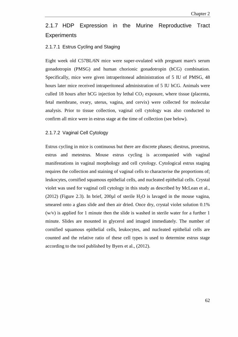
Chapter 2
62
2.1.7 HDP Expression in the Murine Reproductive Tract
Experiments
2.1.7.1 Estrus Cycling and Staging
Eight week old C57BL/6N mice were super-ovulated with pregnant mare's serum
gonadotropin (PMSG) and human chorionic gonadotropin (hCG) combination.
Specifically, mice were given intraperitoneal administration of 5 IU of PMSG, 48
hours later mice received intraperitoneal administration of 5 IU hCG. Animals were
culled 18 hours after hCG injection by lethal CO2 exposure, where tissue (placenta,
fetal membrane, ovary, uterus, vagina, and cervix) were collected for molecular
analysis. Prior to tissue collection, vaginal cell cytology was also conducted to
confirm all mice were in estrus stage at the time of collection (see below).
2.1.7.2 Vaginal Cell Cytology
Estrus cycling in mice is continuous but there are discrete phases; diestrus, proestrus,
estrus and metestrus. Mouse estrus cycling is accompanied with vaginal
manifestations in vaginal morphology and cell cytology. Cytological estrus staging
requires the collection and staining of vaginal cells to characterise the proportions of;
leukocytes, cornified squamous epithelial cells, and nucleated epithelial cells. Crystal
violet was used for vaginal cell cytology in this study as described by McLean et al.,
(2012) (Figure 2.3). In brief, 200µl of sterile H2O is lavaged in the mouse vagina,
smeared onto a glass slide and then air dried. Once dry, crystal violet solution 0.1%
(w/v) is applied for 1 minute then the slide is washed in sterile water for a further 1
minute. Slides are mounted in glycerol and imaged immediately. The number of
cornified squamous epithelial cells, leukocytes, and nucleated epithelial cells are
counted and the relative ratio of these cell types is used to determine estrus stage
according to the tool published by Byers et al., (2012).
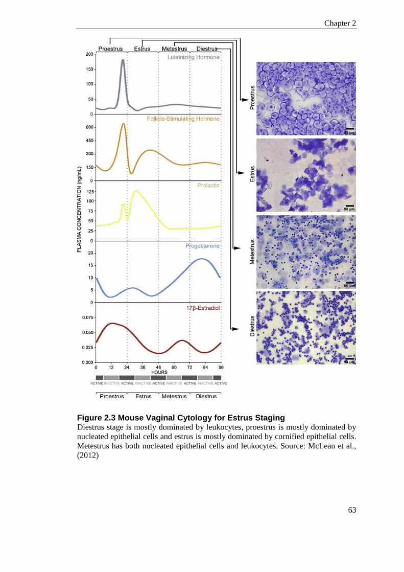
Chapter 2
63
Figure 2.3 Mouse Vaginal Cytology for Estrus Staging Diestrus stage is mostly dominated by leukocytes, proestrus is mostly dominated by
nucleated epithelial cells and estrus is mostly dominated by cornified epithelial cells.
Metestrus has both nucleated epithelial cells and leukocytes. Source: McLean et al.,
(2012)
Chapter 2
64
2.2 Molecular
2.2.1 RNA Extraction and Reverse Transcription
Two methods of RNA extraction were used in this study. All human tissues were
extracted using the TRI reagent method; while all murine tissues were extracted
using the RNA Bee method. The TRI Reagent method is the protocol of choice for
the Edinburgh Reproductive Biobank and was implemented for uniformity across
study-sets. For full details of reagents and supplies, see Appendix on page 314.
2.2.1.1 TRI Reagent Method
Tissue was transferred to 2ml tubes containing 1ml of TRI reagent® with sterile
stainless steel bead. Homogenisation was supported using TissueLyser I for 2 x 3
minutes at 25HZ. Samples were left to incubate for 15 minutes at room temperature
then centrifuged at 14,000 rpm for 15 minutes maintained at 4°C. The supernatant
was then transferred to phase-lock tubes and 200µl of bromochloropropane added,
samples were then left to participate at room temperature for 15 minutes. Samples
were centrifuged at 14,000 rpm for 15 minutes maintained at 4°C; the aqueous phase
was then collected into equal volume of ice cold 70% ethanol. To isolate the RNA
the RNeasy kit was used following the manufactures protocol. Isolated RNA was
quantified on the Nanodrop ND-1000 at 260nm and 280nm as standard; all RNA is
stored at -80°C.
2.2.1.2 RNA Bee Method
Tissue was transferred to 2 ml Lysing Matrix D tubes, containing 1ml of RNA Bee,
then homogenised for 30 seconds at 5000 rpm on the Precellys®24 lyser. 100µl of
chloroform was added and samples were left to incubate on ice for 5 minutes.
Samples were then centrifuged at 10000 rpm for 15 minutes maintained at 4°C. The
subsequent upper aqueous phase was transferred to a new tube containing equal parts
of ice-cold isopropanol. Samples were then incubated at -20°C for 20 minutes and
centrifuged at 10000 rpm for 20 minutes maintained at 4°C to pellet the RNA. Pellets
Chapter 2
65
were washed twice with 75% ice cold ethanol and RNA was re-suspended in 20-
100µl RNase and DNase-free water.
2.2.1.3 Reverse Transcription
High-Capacity cDNA Reverse Transcription Kit was used to reverse transcribe RNA
following the manufactures protocol for a 20µl reaction using oligo (dT) primers and
1µg of each RNA sample. Where possible a representative reverse transcription
negative (RT-NEG) and template negative (NTC-NEG) were included in each batch.
cDNA was stored at -20°C until required.
2.2.2 RT-qPCR
The amount of mRNA transcript was quantified using qPCR, where the cDNA is
amplified for specific genes of interest in the presence of fluorescent reporter probes
and quenchers. The majority of the gene expression analysis in this study was done
using pre-designed Taqman probes, which have been optimised for use on the ABI
PRISM® 7000 Sequence Detection System. These probes were sourced from
Applied Biosystems and are listed in Table 2.1. Some gene expression analysis was
supported on the LightCycler® 480 Real-Time PCR System. For this approach,
primers for specific genes of interest were designed to be compatible with the
universal hydrolysis probes system (UPL), utilising the Universal ProbeLibrary
Assay Design Center (Table 2.2).
Several qPCR negatives (PCR-NTC) and reverse transcription negatives (RT-NEG)
and calibrators were run on every plate. The individual calibrators (CAL) are detailed
in each chapter.
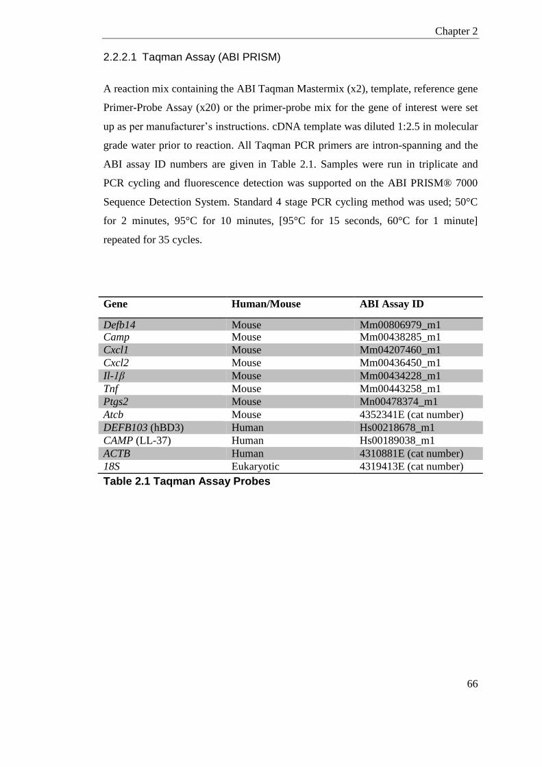
Chapter 2
66
2.2.2.1 Taqman Assay (ABI PRISM)
A reaction mix containing the ABI Taqman Mastermix (x2), template, reference gene
Primer-Probe Assay (x20) or the primer-probe mix for the gene of interest were set
up as per manufacturer’s instructions. cDNA template was diluted 1:2.5 in molecular
grade water prior to reaction. All Taqman PCR primers are intron-spanning and the
ABI assay ID numbers are given in Table 2.1. Samples were run in triplicate and
PCR cycling and fluorescence detection was supported on the ABI PRISM® 7000
Sequence Detection System. Standard 4 stage PCR cycling method was used; 50°C
for 2 minutes, 95°C for 10 minutes, [95°C for 15 seconds, 60°C for 1 minute]
repeated for 35 cycles.
Gene Human/Mouse ABI Assay ID
Defb14 Mouse Mm00806979_m1 Camp Mouse Mm00438285_m1
Cxcl1 Mouse Mm04207460_m1
Cxcl2 Mouse Mm00436450_m1
Il-1β Mouse Mm00434228_m1
Tnf Mouse Mm00443258_m1
Ptgs2 Mouse Mn00478374_m1
Atcb Mouse 4352341E (cat number)
DEFB103 (hBD3) Human Hs00218678_m1
CAMP (LL-37) Human Hs00189038_m1
ACTB Human 4310881E (cat number)
18S Eukaryotic 4319413E (cat number)
Table 2.1 Taqman Assay Probes
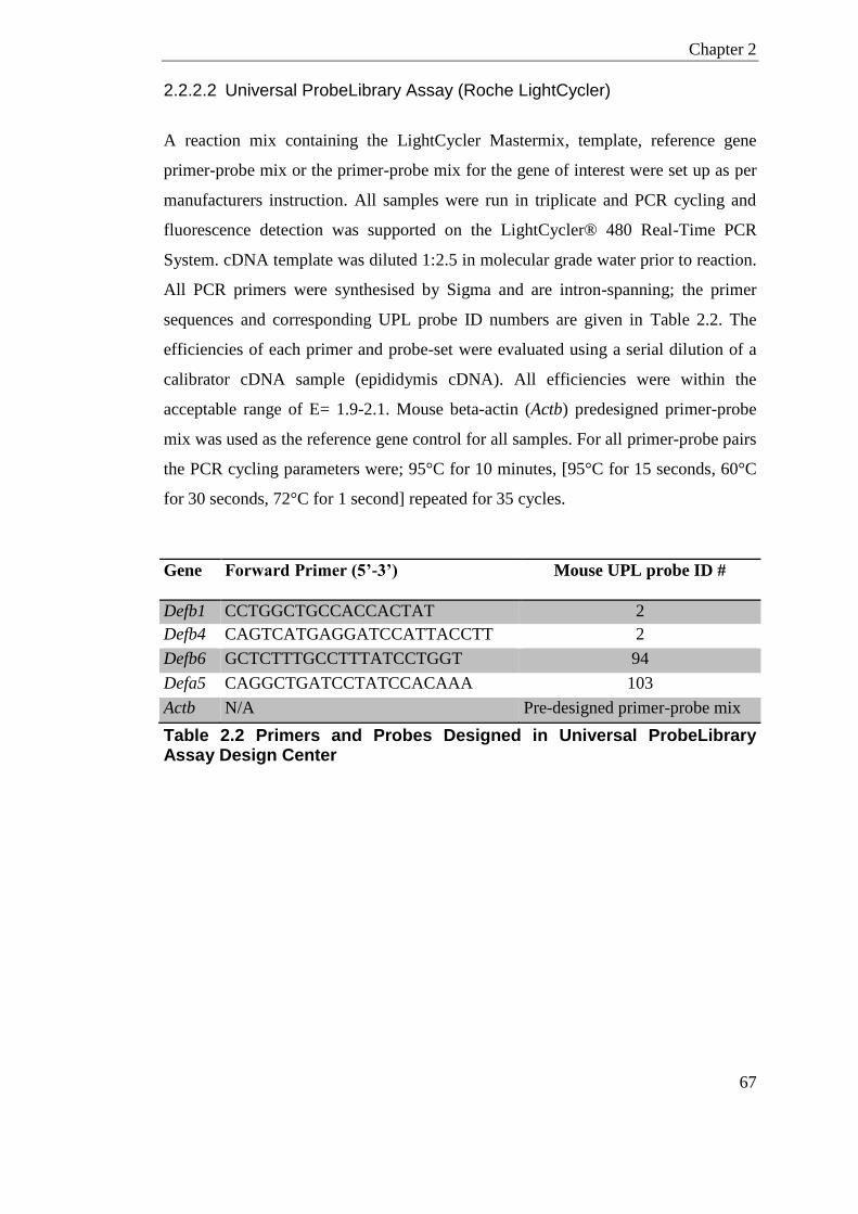
Chapter 2
67
2.2.2.2 Universal ProbeLibrary Assay (Roche LightCycler)
A reaction mix containing the LightCycler Mastermix, template, reference gene
primer-probe mix or the primer-probe mix for the gene of interest were set up as per
manufacturers instruction. All samples were run in triplicate and PCR cycling and
fluorescence detection was supported on the LightCycler® 480 Real-Time PCR
System. cDNA template was diluted 1:2.5 in molecular grade water prior to reaction.
All PCR primers were synthesised by Sigma and are intron-spanning; the primer
sequences and corresponding UPL probe ID numbers are given in Table 2.2. The
efficiencies of each primer and probe-set were evaluated using a serial dilution of a
calibrator cDNA sample (epididymis cDNA). All efficiencies were within the
acceptable range of E= 1.9-2.1. Mouse beta-actin (Actb) predesigned primer-probe
mix was used as the reference gene control for all samples. For all primer-probe pairs
the PCR cycling parameters were; 95°C for 10 minutes, [95°C for 15 seconds, 60°C
for 30 seconds, 72°C for 1 second] repeated for 35 cycles.
Gene
Mouse
Forward Primer (5’-3’)
Reverse Primer (5’-3’)
Mouse UPL probe ID #
Defb1 CCTGGCTGCCACCACTAT
CTTGTGAGAATGCCAACACCT
2
Defb4 CAGTCATGAGGATCCATTACCTT
AATTTGGGTAAAGGCTGCAA
2
Defb6 GCTCTTTGCCTTTATCCTGGT
TGGACTGTTGATTAATTGGGAAA
94
Defa5 CAGGCTGATCCTATCCACAAA
GAAGAGCAGACCCTTCTTGG
103
Actb N/A Pre-designed primer-probe mix
Table 2.2 Primers and Probes Designed in Universal ProbeLibrary Assay Design Center
Chapter 2
68
2.2.2.3 RT-qPCR analysis method
All qPCR in this study were set up in technical triplicates where any intra-sample
outliers were removed. Ct value is the cycle at which the fluorescence signal crosses
a threshold, where the threshold is an estimate of the first point where samples are in
exponential phase above background. Ct values were normalise to the reference gene
and then samples compared to calibrator using the formula 2-ΔΔCT
. This gives a fold
change in gene expression of the target relative to the calibrator. This method also
allows comparison across multiple plates if the same calibrator is used. Where two
reference genes were used as the control, their geometric mean was used to
normalise the Ct value of the gene of interest.
2.2.3 ELISA
ELISA quantitatively measures the amount of a protein of interest in solution by
comparing to a known sample using a serial dilution. Sandwich ELISA involves
coating a plate with monoclonal antibody raised against the protein of interest. After
the protein of interest is bound to this primary antibody, a secondary antibody that
has an epitope for the protein is then bound. These secondary antibodies are coupled
to substrates that allow for detection and quantitative measurement.
All ELISA kits in this study were R&D duosets, the detection range for each protein
is given in Table 2.3. All ELISA in this study were conducted using the following
protocol. Flat-bottom ELISA plates were coated with 50µl of capture antibody,
diluted appropriately in PBS, and incubated overnight at room temperature. Plates
were then washed 3 times in PBS-Tween (0.05%) to remove unbound antibody and
blocked for 2 hours in 0.5% (v/v) Reagent Dilute Concentrate in PBS. After removal
of the blocking solution, 50µl of sample was added to the plate in duplicate. In this
study all blood serum and cell culture supernatants were diluted 1:5 in Reagent
Diluent. Serial dilution of standards were also made in Reagent diluent and added in
duplicate in 50µl volumes. Plates were incubated overnight at 4°C and then washed 3
times in PBS-Tween (0.05%). Appropriate biotinylated detection antibody was
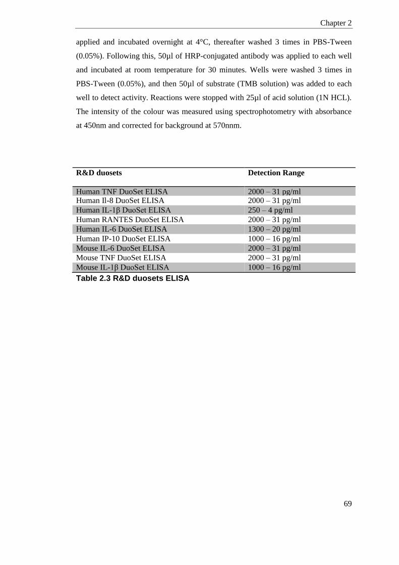
Chapter 2
69
applied and incubated overnight at 4°C, thereafter washed 3 times in PBS-Tween
(0.05%). Following this, 50µl of HRP-conjugated antibody was applied to each well
and incubated at room temperature for 30 minutes. Wells were washed 3 times in
PBS-Tween (0.05%), and then 50µl of substrate (TMB solution) was added to each
well to detect activity. Reactions were stopped with 25µl of acid solution (1N HCL).
The intensity of the colour was measured using spectrophotometry with absorbance
at 450nm and corrected for background at 570nnm.
R&D duosets Detection Range
Human TNF DuoSet ELISA 2000 – 31 pg/ml Human Il-8 DuoSet ELISA 2000 – 31 pg/ml
Human IL-1β DuoSet ELISA 250 – 4 pg/ml
Human RANTES DuoSet ELISA 2000 – 31 pg/ml
Human IL-6 DuoSet ELISA 1300 – 20 pg/ml
Human IP-10 DuoSet ELISA 1000 – 16 pg/ml
Mouse IL-6 DuoSet ELISA 2000 – 31 pg/ml
Mouse TNF DuoSet ELISA 2000 – 31 pg/ml
Mouse IL-1β DuoSet ELISA 1000 – 16 pg/ml
Table 2.3 R&D duosets ELISA
Chapter 2
70
2.3 HDPs in Placental Explants Experiment
Placenta was obtained from women undergoing elective Caesarean section at 38-39
weeks. All women were healthy, non-smokers with a normal BMI. Women had no
indication of infection and all were singleton pregnancies.
Full thickness placental lobule was collected from the central cotyledons region of
the placenta and transported directly to the lab in PBS. Placenta tissue was washed 5
times in sterile PBS and the chorionic surface removed. Any visible connective tissue
deposits and blood clots were also removed. Villous tissue was obtained from the
middle cross-section where 0.05g was placed in each well of a 24-well flat bottom
culture dish. Each well contained 1ml of pre-warmed RPMI 1640 media with 10%
fetal calf serum (FCS) and penicillin and streptomycin [50ug/ml]. Tissues were left
to acclimatise for 1 hour in 5% CO2 at 37°C before treatments applied. Treatments
and concentrations are detailed in section 3.3.2. The scrambled LL-37 peptide
sequence is RSLEGTDRFPFVRLKNSRKLEFKDIKGIKREQFVKIL. All treatments
were applied in triplicate and cultures incubated in 5% CO2 at 37°C for 3 hours,
unless detailed otherwise. Culture supernatant was collected and centrifuged at 800
rpm for 10 minutes maintained at 4°C to remove debris. To further pellet red blood
cells this supernatant was transferred to a new tube and centrifuged at 8000 rpm for
10 minutes maintained at 4°C. Final supernatants were used immediately for LDA
assay or stored in aliquots at -80°C. Placenta tissue was blotted dry on sterile paper
and collected in RNAlater for RNA extraction as described in section 2.2.1.1. For
full details of reagents and supplies, see Appendix on page 314.
2.3.1 LDH assay
Cytotoxicity of each treatment was measured using Pierce™ LDH Cytotoxicity
Assay Kit following the manufactures protocol.

Chapter 2
71
2.4 HDPs in Human Term Labour study
Human myometrium, fetal membrane, and placenta tissue was obtained from the
Edinburgh Reproductive Biobank; REC number 13/ES/0126. Informed consent was
obtained and approved under South East Scotland Scottish Academic Health
Sciences Collaboration (SAHSC) Human Annotated BioResource (HAB). Tissues
were collected from healthy singleton pregnancies undergoing elective Caesarean
section. All samples were collected into RNAlater and stored at -80°C until RNA
extraction. For full details of reagents and supplies, see Appendix on page 314.

72
Chapter 3
Host Defence Peptides
Chapter 3
73
Chapter 3 Host Defence Peptides
3.1 Introduction
Nearly 40% of all preterm deliveries are associated with intrauterine infection and/or
clinical inflammation (Romero et al., 2006). The female reproductive tract has
biochemical and physiological barriers to prevent bacteria ascending from the vagina
into the amniotic cavity during pregnancy. These physiological barriers include the
cervix and fetal membranes which also produce multiple Host Defence Peptides; key
mediators in defence against invading pathogens. HDPs are released by epithelial
cells, trophoblastic cells and immune cells where their action can be by disrupting
bacteria cell membranes, but also by an array of immunomodulatory processes.
Labour is generally accepted to be an inflammatory event where the emphasis from
an anti-inflammatory pregnancy state shifts pro-inflammatory preceding labour
(Youssef et al., 2009).
hBD3 and the murine orthologue DEFB14 have potent anti-inflammatory effects
shown both in-vitro and in-vivo (Semple et al., 2010, Semple et al., 2011). Exposure
of human and mouse primary monocytes to LPS in the presence of hBD3 effectively
inhibited the TLR-4 mediated TNF, IL-6 and type I interferon responses (Semple et
al., 2010). This inhibitory response has been shown to be through both the MyD88
and TRIF pathways. Contrastingly, a study has also suggested hBD3 can be pro-
inflammatory when mediated by TLR1/2 (Funderburg et al., 2007). Funderburg et
al., (2007) displayed that hBD3 increases the production of co-stimulatory molecules
on monocytes and dendritic cells enhancing their chemoattractant potential. The
chemoattractant property of hBD3 has also been shown in many other cell types,
such as T cells, immature dendritic cells and macrophages (Wu et al., 2003, Yang et
al., 1999). Other pro-inflammatory properties of hBD3 are displayed in its potential
to exacerbate the MDA5 cytoplasmic receptor in response to dsRNA, a viral ligand
mimic (Semple et al., 2015).
Chapter 3
74
LL-37 has been shown to have biphasic action, having both pro-inflammatory and
anti-inflammatory functions. In human monocytes, exogenous LL-37 can inhibit the
release of the pro-inflammatory cytokine TNF in response to LPS. This has also been
shown for the mouse peptide CAMP on BMDMs (Pinheiro da Silva et al., 2009). LL-
37 can also inhibit the pro-inflammatory response to PAMPS in other myeloid cells
(Mookherjee et al., 2006). The anti-endotoxic effects of LL-37 and the mouse
orthologue CAMP have been mostly attributed to their ability to directly bind to LPS
(Scott et al., 2000, Scott et al., 2011).
With regard to promoting inflammation, exogenous LL-37 in-vitro increases the
production of cytokines and chemokine such as IL-8 and IL-6 in lung epithelial cells
(Tjabringa et al., 2003, Pistolic et al., 2009), skin cells (Niyonsaba et al., 2007), and
cervical epithelial cells (Frew et al., 2014). LL-37 also preferentially promotes
apoptosis in lung epithelial cells in the presence of an infection (Barlow et al., 2006)
and is a potent modifier of dendritic cell differentiation (Davidson et al., 2004).
In a pregnant state the hypothesis of this chapter is that HDPs have a dual role, in
preventing ascending infection, and also preventing an exacerbated inflammatory
response that can cause preterm birth by initiation of the labour cascade. The
immunomodulatory effects of LL-37 and hBD3 in reproductive tissues in response to
bacterial stimuli has not been well studied. The spatial function most likely reflects
the differing immunological requirements along the length of the female
reproductive tract and pregnancy tissues.
This chapter investigates whether the HDPs, hBD3 and LL-37, are affected by the
inflammatory cascade of normal term labour. This was done by looking at the
mRNA gene expression in the myometrium, amnion/chorion, and placenta of women
in either labour at term or not in labour at term. These tissues were chosen for the
relative ease of collection.
To test the anti-inflammatory properties of HDPs in pregnancy I investigated the
effects of endogenous LL-37 and hBD3 on placental explants in response to bacterial
stimuli. This response could have additionally been tested placental cell lines,

Chapter 3
75
however, it is reported that different trophoblast cell lines express differences in cell
surface markers when cultured in isolation (Nakada et al., 2011, Grasso et al., 2014).
I also determined the expression of the mouse orthologues of LL-37 and hBD3;
Camp and Defb14 respectively, in the mouse female reproductive tract in pregnancy.
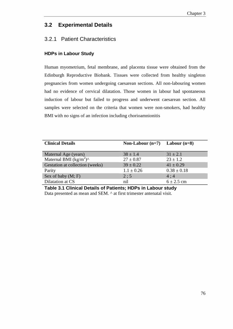
Chapter 3
76
3.2 Experimental Details
3.2.1 Patient Characteristics
HDPs in Labour Study
Human myometrium, fetal membrane, and placenta tissue were obtained from the
Edinburgh Reproductive Biobank. Tissues were collected from healthy singleton
pregnancies from women undergoing caesarean sections. All non-labouring women
had no evidence of cervical dilatation. Those women in labour had spontaneous
induction of labour but failed to progress and underwent caesarean section. All
samples were selected on the criteria that women were non-smokers, had healthy
BMI with no signs of an infection including chorioamnionitis
Clinical Details Non-Labour (n=7) Labour (n=8)
Maternal Age (years) 38 ± 1.4 31 ± 2.1 Maternal BMI (kg/m
2)^ 27 ± 0.87 23 ± 1.2
Gestation at collection (weeks) 39 ± 0.22 41 ± 0.29
Parity 1.1 ± 0.26 0.38 ± 0.18
Sex of baby (M; F) 2 ; 5 4 ; 4
Dilatation at CS nil 6 ± 2.5 cm
Table 3.1 Clinical Details of Patients; HDPs in Labour study Data presented as mean and SEM. ^ at first trimester antenatal visit.
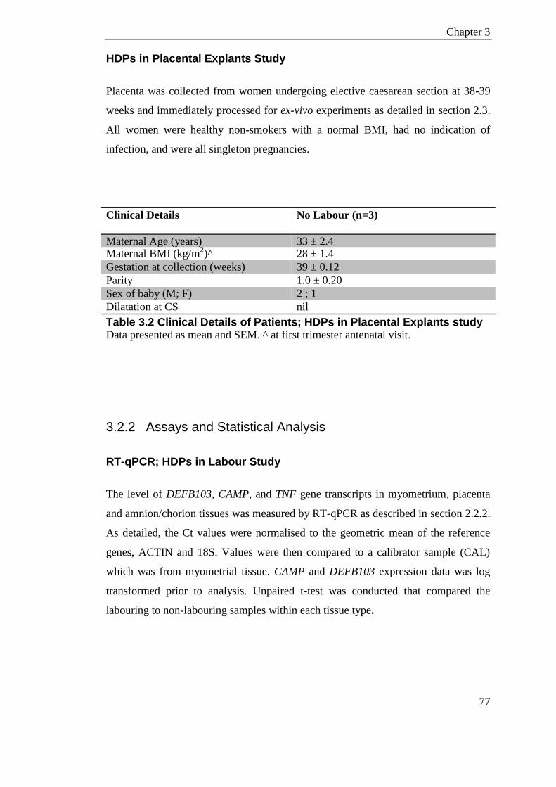
Chapter 3
77
HDPs in Placental Explants Study
Placenta was collected from women undergoing elective caesarean section at 38-39
weeks and immediately processed for ex-vivo experiments as detailed in section 2.3.
All women were healthy non-smokers with a normal BMI, had no indication of
infection, and were all singleton pregnancies.
Clinical Details No Labour (n=3)
Maternal Age (years) 33 ± 2.4 Maternal BMI (kg/m
2)^ 28 ± 1.4
Gestation at collection (weeks) 39 ± 0.12
Parity 1.0 ± 0.20
Sex of baby (M; F) 2 ; 1
Dilatation at CS nil
Table 3.2 Clinical Details of Patients; HDPs in Placental Explants study Data presented as mean and SEM. ^ at first trimester antenatal visit.
3.2.2 Assays and Statistical Analysis
RT-qPCR; HDPs in Labour Study
The level of DEFB103, CAMP, and TNF gene transcripts in myometrium, placenta
and amnion/chorion tissues was measured by RT-qPCR as described in section 2.2.2.
As detailed, the Ct values were normalised to the geometric mean of the reference
genes, ACTIN and 18S. Values were then compared to a calibrator sample (CAL)
which was from myometrial tissue. CAMP and DEFB103 expression data was log
transformed prior to analysis. Unpaired t-test was conducted that compared the
labouring to non-labouring samples within each tissue type.
Chapter 3
78
RT-qPCR; HDPs in Placental Explants Study
The level of DEFB103, CAMP, and TNF gene transcripts in placenta was measured
by RT-qPCR as previously described. Samples were normalised to a calibrator
(CAL) from a placenta explant tissue that received no treatment.
For TNF gene transcript (excluding hBD3 treatment), a two-way ANOVA with
Tukey’s post-test compared to no-peptide treatments within LPS groups was
performed. Significant interactions in LPS treatment were identified with
Bonferroni's post-test compared to no-treatment groups. As there was no hBD3
peptide only group, the effect of hBD3 peptide was measured by paired t-test
between LPS [100ng/ml] alone and LPS [100ng/ml] + hBD3 [25µg/ml] group.
Significant differences in the level of DEFB103 and CAMP in response to LPS were
conducted using a paired t-test comparing no-treatment to LPS alone.
RT-qPCR; HDPs in Murine Reproductive Tract
The mouse beta-defensins; Defb1, Defb4, Defb6, Defb14, the alpha defensin, Defa5,
and the murine cathelicidin, Camp, transcripts were measured in ovary, uterus, cervix
and vagina by RT-qPCR as described in section 2.2.2. The Ct values were
normalised to the reference gene, Actb. Where a calibrator was used to normalise
values; the calibrator was from epididymis tissue. Data presented as mean fold
change and SEM relative to either the calibrator (CAL) or mean expression in the
ovary.
ELISA; HDPs in Placental Explants Study
To detect the amount of secreted protein in culture supernatants from human placenta
explants post-treatment, ELISA’s were conducted as described in section 2.2.3. The
proteins of interest were TNF, IL-8, IL-6, IL-1β, IP-10, and RANTES.
All ELISA data is presented as the mean concentration ± SEM. Two-way ANOVA
was used to highlight the effect of the presence of LPS and LL-37 compared to
control peptide. Significant interactions in peptide treatment were identified with

Chapter 3
79
Tukey’s post-test. A two-way ANOVA with Dunnett’s post-test for matched samples
was conducted on the data looking at LPS effect over time. One-way ANOVA with
Dunnett’s post-test was conducted when comparing dose of LL-37 with LPS
treatment.
As there was no hBD3 peptide only group, the effect of hBD3 peptide was measured
by paired t-test between LPS [100ng/ml] alone and LPS [100ng/ml] + hBD3
[25µg/ml] groups for each secreted cytokine.
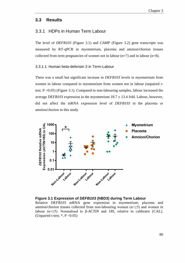
Chapter 3
80
3.3 Results
3.3.1 HDPs in Human Term Labour
The level of DEFB103 (Figure 3.1) and CAMP (Figure 3.2) gene transcripts was
measured by RT-qPCR in myometrium, placenta and amnion/chorion tissues
collected from term pregnancies of women not in labour (n=7) and in labour (n=8).
3.3.1.1 Human beta-defensin 3 in Term-Labour
There was a small but significant increase in DEFB103 levels in myometrium from
women in labour compared to myometrium from women not in labour (unpaired t-
test; P <0.05) (Figure 3.1). Compared to non-labouring samples, labour increased the
average DEFB103 expression in the myometrium 18.7 ± 13.4 fold. Labour, however,
did not affect the mRNA expression level of DEFB103 in the placenta or
amnion/chorion in this study.
Figure 3.1 Expression of DEFB103 (hBD3) during Term Labour Relative DEFB103 mRNA gene expression in myometrium, placenta and
amnion/chorion tissues collected from non-labouring women (n=≥5) and women in
labour (n=≥5). Normalised to β-ACTIN and 18S, relative to calibrator (CAL).
(Unpaired t-test; *, P <0.05)
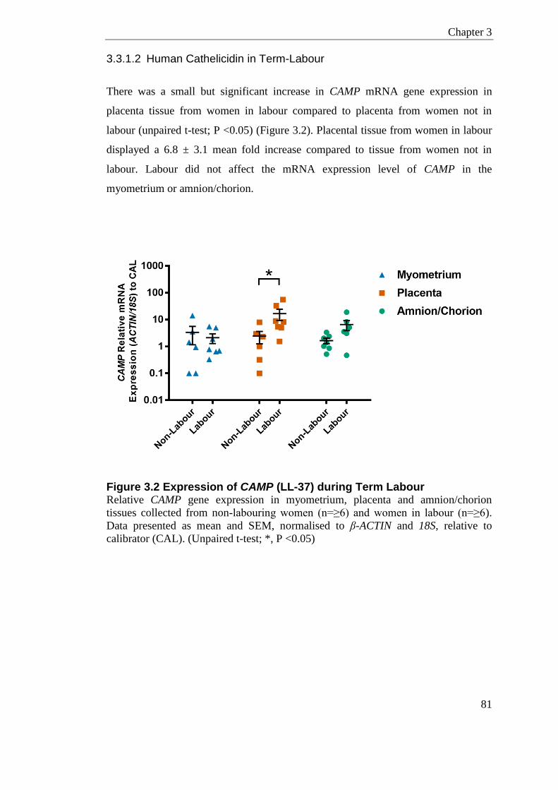
Chapter 3
81
3.3.1.2 Human Cathelicidin in Term-Labour
There was a small but significant increase in CAMP mRNA gene expression in
placenta tissue from women in labour compared to placenta from women not in
labour (unpaired t-test; P <0.05) (Figure 3.2). Placental tissue from women in labour
displayed a 6.8 ± 3.1 mean fold increase compared to tissue from women not in
labour. Labour did not affect the mRNA expression level of CAMP in the
myometrium or amnion/chorion.
Figure 3.2 Expression of CAMP (LL-37) during Term Labour Relative CAMP gene expression in myometrium, placenta and amnion/chorion
tissues collected from non-labouring women (n=≥6) and women in labour (n=≥6).
Data presented as mean and SEM, normalised to β-ACTIN and 18S, relative to
calibrator (CAL). (Unpaired t-test; *, P <0.05)
Chapter 3
82
3.3.2 HDP Anti-inflammatory effect on Placental Explants
To investigate the effects of HDPs on the inflammation caused by infection, placenta
tissue explants were cultured with LPS in combination with exogenous peptides of
hBD3, LL-37 and scrambled LL-37 control. Placenta was collected from women
undergoing caesarean section at term (n=3), but prior to the initiation of labour. The
main read-out of inflammation was the secreted pro-inflammatory cytokine TNF,
which was assessed by ELISA in culture supernatants as described in section 2.2.3.
Other secreted cytokine levels were also determined; IL-8, IL-6, IL-1β, IP-10
(CXCL10), and RANTES (Regulated on Activation, Normal T Cell Expressed and
Secreted) as measured by ELISA. TNF mRNA gene expression levels were assessed
by RT-qPCR as described in section 2.2.2.
Cytotoxicity assay showed that LPS and exogenous HDP treatments did not induce
any significant cell death in the doses and time-points investigated here compared to
background (section 2.3.1; data not shown).
Chapter 3
83
3.3.2.1 LPS Dose and Time Response in Placental Explants
The time and dose-responsive effects of LPS on placental explants was determined
by culturing at two LPS doses, 1µg/ml or 100ng/ml for 24 hours. Culture
supernatants were taken at 1, 3, 6 and 24 hours post treatment, and the amount of the
pro-inflammatory cytokine TNF was measured by ELISA (Figure 3.3).
1µg/ml and 100ng/ml of LPS both increased TNF secretion significantly compared
to non-LPS treated samples at all time-points considered (two-way ANOVA with
Dunnett’s post-test; P <0.0001). 100ng of LPS appeared to produce less secreted
TNF compared to 1µg at all time-points, but this was not significant in this small
sample size with repeated measures. Nonetheless, as the lower dose of 100ng LPS
could elicit a robust TNF response, this dose was chosen for subsequent experiments
to be closer to physiological/clinical levels of LPS (Guo et al., 2013). As TNF
protein levels in this system peak at 6-hours; culture supernatants in subsequent
experiments were collected at 3-hours post treatment to capture initial TNF response
before saturation.
Chapter 3
84
Figure 3.3 Placental Explants; TNF levels in LPS dose and Time Response TNF protein levels from cultures supernatants as measured by ELISA. Presented as
mean and SD of technical replicates, n=3 per group. All treated samples are from one
patient. (Two-way ANOVA with Dunnett’s post-test; P <0.0001 for LPS effect at all
time-points, no significant difference in 1µg compared to 100ng LPS dose at any
time-point; Bonferroni's post-test)
Chapter 3
85
3.3.2.2 LL-37 Dose Response in Placental Explants
The effect of LL-37 on LPS-induced inflammation in the placenta was determined by
culturing explants with increasing doses of exogenous LL-37 peptide in the presence
of 100ng/ml of LPS. Culture supernatants were taken at 3-hours post treatment and
the amount of the secreted pro-inflammatory cytokine TNF was measured by ELISA
(Figure 3.4).
In placental explants from all 3 patients; increasing the dose of LL-37 peptide
reduced the levels of secreted TNF in response to 100ng/ml of LPS. This reached
statistical significance at 25µg/ml of LL-37 (one-way ANOVA with Dunnett’s post-
test; **, P <0.01 compared to LPS alone).
Figure 3.4 Placental Explants; TNF levels in LL-37 Dose Response TNF protein levels from cultures supernatants with 100ng/ml of LPS and increasing
doses of LL-37. All data presented as mean and SD of technical replicates, n=3 per
group. (one-way ANOVA with Dunnett’s post-test; **, P <0.01 compared to LPS
alone)
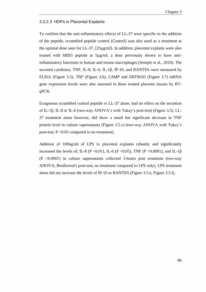
Chapter 3
86
3.3.2.3 HDPs in Placental Explants
To confirm that the anti-inflammatory effects of LL-37 were specific to the addition
of the peptide, scrambled peptide control (Control) was also used as a treatment at
the optimal dose seen for LL-37, [25µg/ml]. In addition, placental explants were also
treated with hBD3 peptide at 5µg/ml; a dose previously shown to have anti-
inflammatory functions in human and mouse macrophages (Semple et al., 2010). The
secreted cytokines; TNF, IL-8, IL-6, IL-1β, IP-10, and RANTES were measured by
ELISA (Figure 3.5). TNF (Figure 3.6), CAMP and DEFB103 (Figure 3.7) mRNA
gene expression levels were also assessed in these treated placenta tissues by RT-
qPCR.
Exogenous scrambled control peptide or LL-37 alone, had no effect on the secretion
of IL-1β, IL-8 or IL-6 (two-way ANOVA’s with Tukey’s post-test) (Figure 3.5). LL-
37 treatment alone however, did show a small but significant decrease in TNF
protein level in culture supernatants (Figure 3.5.c) (two-way ANOVA with Tukey’s
post-test; P <0.05 compared to no treatment).
Addition of 100ng/ml of LPS to placental explants robustly and significantly
increased the levels of; IL-8 (P <0.01), IL-6 (P <0.05), TNF (P <0.0001), and IL-1β
(P <0.0001) in culture supernatants collected 3-hours post treatment (two-way
ANOVA; Bonferroni's post-test; no treatment compared to LPS only). LPS treatment
alone did not increase the levels of IP-10 or RANTES (Figure 3.5.e, Figure 3.5.f).

Chapter 3
87
Figure 3.5 Effects of HDPs on Cytokine Secretion in Placenta Explants Placenta explant cultures (n=3 patients) cytokine secretion after treatment; LPS
[100ng/ml], control scrambled peptide [25µg/ml], LL-37 [25µg/ml] and hBD3
[5µg/ml]. (a) IL-8 (b) IL-6 (c) TNF (d) IL-1β (e) IP-10 and (f) RANTES protein
measured by ELISA in culture supernatants 3 hours post-treatment. All data
presented as mean concentration and SEM. (Two-way ANOVA (excluding hBD3
treatment), with Tukey’s post-test; *, P <0.05, **, P <0.01, ***, P<0.001 compared
to no-peptide treatments within LPS groups. Paired t-test comparing hDB3+LPS
with LPS alone; NS difference)

Chapter 3
88
LL-37 was able to significantly reduce the LPS induced increase in IL-8 (P <0.05),
IL-6 (P <0.0001), TNF (P <0.0001), and IL-1β (P <0.0001). The addition of
scrambled control peptide did not result in any significant change in levels of any
cytokine compared to LPS treatment alone (two-way ANOVA with Tukey’s post-
test; compared to LPS alone).
hBD3 treatment did not have an effect on the levels of any of the cytokines measured
in an environment of 100ng/ml of LPS (paired t-test, hBD3+LPS compared to LPS
alone).
Although IP-10 and RANTES did not respond to LPS treatment, LL-37 peptide
treatment alone did result in increased IP-10 (P <0.001) and RANTES (P <0.001)
compared to no-treatment. As scrambled control peptide was also able to induce a
significant RANTES response this effect does not look peptide specific. In addition
IP-10 levels were at the lower limits of detection in this system and more likely
subject to error.
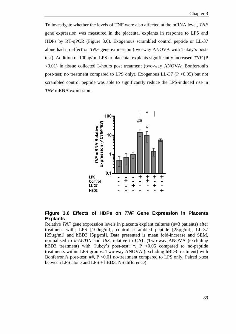
Chapter 3
89
To investigate whether the levels of TNF were also affected at the mRNA level, TNF
gene expression was measured in the placental explants in response to LPS and
HDPs by RT-qPCR (Figure 3.6). Exogenous scrambled control peptide or LL-37
alone had no effect on TNF gene expression (two-way ANOVA with Tukey’s post-
test). Addition of 100ng/ml LPS to placental explants significantly increased TNF (P
<0.01) in tissue collected 3-hours post treatment (two-way ANOVA; Bonferroni's
post-test; no treatment compared to LPS only). Exogenous LL-37 (P <0.05) but not
scrambled control peptide was able to significantly reduce the LPS-induced rise in
TNF mRNA expression.
Figure 3.6 Effects of HDPs on TNF Gene Expression in Placenta Explants Relative TNF gene expression levels in placenta explant cultures (n=3 patients) after
treatment with; LPS [100ng/ml], control scrambled peptide [25µg/ml], LL-37
[25µg/ml] and hBD3 [5µg/ml]. Data presented is mean fold-increase and SEM,
normalised to β-ACTIN and 18S, relative to CAL (Two-way ANOVA (excluding
hBD3 treatment) with Tukey’s post-test; *, P <0.05 compared to no-peptide
treatments within LPS groups. Two-way ANOVA (excluding hBD3 treatment) with
Bonferroni's post-test; ##, P <0.01 no-treatment compared to LPS only. Paired t-test
between LPS alone and LPS + hBD3; NS difference)
Chapter 3
90
Levels of endogenous DEFB103 (hBD3) and CAMP (LL-37) were measured by RT-
qPCR in placental explants from no treatment compared to LPS [100ng/ml]. There
was a trend for both of the HDPs to be increased following LPS treatment but neither
reached significance in the sample size of 3 (paired t-test)
Figure 3.7 Endogenous DEFB103 and CAMP mRNA in Placental Explants Relative (a) DEFB103 and (b) CAMP gene expression levels in placenta explant
cultures (n=3 patients) with no-treatment (NTxC) or LPS [100ng/ml]. Data presented
is mean and SEM, normalised to β-ACTIN and 18S relative to calibrator (CAL).
(Paired t-test: NS difference)
Chapter 3
91
3.3.3 HDPs in the Murine Reproductive Tract
The relative expression of multiple defensin genes and cathelicidin were determined
by RT-qPCR in non-pregnant C57BL/6N mice (n=6) (Figure 3.8). To account for
variation in HDP gene expression during estrus cycling; mice were super-ovulated
with PMSG/hCG combination prior to tissue collection as detailed in section 2.1.7.1.
Vaginal lavages were also taken and stained with crystal violet to confirm vaginal
cell cytology was similar across all mice as described in section 2.1.7.2. The genes
considered were the mouse beta-defensins; Defb1, Defb4, Defb6, Defb14, the alpha
defensin Defa5, and the murine cathelicidin Camp. Relative gene expression results
are displayed compared to samples from the ovary, which was the site of lowest
expression of all the defensins but not cathelicidin.
All beta-defensins investigated in this chapter show spatial expression patterns along
the female reproductive tract. Beta-defensins were found to be highly expressed in
tissues of the lower reproductive tract; the vagina and cervix. Comparatively there
was lower expression of beta-defensins in tissues collected from the upper
reproductive tract; the uterus and ovary. The mouse cathelicidin, Camp, displayed
contrasting mRNA gene expression patterns to that of beta-defensins. Camp was
only expressed at low levels in the vagina and had higher expression in the cervix,
uterus and ovary (Figure 3.8.f).
Defb1 was expressed in vaginal tissues 2840 ± 476 fold higher than tissue from the
ovary (Figure 3.8.a). Similarly, although high variation in a small sample size, the
vaginal expression of Defb6 and Defb4 was on average 377 ± 134 and 5416 ±1476-
fold higher respectively, compared to the expression in the ovary (Figure 3.8.b,
Figure 3.8.d). Defa5 was expressed in all tissues of the reproductive tract but did not
show any spatial expression pattern (Figure 3.8.e).
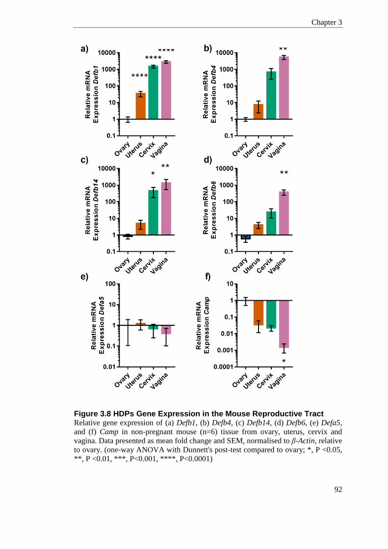
Chapter 3
92
Figure 3.8 HDPs Gene Expression in the Mouse Reproductive Tract Relative gene expression of (a) Defb1, (b) Defb4, (c) Defb14, (d) Defb6, (e) Defa5,
and (f) Camp in non-pregnant mouse (n=6) tissue from ovary, uterus, cervix and
vagina. Data presented as mean fold change and SEM, normalised to β-Actin, relative
to ovary. (one-way ANOVA with Dunnett's post-test compared to ovary; *, P <0.05,
**, P <0.01, ***, P<0.001, ****, P<0.0001)
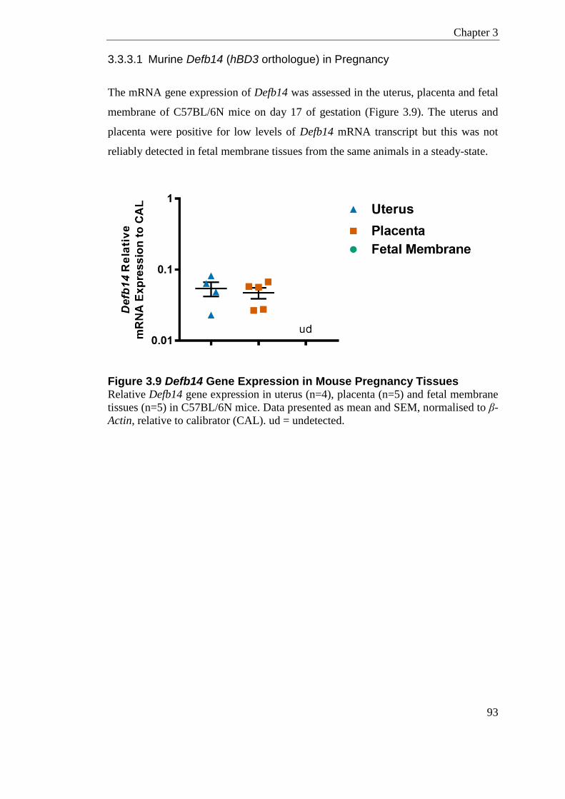
Chapter 3
93
3.3.3.1 Murine Defb14 (hBD3 orthologue) in Pregnancy
The mRNA gene expression of Defb14 was assessed in the uterus, placenta and fetal
membrane of C57BL/6N mice on day 17 of gestation (Figure 3.9). The uterus and
placenta were positive for low levels of Defb14 mRNA transcript but this was not
reliably detected in fetal membrane tissues from the same animals in a steady-state.
Figure 3.9 Defb14 Gene Expression in Mouse Pregnancy Tissues Relative Defb14 gene expression in uterus (n=4), placenta (n=5) and fetal membrane
tissues (n=5) in C57BL/6N mice. Data presented as mean and SEM, normalised to β-
Actin, relative to calibrator (CAL). ud = undetected.
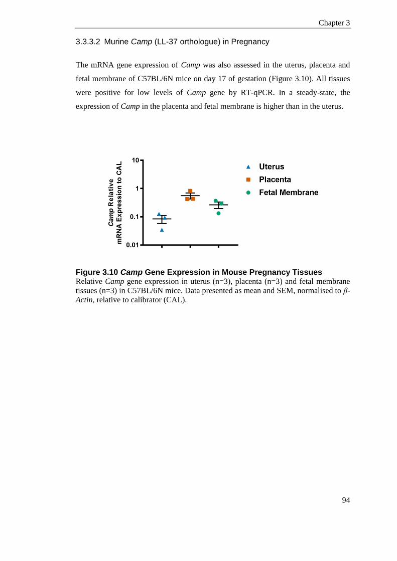
Chapter 3
94
3.3.3.2 Murine Camp (LL-37 orthologue) in Pregnancy
The mRNA gene expression of Camp was also assessed in the uterus, placenta and
fetal membrane of C57BL/6N mice on day 17 of gestation (Figure 3.10). All tissues
were positive for low levels of Camp gene by RT-qPCR. In a steady-state, the
expression of Camp in the placenta and fetal membrane is higher than in the uterus.
Figure 3.10 Camp Gene Expression in Mouse Pregnancy Tissues Relative Camp gene expression in uterus (n=3), placenta (n=3) and fetal membrane
tissues (n=3) in C57BL/6N mice. Data presented as mean and SEM, normalised to β-
Actin, relative to calibrator (CAL).
Chapter 3
95
3.4 Discussion
HDPs in Labour
In tissues and cells not of the reproductive tract, HDPs have been shown to modulate
inflammation. Infection and inflammation are frequently associated with preterm
labour in women. Preterm and term labour share a common terminal pathway
characterised by anatomical, biochemical, and endocrine events (Snegovskikh et al.,
2006). One aim of this chapter was to determine the effect of human term labour on
the expression of HDPs in the fetal membranes, myometrium and placenta of
women.
There was a small but significant increase in DEFB103 levels in labouring tissue of
the myometrium compared to non-labouring tissue. In humans, six of the beta-
defensin genes on chromosome 8 are copy number variable, this includes DEFB103.
Copy number has been shown to correlate with the protein expression and peptide
level (Hollox et al., 2008, Jansen et al., 2009, Fellermann et al., 2006). The beta-
defensin copy number of the women in this chapter was not determined, and could
account for some inter- patient variation in gene transcript levels.
In the placenta there was a slight increase in CAMP gene expression in labour
compared to non-labouring women. No difference in CAMP gene level was seen in
the myometrium or amnion/chorion. This leads to the conclusion that resident
epithelial cells of the myometrium and fetal membrane are not upregulating CAMP
during labour.
LL-37 is a chemoattractant for the stimulation and mobilisation of neutrophils and
eosinophils (Tjabringa et al., 2003). As leukocytes infiltrate uterine tissues around
the time of labour (Osman et al., 2003), it was estimated that the myometrial samples
in this chapter might have shown an increase in CAMP in labouring women.
Although there may be leukocytes infiltrating to the myometrium in labouring
samples; this is not contributing to mRNA gene transcript in this chapter. LL-37
peptide is stored pre-processed in neutrophils so it may have activity in labour at the
protein level. Investigation of protein expression levels of LL-37 and co-localisation
Chapter 3
96
with leukocyte markers would provide evidence for these cells supplying additional
stores of LL-37 during labour. Indeed this is what Lim et al., (2015) had shown in the
fetal membrane and myometrium. Lim et al., (2015) displayed that in labouring
women, immunostaining for hCAP18/LL-37 co-localised with infiltrating leukocytes
but also increased in resident amnion epithelium, chorionic trophoblasts and decidua,
which is not what I found in this chapter at the mRNA gene transcript level.
HDPs in Placenta Explant Cultures
In this chapter, I have shown that LL-37 was able to significantly reduce the LPS
induced increase in IL-8, IL-6, TNF, and IL-1β in placenta explant cultures. This
anti-inflammatory effect of LL-37 ex-vivo is likely due to LL-37 directly binding to
LPS. LL-37 can downregulate signalling through TLR-4 when coadministered with
LPS by preventing the interaction with LPB, essential for TLR-4 activation. Not all
of LL-37 anti-inflammatory properties are due to direct binding to LPS; pre-
treatment with LL-37 prior to LPS can also suppress TNF protein production
(Mookherjee et al., 2006). LL-37 can also act through disruption of TLR-4 receptor
complex function when LPS is already bound to macrophage receptors (Rosenfeld et
al., 2006). All actions can result in lower levels of pro-inflammatory cytokine
production. It would be interesting to further investigate the mechanism of LL-37’s
anti-inflammatory action in placental explants. This could be achieved using
inhibitors of known LL-37 receptors to elude what proportion of the effect might be
due to actions other than LL-37 and LPS binding.
It would be interesting to investigate whether the mouse peptide, CAMP is also anti-
inflammatory on murine placental tissues. This can be achieved by primary culture of
mouse placental tissue with exogenous CAMP and LPS. Camp-/- mice also provide
an additional control to that of human experiments where the endogenous CAMP
contributing to the LPS response can be accounted for. The in-vivo equivalent to this
has already somewhat been done in Chapter 7 of this thesis, where LPS was given
intrauterine during pregnancy in wildtype and Cathelicidin knockout animals. In
placental tissue, with a low dose of intrauterine LPS (1µg), there was a small but
significant reduction in the chemokine Cxcl1 and Ptgs2 (Figure 7.13). The number of
Chapter 3
97
animals in this treatment group was small and this difference was not seen when a
higher dose of 20µg LPS was used.
It was surprising that RANTES was not increased in response to LPS in the placenta
explant cultures in this chapter. Higher expression of RANTES (CCL5) and IL-8
(CXCL8) has been shown in trophoblast cell lines (Swan-71) after stimulation with
LPS (Grasso et al., 2014). Interestingly, after interacting with maternal monocytes,
these trophoblast cells did not modulate the expression of RANTES and IL-8 by LPS
stimulation (Grasso et al., 2014). In this chapter, the placental explant cultures have
high levels of blood, where maternal monocytes could modulate the expression of
these cytokines. Moreover, there are conflicting reports as to whether placenta tissues
express RANTES. Villous trophoblast only express very small amounts of RANTES
(Nakada et al., 2011) whereas trophoblast cell lines (Swan-71, SVneo) express high
amounts of RANTES which is increased in response to LPS and TNF (Grasso et al.,
2014). This might be the reason for the difference in levels of RANTES in placenta
explant cultures seen in this chapter compared to that reported in cell lines.
hBD3 has been shown to have potent anti-inflammatory effects on human and mouse
primary monocytes in response to LPS (Semple et al., 2010, Semple et al., 2011).
However, in this chapter, hBD3 treatment did not have an effect on the levels of any
of the cytokines measured in an environment of 100ng/ml of LPS on placental
explants. Only a single dose of hBD3 peptide [5µg/ml] was used in this chapter as
this has previously been shown to have anti-inflammatory effects in-vitro. It would
be ideal to conduct a dose response for hBD3 to further test any anti-inflammatory
effects in the placenta.
Other defensins are present in the female reproductive tract, including Human beta-
defensin 126 (DEFB126), an epididymis-specific secretory protein, which has the
ability to inhibit LPS-induced inflammation in macrophage cell lines (Liu et al.,
2012).
Chapter 3
98
HDPs in Mouse Pregnancy
There are only a few studies that investigate defensins and cathelicidins in the mouse
female reproductive tract (Hickey et al., 2013, Soboll et al., 2006). Hickey et al.,
(2013) measured the expression of several defensin genes in the murine vagina and
uterus at different stages of estrus. In line with what this paper reported, in this
chapter I have shown that many defensins are expressed at specific spatial patterns
along the reproductive tract. Estrus cycling in mice is continuous but there are
discrete phases; diestrus, proestrus, estrus and metestrus. In this chapter I used
hormones to induced estrus in mice, which coincides with ovulatory follicle growth
and vaginal neutrophil accumulation. I found beta-defensins including Defb14 and
Defb4 were highly expressed in the lower reproductive tract during this phase which
has continuous exposure to bacteria. Most defensins displayed reduced expression
the further up the reproductive tract, which is presumably a sterile environment.
Mice in the estradiol-dominant phase have peak defensin expression and it has been
shown that treating mice directly with estradiol can protect against pathogens
(Kaushic et al., 2003, Kaushic et al., 2000). The load of commensal bacteria in the
vagina is also elevated in estrus compared to other stages (Noguchi et al., 2003).
Estradiol treatment in-vitro has also been shown to enhance the production of HDPs,
namely beta-defensin 2 (human orthologue of Defb4) in uterine epithelial cells.
However, in humans the phase of the menstrual cycle dominated by progesterone has
been shown to display more resistance to infection with various pathogens (reviewed
in Wira et al., (2010). Another reason for lower defensin expression in the uterus
could be to support implantation of the fetus that is immunologically different to the
host.
Defb1 homozygous knockout mice have increased susceptibility to urinary tract
infections whereby they were shown to retain Staphylococcus spp. in the bladder
longer than wildtype mice (Morrison et al., 2002). Defb6 has been shown to have
tissue-specific expression in skeletal muscle along with esophagus, tongue, and
trachea (Yamaguchi et al., 2001). Defa5 has been found in epithelial cells of the
vagina and is highly expressed in Paneth cells, the secretory granules of the small
intestine.
Chapter 3
99
In this study Camp mRNA was expressed highly in the ovary during estrus, the
reasons for this are not clear but as Camp and LL-37 have been shown to be involve
in tissue repair it could be playing a role in proliferation of the ovarian surface.
Pattern recognition receptors are not uniformly expressed along the reproductive
tract and also show temporal expression patterns that coincide with estrus cycling in
mice. The numbers of TLR-2 and TLR-5 molecules are higher in the vagina than
uterus, whereas in general, TLR-4 is higher in the uterus than in the vagina (Hickey
et al., 2013). This is most probably a co-adaption strategy, to allow host tolerances of
commensal and mutualistic species while still being effective to detect invading
pathogens. This has also been shown in humans where TLR-4 and the NOD-2
receptor are found at higher levels in the upper reproductive tract and reduces the
further down the reproductive tract (Pioli et al., 2004, Hart et al., 2009). Other TLRs
are more uniformly expressed along the reproductive tract, namely TLR-9, TLR-8
and TLR-7 (Hart et al., 2009), these TLRs in mice also displayed more consistent
expression in the vagina and uterus regardless of the stage of estrus (Hickey et al.,
2013, Soboll et al., 2006).
Whether changes in HDP levels alter the vaginal microbiome directly has not been
established, but changes in levels are accompanied by alterations of commensal and
pathogenic bacteria in humans and in mice (Mitchell et al., 2013, Fan et al., 2008,
Soboll et al., 2006, Frew et al., 2014).
Chapter 3
100
3.5 Summary
In summary, in this chapter I have shown:
- DEFB103 and CAMP are expressed in myometrium, placenta and
amnion/chorion tissues in term pregnancy.
- DEFB103 expression was upregulated in the myometrium during normal
labour, whereas CAMP expression was upregulated in the placenta.
- Placental explants respond to LPS challenge by increasing production of pro-
inflammatory cytokines.
- LL-37 but not hBD3 peptide was able to modulate LPS induced inflammatory
response by inhibiting the release of pro-inflammatory cytokines in the
placenta.
- Beta-defensins; Defb1, Defb4, Defb6, Defb14, and the cathelicidin Camp
show spatial expression patterns along the female reproductive tract in mice.

101
Chapter 4
Microbiome - 16S rRNA Gene
Sequencing
Chapter 4
102
Chapter 4 Microbiome - 16S rRNA Gene Sequencing
4.1 Introduction
Several methods for bacterial species identification and quantification are available
to researchers; these methods classify bacteria to varying taxonomic levels and all
have their associated advantages and disadvantages. Culture based studies can cause
misrepresentation of some species due to selection in culture conditions. There are
various indirect molecular based methods for measuring cell viability, such as
membrane staining, membrane potential, flow cytometry and reporter gene analysis.
Fluorescent in situ hybridization (FISH) is a commonly used technique for
localisation and quantification of bacterial species. FISH probes, usually for the taxa
specific expanse of the 16S rRNA gene, can be used on tissues sections of interest
for localising bacterial taxa. FISH probes are advancing the specificity of genus-
specific identification, however, the throughput and multiplexing capability is still
limited compared to broad range techniques such as 16S rRNA gene sequencing.
Whole-genome shotgun metagenomic sequencing can also be undertaken for
microbiome analysis, where the entire genomic content in a sample is sequenced.
This is becoming an ever more popular method for public health surveillance in
analysis of outbreaks, but also in the study of antimicrobial resistance mechanisms
and pathogenicity. While full genome sequencing generally can offer researchers the
ability to classify bacteria to a greater taxonomic level, there is often a compromise
with regards to sequencing depth. A metagenomic approach sequences the entire
genomic content of a sample, which depending on how and where the sample is
collected from can contain high levels of host DNA. This is not ideal when trying to
keep the per sample cost low, as greater depth is needed to obtain microbial
sequences. Target gene approaches such as 16S rRNA gene sequencing can offer
increased read depth, which puts this approach in a better position to highlight low-
abundant taxa that may be overlooked in shotgun sequencing.
The objectives of this chapter were to establish a next generation sequencing
workflow for microbial species identification in murine vaginal and fecal samples.
This microbiome analysis utilises the 16S rRNA gene as a target for phylogenetic
Chapter 4
103
reconstruction and species identification. The microbiome field has yet to establish a
gold standard for looking at microbial communities; workflows need to be tailored
and validated for specific sites of interest. Multiple considerations are needed at
almost every point of the workflow. In relation to sample processing, consideration is
needed in; sampling method, sample storage duration, sample storage temperature
and method of DNA extraction. Multiple considerations are also required when
selecting a 16S rRNA gene interrogation method with regards to the amplicon
primers, amplicon length, and sequencing platform limitations. Having sufficient
read quality, read depth and choosing an appropriate method for OTU picking are
also important factors in microbial species identification. Chimeric reads produced
by PCR artifacts can lead to the false discovery of unique organisms and effect
community inference. In this regard optimisation is needed to keep PCR cycles to the
minimum needed for amplification, in addition to the stringent downstream
bioinformatic filtering of proposed chimeric reads. Employing a paired end
sequencing approach has the added benefit of sequence overlap, which helps to
reduce sequencing artefact contribution to community inference. There are many
bioinformatic tools available to researchers interrogating the 16S rRNA gene.
Quantitative Insights in to Microbial Ecology (QIIME) (Caporaso et al., 2010b) and
Mothur (Schloss et al., 2009) are among the most highly used. As 16S rRNA gene
sequencing is becoming an ever popular choice from microbial species analysis there
is an increasing amount of rRNA sequence data accessible in open databases such as
SILVA (Quast et al., 2013), Ribosomal Database Project (RDP) (Cole et al., 2009),
and Greengenes (DeSantis et al., 2006b).
4.2 Targeted 16S rRNA Gene Amplification
The bacterial 16S Ribosomal RNA gene is approximately 1500bp and consists of
nine hyper-variable regions (V1-V9), that when sequenced can be used to identify
bacteria taxonomy to the species or genus level (Figure 4.1). The microbial field has
mixed opinion as to which hypervariable region provides the highest number of
operational taxonomic units (OTUs) when using next generation sequencing
Chapter 4
104
platforms. It is becoming increasingly clear this is dependent on the diversity of
bacteria in the community sampled (Chakravorty et al., 2007). Nossa (2010) looked
at sequence data from all nine hyper-variable regions and proposed that in human
foregut samples the V3 and V4 region is the least effective at class level bacterial
identification. However, Whiteley et al., (2012) regarded sequencing the V6 region
not acceptable for environmental samples and suggested the V3-V4 region provides a
more representative result. Recent in-silico work on human fecal samples
demonstrated that the V3-V4 and V4-V5 regions would most likely give more OTUs
than the other variable regions (Claesson et al., 2010). However, this predicted
advantage was not seen in actual sequencing analysis (Claesson et al., 2010), most
likely due to accumulation bias from the primers and the extraction method
employed.
Comparable to other mucosal body sites, such as the gastrointestinal tract, the human
vagina has considerably less bacterial species in terms of both diversity and bacterial
load (Cho and Blaser, 2012, Ravel et al., 2011). At the current level of sequencing,
the human vaginal microbiome is establishing around 200 bacterial assigned OTUs
across study sets (Ravel et al., 2011, Drell et al., 2013, Martin et al., 2013). A large
study on the human vaginal microbiome suggested that the V1-V2 region has greater
variability than the V3 region for species identification of Lactobacillus and
Prevotella, giving potential higher OTU counts (Ravel et al., 2012). In addition the
V1-V3 region has been reported to have a higher degree of accuracy in classifying
species (Kim et al., 2011), and also less species specific bias (Vilo and Dong, 2012).
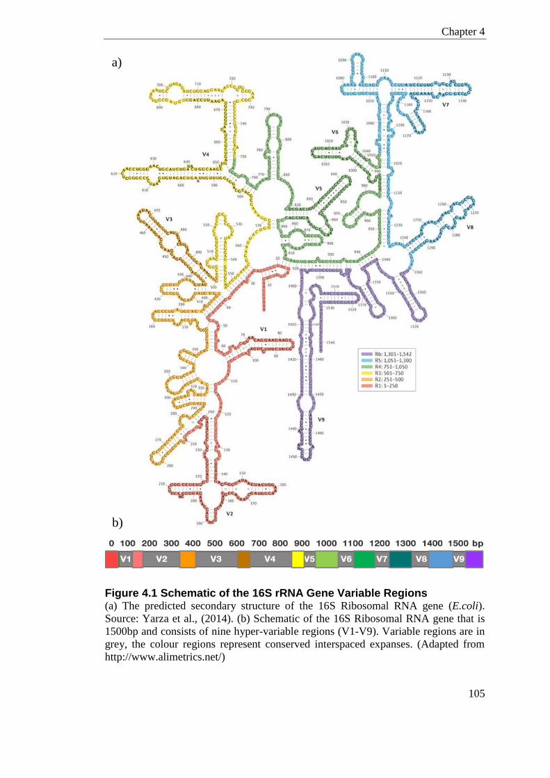
Chapter 4
105
Figure 4.1 Schematic of the 16S rRNA Gene Variable Regions (a) The predicted secondary structure of the 16S Ribosomal RNA gene (E.coli).
Source: Yarza et al., (2014). (b) Schematic of the 16S Ribosomal RNA gene that is
1500bp and consists of nine hyper-variable regions (V1-V9). Variable regions are in
grey, the colour regions represent conserved interspaced expanses. (Adapted from
http://www.alimetrics.net/)
a)
b)
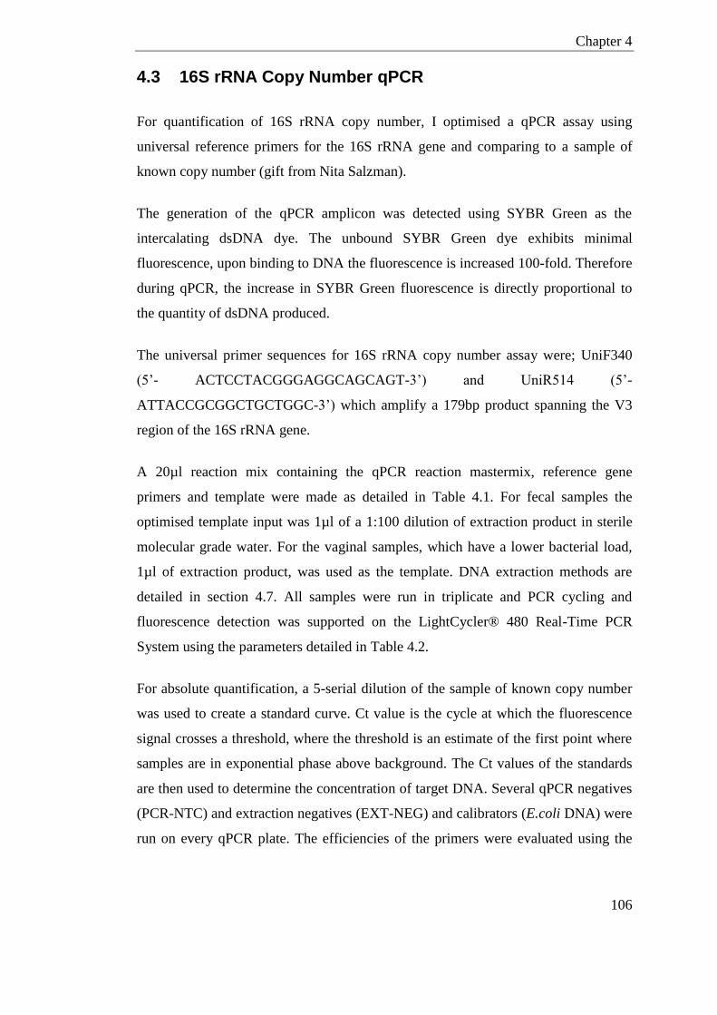
Chapter 4
106
4.3 16S rRNA Copy Number qPCR
For quantification of 16S rRNA copy number, I optimised a qPCR assay using
universal reference primers for the 16S rRNA gene and comparing to a sample of
known copy number (gift from Nita Salzman).
The generation of the qPCR amplicon was detected using SYBR Green as the
intercalating dsDNA dye. The unbound SYBR Green dye exhibits minimal
fluorescence, upon binding to DNA the fluorescence is increased 100-fold. Therefore
during qPCR, the increase in SYBR Green fluorescence is directly proportional to
the quantity of dsDNA produced.
The universal primer sequences for 16S rRNA copy number assay were; UniF340
(5’- ACTCCTACGGGAGGCAGCAGT-3’) and UniR514 (5’-
ATTACCGCGGCTGCTGGC-3’) which amplify a 179bp product spanning the V3
region of the 16S rRNA gene.
A 20µl reaction mix containing the qPCR reaction mastermix, reference gene
primers and template were made as detailed in Table 4.1. For fecal samples the
optimised template input was 1µl of a 1:100 dilution of extraction product in sterile
molecular grade water. For the vaginal samples, which have a lower bacterial load,
1µl of extraction product, was used as the template. DNA extraction methods are
detailed in section 4.7. All samples were run in triplicate and PCR cycling and
fluorescence detection was supported on the LightCycler® 480 Real-Time PCR
System using the parameters detailed in Table 4.2.
For absolute quantification, a 5-serial dilution of the sample of known copy number
was used to create a standard curve. Ct value is the cycle at which the fluorescence
signal crosses a threshold, where the threshold is an estimate of the first point where
samples are in exponential phase above background. The Ct values of the standards
are then used to determine the concentration of target DNA. Several qPCR negatives
(PCR-NTC) and extraction negatives (EXT-NEG) and calibrators (E.coli DNA) were
run on every qPCR plate. The efficiencies of the primers were evaluated using the
Chapter 4
107
serial dilution of the sample of known copy number; this was determined to be
within the acceptable range of E= 1.9-2.1.
Component Final Concentration /
Reaction
20µl
Reaction
LightCycler490 SYBR
mastermix
x1 10 Primer F [20µM] 0.4 µM 0.2
Primer R [20µM] 0.4 µM 0.2
Template Variable 1.0
H20 / 8.6
Table 4.1 qPCR Reaction Mix for 16S rRNA Copy Number Assay
Temperature Time Number of Cycles
50°C 2 minutes x 1 95°C 10 minutes x 1
95°C 30 seconds x 45
63°C 30 seconds
Table 4.2 qPCR Cycle Parameters for 16S rRNA Copy Number Assay
4.4 16S rRNA Gene Sequencing Primer Design
4.4.1 In-silico Variable Region Evaluation
The variable region chosen to sequence for bacterial identification in this study is the
V1-V2 region of the 16S rRNA gene. This region was highlighted as a good
candidate; firstly as literature suggested it has an advantage for discrimination of
Lactobacillus spp. (Ravel et al., 2012), secondly this region displayed higher
discrimination potential in a test-dataset, ‘TEST-SET’, I created in-silico (detailed
below).
The ‘TEST-SET’ was comprised of 949 full length 16S rRNA sequences from
species likely to be encountered in the; murine female reproductive tract (highlighted
in culture studies), murine gastrointestinal tract (highlighted in 16S rRNA studies)
Chapter 4
108
and sequences of species known to colonise the human vagina (highlighted in 16S
rRNA studies). The two regions highlighted as potential candidates were the, V1-V2
region and the V4-V5, as amplicon products from both regions would be less than
500bp, which was a criterion for selection. Three pairwise percent identity matrixes
were calculated in Clustal2.1 (Larkin et al., 2007) for; the full length 16S rRNA, the
V1-V2 and the V4-V5 regions. Pairwise identity above 97% would suggest
sequences are indistinguishable when clustering OTUs for microbial identification.
The identity matrix for the full length 16S rRNA region calculated that 0.003% of the
pairs had sequence similarity above 97%. The V1-V2 region matrix revealed only
0.0009% of the sequence pairs had higher than 97% similarity, whereas for the V4 -
V5 matrix this was 0.01%. Therefore in this test data-set sequencing the V1-V2
region will identify more OTUs than if the V4-V5 region were sequenced. As the
‘TEST-SET’ contains the species which are likely to be encountered in this study the
V1-V2 region was chosen as the desired amplicon region of the 16S rRNA gene.
The 16S rRNA V1-V2 hypervariable region is flanked by more conserved regions
common to all bacteria, this allows for universal primer hybridisation and
amplification. Even in the denoted ‘conserved’ regions no primer sites are totally
conserved; where one primer pair will not amplify 100% of bacteria species even
with miss-matches allowed. Deciding which one/ones to use depends on the
complement of bacteria in the sample and each pair could slightly bias towards a
certain type of bacteria (Milani et al., 2013). In the literature, there are over 30
forward and reverse primers that have been used as universal probes for the V1-V2
region (Klindworth et al., 2013). Klindworth et al., (2013) provides a great review of
common universal 16S rRNA gene primers and conducted in-silico evaluation on
coverage against the SILVA database.
4.4.2 In-silico Variable Region Primer Evaluation
An optimized primer set comprising of 27f-YM (universal forward 5’-
AGAGTTTGATYMTGGCTCAG-3’) and 338r (universal reverse 5’-
TGCTGCCTYCCGTAGGAGT-3’) were chosen to amplify the V1-V2 region based
on the following criteria; the size of product did not exceed 500bp, annealing
Chapter 4
109
temperatures were within 5°C, universal coverage of > 90% 16S rRNA sequences in
SILVA database (v119), and universal coverage of > 90% 16S rRNA sequences in
the ‘TEST-SET’. The forward primer 27f-YM has 92.2% coverage (one mismatch)
and the reverse primer 338r has 95.2% coverage of bacterial sequences in the SILVA
database (one mismatch). The forward primer is commonly referred to as 27f-YM as
it ends at E.coli position 27 of the 16S rRNA gene and has the degenerate bases YM.
The forward 27f-YM represents four discrete primer sequences. The reverse primer
sequence 338r, which ends at E.coli position 388, also has a degenerate base that
represents two discrete primer sequences.
Sequencing in this study was done on two platforms; the Ion Torrent PGM (life
technologies) and the Illumina MiSeq. Each platform has associated platform
specific primer sequences that are incorporated into the fusion primers, detailed in
section 4.8.2 and 4.8.1 respectively. On both platforms 27f-YM (universal forward)
and 338r (universal reverse) were used as the complementary sequence for 16S
rRNA gene amplification.
For the MiSeq primers only, mixtures of four forward 27f primers were used to
maximise discovery of all bacterial species. This mixture has been shown to
eliminate PCR bias and maintain the best ratio of Lactobacillus spp. to Gardnerella
spp. as determined by qPCR (Frank et al., 2008, Romero et al., 2014). The forward
primer mix consisted of 4 parts 27f-YM (the universal forward), and one part of each
27f-Bif, 27f-Bor and 27f-Chl, these primers are specific for the amplification of
Bifidobacteriaceae, Borrelia and Chlamydiales respectively (Table 4.7). The
universal primer 27f-YM alone would not amplify these species in stringent
conditions. The degenerate bases within the forward primer mix provide seven
discrete primer sequences.
For reference, the universal forward primer (27f-YM) best matches ‘S-D-Bact-0008-
b-S-20’, and the universal reverse primer (338r) best matches ‘S-D-Bact-0338-a-A-
19’ in Klindworth et al., (2013) nomenclature.
Chapter 4
110
4.5 Contamination Considerations
An important consideration for microbial sequencing is contamination from
laboratory equipment, molecular grade water, PCR reagents and extraction kits.
Many of the extraction kits used for microbiome analysis are known to have low
level contaminants (Salter et al., 2014). This is of particular importance with low bio-
mass samples such as from the murine vagina in this study. It is often not possible to
exclude these contaminates without additional sequencing of various negative
controls from each kit batch.
In this study extraction kits were ordered in bulk from the same manufacture lot
number to reduce and account for any lot specific contamination. For quality control,
extraction negatives were included in every batch of extractions. To reduce potential
contamination, equipment was cleaned with 70% alcohol, UV treated where suitable,
and then cleaned with RNAzap. Extractions and subsequent PCRs were carried out in
a designated PCR clean-room.
4.6 Sampling method
Fecal Sampling
Clean autoclaved cages were used as collection containers for fecal sampling of each
mouse. One fecal pellet per mouse was immediately collected using disposable
sterile forceps into sterile 2ml tubes and kept at 4°C within 20 minutes and -80°C
within 2 hours. As the bacterial load in murine fecal samples is very high there was
little optimisation needed with regards to sampling approach and yield.
Vaginal Sampling
As the murine vagina has low biomass, different sampling methods were compared
to evaluate bacterial load recovery. One sampling method was a vaginal lavage,
where 200µl of sterile physiological saline [Sodium Chloride 0.9%] is washed in the
vagina 5 times and then extracted as detailed in section 4.7 using the PowerSoil®
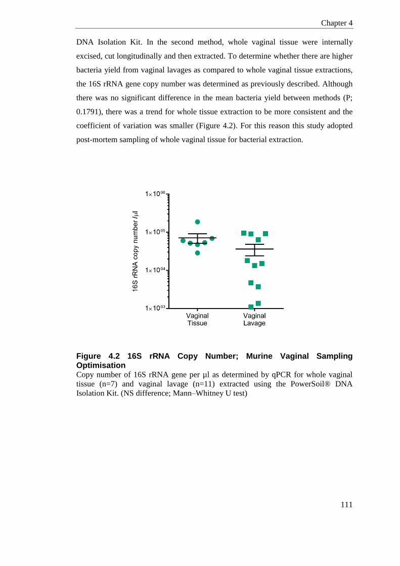
Chapter 4
111
DNA Isolation Kit. In the second method, whole vaginal tissue were internally
excised, cut longitudinally and then extracted. To determine whether there are higher
bacteria yield from vaginal lavages as compared to whole vaginal tissue extractions,
the 16S rRNA gene copy number was determined as previously described. Although
there was no significant difference in the mean bacteria yield between methods (P;
0.1791), there was a trend for whole tissue extraction to be more consistent and the
coefficient of variation was smaller (Figure 4.2). For this reason this study adopted
post-mortem sampling of whole vaginal tissue for bacterial extraction.
Figure 4.2 16S rRNA Copy Number; Murine Vaginal Sampling Optimisation Copy number of 16S rRNA gene per µl as determined by qPCR for whole vaginal
tissue (n=7) and vaginal lavage (n=11) extracted using the PowerSoil® DNA
Isolation Kit. (NS difference; Mann–Whitney U test)

Chapter 4
112
4.7 Bacterial DNA Extraction
The method of bacterial DNA extraction has a profound impact in community
structure as different methods can have bias in the lysis of certain bacterial cell walls
(Milani et al., 2013, Wesolowska-Andersen et al., 2014). Two different extraction
kits were used in this study for microbial DNA isolation, the PowerSoil® Isolation
Kit and the FastDNA® SPIN Kit. DNA extractions using either kit were carried out
according to the manufacturer’s instructions. The PowerSoil® DNA Isolation Kit is
the method of choice for the Human Microbiome Project (Human Microbiome,
2010) and was initially used in this study for samples sequenced on the Ion Torrent
platform. However, recent publication suggested that for low biomass samples, the
FastDNA® SPIN Kit with bead-beating may yield better results and less bias
towards Bacteroidetes phylum than the PowerSoil® DNA Isolation Kit (Milani et al.,
2013, Wesolowska-Andersen et al., 2014). FastDNA® SPIN Kit for DNA extraction
gave more consistent bacterial yields from vaginal tissue than the PowerSoil® DNA
Kit (Figure 4.3). This method was adopted for all samples both fecal and vaginal run
on the MiSeq platform.
Figure 4.3 16S rRNA Copy Number; Extraction Kit Optimisation Copy number of 16S rRNA gene per µl as determined by qPCR for whole vaginal
tissue using the PowerSoil® DNA Isolation Kit (n=9) or the FastDNA® SPIN Kit
(n=6) (NS difference; Mann–Whitney U test)
Chapter 4
113
4.8 Library Preparation
4.8.1 Illumina MiSeq Primer Design and PCR Optimisation
The number of sequence reads generated on an Illumina MiSeq lane vastly exceeds
the number of reads needed per sample for species identification. For this reason and
to reduce per-sample sequencing costs, amplicon libraries were pooled in a 64-plex.
The complementary forward and reverse primer regions have been designed to be as
universal as possible for bacterial species likely to be encountered in biological
specimens from the vagina, but also murine gastrointestinal sites. Specific
consideration has been given to accurately represent bacterial species common to the
vagina by mixing 4 different forward primers in specific proportions (see section
4.4.2).
For multiplexing capability, the forward and reverse primers are indexed with
TruSeq index sequences. Primers have been designed such that they contain the
correctly orientated standard MiSeq sequencing and indexing primer sites that are
compatible with MiSeq HP10, HP11 and HP12. These Illumina sequencing and
indexing primers are contained within the reagent cartridge as standard. To account
for possible low complexity in the library pool, heterogeneity spacers were
incorporated into the designed fusion primers. To reduce oligo crosstalk,
oligonucleotides with TruGrade™ processing were synthesised from IDT (Integrated
DNA Technologies). Full MiSeq primer sequences are given in Table 4.7 and Table
4.8. Primers were checked for possibility of secondary structure formation and
primer dimers.
Optimisation is required to get strong amplification results using fusion primers. To
optimise PCR conditions for MiSeq library preparation several PCR parameters were
compared, namely; template volume, template 16S rRNA copy number, cycle
number, annealing temperature, polymerase, and use of reducing agents.
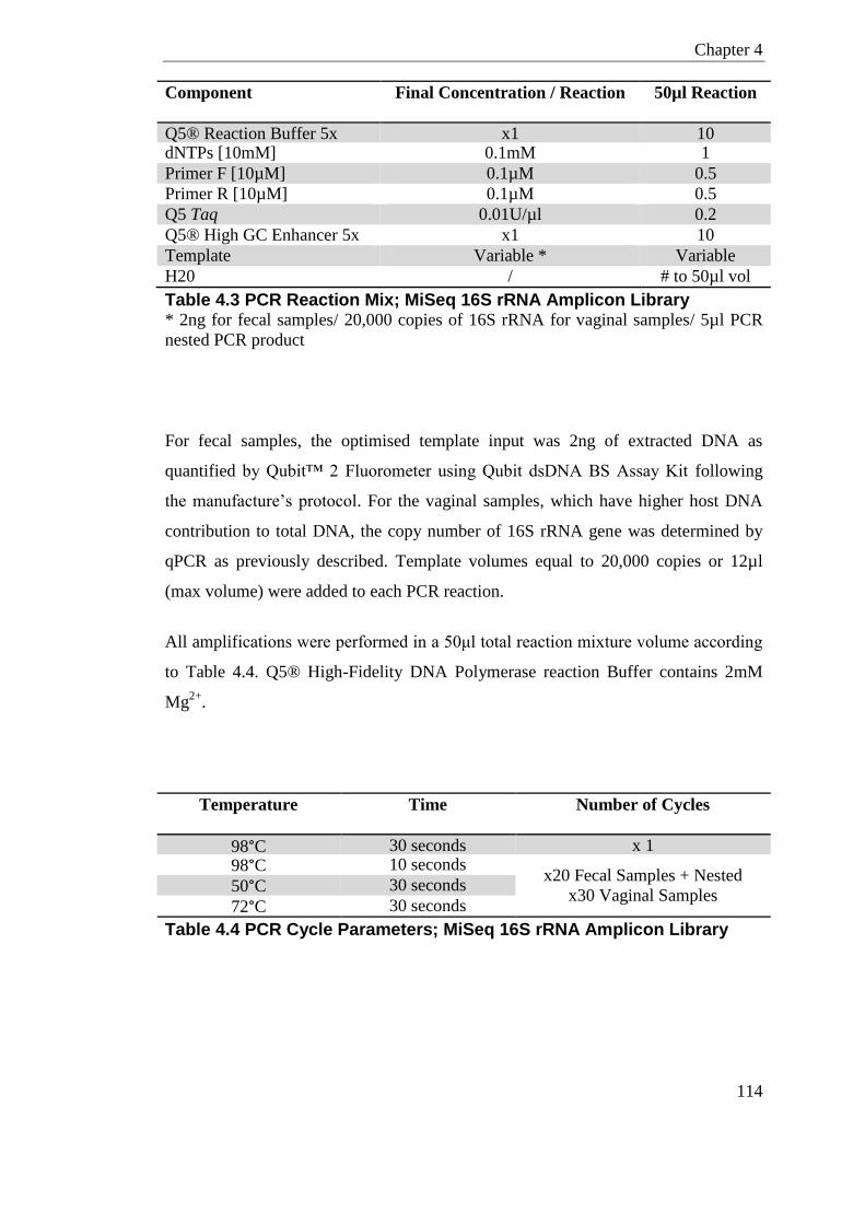
Chapter 4
114
Component Final Concentration / Reaction 50µl Reaction
Q5® Reaction Buffer 5x x1 10 dNTPs [10mM] 0.1mM 1
Primer F [10µM] 0.1µM 0.5
Primer R [10µM] 0.1µM 0.5
Q5 Taq 0.01U/µl 0.2
Q5® High GC Enhancer 5x x1 10
Template Variable * Variable
H20 / # to 50µl vol
Table 4.3 PCR Reaction Mix; MiSeq 16S rRNA Amplicon Library * 2ng for fecal samples/ 20,000 copies of 16S rRNA for vaginal samples/ 5µl PCR
nested PCR product
For fecal samples, the optimised template input was 2ng of extracted DNA as
quantified by Qubit™ 2 Fluorometer using Qubit dsDNA BS Assay Kit following
the manufacture’s protocol. For the vaginal samples, which have higher host DNA
contribution to total DNA, the copy number of 16S rRNA gene was determined by
qPCR as previously described. Template volumes equal to 20,000 copies or 12µl
(max volume) were added to each PCR reaction.
All amplifications were performed in a 50μl total reaction mixture volume according
to Table 4.4. Q5® High-Fidelity DNA Polymerase reaction Buffer contains 2mM
Mg2+
.
Temperature Time Number of Cycles
98°C 30 seconds x 1
98°C 10 seconds x20 Fecal Samples + Nested
x30 Vaginal Samples 50°C 30 seconds
72°C 30 seconds
Table 4.4 PCR Cycle Parameters; MiSeq 16S rRNA Amplicon Library
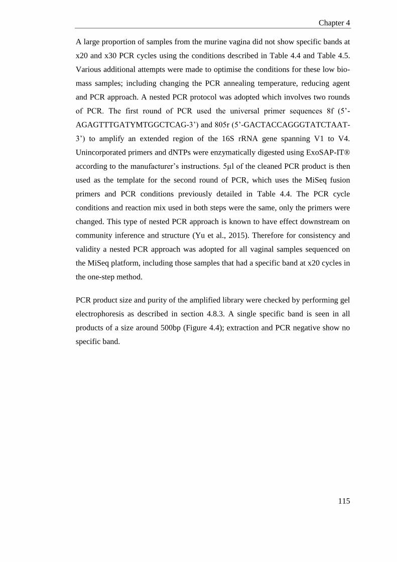
Chapter 4
115
A large proportion of samples from the murine vagina did not show specific bands at
x20 and x30 PCR cycles using the conditions described in Table 4.4 and Table 4.5.
Various additional attempts were made to optimise the conditions for these low bio-
mass samples; including changing the PCR annealing temperature, reducing agent
and PCR approach. A nested PCR protocol was adopted which involves two rounds
of PCR. The first round of PCR used the universal primer sequences 8f (5’-
AGAGTTTGATYMTGGCTCAG-3’) and 805r (5’-GACTACCAGGGTATCTAAT-
3’) to amplify an extended region of the 16S rRNA gene spanning V1 to V4.
Unincorporated primers and dNTPs were enzymatically digested using ExoSAP-IT®
according to the manufacturer’s instructions. 5µl of the cleaned PCR product is then
used as the template for the second round of PCR, which uses the MiSeq fusion
primers and PCR conditions previously detailed in Table 4.4. The PCR cycle
conditions and reaction mix used in both steps were the same, only the primers were
changed. This type of nested PCR approach is known to have effect downstream on
community inference and structure (Yu et al., 2015). Therefore for consistency and
validity a nested PCR approach was adopted for all vaginal samples sequenced on
the MiSeq platform, including those samples that had a specific band at x20 cycles in
the one-step method.
PCR product size and purity of the amplified library were checked by performing gel
electrophoresis as described in section 4.8.3. A single specific band is seen in all
products of a size around 500bp (Figure 4.4); extraction and PCR negative show no
specific band.
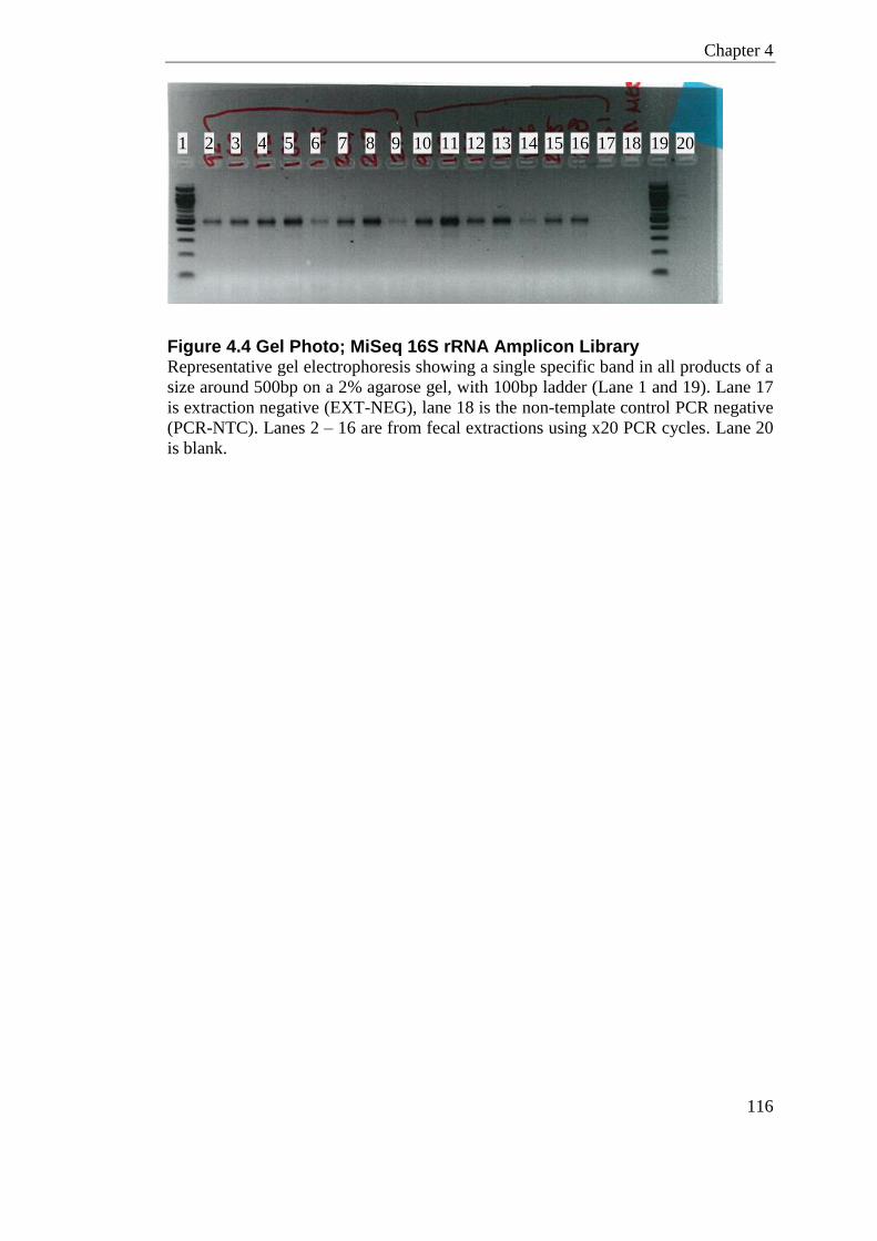
Chapter 4
116
Figure 4.4 Gel Photo; MiSeq 16S rRNA Amplicon Library Representative gel electrophoresis showing a single specific band in all products of a
size around 500bp on a 2% agarose gel, with 100bp ladder (Lane 1 and 19). Lane 17
is extraction negative (EXT-NEG), lane 18 is the non-template control PCR negative
(PCR-NTC). Lanes 2 – 16 are from fecal extractions using x20 PCR cycles. Lane 20
is blank.
1 2 3 5 6 10 11 12 13 14 15 16 17 18 19 20 7 8 9 4
Chapter 4
117
4.8.2 Ion Torrent PGM Primer Design and PCR Optimisation
Microbial sequencing on the Ion Torrent PGM platform using the 400bp Sequencing
Chemistry Kit and PGM 314 chip took advantage of dual-indexed fusion-primers to
reduce per sample cost. Forward and reverse fusion primers were designed to include
the sequences 27f and 338r respectively (as described in section 4.4.2) to amplify the
V1-V2 variable region of the 16S rRNA gene. I designed these primers to include, at
the 5’ end, one of two adaptor sequences (adaptor trP1 or adaptor A) linking to a
unique barcode tag (IonXpressBarcode). Short linkers ‘Key’ and ‘Barcode-Adapter’
sequences were also included. All primers were HPLC synthesised by Sigma® Life
Science; the complete list of Ion Torrent fusion-primers used in this study are shown
in Table 4.9. Primers were checked for possibility of secondary structure formation
and primer dimers.
To optimise PCR conditions for Ion Torrent PGM library preparation, several PCR
parameters were compared as before, namely; template volume, template 16S rRNA
copy number, cycle number, annealing temperature, polymerase, and use of reducing
agents.
For fecal samples the optimised template input was 1.5ng of extracted DNA, as
determined by Qubit™ 2 Fluorometer using Qubit dsDNA BR Assay Kit. For the
vaginal samples, which have higher host DNA contribution to total DNA, the copy
number of 16S rRNA gene was determined by qPCR. Template volumes equal to
20,000 copies or 12µl (max volume) were added to PCR reaction.
All amplifications were performed in a 50μl total reaction mixture volume according
to Table 4.5. Q5® High-Fidelity DNA Polymerase reaction Buffer contains 2mM
Mg2+
.
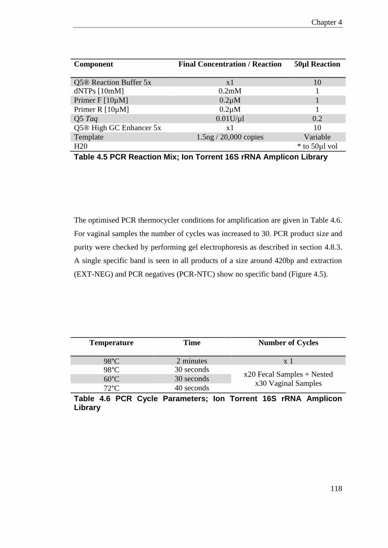
Chapter 4
118
Component Final Concentration / Reaction 50µl Reaction
Q5® Reaction Buffer 5x x1 10 dNTPs [10mM] 0.2mM 1
Primer F [10µM] 0.2µM 1
Primer R [10µM] 0.2µM 1
Q5 Taq 0.01U/µl 0.2
Q5® High GC Enhancer 5x x1 10
Template 1.5ng / 20,000 copies Variable
H20 * to 50µl vol
Table 4.5 PCR Reaction Mix; Ion Torrent 16S rRNA Amplicon Library
The optimised PCR thermocycler conditions for amplification are given in Table 4.6.
For vaginal samples the number of cycles was increased to 30. PCR product size and
purity were checked by performing gel electrophoresis as described in section 4.8.3.
A single specific band is seen in all products of a size around 420bp and extraction
(EXT-NEG) and PCR negatives (PCR-NTC) show no specific band (Figure 4.5).
Temperature Time Number of Cycles
98°C 2 minutes x 1
98°C 30 seconds x20 Fecal Samples + Nested
x30 Vaginal Samples 60°C 30 seconds
72°C 40 seconds
Table 4.6 PCR Cycle Parameters; Ion Torrent 16S rRNA Amplicon Library
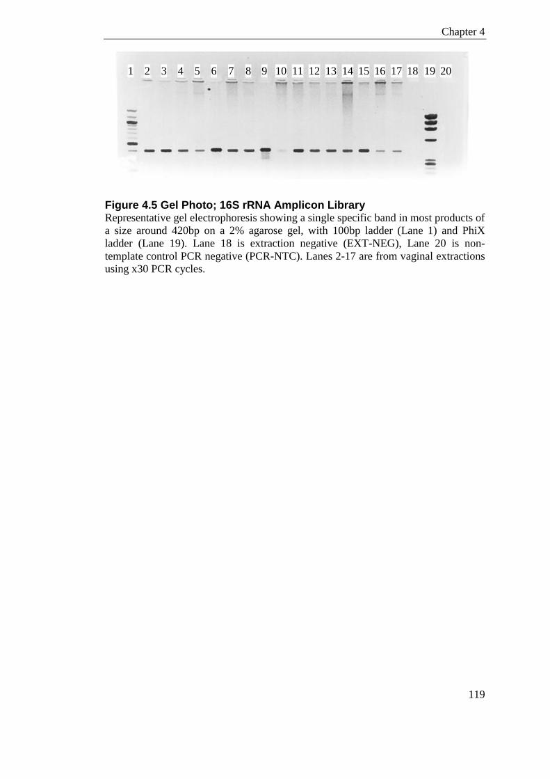
Chapter 4
119
Figure 4.5 Gel Photo; 16S rRNA Amplicon Library Representative gel electrophoresis showing a single specific band in most products of
a size around 420bp on a 2% agarose gel, with 100bp ladder (Lane 1) and PhiX
ladder (Lane 19). Lane 18 is extraction negative (EXT-NEG), Lane 20 is non-
template control PCR negative (PCR-NTC). Lanes 2-17 are from vaginal extractions
using x30 PCR cycles.
1 2 3 5 6 10 11 12 13 14 15 16 17 18 19 20 7 8 9 4
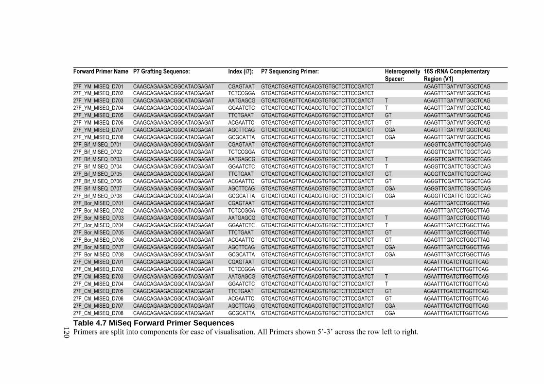
120
Table 4.7 MiSeq Forward Primer Sequences Primers are split into components for ease of visualisation. All Primers shown 5’-3’ across the row left to right.
Forward Primer Name P7 Grafting Sequence: Index (i7): P7 Sequencing Primer: Heterogeneity Spacer:
16S rRNA Complementary Region (V1)
27F_YM_MISEQ_D701 CAAGCAGAAGACGGCATACGAGAT CGAGTAAT GTGACTGGAGTTCAGACGTGTGCTCTTCCGATCT AGAGTTTGATYMTGGCTCAG 27F_YM_MISEQ_D702 CAAGCAGAAGACGGCATACGAGAT TCTCCGGA GTGACTGGAGTTCAGACGTGTGCTCTTCCGATCT AGAGTTTGATYMTGGCTCAG
27F_YM_MISEQ_D703 CAAGCAGAAGACGGCATACGAGAT AATGAGCG GTGACTGGAGTTCAGACGTGTGCTCTTCCGATCT T AGAGTTTGATYMTGGCTCAG
27F_YM_MISEQ_D704 CAAGCAGAAGACGGCATACGAGAT GGAATCTC GTGACTGGAGTTCAGACGTGTGCTCTTCCGATCT T AGAGTTTGATYMTGGCTCAG
27F_YM_MISEQ_D705 CAAGCAGAAGACGGCATACGAGAT TTCTGAAT GTGACTGGAGTTCAGACGTGTGCTCTTCCGATCT GT AGAGTTTGATYMTGGCTCAG
27F_YM_MISEQ_D706 CAAGCAGAAGACGGCATACGAGAT ACGAATTC GTGACTGGAGTTCAGACGTGTGCTCTTCCGATCT GT AGAGTTTGATYMTGGCTCAG
27F_YM_MISEQ_D707 CAAGCAGAAGACGGCATACGAGAT AGCTTCAG GTGACTGGAGTTCAGACGTGTGCTCTTCCGATCT CGA AGAGTTTGATYMTGGCTCAG
27F_YM_MISEQ_D708 CAAGCAGAAGACGGCATACGAGAT GCGCATTA GTGACTGGAGTTCAGACGTGTGCTCTTCCGATCT CGA AGAGTTTGATYMTGGCTCAG
27F_Bif_MISEQ_D701 CAAGCAGAAGACGGCATACGAGAT CGAGTAAT GTGACTGGAGTTCAGACGTGTGCTCTTCCGATCT AGGGTTCGATTCTGGCTCAG
27F_Bif_MISEQ_D702 CAAGCAGAAGACGGCATACGAGAT TCTCCGGA GTGACTGGAGTTCAGACGTGTGCTCTTCCGATCT AGGGTTCGATTCTGGCTCAG
27F_Bif_MISEQ_D703 CAAGCAGAAGACGGCATACGAGAT AATGAGCG GTGACTGGAGTTCAGACGTGTGCTCTTCCGATCT T AGGGTTCGATTCTGGCTCAG
27F_Bif_MISEQ_D704 CAAGCAGAAGACGGCATACGAGAT GGAATCTC GTGACTGGAGTTCAGACGTGTGCTCTTCCGATCT T AGGGTTCGATTCTGGCTCAG
27F_Bif_MISEQ_D705 CAAGCAGAAGACGGCATACGAGAT TTCTGAAT GTGACTGGAGTTCAGACGTGTGCTCTTCCGATCT GT AGGGTTCGATTCTGGCTCAG
27F_Bif_MISEQ_D706 CAAGCAGAAGACGGCATACGAGAT ACGAATTC GTGACTGGAGTTCAGACGTGTGCTCTTCCGATCT GT AGGGTTCGATTCTGGCTCAG
27F_Bif_MISEQ_D707 CAAGCAGAAGACGGCATACGAGAT AGCTTCAG GTGACTGGAGTTCAGACGTGTGCTCTTCCGATCT CGA AGGGTTCGATTCTGGCTCAG
27F_Bif_MISEQ_D708 CAAGCAGAAGACGGCATACGAGAT GCGCATTA GTGACTGGAGTTCAGACGTGTGCTCTTCCGATCT CGA AGGGTTCGATTCTGGCTCAG
27F_Bor_MISEQ_D701 CAAGCAGAAGACGGCATACGAGAT CGAGTAAT GTGACTGGAGTTCAGACGTGTGCTCTTCCGATCT AGAGTTTGATCCTGGCTTAG
27F_Bor_MISEQ_D702 CAAGCAGAAGACGGCATACGAGAT TCTCCGGA GTGACTGGAGTTCAGACGTGTGCTCTTCCGATCT AGAGTTTGATCCTGGCTTAG
27F_Bor_MISEQ_D703 CAAGCAGAAGACGGCATACGAGAT AATGAGCG GTGACTGGAGTTCAGACGTGTGCTCTTCCGATCT T AGAGTTTGATCCTGGCTTAG
27F_Bor_MISEQ_D704 CAAGCAGAAGACGGCATACGAGAT GGAATCTC GTGACTGGAGTTCAGACGTGTGCTCTTCCGATCT T AGAGTTTGATCCTGGCTTAG
27F_Bor_MISEQ_D705 CAAGCAGAAGACGGCATACGAGAT TTCTGAAT GTGACTGGAGTTCAGACGTGTGCTCTTCCGATCT GT AGAGTTTGATCCTGGCTTAG
27F_Bor_MISEQ_D706 CAAGCAGAAGACGGCATACGAGAT ACGAATTC GTGACTGGAGTTCAGACGTGTGCTCTTCCGATCT GT AGAGTTTGATCCTGGCTTAG
27F_Bor_MISEQ_D707 CAAGCAGAAGACGGCATACGAGAT AGCTTCAG GTGACTGGAGTTCAGACGTGTGCTCTTCCGATCT CGA AGAGTTTGATCCTGGCTTAG
27F_Bor_MISEQ_D708 CAAGCAGAAGACGGCATACGAGAT GCGCATTA GTGACTGGAGTTCAGACGTGTGCTCTTCCGATCT CGA AGAGTTTGATCCTGGCTTAG
27F_Chl_MISEQ_D701 CAAGCAGAAGACGGCATACGAGAT CGAGTAAT GTGACTGGAGTTCAGACGTGTGCTCTTCCGATCT AGAATTTGATCTTGGTTCAG
27F_Chl_MISEQ_D702 CAAGCAGAAGACGGCATACGAGAT TCTCCGGA GTGACTGGAGTTCAGACGTGTGCTCTTCCGATCT AGAATTTGATCTTGGTTCAG
27F_Chl_MISEQ_D703 CAAGCAGAAGACGGCATACGAGAT AATGAGCG GTGACTGGAGTTCAGACGTGTGCTCTTCCGATCT T AGAATTTGATCTTGGTTCAG
27F_Chl_MISEQ_D704 CAAGCAGAAGACGGCATACGAGAT GGAATCTC GTGACTGGAGTTCAGACGTGTGCTCTTCCGATCT T AGAATTTGATCTTGGTTCAG
27F_Chl_MISEQ_D705 CAAGCAGAAGACGGCATACGAGAT TTCTGAAT GTGACTGGAGTTCAGACGTGTGCTCTTCCGATCT GT AGAATTTGATCTTGGTTCAG
27F_Chl_MISEQ_D706 CAAGCAGAAGACGGCATACGAGAT ACGAATTC GTGACTGGAGTTCAGACGTGTGCTCTTCCGATCT GT AGAATTTGATCTTGGTTCAG
27F_Chl_MISEQ_D707 CAAGCAGAAGACGGCATACGAGAT AGCTTCAG GTGACTGGAGTTCAGACGTGTGCTCTTCCGATCT CGA AGAATTTGATCTTGGTTCAG
27F_Chl_MISEQ_D708 CAAGCAGAAGACGGCATACGAGAT GCGCATTA GTGACTGGAGTTCAGACGTGTGCTCTTCCGATCT CGA AGAATTTGATCTTGGTTCAG
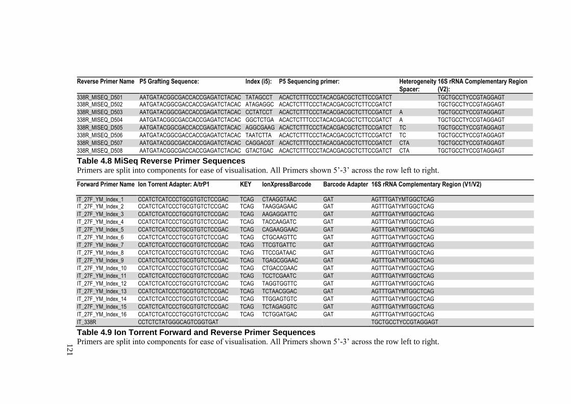
121
Table 4.8 MiSeq Reverse Primer Sequences Primers are split into components for ease of visualisation. All Primers shown 5’-3’ across the row left to right.
Table 4.9 Ion Torrent Forward and Reverse Primer Sequences Primers are split into components for ease of visualisation. All Primers shown 5’-3’ across the row left to right.
Reverse Primer Name P5 Grafting Sequence: Index (i5): P5 Sequencing primer: Heterogeneity Spacer:
16S rRNA Complementary Region (V2):
338R_MISEQ_D501 AATGATACGGCGACCACCGAGATCTACAC TATAGCCT ACACTCTTTCCCTACACGACGCTCTTCCGATCT TGCTGCCTYCCGTAGGAGT 338R_MISEQ_D502 AATGATACGGCGACCACCGAGATCTACAC ATAGAGGC ACACTCTTTCCCTACACGACGCTCTTCCGATCT TGCTGCCTYCCGTAGGAGT
338R_MISEQ_D503 AATGATACGGCGACCACCGAGATCTACAC CCTATCCT ACACTCTTTCCCTACACGACGCTCTTCCGATCT A TGCTGCCTYCCGTAGGAGT
338R_MISEQ_D504 AATGATACGGCGACCACCGAGATCTACAC GGCTCTGA ACACTCTTTCCCTACACGACGCTCTTCCGATCT A TGCTGCCTYCCGTAGGAGT
338R_MISEQ_D505 AATGATACGGCGACCACCGAGATCTACAC AGGCGAAG ACACTCTTTCCCTACACGACGCTCTTCCGATCT TC TGCTGCCTYCCGTAGGAGT
338R_MISEQ_D506 AATGATACGGCGACCACCGAGATCTACAC TAATCTTA ACACTCTTTCCCTACACGACGCTCTTCCGATCT TC TGCTGCCTYCCGTAGGAGT
338R_MISEQ_D507 AATGATACGGCGACCACCGAGATCTACAC CAGGACGT ACACTCTTTCCCTACACGACGCTCTTCCGATCT CTA TGCTGCCTYCCGTAGGAGT
338R_MISEQ_D508 AATGATACGGCGACCACCGAGATCTACAC GTACTGAC ACACTCTTTCCCTACACGACGCTCTTCCGATCT CTA TGCTGCCTYCCGTAGGAGT
Forward Primer Name Ion Torrent Adapter: A/trP1 KEY IonXpressBarcode Barcode Adapter 16S rRNA Complementary Region (V1/V2)
IT_27F_YM_Index_1 CCATCTCATCCCTGCGTGTCTCCGAC TCAG CTAAGGTAAC GAT AGTTTGATYMTGGCTCAG IT_27F_YM_Index_2 CCATCTCATCCCTGCGTGTCTCCGAC TCAG TAAGGAGAAC GAT AGTTTGATYMTGGCTCAG
IT_27F_YM_Index_3 CCATCTCATCCCTGCGTGTCTCCGAC TCAG AAGAGGATTC GAT AGTTTGATYMTGGCTCAG
IT_27F_YM_Index_4 CCATCTCATCCCTGCGTGTCTCCGAC TCAG TACCAAGATC GAT AGTTTGATYMTGGCTCAG
IT_27F_YM_Index_5 CCATCTCATCCCTGCGTGTCTCCGAC TCAG CAGAAGGAAC GAT AGTTTGATYMTGGCTCAG
IT_27F_YM_Index_6 CCATCTCATCCCTGCGTGTCTCCGAC TCAG CTGCAAGTTC GAT AGTTTGATYMTGGCTCAG
IT_27F_YM_Index_7 CCATCTCATCCCTGCGTGTCTCCGAC TCAG TTCGTGATTC GAT AGTTTGATYMTGGCTCAG
IT_27F_YM_Index_8 CCATCTCATCCCTGCGTGTCTCCGAC TCAG TTCCGATAAC GAT AGTTTGATYMTGGCTCAG
IT_27F_YM_Index_9 CCATCTCATCCCTGCGTGTCTCCGAC TCAG TGAGCGGAAC GAT AGTTTGATYMTGGCTCAG
IT_27F_YM_Index_10 CCATCTCATCCCTGCGTGTCTCCGAC TCAG CTGACCGAAC GAT AGTTTGATYMTGGCTCAG
IT_27F_YM_Index_11 CCATCTCATCCCTGCGTGTCTCCGAC TCAG TCCTCGAATC GAT AGTTTGATYMTGGCTCAG
IT_27F_YM_Index_12 CCATCTCATCCCTGCGTGTCTCCGAC TCAG TAGGTGGTTC GAT AGTTTGATYMTGGCTCAG
IT_27F_YM_Index_13 CCATCTCATCCCTGCGTGTCTCCGAC TCAG TCTAACGGAC GAT AGTTTGATYMTGGCTCAG
IT_27F_YM_Index_14 CCATCTCATCCCTGCGTGTCTCCGAC TCAG TTGGAGTGTC GAT AGTTTGATYMTGGCTCAG
IT_27F_YM_Index_15 CCATCTCATCCCTGCGTGTCTCCGAC TCAG TCTAGAGGTC GAT AGTTTGATYMTGGCTCAG
IT_27F_YM_Index_16 CCATCTCATCCCTGCGTGTCTCCGAC TCAG TCTGGATGAC GAT AGTTTGATYMTGGCTCAG
IT_338R CCTCTCTATGGGCAGTCGGTGAT TGCTGCCTYCCGTAGGAGT
Chapter 4
122
4.8.3 Gel Electrophoresis
To qualitatively confirm products were of a desired size, PCR products were loaded
on an agarose gel (2%; w/v) containing ethidium bromide and exposed to
electrophoresis at 110 V for 1 hour in TBE buffer. Gels were visualised under UV
light to determine the presence and size of PCR amplicons. For all samples, a 12µl
volume was loaded into each well; 5µl of PCR product mixed with 5µl of sterile
water and 2µl of loading dye. For size comparison PCR products were compared
against 100bp ladder and/or PhiX.
4.8.4 Sample clean-up
Post-PCR samples were purified by one round of Agencourt AMPure XP (Beckman
Coulter) DNA purification beads to remove primer dimers and short fragments.
Manufacturer’s instructions were followed apart from the ratio of DNA to beads. A
ratio of 1: 0.8 DNA to beads was used for better size selection of larger fragments.
4.8.5 DNA Quantification, Quality Control, and Equimolar Pool
The integrity of the clean-up PCR-product was subsequently analysed on the 2100
Bioanalyzer Instrument using an Agilent DNA 1000 Chip Kit for size determination.
Total dsDNA was measured on the Qubit™ 2 Fluorometer using Qubit dsDNA HS
Assay Kit following the manufacturer’s instructions. For Illumina MiSeq samples
only, an additional qPCR using KAPA Library Quantification Kit was performed
following the manufacturer’s instructions. This qPCR is specific to the Illumina
sequence primers and gives an accurate measure of copies of DNA amplicons with
Illumina adapters. KAPA qPCR was conducted with three technical replicates at two
dilutions per library; 1:1000 and 1:10000 as suggested by the manufacturer’s
protocol.
The concentration (determined by Qubit and/or KAPA qPCR) and average size
(determined by Bioanalyzer) of each sample was used to make an equimolar
amplicon library pool which was diluted appropriately prior to sequencing. The

Chapter 4
123
concentration and average size of each sample was also used to highlight samples
below threshold that were subsequently dropped from processing. Purity of the
equimolar pool was determined as before using the KAPA Library Quantification kit
and size determined using the Bioanalyzer Instrument.

Chapter 4
124
4.9 Sequencing and Base Calling
For MiSeq libraries, bridge amplification was supported on the MiSeq platform in 2
x 300 Paired End (PE) mode using the 600-cycle MiSeq Reagent v3 kit, at Edinburgh
Genomics; University of Edinburgh. After sequencing, the FASTQ reads were
filtered by the Illumina software to remove TruSeq indexes and demultiplexed.
For Ion Torrent libraries, emulsion PCR was supported on the Ion OneTouchTM 400
Template Kit v2 according to the manufacturer’s instructions. Sequencing of the
library was carried out on the 314 chip using the Ion Torrent Personal Genomic
Machine (PGM) system and using the Ion Sequencing 400 kit at the MRC-Human
Genetics Unit, Edinburgh. After sequencing, the FASTQ reads were filtered by the
PGM software to remove sub-threshold and polyclonal sequences.
4.10 QIIME and Mothur
Quantitative Insights into Microbial Ecology (QIIME) is a Linux-based open source
software package that aids analysis of microbial communities from high throughput
sequencing (HTS) technologies (Caporaso et al., 2010b). QIIME packaged utilises
many other open source tools including; FastTree 2.1.3 (Price et al., 2010), RDP
Classifier (Wang et al., 2007), PyNAST algorithm (Caporaso et al., 2010a) and
UCHIME (Edgar et al., 2011). QIIME allows for; demultiplexing, aligning,
screening, assigning OTUs, calculating diversity, hypothesis testing, and data
visualisation.
Mothur is also an open-source software program for microbial bioinformatics
(Schloss et al., 2009). Mothur has all the similar functions to QIIME for data
processing with built in function for RDP Classifier (Wang et al., 2007), NAST
algorithm (DeSantis et al., 2006a) and UCHIME (Edgar et al., 2011).
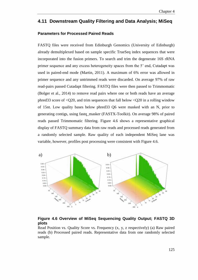
Chapter 4
125
4.11 Downstream Quality Filtering and Data Analysis; MiSeq
Parameters for Processed Paired Reads
FASTQ files were received from Edinburgh Genomics (University of Edinburgh)
already demultiplexed based on sample specific TrueSeq index sequences that were
incorporated into the fusion primers. To search and trim the degenerate 16S rRNA
primer sequence and any excess heterogeneity spaces from the 5’ end, Cutadapt was
used in paired-end mode (Martin, 2011). A maximum of 6% error was allowed in
primer sequence and any untrimmed reads were discarded. On average 97% of raw
read-pairs passed Cutadapt filtering. FASTQ files were then passed to Trimmomatic
(Bolger et al., 2014) to remove read pairs where one or both reads have an average
phred33 score of <Q20, and trim sequences that fall below <Q20 in a rolling window
of 15nt. Low quality bases below phred33 Q6 were masked with an N, prior to
generating contigs, using fastq_masker (FASTX-Toolkit). On average 98% of paired
reads passed Trimmomatic filtering. Figure 4.6 shows a representative graphical
display of FASTQ summary data from raw reads and processed reads generated from
a randomly selected sample. Raw quality of each independent MiSeq lane was
variable, however, profiles post processing were consistent with Figure 4.6.
Figure 4.6 Overview of MiSeq Sequencing Quality Output; FASTQ 3D plots Read Position vs. Quality Score vs. Frequency (x, y, z respectively) (a) Raw paired
reads (b) Processed paired reads. Representative data from one randomly selected
sample.
a) b)
Chapter 4
126
Contigs, Chimera filtering, OTU Picking and Tree building
Contigs from paired-end reads were generated in Mothur (Schloss et al., 2009). To
ensure sequence reads are a true representation of 16S rRNA gene and not
sequencing artifacts; sequences with ambiguous bases, homopolymers of >6, and a
contig length above 350bp were removed.
To ensure sequence reads are a true representation of 16S rRNA gene and not PCR
artifacts, reads were screened for chimeras using Mothur (Schloss et al., 2011) which
is based on UCHIME (Edgar et al., 2011) and USEARCH (Edgar, 2010). The
operational taxonomic units (OTUs) were picked de novo and clustered at 97%
sequence similarity using the Nearest Alignment Space Termination (NAST)
algorithm for alignment (DeSantis et al., 2006a). The taxonomy of each OTU was
assigned using naive Bayesian classification that is based on the RDP Classifier 8-
kmers method (Wang et al., 2007) which is integrated in Mothur (bootstrap threshold
80%) using SILVA (v119) database. FastTree 2.1.3 (Price et al., 2010) intergraded in
QIIME (Caporaso et al., 2010b), was used to build approximately-maximum-
likelihood phylogenetic trees.
Chapter 4
127
4.12 Downstream Quality Filtering and Data Analysis; Ion
Torrent
Parameters for Processed Reads
Ion Torrent raw FASTQ sequencing reads were first quality filtered using tools in
Galaxy (web-based platform for biological data) then directed into QIIME (version
1.8.0), for OTU calling, taxonomy identification and diversity measurements. To
ensure sequence reads are a true representation of 16S rRNA gene and not
sequencing artifacts reads were quality filtered and trimmed. The workflow I
designed in Galaxy quality filters and trims the sequence reads based on the
following criteria (see appendix for full details).
i) Only reads with perfect match to a barcode sequence are kept; barcode is
trimmed
ii) Only reads with a maximum of 2 mismatches in the complementary primer
sequence are kept; complementary primer trimmed
iii) Reads are trimmed when the average quality score drops below Phred score 25
in a sliding window of 6bp.
iv) Reads outside the rage of 150–500bp post trimming are discarded.
v) Reads containing homopolymers of more than 8bp are discarded.
Sequences are then binned by sample (demultiplexed) and the remaining high quality
FASTQ sequences are sent to QIIME for chimera filtering, OTU picking and tree
building.
Chapter 4
128
Chimera filtering, OTU Picking and Tree building
Before OTU picking, reference-based and de novo based chimera detection was
carried out using USEARCH 6.1 (Edgar, 2010) against the full Greengenes database
(13_8 release). OTUs were picked at 97% sequence similarity using an open-
reference approach. This approach first clusters the sequences against the
Greengenes database using UCLUST (Edgar, 2010), those sequences which do not
cluster are then sent for de novo OTU picking. Representative FASTA sequences for
each OTU were aligned to the Greengenes database (13_8 release) using PyNAST
(re-implemented NAST aligner (Caporaso et al., 2010a)). Taxonomy was assigned
using the RDP Classifier (Wang et al., 2007) with a bootstrap threshold of 60%.
FastTree 2.1.3 (Price et al., 2010) intergraded in QIIME, was used to build
approximately-maximum-likelihood phylogenetic trees.
4.13 R Package
The resulting OTU tables were imported into R for filtering and statistical analysis
using multiple R packages for data manipulation, such as; Phyloseq, ggplot2, and
Hclust. Phyloseq is an R-Package which manipulates the raw OTU data generated
elsewhere (such as in QIIME or Mothur), and together with analytical and graphical
packages (such as vegan, ape, picante and ggplot2) provides a tool for analysis and
visualisation of phylogenetic data. Phyloseq also has functions to rarefy data and
calculate diversity, however, for the most part these functions were used in QIIME
and phyloseq was primarily used in this study for data manipulation and generating
2D PCoA plots.

Chapter 4
129
4.14 Rarefaction Curves
Sufficient sequencing depth is needed to capture the full representation of bacterial
species in a sample. To establish whether the depth of sequencing truly captures the
community, a method commonly called rarefaction can be employed. Rarefaction is
a technique used in ecology to measure species richness as a function of sampling
effort. Rarefaction can be thought of as a method for random resampling, whereby
diversity measures are then calculated for each simulated sequencing effort. The rate
of increase in diversity measure is an inherent property of the microbial community
of that sample, whereby a plateau suggest the majority of the species in the
community have been found.
In this study multiple rarefactions are done for each sample, diversity measures
calculated and shown graphically on rarefaction curve plots. The diversity measures
calculated for rarefaction were; species richness (Sobs), Chao1 Estimate (Chao, 1987),
Shannon Diversity (Shannon, 1948) and Inverse Simpsons Index (Simpson, 1949);
these diversity measures are described in more detail below.
Chapter 4
130
4.15 Alpha Diversity Analysis
Bacterial diversity at the community level is evaluated using two principle measures;
alpha and beta diversity. Alpha diversity is a measure of diversity ‘within’ a sample
and can be assessed using various metrics. Most measures take into account both
species richness and evenness. Richness refers to how many different species
(OTUs) there are present, whereas evenness describes their distribution.
The alpha diversity measures used in this study are; Observed species (Sobs), Chao1
Estimate, Shannon Diversity and Inverse Simpsons Index. Observed species is a
measure of the unique OTUs in each sample and Chao1 is an estimate of species
richness which uses the ratio of singletons to doubletons (Chao, 1987). Low
abundant OTUs should therefore not be removed prior to analysis. Simpson’s index
is a measure of the probability that upon randomly selecting an individual from a
community, the individual has already been observed (Simpson, 1949). Shannon
Diversity measure is more sensitive to species richness, giving higher weight to low
abundant OTUs/species (Shannon, 1948).
4.15.1 Statistical analysis of alpha diversity
Where alpha diversity between two groups were compared (e.g. Fecal vs. Vaginal); a
unpaired student t-test was performed. Continuous parametric data between more
than two groups was compared using a one way analysis of variance (ANOVA).
Longitudinal data within-subject across the 4 time-points was compared using
repeated measures ANOVA (RM-ANOVA) and Tukey's post-test. Linear mixed
regression was applied to the Alpha Diversity measures across collection time-points.
A P value of less than 0.05 was determined to be significant for all measures.
Chapter 4
131
4.16 Beta Diversity Analysis
While Alpha Diversity is descriptive for inferring community structure, it represents
only the dispersion pattern of the OTUs. Alpha Diversity will not tell us if the same
bacteria are shared between samples. Beta diversity measures are used to describe the
similarity or dissimilarity between two different samples. Beta diversity, like alpha
diversity is dependent on the depth of sequencing and the metric used. A popular
method which uses the phylogenetic tree as the measure for similarity is a technique
called UniFrac (Lozupone and Knight, 2005). UniFrac takes into account the shared
species between two samples, their abundance and their phylogenetic relationship. It
measures pairwise distances and returns a value between 0, where two communities
are identical, and 1, where communities are totally unrelated. The higher the beta
diversity, the less similar the communities are. Unweighted UniFrac distance is a
qualitative description of similarity whereas weighted UniFrac distance takes into
account the quantitative occurrence of the OTUs/species. Weighted UniFrac distance
therefore attributes more power to highly abundance OTUs/species than unweighted
UniFrac measure.
The UniFrac distances from all samples are approximated visually using Principle
Co-ordinate Analysis (PCoA). PCoA is a method of dimensionality reduction that
aims to recapture whatever patterns are in the data but reduce the number of
dimensions using a new co-ordinate system. PCoA is related to the commonly used
statistical method principle component analysis (PCA) which is analogous to
Euclidian distance and not UniFrac measures. 2D PCoA plots in this thesis were
created and visualised using phyloseq; an R package (McMurdie and Holmes, 2013).
Ultimately ordination is a visualisation method, but any spatial separation seen in 2D
PCoA plots can be tested for statistical significance using hypothesis testing on the
raw data (described below).
Chapter 4
132
4.16.1 Statistical analysis of beta diversity
Where beta diversity between two groups was compared, a parametric two sample t-
test with Bonferroni correction was performed. This was supported with non-
parametric testing using 999 Monte Carlo permutations and Bonferroni-corrected.
Adonis
Adonis is a nonparametric statistical method that first identifies the central points in
the data, centroids, and then radially calculates the squared deviations out from these
points. This produces an effect size value (r2) which is the level of variance in the
data that can be attributed to the specified category (e.g. genotype or gestation). In
addition this method provides a P value from permutations of the raw data (999)
using multiple F-tests (sequential sums of squares).
ANOSIM
ANOSIM is a test which determines whether two or more groups of samples (e.g.
C57BL/6J vs. Camp-/-) are significantly different by relating the distances ‘between’
the groups, to the distances ‘within’ the groups, and providing an r value. A positive
r value close to 1.0 signifies that the groups are highly dissimilar. Significance is
supported by 999 permutations of the similarity matrix.
Chapter 4
133
4.16.2 Jack-knife and UPGMA bootstrapped trees
A rooted dendrogram (tree) is constructed from the pairwise UniFrac distance matrix
using Unweighted Pair Group Method with Arithmetic Means algorithm (UPGMA)
to infer structure. Jack-knife replicates are used to estimate uncertainty in distance
matrix and involves dropping one/many observations from a sample each time and
recalculating the distance. Together with bootstrapped hierarchical clustering,
UPGMA provides an estimate to the uncertainty of the points in PCoA plots. Jack-
knifing in this study was set at 100 replicates at 45,000 sequences (75% per sample
sequences). Those nodes which are consistent across jack-knife replicates are robust
for microbial inference. Bootstrap values on the branch indicate how much support is
given to that branch on a phylogenetic tree.
4.17 Significant Taxa (LEfSe)
Linear Discriminant Analysis with Effect Size (LEfSe) is an all-against-all multi-
class comparison tool used to highlight biological consistency and estimate effect
size (Segata et al., 2011). LEfSe was used to detect which taxa were enriched in
different genotypes; C57BL/6J vs. Camp-/-. LEfSe first applies a nonparametric
(Kruskal -Wallis) rank sum test to the OTU data to detect OTUs and classifications
with significant differential abundances. Stringent alpha values of 0.01 for Kruskal-
Wallis were set in this study. This is then followed by a biological consistency
estimate which involves pairwise rank sum tests (Wilcoxon). Linear Discriminant
Analysis is then used to estimate an effect size of the differentially abundant taxa;
threshold of 3.0 was chosen for a logarithmic LDA score.
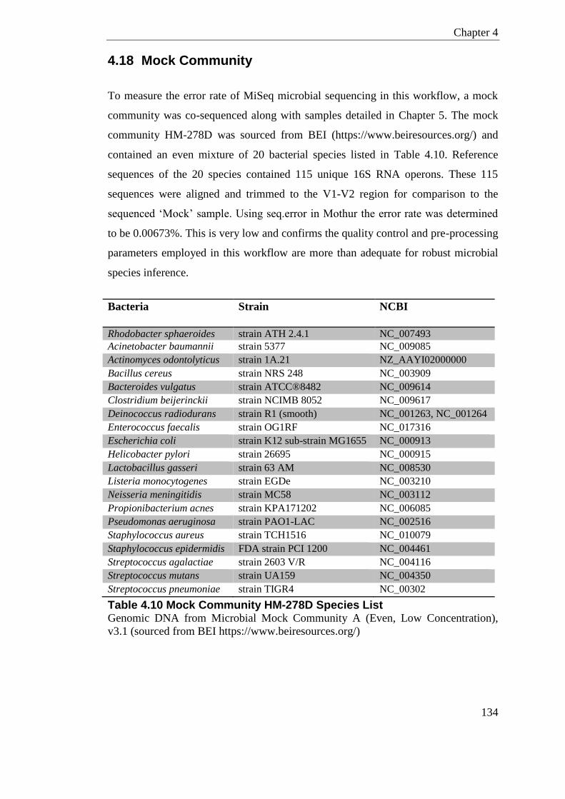
Chapter 4
134
4.18 Mock Community
To measure the error rate of MiSeq microbial sequencing in this workflow, a mock
community was co-sequenced along with samples detailed in Chapter 5. The mock
community HM-278D was sourced from BEI (https://www.beiresources.org/) and
contained an even mixture of 20 bacterial species listed in Table 4.10. Reference
sequences of the 20 species contained 115 unique 16S RNA operons. These 115
sequences were aligned and trimmed to the V1-V2 region for comparison to the
sequenced ‘Mock’ sample. Using seq.error in Mothur the error rate was determined
to be 0.00673%. This is very low and confirms the quality control and pre-processing
parameters employed in this workflow are more than adequate for robust microbial
species inference.
Bacteria Strain NCBI
Rhodobacter sphaeroides strain ATH 2.4.1 NC_007493
Acinetobacter baumannii strain 5377 NC_009085
Actinomyces odontolyticus strain 1A.21 NZ_AAYI02000000
Bacillus cereus strain NRS 248 NC_003909
Bacteroides vulgatus strain ATCC®8482 NC_009614
Clostridium beijerinckii strain NCIMB 8052 NC_009617
Deinococcus radiodurans strain R1 (smooth) NC_001263, NC_001264
Enterococcus faecalis strain OG1RF NC_017316
Escherichia coli strain K12 sub-strain MG1655 NC_000913
Helicobacter pylori strain 26695 NC_000915
Lactobacillus gasseri strain 63 AM NC_008530
Listeria monocytogenes strain EGDe NC_003210
Neisseria meningitidis strain MC58 NC_003112
Propionibacterium acnes strain KPA171202 NC_006085
Pseudomonas aeruginosa strain PAO1-LAC NC_002516
Staphylococcus aureus strain TCH1516 NC_010079
Staphylococcus epidermidis FDA strain PCI 1200 NC_004461
Streptococcus agalactiae strain 2603 V/R NC_004116
Streptococcus mutans strain UA159 NC_004350
Streptococcus pneumoniae strain TIGR4 NC_00302
Table 4.10 Mock Community HM-278D Species List Genomic DNA from Microbial Mock Community A (Even, Low Concentration),
v3.1 (sourced from BEI https://www.beiresources.org/)
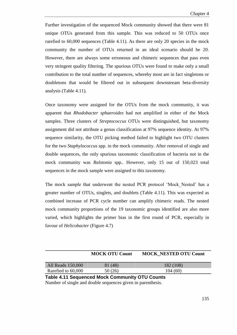
Chapter 4
135
Further investigation of the sequenced Mock community showed that there were 81
unique OTUs generated from this sample. This was reduced to 50 OTUs once
rarefied to 60,000 sequences (Table 4.11). As there are only 20 species in the mock
community the number of OTUs returned in an ideal scenario should be 20.
However, there are always some erroneous and chimeric sequences that pass even
very stringent quality filtering. The spurious OTUs were found to make only a small
contribution to the total number of sequences, whereby most are in fact singletons or
doubletons that would be filtered out in subsequent downstream beta-diversity
analysis (Table 4.11).
Once taxonomy were assigned for the OTUs from the mock community, it was
apparent that Rhodobacter sphaeroides had not amplified in either of the Mock
samples. Three clusters of Streptococcus OTUs were distinguished, but taxonomy
assignment did not attribute a genus classification at 97% sequence identity. At 97%
sequence similarity, the OTU picking method failed to highlight two OTU clusters
for the two Staphylococcus spp. in the mock community. After removal of single and
double sequences, the only spurious taxonomic classification of bacteria not in the
mock community was Ralstonia spp.. However, only 15 out of 150,023 total
sequences in the mock sample were assigned to this taxonomy.
The mock sample that underwent the nested PCR protocol ‘Mock_Nested’ has a
greater number of OTUs, singlets, and doublets (Table 4.11). This was expected as
combined increase of PCR cycle number can amplify chimeric reads. The nested
mock community proportions of the 19 taxonomic groups identified are also more
varied, which highlights the primer bias in the first round of PCR, especially in
favour of Helicobacter (Figure 4.7)
MOCK OTU Count MOCK_NESTED OTU Count
All Reads 150,000 81 (48) 182 (108)
Rarefied to 60,000 50 (26) 104 (60)
Table 4.11 Sequenced Mock Community OTU Counts Number of single and double sequences given in parenthesis.
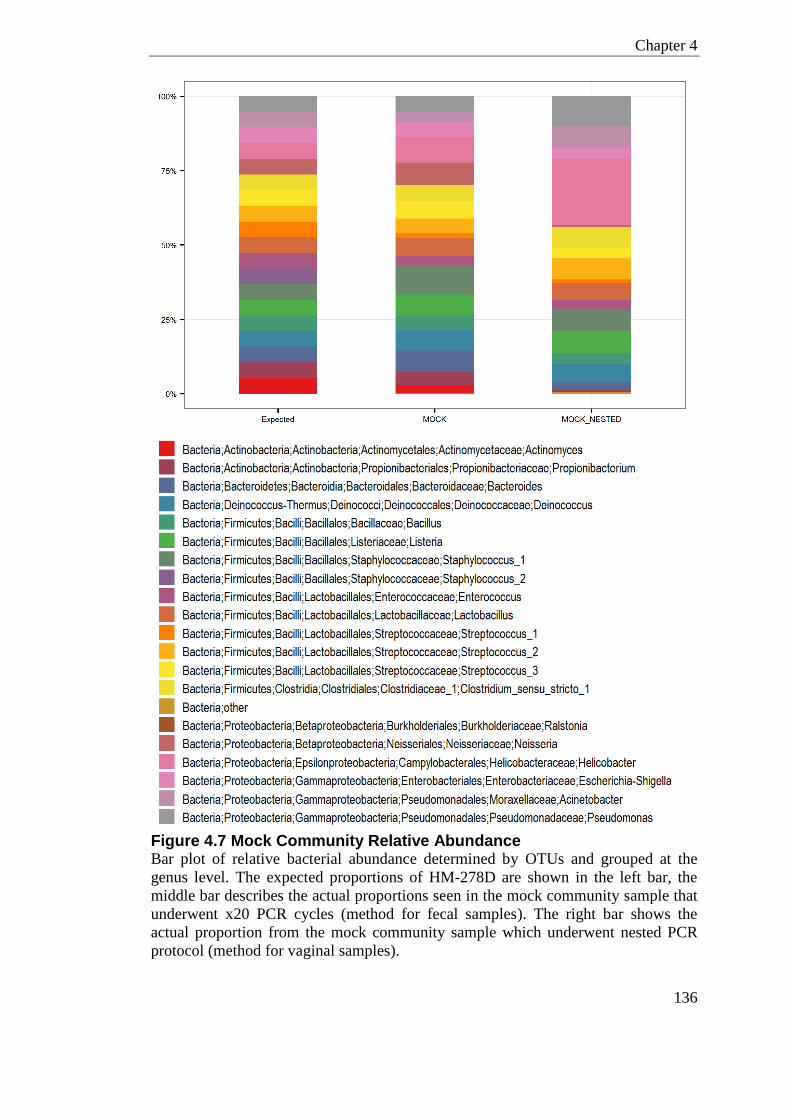
Chapter 4
136
Figure 4.7 Mock Community Relative Abundance Bar plot of relative bacterial abundance determined by OTUs and grouped at the
genus level. The expected proportions of HM-278D are shown in the left bar, the
middle bar describes the actual proportions seen in the mock community sample that
underwent x20 PCR cycles (method for fecal samples). The right bar shows the
actual proportion from the mock community sample which underwent nested PCR
protocol (method for vaginal samples).
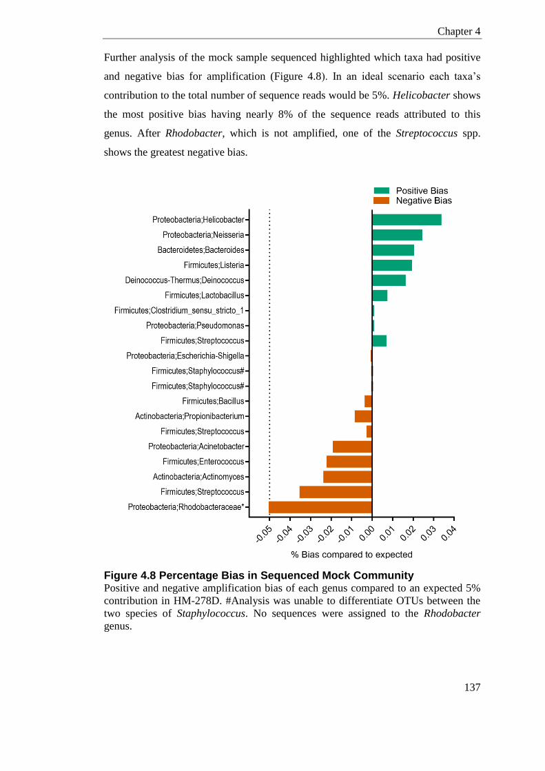
Chapter 4
137
Further analysis of the mock sample sequenced highlighted which taxa had positive
and negative bias for amplification (Figure 4.8). In an ideal scenario each taxa’s
contribution to the total number of sequence reads would be 5%. Helicobacter shows
the most positive bias having nearly 8% of the sequence reads attributed to this
genus. After Rhodobacter, which is not amplified, one of the Streptococcus spp.
shows the greatest negative bias.
Figure 4.8 Percentage Bias in Sequenced Mock Community Positive and negative amplification bias of each genus compared to an expected 5%
contribution in HM-278D. #Analysis was unable to differentiate OTUs between the
two species of Staphylococcus. No sequences were assigned to the Rhodobacter
genus.
Chapter 4
138
4.19 Discussion
This chapter details a method for looking at the microbiome across body sites and
has been optimised to consider the murine fecal and vaginal bacterial community.
After review of the literature and in-silico work I deduced the V1-V2 region would
provide the highest discrimination potential for species identification in the murine
vagina and gastrointestinal tract. In this chapter I conducted in-silico primer
evaluation to generate and confirm the suitability of universal sequencing primers to
amplify the V1-V2 region of the 16S rRNA gene.
As the murine vagina has a low bacteria burden, different sampling methods were
evaluated for 16S rRNA gene recovery, this highlighted that whole vaginal tissue
provides the most consistent bacterial yield.
Different extraction methods used to isolate and extract bacterial DNA from
ecological samples can give a varied representation of species diversity and
abundance (Milani et al., 2013). Milani et al., (2013) found large variations in
proportions of the contributing bacteria depending on the extraction method used;
mechanical and/or enzymatic. Milani et al., (2013) concluded that Faecalibacterium
and Bacteroides (gut microbial taxa), might be over-represented in human gut
metagenomic studies as a result of this bias. Although I did not directly compare the
bias in extraction methods in this study, I favoured the FastDNA® SPIN Kit with
bead-beating over the PowerSoil® DNA Isolation Kit due to this reported reduction
in bias towards Bacteroidetes phylum.
Reagent contamination is emerging as a key factor in microbiome studies especially
in low-biomass sites. Salter et al., (2014) demonstrated this using low concentrations
of Salmonella DNA, where low biomass was associated with inflated spurious
sequences of various taxonomic assignments. Ultra-pure polymerases and ultra-pure
molecular grade water were used in this study with the aim of reducing potential
contaminants.
Co-sequencing of a mock community on the MiSeq platform highlighted that the
error rate in sequencing microbial communities in this optimised workflow was
Chapter 4
139
extremely low, only 0.00673%. The mock community also highlights Ralstonia as a
species for possible PCR reagent contamination, although only 15 out of 150,023
total sequences in the mock sample were attributed to this clade. Molecular grade
water is a common source of this bacterium (Grahn et al., 2003, Barton et al., 2006,
Laurence et al., 2014). The mock community also highlights that the primers in this
study are not suitable for picking up bacteria in the Rhodobacter genus as no OTUs
were assigned to this taxonomy.
Consideration was given in this study to keep the number of PCR cycles to a
minimum as to reduce the affliction of PCR artifacts, such as chimeras, inflating
OTU clusters (Acinas et al., 2005). A study comparing 15x and 30x PCR cycles
showed a 30% inflation of chimeric amplicons with increased cycle number (Ahn et
al., 2012).
In this study, for the low biomass vaginal samples, a nested PCR approach was
adopted. This approach was a compromise of controlling the number of chimeric
reads while allowing for workable concentrations of PCR product to be obtained.
Sequencing artifacts are also kept to a minimum by doing paired end sequencing
(MiSeq only) and employing stringent downstream chimera checking. Using the
most current database for taxonomic assignment is essential but there are still
sequences that are poorly annotated.
A limitation in this study was in the optimisation required to get strong amplification
results using fusion-primers; which are known to be difficult due to the extended
primer length and off target amplification. There are similar low cost methods of
multiplexing that allow barcodes and sequencing adaptors to be added without
having to incorporate target-specific sequences for each fusion primer. These
methods employ a two-step PCR protocol, where shorter ‘tail’ sequences are
incorporated in the first universal 16S rRNA amplification. The second PCR is then
specific to the tail sequence allowing for barcodes and sequencing adapters to be
added at this stage. Any processes involving two PCR steps, as I have shown in this
chapter, has the disadvantage of potential inflation of PCR artifacts and amplification
Chapter 4
140
bias. A robust study comparing multiplexing approaches for 16S rRNA sequences
has yet to determine the associated bias in the various approaches.
OTUs were clustered using a cut-off of 97% sequence identity to seed in database,
which is generally accepted to represent species level. There is however some debate
whether this cut-off is too conservative (Mizrahi-Man et al., 2013). Collectively, for
the MiSeq data-set, only 0.01630% of the sequences did not align with the V1-V2
region of the 16S rRNA gene, of all those sequences that did align, only 0.09143%
were identified as non-bacterial (e.g. chloroplast, mitochondria and Archaea and
unknown) with a cut off of 80% to bacteria kingdom. Collectively this means that
only 0.5418% of total sequences passing quality filtering were not identifiable as
bacterial. This low value is evidence of the specificity of the primers used in this
study and appropriate quality filtering.
Low nucleotide diversity in the first few cycles of sequencing on the Illumina
platform is known to cause problems with base calling and the cluster identification
algorithms, which impacts greatly on sequence quality (Krueger et al., 2011). To
bypass/rectify this problem researchers often “spike-in” a PhiX shotgun library to
increase diversity and gain better sequence quality. The amount of PhiX spike can
range for 10% but some times as high as 50-80% depending on the complexity of
targeted library (Mitra et al., 2015). This obviously has enormous impact on the per
sample cost, as the majority of sequences are being assigned to PhiX and not the 16S
rRNA gene of interest. In this study I attempt to reduce this problem by the
incorporation of random nucleotides of varying lengths into the forward and reverse
fusion-primers, known as ‘heterogeneity’ spacers. Cluster density of all MiSeq runs
in this study was respectable, and raw sequence quality was above threshold; with
>78% raw reads having an average quality of >Q20. This is suggestive that the
heterogeneity spacers employed have reduced possible issues with low complexity
and improved sequence quality.
Cross-study comparisons with regards to 16S rRNA gene studies should be
approached with some caution. Although the discussed factors in 16S rRNA
protocols are important to consider, variation attributed to the choice of extraction
Chapter 4
141
method and variable regions were still shown to be less than the variation attributed
to differences between individual samples and body sites (Wesolowska-Andersen et
al., 2014, Lozupone et al., 2013). There is however still a need for a gold standard
library preparation and bioinformatic protocol for looking at microbial communities.
4.20 Summary
In this chapter I have detailed a method for looking at the microbiome across body
sites; specifically optimising protocols to consider the murine fecal and vaginal
bacterial communities. Co-sequencing of a mock community highlights this
workflow is robust for broad taxonomic classification, but is not suitable for picking
up bacteria in the Rhodobacter genus. The mock community also highlights
Ralstonia as a species for possible low level PCR reagent contamination.

142
Chapter 5
Modulation of the Microbiome
Chapter 5
143
Chapter 5 Modulation of the Microbiome
5.1 Introduction
Animals offer researchers a vital tool by providing a model to test out host-
microbiome interactions under controlled environmental conditions. They also allow
for infectious models that help understand pathogenicity. Gastrointestinal bacteria
have an important role in maintaining multiple physiological processes. LL-37/Camp
is upregulated during ileitis/colitis dependent on the level of inflammation (Schauber
et al., 2006), and Camp null mice have more severe phenotype in a DSS induced
colitis model (Koon et al., 2011). Addition of exogenous Camp could reverse some
of the phenotypic severity of DSS treatment in Camp-/- animals, such as cytokine
production, apoptosis and mucus production (Tai et al., 2013). In this chapter I look
at the direct consequences of the cathelicidin gene on gut microbial diversity and
structure, by interrogating the microbiome of Camp-/- animals.
There are vast metabolic changes that occur during pregnancy, especially in late
gestation, which could impact on microbial communities. Indeed, alterations in the
maternal gastrointestinal bacteria have been implicated in metabolic adaptations to
pregnancy (Koren et al., 2012, Collado et al., 2008). However, there are conflicting
reports with regards to whether pregnancy causes the modulation of gut bacterial
composition, with a large longitudinal study failing to show significant remodelling
of the gut microbiota in pregnant women (DiGiulio et al., 2015). This study
investigates whether there is remodelling in the diversity and composition of the
murine gastrointestinal and vaginal microbiome in pregnancy.

Chapter 5
144
The aims of this study were;
1) To establish whether the microbiome of animals lacking cathelicidin are altered
across body sites.
2) To investigate whether there are gestational related changes in the female murine
gut microbiome.
3) To establish whether the vaginal microbiome is affected by pregnancy, results and
discussion presented in Chapter 6
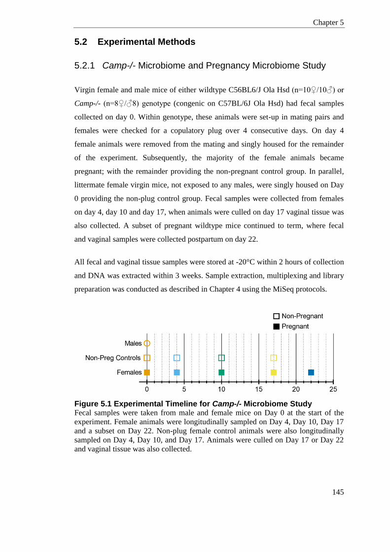
Chapter 5
145
5.2 Experimental Methods
5.2.1 Camp-/- Microbiome and Pregnancy Microbiome Study
Virgin female and male mice of either wildtype C56BL6/J Ola Hsd (n=10♀/10♂) or
Camp-/- (n=8♀/♂8) genotype (congenic on C57BL/6J Ola Hsd) had fecal samples
collected on day 0. Within genotype, these animals were set-up in mating pairs and
females were checked for a copulatory plug over 4 consecutive days. On day 4
female animals were removed from the mating and singly housed for the remainder
of the experiment. Subsequently, the majority of the female animals became
pregnant; with the remainder providing the non-pregnant control group. In parallel,
littermate female virgin mice, not exposed to any males, were singly housed on Day
0 providing the non-plug control group. Fecal samples were collected from females
on day 4, day 10 and day 17, when animals were culled on day 17 vaginal tissue was
also collected. A subset of pregnant wildtype mice continued to term, where fecal
and vaginal samples were collected postpartum on day 22.
All fecal and vaginal tissue samples were stored at -20°C within 2 hours of collection
and DNA was extracted within 3 weeks. Sample extraction, multiplexing and library
preparation was conducted as described in Chapter 4 using the MiSeq protocols.
Figure 5.1 Experimental Timeline for Camp-/- Microbiome Study Fecal samples were taken from male and female mice on Day 0 at the start of the
experiment. Female animals were longitudinally sampled on Day 4, Day 10, Day 17
and a subset on Day 22. Non-plug female control animals were also longitudinally
sampled on Day 4, Day 10, and Day 17. Animals were culled on Day 17 or Day 22
and vaginal tissue was also collected.
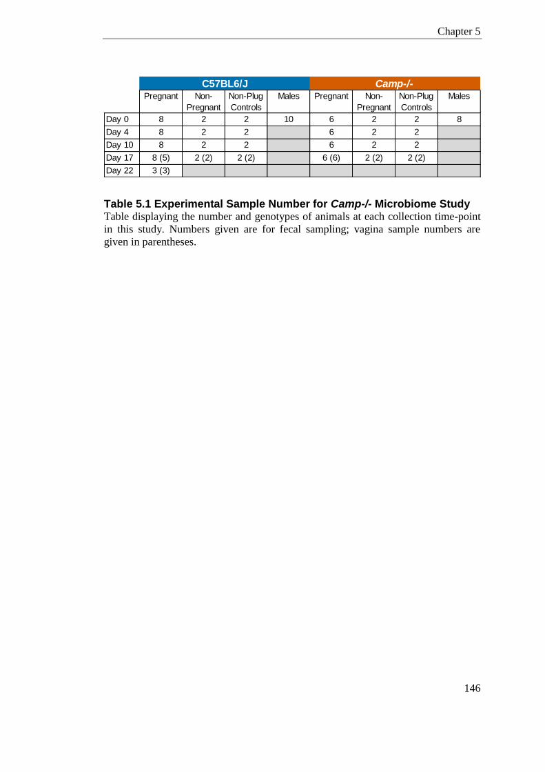
Chapter 5
146
Table 5.1 Experimental Sample Number for Camp-/- Microbiome Study Table displaying the number and genotypes of animals at each collection time-point
in this study. Numbers given are for fecal sampling; vagina sample numbers are
given in parentheses.
Pregnant Non-
Pregnant
Non-Plug
Controls
Males Pregnant Non-
Pregnant
Non-Plug
Controls
Males
Day 0 8 2 2 10 6 2 2 8
Day 4 8 2 2 6 2 2
Day 10 8 2 2 6 2 2
Day 17 8 (5) 2 (2) 2 (2) 6 (6) 2 (2) 2 (2)
Day 22 3 (3)
C57BL6/J Camp-/-

Chapter 5
147
5.3 Results
5.3.1 Data Summary
The full 16S rRNA gene sequencing performed on the Illumina MiSeq platform
consisted of 139 samples; two mock communities, 114 murine fecal samples and 22
murine vaginal samples. Amplicon libraries were sequenced on 6 independent MiSeq
lanes. Bioinformatic and quality filtering done on the raw data are described in
section 4.11, but in brief; runs were demultiplexed and barcodes and indexes
removed using Illumina software. Raw sequence read quality filtering and primer
trimming was done using Cutadapt and Trimmomatic. The total number of raw reads
prior and post data processing is given in Table 5.2. Quality filtering the raw reads
reduced the number of paired end reads from 31,640,538 (raw paired reads) to
30,279,366 reads (processed paired reads) (Figure 5.2).
Contigs were made in Mothur and screened as detailed in section 4.11. Chimeras
were removed by UCHIME and operational taxonomic units (OTUs) were picked de
novo and clustered at 97% sequence similarity using the NAST algorithm. The
taxonomy was assigned by RDP classifier (Bootstrap threshold 80%) using SILVA
(v119) as the reference database. This further quality screening reduced the number
of processed paired end reads from 30,279,366 (processed paired reads) to
24,306,711 contig sequences (filtered contigs) (Figure 5.2).
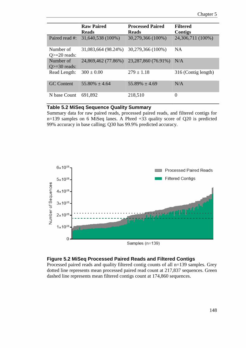
Chapter 5
148
Raw Paired
Reads
Processed Paired
Reads
Filtered
Contigs
Paired read #: 31,640,538 (100%) 30,279,366 (100%) 24,306,711 (100%)
Number of
Q>=20 reads:
31,083,664 (98.24%) 30,279,366 (100%) NA
Number of
Q>=30 reads:
24,869,462 (77.86%) 23,287,860 (76.91%) N/A
Read Length: 300 ± 0.00 279 ± 1.18 316 (Contig length)
GC Content 55.80% ± 4.64 55.89% ± 4.69 N/A
N base Count 691,892 218,510 0
Table 5.2 MiSeq Sequence Quality Summary Summary data for raw paired reads, processed paired reads, and filtered contigs for
n=139 samples on 6 MiSeq lanes. A Phred +33 quality score of Q20 is predicted
99% accuracy in base calling; Q30 has 99.9% predicted accuracy.
Figure 5.2 MiSeq Processed Paired Reads and Filtered Contigs Processed paired reads and quality filtered contig counts of all n=139 samples. Grey
dotted line represents mean processed paired read count at 217,837 sequences. Green
dashed line represents mean filtered contigs count at 174,860 sequences.
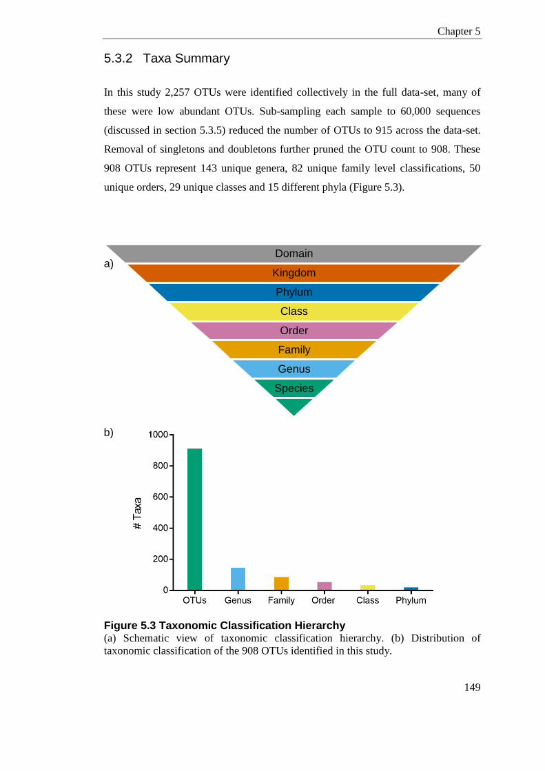
Chapter 5
149
5.3.2 Taxa Summary
In this study 2,257 OTUs were identified collectively in the full data-set, many of
these were low abundant OTUs. Sub-sampling each sample to 60,000 sequences
(discussed in section 5.3.5) reduced the number of OTUs to 915 across the data-set.
Removal of singletons and doubletons further pruned the OTU count to 908. These
908 OTUs represent 143 unique genera, 82 unique family level classifications, 50
unique orders, 29 unique classes and 15 different phyla (Figure 5.3).
Figure 5.3 Taxonomic Classification Hierarchy (a) Schematic view of taxonomic classification hierarchy. (b) Distribution of
taxonomic classification of the 908 OTUs identified in this study.
Domain
Kingdom
Phylum
Class
Order
Family
Genus
Species
a)
b)
Chapter 5
150
5.3.3 Subsets
Subset 1 - Fecal vs. Vaginal
To examine the murine fecal microbiome in relation to vaginal bacterial
communities, Subset 1 consisted of the full data-set minus the two mock community
samples, which are evaluated in section 4.18. This subset includes the full data of
vaginal (n=22) and fecal samples (n=115).
Subset 2 - Camp-/- vs. C57BL/6J
To determine whether there is any genotypic difference in the fecal microbiome in
Camp-/- compared to wildtype C57BL/6J animals, the data was further partitioned to
only include fecal samples from the Camp-/- pregnancy microbiome study (n=99; 42
Camp-/- , 57 C57BL/6J).
Subset 3 - C57BL/6J; Gestational effect
To examine whether the fecal microbiome varied longitudinally both in a non-
pregnant and pregnant state, fecal samples were further partitioned to only include
the wildtype C57BL/6J animals which were longitudinally sampled at 4 time-points
over 17 days (n=44 samples; 16 non-pregnant, 28 pregnant).
Subset 4 - Vaginal Microbiome; Camp-/- vs. C57BL/6J, Pregnant vs.
Non-pregnant
This subset included only the vaginal samples (n=22), and is detailed and discussed
in Chapter 6.
Chapter 5
151
5.3.4 Rarefaction Curves
As deep sequencing was performed in this study, it would be assumed that the depth
of sequencing is sufficient for robust conclusions to be made. However, this needs to
be empirically established as a true representation is dependent on community
complexity. To establish whether the depth of sequencing truly captures the
community, a method commonly called rarefaction was employed (discussed in
section 4.14).
Multiple rarefactions to a maximum sampling depth of 60,000 sequences were
calculated for each sample and species richness (Sobs), Chao1 Estimate, Shannon
Diversity, and Inverse Simpsons Index, were computed for each simulated
sequencing effort (Figure 5.4). Specifically, 10 rarefactions at 10 increasing depths
(6,000 integers) were conducted on the data. Maximum rarefaction at 60,000 was
chosen as this is the number of sequences from the lowest sample.
Visual assessment of the rarefaction plots reveal that the diversity and complexity of
the vaginal community is considerably lower than the fecal samples, as expected,
displayed by lower amplitude on the rarefaction curves (Figure 5.4). A common
trend was seen in all mean diversity measures in both fecal and vaginal samples,
whereby a steep increase up-to 6,000 is observed followed by a plateau to varying
degrees (Figure 5.4). The number of observed OTUs (Sobs) can sometimes never
reach a true plateau, as this is often a factor of sequencing chemistry error and
stringency of OTU alignment. Chao1 estimates species richness based on the ratio of
singletons to doubletons in the sample and is therefore also a factor of Sobs. Chao1
and Sobs show a steady increase with sampling effort after 6,000 sequences but this is
not adding to the complexity of community structure. This can be seen by the
Shannon Diversity and Inverse Simpson Index, which are stable past 6,000
sequences. This suggests that many OTUs are present in low abundance. Overall the
curves reach an acceptable plateau somewhere before 6,000, highlighting that
sequence depth >6,000 is more than adequate to capture a true representation of the
community. Subsequent beta-diversity measures were calculated on a single
subsampled population of 60,000 sequences per sample.
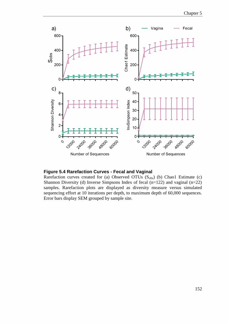
Chapter 5
152
Figure 5.4 Rarefaction Curves - Fecal and Vaginal Rarefaction curves created for (a) Observed OTUs (Sobs) (b) Chao1 Estimate (c)
Shannon Diversity (d) Inverse Simpsons Index of fecal (n=122) and vaginal (n=22)
samples. Rarefaction plots are displayed as diversity measure versus simulated
sequencing effort at 10 iterations per depth, to maximum depth of 60,000 sequences.
Error bars display SEM grouped by sample site.

Chapter 5
153
5.3.5 Subsampling; Single Rarefaction
Although library quantification steps, such as KAPA qPCR (detailed in section
4.8.5), were used in this study, there was still a degree of unequal contribution of
each library to the sequencing reads (Figure 5.2). In addition, as sequencing in this
chapter was conducted over 6 independent MiSeq lanes, there is additional
variability in depth due to lane specific characteristics. Although there is some debate
about whether subsampling (single rarefaction) to normalise 16S rRNA amplicons is
essential (McMurdie and Holmes, 2014); to minimise artificial inflation of diversity
from samples with greater coverage I performed subsampling to a depth of 60,000.
This was done as the biological questions I will be asking are based on beta-diversity
measures, which have sensitivity to sample size. This subsampling depth was chosen
as it is the number of sequences of the lowest sample.
5.3.6 The Murine Fecal Microbiome in Relation to Vaginal
Bacterial Communities (Subset 1)
5.3.6.1 Alpha Diversity
Alpha diversity is a measure of diversity ‘within’ a sample and can be assessed using
various metrics. Observed species (Sobs), Shannon Diversity, and Inverse Simpson
Index were calculated for each sample on the rarefied but unfiltered OTU data
(Figure 5.5). As expected the vaginal community has a significantly lower number of
observed species, lower Shannon Diversity and lower Inverse Simpsons Index than
the fecal samples (P<0.0001 for all). The mean Shannon Diversity measure for fecal
sample is 5.98 ± 0.06, whereas the mean value for vaginal samples is 1.06 ± 0.1. This
indicates that the fecal microbiome is significantly more complex than the vaginal
community. This is a result of the vaginal microbiome being dominated by just a few
high abundant OTUs. The most abundant genera found in the vaginal samples are
Ralstonia, Streptococcus and Staphylococcus.
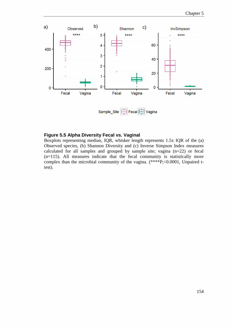
Chapter 5
154
Figure 5.5 Alpha Diversity Fecal vs. Vaginal Boxplots representing median, IQR, whisker length represents 1.5x IQR of the (a)
Observed species, (b) Shannon Diversity and (c) Inverse Simpson Index measures
calculated for all samples and grouped by sample site; vagina (n=22) or fecal
(n=115). All measures indicate that the fecal community is statistically more
complex than the microbial community of the vagina. (****P;<0.0001, Unpaired t-
test).
a) b) c)
**** ****
****
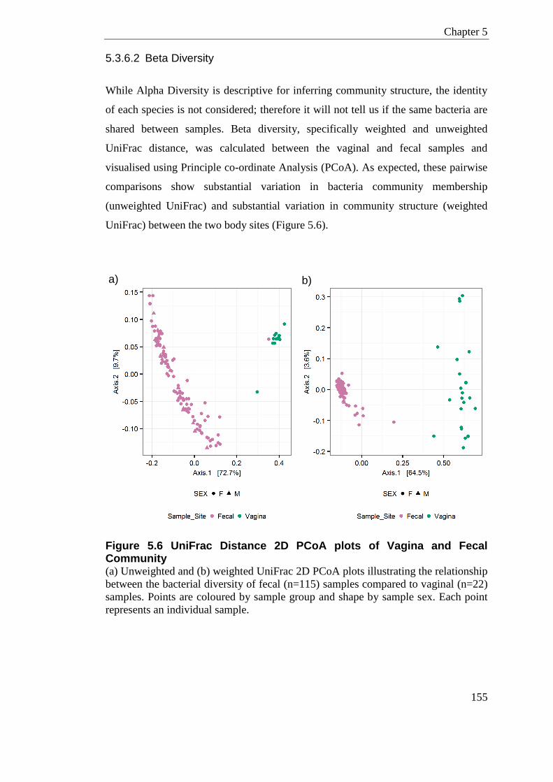
Chapter 5
155
5.3.6.2 Beta Diversity
While Alpha Diversity is descriptive for inferring community structure, the identity
of each species is not considered; therefore it will not tell us if the same bacteria are
shared between samples. Beta diversity, specifically weighted and unweighted
UniFrac distance, was calculated between the vaginal and fecal samples and
visualised using Principle co-ordinate Analysis (PCoA). As expected, these pairwise
comparisons show substantial variation in bacteria community membership
(unweighted UniFrac) and substantial variation in community structure (weighted
UniFrac) between the two body sites (Figure 5.6).
Figure 5.6 UniFrac Distance 2D PCoA plots of Vagina and Fecal Community (a) Unweighted and (b) weighted UniFrac 2D PCoA plots illustrating the relationship
between the bacterial diversity of fecal (n=115) samples compared to vaginal (n=22)
samples. Points are coloured by sample group and shape by sample sex. Each point
represents an individual sample.
a) b)
a)
)
Chapter 5
156
5.3.6.3 Sample Cross Contamination
Due to the close proximity of the murine vagina and anus it is possible that cross-
contamination during sampling can occur. To determine whether this is a common
occurrence the shared taxa between the two sites were compared. There were 188
overlapping OTUs between the vaginal and fecal samples (Figure 5.7.a). This portion
is higher than expected, inferred from what Barfod et al., (2013) had detailed
comparing the cecum and vagina. Due to the high depth of sequencing I conducted in
this study, many of these shared OTUs are low abundant; less than 100 sequences. If
only OTUs with an abundance of >0.1% are considered, the number of shared OTUs
is reduced to 50 (data not shown), these shared OTUs represent only 4 different
genus level classifications (Figure 5.7.b); Staphylococcus, Lactobacillus, S24-7-
Family, and Pseudomonas. This is more in keeping with the proportions of shared
taxa detailed in Barfod et al., (2013).
The contribution of Pseudomonas to the fecal community is highly skewed by one
animal that had predominately Pseudomonas sequences. Pseudomonas should
therefore not be defined as a common fecal bacterium in this study. Staphylococcus,
Lactobacillus, and Pseudomonas have previously been reported in the vagina of
laboratory and wild mice (MacManes, 2011, Barfod et al., 2013). S24-7 is mostly a
mouse gut associated bacteria, sequences from this genus are most likely due to low
level contamination from fecal material. The contribution of S24-7 sequences in the
vagina is 0.22% compared to fecal sample where 44.6% of sequences are from this
clade. To conclude there is very little overlap between fecal and vaginal microbiota,
which would be indicative of high levels of cross-contamination during sampling.
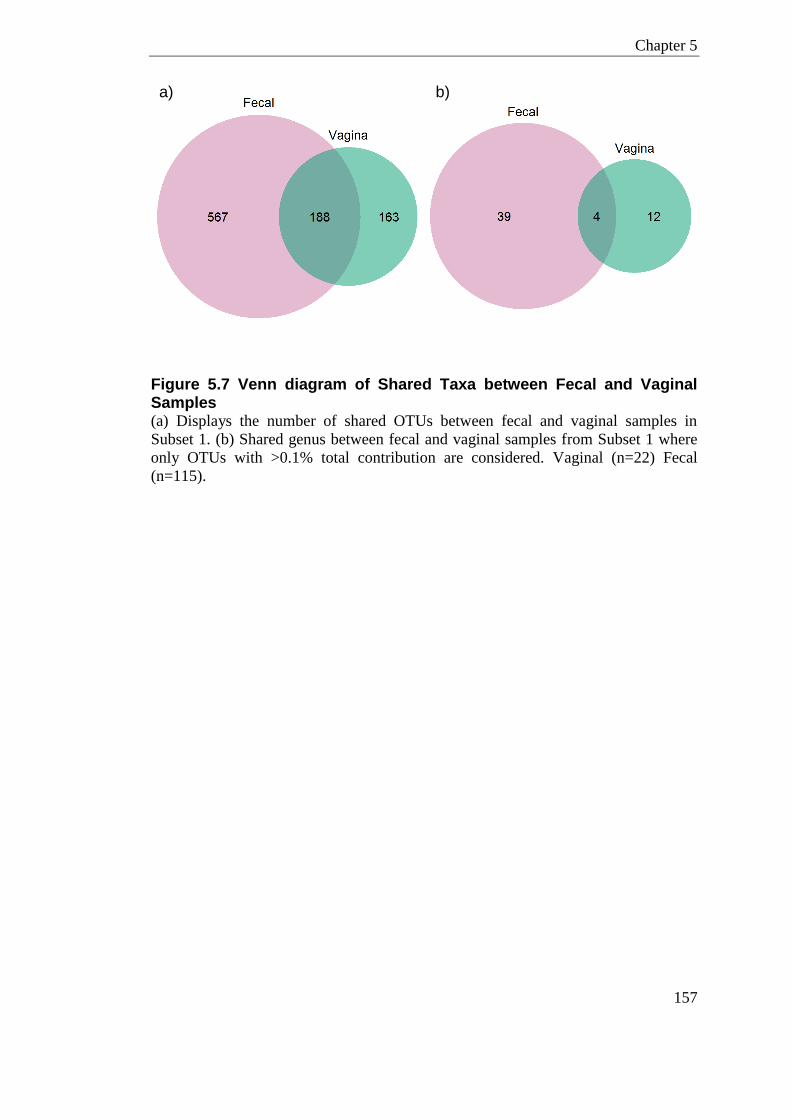
Chapter 5
157
Figure 5.7 Venn diagram of Shared Taxa between Fecal and Vaginal Samples (a) Displays the number of shared OTUs between fecal and vaginal samples in
Subset 1. (b) Shared genus between fecal and vaginal samples from Subset 1 where
only OTUs with >0.1% total contribution are considered. Vaginal (n=22) Fecal
(n=115).
a) b)
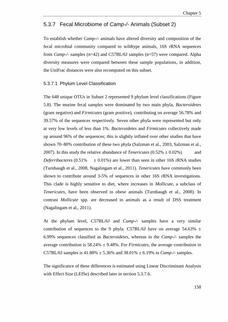
Chapter 5
158
5.3.7 Fecal Microbiome of Camp-/- Animals (Subset 2)
To establish whether Camp-/- animals have altered diversity and composition of the
fecal microbial community compared to wildtype animals, 16S rRNA sequences
from Camp-/- samples (n=42) and C57BL/6J samples (n=57) were compared. Alpha
diversity measures were compared between these sample populations, in addition,
the UniFrac distances were also recomputed on this subset.
5.3.7.1 Phylum Level Classification
The 648 unique OTUs in Subset 2 represented 9 phylum level classifications (Figure
5.8). The murine fecal samples were dominated by two main phyla, Bacteroidetes
(gram negative) and Firmicutes (gram positive), contributing on average 56.78% and
39.57% of the sequences respectively. Seven other phyla were represented but only
at very low levels of less than 1%. Bacteroidetes and Firmicutes collectively made
up around 96% of the sequences; this is slightly inflated over other studies that have
shown 70–80% contribution of these two phyla (Salzman et al., 2003, Salzman et al.,
2007). In this study the relative abundance of Tenericutes (0.52% ± 0.02%) and
Deferribacteres (0.51% ± 0.01%) are lower than seen in other 16S rRNA studies
(Turnbaugh et al., 2008, Nagalingam et al., 2011). Tenericutes have commonly been
shown to contribute around 3-5% of sequences in other 16S rRNA investigations.
This clade is highly sensitive to diet, where increases in Mollicute, a subclass of
Tenericutes, have been observed in obese animals (Turnbaugh et al., 2008). In
contrast Mollicute spp. are decreased in animals as a result of DSS treatment
(Nagalingam et al., 2011).
At the phylum level, C57BL/6J and Camp-/- samples have a very similar
contribution of sequences to the 9 phyla. C57BL/6J have on average 54.63% ±
6.99% sequences classified as Bacteroidetes, whereas in the Camp-/- samples the
average contribution is 58.24% ± 9.48%. For Firmicutes, the average contribution in
C57BL/6J samples is 41.88% ± 5.36% and 38.01% ± 6.19% in Camp-/- samples.
The significance of these differences is estimated using Linear Discriminant Analysis
with Effect Size (LEfSe) described later in section 5.3.7.6.
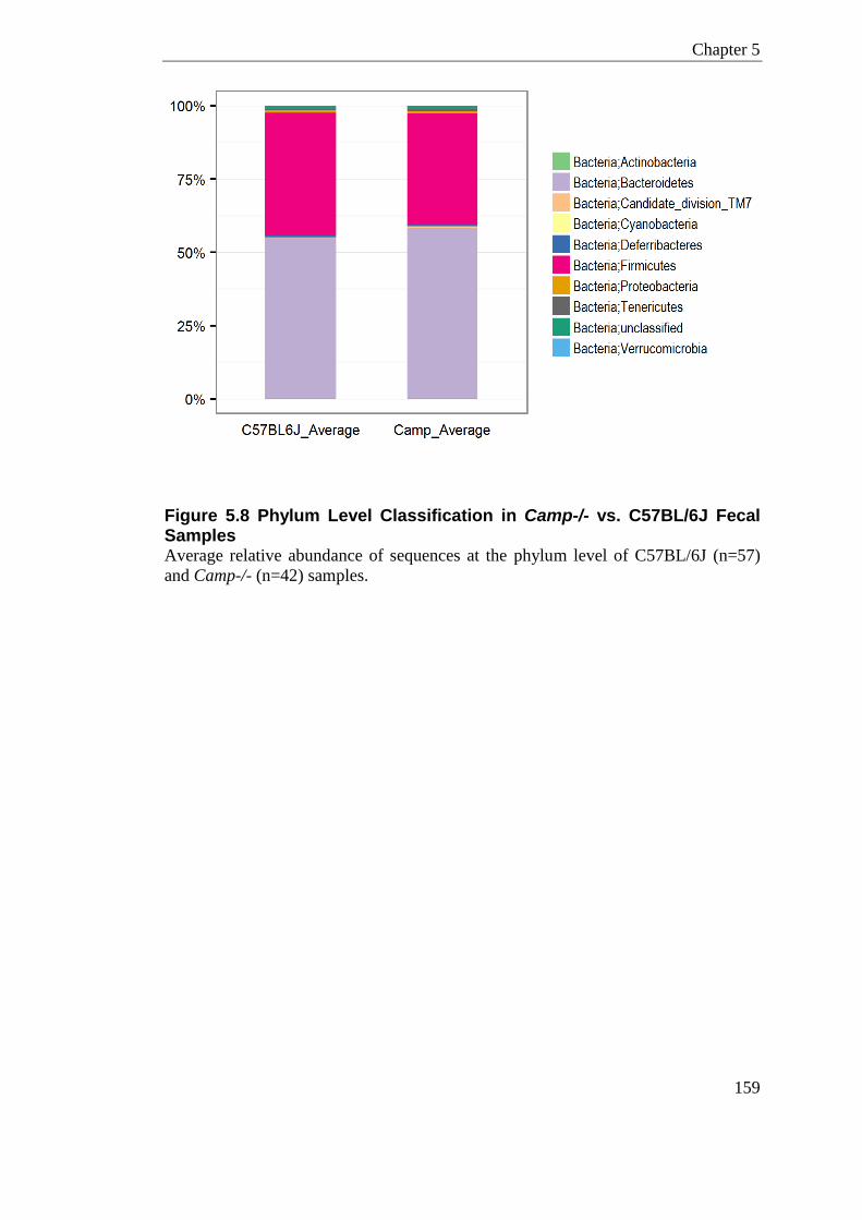
Chapter 5
159
Figure 5.8 Phylum Level Classification in Camp-/- vs. C57BL/6J Fecal Samples Average relative abundance of sequences at the phylum level of C57BL/6J (n=57)
and Camp-/- (n=42) samples.
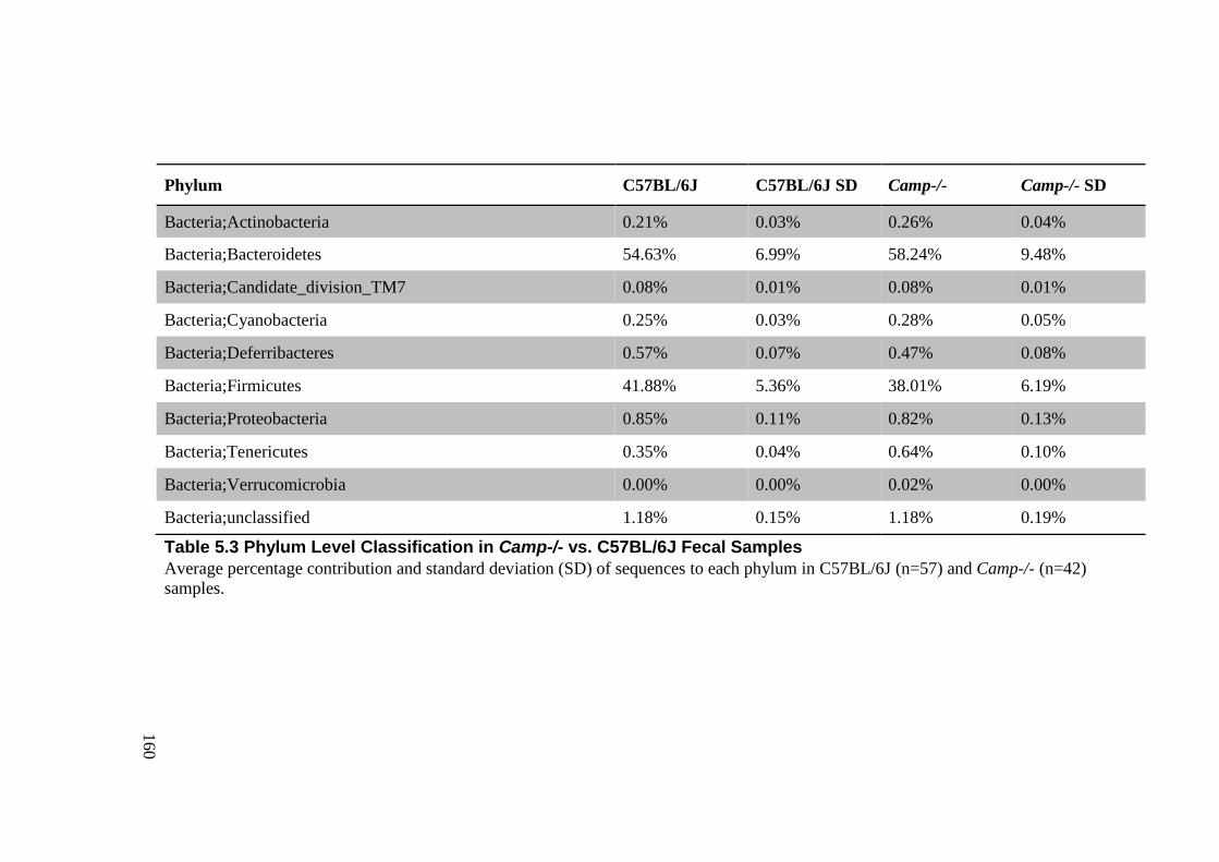
160
Phylum C57BL/6J C57BL/6J SD Camp-/- Camp-/- SD
Bacteria;Actinobacteria 0.21% 0.03% 0.26% 0.04%
Bacteria;Bacteroidetes 54.63% 6.99% 58.24% 9.48%
Bacteria;Candidate_division_TM7 0.08% 0.01% 0.08% 0.01%
Bacteria;Cyanobacteria 0.25% 0.03% 0.28% 0.05%
Bacteria;Deferribacteres 0.57% 0.07% 0.47% 0.08%
Bacteria;Firmicutes 41.88% 5.36% 38.01% 6.19%
Bacteria;Proteobacteria 0.85% 0.11% 0.82% 0.13%
Bacteria;Tenericutes 0.35% 0.04% 0.64% 0.10%
Bacteria;Verrucomicrobia 0.00% 0.00% 0.02% 0.00%
Bacteria;unclassified 1.18% 0.15% 1.18% 0.19%
Table 5.3 Phylum Level Classification in Camp-/- vs. C57BL/6J Fecal Samples
Average percentage contribution and standard deviation (SD) of sequences to each phylum in C57BL/6J (n=57) and Camp-/- (n=42)
samples.
Chapter 5
161
5.3.7.2 Genus Level Classification
The 648 unique OTUs in Subset 2 represented 78 genus level classifications. Figure
5.9.a and Table 5.4 show the relative abundance of these genera in each sample
(n=99). Genera with a contribution of <0.5% sequences have been grouped and
represented as ‘low OTUs’ for ease of visualisation.
Bacteroidetes
Ten Bacteroidetes genera were identified which represent six different families. The
most dominant Bacteroidetes in the fecal samples were found to be S24-7 family of
bacteria, contributing on average 44.98% of the sequences. The next abundant genera
of Bacteroidetes were, Alistipes, Bacteroides and RC9 gut group, contributing
3.65%, 2.74% and 1.29% average sequences respectively.
S24-7 family, a butyrate-producing bacterium predominantly only seen in rodents,
covers the largest component of mouse fecal sequences assigned in the Bacteroidetes
phylum. Alistipes spp. are found in the gastrointestinal tract of both humans and
mouse. It has been reported to be the most significantly overrepresented taxa in older
mice (Langille et al., 2014), which is consistent to what has been shown in humans,
suggesting parallel age related microbial shifts in this taxa (Claesson et al., 2012).
Gastrointestinal Bacteroides are an abundant and well-studied member of the
mammalian commensal microbiota providing many beneficial activities, including
the metabolism of complex carbohydrates and glycosylation (Comstock, 2009,
Hooper et al., 2001).
Firmicutes
Forty Firmicutes genera were identified, representing nineteen different families. The
most dominant Firmicutes were Lactobacillus with 11.23% contribution, followed by
Blautia, an unclassified Lachnospiraceae, and an unclassified Ruminococcaceae,
with 8.99%, 6.31% and 3.27% average sequence contributions respectively.
Chapter 5
162
Lactobacillus spp. colonise the gut and mucosal sites of most mammals where they
can adhere to the non-secreting stomach epithelium, continuously seeding the content
of the stomach. Interestingly the absence of Lactobacillus reuteri in the gut of mice
causes autism-like behaviours, which can be reversed by probiotic inoculation of this
bacterium (Buffington et al., 2016). The mechanisms of this are still unclear but
Lactobacillus reuteri has been shown to increase the levels of the oxytocin hormone
(Buffington et al., 2016). Other Lactobacillus species have also been found to have
cognitive effects in mice (Bravo et al., 2011).
Blautia is a genera often associated with the production of beneficial Short-chain
fatty acids (SCFAs), which are also thought to work in helping suppress
inflammation. Interestingly giving Blautia spp. to mice could protect them from graft
versus host disease (Jenq et al., 2015).
Lachnospiraceae family is abundant in the digestive tracts of many mammals but is
rarely found at other sites, it is also a group often associated with the production of
SCFAs. Members of this family in humans have been linked to obesity and colon
cancer, mainly due to the association with the production of Butyric acid. Although
this property is not conserved as not all species of Lachnospiraceae have genes
related to Butyric acid production (Meehan and Beiko, 2014).
Ruminococcaceae is a family normally found just in the ruminant gut, although
species within this family have also been seen in human gastrointestinal samples
(Wang et al., 2004a).
Shared Taxa
77 of the 78 genera were common to C57BL/6J and Camp-/- samples. The only
genera not shared were the low abundant Akkermansia. C57BL/6J samples
collectively had no sequences from this clade; Camp-/- animals had 0.02% average
Akkermansia contribution. Although, this clade was not consistently seen in all
Camp-/- animals; only 11/42 had Akkermansia sequences. The most abundant
genera, S24-7 family, are higher in the Camp-/- samples; 47.32% compared to
C57BL/6J samples which have on average 43.25%. Lactobacillus was higher in the

Chapter 5
163
wildtypes displaying 11.82% compared to Camp-/- samples which had 10.43%
average contribution. Lower abundant genera also show differences such as
Allobaculum, which is 2.02% in C57BL/6J and 0.95% in Camp-/-mice. The
significance of these differences, and all other OTUs, is estimated using Linear
Discriminant Analysis with Effect Size (LEfSe) as described later in section 5.3.7.6.
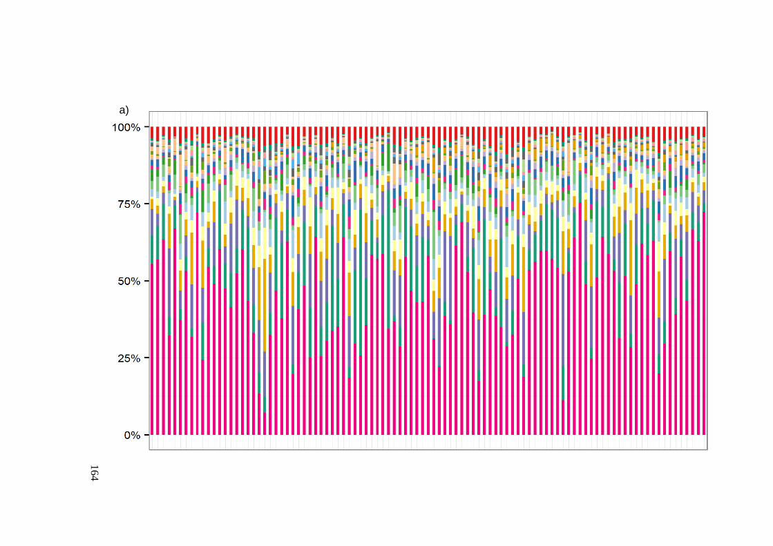
164
a)
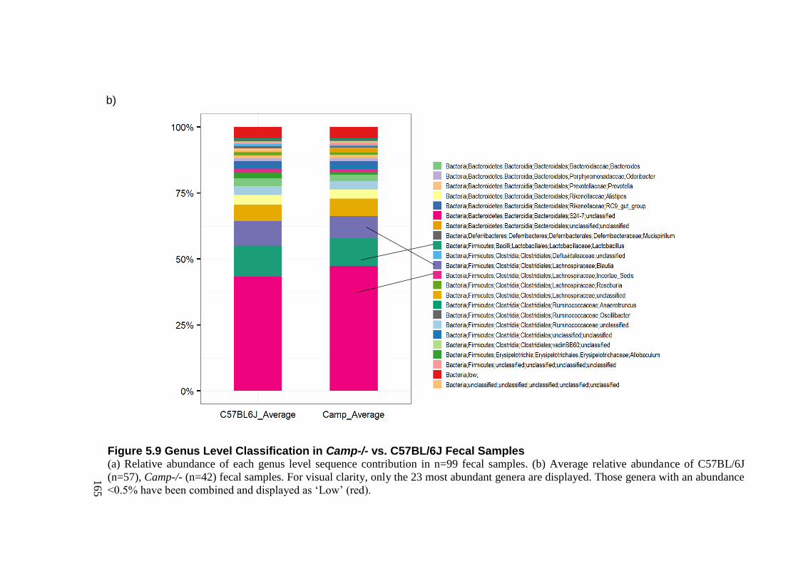
165
Figure 5.9 Genus Level Classification in Camp-/- vs. C57BL/6J Fecal Samples (a) Relative abundance of each genus level sequence contribution in n=99 fecal samples. (b) Average relative abundance of C57BL/6J
(n=57), Camp-/- (n=42) fecal samples. For visual clarity, only the 23 most abundant genera are displayed. Those genera with an abundance
<0.5% have been combined and displayed as ‘Low’ (red).
b)
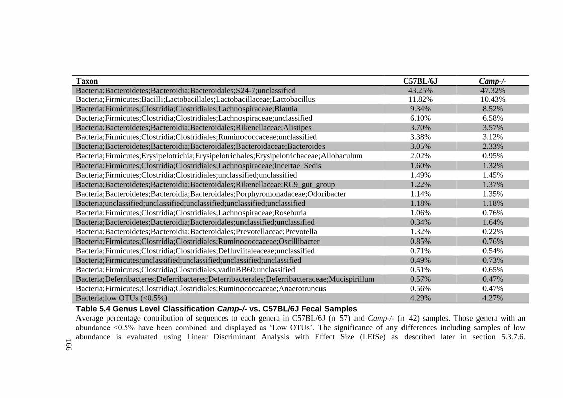
166
Taxon C57BL/6J Camp-/-
Bacteria;Bacteroidetes;Bacteroidia;Bacteroidales;S24-7;unclassified 43.25% 47.32% Bacteria;Firmicutes;Bacilli;Lactobacillales;Lactobacillaceae;Lactobacillus 11.82% 10.43%
Bacteria;Firmicutes;Clostridia;Clostridiales;Lachnospiraceae;Blautia 9.34% 8.52%
Bacteria;Firmicutes;Clostridia;Clostridiales;Lachnospiraceae;unclassified 6.10% 6.58%
Bacteria;Bacteroidetes;Bacteroidia;Bacteroidales;Rikenellaceae;Alistipes 3.70% 3.57%
Bacteria;Firmicutes;Clostridia;Clostridiales;Ruminococcaceae;unclassified 3.38% 3.12%
Bacteria;Bacteroidetes;Bacteroidia;Bacteroidales;Bacteroidaceae;Bacteroides 3.05% 2.33%
Bacteria;Firmicutes;Erysipelotrichia;Erysipelotrichales;Erysipelotrichaceae;Allobaculum 2.02% 0.95%
Bacteria;Firmicutes;Clostridia;Clostridiales;Lachnospiraceae;Incertae_Sedis 1.60% 1.32%
Bacteria;Firmicutes;Clostridia;Clostridiales;unclassified;unclassified 1.49% 1.45%
Bacteria;Bacteroidetes;Bacteroidia;Bacteroidales;Rikenellaceae;RC9_gut_group 1.22% 1.37%
Bacteria;Bacteroidetes;Bacteroidia;Bacteroidales;Porphyromonadaceae;Odoribacter 1.14% 1.35%
Bacteria;unclassified;unclassified;unclassified;unclassified;unclassified 1.18% 1.18%
Bacteria;Firmicutes;Clostridia;Clostridiales;Lachnospiraceae;Roseburia 1.06% 0.76%
Bacteria;Bacteroidetes;Bacteroidia;Bacteroidales;unclassified;unclassified 0.34% 1.64%
Bacteria;Bacteroidetes;Bacteroidia;Bacteroidales;Prevotellaceae;Prevotella 1.32% 0.22%
Bacteria;Firmicutes;Clostridia;Clostridiales;Ruminococcaceae;Oscillibacter 0.85% 0.76%
Bacteria;Firmicutes;Clostridia;Clostridiales;Defluviitaleaceae;unclassified 0.71% 0.54%
Bacteria;Firmicutes;unclassified;unclassified;unclassified;unclassified 0.49% 0.73%
Bacteria;Firmicutes;Clostridia;Clostridiales;vadinBB60;unclassified 0.51% 0.65%
Bacteria;Deferribacteres;Deferribacteres;Deferribacterales;Deferribacteraceae;Mucispirillum 0.57% 0.47%
Bacteria;Firmicutes;Clostridia;Clostridiales;Ruminococcaceae;Anaerotruncus 0.56% 0.47%
Bacteria;low OTUs (<0.5%) 4.29% 4.27%
Table 5.4 Genus Level Classification Camp-/- vs. C57BL/6J Fecal Samples Average percentage contribution of sequences to each genera in C57BL/6J (n=57) and Camp-/- (n=42) samples. Those genera with an
abundance <0.5% have been combined and displayed as ‘Low OTUs’. The significance of any differences including samples of low
abundance is evaluated using Linear Discriminant Analysis with Effect Size (LEfSe) as described later in section 5.3.7.6.
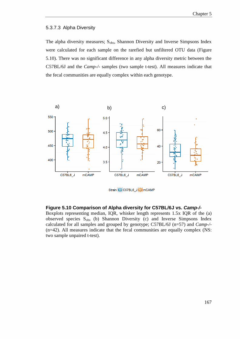
Chapter 5
167
5.3.7.3 Alpha Diversity
The alpha diversity measures; Sobs, Shannon Diversity and Inverse Simpsons Index
were calculated for each sample on the rarefied but unfiltered OTU data (Figure
5.10). There was no significant difference in any alpha diversity metric between the
C57BL/6J and the Camp-/- samples (two sample t-test). All measures indicate that
the fecal communities are equally complex within each genotype.
Figure 5.10 Comparison of Alpha diversity for C57BL/6J vs. Camp-/- Boxplots representing median, IQR, whisker length represents 1.5x IQR of the (a)
observed species Sobs (b) Shannon Diversity (c) and Inverse Simpsons Index
calculated for all samples and grouped by genotype; C57BL/6J (n=57) and Camp-/-
(n=42). All measures indicate that the fecal communities are equally complex (NS:
two sample unpaired t-test).
a) b)
c)

Chapter 5
168
5.3.7.4 Beta Diversity
The pairwise qualitative difference between sample diversities (unweighted UniFrac)
distances reveals there is a highly significant difference in the diversity within
genotypes compared to between genotypes (P<0.0001), whereby Camp-/- knockout
animals are significantly more diverse than the wildtype controls (P<0.0001) (Figure
5.11). The diversity between genotypes is also significantly more than the diversity
within each genotype respectively (P <0.0001 for both). Nonparametric p-values
were also calculated using 999 Monte Carlo permutations and Bonferroni-corrected,
all tested pairs give P <0.01.
That is to say animals are more similar to other animals of the same genotype than
they are to another animal of the opposite genotype. In addition the Camp-/- animals
have more bacterial diversity than the wildtype animals.
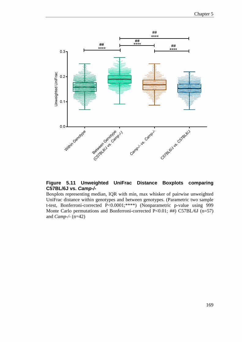
Chapter 5
169
Figure 5.11 Unweighted UniFrac Distance Boxplots comparing C57BL/6J vs. Camp-/- Boxplots representing median, IQR with min, max whisker of pairwise unweighted
UniFrac distance within genotypes and between genotypes. (Parametric two sample
t-test, Bonferroni-corrected P<0.0001;****) (Nonparametric p-value using 999
Monte Carlo permutations and Bonferroni-corrected P<0.01; ##) C57BL/6J (n=57)
and Camp-/- (n=42)
Chapter 5
170
The pairwise UniFrac distances were visualised using Principle co-ordinate Analysis
(PCoA) (Figure 5.12). Principal coordinate 1 (Axis 1; percent variation explained:
19.6%) of the unweighted UniFrac distances separates the two genotypes, while
Principal coordinate 2 (Axis 2; percent variation explained: 9.7%) intercepts the
genotypes and appears to be driven partly by cage variation, especially for the
wildtype cluster (Figure 5.13). In the weighted UniFrac plot, there is no apparent
separation of the samples by genotype or cage, and therefore does not highlight
substantial variation in community structure. The clustering of samples by genotype
in the unweighted UniFrac suggests a strong relationship in terms of qualitative
community membership.
In other terms, it is the presence or absence of certain OTUs that is driving the
clustering by genotype, more so than the abundance of the OTUs that are shared.
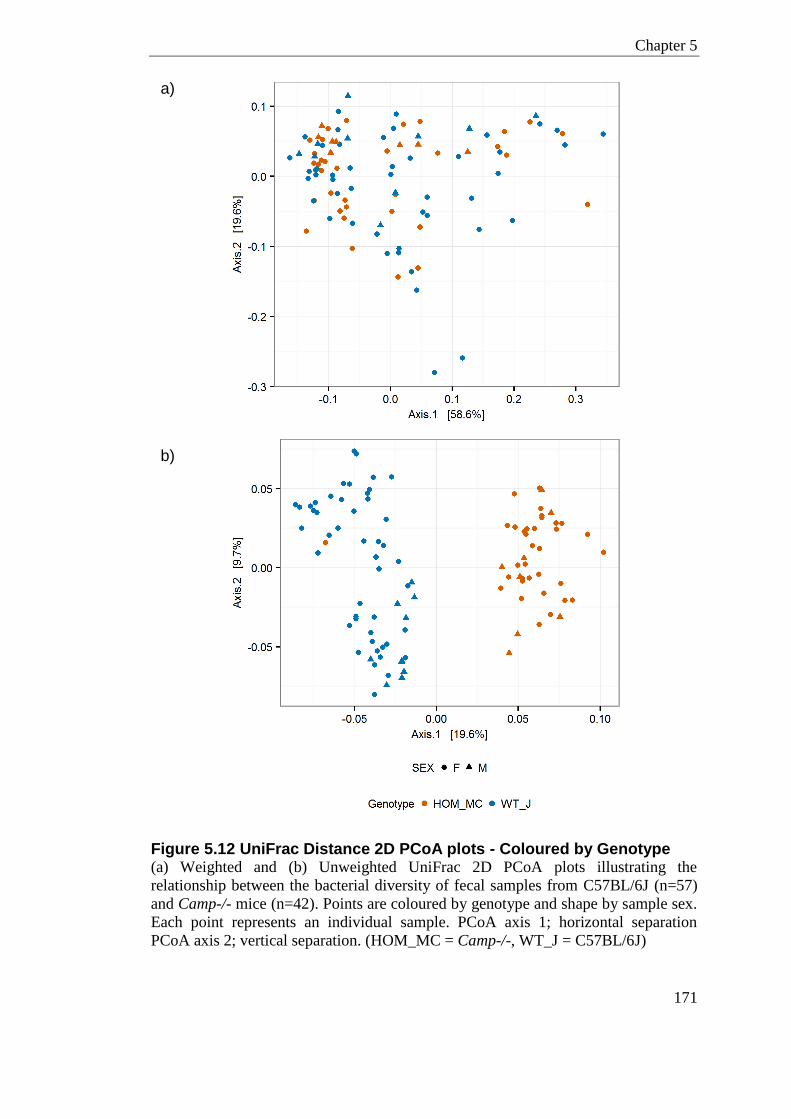
Chapter 5
171
Figure 5.12 UniFrac Distance 2D PCoA plots - Coloured by Genotype (a) Weighted and (b) Unweighted UniFrac 2D PCoA plots illustrating the
relationship between the bacterial diversity of fecal samples from C57BL/6J (n=57)
and Camp-/- mice (n=42). Points are coloured by genotype and shape by sample sex.
Each point represents an individual sample. PCoA axis 1; horizontal separation
PCoA axis 2; vertical separation. (HOM_MC = Camp-/-, WT_J = C57BL/6J)
a)
b)
a)
)
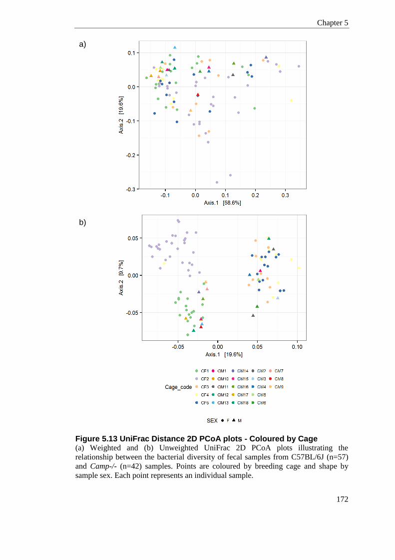
Chapter 5
172
Figure 5.13 UniFrac Distance 2D PCoA plots - Coloured by Cage (a) Weighted and (b) Unweighted UniFrac 2D PCoA plots illustrating the
relationship between the bacterial diversity of fecal samples from C57BL/6J (n=57)
and Camp-/- (n=42) samples. Points are coloured by breeding cage and shape by
sample sex. Each point represents an individual sample.
a)
b)
a)
)
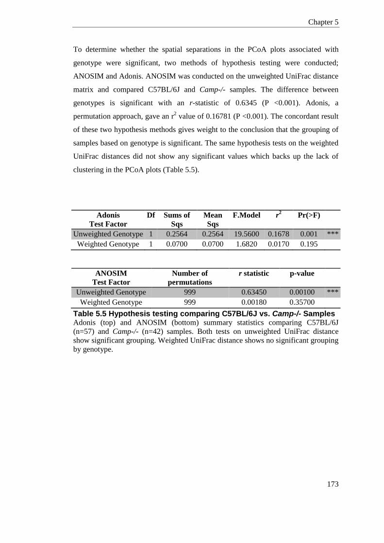
Chapter 5
173
To determine whether the spatial separations in the PCoA plots associated with
genotype were significant, two methods of hypothesis testing were conducted;
ANOSIM and Adonis. ANOSIM was conducted on the unweighted UniFrac distance
matrix and compared C57BL/6J and Camp-/- samples. The difference between
genotypes is significant with an r-statistic of 0.6345 (P <0.001). Adonis, a
permutation approach, gave an r2 value of 0.16781 (P <0.001). The concordant result
of these two hypothesis methods gives weight to the conclusion that the grouping of
samples based on genotype is significant. The same hypothesis tests on the weighted
UniFrac distances did not show any significant values which backs up the lack of
clustering in the PCoA plots (Table 5.5).
Adonis
Test Factor
Df Sums of
Sqs
Mean
Sqs
F.Model r2 Pr(>F)
Unweighted Genotype 1 0.2564 0.2564 19.5600 0.1678 0.001 ***
Weighted Genotype 1 0.0700 0.0700 1.6820 0.0170 0.195
ANOSIM
Test Factor
Number of
permutations
r statistic p-value
Unweighted Genotype 999 0.63450 0.00100 ***
Weighted Genotype 999 0.00180 0.35700
Table 5.5 Hypothesis testing comparing C57BL/6J vs. Camp-/- Samples Adonis (top) and ANOSIM (bottom) summary statistics comparing C57BL/6J
(n=57) and Camp-/- (n=42) samples. Both tests on unweighted UniFrac distance
show significant grouping. Weighted UniFrac distance shows no significant grouping
by genotype.
Chapter 5
174
As a portion of samples in this study were taken from the same mouse across 4 time-
points, this raises the question of whether intra-mouse correlations are artificially
increasing the similarity within genotype. To address this question, I examined the
average beta-diversity from the same mouse and compared to the beta-diversity
between mice, within the same genotype (Figure 5.14). The mean unweighted
UniFrac distance within animals is statistically lower than the mean UniFrac distance
between animals in both genotypes (P; <0.0001; parametric two sample t-test,
Bonferroni-corrected). Overall this suggests the phylogenetic diversity within the
same-animal at the different time-points is more similar than between samples at the
various time-points. That is to say animals are more similar to themselves than they
are to another animal within genotype. This difference suggests no substantial intra-
mouse changes in the microbiome composition over the short period of time (17
days). This will however inflate the apparent similarity within genotype; therefore a
further subset of the data is required to only include mice that have been sampled
once to confirm this separation by genotype is significant. Sub-setting to only include
a single time-point (Day 0) will also reduce any confounding signals relating to
pregnancy.
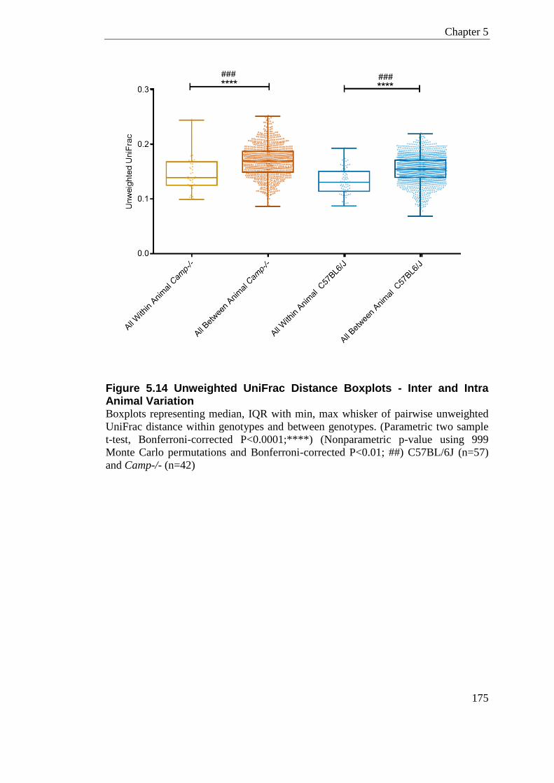
Chapter 5
175
Figure 5.14 Unweighted UniFrac Distance Boxplots - Inter and Intra Animal Variation Boxplots representing median, IQR with min, max whisker of pairwise unweighted
UniFrac distance within genotypes and between genotypes. (Parametric two sample
t-test, Bonferroni-corrected P<0.0001;****) (Nonparametric p-value using 999
Monte Carlo permutations and Bonferroni-corrected P<0.01; ##) C57BL/6J (n=57)
and Camp-/- (n=42)
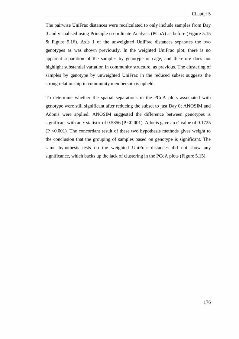
Chapter 5
176
The pairwise UniFrac distances were recalculated to only include samples from Day
0 and visualised using Principle co-ordinate Analysis (PCoA) as before (Figure 5.15
& Figure 5.16). Axis 1 of the unweighted UniFrac distances separates the two
genotypes as was shown previously. In the weighted UniFrac plot, there is no
apparent separation of the samples by genotype or cage, and therefore does not
highlight substantial variation in community structure, as previous. The clustering of
samples by genotype by unweighted UniFrac in the reduced subset suggests the
strong relationship in community membership is upheld.
To determine whether the spatial separations in the PCoA plots associated with
genotype were still significant after reducing the subset to just Day 0; ANOSIM and
Adonis were applied. ANOSIM suggested the difference between genotypes is
significant with an r-statistic of 0.5856 (P <0.001). Adonis gave an r2 value of 0.1725
(P <0.001). The concordant result of these two hypothesis methods gives weight to
the conclusion that the grouping of samples based on genotype is significant. The
same hypothesis tests on the weighted UniFrac distances did not show any
significance, which backs up the lack of clustering in the PCoA plots (Figure 5.15).

Chapter 5
177
Figure 5.15 UniFrac Distance 2D PCoA plots - Coloured by Genotype; Day 0 Subset
(a) Weighted and (b) Unweighted UniFrac 2D PCoA plots illustrating the
relationship between the bacterial diversity of fecal samples from C57BL/6J (n=22)
and Camp-/- (n=17) animals. Points are coloured by genotype and shape by sample
sex. Each point represents an individual animal. (HOM_MC = Camp-/-, WT_J =
C57BL/6J)
a)
b)
a)
)
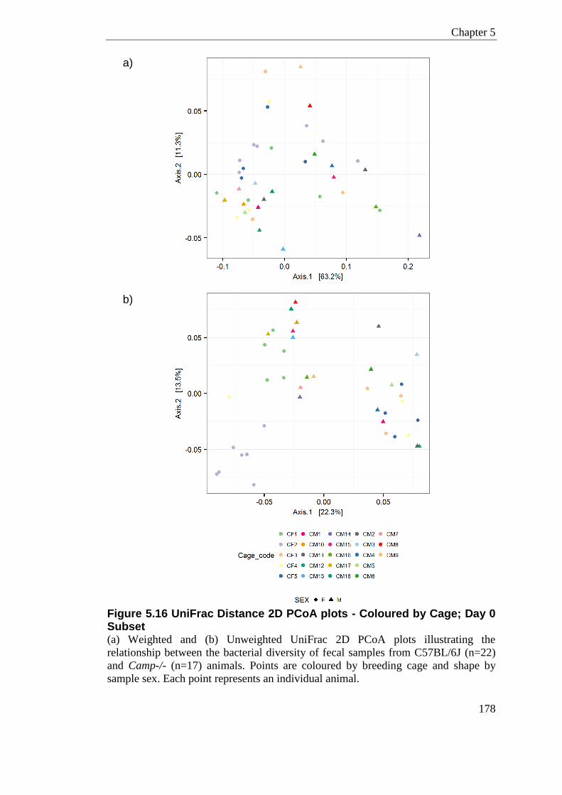
Chapter 5
178
Figure 5.16 UniFrac Distance 2D PCoA plots - Coloured by Cage; Day 0 Subset (a) Weighted and (b) Unweighted UniFrac 2D PCoA plots illustrating the
relationship between the bacterial diversity of fecal samples from C57BL/6J (n=22)
and Camp-/- (n=17) animals. Points are coloured by breeding cage and shape by
sample sex. Each point represents an individual animal.
a)
b)
a)
)

Chapter 5
179
Adonis Test
Factor
Df Sums of
Sqs
Mean
Sqs
F.Model r2 Pr(>F)
Unweighted
Genotype
1 (38) 0.0983 0.09836 7.9216 0.1725 0.001 ***
Weighted
Genotype
1 (38) 0.0350 0.03507 1.3028 0.0331 0.233
ANOSIM Test
Factor
Number of
permutations
r statistic p-value
Unweighted Genotype 999 0.5856 0.001 ***
Weighted Genotype 999 0.0056 0.310
Table 5.6 Hypothesis testing comparing C57BL/6J vs. Camp-/- Animals; Day 0 subset Adonis (top) and ANOSIM (bottom) summary statistics comparing C57BL/6J
(n=22) and Camp-/- (n=17) animals. Both tests on unweighted UniFrac distance
show significant grouping. The weighted UniFrac distance shows no significant
grouping by genotype in either test.

Chapter 5
180
5.3.7.5 Jack-knife Support; UPMGA bootstrapping
To estimate the uncertainty in the PCoA plots, Jack-knife bootstrapping was
performed, whereby samples are hierarchically clustered based on inter-sample
variation in UniFrac distances. Specifically, 100 jack-knife replicates at 45,000
sequences were used to generate a bootstrapped Unweighted-Pair Group Method
with Arithmetic mean (UPGMA) tree for the unweighted distance matrix (Figure
5.17). Tree generally supports the clustering seen in the PCoA plots; phylogenetic
clustering of samples by genotype with the exception of one outlier and root.
Figure 5.17 Jack-knife Unweighted UniFrac UPGMA Bootstrapped Tree Unweighted UniFrac pair group method with arithmetic mean (UPGMA) of Jack-
knife distance, re-sampling 100 times at 45,000 sequences. Fecal Samples are
coloured according to genotype. (mCAMP = Camp-/- (red), C57BL6_J (blue))
Chapter 5
181
5.3.7.6 Significant Taxa (LEfSe)
Using Linear Discriminant Analysis with Effect Size (LEfSe), an all-against-all
multi-class comparison of C57BL/6J and Camp-/- fecal samples was performed
(Figure 5.18). This highlighted several taxa were specifically modulated by
genotype.
A total of three core microbial groups at the family level were identified with
enrichment; Erysipelotrichaceae, Anaeroplasmataceae and Prevotellaceae. The most
notable OTU assigned to be enriched in Camp-/- samples was OTU0003, which was
classified as S24-7 family of bacteria, having an LDA score of 4.12. At the family
level this bacterium is not shown to have significant enrichment in Camp-/- samples.
OTUs within Lachnospiraceae and Blautia genera were also enriched in Camp-/-
animals.
C57BL/6J samples show an enrichment of Prevotella, this is very interesting as the
Camp-/- samples are nearly devoid of Prevotella clade. Certain Blautia OTUs and
Allobaculum are also enriched in the C57BL/6J samples.
The only enriched ‘class’-level taxa was Erysipelotrichia (p.Firmicutes), which was
mainly attributed to the genera Allobaculum. Allobaculum was significantly enriched
in C57BL/6J compared to Camp-/- samples. The other main genera showing multi-
level enrichment were Anaeroplasma, which were enriched in Camp-/- samples. As
one of the few Tenericutes observed in this study, Anaeroplasma contributed to the
enrichment back to the ‘order’-level of classification. Prevotella was highlighted as
an enriched genera in the C57BL/6J animals, this genus (and family) were also
notably absent from the Camp-/- samples with the exception of one outlier sample.
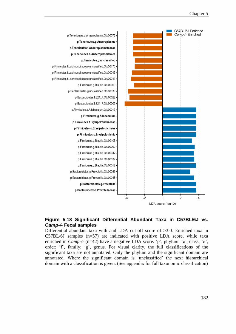
Chapter 5
182
Figure 5.18 Significant Differential Abundant Taxa in C57BL/6J vs. Camp-/- Fecal samples Differential abundant taxa with and LDA cut-off score of >3.0. Enriched taxa in
C57BL/6J samples (n=57) are indicated with positive LDA score, while taxa
enriched in Camp-/- (n=42) have a negative LDA score. ‘p’, phylum; ‘c’, class; ‘o’,
order; ‘f’, family; ‘g’, genus. For visual clarity, the full classifications of the
significant taxa are not annotated. Only the phylum and the significant domain are
annotated. Where the significant domain is ‘unclassified’ the next hierarchical
domain with a classification is given. (See appendix for full taxonomic classification)
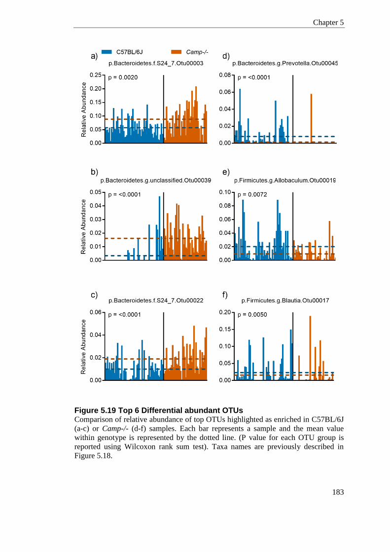
Chapter 5
183
Figure 5.19 Top 6 Differential abundant OTUs Comparison of relative abundance of top OTUs highlighted as enriched in C57BL/6J
(a-c) or Camp-/- (d-f) samples. Each bar represents a sample and the mean value
within genotype is represented by the dotted line. (P value for each OTU group is
reported using Wilcoxon rank sum test). Taxa names are previously described in
Figure 5.18.
Chapter 5
184
5.3.8 Pregnancy Modulation of the Microbiome (Subset 3)
To examine whether the fecal microbiome varied longitudinally both in a non-
pregnant and pregnant state, samples were partitioned to only include the wildtype
C57BL/6J animals which were sampled at 4 time-points over 17 days (n=44 samples;
16 non-pregnant, 28 pregnant).
5.3.8.1 The Stability of the Fecal Microbiome- Non Pregnant
Establishing baseline instability in a population is important when trying to infer
microbial shifts that are truly related to pregnancy. It is therefore important to
establish the diversity and composition of the bacterial community in a steady sate.
To determine the stability of the fecal microbiome over 17 days, longitudinal
samples of female C57BL/6J animals (n=4 animals, n=16 samples) were analysed for
alpha and beta diversity measures. The 4 mice investigated here (Animal ID: F01,
F04, F05, and F12) had very similar bacterial communities, mostly dominated by
S24-7 family of bacteria but also high abundances of Lactobacillus and Blautia spp.
(Figure 5.20).
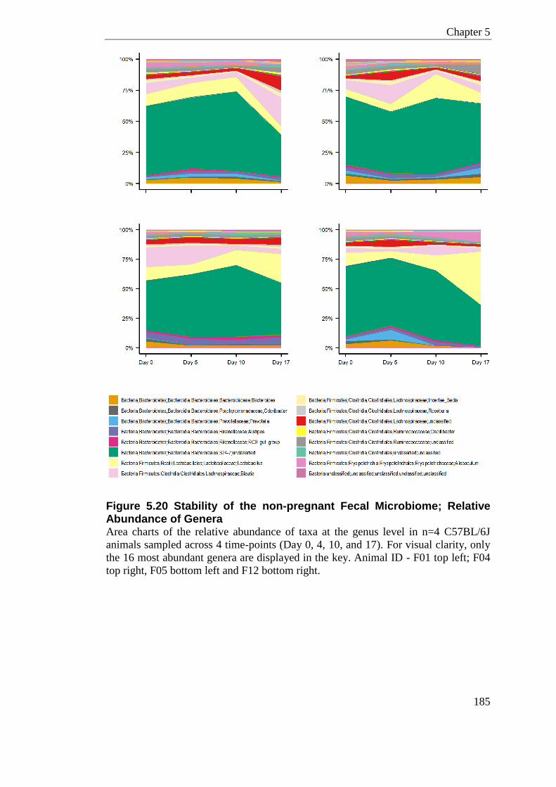
Chapter 5
185
Figure 5.20 Stability of the non-pregnant Fecal Microbiome; Relative Abundance of Genera Area charts of the relative abundance of taxa at the genus level in n=4 C57BL/6J
animals sampled across 4 time-points (Day 0, 4, 10, and 17). For visual clarity, only
the 16 most abundant genera are displayed in the key. Animal ID - F01 top left; F04
top right, F05 bottom left and F12 bottom right.
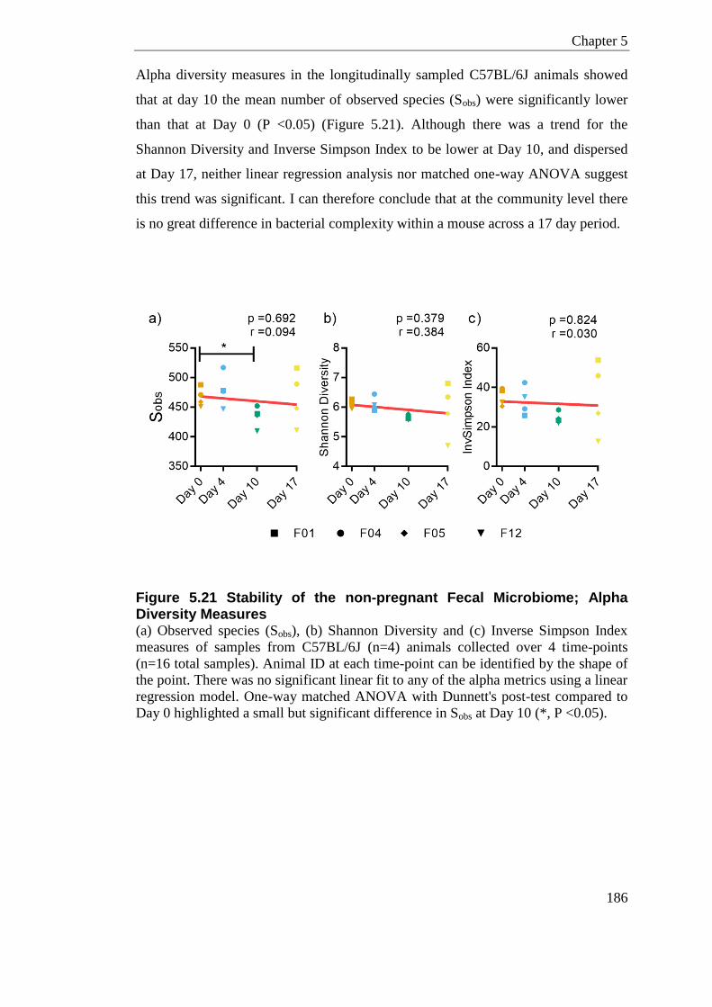
Chapter 5
186
Alpha diversity measures in the longitudinally sampled C57BL/6J animals showed
that at day 10 the mean number of observed species (Sobs) were significantly lower
than that at Day 0 (P <0.05) (Figure 5.21). Although there was a trend for the
Shannon Diversity and Inverse Simpson Index to be lower at Day 10, and dispersed
at Day 17, neither linear regression analysis nor matched one-way ANOVA suggest
this trend was significant. I can therefore conclude that at the community level there
is no great difference in bacterial complexity within a mouse across a 17 day period.
Figure 5.21 Stability of the non-pregnant Fecal Microbiome; Alpha Diversity Measures (a) Observed species (Sobs), (b) Shannon Diversity and (c) Inverse Simpson Index
measures of samples from C57BL/6J (n=4) animals collected over 4 time-points
(n=16 total samples). Animal ID at each time-point can be identified by the shape of
the point. There was no significant linear fit to any of the alpha metrics using a linear
regression model. One-way matched ANOVA with Dunnett's post-test compared to
Day 0 highlighted a small but significant difference in Sobs at Day 10 (*, P <0.05).
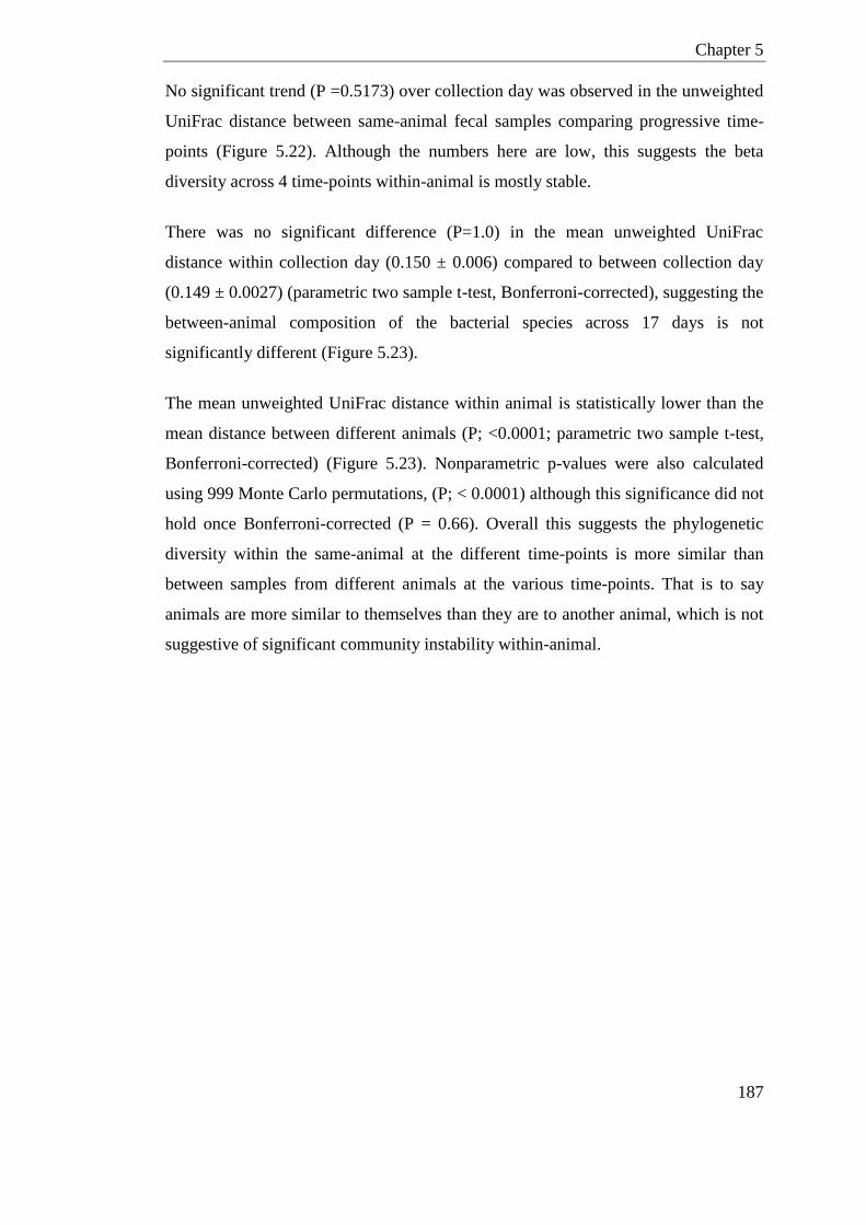
Chapter 5
187
No significant trend (P =0.5173) over collection day was observed in the unweighted
UniFrac distance between same-animal fecal samples comparing progressive time-
points (Figure 5.22). Although the numbers here are low, this suggests the beta
diversity across 4 time-points within-animal is mostly stable.
There was no significant difference (P=1.0) in the mean unweighted UniFrac
distance within collection day (0.150 ± 0.006) compared to between collection day
(0.149 ± 0.0027) (parametric two sample t-test, Bonferroni-corrected), suggesting the
between-animal composition of the bacterial species across 17 days is not
significantly different (Figure 5.23).
The mean unweighted UniFrac distance within animal is statistically lower than the
mean distance between different animals (P; <0.0001; parametric two sample t-test,
Bonferroni-corrected) (Figure 5.23). Nonparametric p-values were also calculated
using 999 Monte Carlo permutations, (P; < 0.0001) although this significance did not
hold once Bonferroni-corrected (P = 0.66). Overall this suggests the phylogenetic
diversity within the same-animal at the different time-points is more similar than
between samples from different animals at the various time-points. That is to say
animals are more similar to themselves than they are to another animal, which is not
suggestive of significant community instability within-animal.
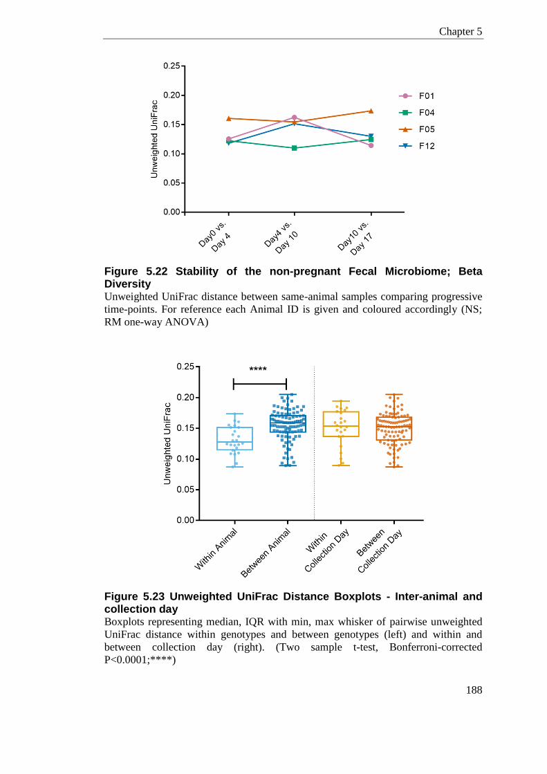
Chapter 5
188
Figure 5.22 Stability of the non-pregnant Fecal Microbiome; Beta Diversity Unweighted UniFrac distance between same-animal samples comparing progressive
time-points. For reference each Animal ID is given and coloured accordingly (NS;
RM one-way ANOVA)
Figure 5.23 Unweighted UniFrac Distance Boxplots - Inter-animal and collection day Boxplots representing median, IQR with min, max whisker of pairwise unweighted
UniFrac distance within genotypes and between genotypes (left) and within and
between collection day (right). (Two sample t-test, Bonferroni-corrected
P<0.0001;****)
Chapter 5
189
5.3.8.2 Pregnancy Related Microbial Change; Alpha Diversity
To examine whether the fecal microbiome varied longitudinally with gestation, I
applied a linear regression to the Observed species, Shannon Diversity and Inverse
Simpson Index (Figure 5.24) both in a non-pregnant and pregnant state. The only
linear trend was seen in the pregnant Inverse Simpson index (P =0.024, r = 0.180)
(Figure 5.24.b). However, there is no significant difference in this regression
compared to the non-pregnant control slope (P =0.192). In a similar regard, there was
no significant difference between the slopes from pregnant and non-pregnant mice in
either the Sobs (P =0.525; Figure 5.24.a) or Inverse Simpson Index (P=0.798; Figure
5.24.c). This indicates the diversity in microbial community did not progressively
change in terms of complexity throughout gestation.
The prior tested hypothesis is that microbial changes in pregnancy are accumulative
and progressive with gestation. However this need not be the case; many of the
metabolic and immunological changes that could impact the microbiome are
temporal during pregnancy. To test a non-linear association between alpha diversity
and gestation, a two-way ANOVA was also conducted. The two-way ANOVA
showed no significant association with time-point in either the pregnant (i.e.
gestation) or non-pregnant samples (i.e. time-point). This gives further support to the
conclusion that the complexity of the microbial communities is similar in a pregnant
and non-pregnant state.

Chapter 5
190
Figure 5.24 Pregnancy Influence on Alpha Diversity Measures Observed species (a) Shannon Diversity (b) and Inverse Simpson Index (c) measures
in samples collected over 4 time-points in pregnant and non-pregnant animals. P-
values displayed are for the difference between the Non-Pregnant vs. Pregnant
slopes. The only linear trend was seen in the pregnant Inverse Simpson index (b), P-
value and slope are given in parentheses. A two-way ANOVA did not highlight any
significant association of gestation or pregnancy status with alpha diversity
comparing to Day 0 groups.
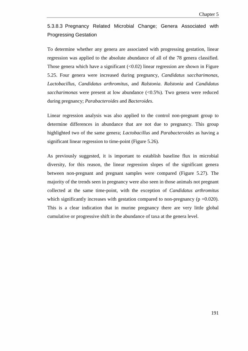
Chapter 5
191
5.3.8.3 Pregnancy Related Microbial Change; Genera Associated with
Progressing Gestation
To determine whether any genera are associated with progressing gestation, linear
regression was applied to the absolute abundance of all of the 78 genera classified.
Those genera which have a significant (<0.02) linear regression are shown in Figure
5.25. Four genera were increased during pregnancy, Candidatus saccharimonas,
Lactobacillus, Candidatus arthromitus, and Ralstonia. Ralstonia and Candidatus
saccharimonas were present at low abundance (<0.5%). Two genera were reduced
during pregnancy; Parabacteroides and Bacteroides.
Linear regression analysis was also applied to the control non-pregnant group to
determine differences in abundance that are not due to pregnancy. This group
highlighted two of the same genera; Lactobacillus and Parabacteroides as having a
significant linear regression to time-point (Figure 5.26).
As previously suggested, it is important to establish baseline flux in microbial
diversity, for this reason, the linear regression slopes of the significant genera
between non-pregnant and pregnant samples were compared (Figure 5.27). The
majority of the trends seen in pregnancy were also seen in those animals not pregnant
collected at the same time-point, with the exception of Candidatus arthromitus
which significantly increases with gestation compared to non-pregnancy (p =0.020).
This is a clear indication that in murine pregnancy there are very little global
cumulative or progressive shift in the abundance of taxa at the genera level.
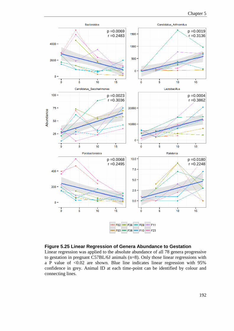
Chapter 5
192
Figure 5.25 Linear Regression of Genera Abundance to Gestation Linear regression was applied to the absolute abundance of all 78 genera progressive
to gestation in pregnant C57BL/6J animals (n=8). Only those linear regressions with
a P value of <0.02 are shown. Blue line indicates linear regression with 95%
confidence in grey. Animal ID at each time-point can be identified by colour and
connecting lines.
p =0.0180 r =0.2248
p =0.0069 r =0.2483
p =0.0019 r =0.3136
p =0.0004 r =0.3862
p =0.0068 r =0.2495
p =0.0023 r =0.3036
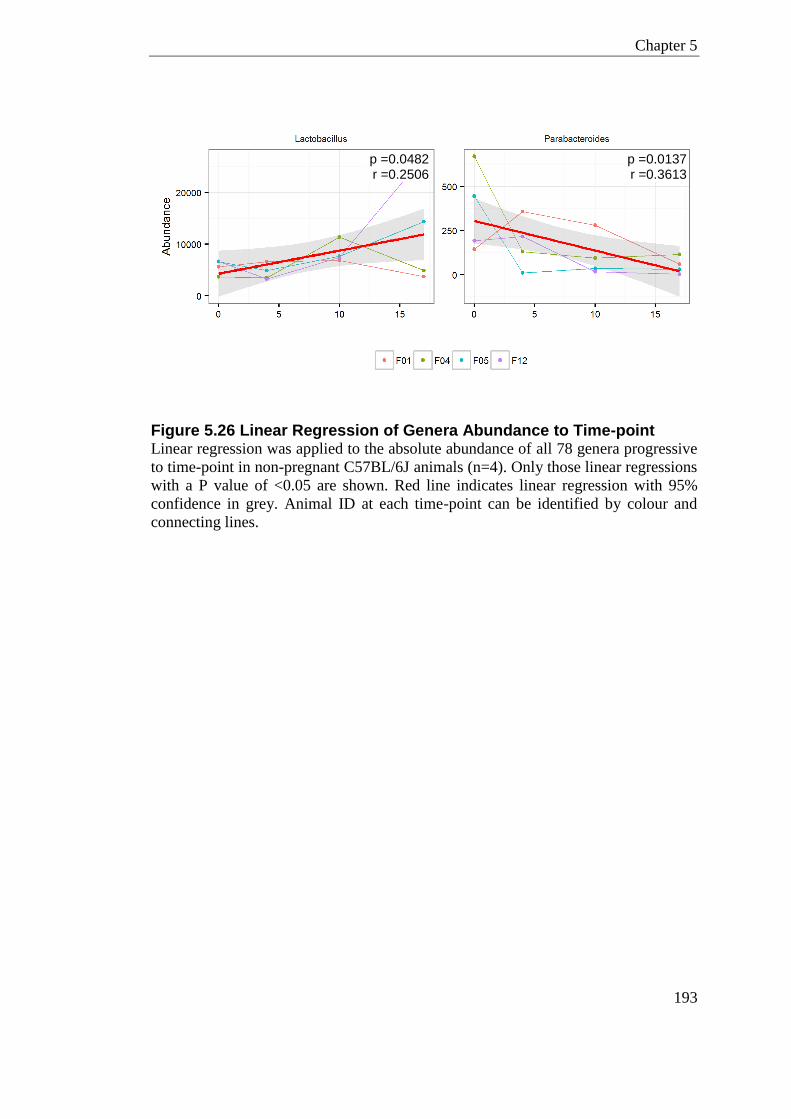
Chapter 5
193
Figure 5.26 Linear Regression of Genera Abundance to Time-point Linear regression was applied to the absolute abundance of all 78 genera progressive
to time-point in non-pregnant C57BL/6J animals (n=4). Only those linear regressions
with a P value of <0.05 are shown. Red line indicates linear regression with 95%
confidence in grey. Animal ID at each time-point can be identified by colour and
connecting lines.
p =0.0482 r =0.2506
p =0.0137 r =0.3613
Chapter 5
194
Figure 5.27 Linear Regression of Genera Abundance; Pregnant vs. Non-Pregnant Linear regression comparison of genera progressive to gestation/time-point in non-
pregnant (n=4) and pregnant (n=8) C57BL/6J animals. Only those linear regressions
with a P value of <0.05 in either pregnant or non-pregnant group are shown. Red line
indicates linear regression in non-pregnant (NP) samples. Blue line indicates linear
regression in pregnant samples (PR). Animal ID at each time-point can be identified
by colour of point and the shape of point corresponds to pregnancy status.
p =0.463 p =0.020 *
p =0.261 p =0.487
p =0.573 p =0.225
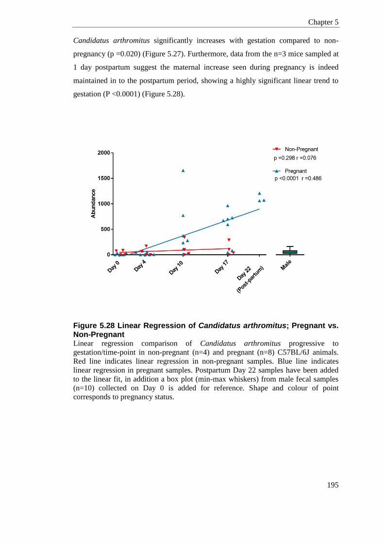
Chapter 5
195
Candidatus arthromitus significantly increases with gestation compared to non-
pregnancy (p =0.020) (Figure 5.27). Furthermore, data from the n=3 mice sampled at
1 day postpartum suggest the maternal increase seen during pregnancy is indeed
maintained in to the postpartum period, showing a highly significant linear trend to
gestation (P <0.0001) (Figure 5.28).
Figure 5.28 Linear Regression of Candidatus arthromitus; Pregnant vs. Non-Pregnant Linear regression comparison of Candidatus arthromitus progressive to
gestation/time-point in non-pregnant (n=4) and pregnant (n=8) C57BL/6J animals.
Red line indicates linear regression in non-pregnant samples. Blue line indicates
linear regression in pregnant samples. Postpartum Day 22 samples have been added
to the linear fit, in addition a box plot (min-max whiskers) from male fecal samples
(n=10) collected on Day 0 is added for reference. Shape and colour of point
corresponds to pregnancy status.
Chapter 5
196
As mentioned previously the hypothesis for linear regression analysis is that the
community is driven by progressive and accumulative microbial shifts as pregnancy
progresses. To test for non-linear association of the gestation/time-point on
abundance of genera, a non-parametric Kurskal-Wallis test was used to identify
significant genera associated with gestation/time-point in pregnant and non-pregnant
animals respectively.
Several genera were highlighted as having a significant association to gestation/time-
point, including; Anaeroplasma, Bacteroides, Blautia, Candidatus arthromitus,
Candidatus saccharimonas, Parabacteroides, Prevotella, and Oscillibacter (Figure
5.29). However, when adjusted for multiple comparisons none of these genera were
statistically significant. Moreover, an ANOVA did not show any significant
differences between pregnant and non-pregnant abundance of any of these genera
across all time-points. This suggests that although there is variation in the relative
abundance of genera over time, the majority is not attributed to pregnancy and is
most likely normal background community variation.
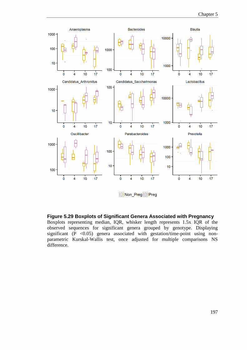
Chapter 5
197
Figure 5.29 Boxplots of Significant Genera Associated with Pregnancy Boxplots representing median, IQR, whisker length represents 1.5x IQR of the
observed sequences for significant genera grouped by genotype. Displaying
significant (P <0.05) genera associated with gestation/time-point using non-
parametric Kurskal-Wallis test, once adjusted for multiple comparisons NS
difference.
Chapter 5
198
5.4 Discussion
Fecal Microbiome of Camp-/- Animals
Microbes in the gut are essential for the break down of complex carbohydrates where
they can manipulate host metabolism by altering nutrient availability. The presence
and abundance of each bacterial group is fundamental since different species
differentially metabolise these nutrients. The host antimicrobial defence system has
co-evolved with complex communities of bacteria that have sought to create an
environment for commensal and mutualistic cohabitation. The mammalian
gastrointestinal tract is dominated by two main phyla, Firmicutes and Bacteroidetes.
Shifts in the ratio of these two phyla have been observed in many conditions such as
obesity (Ley et al., 2006) and diabetes (Wen et al., 2008). Diet and host genetics can
also influence on microbial community composition (Blanton et al., 2016, Ussar et
al., 2015). Host Defence Peptides provide the first line of defence against infectious
pathogens in the gastrointestinal tract. In the mouse and human colon, cathelicidin is
expressed in epithelial cells and immune cells such as macrophages.
In this chapter, I evaluate the direct influence of Cathelicidin on the fecal
microbiome by utilising Camp-/- knockout animals. The microbiome was
interrogated by 16S rRNA gene sequencing for bacterial classification. After
subsampling and removal of rare sequences, 908 OTUs were identified, which
represent 143 unique genera, 82 unique family level classifications, 50 unique orders,
29 unique classes and 15 different phyla (Figure 5.3).
Direct comparison of the phyla classifications for the Camp-/- and C57BL/6J
samples showed similar overall compositions; there were no differences in relative
abundance at the phylum level (Figure 5.8). As CAMP is a potent broad range
antimicrobial which has been shown to modulate mucosal immune responsiveness, it
was unexpected that knocking out this gene in mice would not confer shifts in
bacteria at the phylum level. Although, cathelicidin is only expressed at moderate
levels in the gut without inflammation or infection, moreover it has a higher potency
Chapter 5
199
for pathogenic species; these factors might be responsible for the lack of huge shifts
at the phylum level.
At the genera and OTU level of classification, Camp-/- and C57BL/6J mice
demonstrated disparate gut microbiota as indicated by statistically significant
taxonomic differences (Figure 5.18). At the genus level Allobaculum (p.Firmicutes
o.Erysipelotrichales), was significantly enriched in C57BL/6J samples, whereas
Anaeroplasma (p.Tenericutes o.Anaeroplasmatales) was enriched in samples from
Camp-/- animals. The most notable OTU assigned to be enriched in Camp-/- samples
was OTU0003, which was classified as S24-7 family of bacteria. C57BL/6J animals
show an enrichment of Prevotella, which is interesting as the Camp-/- samples are
nearly totally devoid of this clade (Figure 5.19).
Allobaculum is a low abundant genera significantly enriched in C57BL/6J animals.
On average the sequence contribution of this genus in C57BL/6J samples was 2.02%
compared to 0.95% in Camp-/- mice. Allobaculum is considered beneficial to host
physiology; low fat intake has been associated with increases in this genus, compared
to high-fat intake where this genus is reduced (Cani et al., 2008, Ravussin et al.,
2012). In this study, diet was kept consistent; indicating that innate immune genotype
is also able to confer changes in relative abundance of this genus.
As one of the few Tenericutes observed in this study, Anaeroplasma, an anaerobic
Mycoplasmatales, contributed to the enrichment back to the ‘order’-level of
classification in Camp-/- samples.
S24-7 family, a butyrate-producing bacterium only normally seen in rodents covers
the largest component of mouse fecal sequences assigned in the Bacteroidetes
phylum in this study. Many of the Bacteroidetes species in mice do-not fit into
existing genera well. The strain reference for S24-7 family (AJ400263), deposited by
Salzman et al., (2002a) is distinct but closely related to Prevotella, Bacteroides, and
Porphyromonas. Exercise has been shown to significantly increase the relative
proportion of S24-7 in mice, whereas sedentary mice on a high-fat diet had lower
levels of S24-7 (Evans et al., 2014). Interestingly antibiotics, and antibiotic prodrug
treatments suppresses S24-7 in mice (Yao et al., 2016, Zanvit et al., 2015, Rajpal et
Chapter 5
200
al., 2015). Only one study has found S24-7 in humans (Clemente et al., 2015), S24-7
is however closely related to Bacteroidetes which are abundant in humans.
Interestingly in humans, Bacteroidetes levels in the gut are lower in obese
individuals then those of healthy weight (Turnbaugh et al., 2008, Turnbaugh et al.,
2009).
The increase in S24-7 in the Camp-/- mice could be due to the direct microbicidal
activity of CAMP targeted to this genus. As mostly commensals, Bacteroidetes have
adopted mechanisms of increased resistance to bacterial killing to LL-37 and CAMP
(Cullen et al., 2015).
Prevotella was highlighted as an enriched genus in the C57BL/6J animals, this genus
(and family) were also notably absent from the Camp-/- samples with the exception
of one sample. The one outlier sample clusters with the C57BL/6J samples in the
PCoA plots (Figure 5.12). This mouse has been sampled multiple times, as all other
samples from this animal cluster with the Camp-/- samples I would suggest this is a
sampling or processing error and can be excluded.
Prevotella spp. have mainly been isolated from the mammalian oral cavity but are
also present in the gut, where Prevotella copri is generally the more abundant species
(Gupta et al., 2015). Prevotella has been associated with some beneficial activities
but has also been linked with inflammatory conditions (Scher et al., 2013, Dillon et
al., 2016). Most Prevotella spp. produce succinate and this genus has been positively
associated with a Mediterranean diet (De Filippis et al., 2015). This bacterium has
also been described to improve glucose metabolism in healthy human individuals
(Kovatcheva-Datchary et al., 2015). However, Prevotella in the gut has been
positively associated with HIV, where exposure of key dendritic cells to Prevotella
copri boosted cytokine production in-vitro (Dillon et al., 2016). Dillon et al., (2016)
suggested that increased levels of Prevotella copri might contribute to driving the
chronic inflammation seen in individuals infected with HIV.
Prevotella copri has been used in many in-vivo studies looking at host-microbe
interactions. Mice treated with Prevotella copri had increased hepatic glycogen
storage and showed improved glucose tolerance. On the other hand Prevotella copri
Chapter 5
201
post-antibiotic treatment has been shown to increase murine susceptibility to colitis
induced by dextran sulfate sodium (DSS). Camp-/- animals are also shown to have
increased susceptibility to DSS colitis (Tai et al., 2013, Koon et al., 2011), which is
interesting as in this study I have shown they have reduced levels of Prevotella. This
is contradictory to the observation that Prevotella increases the host susceptibility to
colitis. Prevotella is however a very large diverse genus with large genomic diversity
between strains. It is most probable that the species of Prevotella in this study is not
the highly used in-vivo strain Prevotella copri and therefore can have very different
properties. It is also possible that these specific members of the Prevotella genus
have important anti-inflammatory functions in the gastrointestinal tract; loss of such
anti-inflammatory genera could result in exacerbated inflammation during exposure
to DSS, conferring the amplified phenotype.
It is not known why removing Camp results in a lack of Prevotella, it is possible that
this decrease is a result of another species inhibiting or out competing for space. As
OTUs from the S24-7 family of bacteria are enriched in Camp-/- mice I would
suggest this genera is an ideal candidate. Another possible reason for the differences
in gut microbiome of the Camp-/- mice compared to wildtype mice, is the influence
of gut architecture.
There are two reports that confer a low-level change in steady state gut morphology
in Camp-/- mice. While investigating induced inflammation via TLR-9 signalling,
Koon et al., (2011) demonstrated that Camp-/- mice developed very mild
inflammation when given just water. Another study suggested that Camp-/- mice
have a 32% thinner mucus layer in the colon (Chromek et al., 2012). This is
supported by in-vitro work on human colon cells which showed LL-37 can directly
stimulate mucus synthesis by the increased expression of mucins, MUC1 and MUC2
(Tai et al., 2008). By use of Muc2 knockout mice, this mucin was shown to be
essential for limiting the number of pathogenic but also commensal bacteria that can
interact with the colonic mucus layer (Bergstrom et al., 2010). The mucus layer is a
very dynamic environment, which is rapidly renewing, therefore bacteria need to be
tough enough to repopulate at a similar rate while also competing for resources.
Chapter 5
202
The mucus layer development of the Camp-/- animals was not investigated in this
study. As it has been reported that different animal facilities and background strain
can influence mucus development (Jakobsson et al., 2015), it would be essential to
establish whether the Camp-/- mice which I have shown have an altered microbiome,
also have the mucus layer phenotype as described by Chromek et al., (2012).
Jakobsson et al., (2015) showed that two mice colonies within the same animal unit
can display different mucus permeability to bacteria. Interestingly, although the
authors did not conclude a specific bacteria was responsible for normal mucus
development; higher relative abundances of Bacteroides were found in mice with
less developed mucus. In addition, the authors found that Erysipelotrichia was
increased in those animals with normal impenetrable mucus. I have shown wildtype
mice in this study have enrichment for Erysipelotrichia and the Camp-/- mice are
enriched in S24-7, closely relate to Bacteroides, this could be inducing or a result of
Camp-/- mice having a under developed mucus layer. In addition to histological
evidence for mucus layer thickness, it would be of interest to quantify secreted
mucus glycoprotein and also transcript levels of key mucins in Camp-/- mice to
support this theory. Definitive proof of the involvement of these mechanisms would
aid understanding of how cathelicidin maintains gut homeostasis by the maintenance
of mucus integrity.
It would also be interesting to see whether giving Camp-/- mice exogenous CAMP
peptide could restore the levels of Prevotella and Erysipelotrichia in the gut. One
study has given exogenous LL-37 treatment to wildtype rats and explored gut
microbial populations (Pound et al., 2015). Although the authors did not find global
alterations in gastrointestinal bacterial communities in response to LL-37 peptide,
there were increases in select lineages, including Adlercreutzia (p. Actinobacteria),
but also Lactobacillus spp.. These species were not consistently increased in another
rat strain suggesting there are possible additional genotypic interactions of LL-37 and
the microbiome (Pound et al., 2015).
In this chapter, I compared the alpha diversity (the diversity within samples) and beta
diversity (the diversity between samples) in the C57BL/6J and Camp-/- animals.
Chapter 5
203
There was no significant difference in any alpha diversity metric between these two
genotypes. These alpha diversity measures assume all species are equally related to
each other and has no consideration to highly divergent species. UniFrac takes into
account the shared species between two groups, their abundance and their
phylogenetic relationship. Applying qualitative and quantitative measures allude to
different assumptions about the key factors that are driving microbial diversity
(Lozupone et al., 2007). It is therefore important to use a variety of distance
calculation methods to highlight patterns in the data. Both weighted and unweighted
UniFrac measures have specific functions in microbial community analysis.
Unweighted UniFrac, a qualitative measure of similarity, is best positioned in
revealing communities that predominantly differ by what can sustain growth.
Weighted UniFrac distance is a quantitative description of similarity, best suited in
revealing community variations that are due to changes in relative abundance of
species.
In this study hypothesis tests on the unweighted UniFrac distances, grouping by
genotype, was found to be highly significant using multiple methods of hypothesis
testing (Table 5.6). Conversely, hypothesis testing on the weighted UniFrac distances
did not show any significant trend, which backs up the lack of clustering in the
weighted UniFrac PCoA plots (Figure 5.15). The clustering of samples by genotype
by unweighted UniFrac suggests a strong relationship in terms of qualitative
community membership. Therefore the incidence of particular OTUs is a higher
factor separating the samples by genotype as opposed to the flourishment of shared
species. Mechanistically this suggests the direct microbicidal activity of CAMP could
be targeting certain genera more than others.
The unweighted UniFrac pairwise distances show Camp-/- knockout animals are
significantly more diverse than the wildtype controls (P<0.0001). The weighted and
unweighted UniFrac UPMGA trees generally support the clustering seen in the
PCoA plots (Figure 5.17).
Supplier, housing laboratory, strain, diet, and gender can all affect diversity and be
compounding factors in microbial studies (reviewed in Laukens et al., (2016)). The
Chapter 5
204
background animal strain can regulate many experimental phenotypes that also might
play a role in mediating microbial communities (Turnbaugh et al., 2006, Hooper and
Gordon, 2001). Indeed background strain even within animal facility can all have
huge impact on microbial community structure (Friswell et al., 2010, Campbell et al.,
2012). Different suppliers of the same strain have also been shown to have different
microbial complexity. For this reason, control animals in this study were sourced ‘in-
house’ from backcrossing Camp-/- to C57BL/6J animals. Although control
C57BL/6J animals here were from backcrosses, they were not littermate controls. It
is possible in the few generations the Camp-/- and C57BL/6J animals were bred in
isolation there has been some amplification of divergent bacterium caused either by
mutation or exposure. Looking at littermate controls is also an option in this study,
but this too can be complicated by what mice are exposed to dependent upon their
parents and grandparents, cage effects can also possibly hide the effects of the
genotype. It would be of interest to determine the transmissibility of the microbiome
by co-housing wildtype with Camp-/- mice and including a larger number of animals.
A strength of this study design was that samples were collected from multiple cages,
multiples sexes, and at multiple time-points. Regardless of sex and pregnancy status,
the clustering of samples by genotype was robust at every time-point considered.
Camp-/- and C57BL/6J mice demonstrated disparate gut microbiota. This microbial
change, as the data in this chapter suggests, is driven by community-wide effects
involving the loss and gain of multiple bacteria. I hypothesise that alteration in
relative abundance of these bacterial populations maybe a result of the thinning of
the protective mucin layer. Disruption of this mucin layer has been shown to enhance
the ability of opportunistic pathogens to invade the mucosa. This could be the
attributing factor to why Camp-/- mice are more susceptible to DSS colitis and
infection with pathogenic organisms. Identifying the mechanisms behind this will be
important for addressing how cathelicidin maintains gut homeostasis.
Chapter 5
205
Pregnancy modulation of the microbiome
There are great metabolic changes in pregnancy, especially in late gestation, which
could have impact on microbial communities. Late gestation is associated with
remodelling of adipose tissue deposits which aid growth of the placenta and fetus
(Resi et al., 2012). There has been conflicting reports in humans whether pregnancy
is associated with shifts in gut bacteria (Collado et al., 2008, Koren et al., 2012,
DiGiulio et al., 2010). It has been reported that the bio-mass of the bacteria in the
mother’s gut increases during gestation and this was associated with an increase in
bacterial diversity (Koren et al., 2012). Koren et al., (2012) went on to suggest that
the pregnancy related changes in the gut microbiome are akin to that seen in obesity.
In this chapter, I investigate microbial shifts in murine pregnancy by longitudinally
sampling mice throughout gestation and interrogating the microbiome by 16S rRNA
gene sequencing.
Akkermenisa and Bifidobacterium have been related to pregnancy in mice. Gohir et
al., (2015) displayed that these genera were increased after conception. In this study I
found no Akkermenisa or Bifidobacterium in wildtype female C57BL/6J animals.
This could obviously be a reflection of the differences in bacteria burden between
animal facilities. Gohir et al., (2015) uses the same animal sampled prior to
pregnancy as the non-pregnant control group. Indeed in this study I also sample
animals prior to conception to account for intra and inter-animal variation. Strength
of my study design was that I also collected samples from non-pregnant littermate
control mice in parallel, to account for environmental changes that might occur
within the time period of the study. In addition, it is not known whether singly
housing females for experiment would impact on the microbiome. The majority of
the trends seen in pregnancy were also seen in non-pregnant littermate control
samples collected at the same time-point, with the exception of Candidatus
arthromitus which significantly increases with gestation compared to non-pregnancy
(p =0.020). This is a clear indication that in murine pregnancy there are very little
globally cumulative or progressive shifts at the genera level.
Chapter 5
206
Candidatus arthromitus is an ideal candidate to be a true pregnancy associated
bacteria. Candidatus arthromitus, also known as segmented filamentous bacteria
(SFB), colonises both the gut of human and mice in an age dependent manor (Jiang
et al., 2001, Yin et al., 2013). In humans, colonisation occurs within 2 years of age
but then disappears by the age of 3 (Yin et al., 2013). In mice, pups are colonised
from birth, where SFB peaks at around day 12 then dramatically drops post weaning
to adult levels (Jiang et al., 2001). SFB is unique, whereby it is one of the only
bacteria that can be in direct contact with intestinal epithelial cells. Its presence and
abundance has been correlated with reduced colonisation of many different
pathogenic bacteria (Gauguet et al., 2015, Heczko et al., 2000). Most interestingly it
has been shown that SFB serves a protective role in shaping adult metabolism. Cox
et al., (2014) showed that giving mice low dose antibiotic treatment at birth alters
metabolic hormones, expression of immunity genes and adult fat mass. Importantly
the authors showed that the mice treated at birth with antibiotics consistently
displayed reduced levels of SFB but also Lactobacillus. Moreover, young mice
populated with SFB developed less severe Staphylococcus aureus pneumonia
(Gauguet et al., 2015). Co-housing SFB-negative and SFB-positive mice for two
weeks transferred the SFB and also this protective phenotype. This SFB mediated
resistance to infection was associated with a type 17 innate immune response (Ivanov
et al., 2009). It is accepted that the maternal microbiota can be passed onto offspring
by direct colonisation at or around the time of birth. Collectively it would therefore
be beneficial for the nursing dam to maintain high levels of SFB in their gut for
continuous seeding to pups in the critical first few weeks of life. To support this it
would be important to establish whether the levels of SFB remain high in the
mother’s gut postpartum well into the weening stage. The preliminary data I have
from n=3 mice sampled at 1 day postpartum suggest the maternal increase seen
during pregnancy might be indeed be maintained, but more time-points are needed to
establish this.
A slight caveat to this study design is the unequal proportion of non-pregnant (n=4)
to pregnant animals (n=8); greater power could also be given to conclusions if the
animal numbers were increased.
Chapter 5
207
I have shown that SFB is increased in mid to late gestation, which is fitting that
bacteria which occur early in life, and has beneficial activity would be increased in
the maternal microbiome. Clearly further studies are required to examine both the
causal mechanism for the increase in SFB, and the influence this has on the
metabolic function of the offspring. Key candidates for causal mechanisms are sex
hormones and preparations for the metabolic demands of lactation. In humans,
during the first week of the postpartum period oestrogen levels fall dramatically.
Alterations in this postpartum phase have been shown in the vaginal microbiome,
where a high proportion of mothers displayed depleted Lactobacillus spp. 3 weeks
after labour (MacIntyre et al., 2015).
5.5 Summary
In summary, in this chapter I have shown:
- Camp-/- and wildtype mice demonstrated disparate gut microbiota as
indicated by statistically significant taxonomic differences. This community-
wide effect involves the loss and gain of multiple bacteria. Most notably
Camp-/- mice show an enrichment of S24-7 family of bacteria and a depletion
of Prevotella.
- Camp-/- mice have an altered microbiome and they are more diverse than
wildtype controls.
- In murine pregnancy, there are very little globally cumulative or progressive
shifts in bacteria, with the exception of Candidatus arthromitus.
- Candidatus arthromitus significantly increases with gestation compared to
non-pregnancy and is maintained at high levels 1 day postpartum.

208
Chapter 6
Murine Vaginal Microbiome
Chapter 6
209
Chapter 6 Murine Vaginal Microbiome
6.1 Introduction
In contrast to humans it has been proposed only a small percentage of female mice
harbour Lactobacillus species at the vaginal mucosa (McGrory and Garber, 1992).
Bacterial species identified to reside in the murine reproductive tract have mostly
been through culture-dependent techniques (Noguchi et al., 2003). Very little is
known about the species level richness of bacteria that colonise the murine vaginal
mucosa. MacManes (2011) conducted a study looking at two different strains of wild
mice, and showed there was a greater degree of diversity in the vaginal bacteria of a
promiscuous strain of mice. Only one study has used a bacterial next generation
sequencing approach to detail this microbial environment (Barfod et al., 2013).
This chapter investigates microbial change of vaginal bacteria in mice. The first main
interest of this chapter was to discover what bacteria are present in the vaginal
mucosa, how consistent the communities are and what level of sequencing depth is
required to gain insight on microbial diversity. The next progression was to establish
whether the vaginal microbiome of animals lacking cathelicidin is altered in
pregnancy compared to wildtypes.
Chapter 6
210
6.2 Results
6.2.1 Murine Non-pregnant Vaginal Microbiome
Vaginal lavages were collected from eight wildtype C57BL/6J mice housed in two
separate cages. Bacterial DNA was extracted with the PowerSoil® DNA Isolation
Kit following the manufactures protocol. Sample processing, multiplexing and
library preparation was conducted as described in section 4.8 using the Ion Torrent
Protocol. Emulsion PCR was supported on the Ion OneTouch® using the 400
Template Kit (v2) according to the manufacturer’s instructions. Sequencing and base
calling was conducted on a 314 chip, supported on the Ion Torrent PGM. After base
calling, the PGM software filters individual reads to discard sub-threshold and
polyclonal sequences. The raw sequence count from this 314 PGM chip was 509,851
reads. Sequences were then binned by sample using the sample-specific barcodes
that were incorporated into the fusion primers. Quality filtering was done in Galaxy,
using a workflow detailed in the appendix. In brief, reads were quality filtered on the
following criteria; i) identical match to a barcode sequence, ii) maximum of 2
mismatches in the complementary primer sequence, iii) average quality score of
>=25 in a sliding window of 6bp, iv) read-length after trimming between 150–500bp.
This quality filtering reduced the raw read count from 509,851 to 192,888 sequences,
which was then used as the input for QIIME analysis. Before OTU picking,
reference-based and de novo based chimera detection was carried out using
USEARCH 6.1. This highlighted 2,632 reference based chimeric reads and 496 de
novo chimeric reads, which were discarded. OTUs were clustered at 97% similarity
using UCLUST, and candidate sequences for each OTU were aligned using
PyNAST. The taxonomy was assigned using RDP Classifier with a confidence value
threshold of 60%. To exclude potentially contaminating taxa while preserving actual
OTUs in the community, singletons were removed (2.9% total reads and 40.3% of
clusters). The final resulting OTU count was 2738.
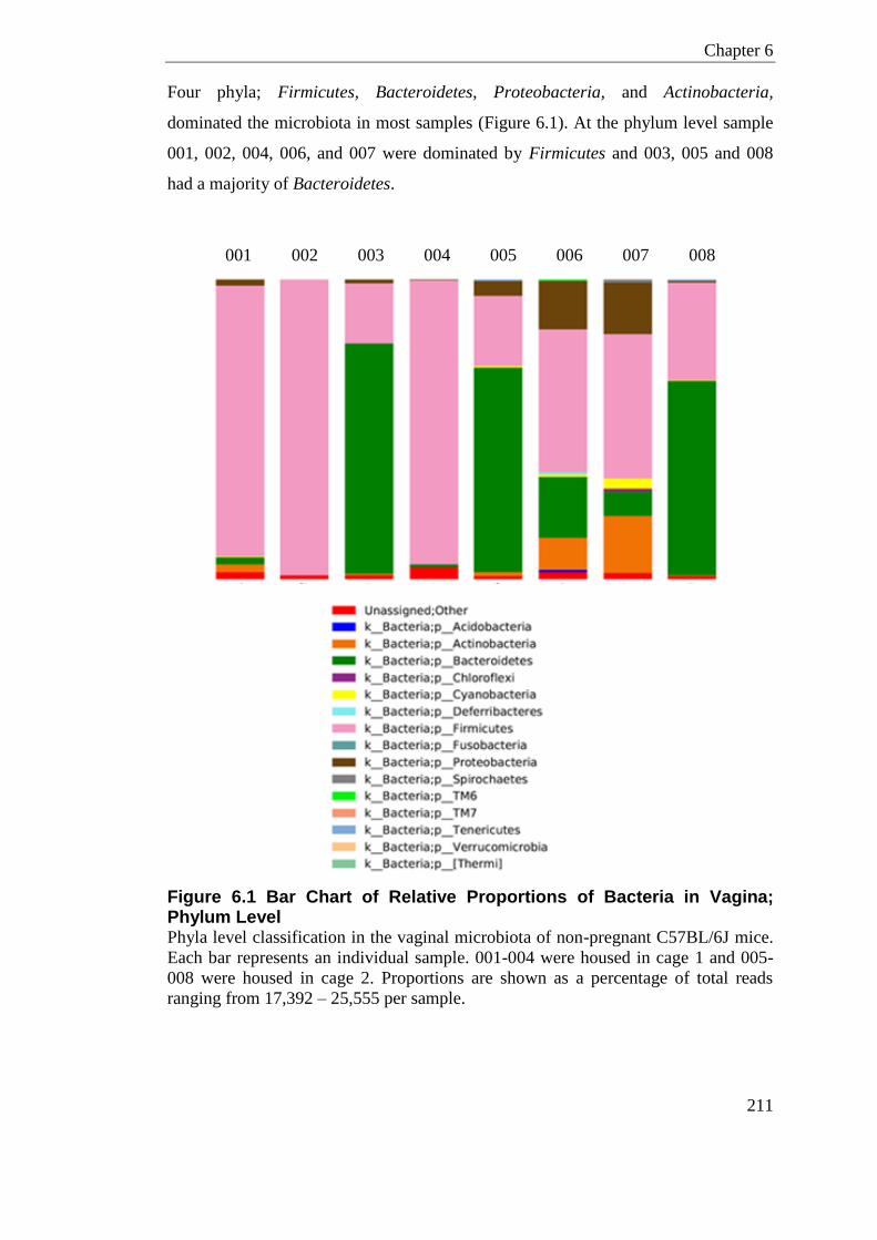
Chapter 6
211
Four phyla; Firmicutes, Bacteroidetes, Proteobacteria, and Actinobacteria,
dominated the microbiota in most samples (Figure 6.1). At the phylum level sample
001, 002, 004, 006, and 007 were dominated by Firmicutes and 003, 005 and 008
had a majority of Bacteroidetes.
001 002 003 004 005 006 007 008
Figure 6.1 Bar Chart of Relative Proportions of Bacteria in Vagina; Phylum Level Phyla level classification in the vaginal microbiota of non-pregnant C57BL/6J mice.
Each bar represents an individual sample. 001-004 were housed in cage 1 and 005-
008 were housed in cage 2. Proportions are shown as a percentage of total reads
ranging from 17,392 – 25,555 per sample.
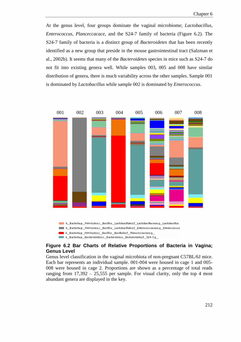
Chapter 6
212
At the genus level, four groups dominate the vaginal microbiome; Lactobacillus,
Enterococcus, Planceccacace, and the S24-7 family of bacteria (Figure 6.2). The
S24-7 family of bacteria is a distinct group of Bacteroidetes that has been recently
identified as a new group that preside in the mouse gastrointestinal tract (Salzman et
al., 2002b). It seems that many of the Bacteroidetes species in mice such as S24-7 do
not fit into existing genera well. While samples 003, 005 and 008 have similar
distribution of genera, there is much variability across the other samples. Sample 001
is dominated by Lactobacillus while sample 002 is dominated by Enterococcus.
001 002 003 004 005 006 007 008
Figure 6.2 Bar Charts of Relative Proportions of Bacteria in Vagina; Genus Level Genus level classification in the vaginal microbiota of non-pregnant C57BL/6J mice.
Each bar represents an individual sample. 001-004 were housed in cage 1 and 005-
008 were housed in cage 2. Proportions are shown as a percentage of total reads
ranging from 17,392 – 25,555 per sample. For visual clarity, only the top 4 most
abundant genera are displayed in the key.
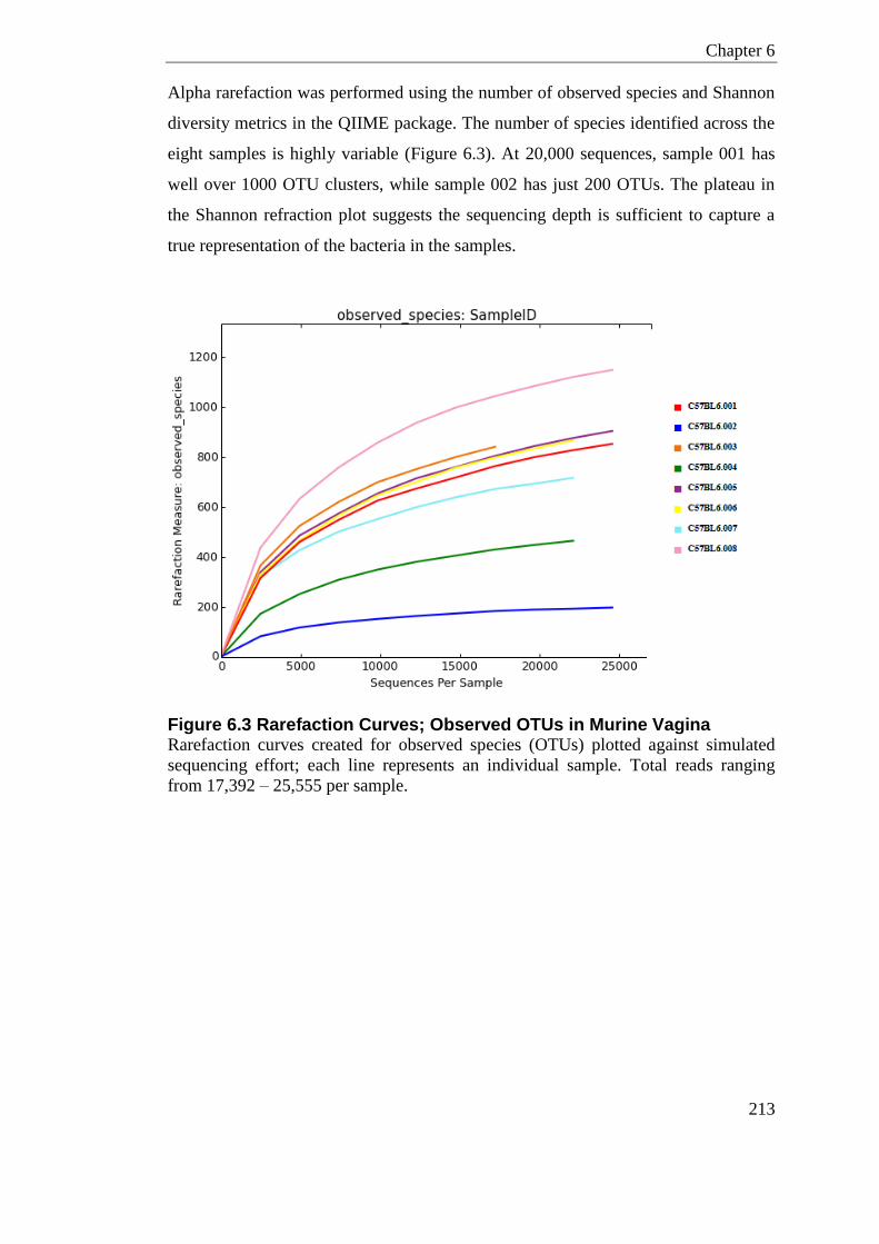
Chapter 6
213
Alpha rarefaction was performed using the number of observed species and Shannon
diversity metrics in the QIIME package. The number of species identified across the
eight samples is highly variable (Figure 6.3). At 20,000 sequences, sample 001 has
well over 1000 OTU clusters, while sample 002 has just 200 OTUs. The plateau in
the Shannon refraction plot suggests the sequencing depth is sufficient to capture a
true representation of the bacteria in the samples.
Figure 6.3 Rarefaction Curves; Observed OTUs in Murine Vagina Rarefaction curves created for observed species (OTUs) plotted against simulated
sequencing effort; each line represents an individual sample. Total reads ranging
from 17,392 – 25,555 per sample.
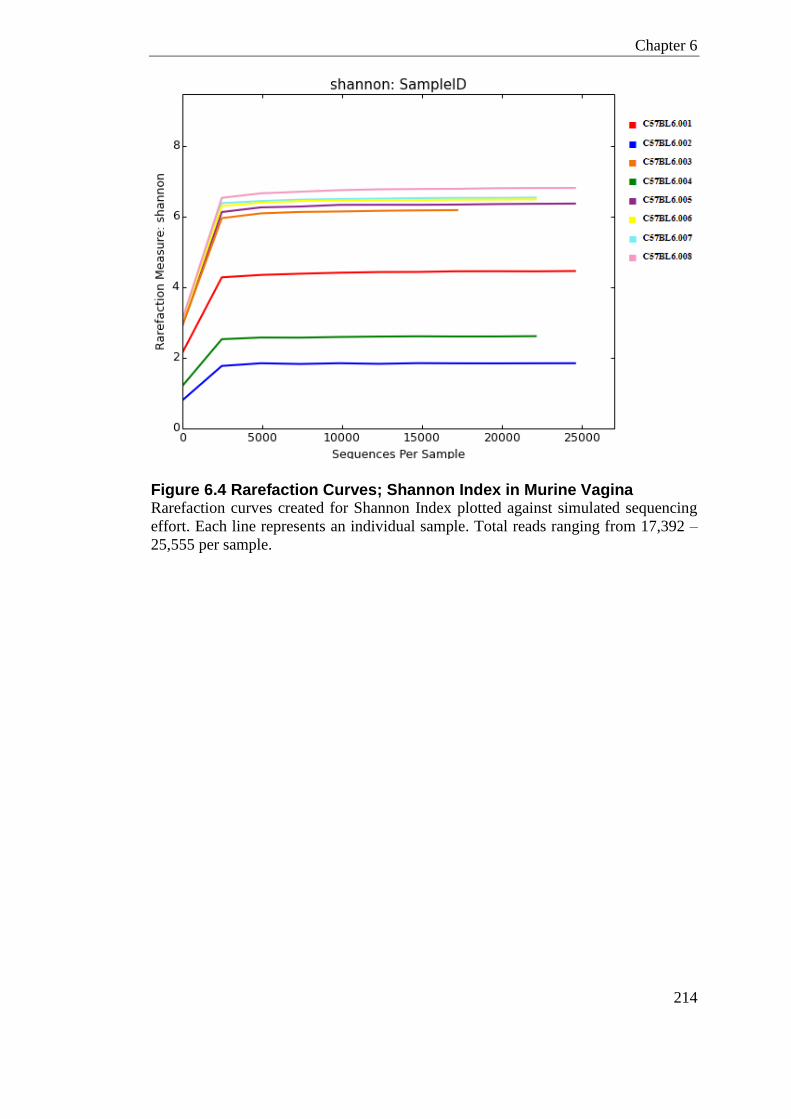
Chapter 6
214
Figure 6.4 Rarefaction Curves; Shannon Index in Murine Vagina Rarefaction curves created for Shannon Index plotted against simulated sequencing
effort. Each line represents an individual sample. Total reads ranging from 17,392 –
25,555 per sample.
Chapter 6
215
6.2.1.1 Conclusion
This first Ion Torrent run, sequencing wildtype non-pregnant vaginal communities,
aimed to establish, i) what bacteria are present at the vaginal mucosa ii) what level of
sequencing is required to gain insight on microbial diversity.
There was a notable amount of variability in the genera seen across the 8 samples
taken from vaginal lavages (Figure 6.2). At the phylum level, 5 of the animals were
dominated by Firmicutes (001, 002, 004, 006, and 007), whereas 3 animals had a
majority of Bacteroidetes (003, 005 and 008) (Figure 6.1). In women, the
composition in the vagina is mostly dominated by Firmicutes, while some women
have a dominance of Actinobacteria phyla; Bacteroidetes are rarely seen. In contrast
to humans it has been proposed only a small percentage of female mice harbour
Lactobacillus species at the vaginal mucosa (McGrory and Garber, 1992). In this
study, only 6 of the 8 mice had OTUs classified as Lactobacillus, in 5 of these 6 mice
Lactobacillus was a minor component. Enterococcus, Planceccacace, and the S24-7
family were other highly abundant species.
The plateau in the Shannon Index prior to 5,000 sequences (Figure 6.4) suggests the
sequencing effort in this run has adequately sampled the population. The number of
OTUs across the study-set is highly inflated over what I had expected. At the current
level of sequencing, the human vaginal microbiome is establishing around 200
bacterial assigned OTUs across study sets (Ravel et al., 2011, Drell et al., 2013,
Martin et al., 2013). After chimera checking there were 2738 OTUs in this study,
while some of these are true OTUs, many could be spurious clusters from PCR or
experimental artefacts. Some of these are likely to be chimeric reads that have not
been detected. In addition, the premature truncation of reads on the PGM platform
and at quality filtering could be inflating unique clusters. The Ion Torrent chemistry
offers the continuous sequencing of reads up to 400bp, which has the potential to
span the full 16S rRNA V1-V2 target region. The V1-V2 region was only sequenced
in this study in the forward direction. The standard Ion Torrent chemistry does not
support paired end sequencing, however, the reverse direction can be sequenced as
an independent sequencing run. Premature sequence truncation is specific to
Chapter 6
216
semiconductor sequencing; this can inflate OTUs as the partial sequence fragments
may not contain enough taxonomic information for robust classification. In addition,
as the base calling quality drops toward the end of the read, many sequences are
additionally truncated in the Galaxy workflow (see appendix).
It is well documented that the Ion Torrent platform exhibits a higher rate of
sequencing errors than Illumina sequencing technologies. I chose to pursue the
Illumina MiSeq platform for subsequent investigations of the vaginal microbiome
due to the added benefit of paired end sequencing, in addition to this known
advantage in base calling. Illumina sequencing chemistry can sequence 300bp
paired-end reads, which covers the majority of the approximately 340-370bp V1-V2
amplicon. The non-overlapping region is proximal on both sequencing reads; where
quality is normally the highest.
In addition to using the MiSeq Illumina platform, several other optimisation steps
were attempted in aim of reducing the variability in the vaginal community. These
optimisation steps included different sampling methods and extraction protocols.
These are detailed in section 4.6 and 4.7, but to summarise, whole tissue compared to
vaginal lavage provided more consistent bacterial yield. For this reason, post-mortem
sampling of whole vaginal tissue for bacterial extraction was adopted. In addition,
the FastDNA® SPIN Kit was chosen as it provided more consistency in bacterial
yields from vaginal tissue than the PowerSoil® DNA Kit.
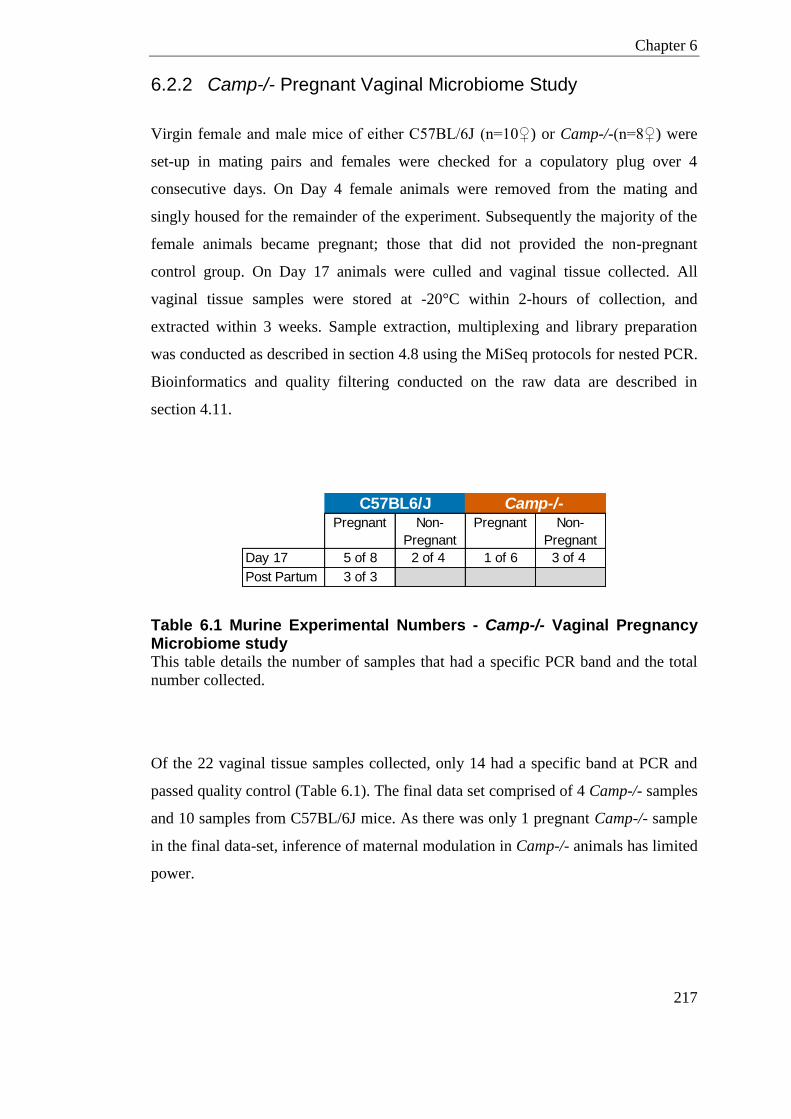
Chapter 6
217
6.2.2 Camp-/- Pregnant Vaginal Microbiome Study
Virgin female and male mice of either C57BL/6J (n=10♀) or Camp-/-(n=8♀) were
set-up in mating pairs and females were checked for a copulatory plug over 4
consecutive days. On Day 4 female animals were removed from the mating and
singly housed for the remainder of the experiment. Subsequently the majority of the
female animals became pregnant; those that did not provided the non-pregnant
control group. On Day 17 animals were culled and vaginal tissue collected. All
vaginal tissue samples were stored at -20°C within 2-hours of collection, and
extracted within 3 weeks. Sample extraction, multiplexing and library preparation
was conducted as described in section 4.8 using the MiSeq protocols for nested PCR.
Bioinformatics and quality filtering conducted on the raw data are described in
section 4.11.
Table 6.1 Murine Experimental Numbers - Camp-/- Vaginal Pregnancy Microbiome study This table details the number of samples that had a specific PCR band and the total
number collected.
Of the 22 vaginal tissue samples collected, only 14 had a specific band at PCR and
passed quality control (Table 6.1). The final data set comprised of 4 Camp-/- samples
and 10 samples from C57BL/6J mice. As there was only 1 pregnant Camp-/- sample
in the final data-set, inference of maternal modulation in Camp-/- animals has limited
power.
Pregnant Non-
Pregnant
Pregnant Non-
Pregnant
Day 17 5 of 8 2 of 4 1 of 6 3 of 4
Post Partum 3 of 3
C57BL6/J Camp-/-
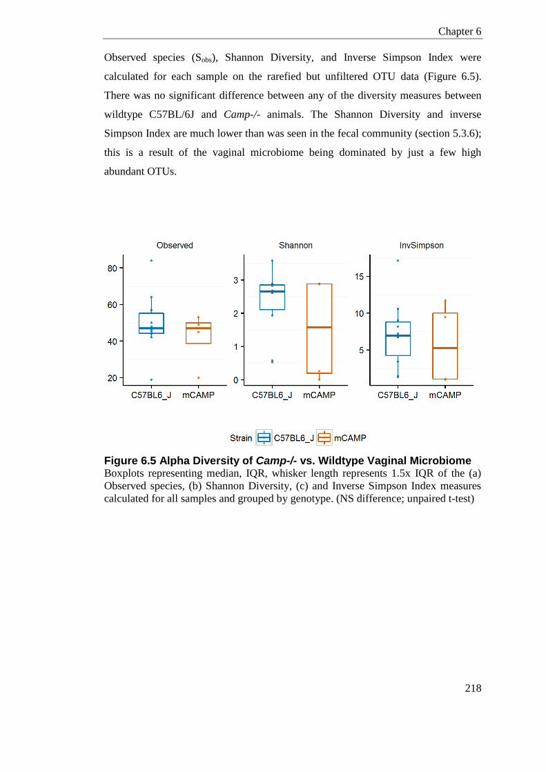
Chapter 6
218
Observed species (Sobs), Shannon Diversity, and Inverse Simpson Index were
calculated for each sample on the rarefied but unfiltered OTU data (Figure 6.5).
There was no significant difference between any of the diversity measures between
wildtype C57BL/6J and Camp-/- animals. The Shannon Diversity and inverse
Simpson Index are much lower than was seen in the fecal community (section 5.3.6);
this is a result of the vaginal microbiome being dominated by just a few high
abundant OTUs.
Figure 6.5 Alpha Diversity of Camp-/- vs. Wildtype Vaginal Microbiome Boxplots representing median, IQR, whisker length represents 1.5x IQR of the (a)
Observed species, (b) Shannon Diversity, (c) and Inverse Simpson Index measures
calculated for all samples and grouped by genotype. (NS difference; unpaired t-test)

Chapter 6
219
Figure 6.6 shows relative abundance of the top 25 most abundant OTUs classified at
the genus level. The highest abundant OTU in most samples was classified as
Ralstonia spp. One Camp-/- sample had a dominance of Staphylococcus, and two
C57BL/6J samples had high proportions of Streptococcus.
Figure 6.6 Heat map of most Abundant OTUs showing their Genus Level Classification in Vaginal Samples from Camp-/- and C57BL/6J mice Relative abundance of n=14 vaginal samples of the top 25 OTUs, labelled by Genus.

Chapter 6
220
Ralstonia spp. are known contaminants in molecular grade water (Grahn et al., 2003,
Barton et al., 2006, Laurence et al., 2014), and I have shown this species is a
potential source of contamination by sequencing a mock community (detailed in
section 4.18). For this reason, OTUs classified as Ralstonia were removed from the
data-set prior to beta-diversity distance calculations. The pairwise UniFrac distances
were visualised using Principle co-ordinate Analysis (PCoA) (Figure 6.7 and Figure
6.8). In both the weighted and unweighted PCoA plots, there was no clear separation
by genotype (Figure 6.7) or pregnancy status (Figure 6.8).
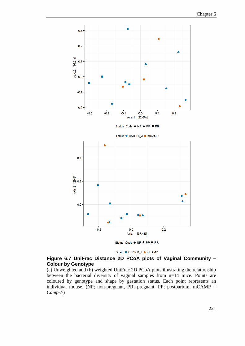
Chapter 6
221
Figure 6.7 UniFrac Distance 2D PCoA plots of Vaginal Community – Colour by Genotype (a) Unweighted and (b) weighted UniFrac 2D PCoA plots illustrating the relationship
between the bacterial diversity of vaginal samples from n=14 mice. Points are
coloured by genotype and shape by gestation status. Each point represents an
individual mouse. (NP; non-pregnant, PR; pregnant, PP; postpartum, mCAMP =
Camp-/-)
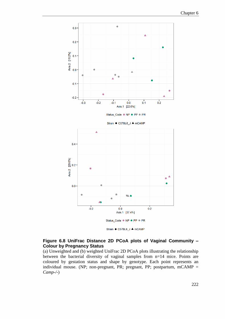
Chapter 6
222
Figure 6.8 UniFrac Distance 2D PCoA plots of Vaginal Community – Colour by Pregnancy Status (a) Unweighted and (b) weighted UniFrac 2D PCoA plots illustrating the relationship
between the bacterial diversity of vaginal samples from n=14 mice. Points are
coloured by gestation status and shape by genotype. Each point represents an
individual mouse. (NP; non-pregnant, PR; pregnant, PP; postpartum, mCAMP =
Camp-/-)

Chapter 6
223
6.2.2.1 Conclusion
The vaginal microbiome was problematic due to low bacterial yields. Of the 22
samples collected, only 14 had a specific PCR band and could be carried forward for
microbial sequencing. This left n=10 wildtype samples and n=4 Camp-/- samples, of
which only 1 was from a pregnant animal. Microbial sequencing revealed Ralstonia
species as the most highly abundant genera in the vaginal samples. Ralstonia is a
known contaminate of molecular grade water and I have also demonstrated this
species is a potential contaminate in the mock community. As both the extraction
negative and PCR negative did not display any PCR product, it was surprising to see
contaminates in the samples from the vagina. After removal of the contaminating
sequences, there is no clear grouping with regard to the genotype of the mouse like
there was in the fecal samples. There was also no clear clustering with regards to
whether the samples were taken from pregnant or non-pregnant mice, or if they were
taken postpartum.
Chapter 6
224
6.3 Discussion
Under normal conditions it is common for female animals caged together to
synchronise estrus cycling. The eight non-pregnant mice from experiment detailed in
section 6.2.1 were housed in two separate cages but there was no apparent similarity
of vaginal community within cage. The estrus stage of the mice used in this
experiment was not determined, so it cannot be confirmed that all mice in the same
cage were synchronised. It has previously been shown that the total numbers of
bacteria are statistically higher during estrus than diestrus or metestrus in the murine
vagina (Noguchi et al., 2003). In addition, Noguchi et al., (2003) showed the number
of aerobes was higher than the total number of anaerobes during estrus.
Synchronising the mice stage of estrus would perhaps result in higher potential
bacterial recovery and more uniformity across samples. Hormones could be used in
non-pregnant mice to synchronise estrus in aim of reducing this variability.
In section 6.2.2, I collected vaginal samples from animals at the same stage of
pregnancy. In addition to gaining insight into the maternal vaginal community,
sampling during pregnancy aimed to reduce the potential estrus cycling variability.
The pregnant samples did not show any distinct clustering in the UniFrac PCoA plots
(Figure 6.8), suggesting the vaginal microbiome while pregnant is also highly
variable.
After review of the literature, only one other study attempts to categorise the murine
vaginal commensal microbiome using 16S rRNA gene sequencing (Barfod et al.,
2013). Similarly, Barfod et al., (2013) highlighted that the vaginal community of
BALB/c mice was highly variable. Although the authors only had 8 samples they
showed the vaginal community had two sub-clusters. One sub-cluster appeared to be
closely related to the lung microbiota, whereas the other sub-cluster did not show
similarity to either of the other two sample sites the authors investigated (lung and
cecum). Barfod et al., (2013) attributed Streptococcus (Gram-positive) as the species
driving the separation of the two clusters; the authors also listed Acinetobacter,
Sphinogmonas, Enterococcus and Polaromonas as genera from the vaginal mucosa.
Culture-dependent studies have listed Streptococci, gram-negative rods,

Chapter 6
225
Bacteroidaceae and Staphylococci as found in the non-pregnant vagina, however,
these observations were not consistent and were highly variable (Noguchi et al.,
2003).
Further work is needed to refine and understand the commensal vaginal bacteria in
mice. Due to the differences in commensal bacteria between women and mice, it is
imaginable that an approach inoculating germ-free mice could recreate the human
vaginal microbiome, providing a model for testing exogenous disturbances.

226
Chapter 7
Role of Host Defence Peptides
in an Infection/Inflammation
Induced Mouse Model of
Preterm Labour
Chapter 7
227
Chapter 7 Role of Host Defence Peptides in an Infection/
Inflammation Induced Mouse Model of Preterm Labour
7.1 Introduction
Local inflammation in the decidua and chorion is thought to be important in the
initiation of preterm labour (Galinsky et al., 2013, Vrachnis et al., 2010). This
inflammation is frequently caused by ascending opportunistic commensal bacteria
from the vagina, through the cervix and into the fetal membranes (Allam et al., 2013,
DiGiulio et al., 2010). In humans, amniotic fluid cultures from most women with
preterm labour are negative for bacterial growth, although amniotic fluid
inflammatory cytokines and LPS binding proteins have been shown to be increased
(Gardella et al., 2001). This lack of bacterial detection could be due to the limitations
of the culture-dependent techniques.
As most women presenting with preterm labour do not have elevated white blood
cells or symptoms of illness, exploring a localised model of inflammation at the
maternal-fetal interface is used to mimic conditions present in PTL.
Stimulation of the TLR-4 pathway with LPS has been a central method of invoking
preterm labour in animal models. Delivery of LPS intrauterine, between two
amniotic sacs, in pregnant mice has been reported to induce high rates of preterm
labour, resulting in minimum maternal mortality and showing high reproducibility
(Rinaldi et al., 2014, Rinaldi et al., 2015, Sykes et al., 2013).
Cells in the female reproductive tract are a major source of multiple microbicides
including the defensins and cathelicidin. LL-37 is found at high levels in fetal skin,
vaginal epithelial cells, amniotic fluids, vaginal fluid and the vernix caseosa
(reviewed in Yarbrough et al., (2015)). Steady state defensin expression is cell
specific and compartmentalised in the female reproductive tract in pregnant and non-
pregnant women (King et al., 2003b, Fleming et al., 2003, Quayle et al., 1998).
Further work on human extra-placental membranes in ex-vivo cultures has shown
increased levels of hBD2 after exposure to bacteria (Boldenow et al., 2013).
Chapter 7
228
hBD3 and DEFB14 have potent anti-inflammatory effects on human and mouse
primary monocyte derived macrophages (Semple et al., 2010). hBD3 and DEFB14
effectively inhibit the LPS-activated TLR4-induced TNF and IL-6 response through
both the MyD88 and TRIF pathways, shown both in-vitro and in-vivo (Semple et al.,
2010, Semple et al., 2011). Preliminary unpublished data from Fiona Semple in the
Dorin lab indicates that in-vivo, Defb14-/- mice have an increased sensitivity to LPS
when delivered intraperitoneally. Interestingly, hBD3 can act in synergy with LL-37
reducing the LPS-induced secretion of cytokines in gingival cells (Bedran et al.,
2014).
LL-37/hCAP18 has numerous immune modulatory properties that can contribute to
infection and inflammatory phenotypes (discussed in section 1.2.2). Mice null for
Camp, the murine orthologue of LL-37/hCAP18, have many inducible phenotypes;
showing increased susceptibility to bacterial infections across many body sites,
detailed in section 1.2.2.
As hBD3 and LL-37 suppress the inflammatory response of cells to LPS through
TLR-4, the hypothesis is that in-vivo they might offer protection from preterm birth
caused by inflammation.
In this chapter I use LPS, a cell wall component of gram negative bacteria, as the
agent to induce inflammation and subsequent preterm labour in mice. Initially this
agent was administered intrauterine (between two amniotic sacs) by aid of a
laparotomy, but subsequently a more elegant approach of delivering the intrauterine
injection guided by ultrasound sonography (USS) was utilised (described in Rinaldi
et al.,(2015)). Animals lacking the host defence peptide genes Defb14 (Defb14-/-) or
Camp (Camp-/-) were given LPS intrauterine during late gestation to determine
whether these genes are critical in the continuation of pregnancy and timing of
delivery.
Chapter 7
229
7.2 Experimental Details
7.2.1 Fertility in Host Defence Peptide Knockout Mice
The fertility of Camp-/- (n=46) and Defb14-/- mice (n=18) was determined by
looking at several pregnancy indices; plug rate, pregnancy rate and first litter size.
The plug rate was determined as the fraction of female mice where a copulatory plug
was found (within 4-days of being placed with a male), compared to the total number
set-up. Subsequent pregnancy rate is the fraction of the plugged females who
continued pregnancy to gestational day 17, compared to the total number plugged.
First litter sizes are described here as the number of viable fetus found in-utero on
day 17 of gestation.
7.2.2 Timed Collection Experiment
Detailed experimental method for timed collection experiment is given in section
2.1.5. In brief, at day 17 of murine gestation, 1µg or 20µg of LPS was injected
between two fetal-sacs as guided by ultrasound sonography. Animals were then
culled 6-hours post treatment where tissue and blood serum were collected for
molecular analysis. In parallel non-anaesthetic non-treatment day 17 pregnant
females (NTxC) were also culled for control tissue and blood serum for comparison.
The number of animals in each treatment group is detailed in Table 7.1.
C57BL/6J Camp-/-
NTxC n = 3 n = 3
LPS [1µg] n = 3 n = 3
LPS [20µg] n = 6 n = 6
Table 7.1 Timed Collection Experiment - Animal Numbers and Treatment Treatments and genotype of animals for timed collection experiment using USS
guided model of PTL.

Chapter 7
230
7.2.3 Laparotomy Model of PTL Experiment
Detailed experimental method for laparotomy model of PTL experiment is given in
section 2.1.2. In brief, at day 17 of murine gestation a laparotomy was performed to
expose the uterine horns, 20µg of LPS or sterile physiological saline was then
injected between two fetal-sacs. Uterine horns are then returned to the abdominal
cavity and the incision was sutured closed. Animals were left to recover and the
interval to delivery was recorded in hours. The number of live born pups was also
recorded at the first observation after the initiation of labour. Animals that have
preterm labour delivered within 36 hours while those that go to term delivered
between 36 and 96 hours post-injection. In parallel non-surgery non-treatment
control day 17 pregnant females (NTxC) were also set-up to establish normal interval
to delivery. The number of animals in each treatment group is detailed in Table 7.2.
C57BL/6N Defb14-/-
NTxC n = 3 n = 6
Saline n = 9 n = 6
LPS [20µg] n = 8 n = 10
Table 7.2 Laparotomy Model of PTL Experiment Numbers Treatments and genotype of animals for laparotomy model of PTL experiment.
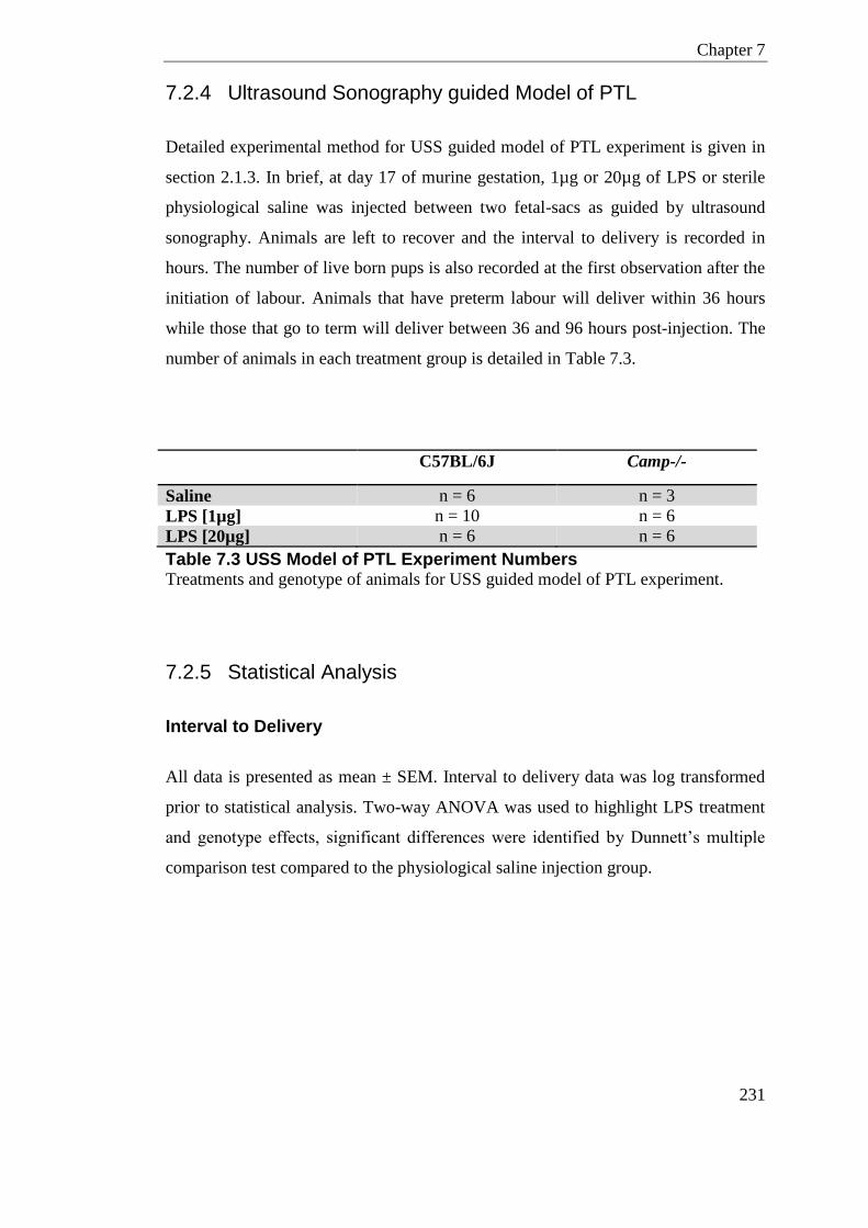
Chapter 7
231
7.2.4 Ultrasound Sonography guided Model of PTL
Detailed experimental method for USS guided model of PTL experiment is given in
section 2.1.3. In brief, at day 17 of murine gestation, 1µg or 20µg of LPS or sterile
physiological saline was injected between two fetal-sacs as guided by ultrasound
sonography. Animals are left to recover and the interval to delivery is recorded in
hours. The number of live born pups is also recorded at the first observation after the
initiation of labour. Animals that have preterm labour will deliver within 36 hours
while those that go to term will deliver between 36 and 96 hours post-injection. The
number of animals in each treatment group is detailed in Table 7.3.
C57BL/6J Camp-/-
Saline n = 6 n = 3
LPS [1µg] n = 10 n = 6
LPS [20µg] n = 6 n = 6
Table 7.3 USS Model of PTL Experiment Numbers Treatments and genotype of animals for USS guided model of PTL experiment.
7.2.5 Statistical Analysis
Interval to Delivery
All data is presented as mean ± SEM. Interval to delivery data was log transformed
prior to statistical analysis. Two-way ANOVA was used to highlight LPS treatment
and genotype effects, significant differences were identified by Dunnett’s multiple
comparison test compared to the physiological saline injection group.
Chapter 7
232
Number of Live Born Pups
All data is presented as mean ± SEM. Live born pups fractions were ArcSin
transformed prior to analysis. Two-way ANOVA was used to highlight LPS
treatment and genotype effects, significant differences were identified by Dunnett’s
multiple comparison test compared to the physiological saline injection group.
RT-qPCR
All data is presented as mean ± SEM normalised to β-Actin and relative to CAL. The
calibrator used in this chapter was a cDNA pool from mouse placenta, uterus and
fetal membrane from a single animal undergoing no treatment. Data was log
transformed prior to analysis.
Where comparing two groups, NTxC and LPS, an unpaired t-test was performed.
Two-way ANOVA was used to highlight LPS treatment and genotype effects,
significant differences in genotype were identified with Bonferroni’s post-test, and
significant differences in LPS treatment identified with Tukey’s post-test.
Camp mRNA gene expression significance was measured by one-way ANOVA with
Tukey’s post-test in timed-collection experiment.
Circulating Serum Cytokines
All data is presented as mean ± SEM. Two-way ANOVA was used to highlight LPS
treatment and genotype effects, significant differences in genotype were identified
with Bonferroni’s post-test, and significant differences in LPS treatment identified
with Tukey’s post-test.
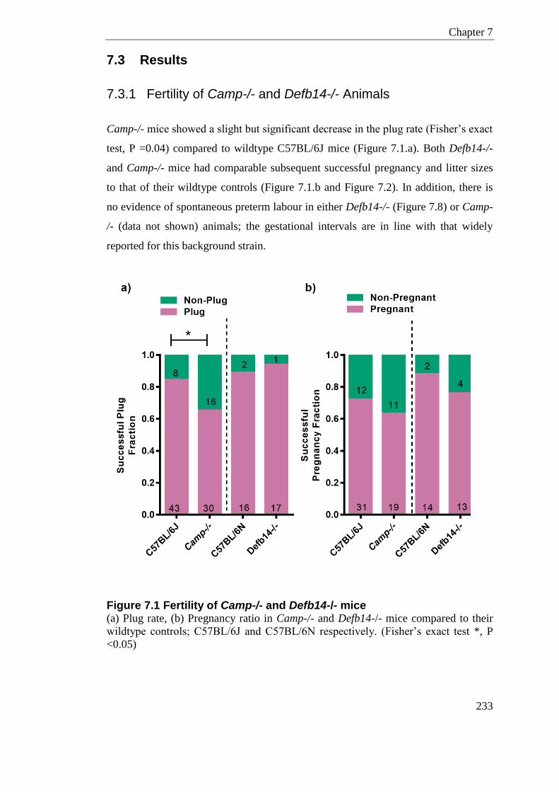
Chapter 7
233
7.3 Results
7.3.1 Fertility of Camp-/- and Defb14-/- Animals
Camp-/- mice showed a slight but significant decrease in the plug rate (Fisher’s exact
test, P =0.04) compared to wildtype C57BL/6J mice (Figure 7.1.a). Both Defb14-/-
and Camp-/- mice had comparable subsequent successful pregnancy and litter sizes
to that of their wildtype controls (Figure 7.1.b and Figure 7.2). In addition, there is
no evidence of spontaneous preterm labour in either Defb14-/- (Figure 7.8) or Camp-
/- (data not shown) animals; the gestational intervals are in line with that widely
reported for this background strain.
Figure 7.1 Fertility of Camp-/- and Defb14-/- mice (a) Plug rate, (b) Pregnancy ratio in Camp-/- and Defb14-/- mice compared to their
wildtype controls; C57BL/6J and C57BL/6N respectively. (Fisher’s exact test *, P
<0.05)
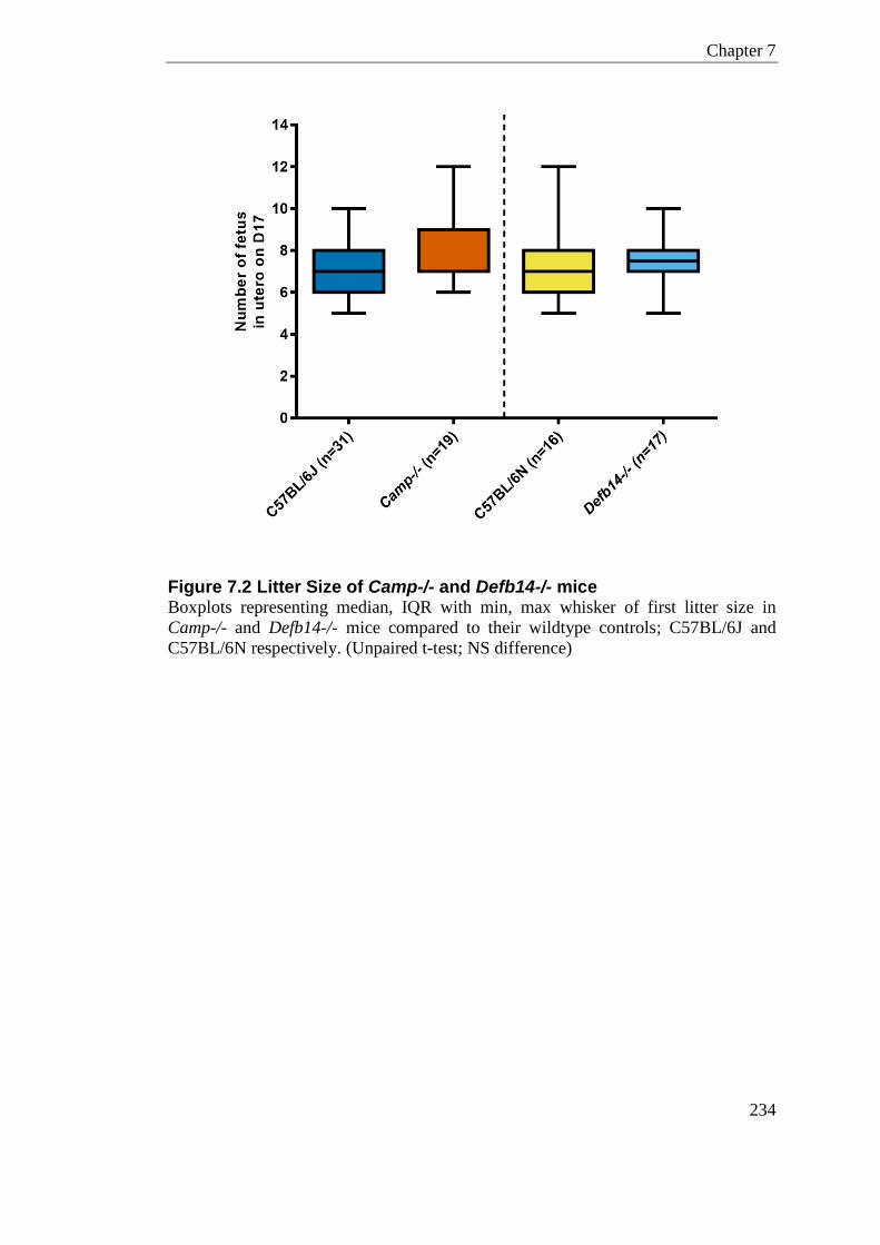
Chapter 7
234
Figure 7.2 Litter Size of Camp-/- and Defb14-/- mice Boxplots representing median, IQR with min, max whisker of first litter size in
Camp-/- and Defb14-/- mice compared to their wildtype controls; C57BL/6J and
C57BL/6N respectively. (Unpaired t-test; NS difference)
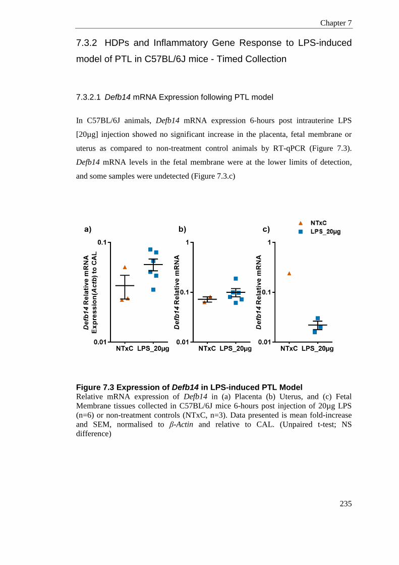
Chapter 7
235
7.3.2 HDPs and Inflammatory Gene Response to LPS-induced
model of PTL in C57BL/6J mice - Timed Collection
7.3.2.1 Defb14 mRNA Expression following PTL model
In C57BL/6J animals, Defb14 mRNA expression 6-hours post intrauterine LPS
[20µg] injection showed no significant increase in the placenta, fetal membrane or
uterus as compared to non-treatment control animals by RT-qPCR (Figure 7.3).
Defb14 mRNA levels in the fetal membrane were at the lower limits of detection,
and some samples were undetected (Figure 7.3.c)
Figure 7.3 Expression of Defb14 in LPS-induced PTL Model Relative mRNA expression of Defb14 in (a) Placenta (b) Uterus, and (c) Fetal
Membrane tissues collected in C57BL/6J mice 6-hours post injection of 20µg LPS
(n=6) or non-treatment controls (NTxC, n=3). Data presented is mean fold-increase
and SEM, normalised to β-Actin and relative to CAL. (Unpaired t-test; NS
difference)
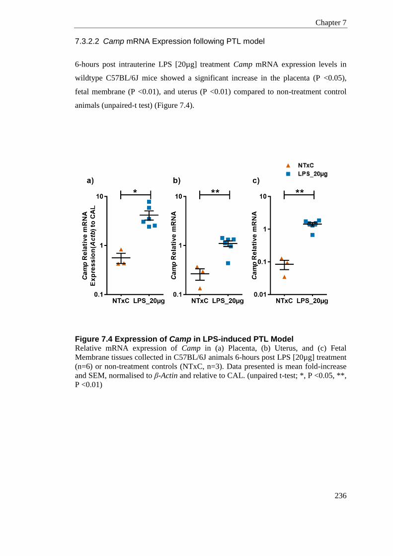
Chapter 7
236
7.3.2.2 Camp mRNA Expression following PTL model
6-hours post intrauterine LPS [20µg] treatment Camp mRNA expression levels in
wildtype C57BL/6J mice showed a significant increase in the placenta (P <0.05),
fetal membrane (P <0.01), and uterus (P <0.01) compared to non-treatment control
animals (unpaired-t test) (Figure 7.4).
Figure 7.4 Expression of Camp in LPS-induced PTL Model Relative mRNA expression of Camp in (a) Placenta, (b) Uterus, and (c) Fetal
Membrane tissues collected in C57BL/6J animals 6-hours post LPS [20µg] treatment
(n=6) or non-treatment controls (NTxC, n=3). Data presented is mean fold-increase
and SEM, normalised to β-Actin and relative to CAL. (unpaired t-test; *, P <0.05, **,
P <0.01)
Chapter 7
237
7.3.2.3 Inflammatory and Immune Cell Mediators in Placenta
Intrauterine LPS [20µg] injection significantly increased the expression of all
investigated inflammatory and immune cell mediators in placental tissues of
C57BL/6J mice (Figure 7.5).
The neutrophil chemokines, Cxcl1 and Cxcl2, in placental tissues showed significant
increases after LPS [20µg] injection compared to non-treatment controls. Cxcl1
mRNA expression displayed a mean 45.16 ± 7.06 fold-increase (P <0.001),
compared to non-treatment animals. Cxcl2 mRNA expression increased on average
83.02 ± 20.09 fold, compared to those animals not-treated (P <0.01).
Similarly, the pro-inflammatory cytokines, Il-1β and Tnf, are also significantly
increased in placental tissues following LPS [20µg] treatment; displaying a 14.43 ±
3.15 mean fold-increase (P <0.01) and 6.5 ± 1.46 mean fold-increase (P <0.01)
respectively.
Placental mRNA expression of Ptgs2 (Cox-2) showed a 3.61 ± 0.94 fold-increase in
the placenta (P <0.01) in response to LPS [20µg] as compared to non-treatment
controls.
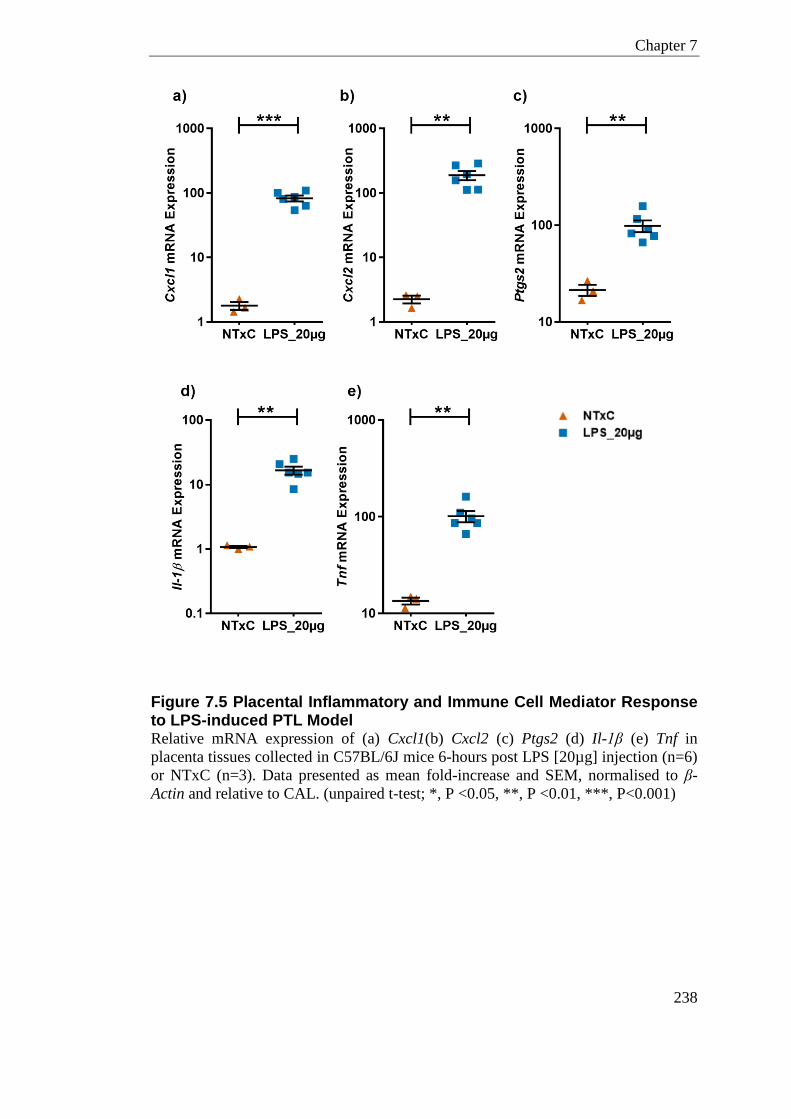
Chapter 7
238
Figure 7.5 Placental Inflammatory and Immune Cell Mediator Response to LPS-induced PTL Model Relative mRNA expression of (a) Cxcl1(b) Cxcl2 (c) Ptgs2 (d) Il-1β (e) Tnf in
placenta tissues collected in C57BL/6J mice 6-hours post LPS [20µg] injection (n=6)
or NTxC (n=3). Data presented as mean fold-increase and SEM, normalised to β-
Actin and relative to CAL. (unpaired t-test; *, P <0.05, **, P <0.01, ***, P<0.001)
Chapter 7
239
7.3.2.4 Inflammatory and Immune Cell Mediators in Uterus
Intrauterine LPS [20µg] injection also significantly increased the expression of
several inflammatory and immune cell mediators in uterine tissues of C57BL/6J mice
compared to those not treated (Figure 7.6).
The neutrophil chemokines, Cxcl1 and Cxcl2, in uterine tissue showed significant
increase after LPS [20µg] injection compared to non-treatment controls (P <0.0001
and P <0.01 respectively). mRNA Cxcl1 expression displayed a mean 443.6 ± 52.89
fold-increase compared to non-treatment animals. Cxcl2 mRNA expression increased
on average 319.1 ± 84.93 fold compared to those animals not-treated
Similarly, the pro-inflammatory cytokines, Il-1β and Tnf, are also increased in
uterine tissues following LPS [20µg] treatment (P <0.001 and P <0.0001
respectively). Il-β exhibited a mean fold increase of 49.35 ± 9.14, and on average Tnf
increased 13.96 ± 1.425 fold compared to the mean of those animals not treated.
Uterine mRNA expression of Ptgs2 (Cox-2) showed a significant (P <0.01) mean
6.18 ± 1.69 fold-increase in the uterus in response to LPS [20µg] compared to non-
treatment controls.
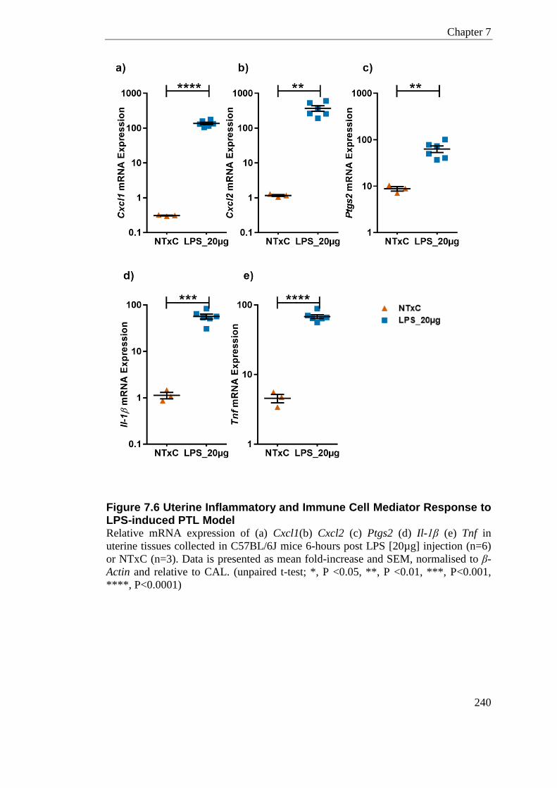
Chapter 7
240
Figure 7.6 Uterine Inflammatory and Immune Cell Mediator Response to LPS-induced PTL Model Relative mRNA expression of (a) Cxcl1(b) Cxcl2 (c) Ptgs2 (d) Il-1β (e) Tnf in
uterine tissues collected in C57BL/6J mice 6-hours post LPS [20µg] injection (n=6)
or NTxC (n=3). Data is presented as mean fold-increase and SEM, normalised to β-
Actin and relative to CAL. (unpaired t-test; *, P <0.05, **, P <0.01, ***, P<0.001,
****, P<0.0001)
Chapter 7
241
7.3.2.5 Inflammatory and Immune Cell Mediators in Fetal Membrane
Intrauterine LPS [20µg] injection significantly increased the expression of neutrophil
chemokines, Cxcl1 and Cxcl2, and Ptgs2 in fetal membrane tissues of C57BL/6J
mice (Figure 7.7). mRNA Cxcl1 expression was significantly increased (P <0.05) in
fetal membranes, showing a mean 11.32 ± 5.53 fold-increase in LPS treatment
compared to non-treatment animals. Cxcl2 mRNA expression increased 3.22 ± 1.56 -
fold, compared to those animals not-treated (P <0.05).
Fetal membrane mRNA expression of Ptgs2 (Cox-2) showed a small but significant
(P <0.05) increase of 1.64 ± 0.57 in response to LPS [20µg] treatment as compared
to non-treatment animals.
The pro-inflammatory cytokines, Il-1β and Tnf, did not show a significant increase in
the fetal membrane post LPS treatment (Figure 7.7d and Figure 7.7.e).
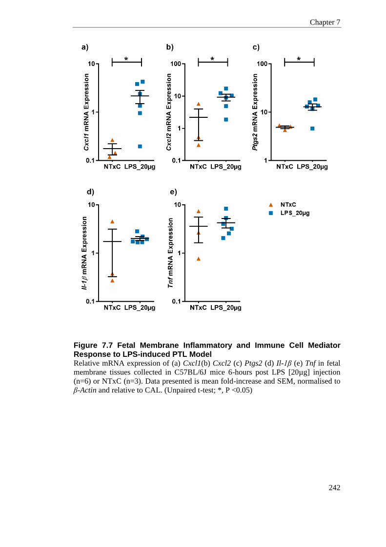
Chapter 7
242
Figure 7.7 Fetal Membrane Inflammatory and Immune Cell Mediator Response to LPS-induced PTL Model Relative mRNA expression of (a) Cxcl1(b) Cxcl2 (c) Ptgs2 (d) Il-1β (e) Tnf in fetal
membrane tissues collected in C57BL/6J mice 6-hours post LPS [20µg] injection
(n=6) or NTxC (n=3). Data presented is mean fold-increase and SEM, normalised to
β-Actin and relative to CAL. (Unpaired t-test; *, P <0.05)
Chapter 7
243
7.3.3 Defb14 in LPS Induced Preterm Labour; Laparotomy
Model
7.3.3.1 Interval to Delivery
C57BL/6N and Defb14-/- mice received an intrauterine injection of LPS [20µg],
physiological sterile saline or no-treatment on day 17 of gestation and the interval to
delivery was recorded in hours (Figure 7.8).
The normal gestational interval of C57BL/6N and Defb14-/- mice undergoing no-
treatment was comparable. C57BL/6N mice deliver on average 62.0 ± 1.5 (n=3) and
Defb14-/- mice deliver 57.1 ± 3.3 (n=6) hours after being set-up on day 17 with no
other intervention. This is equivalent to labour on day 19.5 of gestation, which is in
line with what has widely been reported for this background strain.
Intrauterine injection of LPS [20µg] on day 17 of gestation caused preterm labour
(defined as delivery within 36 hours) in 17 out of the 18 total animals treated at this
dose. LPS [20µg] injection significantly reduces the interval to delivery in
C57BL/6N and Defb14-/- mice compared to saline injection controls (two-way
ANOVA; Dunnett’s post-test; P<0.0001 at both genotypes). However, there was no
significant difference in the interval to delivery between C57BL/6N and Defb14-/-
mice (two-way ANOVA). C57BL/6N mice deliver on average 14.1 ± 2.0 (n=8) and
Defb14-/- mice deliver 18.4 ± 6.4 (n=10) hours post LPS [20µg] injection.
Injection of physiological sterile saline by laparotomy induced some variation in the
interval to delivery, where 2 of the 10 C57BL/6N mice had preterm labour (defined
as delivery within 36 hours). This did not reach statistical significance in C57BL/6N
or Defb14-/- mice when compared to the respective animals not treated (two-way
ANOVA; Dunnett’s post-test).
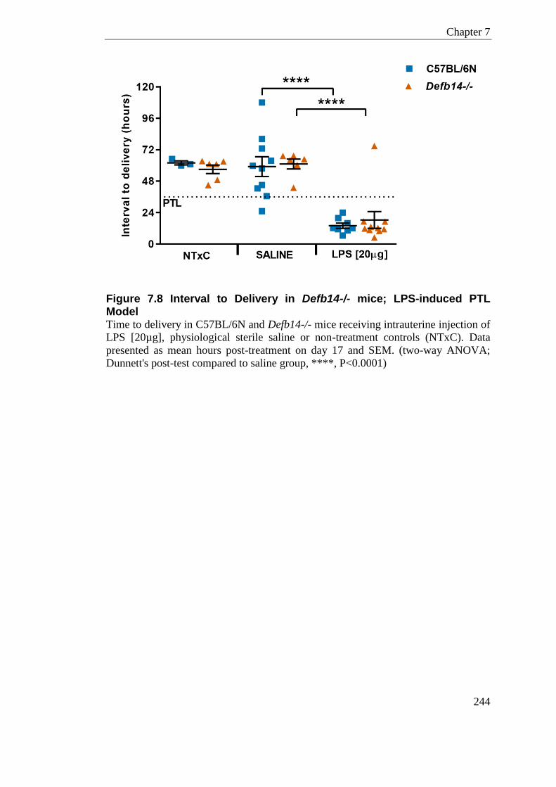
Chapter 7
244
Figure 7.8 Interval to Delivery in Defb14-/- mice; LPS-induced PTL Model Time to delivery in C57BL/6N and Defb14-/- mice receiving intrauterine injection of
LPS [20µg], physiological sterile saline or non-treatment controls (NTxC). Data
presented as mean hours post-treatment on day 17 and SEM. (two-way ANOVA;
Dunnett's post-test compared to saline group, ****, P<0.0001)
Chapter 7
245
7.3.3.2 Pup Survival
Post-delivery the number of live born pups in a litter was recorded for C57BL/6N
and Defb14-/- animals and presented as a ratio to the number of viable fetuses
counted at the time of surgery on day 17 of gestation (Figure 7.9). As the number of
viable fetuses in NTxC animals is not determined at the time of surgery, the fraction
of live born pups presented is calculated against the total number of pups delivered.
Injection of LPS [20µg] by laparotomy resulted in significant amounts of fetal death
in both C57BL/6N and Defb14-/-mice; where no pups were found born live in this
treatment group (Two-way ANOVA; Dunnett’s post-test; P<0.0001 at both
genotypes).
Injection of physiological sterile saline by laparotomy results in an increase in the
amount of fetal death seen compared to those animals undergoing no-treatment. This
reached significance in just the C57BL/6N animals (two-way ANOVA; Dunnett's
post-test compared to saline group; P <0.01), where those receiving sterile saline
(n=10) had on average 0.54 ± 0.13 live born pups compared to those undergoing no-
treatment (n=3) where 1 ± 0 pups are born live (two-way ANOVA).
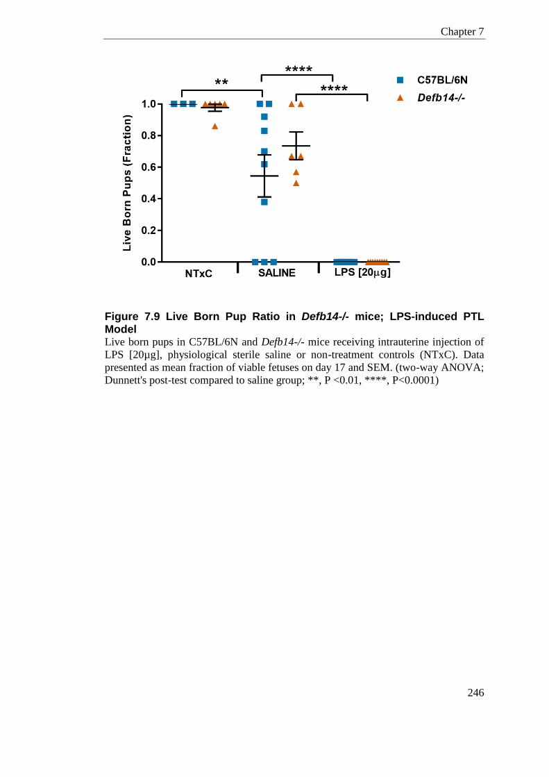
Chapter 7
246
Figure 7.9 Live Born Pup Ratio in Defb14-/- mice; LPS-induced PTL Model Live born pups in C57BL/6N and Defb14-/- mice receiving intrauterine injection of
LPS [20µg], physiological sterile saline or non-treatment controls (NTxC). Data
presented as mean fraction of viable fetuses on day 17 and SEM. (two-way ANOVA;
Dunnett's post-test compared to saline group; **, P <0.01, ****, P<0.0001)
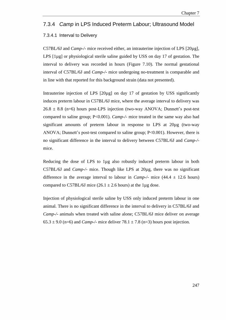
Chapter 7
247
7.3.4 Camp in LPS Induced Preterm Labour; Ultrasound Model
7.3.4.1 Interval to Delivery
C57BL/6J and Camp-/- mice received either, an intrauterine injection of LPS [20µg],
LPS [1µg] or physiological sterile saline guided by USS on day 17 of gestation. The
interval to delivery was recorded in hours (Figure 7.10). The normal gestational
interval of C57BL/6J and Camp-/- mice undergoing no-treatment is comparable and
in line with that reported for this background strain (data not presented).
Intrauterine injection of LPS [20µg] on day 17 of gestation by USS significantly
induces preterm labour in C57BL/6J mice, where the average interval to delivery was
26.8 ± 8.8 (n=6) hours post-LPS injection (two-way ANOVA; Dunnett’s post-test
compared to saline group; P<0.001). Camp-/- mice treated in the same way also had
significant amounts of preterm labour in response to LPS at 20µg (two-way
ANOVA; Dunnett’s post-test compared to saline group; P<0.001). However, there is
no significant difference in the interval to delivery between C57BL/6J and Camp-/-
mice.
Reducing the dose of LPS to 1µg also robustly induced preterm labour in both
C57BL/6J and Camp-/- mice. Though like LPS at 20µg, there was no significant
difference in the average interval to labour in Camp-/- mice (44.4 ± 12.6 hours)
compared to C57BL/6J mice (26.1 ± 2.6 hours) at the 1µg dose.
Injection of physiological sterile saline by USS only induced preterm labour in one
animal. There is no significant difference in the interval to delivery in C57BL/6J and
Camp-/- animals when treated with saline alone; C57BL/6J mice deliver on average
65.3 ± 9.0 (n=6) and Camp-/- mice deliver 78.1 ± 7.8 (n=3) hours post injection.
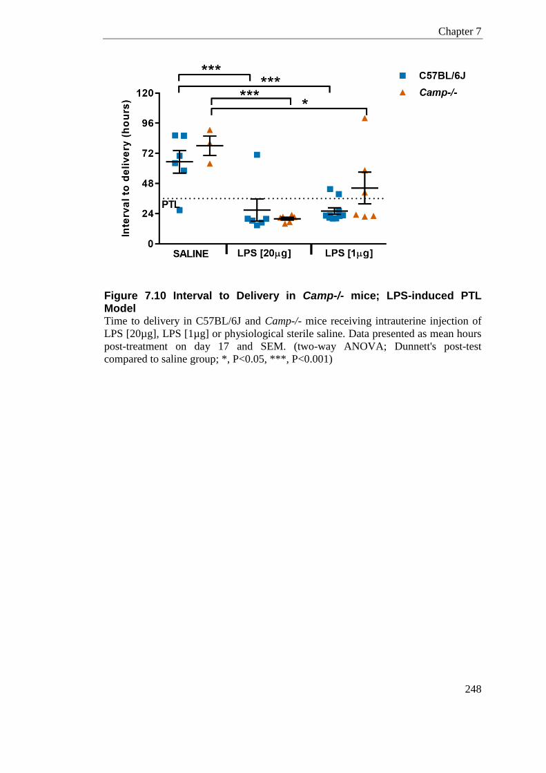
Chapter 7
248
Figure 7.10 Interval to Delivery in Camp-/- mice; LPS-induced PTL Model Time to delivery in C57BL/6J and Camp-/- mice receiving intrauterine injection of
LPS [20µg], LPS [1µg] or physiological sterile saline. Data presented as mean hours
post-treatment on day 17 and SEM. (two-way ANOVA; Dunnett's post-test
compared to saline group; *, P<0.05, ***, P<0.001)
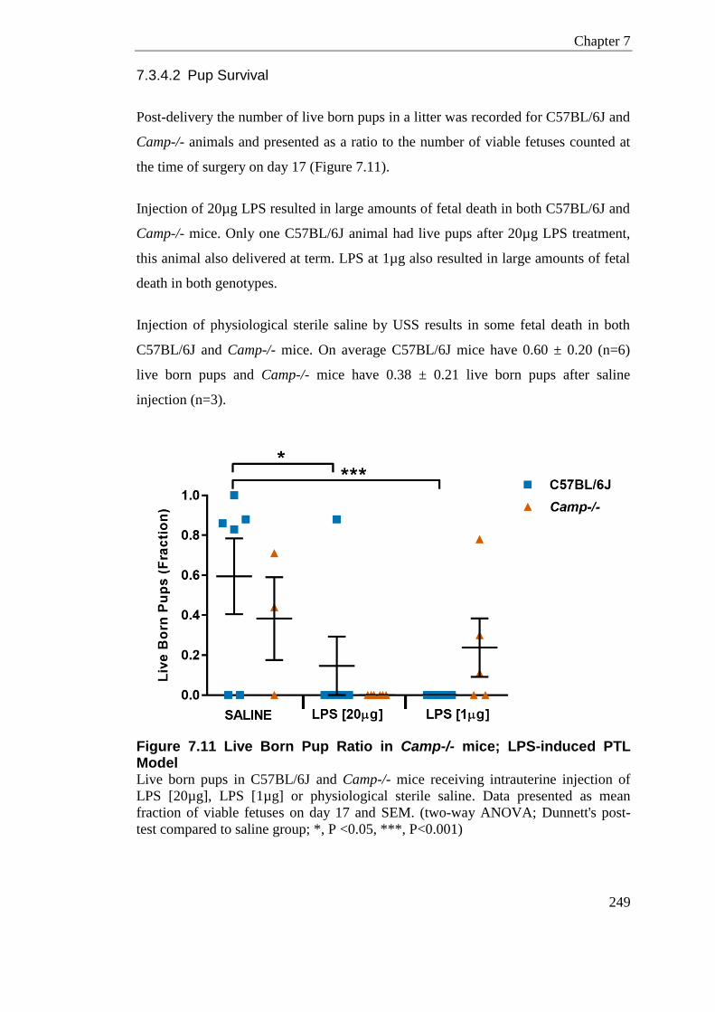
Chapter 7
249
7.3.4.2 Pup Survival
Post-delivery the number of live born pups in a litter was recorded for C57BL/6J and
Camp-/- animals and presented as a ratio to the number of viable fetuses counted at
the time of surgery on day 17 (Figure 7.11).
Injection of 20µg LPS resulted in large amounts of fetal death in both C57BL/6J and
Camp-/- mice. Only one C57BL/6J animal had live pups after 20µg LPS treatment,
this animal also delivered at term. LPS at 1µg also resulted in large amounts of fetal
death in both genotypes.
Injection of physiological sterile saline by USS results in some fetal death in both
C57BL/6J and Camp-/- mice. On average C57BL/6J mice have 0.60 ± 0.20 (n=6)
live born pups and Camp-/- mice have 0.38 ± 0.21 live born pups after saline
injection (n=3).
Figure 7.11 Live Born Pup Ratio in Camp-/- mice; LPS-induced PTL Model Live born pups in C57BL/6J and Camp-/- mice receiving intrauterine injection of
LPS [20µg], LPS [1µg] or physiological sterile saline. Data presented as mean
fraction of viable fetuses on day 17 and SEM. (two-way ANOVA; Dunnett's post-
test compared to saline group; *, P <0.05, ***, P<0.001)
Chapter 7
250
7.3.4.3 Inflammatory and Immune Cell Mediators in Placenta, Uterus and
Fetal Membrane in response to LPS induced PTL
Placental, uterine and fetal membrane tissue were analysed for the mRNA gene
expression of several key inflammatory and immune cell mediators in LPS-induced
PTL in Camp-/- and wildtype animals. The genes investigated were the pro-
inflammatory cytokines, Tnf and Il-β, the strong neutrophil chemoattractants, Cxcl1
and Cxcl2, Ptgs2, which is important in the degradation of prostaglandins, and the
Host Defence Peptide, Camp.
As expected, placental, uterus and fetal membrane tissue collected 6-hours post
treatment in all Camp-/- mice showed no expression of the Camp gene, confirming
these animals are null (Figure 7.13.b, Figure 7.15.b, and Figure 7.17.b)
7.3.4.3.1 Placenta
Chemoattractants
There was a small but significant difference in the expression of the neutrophil
chemoattractant Cxcl1 between C57BL/6J and Camp-/- mice following injection of
LPS at 1µg (Two-way ANOVA with Bonferroni post-test; P =0.03) (Figure 7.12.a).
In response to 1µg of LPS there was 13.6 ± 0.8 fold increase in expression of Cxcl1
compared to NTxC in C57BL/6J mice. Whereas 1µg of LPS injected Camp-/- mice
displayed a mean fold increase of 5.5 ± 3.0 in Cxcl1 compared to NTxC. Sample size
in this group is n=3; this genotypic difference in Cxcl1 was not seen at a dose of
20µg of LPS which has a larger samples size of n=5/6.
In C57BL/6J and Camp-/- mice, LPS injection had a significant effect (two-way
ANOVA; P <0.0001) on Cxcl1 expression levels measured by RT-qPCR in placental
tissue compared to NTxC. LPS increased Cxcl1 expression in a dose-responsive
manner in both C57BL/6J and Camp-/- mice. Cxcl1 expression level of 20µg LPS
injected mice compared to 1µg LPS injection is statistically significant in C57BL/6J
animals (two-way ANOVA, with Tukey’s post-test; P <0.05). In response to 1µg of
LPS there was 13.6 ± 0.8 fold increase in expression of Cxcl1 compared to NTxC in
Chapter 7
251
C57BL/6J mice. Whereas 20µg of LPS injected in C57BL/6J mice displayed a 42.2
± 4.8 fold increase in Cxcl1 compared to NTxC. This significant dose-response was
also seen in the Camp-/- mice, which had a 5.5 ± 3.0 fold and 31.9 ± 7.4 fold
increase in Cxcl1 gene expression in response to 1µg and 20µg of LPS respectively
(two-way ANOVA, with Tukey’s post-test; (P <0.001)).
Cxcl2 expression was significantly increased in response to all doses of LPS in both
C57BL/6J and Camp-/- mice compared to NTxC (two-way ANOVA; P <0.0001 for
both 1µg and 20µg) (Figure 7.12.b). The increase in Cxcl2 expression in 1µg
compared to 20µg LPS dose was statistically significant in C57BL/6J (P <0.01) and
Camp-/- (P <0.001) mouse placental tissue.
Pro-inflammatory Cytokines
There was no difference in the expression of the pro-inflammatory cytokines Tnf or
Il-β between C57BL/6J and Camp-/- mice following injection of LPS either at 20µg
or 1µg dose (two-way ANOVA) (Figure 7.12c, Figure 7.12.d).
In C57BL/6J and Camp-/- mice, LPS injection had a significant effect (two-way
ANOVA; P <0.0001) in Tnf expression levels measured by RT-qPCR in the placenta.
Tnf expression in the placenta was significantly elevated in response to LPS in
C57BL/6J mice (P <0.0001 at both doses). 20µg of LPS injection in C57BL/6J mice
displayed an 84.0 ± 13.0 fold increase in Tnf levels compared to NTxC. This
significant increase in Tnf gene expression was also seen in the Camp-/- mice, (P
<0.0001 at both doses) where 20µg of LPS increase Tnf gene expression 60.1 ± 8.1
fold compared to the NTxC group.
Similarly in C57BL/6J and Camp-/- mice, LPS injection had a significant effect
(two-way ANOVA; P <0.0001) in Il-1β expression levels measured by RT-qPCR in
the placenta. Il-1β expression in the placenta was significantly elevated in response
to LPS in C57BL/6J mice (P <0.05 at 1µg dose, P <0.0001 at 20µg). 20µg of LPS in
C57BL/6J mice displayed a 15.4 ± 2.2 fold increase in Il-1β compared to NTxC. This
significant increase in Il-1β gene expression was also seen in the Camp-/- mice, (P
Chapter 7
252
<0.0001 at 1µg, P <0.001 at 20µg) where at 20µg there was a 20.4 ± 2.7 fold
increase (Two-way ANOVA, with Tukey’s post-test).
Ptgs2 (Cox-2)
In C57BL/6J and Camp-/- mice, LPS injection had a significant effect (two-way
ANOVA; P <0.0001) on Ptgs2 expression levels measured by RT-qPCR in placental
tissue (Figure 7.13.a). There was a small but significant interaction between LPS and
genotype (Two-way ANOVA; P = 0.02). Ptgs2 expression in the placenta was
significantly elevated in response to LPS in C57BL/6J mice (P <0.01 at 1 µg, P
<0.0001 at 20 µg). 20µg of LPS injected in C57BL/6J mice displayed a 4.6 ± 0.3
fold increase in Ptgs2 compared to the NTxC group. A significant increase in Ptgs2
gene expression was only seen in the Camp-/- mice treated with 20µg of LPS (P
<0.001), where 20µg of LPS increases Ptgs2 gene expression 4.8 ± 0.7 fold
compared to NTxC.
Given the significant interaction effect of LPS and Genotype a two-way ANOVA
with Tukey’s post-test indicated a significant dose-responsive LPS effect in only
Camp-/- mice (two-way ANOVA, with Tukey’s post-test, P <0.001). Camp-/- mice
had a 1.4 ± 0.5 fold and 4.8 ± 0.7 fold increase in Ptgs2 gene expression in response
to 1µg and 20µg of LPS respectively.
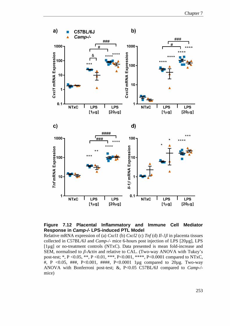
Chapter 7
253
Figure 7.12 Placental Inflammatory and Immune Cell Mediator Response in Camp-/- LPS-induced PTL Model Relative mRNA expression of (a) Cxcl1 (b) Cxcl2 (c) Tnf (d) Il-1β in placenta tissues
collected in C57BL/6J and Camp-/- mice 6-hours post injection of LPS [20µg], LPS
[1µg] or no-treatment controls (NTxC). Data presented is mean fold-increase and
SEM, normalised to β-Actin and relative to CAL. (Two-way ANOVA with Tukey’s
post-test; *, P <0.05, **, P <0.01, ***, P<0.001, ****, P<0.0001 compared to NTxC,
#, P <0.05, ###, P<0.001, ####, P<0.0001 1µg compared to 20µg. Two-way
ANOVA with Bonferroni post-test; &, P<0.05 C57BL/6J compared to Camp-/-
mice)
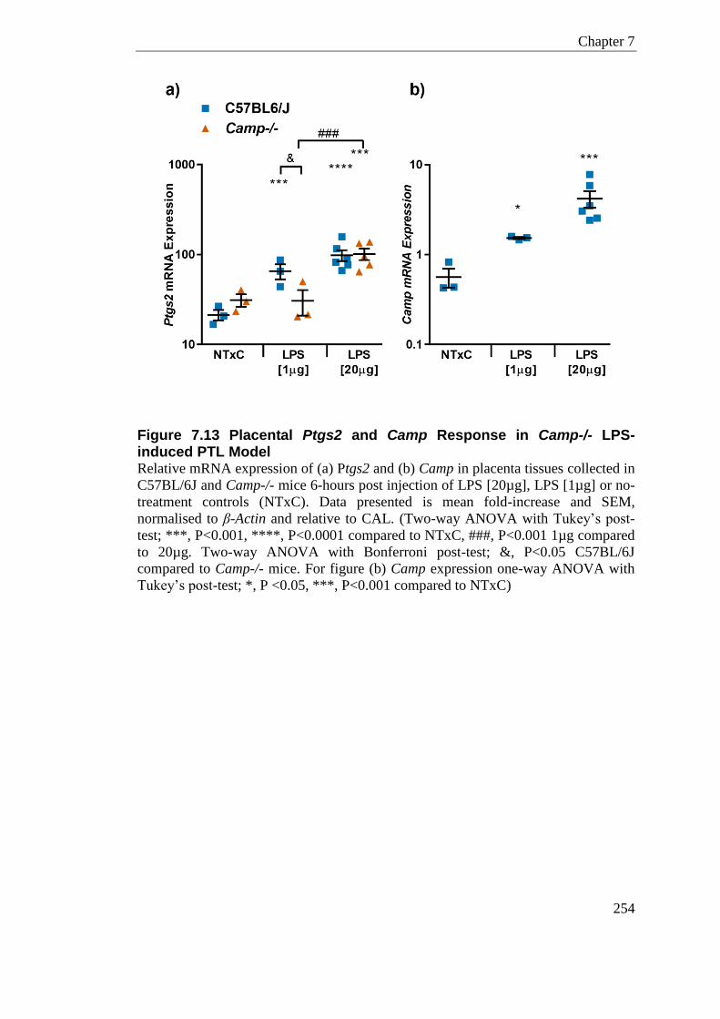
Chapter 7
254
Figure 7.13 Placental Ptgs2 and Camp Response in Camp-/- LPS-induced PTL Model Relative mRNA expression of (a) Ptgs2 and (b) Camp in placenta tissues collected in
C57BL/6J and Camp-/- mice 6-hours post injection of LPS [20µg], LPS [1µg] or no-
treatment controls (NTxC). Data presented is mean fold-increase and SEM,
normalised to β-Actin and relative to CAL. (Two-way ANOVA with Tukey’s post-
test; ***, P<0.001, ****, P<0.0001 compared to NTxC, ###, P<0.001 1µg compared
to 20µg. Two-way ANOVA with Bonferroni post-test; &, P<0.05 C57BL/6J
compared to Camp-/- mice. For figure (b) Camp expression one-way ANOVA with
Tukey’s post-test; *, P <0.05, ***, P<0.001 compared to NTxC)
Chapter 7
255
7.3.4.3.2 Uterus
Chemoattractants
There was no difference in the expression of the neutrophil chemoattractants Cxcl1
or Cxcl2 between C57BL/6J and Camp-/- mice following injection of LPS at either
20µg or 1µg dose (two-way ANOVA) (Figure 7.14.a, Figure 7.14,b).
In C57BL/6J and Camp-/- mice, LPS injection had a significant effect (two-way
ANOVA; P <0.0001) on Cxcl1 expression levels measured by RT-qPCR in uterine
tissue compared to NTxC. LPS increased Cxcl1 expression in a dose-responsive
manner in both C57BL/6J and Camp-/- mice. Cxcl1 expression level of 20µg LPS
injected mice compared to 1µg LPS injection is statistically significant in C57BL/6J
animals (two-way ANOVA, with Tukey’s post-test; P <0.05). In response to 1µg of
LPS there was 57.5 ± 22 fold increase in expression of Cxcl1 compared to NTxC in
C57BL/6J mice. Whereas 20µg of LPS injected in C57BL/6J mice displayed a 444.6
± 36.1 fold increase in Cxcl1 compared to NTxC. This significant dose-response was
also seen in the Camp-/- mice, which had a 91.9 ± 47.9 fold and 420.2 ± 121.6 fold
increase in Cxcl1 gene expression in response to 1µg and 20µg of LPS respectively
(two-way ANOVA, with Tukey’s post-test; P <0.05).
Cxcl2 expression was significantly increased in response to all doses of LPS in both
C57BL/6J and Camp-/- mice compared to NTxC (two-way ANOVA; P <0.0001 for
both 1µg and 20µg). The dose-responsive manner of LPS in Cxcl2 response did not
reach statistical significance in either, C57BL/6J or Camp-/- mice.
Pro-inflammatory cytokines
There was no difference in the expression of the pro-inflammatory cytokines Tnf or
Il-β between C57BL/6J and Camp-/- mice following injection of LPS at either 20µg
or 1µg dose (two-way ANOVA) (Figure 7.14.c, Figure 7.14.d).
In C57BL/6J and Camp-/- mice, LPS injection had a significant effect (two-way
ANOVA; P <0.0001) in Tnf expression levels measured by RT-qPCR in the uterus.
Tnf expression in the uterus was significantly elevated in response to LPS in
Chapter 7
256
C57BL/6J mice (P <0.001 at 1µg and P <0.0001 at 20µg). 20µg of LPS in C57BL/6J
mice displayed a 15.0 ± 1.0 fold increase in Tnf compared to NTxC. This significant
increase in Tnf gene expression was also seen in the Camp-/- mice, (P <0.0001 at
both doses) where 20µg LPS injection increase Tnf gene expression 15.0 ± 2.6 fold
compared to NTxC. Similarly in C57BL/6J and Camp-/- mice, LPS injection had a
significant effect (two-way ANOVA; P <0.0001) in Il-1β expression levels measured
by RT-qPCR in the uterus.
Il-1β expression in the uterus was significantly elevated in response to LPS in
C57BL/6J mice (P <0.001 both doses). 20µg of LPS injection in C57BL/6J mice
displayed a 50.3 ± 6.2 fold increase in Il-1β compared to NTxC. This significant
increase in Il-1β gene expression was also seen in the Camp-/- mice, (P <0.0001)
given 20µg of LPS (Two-way ANOVA, with Tukey’s post-test).
Ptgs2 (Cox-2)
In C57BL/6J and Camp-/- mice, LPS injection had a significant effect (two-way
ANOVA; P <0.01) on Ptgs2 expression levels measured by RT-qPCR in uterine
tissue. A significant increase in Ptgs2 was only seen at the 20µg LPS dose in
C57BL/6J and Camp-/- mice (P <0.05 for both genotypes) compared to NTxC (two-
way ANOVA, with Tukey’s post-test; P <0.05). In response to 20µg of LPS in
C57BL/6J and Camp-/- mice there was a 7.2 ± 1.2 fold increase and 9.6 ± 3.1 fold
increase in Ptgs2 respectively compared to NTxC. Two-way ANOVA with Tukey’s
post-test indicated a small (P = 0.41) but significant difference in Ptgs2 expression
between 1µg and 20µg treated C57BL/6J mice.
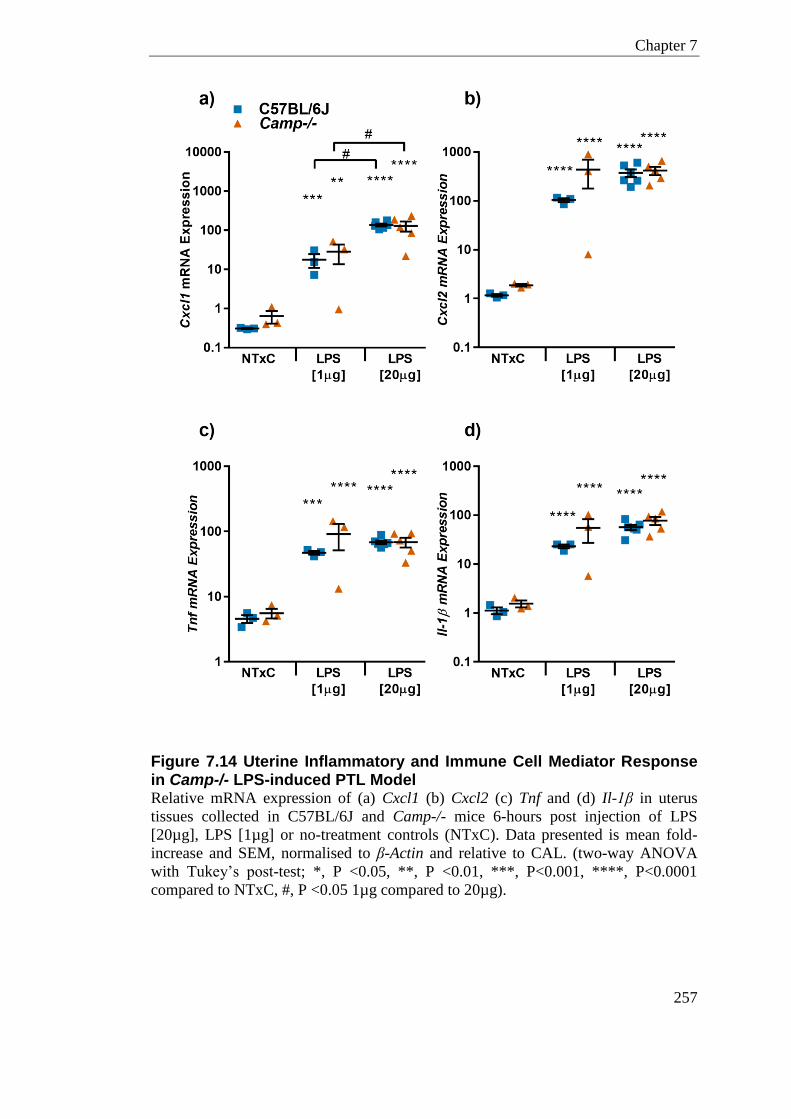
Chapter 7
257
Figure 7.14 Uterine Inflammatory and Immune Cell Mediator Response in Camp-/- LPS-induced PTL Model Relative mRNA expression of (a) Cxcl1 (b) Cxcl2 (c) Tnf and (d) Il-1β in uterus
tissues collected in C57BL/6J and Camp-/- mice 6-hours post injection of LPS
[20µg], LPS [1µg] or no-treatment controls (NTxC). Data presented is mean fold-
increase and SEM, normalised to β-Actin and relative to CAL. (two-way ANOVA
with Tukey’s post-test; *, P <0.05, **, P <0.01, ***, P<0.001, ****, P<0.0001
compared to NTxC, #, P <0.05 1µg compared to 20µg).
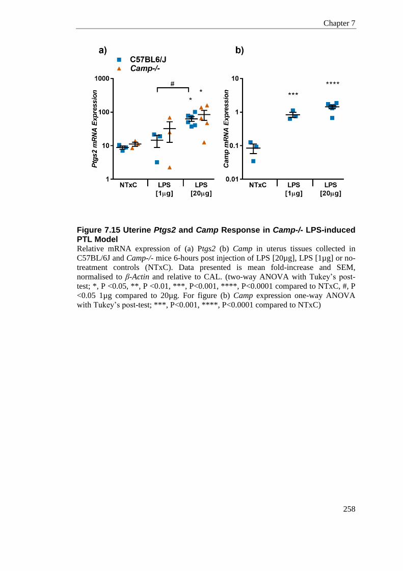
Chapter 7
258
Figure 7.15 Uterine Ptgs2 and Camp Response in Camp-/- LPS-induced PTL Model Relative mRNA expression of (a) Ptgs2 (b) Camp in uterus tissues collected in
C57BL/6J and Camp-/- mice 6-hours post injection of LPS [20µg], LPS [1µg] or no-
treatment controls (NTxC). Data presented is mean fold-increase and SEM,
normalised to β-Actin and relative to CAL. (two-way ANOVA with Tukey’s post-
test; *, P <0.05, **, P <0.01, ***, P<0.001, ****, P<0.0001 compared to NTxC, #, P
<0.05 1µg compared to 20µg. For figure (b) Camp expression one-way ANOVA
with Tukey’s post-test; ***, P<0.001, ****, P<0.0001 compared to NTxC)
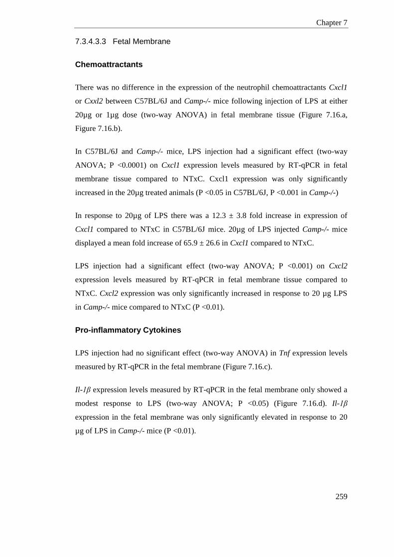
Chapter 7
259
7.3.4.3.3 Fetal Membrane
Chemoattractants
There was no difference in the expression of the neutrophil chemoattractants Cxcl1
or Cxxl2 between C57BL/6J and Camp-/- mice following injection of LPS at either
20µg or 1µg dose (two-way ANOVA) in fetal membrane tissue (Figure 7.16.a,
Figure 7.16.b).
In C57BL/6J and Camp-/- mice, LPS injection had a significant effect (two-way
ANOVA; P <0.0001) on Cxcl1 expression levels measured by RT-qPCR in fetal
membrane tissue compared to NTxC. Cxcl1 expression was only significantly
increased in the 20µg treated animals (P <0.05 in C57BL/6J, P <0.001 in Camp-/-)
In response to 20µg of LPS there was a 12.3 ± 3.8 fold increase in expression of
Cxcl1 compared to NTxC in C57BL/6J mice. 20µg of LPS injected Camp-/- mice
displayed a mean fold increase of 65.9 ± 26.6 in Cxcl1 compared to NTxC.
LPS injection had a significant effect (two-way ANOVA; P <0.001) on Cxcl2
expression levels measured by RT-qPCR in fetal membrane tissue compared to
NTxC. Cxcl2 expression was only significantly increased in response to 20 µg LPS
in Camp-/- mice compared to NTxC (P <0.01).
Pro-inflammatory Cytokines
LPS injection had no significant effect (two-way ANOVA) in Tnf expression levels
measured by RT-qPCR in the fetal membrane (Figure 7.16.c).
Il-1β expression levels measured by RT-qPCR in the fetal membrane only showed a
modest response to LPS (two-way ANOVA; P <0.05) (Figure 7.16.d). Il-1β
expression in the fetal membrane was only significantly elevated in response to 20
µg of LPS in Camp-/- mice (P <0.01).

Chapter 7
260
Ptgs2 (Cox-2)
LPS injection had a significant effect (two-way ANOVA; P <0.0001) on Ptgs2
expression levels measured by RT-qPCR in fetal membrane tissue (Figure 7.17.a). Il-
1β expression in the fetal membrane was only significantly elevated in response to 20
µg of LPS in C57BL/6J (P <0.05) and Camp-/- mice (P <0.01). 20µg of LPS in
C57BL/6J mice displayed a 2.6 ± 0.4 fold increase in Ptgs2 compared to NTxC.
Camp-/- mice treated with 20 µg LPS increase Ptgs2 gene expression 5.6 ± 1.5 fold
compared to NTxC.
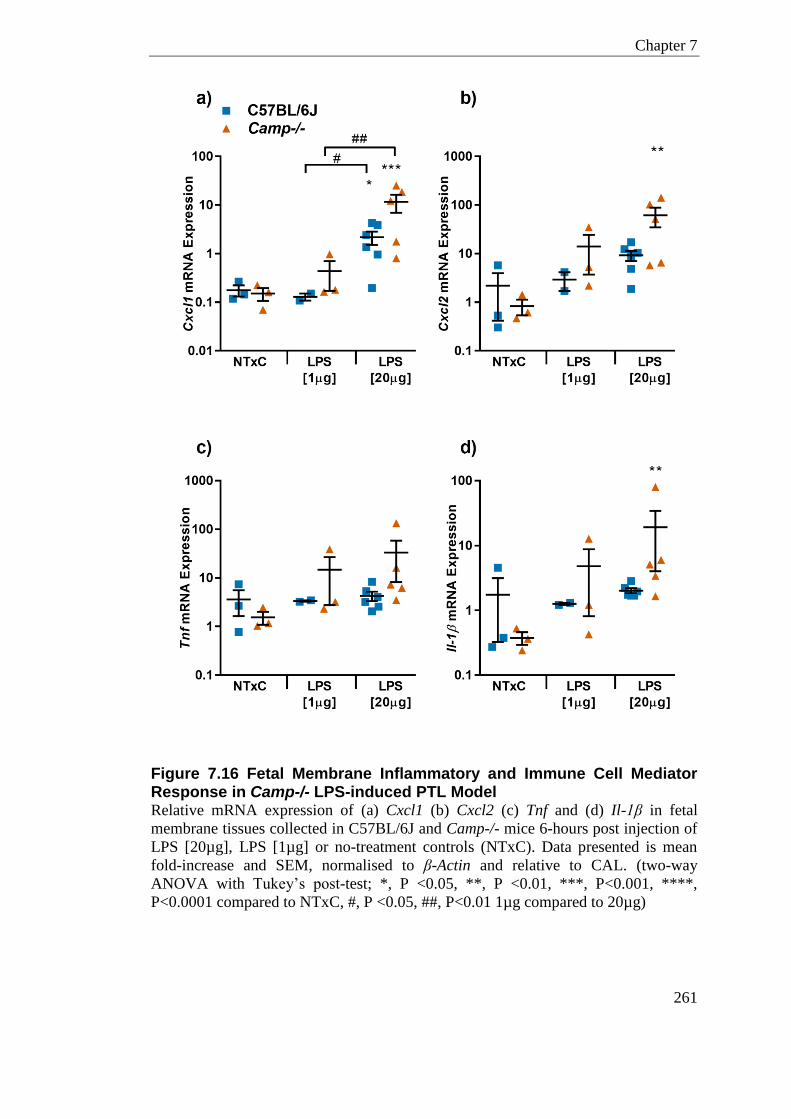
Chapter 7
261
Figure 7.16 Fetal Membrane Inflammatory and Immune Cell Mediator Response in Camp-/- LPS-induced PTL Model Relative mRNA expression of (a) Cxcl1 (b) Cxcl2 (c) Tnf and (d) Il-1β in fetal
membrane tissues collected in C57BL/6J and Camp-/- mice 6-hours post injection of
LPS [20µg], LPS [1µg] or no-treatment controls (NTxC). Data presented is mean
fold-increase and SEM, normalised to β-Actin and relative to CAL. (two-way
ANOVA with Tukey’s post-test; *, P <0.05, **, P <0.01, ***, P<0.001, ****,
P<0.0001 compared to NTxC, #, P <0.05, ##, P<0.01 1µg compared to 20µg)
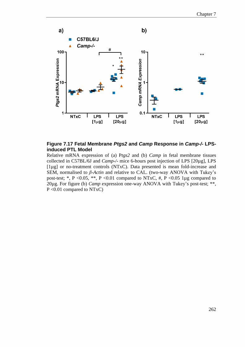
Chapter 7
262
Figure 7.17 Fetal Membrane Ptgs2 and Camp Response in Camp-/- LPS-induced PTL Model Relative mRNA expression of (a) Ptgs2 and (b) Camp in fetal membrane tissues
collected in C57BL/6J and Camp-/- mice 6-hours post injection of LPS [20µg], LPS
[1µg] or no-treatment controls (NTxC). Data presented is mean fold-increase and
SEM, normalised to β-Actin and relative to CAL. (two-way ANOVA with Tukey’s
post-test; *, P <0.05, **, P <0.01 compared to NTxC, #, P <0.05 1µg compared to
20µg. For figure (b) Camp expression one-way ANOVA with Tukey’s post-test; **,
P <0.01 compared to NTxC)
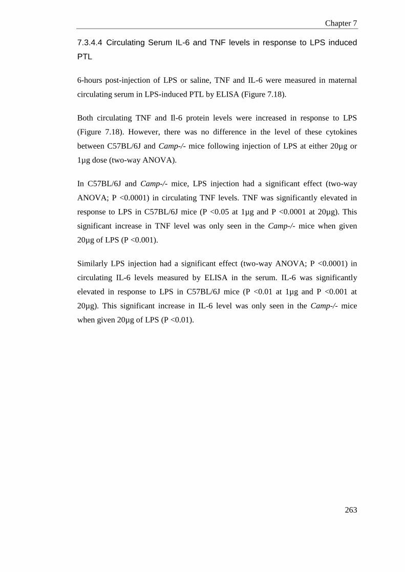
Chapter 7
263
7.3.4.4 Circulating Serum IL-6 and TNF levels in response to LPS induced
PTL
6-hours post-injection of LPS or saline, TNF and IL-6 were measured in maternal
circulating serum in LPS-induced PTL by ELISA (Figure 7.18).
Both circulating TNF and Il-6 protein levels were increased in response to LPS
(Figure 7.18). However, there was no difference in the level of these cytokines
between C57BL/6J and Camp-/- mice following injection of LPS at either 20µg or
1µg dose (two-way ANOVA).
In C57BL/6J and Camp-/- mice, LPS injection had a significant effect (two-way
ANOVA; P <0.0001) in circulating TNF levels. TNF was significantly elevated in
response to LPS in C57BL/6J mice (P <0.05 at 1µg and P <0.0001 at 20µg). This
significant increase in TNF level was only seen in the Camp-/- mice when given
20µg of LPS (P <0.001).
Similarly LPS injection had a significant effect (two-way ANOVA; P <0.0001) in
circulating IL-6 levels measured by ELISA in the serum. IL-6 was significantly
elevated in response to LPS in C57BL/6J mice (P <0.01 at 1µg and P <0.001 at
20µg). This significant increase in IL-6 level was only seen in the Camp-/- mice
when given 20µg of LPS (P <0.01).
Chapter 7
264
Figure 7.18 IL-6 and TNF in Circulating Serum in Camp-/- LPS-induced PTL Model (a) IL-6 and (b) TNF protein levels in circulating serum collected in C57BL/6J and
Camp-/- mice 6-hours post injection of LPS [20µg], LPS [1µg] or non-treatment
controls (NTxC) and measured by ELISA. Data presented is mean pg/ml and SEM
(two-way ANOVA with Tukey’s post-test; *, P <0.05, **, P <0.01, ***, P<0.001,
****, P<0.0001 compared to NTxC, #, P <0.05, ##, P<0.01 1µg compared to 20µg)
Chapter 7
265
7.4 Discussion
Interval to Delivery and Pup Survival
In this chapter, I have addressed the murine expression of the HDPs, Defb14 and
Camp, in the reproductive tract and whether they have a role modulating the
inflammation that maybe the inducing factor of preterm labour. Defb14-/- and Camp-
/- mice have no marked reduction in fertility neither do they display any evidence of
spontaneous PTL. I found that a dose of 20µg LPS injected intrauterine on day 17 of
gestation by laparotomy significantly and robustly induces preterm labour in
C57BL/6N mice. The Defb14-/- mice that were treated the same way showed no
significant difference from the C57BL/6N mice in the interval to delivery (Figure
7.8), or in the number of live born pups (Figure 7.9). Thus at this concentration of
LPS delivered by the laparotomy technique there was no evidence for an influence of
Defb14 on preterm labour in mice.
Injection of 20µg and 1µg of LPS intrauterine on day 17 of gestation by USS
significantly induces preterm labour in C57BL/6J animals. Camp-/- mice receiving
the same treatment displayed no significant difference in the interval to delivery or
the number of live born pups to that of C57BL/6J mice (Figure 7.10; Figure 7.11). At
20µg and 1µg LPS dose, delivered by the ultrasound technique, there was no
evidence for an influence of Camp on preterm labour in mice. Some of the Camp-/-
animals given 1µg of LPS went on to deliver at term and had associated increase in
pup survival. However there was no significant difference in pup survival in Camp-/-
animals between the Saline and LPS treatment group; more animal numbers are
needed here to fully exclude effect.
Pup Survival and Model Refinement
Some of the physiological saline injection control mice delivered preterm and
showed a large number of fetal deaths when treatment was given by laparotomy.
This model of preterm labour was refined for subsequent experiments where
injection of treatments was adapted to be guided by ultrasound sonography. This
refined method is less invasive and reported to negate the laparotomy induced
Chapter 7
266
neutrophil influx to the uterus (Rinaldi et al., 2015, Rinaldi et al., 2014). This model
displayed a small improvement in the number of live-born pups in the saline
injection group of Camp-/- and C57BL/6J control mice. Due to the slight difference
in background strain of the Defb14-/- and Camp-/- animals (C57BL/6N and
C57BL/6J respectively), direct comparisons between modes of treatment delivery
would not be valid here (see Rinaldi et al., (2015) for a direct and robust
comparison). Moreover, the laparotomy and USS model experiments in this chapter
had been conducted in different SPF animal facilities. The effect of differential
bacterial burden between animal facilities on steady state immune profiles remains to
be established.
hBD3 and Defb14
hBD3 expression is known to be upregulated in many cell types when treated with
bacterial stimuli. Oral squamous cells (Shuyi et al., 2011), gingival epithelial cells
and dendritic cells (Yin et al., 2010) all show a marked increase in hBD3 in response
to LPS treatment in-vitro. Garcia-Lopez et al., (2010) displayed that hBD3 secretion
was increased in fetal membranes in a tissue-specific manner; application of E.coli to
the choriodecidua increased both gene and protein expression. Mouse bone marrow
dendritic cells (BMDCs) treated with LPS in-vitro increase the expression of Defb14
gene and protein (Semple et al., 2010). Mouse lung epithelial cells also increase
Defb14 expression in response to TLR stimuli (Rohrl et al., 2008). It is therefore
unexpected that in this chapter LPS treatment intrauterine did not result in increased
endogenous gene expression of Defb14 in response to PTL.
Gene level mRNA for Defb14 can also be stimulated by the pro-inflammatory
cytokines TNF and INF-γ (Rohrl et al., 2008). In this chapter, mice treated with LPS
had increases in circulating TNF protein level and local Tnf mRNA expression in
utero-placenta tissues, nonetheless this had no effect on Defb14 gene expression in
tissues collected 6-hours post injection. It is possible that increase in transcription of
Defb14 comes into effect later than the 6-hours investigated here. Yin et al., (2010)
had shown that LPS was not effective in upregulating hBD3 gene expression in
Chapter 7
267
epithelial cells at 24-hours post treatment in-vitro, but showed an increase at 48-
hours at the protein level.
Defb14 in mouse pregnancy has not been well studied. Hickey et al., (2013) found
that in non-pregnant animals Defb14 had specific estrus cycle mRNA expression
profiles and the level in the vagina was much higher than that in the uterus. Indeed,
in Chapter 2, I show mRNA level of Defb14 to be 1368 fold higher in the vagina
than in the uterus. It is therefore possible that Defb14 has a greater importance in the
lower reproductive tract, which has constant exposure to bacteria.
Dose of LPS in PTL
The hypothesis of this chapter was that mice null for Defb14 or Camp would have
increased rates of preterm labour and the interval to delivery would be shorter than
wildtype animals. However, it is possible that 20µg of LPS is excessive in this
system and any effects of the host defence peptides might be masked by this bolus of
endotoxin. Rinaldi et al., (2014) had previously shown that reducing the LPS
concentration below 15µg delivered by laparotomy caused no-significant increase in
the rate of animals delivering preterm. In the less invasive USS mode of LPS
delivery the lower dose threshold for causing preterm labour has yet to be
established.
Given the published dose response data from the laparotomy model the experiments
conducted in this chapter, using a lower dose of 1µg of LPS induced preterm labour
rates higher than expected in the C57BL/6J animals. Eight out of 10 C57BL/6J
animals delivered preterm, and three out of five of the Camp-/- animals had PTL in
response to 1µg of LPS (Figure 7.10).
To further refine this model it would have been ideal to use LPS at a dose sufficient
to only induce preterm labour in 50% of the wildtype animals, thus highlighting
whether the rates of preterm labour in the HDP knockout mice are higher. I sought to
achieve this by lowering the dose to 1µg of LPS but in this system eight out of 10
wildtype animals delivered preterm (80%), thus conducting a dose response lower
than 1µg would be needed.
Chapter 7
268
Inflammatory and Immune Cell Mediators in Camp-/- PTL
Given the anti-inflammatory effects of human LL-37 on placental explants I have
shown in Chapter 2, and the known ability of CAMP to bind to LPS, I investigated
differences in immune response of Camp-/- mice compared to wildtype in LPS-
induced PTL model. Placental, uterine and fetal membrane tissue were analysed for
the mRNA gene expression of several key inflammatory and immune cell mediators;
Tnf, Il-β, Cxcl1, Cxcl2 and Ptgs2. Broadly most of these mediators showed no
difference in expression between Camp-/- and wildtype animals at either the 1µg or
20µg dose in utero-placental tissue. Cxcl1 and Ptgs2 expression in the placenta at
1µg LPS injection showed a small but significant (P<0.05) difference in the mRNA
expression level between Camp-/- and wildtype animals, although the sample sizes
here are small (n=3 per group).
A 6-hour post-injection time-point was chosen for tissue and blood serum collection
to highlight effects of LPS on inflammatory and immune cell mediators in pregnancy
tissues and circulating serum. This was an intermediate stage between treatment and
the initiation of labour, which would be variable between mice. As many cytokines
and immune cell mediators have unique temporal and presumably spatial patterning
in response to endotoxin, a single time-point is a limiting factor in this study.
Rinaldi et al., (unpublished) had previously shown that saline injection by USS in
C57BL/6J mice did not result in any significant increase in key immune markers
when compared to non-treated animals of the same gestation. Therefore, in this
chapter, the immune marker levels of LPS [20µg/1µg] treatment was compared to
non-treatment animals.
In humans, labour at term and preterm has been associated with pro-inflammatory
mediators in amniotic fluid and at the local maternal fetal interface (Gardella et al.,
2001, Galinsky et al., 2013, Vrachnis et al., 2010). These inflammatory cytokines can
stimulate prostaglandin production and propagate neutrophil chemotaxis, infiltration,
and activation, resulting in metalloproteases (MMPs) synthesis and release.
Prostaglandins can directly stimulate myometrial contractions, in addition to indirect
stimulation by the up regulation of the oxytocin receptor (COX-2) (Liggins, 1989,
Chapter 7
269
Crankshaw and Dyal, 1994). COX-2 is an inducible labour associated gene, which is
expressed at term in the human myometrium; while MMPs can aid tissue
remodelling in the cervix and fetal membrane (Heng et al., 2012)
Chemokines in term labour in both humans (Hamilton et al., 2013) and mice
(Shynlova et al., 2013a) have shown increased production. To investigate the role of
these immune mediators in the mouse model of preterm labour I looked at Cxcl1 and
Cxcl2 the strong neutrophil chemoattractants in response to different doses of LPS.
These chemokines increased in utero-placental tissue in a mostly dose-responsive
manner, the consequences of this are likely to include neutrophil influx. Leucocytes
are a large source of pro-inflammatory cytokines and prostaglandins thus are capable
of initiating or strengthening an inflammatory cascade and triggering labour by
activating the fetal membrane tissues and uterine epithelia. However, Rinaldi et al
(2014) showed that neutrophil influx to the decidua is not necessary for initiation of
preterm labour in an animal model of PTL.
Many chemokines are reported to be increased in normal mouse labour (Shynlova et
al., 2013a), but how the levels in response to LPS-induced PTL compares to
spontaneous term-labour was not studied in this chapter.
Prostaglandins are important inflammatory mediators involved in parturition. PTGS2
(COX-2) is expressed at term in the human myometrium and also in animal cervical
fibroblasts (Sato et al., 2001); there is also some evidence that low expression may
be protective against preterm labour (Hirsch et al., 2006). To investigate the role of
Ptgs2 in the mouse model of preterm labour I looked at Ptgs2 gene expression in
utero-placental tissues; this mediator increased in a mostly dose-responsive manner
to LPS.
Cytokines in Blood Serum
6-hours post-injection of LPS or saline, TNF and IL-6 were measured in maternal
circulating serum in LPS-induced PTL by ELISA. Elevated serum cytokines have
been associated with PTL, although most women with utero-placental infection do
not present with symptoms of systemic illness. It is often reported that LPS induces a
Chapter 7
270
rapid induction of pro-inflammatory cytokines after intraperitoneal injection in non-
pregnant mice. In one study, IL-6 and TNF in the serum increased sharply, peaking
at 2-hours and returned to baseline levels by 6-hours post injection (Copeland et al.,
2005). In this chapter, I found that the increase of TNF and IL-6 in maternal serum
was still detectable 6-hours post LPS-treatment. The timing of peak response and
duration before returning to baseline of these cytokines in LPS-induced PTL remains
to be established.
Mice as a model for PTL
There is known variability in the maternal and fetal inflammatory response to
different serotypes of LPS used to induce preterm labour (Migale et al., 2015). In this
study the serotype of LPS (O111:B4) was kept constant, nonetheless cross-study
comparisons will be problematic. There are limitations of using mouse as a model for
preterm labour, due to the physiological differences to human pregnancy. One main
difference between humans and mice is progesterone production. Humans first
maintain pregnancy by the production of progesterone in the corpus lutum; this is
then superseded by placental progesterone production. In humans, labour is not
associated with a systemic withdrawal of progesterone (Elovitz and Mrinalini, 2004),
however emerging evidence suggests there may be local functional progesterone
withdrawal prior to the onset of labour (Mesiano et al., 2002, Pieber et al., 2001). In
mice however, the contribution of placental progesterone is not thought to supersede
that of the corpus lutum, and removing the ovaries during pregnancy induces labour
(Condon et al., 2004). It has been proposed that Surfactant Protein A (SP-A) released
from the pup lung initiates labour in mice as it causes activation and migration of
amniotic fluid macrophages into the serum, leading to activation of pro-inflammatory
factors such as NF-𝜅B and IL-1β (Condon et al., 2004, Mendelson and Condon,
2005). However, the causative factor that initiates the inflammatory cascade in
human term labour still remains unclear.
Chapter 7
271
7.5 Summary
In this chapter, I have shown that:
- Intrauterine injection of LPS induces preterm labour in wildtype C57BL/6J
and C57BL/6N mice.
- Defb14-/- and Camp-/- mice do not have an increased rate of preterm labour in
a LPS induced inflammatory model.
- Camp but not Defb14 is upregulated in utero-placental tissues in response to
intrauterine LPS injection.
- The inflammatory mediators; Cxcl1, Cxcl2, Tnf, and Il-1β are increased in
response to LPS-induced PTL, some in a dose-responsive manner.
- Camp-/- animals have a similar inflammatory response as wildtypes
C57BL/6J mice when given LPS at 20µg or 1µg during pregnancy.
In this model of preterm labour, it is not clear whether the HDPs, Defb14 and Camp
are fundamental to immune defence of the reproductive tract in pregnancy,
preventing excessive inflammation that can cause preterm labour.

272
Chapter 8
Discussion

Chapter 8
273
Chapter 8 Discussion
Summary of Findings
In a pregnant state, the hypothesis of this thesis is that HDPs have a dual role in
preventing ascending infection, but also preventing an exacerbated inflammatory
response that can cause preterm birth by initiation of the labour cascade.
To explore this I determine whether bacterial stimuli can regulate HDPs expression
in pregnancy tissues. I also explore what interactions HDPs have on the
production/induction of important cytokines, which are vital to the inflammatory
response. As hBD3 and LL-37 suppress the inflammatory response of cells to LPS
through TLR-4, the hypothesis is that in-vivo they might offer protection from
preterm birth caused by inflammation. With the aid of HDP knockout mice, the role
of these peptides in infection/inflammation and continuation of pregnancy is
investigated in a mouse-model of induced preterm-labour. Infection-induced preterm
labour in women is associated with vaginal disruption of commensal bacteria as well
as invading pathogens. To understand how ascending infection might be controlled
by HDPs in pregnancy, I explore how HDPs regulate commensal bacteria. This is
achieved by interrogating the maternal microbiome at mucosal sites in HDP
knockout mice, utilising the bacterial 16S rRNA gene and next generation
sequencing.
In summary, in this thesis I have shown:
- DEFB103 and CAMP are expressed in myometrium, placenta and amnion/chorion
tissues in term human pregnancy.
- Human placental explants respond to LPS challenge by increasing the production
of pro-inflammatory cytokines.
- LL-37 but not hBD3 peptide was able to modulate LPS induced inflammatory
response by inhibiting the release of pro-inflammatory cytokines in the placenta.
Chapter 8
274
- Beta-defensins; Defb1, Defb4, Defb6, Defb14, and the cathelicidin, Camp, show
spatial expression patterns along the female reproductive tract in mice.
- Camp-/- and wildtype mice demonstrated disparate gut microbiota as indicated by
statistically significant taxonomic differences. This community-wide effect involves
the loss and gain of multiple bacteria.
- Camp-/- mice have a more diverse gut microbiome than wildtype controls.
- In murine pregnancy, there are very little global cumulative or progressive shifts
in bacteria, with the exception of Candidatus arthromitus.
- Candidatus arthromitus significantly increases with gestation compared to non-
pregnancy and is maintained at high levels 1 day postpartum.
- Defb14-/- and Camp-/- mice do not have an increased rate of preterm labour in a
LPS induced inflammatory model.
- Camp-/- animals have a similar inflammatory response as wildtype mice when
given LPS at 20µg or 1µg during pregnancy.
Chapter 8
275
Preterm Birth
Preterm birth (<37 weeks gestation) is the main cause of morbidity and mortality in
newborn infants, resulting in a multitude of developmental, neurological and
cognitive problems (Galinsky et al., 2013). Although research into preterm labour
has vastly increased in the last decade, this has not translated into a reduction in the
incidence of preterm labour and improved neonatal outcomes. Preterm labour is
complex and has multifactorial aetiologies; while some preterm labours are
idiopathic, others can be brought on by rupture of the fetal membranes, cervical
insufficiency, infection or medically indicated delivery (iatrogenic).
The upper genital tract was generally considered sterile prior to the onset of labour
(Jones et al., 2009). However, studies investigating bacterial DNA in placenta and
fetal membranes discovered that women who present with preterm labour had a
greater diversity of bacteria in these tissues (Prince et al., 2016, Aagaard et al.,
2014). Indeed, nearly 40% of preterm deliveries are associated with intrauterine
infection and/or clinical inflammation (Romero et al., 2006). The proposed
mechanisms by which infectious agents can transfer to the fetus is either by systemic
maternal-fetal transfer, or more likely initiated by ascending opportunistic
commensal or pathogenic bacteria from the vagina, through the cervix and into the
fetal membrane (Allam et al., 2013, DiGiulio et al., 2010). Cells in the female
reproductive tract, including vaginal epithelial cells, cervical glands, and neutrophils,
are a major source of multiple microbicides including HDPs. Evidence of their
importance has come from many animal and in-vitro studies. For example, mice null
for Camp, the murine orthologue of LL-37/hCAP18 have increased susceptibility to
bacterial urinary tract infections (Chromek et al., 2006). Defb1 homozygous
knockout mice also have increased susceptibility to urinary tract infections whereby
they were shown to retain Staphylococcus spp. in the bladder longer than wildtype
mice (Morrison et al., 2002). In Chapter 3, I show DEFB103, which encodes hBD3,
and CAMP, which encodes LL-37, are expressed in human term myometrium,
placenta and amnion/chorion and there are tissue-specific increases during normal
term labour without signs of infection. This suggests these HDPs could be having a
role in processes of parturition not induced by infection. It is possible their role is

Chapter 8
276
providing additional microbicidal protection at this vulnerable phase of pregnancy or
in the case of the myometrium; HDPs could be involved in tissue remodelling and
repair.
Steady state HDP expression is cell specific and compartmentalised in the female
reproductive tract in pregnant and non-pregnant women. hBD1-4 and human alpha
defensin 5 (HD5) have all been reported to be expressed with distinctive temporal
profiles in the endometrial epithelium, vagina and cervix (King et al., 2003a,
Fleming et al., 2003, Quayle et al., 1998). Similarly, in Chapter 3 I have shown that
many mouse HDPs, specifically, Defb1, Defb4, Defb6, Defb14 and Camp are not
uniformly expressed in the reproductive tract and show spatial patterns. The kinetics
of this differential expression most likely reflects the differing immunological
requirements along the length of the female reproductive tract. This confirms what
Hickey et al., (2013) found in defensin mRNA expression profiles in the vagina and
uterus of lab mice.
In a steady state, I have shown Defb14 is highly expressed in the vagina, which
suggests that this peptide is having a continuous role here, possibly in the
maintenance of commensal microbiota and pathogen clearance. Indeed, lower levels
of the human orthologue, hBD3 have been shown in women with vaginal microbiota
dysbiosis (bacterial vaginosis) and colonisation with BV associated bacteria
(Mitchell et al., 2013). At other mucosal sites, lower hBD3 levels have also been
associated with infection, for example in periodontitis patients. hBD3 levels in
gingival fluid in periodontitis patients were found to be lower than the levels in
normal control individuals (Brancatisano et al., 2011). Interestingly, periodontitis is a
risk factor for preterm birth and having a low birthweight baby. The causality of this
is still unclear but systemic and fetomaternal increases in pro-inflammatory
cytokines, which are deleterious to the fetus, are thought to be involved (Walia and
Saini, 2015). It is not known what causes the reduction in hBD3 levels seen in
mucosal dysbiosis; this could be due to host genetic variation or possible bacterial
inhibition or degradation. Klebsiella pneumoniae is known to inhibit the production
of hBD3 and cysteine proteases can directly degrade hBD3 peptide (Cogen et al.,
2010). hBD3 is copy number variable and the copy number of this loci has been
Chapter 8
277
linked to the inflammatory disease psoriasis (Hollox et al., 2008). An interesting
study looking at the beta-defensin copy number and the nasopharynx microbiota
from otitis media prone children showed that copy number was associated with the
composition of specific pathogens in these children (Jones et al., 2014). Collectively,
it would therefore be valid to investigate beta-defensin copy number in association
with the vaginal microbiome in women. It is possible that pregnancy modifies the
interaction between bacteria and the host mucosa. Therefore correlating the CNV of
the beta-defensin loci and vagina microbiome in pregnancy would provide insight
into maternal microbiome modulation. The commensal vaginal microbiome of
Defb14 null mice was not studied in this thesis; this could well be an avenue for
investigation. Due to concerns with low bacterial yield and variability, an equally
valid approach would be looking at in-vivo vaginal pathogen clearance and in-vitro
bacterial killing from vaginal lavages. Given the impact on outcomes of pregnancy
caused by vaginal bacteria, it is fundamentally essential that we continue to research
vaginal microbiome modulation and dysbiosis to improve female reproductive
health.
Although in Chapter 7 I show Defb14-/- mice do not have an increased rate of PTL
using an LPS-induced model, it would be interesting to investigate the role of Defb14
in the lower reproductive tract in response to pathogens during pregnancy by using a
model of ascending infection. In-vivo inoculation of pathogenic organisms into the
vagina of Defb14 null mice while pregnant would be a valid investigation. It has
been demonstrated that during murine pregnancy the cervical plug prevents from
ascending infection of E.coli (Racicot et al., 2013). It is highly plausible that Defb14
null animals would have defective bacterial killing that could allow bacteria to
colonise in the vagina, transverse the cervix, influencing the continuation of
pregnancy. This would allow for a better understanding in the role Defb14/hBD3
plays in protecting the genital tract during pregnancy. Defensins are however a
multigene family and they may well have redundant compensatory roles in the
vagina mucosa. Indeed, in Chapter 3 I found Defb4, mouse orthologue of hBD2, was
also highly expressed in the vagina in a steady state. To determine the collective role
of these peptides it would be appropriate to knockout multiple highly expressed
defensin genes. The Dorin lab has a genetically modified mouse with a complete
Chapter 8
278
deletion of the beta-defensin region on chromosome 8 that would be suited for such
an investigation. The female mice are sub-fertile but further investigation has not
been carried out.
Some HDPs are inducible upon stimulation with pathogenic agents, which is when
they are most likely to be expressed. It was surprising that Defb14, the murine
orthologue of DEFB103, was not increased in the placenta and uterus 6 hours after
LPS exposure in wildtype pregnant mice. In humans, hBD3 is known to be inducible
upon stimulation. In gingival fibroblasts, hBD3 expression increases in response to
bacterial infection with Candida albicans (Gursoy and Kononen, 2012). Oral
squamous cells (Shuyi et al., 2011), gingival epithelial cells and dendritic cells (Yin
et al., 2010) all show a marked increase in hBD3 in response to LPS treatment in-
vitro. The lack of Defb14 increase in response to LPS in this thesis could be a factor
of the time-point that the tissues were collected in the experiment. A 6-hour post-
injection time-point was chosen for tissue and blood serum collection. This was an
intermediate stage between treatment and the initiation of labour, which would be
variable between mice. As many cytokines and immune cell mediators have unique
temporal and presumably spatial patterning in response to endotoxin, a single time-
point is a limiting factor in this study.
Although inflammation is a crucial mechanism in response to injury and pathogen
clearance, inappropriate or excessive induction of the inflammatory response in
pregnancy can cause initiation of the labour cascade and subsequent preterm
delivery. There is therefore a great need to understand the pathophysiology of
infection/inflammation in the context of preterm labour concerning both host-genetic
and environmental risk factors. The immunomodulatory effect of HDPs in
reproductive tissues in response to infection/inflammation has not been well studied.
In a pregnant state, the hypothesis of this thesis is that HDPs prevent an exacerbated
inflammatory response that can cause preterm birth by initiation of the labour
cascade. To explore this I determine whether bacterial stimuli can regulate HDPs
expression in pregnancy tissues. I also explore what interactions HDPs have on the
production/induction of important cytokines, which are vital to the inflammatory
response. Placental explants respond to LPS challenge by increasing production of
Chapter 8
279
pro-inflammatory cytokines. LL-37 but not hBD3 peptide was able to modulate this
inflammation by inhibiting the release of these pro-inflammatory cytokines. It is of
importance to understand how the inflammatory process leads to preterm labour with
the end goal of delaying or preventing this process in women. Further study could
lead to the development of new antimicrobial therapies for infection and threatened
preterm birth.
Mouse Model of PTL
Ethically and practically the number of samples that can be obtained from women
during pregnancy is limited. In addition, there are multiple compounding factors
concerning ethnicity, BMI, smoking status and the multifactorial aetiologies of
preterm labour. Many different animals have been used to understand the
mechanisms of labour and to search for therapeutics that might have benefit to both
the mother and neonate. Each animal has associated limitations and benefits as a
model for parturition, concerning the correlation to human reproductive biology
(reviewed in Elovitz and Mrinalini, (2004) and Bezold et al., (2013)). Mice have
extensively been used in labour studies as they have the advantage that tissue can be
collected at multiple time-points, in addition to the advantage of their short
gestational period. The immune profile of mice is reasonably well annotated and the
relative ease of genetic modification makes it possible to elude causative effects.
There are some notable differences in mouse physiology that are limitations in using
mouse as a model for parturition; one main feature being the production of
progesterone. Humans first maintain pregnancy by the production of progesterone in
the corpus lutum; this is then superseded by placental progesterone production
(Elovitz and Mrinalini, 2004). In contrast, in mice, placental progesterone is not
thought to supersede that of the corpus lutum and so removal of the ovaries during
pregnancy induces labour (Condon et al., 2004). That said, there is emerging
evidence in humans that suggests there may be local functional progesterone
withdrawal prior to the onset of labour (Pieber et al., 2001, Mesiano et al., 2002).
Mice also have multiple pregnancies and deliver fetus still in their gestational sac,
which is not always ruptured at labour. Contrastingly, in women, the fetal
membranes and placenta are retained for a short duration after labour. Mouse preterm
Chapter 8
280
labour models have focused on stimulation with agents to create an infection and/or
inflammatory state. Stimulation of the TLR4 pathways with LPS has been the central
method of invoking preterm labour in animal models. To establish whether HDPs are
critical in the continuation of pregnancy I used a LPS induced mouse–model of
preterm labour. The initial model of PTL adopted in this thesis was highly invasive
in nature; involving a laparotomy to expose the uterine horns for intrauterine
injection. This was a limitation of this model as some mice receiving only saline
control went on to have preterm labour. An approach established by Sara Rinaldi and
Sarah Stock at the University of Edinburgh, used ultrasound sonography to guide
intrauterine injection. This refined model was adopted in the later experiments in this
thesis, reducing the need for invasive surgery. This is in keeping with the “3Rs”
which is a framework aiming to ‘refine, reduce and replace’ animals used in
research.
Intrauterine injection of LPS induced preterm labour in wildtype mice but the
Defb14-/- and Camp-/- mice did not have an increased rate of preterm labour. Key
inflammatory mediators are increased in response to LPS-induced PTL, some in a
dose-responsive manner, but knockout animals have a similar inflammatory response
as wildtype mice when given LPS during pregnancy. At this dose of LPS in this
model of preterm labour, is not clear whether the HDPs, Defb14 and Camp are
fundamental to immune defence of the reproductive tract in pregnancy, preventing
excessive inflammation that can cause preterm labour. As previously discussed,
additional studies with lower doses of LPS are needed to fully establish the role of
HDP in LPS-induced PTL.
Microbiome
Being able to survey all the microbes in a community is important especially for
applications looking at low abundant species, slow-growing bacteria or bacteria that
cannot easily be cultured. The use of 16S rRNA gene sequencing is becoming a more
popular method for species identification in research, but also in clinical settings for
the discovery of novel bacteria in culture-negative infections (reviewed in Woo et al.,
(2008)). Animals offer researchers a tool to study the role and function of the
Chapter 8
281
microbiome under controlled conditions. Understanding host-microbiome interaction
is vital when looking at disease causality. The advantage of the mouse is that we can
manipulate host genetics to elude gene function. In addition, animals allow for select
colonisation and perturbations such as antibiotics and dietary changes. As the
number and diversity of species we can identify is expanding, there needs to be a
parallel advancement of annotation of bacterial genome databases. The microbiome
field has yet to establish a gold standard for looking at microbial communities. This
thesis has put into place a number of techniques and experimental protocols for 16S
rRNA gene sequencing on next-generation sequencing platforms. Using this
approach I was able to show the mucosal microbiome of Camp-/- animals were
significantly different to that of wildtype controls, showing increased diversity in the
microbes present. While this thesis has displayed a relatively comprehensive
understanding of the major players present in the microbiome in Camp-/- mice, it has
not addressed what these genotype related bacteria are doing. Metagenomics will
help to understand the interactions between the gene of the bacteria and the
microbiota. It would be of interest to study the metabolome in combination with
transcriptomics to gain insight into how these differences in bacterial populations
maybe interacting with the host. Determining the transmissibility of the microbiome
is also a key avenue for investigation. This can be achieved by co-housing wildtype
with Camp-/- mice and interrogating the microbiome. As Camp-/- animals are known
to have increased susceptibility to DSS colitis (Tai et al., 2013, Koon et al., 2011) it
is possible that this could be a result of having an altered microbiome as has been
demonstrated in Chapter 5. Testing the susceptibility to DSS of animals that have
been co-housed would highlight whether this susceptibility phenotype can be
transferred by the transfer of microbes.
There has been much interest on the maternal microbiome during pregnancy and in
the preconception period, but little is known about the postpartum microbiome and
the effects of diet during the lactation stage. As baby’s first colonisation and re-
seeding in the first few weeks of life comes from the mother, promoting postpartum
microbial health could improve the metabolic functions in offspring later in life. In
murine pregnancy, there were very little global cumulative or progressive shifts in
bacteria, with the exception of Candidatus arthromitus, which significantly increases
Chapter 8
282
with gestation compared to non-pregnancy. Candidatus arthromitus, also known as
segmented filamentous bacteria has been shown to serve a protective role in shaping
adult metabolism (Jiang et al., 2001). I have shown that SFB is increased in mid to
late gestation, which is fitting that bacteria which occur early in life and has
beneficial activity would be increased in the maternal microbiome. Clearly further
studies are required to examine both the causal mechanism for the increase in SFB,
and the influence this has on the metabolic function of the offspring.
Apart from primates, which are expensive to work with, there are very few animal
models to test the effects of exogenous perturbation on commensal vaginal
communities. Mice could provide a useful tool to develop and test concepts although
there are clear differences in physiology that make this challenging. In contrast to
humans it has been proposed only a small percentage of female mice harbour
Lactobacillus species at the vaginal mucosa (McGrory and Garber, 1992). In Chapter
6, I have shown that the commensal vaginal microbiome in mice is highly variable.
After review of the literature, this is only the second study to look at laboratory
murine vaginal microbiome by high throughput sequencing (Barfod et al., 2013).
Concordantly Barfod et al., (2013) also concluded there was high variability in
vaginal communities. Collectively this suggests that the murine vagina is perhaps not
an ideal fit for looking at commensal bacterial disturbances in small sample sizes.
Treatments for Infection Associated PTL
In the context of infection, delaying myometrial contractions by the use of anti-
inflammatory agents to target inflammatory signalling pathways might actually be
doing more harm. Initiation of the labour cascade could be a conserved mechanism
to deliver the fetus out of the inflamed in-utero environment. That said, anti-
inflammatory agents could also resolve the intrauterine inflammation that threatens a
preterm delivery. There is therefore a delicate balance in the treatment of PTL,
between dampening the inflammatory environment enough that any negative effects
of retaining pregnancy improve neonatal outcome. Preventing infection in the first
instance is going to be a vital mode for improving neonatal outcome. As it are
commonly bacteria from the vagina that cause infection during pregnancy, vaginal
Chapter 8
283
microbial health even prior to conception will be paramount. The combination of
antimicrobial and anti-inflammatory properties of HDPs make these peptides
attractive as a treatment. The biphasic pro- and anti-inflammatory properties of
HDPs are a benefit, but could also be a concern for therapeutic use. Further studies
are needed to investigate HDP and pro-inflammatory processes such as chemotaxis
in pregnancy.
Conclusion
The development of preventative therapies for preterm labour requires a mechanistic
understanding of how the pro-inflammatory profile is induced. HDPs have been
shown to have multiple roles in this pro-inflammatory process. This thesis has
demonstrated that Host Defence Peptides are expressed in pregnancy tissues and
have anti-inflammatory properties in response to bacterial stimuli. It is not clear
whether the HDP, hBD3 and LL-37 are fundamental to the immune defence in
pregnancy by preventing excessive inflammation. I have shown LL-37 may have a
role in modulation of the maternal microbiota. Given the deleterious impact on
outcomes of pregnancy caused by the vaginal bacteria, it is fundamentally essential
that we continue to apply the findings of this thesis to improve female reproductive
health
References
284
References
AAGAARD, K., MA, J., ANTONY, K. M., GANU, R., PETROSINO, J. & VERSALOVIC, J. 2014. The placenta harbors a unique microbiome. Sci Transl Med, 6, 237ra65.
AAGAARD, K., RIEHLE, K., MA, J., SEGATA, N., MISTRETTA, T. A., COARFA, C., RAZA, S., ROSENBAUM, S., VAN DEN VEYVER, I., MILOSAVLJEVIC, A., GEVERS, D., HUTTENHOWER, C., PETROSINO, J. & VERSALOVIC, J. 2012. A metagenomic approach to characterization of the vaginal microbiome signature in pregnancy. PLoS One, 7, e36466.
ABOU ALAIWA, M. H., REZNIKOV, L. R., GANSEMER, N. D., SHEETS, K. A., HORSWILL, A. R., STOLTZ, D. A., ZABNER, J. & WELSH, M. J. 2014. pH modulates the activity and synergism of the airway surface liquid antimicrobials beta-defensin-3 and LL-37. Proc Natl Acad Sci U S A, 111, 18703-8.
ACINAS, S. G., SARMA-RUPAVTARM, R., KLEPAC-CERAJ, V. & POLZ, M. F. 2005. PCR-induced sequence artifacts and bias: insights from comparison of two 16S rRNA clone libraries constructed from the same sample. Appl Environ Microbiol, 71, 8966-9.
AHN, J. H., KIM, B. Y., SONG, J. & WEON, H. Y. 2012. Effects of PCR cycle number and DNA polymerase type on the 16S rRNA gene pyrosequencing analysis of bacterial communities. J Microbiol, 50, 1071-4.
AISEMBERG, J., VERCELLI, C. A., BARIANI, M. V., BILLI, S. C., WOLFSON, M. L. & FRANCHI, A. M. 2013. Progesterone is essential for protecting against LPS-induced pregnancy loss. LIF as a potential mediator of the anti-inflammatory effect of progesterone. PLoS ONE, 8, e56161-e56161.
AKIRA, S. & TAKEDA, K. 2004. Toll-like receptor signalling. Nat Rev Immunol, 4, 499-511.
ALDUNATE, M., SRBINOVSKI, D., HEARPS, A. C., LATHAM, C. F., RAMSLAND, P. A., GUGASYAN, R., CONE, R. A. & TACHEDJIAN, G. 2015. Antimicrobial and immune modulatory effects of lactic acid and short chain fatty acids produced by vaginal microbiota associated with eubiosis and bacterial vaginosis. Front Physiol, 6, 164.
ALLAM, A. B., VON CHAMIER, M., BROWN, M. B. & REYES, L. 2013. Immune Profiling of BALB/C and C57BL/6 Mice Reveals a Correlation Between Ureaplasma parvum-Induced Fetal Inflammatory Response Syndrome-Like Pathology and Increased Placental Expression of TLR2 and CD14. American journal of reproductive immunology (New York, N.Y. : 1989), 1-11.
ALVAREZ-OLMOS, M. I., BAROUSSE, M. M., RAJAN, L., VAN DER POL, B. J., FORTENBERRY, D., ORR, D. & FIDEL, P. L., JR. 2004. Vaginal lactobacilli in adolescents: presence and relationship to local and systemic immunity, and to bacterial vaginosis. Sex Transm Dis, 31, 393-400.
ARAKI, A., KANAI, T., ISHIKURA, T., MAKITA, S., URAUSHIHARA, K., IIYAMA, R., TOTSUKA, T., TAKEDA, K., AKIRA, S. & WATANABE, M. 2005. MyD88-deficient mice develop severe intestinal inflammation in dextran sodium sulfate colitis. J Gastroenterol, 40, 16-23.
ARCURI, F., TOTI, P., BUCHWALDER, L., CASCIARO, A., CINTORINO, M., SCHATZ, F., RYBALOV, B. & LOCKWOOD, C. J. 2009. Mechanisms of leukocyte accumulation and activation in chorioamnionitis: interleukin 1 beta and tumor necrosis factor alpha enhance colony stimulating factor 2 expression in term decidua. Reprod Sci, 16, 453-61.
ARON-WISNEWSKY, J. & CLEMENT, K. 2016. The gut microbiome, diet, and links to cardiometabolic and chronic disorders. Nat Rev Nephrol, 12, 169-81.
BACH, J. F. 2002. The effect of infections on susceptibility to autoimmune and allergic diseases. N Engl J Med, 347, 911-20.
References
285
BALS, R., WANG, X., MEEGALLA, R. L., WATTLER, S., WEINER, D. J., NEHLS, M. C. & WILSON, J. M. 1999. Mouse beta-defensin 3 is an inducible antimicrobial peptide expressed in the epithelia of multiple organs. Infection and immunity, 67, 3542-7.
BALU, R. B., SAVITZ, D. A., ANANTH, C. V., HARTMANN, K. E., MILLER, W. C., THORP, J. M. & HEINE, R. P. 2003. Bacterial vaginosis, vaginal fluid neutrophil defensins, and preterm birth. Obstetrics and gynecology, 101, 862-8.
BARFOD, K. K., ROGGENBUCK, M., HANSEN, L. H., SCHJORRING, S., LARSEN, S. T., SORENSEN, S. J. & KROGFELT, K. A. 2013. The murine lung microbiome in relation to the intestinal and vaginal bacterial communities. BMC Microbiol, 13, 303.
BARLOW, P. G., LI, Y., WILKINSON, T. S., BOWDISH, D. M., LAU, Y. E., COSSEAU, C., HASLETT, C., SIMPSON, A. J., HANCOCK, R. E. & DAVIDSON, D. J. 2006. The human cationic host defense peptide LL-37 mediates contrasting effects on apoptotic pathways in different primary cells of the innate immune system. J Leukoc Biol, 80, 509-20.
BARNES, P. J. 2006. How corticosteroids control inflammation: Quintiles Prize Lecture 2005. Br J Pharmacol.
BARTON, H. A., TAYLOR, N. M., LUBBERS, B. R. & PEMBERTON, A. C. 2006. DNA extraction from low-biomass carbonate rock: an improved method with reduced contamination and the low-biomass contaminant database. J Microbiol Methods, 66, 21-31.
BAUER, F., SCHWEIMER, K., KLUVER, E., CONEJO-GARCIA, J. R., FORSSMANN, W. G., ROSCH, P., ADERMANN, K. & STICHT, H. 2001. Structure determination of human and murine beta-defensins reveals structural conservation in the absence of significant sequence similarity. Protein Sci, 10, 2470-9.
BAUMANN, P., ROMERO, R., BERRY, S., GOMEZ, R., MCFARLIN, B., ARANEDA, H., COTTON, D. B. & FIDEL, P. 1993. Evidence of participation of the soluble tumor necrosis factor receptor I in the host response to intrauterine infection in preterm labor. American journal of reproductive immunology (New York, N.Y. : 1989), 30, 184-93.
BEDRAN, T. B., MAYER, M. P., SPOLIDORIO, D. P. & GRENIER, D. 2014. Synergistic anti-inflammatory activity of the antimicrobial peptides human beta-defensin-3 (hBD-3) and cathelicidin (LL-37) in a three-dimensional co-culture model of gingival epithelial cells and fibroblasts. PLoS One, 9, e106766.
BERGMAN, P., WALTER-JALLOW, L., BROLIDEN, K., AGERBERTH, B. & SODERLUND, J. 2007. The antimicrobial peptide LL-37 inhibits HIV-1 replication. Curr HIV Res, 5, 410-5.
BERGSTROM, K. S. B., KISSOON-SINGH, V., GIBSON, D. L., MA, C., MONTERO, M., SHAM, H. P., RYZ, N., HUANG, T., VELCICH, A., FINLAY, B. B., CHADEE, K. & VALLANCE, B. A. 2010. Muc2 Protects against Lethal Infectious Colitis by Disassociating Pathogenic and Commensal Bacteria from the Colonic Mucosa. PLoS Pathog.
BETHEA, C. L., GRAVETT, M. G., SADOWSKY, D. W., HALUSKA, G. J., AXTHELM, M. K. & NOVY, M. J. 1998. Amniotic fluid prolactin is decreased by experimental intrauterine infection or interleukin-1beta infusion but not via prostaglandins in pregnant rhesus macaques. Biol Reprod, 58, 1385-93.
BEZOLD, K. Y., KARJALAINEN, M. K., HALLMAN, M., TERAMO, K. & MUGLIA, L. J. 2013. The genomics of preterm birth: from animal models to human studies. Genome Med, 5, 34.
BISCHOFF, F. Z., LEWIS, D. E. & SIMPSON, J. L. 2005. Cell-free fetal DNA in maternal blood: kinetics, source and structure. Human reproduction update, 11, 59-67.
BLANTON, L. V., CHARBONNEAU, M. R., SALIH, T., BARRATT, M. J., VENKATESH, S., ILKAVEYA, O., SUBRAMANIAN, S., MANARY, M. J., TREHAN, I., JORGENSEN, J. M., FAN, Y. M., HENRISSAT, B., LEYN, S. A., RODIONOV, D. A., OSTERMAN, A. L.,
References
286
MALETA, K. M., NEWGARD, C. B., ASHORN, P., DEWEY, K. G. & GORDON, J. I. 2016. Gut bacteria that prevent growth impairments transmitted by microbiota from malnourished children. Science, 351.
BOLDENOW, E., JONES, S., LIEBERMAN, R. W., CHAMES, M. C., ARONOFF, D. M., XI, C. & LOCH-CARUSO, R. 2013. Antimicrobial peptide response to group B Streptococcus in human extraplacental membranes in culture. Placenta, 34, 480-5.
BOLGER, A. M., LOHSE, M. & USADEL, B. 2014. Trimmomatic: a flexible trimmer for Illumina sequence data. Bioinformatics, 30, 2114-20.
BOLLAPRAGADA, S., YOUSSEF, R., JORDAN, F., GREER, I., NORMAN, J. & NELSON, S. 2009. Term labor is associated with a core inflammatory response in human fetal membranes, myometrium, and cervix. Am J Obstet Gynecol, 200, 104 e1-11.
BOWDISH, D. M., DAVIDSON, D. J., SPEERT, D. P. & HANCOCK, R. E. 2004. The human cationic peptide LL-37 induces activation of the extracellular signal-regulated kinase and p38 kinase pathways in primary human monocytes. J Immunol, 172, 3758-65.
BRADSHAW, C. S. & BROTMAN, R. M. 2015. Making inroads into improving treatment of bacterial vaginosis - striving for long-term cure. BMC Infect Dis, 15, 292.
BRANCATISANO, F. L., MAISETTA, G., BARSOTTI, F., ESIN, S., MICELI, M., GABRIELE, M., GIUCA, M. R., CAMPA, M. & BATONI, G. 2011. Reduced human beta defensin 3 in individuals with periodontal disease. J Dent Res, 90, 241-5.
BRAVO, J. A., FORSYTHE, P., CHEW, M. V., ESCARAVAGE, E., SAVIGNAC, H. M., DINAN, T. G., BIENENSTOCK, J. & CRYAN, J. F. 2011. Ingestion of Lactobacillus strain regulates emotional behavior and central GABA receptor expression in a mouse via the vagus nerve. Proc Natl Acad Sci U S A, 108, 16050-5.
BRIERY, C. M., VEILLON, E. W., KLAUSER, C. K., MARTIN, R. W., MAGANN, E. F., CHAUHAN, S. P. & MORRISON, J. C. 2011. Women with preterm premature rupture of the membranes do not benefit from weekly progesterone. Am J Obstet Gynecol, 204, 54.e1-5.
BUFFINGTON, S. A., DI PRISCO, G. V., AUCHTUNG, T. A., AJAMI, N. J., PETROSINO, J. F. & COSTA-MATTIOLI, M. 2016. Microbial Reconstitution Reverses Maternal Diet-Induced Social and Synaptic Deficits in Offspring. Cell, 165, 1762-75.
BUHIMSCHI, I., ALI, M., JAIN, V., CHWALISZ, K. & GARFIELD, R. E. 1996. Differential regulation of nitric oxide in the rat uterus and cervix during pregnancy and labour. Hum Reprod, 11, 1755-66.
BURD, I., BROWN, A., GONZALEZ, J. M., CHAI, J. & ELOVITZ, M. A. 2011. A mouse model of term chorioamnionitis: unraveling causes of adverse neurological outcomes. Reproductive sciences (Thousand Oaks, Calif.), 18, 900-7.
BURRIS, H. H., COLLINS, J. W. & WRIGHT, R. O. 2011. Racial/ethnic disparities in preterm birth: clues from environmental exposures. Curr Opin Pediatr, 23, 227-32.
BYERS, S. L., WILES, M. V., DUNN, S. L. & TAFT, R. A. 2012. Mouse estrous cycle identification tool and images. PLoS ONE, 7, e35538-e35538.
CAMPBELL, J. H., FOSTER, C. M., VISHNIVETSKAYA, T., CAMPBELL, A. G., YANG, Z. K., WYMORE, A., PALUMBO, A. V., CHESLER, E. J. & PODAR, M. 2012. Host genetic and environmental effects on mouse intestinal microbiota. Isme j, 6, 2033-44.
CANI, P. D., BIBILONI, R., KNAUF, C., WAGET, A., NEYRINCK, A. M., DELZENNE, N. M. & BURCELIN, R. 2008. Changes in gut microbiota control metabolic endotoxemia-induced inflammation in high-fat diet-induced obesity and diabetes in mice. Diabetes, 57, 1470-81.
CAPORASO, J. G., BITTINGER, K., BUSHMAN, F. D., DESANTIS, T. Z., ANDERSEN, G. L. & KNIGHT, R. 2010a. PyNAST: a flexible tool for aligning sequences to a template alignment. Bioinformatics, 26, 266-7.
References
287
CAPORASO, J. G., KUCZYNSKI, J., STOMBAUGH, J., BITTINGER, K., BUSHMAN, F. D., COSTELLO, E. K., FIERER, N., PEÑA, A. G., GOODRICH, J. K., GORDON, J. I., HUTTLEY, G. A., KELLEY, S. T., KNIGHTS, D., KOENIG, J. E., LEY, R. E., LOZUPONE, C. A., MCDONALD, D., MUEGGE, B. D., PIRRUNG, M., REEDER, J., SEVINSKY, J. R., TURNBAUGH, P. J., WALTERS, W. A., WIDMANN, J., YATSUNENKO, T., ZANEVELD, J. & KNIGHT, R. 2010b. QIIME allows analysis of high-throughput community sequencing data. Nat Methods, 7, 335-6.
CARDING, S., VERBEKE, K., VIPOND, D. T., CORFE, B. M. & OWEN, L. J. 2015. Dysbiosis of the gut microbiota in disease. Microb Ecol Health Dis.
CARRETERO, M., ESCAMEZ, M. J., GARCIA, M., DUARTE, B., HOLGUIN, A., RETAMOSA, L., JORCANO, J. L., RIO, M. D. & LARCHER, F. 2008. In vitro and in vivo wound healing-promoting activities of human cathelicidin LL-37. J Invest Dermatol, 128, 223-36.
CATALANO, R. D., LANNAGAN, T. R. M., GOROWIEC, M., DENISON, F. C., NORMAN, J. E. & JABBOUR, H. N. 2010. Prokineticins: novel mediators of inflammatory and contractile pathways at parturition? Molecular human reproduction, 16, 311-9.
CDC 2006. Community-associated methicillin-resistant Staphylococcus aureus infection among healthy newborns--Chicago and Los Angeles County, 2004. MMWR Morb Mortal Wkly Rep, 55, 329-32.
CHAKRAVORTY, S., HELB, D., BURDAY, M., CONNELL, N. & ALLAND, D. 2007. A detailed analysis of 16S ribosomal RNA gene segments for the diagnosis of pathogenic bacteria. Journal of microbiological methods, 69, 330-9.
CHAMORRO, C. I., WEBER, G., GRONBERG, A., PIVARCSI, A. & STAHLE, M. 2009. The human antimicrobial peptide LL-37 suppresses apoptosis in keratinocytes. J Invest Dermatol, 129, 937-44.
CHAO, A. 1987. Estimating the population size for capture-recapture data with unequal catchability. Biometrics, 43, 783-91.
CHERTOV, O., UEDA, H., XU, L. L., TANI, K., MURPHY, W. J., WANG, J. M., HOWARD, O. M., SAYERS, T. J. & OPPENHEIM, J. J. 1997. Identification of human neutrophil-derived cathepsin G and azurocidin/CAP37 as chemoattractants for mononuclear cells and neutrophils. J Exp Med, 186, 739-47.
CHEUNG, P. Y., WALTON, J. C., TAI, H. H., RILEY, S. C. & CHALLIS, J. R. 1992. Localization of 15-hydroxy prostaglandin dehydrogenase in human fetal membranes, decidua, and placenta during pregnancy. Gynecol Obstet Invest, 33, 142-6.
CHO, I. & BLASER, M. J. 2012. The human microbiome: at the interface of health and disease. Nat Rev Genet, 13, 260-70.
CHOI, K. Y., CHOW, L. N. & MOOKHERJEE, N. 2012. Cationic host defence peptides: multifaceted role in immune modulation and inflammation. J Innate Immun, 4, 361-70.
CHROMEK, M., ARVIDSSON, I. & KARPMAN, D. 2012. The antimicrobial peptide cathelicidin protects mice from Escherichia coli O157:H7-mediated disease. PLoS One, 7, e46476.
CHROMEK, M., SLAMOVA, Z., BERGMAN, P., KOVACS, L., PODRACKA, L., EHREN, I., HOKFELT, T., GUDMUNDSSON, G. H., GALLO, R. L., AGERBERTH, B. & BRAUNER, A. 2006. The antimicrobial peptide cathelicidin protects the urinary tract against invasive bacterial infection. Nat Med, 12, 636-41.
CHU, H., PAZGIER, M., JUNG, G., NUCCIO, S. P., CASTILLO, P. A., DE JONG, M. F., WINTER, M. G., WINTER, S. E., WEHKAMP, J., SHEN, B., SALZMAN, N. H., UNDERWOOD, M. A., TSOLIS, R. M., YOUNG, G. M., LU, W., LEHRER, R. I., BAUMLER, A. J. & BEVINS, C. L. 2012. Human alpha-defensin 6 promotes mucosal innate immunity through self-assembled peptide nanonets. Science, 337, 477-81.
References
288
CIORNEI, C. D., SIGURDARDOTTIR, T., SCHMIDTCHEN, A. & BODELSSON, M. 2005. Antimicrobial and chemoattractant activity, lipopolysaccharide neutralization, cytotoxicity, and inhibition by serum of analogs of human cathelicidin LL-37. Antimicrob Agents Chemother, 49, 2845-50.
CLAESSON, M. J., JEFFERY, I. B., CONDE, S., POWER, S. E., O'CONNOR, E. M., CUSACK, S., HARRIS, H. M., COAKLEY, M., LAKSHMINARAYANAN, B., O'SULLIVAN, O., FITZGERALD, G. F., DEANE, J., O'CONNOR, M., HARNEDY, N., O'CONNOR, K., O'MAHONY, D., VAN SINDEREN, D., WALLACE, M., BRENNAN, L., STANTON, C., MARCHESI, J. R., FITZGERALD, A. P., SHANAHAN, F., HILL, C., ROSS, R. P. & O'TOOLE, P. W. 2012. Gut microbiota composition correlates with diet and health in the elderly. Nature, 488, 178-84.
CLAESSON, M. J., WANG, Q., O'SULLIVAN, O., GREENE-DINIZ, R., COLE, J. R., ROSS, R. P. & O'TOOLE, P. W. 2010. Comparison of two next-generation sequencing technologies for resolving highly complex microbiota composition using tandem variable 16S rRNA gene regions. Nucleic Acids Res, 38, e200.
CLEMENTE, J. C., PEHRSSON, E. C., BLASER, M. J., SANDHU, K., GAO, Z., WANG, B., MAGRIS, M., HIDALGO, G., CONTRERAS, M., NOYA-ALARCON, O., LANDER, O., MCDONALD, J., COX, M., WALTER, J., OH, P. L., RUIZ, J. F., RODRIGUEZ, S., SHEN, N., SONG, S. J., METCALF, J., KNIGHT, R., DANTAS, G. & DOMINGUEZ-BELLO, M. G. 2015. The microbiome of uncontacted Amerindians. Sci Adv, 1.
COGEN, A. L., YAMASAKI, K., MUTO, J., SANCHEZ, K. M., CROTTY ALEXANDER, L., TANIOS, J., LAI, Y., KIM, J. E., NIZET, V. & GALLO, R. L. 2010. Staphylococcus epidermidis antimicrobial delta-toxin (phenol-soluble modulin-gamma) cooperates with host antimicrobial peptides to kill group A Streptococcus. PLoS One, 5, e8557.
COLE, J. R., WANG, Q., CARDENAS, E., FISH, J., CHAI, B., FARRIS, R. J., KULAM-SYED-MOHIDEEN, A. S., MCGARRELL, D. M., MARSH, T., GARRITY, G. M. & TIEDJE, J. M. 2009. The Ribosomal Database Project: improved alignments and new tools for rRNA analysis. Nucleic Acids Res.
COLLADO, M. C., ISOLAURI, E., LAITINEN, K. & SALMINEN, S. 2008. Distinct composition of gut microbiota during pregnancy in overweight and normal-weight women. Am J Clin Nutr, 88, 894-9.
COMSTOCK, L. E. 2009. Importance of glycans to the host-bacteroides mutualism in the mammalian intestine. Cell Host Microbe, 5, 522-6.
CONDON, J. C., JEYASURIA, P., FAUST, J. M. & MENDELSON, C. R. 2004. Surfactant protein secreted by the maturing mouse fetal lung acts as a hormone that signals the initiation of parturition. Proc Natl Acad Sci U S A, 101, 4978-83.
COPELAND, S., WARREN, H. S., LOWRY, S. F., CALVANO, S. E., REMICK, D. & INVESTIGATORS, T. I. A. T. H. R. T. I. 2005. Acute Inflammatory Response to Endotoxin in Mice and Humans.
COX, L. M., YAMANISHI, S., SOHN, J., ALEKSEYENKO, A. V., LEUNG, J. M., CHO, I., KIM, S. G., LI, H., GAO, Z., MAHANA, D., ZARATE RODRIGUEZ, J. G., ROGERS, A. B., ROBINE, N., LOKE, P. & BLASER, M. J. 2014. Altering the intestinal microbiota during a critical developmental window has lasting metabolic consequences. Cell, 158, 705-21.
CRANKSHAW, D. J. & DYAL, R. 1994. Effects of some naturally occurring prostanoids and some cyclooxygenase inhibitors on the contractility of the human lower uterine segment in vitro. Can J Physiol Pharmacol, 72, 870-4.
CULLEN, T. W., SCHOFIELD, W. B., BARRY, N. A., PUTNAM, E. E., RUNDELL, E. A., TRENT, M. S., DEGNAN, P. H., BOOTH, C. J., YU, H. & GOODMAN, A. L. 2015. Gut microbiota. Antimicrobial peptide resistance mediates resilience of prominent gut commensals during inflammation. Science, 347, 170-5.
References
289
CURRIE, S. M., GWYER FINDLAY, E., MCFARLANE, A. J., FITCH, P. M., BÖTTCHER, B., COLEGRAVE, N., PARAS, A., JOZWIK, A., CHIU, C., SCHWARZE, J. & DAVIDSON, D. J. 2016. Cathelicidins Have Direct Antiviral Activity against Respiratory Syncytial Virus In Vitro and Protective Function In Vivo in Mice and Humans. J Immunol.
DA FONSECA, E. B., BITTAR, R. E., CARVALHO, M. H. & ZUGAIB, M. 2003. Prophylactic administration of progesterone by vaginal suppository to reduce the incidence of spontaneous preterm birth in women at increased risk: a randomized placebo-controlled double-blind study. Am J Obstet Gynecol, 188, 419-24.
DAS, S., VINCE, G. S., LEWIS-JONES, I., BATES, M. D. & GAZVANI, R. 2007. The expression of human alpha and beta defensin in the endometrium and their effect on implantation. J Assist Reprod Genet, 24, 533-9.
DAVIDSON, D. J., CURRIE, A. J., REID, G. S., BOWDISH, D. M., MACDONALD, K. L., MA, R. C., HANCOCK, R. E. & SPEERT, D. P. 2004. The cationic antimicrobial peptide LL-37 modulates dendritic cell differentiation and dendritic cell-induced T cell polarization. J Immunol, 172, 1146-56.
DE FILIPPIS, F., PELLEGRINI, N., VANNINI, L., JEFFERY, I. B., LA STORIA, A., LAGHI, L., SERRAZANETTI, D. I., DI CAGNO, R., FERROCINO, I., LAZZI, C., TURRONI, S., COCOLIN, L., BRIGIDI, P., NEVIANI, E., GOBBETTI, M., O'TOOLE, P. W. & ERCOLINI, D. 2015. High-level adherence to a Mediterranean diet beneficially impacts the gut microbiota and associated metabolome. Gut.
DE, Y., CHEN, Q., SCHMIDT, A. P., ANDERSON, G. M., WANG, J. M., WOOTERS, J., OPPENHEIM, J. J. & CHERTOV, O. 2000. Ll-37, the Neutrophil Granule–And Epithelial Cell–Derived Cathelicidin, Utilizes Formyl Peptide Receptor–Like 1 (Fprl1) as a Receptor to Chemoattract Human Peripheral Blood Neutrophils, Monocytes, and T Cells. J Exp Med.
DEAN, S. N., BISHOP, B. M. & VAN HOEK, M. L. 2011. Natural and synthetic cathelicidin peptides with anti-microbial and anti-biofilm activity against Staphylococcus aureus. BMC Microbiol, 11, 114.
DERUAZ, M. & LUSTER, A. D. 2013. BLT humanized mice as model to study HIV vaginal transmission. J Infect Dis, 208 Suppl 2, S131-6.
DESANTIS, T. Z., HUGENHOLTZ, P., KELLER, K., BRODIE, E. L., LARSEN, N., PICENO, Y. M., PHAN, R. & ANDERSEN, G. L. 2006a. NAST: a multiple sequence alignment server for comparative analysis of 16S rRNA genes. Nucleic Acids Res, 34, W394-9.
DESANTIS, T. Z., HUGENHOLTZ, P., LARSEN, N., ROJAS, M., BRODIE, E. L., KELLER, K., HUBER, T., DALEVI, D., HU, P. & ANDERSEN, G. L. 2006b. Greengenes, a Chimera-Checked 16S rRNA Gene Database and Workbench Compatible with ARB. Appl Environ Microbiol.
DETHLEFSEN, L., MCFALL-NGAI, M. & RELMAN, D. A. 2007. An ecological and evolutionary perspective on human-microbe mutualism and disease. Nature, 449, 811-8.
DHOPLE, V., KRUKEMEYER, A. & RAMAMOORTHY, A. 2006. The human beta-defensin-3, an antibacterial peptide with multiple biological functions. Biochim Biophys Acta, 1758, 1499-512.
DIGIULIO, D. B., CALLAHAN, B. J., MCMURDIE, P. J., COSTELLO, E. K., LYELL, D. J., ROBACZEWSKA, A., SUN, C. L., GOLTSMAN, D. S., WONG, R. J., SHAW, G., STEVENSON, D. K., HOLMES, S. P. & RELMAN, D. A. 2015. Temporal and spatial variation of the human microbiota during pregnancy. Proc Natl Acad Sci U S A, 112, 11060-5.
DIGIULIO, D. B., ROMERO, R., KUSANOVIC, J. P., GÓMEZ, R., KIM, C. J., SEOK, K. S., GOTSCH, F., MAZAKI-TOVI, S., VAISBUCH, E., SANDERS, K., BIK, E. M., CHAIWORAPONGSA, T., OYARZÚN, E. & RELMAN, D. A. 2010. Prevalence and diversity of microbes in the amniotic fluid, the fetal inflammatory response, and
References
290
pregnancy outcome in women with preterm pre-labor rupture of membranes. American journal of reproductive immunology (New York, N.Y. : 1989), 64, 38-57.
DILLON, S. M., LEE, E. J., KOTTER, C. V., AUSTIN, G. L., GIANELLA, S., SIEWE, B., SMITH, D. M., LANDAY, A. L., MCMANUS, M. C., ROBERTSON, C. E., FRANK, D. N., MCCARTER, M. D. & WILSON, C. C. 2016. Gut dendritic cell activation links an altered colonic microbiome to mucosal and systemic T-cell activation in untreated HIV-1 infection. Mucosal Immunol, 9, 24-37.
DOMINGUEZ-BELLO, M. G., COSTELLO, E. K., CONTRERAS, M., MAGRIS, M., HIDALGO, G., FIERER, N. & KNIGHT, R. 2010. Delivery mode shapes the acquisition and structure of the initial microbiota across multiple body habitats in newborns. Proceedings of the National Academy of Sciences of the United States of America, 107, 11971-5.
DONG, W. & YIN, L. 2014. Expression of lipoxin A4, TNFalpha and IL-1beta in maternal peripheral blood, umbilical cord blood and placenta, and their significance in pre-eclampsia. Hypertens Pregnancy, 33, 449-56.
DRELL, T., LILLSAAR, T., TUMMELEHT, L., SIMM, J., AASPÕLLU, A., VÄIN, E., SAARMA, I., SALUMETS, A., DONDERS, G. G. G. & METSIS, M. 2013. Characterization of the Vaginal Micro- and Mycobiome in Asymptomatic Reproductive-Age Estonian Women. PLoS ONE, 8, e54379-e54379.
DUITS, L. A., RADEMAKER, M., RAVENSBERGEN, B., VAN STERKENBURG, M. A., VAN STRIJEN, E., HIEMSTRA, P. S. & NIBBERING, P. H. 2001. Inhibition of hBD-3, but not hBD-1 and hBD-2, mRNA expression by corticosteroids. Biochem Biophys Res Commun, 280, 522-5.
DURNWALD, C. P., MOMIROVA, V., ROUSE, D. J., CARITIS, S. N., PEACEMAN, A. M., SCISCIONE, A., VARNER, M. W., MALONE, F. D., MERCER, B. M., THORP, J. M., JR., SOROKIN, Y., CARPENTER, M. W., LO, J., RAMIN, S. M., HARPER, M. & SPONG, C. Y. 2010. Second trimester cervical length and risk of preterm birth in women with twin gestations treated with 17-alpha hydroxyprogesterone caproate. J Matern Fetal Neonatal Med, 23, 1360-4.
DURR, U. H., SUDHEENDRA, U. S. & RAMAMOORTHY, A. 2006. LL-37, the only human member of the cathelicidin family of antimicrobial peptides. Biochim Biophys Acta, 1758, 1408-25.
ECKBURG, P. B., BIK, E. M., BERNSTEIN, C. N., PURDOM, E., DETHLEFSEN, L., SARGENT, M., GILL, S. R., NELSON, K. E. & RELMAN, D. A. 2005. Diversity of the Human Intestinal Microbial Flora. Science, 308, 1635-8.
EDGAR, R. C. 2010. Search and clustering orders of magnitude faster than BLAST. Bioinformatics, 26, 2460-1.
EDGAR, R. C., HAAS, B. J., CLEMENTE, J. C., QUINCE, C. & KNIGHT, R. 2011. UCHIME improves sensitivity and speed of chimera detection. Bioinformatics.
ELLIOTT, C. L., ALLPORT, V. C., LOUDON, J. A., WU, G. D. & BENNETT, P. R. 2001. Nuclear factor-kappa B is essential for up-regulation of interleukin-8 expression in human amnion and cervical epithelial cells. Mol Hum Reprod, 7, 787-90.
ELOVITZ, M. A. & MRINALINI, C. 2004. Animal models of preterm birth. Trends in endocrinology and metabolism: TEM, 15, 479-87.
ELOVITZ, M. A., WANG, Z., CHIEN, E. K., RYCHLIK, D. F. & PHILLIPPE, M. 2003. A New Model for Inflammation-Induced Preterm Birth: The Role of Platelet-Activating Factor and Toll-Like Receptor-4. Am J Pathol.
ERIGUCHI, Y., TAKASHIMA, S., OKA, H., SHIMOJI, S., NAKAMURA, K., URYU, H., SHIMODA, S., IWASAKI, H., SHIMONO, N., AYABE, T., AKASHI, K. & TESHIMA, T. 2012. Graft-versus-host disease disrupts intestinal microbial ecology by inhibiting Paneth cell production of alpha-defensins. Blood, 120, 223-31.
References
291
EVANS, C. C., LEPARD, K. J., KWAK, J. W., STANCUKAS, M. C., LASKOWSKI, S., DOUGHERTY, J., MOULTON, L., GLAWE, A., WANG, Y., LEONE, V., ANTONOPOULOS, D. A., SMITH, D., CHANG, E. B. & CIANCIO, M. J. 2014. Exercise prevents weight gain and alters the gut microbiota in a mouse model of high fat diet-induced obesity. PLoS One, 9, e92193.
FAN, S. R., LIU, X. P. & LIAO, Q. P. 2008. Human defensins and cytokines in vaginal lavage fluid of women with bacterial vaginosis. International journal of gynaecology and obstetrics: the official organ of the International Federation of Gynaecology and Obstetrics, 103, 50-4.
FARINA, A., LESHANE, E. S., ROMERO, R., GOMEZ, R., CHAIWORAPONGSA, T., RIZZO, N. & BIANCHI, D. W. 2005. High levels of fetal cell-free DNA in maternal serum: a risk factor for spontaneous preterm delivery. American journal of obstetrics and gynecology, 193, 421-5.
FELLERMANN, K., STANGE, D. E., SCHAEFFELER, E., SCHMALZL, H., WEHKAMP, J., BEVINS, C. L., REINISCH, W., TEML, A., SCHWAB, M., LICHTER, P., RADLWIMMER, B. & STANGE, E. F. 2006. A chromosome 8 gene-cluster polymorphism with low human beta-defensin 2 gene copy number predisposes to Crohn disease of the colon. Am J Hum Genet, 79, 439-48.
FIDEL, P. L., ROMERO, R., WOLF, N., CUTRIGHT, J., RAMIREZ, M., ARANEDA, H. & COTTON, D. B. 1994. Systemic and local cytokine profiles in endotoxin-induced preterm parturition in mice. American journal of obstetrics and gynecology, 170, 1467-75.
FLEMING, D. C., KING, A. E., WILLIAMS, A. R. W., CRITCHLEY, H. O. D. & KELLY, R. W. 2003. Hormonal contraception can suppress natural antimicrobial gene transcription in human endometrium. Fertility and sterility, 79, 856-63.
FRANK, J. A., REICH, C. I., SHARMA, S., WEISBAUM, J. S., WILSON, B. A. & OLSEN, G. J. 2008. Critical evaluation of two primers commonly used for amplification of bacterial 16S rRNA genes. Applied and environmental microbiology, 74, 2461-70.
FRANTZ, A. L., ROGIER, E. W., WEBER, C. R., SHEN, L., COHEN, D. A., FENTON, L. A., BRUNO, M. E. C. & KAETZEL, C. S. 2012. Targeted deletion of MyD88 in intestinal epithelial cells results in compromised antibacterial immunity associated with downregulation of polymeric immunoglobulin receptor, mucin-2, and antibacterial peptides. Mucosal Immunol.
FREW, L. 2013. The production and function of cervical hCAP18/LL-37 in pregnancy. PhD, University of Edinburgh.
FREW, L., MAKIEVA, S., MCKINLAY, A. T., MCHUGH, B. J., DOUST, A., NORMAN, J. E., DAVIDSON, D. J. & STOCK, S. J. 2014. Human cathelicidin production by the cervix. PLoS One, 9, e103434.
FRISWELL, M. K., GIKA, H., STRATFORD, I. J., THEODORIDIS, G., TELFER, B., WILSON, I. D. & MCBAIN, A. J. 2010. Site and strain-specific variation in gut microbiota profiles and metabolism in experimental mice. PLoS One, 5, e8584.
FUNDERBURG, N., LEDERMAN, M. M., FENG, Z., DRAGE, M. G., JADLOWSKY, J., HARDING, C. V., WEINBERG, A. & SIEG, S. F. 2007. Human -defensin-3 activates professional antigen-presenting cells via Toll-like receptors 1 and 2. Proc Natl Acad Sci U S A, 104, 18631-5.
GAJER, P., BROTMAN, R. M., BAI, G., SAKAMOTO, J., SCHÜTTE, U. M. E., ZHONG, X., KOENIG, S. S. K., FU, L., MA, Z. S., ZHOU, X., ABDO, Z., FORNEY, L. J. & RAVEL, J. 2012. Temporal dynamics of the human vaginal microbiota. Science translational medicine, 4, 132ra52-132ra52.
GALINSKY, R., POLGLASE, G. R., HOOPER, S. B., BLACK, M. J. & MOSS, T. J. M. 2013. The consequences of chorioamnionitis: preterm birth and effects on development. Journal of pregnancy, 2013, 412831-412831.
References
292
GARCIA-LOPEZ, G., FLORES-ESPINOSA, P. & ZAGA-CLAVELLINA, V. 2010. HBD2 , and HBD3 secretion from human extra-placental membranes stimulated with Escherichia coli. 1-9.
GARCIA-LOPEZ, G., FLORES-ESPINOSA, P. & ZAGA-CLAVELLINA, V. 2010. Tissue-specific human beta-defensins (HBD)1, HBD2, and HBD3 secretion from human extra-placental membranes stimulated with Escherichia coli. Reproductive biology and endocrinology : RB&E, 8, 146-146.
GARDELLA, C., HITTI, J., MARTIN, T. R., RUZINSKI, J. T. & ESCHENBACH, D. 2001. Amniotic fluid lipopolysaccharide-binding protein and soluble CD14 as mediators of the inflammatory response in preterm labor. Am J Obstet Gynecol, 184, 1241-8.
GARNIER, Y., KADYROV, M., GANTERT, M., EINIG, A., RATH, W. & HUPPERTZ, B. 2008. Proliferative responses in the placenta after endotoxin exposure in preterm fetal sheep. Eur J Obstet Gynecol Reprod Biol, 138, 152-7.
GAUGUET, S., D'ORTONA, S., AHNGER-PIER, K., DUAN, B., SURANA, N. K., LU, R., CYWES-BENTLEY, C., GADJEVA, M., SHAN, Q., PRIEBE, G. P. & PIER, G. B. 2015. Intestinal Microbiota of Mice Influences Resistance to Staphylococcus aureus Pneumonia. Infect Immun, 83, 4003-14.
GHOSH, S. & BALTIMORE, D. 1990. Activation in vitro of NF-kappa B by phosphorylation of its inhibitor I kappa B. Nature, 344, 678-82.
GILBERT, N. M., LEWIS, W. G. & LEWIS, A. L. 2013. Clinical Features of Bacterial Vaginosis in a Murine Model of Vaginal Infection with Gardnerella vaginalis. PLoS ONE, 8, e59539-e59539.
GILLIET, M. & LANDE, R. 2008. Antimicrobial peptides and self-DNA in autoimmune skin inflammation. Curr Opin Immunol, 20, 401-7.
GOHIR, W., WHELAN, F. J., SURETTE, M. G., MOORE, C., SCHERTZER, J. D. & SLOBODA, D. M. 2015. Pregnancy-related changes in the maternal gut microbiota are dependent upon the mother's periconceptional diet. Gut Microbes, 6, 310-20.
GOLDENBERG, R. L., HAUTH, J. C. & ANDREWS, W. W. 2000. Intrauterine infection and preterm delivery. N Engl J Med, 342, 1500-7.
GOMEZ-LOPEZ, N., GUILBERT, L. J. & OLSON, D. M. 2010. Invasion of the leukocytes into the fetal-maternal interface during pregnancy. J Leukoc Biol, 88, 625-33.
GOMEZ-LOPEZ, N., STLOUIS, D., LEHR, M. A., SANCHEZ-RODRIGUEZ, E. N. & ARENAS-HERNANDEZ, M. 2014. Immune cells in term and preterm labor. Cell Mol Immunol, 11, 571-81.
GONZALEZ, J. M., DONG, Z., ROMERO, R. & GIRARDI, G. 2011a. Cervical remodeling/ripening at term and preterm delivery: the same mechanism initiated by different mediators and different effector cells. PloS one, 6, e26877-e26877.
GONZALEZ, J. M., FRANZKE, C.-W., YANG, F., ROMERO, R. & GIRARDI, G. 2011b. Complement activation triggers metalloproteinases release inducing cervical remodeling and preterm birth in mice. The American journal of pathology, 179, 838-49.
GRAHN, N., OLOFSSON, M., ELLNEBO-SVEDLUND, K., MONSTEIN, H. J. & JONASSON, J. 2003. Identification of mixed bacterial DNA contamination in broad-range PCR amplification of 16S rDNA V1 and V3 variable regions by pyrosequencing of cloned amplicons. FEMS Microbiol Lett, 219, 87-91.
GRASSO, E., PAPARINI, D., HAUK, V., SALAMONE, G., LEIROS, C. P. & RAMHORST, R. 2014. Differential Migration and Activation Profile of Monocytes after Trophoblast Interaction. PLoS One.
GRAVETT, M. G., HUMMEL, D., ESCHENBACH, D. A. & HOLMES, K. K. 1986. Preterm labor associated with subclinical amniotic fluid infection and with bacterial vaginosis. Obstet Gynecol, 67, 229-37.
References
293
GUO, C., SINNOTT, B., NIU, B., LOWRY, M. B., FANTACONE, M. L. & GOMBART, A. F. 2013. Synergistic induction of human cathelicidin antimicrobial peptide gene expression by vitamin D and stilbenoids. Molecular nutrition & food research, 3, 1-9.
GUPTA, V. K., CHAUDHARI, N. M., ISKEPALLI, S. & DUTTA, C. 2015. Divergences in gene repertoire among the reference Prevotella genomes derived from distinct body sites of human. BMC Genomics.
GURSOY, U. K. & KONONEN, E. 2012. Understanding the roles of gingival beta-defensins. J Oral Microbiol, 4.
HAMILTON, S., OOMOMIAN, Y., STEPHEN, G., SHYNLOVA, O., TOWER, C. L., GARROD, A., LYE, S. J. & JONES, R. L. 2012. Macrophages infiltrate the human and rat decidua during term and preterm labor: evidence that decidual inflammation precedes labor. Biol Reprod, 86, 39.
HAMILTON, S. A., TOWER, C. L. & JONES, R. L. 2013. Identification of Chemokines Associated with the Recruitment of Decidual Leukocytes in Human Labour: Potential Novel Targets for Preterm Labour. PLoS One.
HAMMERSCHLAG, M. R., ALPERT, S., ONDERDONK, A. B., THURSTON, P., DRUDE, E., MCCORMACK, W. M. & BARTLETT, J. G. 1978. Anaerobic microflora of the vagina in children. Am J Obstet Gynecol, 131, 853-6.
HAN, Y. W., REDLINE, R. W., LI, M. & YIN, L. 2004. Fusobacterium nucleatum induces premature and term stillbirths in pregnant mice: implication of oral bacteria in preterm birth. Infection and …, 72, 2272-2279.
HARDER, J., BARTELS, J., CHRISTOPHERS, E. & SCHRODER, J. M. 2001. Isolation and characterization of human beta -defensin-3, a novel human inducible peptide antibiotic. J Biol Chem, 276, 5707-13.
HARDER, J., MEYER-HOFFERT, U., WEHKAMP, K., SCHWICHTENBERG, L. & SCHRODER, J. M. 2004. Differential gene induction of human beta-defensins (hBD-1, -2, -3, and -4) in keratinocytes is inhibited by retinoic acid. J Invest Dermatol, 123, 522-9.
HART, K. M., MURPHY, A. J., BARRETT, K. T., WIRA, C. R., GUYRE, P. M. & PIOLI, P. A. 2009. Functional Expression of Pattern Recognition Receptors in Tissues of the Human Female Reproductive Tract. J Reprod Immunol, 80, 33-40.
HAYDEN, M. S. & GHOSH, S. 2004. Signaling to NF-kappaB. Genes Dev, 18, 2195-224.
HAZRATI, E., GALEN, B., LU, W., WANG, W., OUYANG, Y., KELLER, M. J., LEHRER, R. I. & HEROLD, B. C. 2006. Human alpha- and beta-defensins block multiple steps in herpes simplex virus infection. J Immunol, 177, 8658-66.
HECZKO, U., ABE, A. & FINLAY, B. B. 2000. Segmented filamentous bacteria prevent colonization of enteropathogenic Escherichia coli O103 in rabbits. J Infect Dis, 181, 1027-33.
HELL, E., GISKE, C. G., NELSON, A., ROMLING, U. & MARCHINI, G. 2010. Human cathelicidin peptide LL37 inhibits both attachment capability and biofilm formation of Staphylococcus epidermidis. Lett Appl Microbiol, 50, 211-5.
HENG, Y. J., DI QUINZIO, M. K. W., LIONG, S., PERMEZEL, M., RICE, G. E. & GEORGIOU, H. M. 2012. Temporal investigation of matrix metalloproteinases and their inhibitors in human cervicovaginal fluid in late pregnancy and labor. Reproductive sciences (Thousand Oaks, Calif.), 19, 55-63.
HENZLER WILDMAN, K. A., LEE, D. K. & RAMAMOORTHY, A. 2003. Mechanism of lipid bilayer disruption by the human antimicrobial peptide, LL-37. Biochemistry, 42, 6545-58.
HICKEY, D. K., FAHEY, J. V. & WIRA, C. R. 2013. Mouse estrous cycle regulation of vaginal versus uterine cytokines, chemokines, α-/β-defensins and TLRs. Innate immunity, 19, 121-31.
References
294
HILL, G. B., ST CLAIRE, K. K. & GUTMAN, L. T. 1995. Anaerobes predominate among the vaginal microflora of prepubertal girls. Clin Infect Dis, 20 Suppl 2, S269-70.
HING, T. C., HO, S., SHIH, D. Q., ICHIKAWA, R., CHENG, M., CHEN, J., CHEN, X., LAW, I., NAJARIAN, R., KELLY, C. P., GALLO, R. L., TARGAN, S. R., POTHOULAKIS, C. & KOON, H. W. 2013. The antimicrobial peptide cathelicidin modulates Clostridium difficile-associated colitis and toxin A-mediated enteritis in mice. Gut, 62, 1295-305.
HINRICHSEN, K., PODSCHUN, R., SCHUBERT, S., SCHRÖDER, J. M., HARDER, J. & PROKSCH, E. 2008. Mouse beta-defensin-14, an antimicrobial ortholog of human beta-defensin-3. Antimicrobial agents and chemotherapy, 52, 1876-9.
HIRATA, T., OSUGA, Y., HAMASAKI, K., HIROTA, Y., NOSE, E., MORIMOTO, C., HARADA, M., TAKEMURA, Y., KOGA, K., YOSHINO, O., TAJIMA, T., HASEGAWA, A., YANO, T. & TAKETANI, Y. 2007. Expression of toll-like receptors 2, 3, 4, and 9 genes in the human endometrium during the menstrual cycle. J Reprod Immunol, 74, 53-60.
HIRSCH, E., FILIPOVICH, Y. & MAHENDROO, M. 2006. Signaling via the type I IL-1 and TNF receptors is necessary for bacterially induced preterm labor in a murine model. American journal of obstetrics and gynecology, 194, 1334-40.
HOLLOX, E. J., HUFFMEIER, U., ZEEUWEN, P. L., PALLA, R., LASCORZ, J., RODIJK-OLTHUIS, D., VAN DE KERKHOF, P. C., TRAUPE, H., DE JONGH, G., DEN HEIJER, M., REIS, A., ARMOUR, J. A. & SCHALKWIJK, J. 2008. Psoriasis is associated with increased beta-defensin genomic copy number. Nat Genet, 40, 23-5.
HOOPER, L. V. & GORDON, J. I. 2001. Commensal host-bacterial relationships in the gut. Science, 292, 1115-8.
HOOPER, L. V., WONG, M. H., THELIN, A., HANSSON, L., FALK, P. G. & GORDON, J. I. 2001. Molecular analysis of commensal host-microbial relationships in the intestine. Science, 291, 881-4.
HOOVER, D. M., BOULEGUE, C., YANG, D., OPPENHEIM, J. J., TUCKER, K., LU, W. & LUBKOWSKI, J. 2002. The structure of human macrophage inflammatory protein-3alpha /CCL20. Linking antimicrobial and CC chemokine receptor-6-binding activities with human beta-defensins. J Biol Chem, 277, 37647-54.
HUANG, L. C., REINS, R. Y., GALLO, R. L. & MCDERMOTT, A. M. 2007. Cathelicidin-deficient (Cnlp -/- ) mice show increased susceptibility to Pseudomonas aeruginosa keratitis. Invest Ophthalmol Vis Sci, 48, 4498-508.
HUMAN MICROBIOME, P. 2010. Human Microbiome Project.
HURTADO, P. & PEH, C. A. 2010. LL-37 promotes rapid sensing of CpG oligodeoxynucleotides by B lymphocytes and plasmacytoid dendritic cells. J Immunol, 184, 1425-35.
HYMAN, R. W., FUKUSHIMA, M., JIANG, H., FUNG, E., RAND, L., JOHNSON, B., VO, K. C., CAUGHEY, A. B., HILTON, J. F., DAVIS, R. W. & GIUDICE, L. C. 2014. Diversity of the vaginal microbiome correlates with preterm birth. Reprod Sci, 21, 32-40.
IVANOV, II, ATARASHI, K., MANEL, N., BRODIE, E. L., SHIMA, T., KARAOZ, U., WEI, D., GOLDFARB, K. C., SANTEE, C. A., LYNCH, S. V., TANOUE, T., IMAOKA, A., ITOH, K., TAKEDA, K., UMESAKI, Y., HONDA, K. & LITTMAN, D. R. 2009. Induction of intestinal Th17 cells by segmented filamentous bacteria. Cell, 139, 485-98.
JAKOBSEN, T. R., CLAUSEN, F. B., RODE, L., DZIEGIEL, M. H. & TABOR, A. 2012. High levels of fetal DNA are associated with increased risk of spontaneous preterm delivery. Prenatal diagnosis, 32, 840-5.
JAKOBSSON, H. E., RODRÍGUEZ-PIÑEIRO, A. M., SCHÜTTE, A., ERMUND, A., BOYSEN, P., BEMARK, M., SOMMER, F., BÄCKHED, F., HANSSON, G. C. & JOHANSSON, M. E. 2015. The composition of the gut microbiota shapes the colon mucus barrier. EMBO Rep, 16, 164-77.
References
295
JAMES, C., BAJAJ-ELLIOTT, M., SYNGELAKI, A., KLEIN, N., NICOLAIDES, K. & PEEBLES, D. 2013. Variation in Beta Defensin 1 Genotype is Associated with Preterm Birth. Archives of Disease in Childhood - Fetal and Neonatal Edition, 98, A4-A4.
JANSEN, P. A., RODIJK-OLTHUIS, D., HOLLOX, E. J., KAMSTEEG, M., TJABRINGA, G. S., DE JONGH, G. J., VAN VLIJMEN-WILLEMS, I. M., BERGBOER, J. G., VAN ROSSUM, M. M., DE JONG, E. M., DEN HEIJER, M., EVERS, A. W., BERGERS, M., ARMOUR, J. A., ZEEUWEN, P. L. & SCHALKWIJK, J. 2009. Beta-defensin-2 protein is a serum biomarker for disease activity in psoriasis and reaches biologically relevant concentrations in lesional skin. PLoS One, 4, e4725.
JENQ, R. R., TAUR, Y., DEVLIN, S. M., PONCE, D. M., GOLDBERG, J. D., AHR, K. F., LITTMANN, E. R., LING, L., GOBOURNE, A. C., MILLER, L. C., DOCAMPO, M. D., PELED, J. U., ARPAIA, N., CROSS, J. R., PEETS, T. K., LUMISH, M. A., SHONO, Y., DUDAKOV, J. A., POECK, H., HANASH, A. M., BARKER, J. N., PERALES, M. A., GIRALT, S. A., PAMER, E. G. & VAN DEN BRINK, M. R. 2015. Intestinal Blautia Is Associated with Reduced Death from Graft-versus-Host Disease. Biol Blood Marrow Transplant, 21, 1373-83.
JIANG, H. Q., BOS, N. A. & CEBRA, J. J. 2001. Timing, Localization, and Persistence of Colonization by Segmented Filamentous Bacteria in the Neonatal Mouse Gut Depend on Immune Status of Mothers and Pups. Infect Immun.
JONES, E. A., KANANURAK, A., BEVINS, C. L., HOLLOX, E. J. & BAKALETZ, L. O. 2014. Copy number variation of the beta defensin gene cluster on chromosome 8p influences the bacterial microbiota within the nasopharynx of otitis-prone children. PLoS One, 9, e98269.
JONES, H. E., HARRIS, K. A., AZIZIA, M., BANK, L., CARPENTER, B., HARTLEY, J. C., KLEIN, N. & PEEBLES, D. 2009. Differing prevalence and diversity of bacterial species in fetal membranes from very preterm and term labor. PLoS One, 4, e8205.
KAI-LARSEN, Y., LUTHJE, P., CHROMEK, M., PETERS, V., WANG, X., HOLM, A., KADAS, L., HEDLUND, K. O., JOHANSSON, J., CHAPMAN, M. R., JACOBSON, S. H., ROMLING, U., AGERBERTH, B. & BRAUNER, A. 2010. Uropathogenic Escherichia coli modulates immune responses and its curli fimbriae interact with the antimicrobial peptide LL-37. PLoS Pathog, 6, e1001010.
KAUSHIC, C., ASHKAR, A. A., REID, L. A. & ROSENTHAL, K. L. 2003. Progesterone Increases Susceptibility and Decreases Immune Responses to Genital Herpes Infection. J Virol.
KAUSHIC, C., ZHOU, F., MURDIN, A. D. & WIRA, C. R. 2000. Effects of estradiol and progesterone on susceptibility and early immune responses to Chlamydia trachomatis infection in the female reproductive tract. Infect Immun, 68, 4207-16.
KIDA, Y., SHIMIZU, T. & KUWANO, K. 2006. Sodium butyrate up-regulates cathelicidin gene expression via activator protein-1 and histone acetylation at the promoter region in a human lung epithelial cell line, EBC-1. Mol Immunol, 43, 1972-81.
KIM, M., MORRISON, M. & YU, Z. 2011. Evaluation of different partial 16S rRNA gene sequence regions for phylogenetic analysis of microbiomes. J Microbiol Methods, 84, 81-7.
KIM, Y. M., ROMERO, R., CHAIWORAPONGSA, T., KIM, G. J., KIM, M. R., KUIVANIEMI, H., TROMP, G., ESPINOZA, J., BUJOLD, E., ABRAHAMS, V. M. & MOR, G. 2004. Toll-like receptor-2 and -4 in the chorioamniotic membranes in spontaneous labor at term and in preterm parturition that are associated with chorioamnionitis. Am J Obstet Gynecol, 191, 1346-55.
KINDINGER, L. M., MACINTYRE, D. A., LEE, Y. S., MARCHESI, J. R., SMITH, A., MCDONALD, J. A., TERZIDOU, V., COOK, J. R., LEES, C., ISRAFIL-BAYLI, F., FAIZA, Y., TOOZS-HOBSON, P., SLACK, M., CACCIATORE, S., HOLMES, E., NICHOLSON, J. K., TEOH, T. G. & BENNETT, P. R. 2016. Relationship between vaginal microbial
References
296
dysbiosis, inflammation, and pregnancy outcomes in cervical cerclage. Sci Transl Med, 8, 350ra102.
KING, A. E., CRITCHLEY, H. O. & KELLY, R. W. 2003a. Innate immune defences in the human endometrium. Reprod Biol Endocrinol, 1, 116.
KING, A. E., FLEMING, D. C., CRITCHLEY, H. O. D. & KELLY, R. W. 2003b. Differential expression of the natural antimicrobials, beta-defensins 3 and 4, in human endometrium. Journal of reproductive immunology, 59, 1-16.
KING, J. & FLENADY, V. 2002. Prophylactic antibiotics for inhibiting preterm labour with intact membranes. Cochrane Database Syst Rev, CD000246.
KLINDWORTH, A., PRUESSE, E., SCHWEER, T., PEPLIES, J., QUAST, C., HORN, M. & GLÖCKNER, F. O. 2013. Evaluation of general 16S ribosomal RNA gene PCR primers for classical and next-generation sequencing-based diversity studies. Nucleic Acids Research, 41, e1-e1.
KOCSIS, A. K., LAKATOS, P. L., SOMOGYVARI, F., FUSZEK, P., PAPP, J., FISCHER, S., SZAMOSI, T., LAKATOS, L., KOVACS, A., HOFNER, P. & MANDI, Y. 2008. Association of beta-defensin 1 single nucleotide polymorphisms with Crohn's disease. Scand J Gastroenterol, 43, 299-307.
KOCZULLA, R., VON DEGENFELD, G., KUPATT, C., KROTZ, F., ZAHLER, S., GLOE, T., ISSBRUCKER, K., UNTERBERGER, P., ZAIOU, M., LEBHERZ, C., KARL, A., RAAKE, P., PFOSSER, A., BOEKSTEGERS, P., WELSCH, U., HIEMSTRA, P. S., VOGELMEIER, C., GALLO, R. L., CLAUSS, M. & BALS, R. 2003. An angiogenic role for the human peptide antibiotic LL-37/hCAP-18. J Clin Invest, 111, 1665-72.
KOON, H. W., SHIH, D. Q., CHEN, J., BAKIRTZI, K., HING, T. C., LAW, I., HO, S., ICHIKAWA, R., ZHAO, D., XU, H., GALLO, R., DEMPSEY, P., CHENG, G., TARGAN, S. R. & POTHOULAKIS, C. 2011. Cathelicidin signaling via the Toll-like receptor protects against colitis in mice. Gastroenterology, 141, 1852-63.e1-3.
KOREN, O., GOODRICH, J. K., CULLENDER, T. C., SPOR, A., LAITINEN, K., BÄCKHED, H. K., GONZALEZ, A., WERNER, J. J., ANGENENT, L. T., KNIGHT, R., BÄCKHED, F., ISOLAURI, E., SALMINEN, S. & LEY, R. E. 2012. Host remodeling of the gut microbiome and metabolic changes during pregnancy. Cell, 150, 470-80.
KOVACH, M. A., BALLINGER, M. N., NEWSTEAD, M. W., ZENG, X., BHAN, U., YU, F., MOORE, B. B., GALLO, R. L. & STANDIFORD, T. J. 2012. Cathelicidin Related Antimicrobial Peptide is Required for Effective Lung Mucosal Immunity in Gram-negative Bacterial Pneumonia. J Immunol, 189, 304-11.
KOVATCHEVA-DATCHARY, P., NILSSON, A., AKRAMI, R., LEE, Y. S., DE VADDER, F., ARORA, T., HALLEN, A., MARTENS, E., BJORCK, I. & BACKHED, F. 2015. Dietary Fiber-Induced Improvement in Glucose Metabolism Is Associated with Increased Abundance of Prevotella. Cell Metab, 22, 971-82.
KREUTER, A., JAOUHAR, M., SKRYGAN, M., TIGGES, C., STUCKER, M., ALTMEYER, P., GLASER, R. & GAMBICHLER, T. 2011. Expression of antimicrobial peptides in different subtypes of cutaneous lupus erythematosus. J Am Acad Dermatol, 65, 125-33.
KRUEGER, F., ANDREWS, S. R. & OSBORNE, C. S. 2011. Large scale loss of data in low-diversity illumina sequencing libraries can be recovered by deferred cluster calling. PLoS One, 6, e16607.
KUMAR, N., BEHERA, B., SAGIRI, S. S., PAL, K., RAY, S. S. & ROY, S. 2011. Bacterial vaginosis: Etiology and modalities of treatment—A brief note. J Pharm Bioallied Sci.
LAMBERT, J. A., JOHN, S., SOBEL, J. D. & AKINS, R. A. 2013. Longitudinal analysis of vaginal microbiome dynamics in women with recurrent bacterial vaginosis: recognition of the conversion process. PLoS ONE, 8, e82599-e82599.
LAMONT, R. F. 2015. Advances in the Prevention of Infection-Related Preterm Birth. Front Immunol, 6, 566.
References
297
LANDE, R., GANGULY, D., FACCHINETTI, V., FRASCA, L., CONRAD, C., GREGORIO, J., MELLER, S., CHAMILOS, G., SEBASIGARI, R., RICCIERI, V., BASSETT, R., AMURO, H., FUKUHARA, S., ITO, T., LIU, Y. J. & GILLIET, M. 2011. Neutrophils activate plasmacytoid dendritic cells by releasing self-DNA-peptide complexes in systemic lupus erythematosus. Sci Transl Med, 3, 73ra19.
LANGDON, A., CROOK, N. & DANTAS, G. 2016. The effects of antibiotics on the microbiome throughout development and alternative approaches for therapeutic modulation. Genome Med, 8, 39.
LANGILLE, M. G., MEEHAN, C. J., KOENIG, J. E., DHANANI, A. S., ROSE, R. A., HOWLETT, S. E. & BEIKO, R. G. 2014. Microbial shifts in the aging mouse gut. Microbiome.
LAPPAS, M., PERMEZEL, M. & RICE, G. E. 2003. N-Acetyl-cysteine inhibits phospholipid metabolism, proinflammatory cytokine release, protease activity, and nuclear factor-kappaB deoxyribonucleic acid-binding activity in human fetal membranes in vitro. J Clin Endocrinol Metab, 88, 1723-9.
LAPPAS, M. & RICE, G. E. 2009. Transcriptional regulation of the processes of human labour and delivery. Placenta, 30 Suppl A, S90-5.
LARKIN, M. A., BLACKSHIELDS, G., BROWN, N. P., CHENNA, R., MCGETTIGAN, P. A., MCWILLIAM, H., VALENTIN, F., WALLACE, I. M., WILM, A., LOPEZ, R., THOMPSON, J. D., GIBSON, T. J. & HIGGINS, D. G. 2007. Clustal W and Clustal X version 2.0. Bioinformatics, 23, 2947-8.
LAUKENS, D., BRINKMAN, B. M., RAES, J., DE VOS, M. & VANDENABEELE, P. 2016. Heterogeneity of the gut microbiome in mice: guidelines for optimizing experimental design. FEMS Microbiol Rev, 40, 117-32.
LAURENCE, M., HATZIS, C. & BRASH, D. E. 2014. Common contaminants in next-generation sequencing that hinder discovery of low-abundance microbes. PLoS One, 9, e97876.
LAVELLE, E. C., MURPHY, C., O'NEILL, L. A. & CREAGH, E. M. 2010. The role of TLRs, NLRs, and RLRs in mucosal innate immunity and homeostasis. Mucosal Immunol, 3, 17-28.
LAY, C., RIGOTTIER-GOIS, L., HOLMSTROM, K., RAJILIC, M., VAUGHAN, E. E., DE VOS, W. M., COLLINS, M. D., THIEL, R., NAMSOLLECK, P., BLAUT, M. & DORE, J. 2005. Colonic microbiota signatures across five northern European countries. Appl Environ Microbiol, 71, 4153-5.
LEE, C. C., SUN, Y., QIAN, S. & HUANG, H. 2011. Transmembrane Pores Formed by Human Antimicrobial Peptide LL-37. Biophys J.
LEE, J. K., CHANG, S. W., PERINPANAYAGAM, H., LIM, S. M., PARK, Y. J., HAN, S. H., BAEK, S. H., ZHU, Q., BAE, K. S. & KUM, K. Y. 2013. Antibacterial efficacy of a human beta-defensin-3 peptide on multispecies biofilms. J Endod, 39, 1625-9.
LEY, R. E., BACKHED, F., TURNBAUGH, P., LOZUPONE, C. A., KNIGHT, R. D. & GORDON, J. I. 2005. Obesity alters gut microbial ecology. Proc Natl Acad Sci U S A, 102, 11070-5.
LEY, R. E., TURNBAUGH, P. J., KLEIN, S. & GORDON, J. I. 2006. Microbial ecology: human gut microbes associated with obesity. Nature, 444, 1022-3.
LIGGINS, G. C. 1989. Initiation of labour. Biol Neonate, 55, 366-75.
LIM, J. H., KIM, M. H., HAN, Y. J., LEE, D. E., PARK, S. Y., HAN, J. Y., KIM, M. Y. & RYU, H. M. 2013. Cell-free fetal DNA and cell-free total DNA levels in spontaneous abortion with fetal chromosomal aneuploidy. PLoS ONE, 8, e56787-e56787.
LIM, R., BARKER, G. & LAPPAS, M. 2015. Human cathelicidin antimicrobial protein 18 (hCAP18/LL-37) is increased in foetal membranes and myometrium after spontaneous labour and delivery. J Reprod Immunol, 107, 31-42.

References
298
LINDSTRÖM, T. M. & BENNETT, P. R. 2005. The role of nuclear factor kappa B in human labour. Reproduction (Cambridge, England), 130, 569-81.
LINHARES, I. M., SUMMERS, P. R., LARSEN, B., GIRALDO, P. C. & WITKIN, S. S. 2011. Contemporary perspectives on vaginal pH and lactobacilli. Am J Obstet Gynecol, 204, 120.e1-5.
LIU, H., YU, H., GU, Y., XIN, A., ZHANG, Y., DIAO, H. & LIN, D. 2012. Human beta-defensin DEFB126 is capable of inhibiting LPS-mediated inflammation. Applied microbiology and biotechnology.
LIU, N., KAPLAN, A., LOW, J., NGUYEN, L., LIU, G., EQUILS, O. & HEWISON, M. 2009. Vitamin D Induces Innate Antibacterial Responses in Human Trophoblasts via an Intracrine Pathway1. Biol Reprod, 80, 398-406.
LOCKWOOD, C. J., ARCURI, F., TOTI, P., FELICE, C. D., KRIKUN, G., GULLER, S., BUCHWALDER, L. F. & SCHATZ, F. 2006. Tumor necrosis factor-alpha and interleukin-1beta regulate interleukin-8 expression in third trimester decidual cells: implications for the genesis of chorioamnionitis. Am J Pathol, 169, 1294-302.
LOZUPONE, C. & KNIGHT, R. 2005. UniFrac: a new phylogenetic method for comparing microbial communities. Appl Environ Microbiol, 71, 8228-35.
LOZUPONE, C. A., HAMADY, M., KELLEY, S. T. & KNIGHT, R. 2007. Quantitative and qualitative beta diversity measures lead to different insights into factors that structure microbial communities. Appl Environ Microbiol, 73, 1576-85.
MACINTYRE, D. A., CHANDIRAMANI, M., LEE, Y. S., KINDINGER, L., SMITH, A., ANGELOPOULOS, N., LEHNE, B., ARULKUMARAN, S., BROWN, R., TEOH, T. G., HOLMES, E., NICOHOLSON, J. K., MARCHESI, J. R. & BENNETT, P. R. 2015. The vaginal microbiome during pregnancy and the postpartum period in a European population. Sci Rep, 5, 8988.
MACINTYRE, D. A., LEE, Y. S., MIGALE, R., HERBERT, B. R., WADDINGTON, S. N., PEEBLES, D., HAGBERG, H., JOHNSON, M. R. & BENNETT, P. R. 2014. Activator protein 1 is a key terminal mediator of inflammation-induced preterm labor in mice. Faseb j, 28, 2358-68.
MACKLER, A. M., IEZZA, G., AKIN, M. R., MCMILLAN, P. & YELLON, S. M. 1999. Macrophage trafficking in the uterus and cervix precedes parturition in the mouse. Biol Reprod, 61, 879-83.
MACMANES, M. D. 2011. Promiscuity in mice is associated with increased vaginal bacterial diversity. Naturwissenschaften, 98, 951-60.
MACONES, G. A., PARRY, S., ELKOUSY, M., CLOTHIER, B., URAL, S. H. & STRAUSS, J. F., 3RD 2004. A polymorphism in the promoter region of TNF and bacterial vaginosis: preliminary evidence of gene-environment interaction in the etiology of spontaneous preterm birth. Am J Obstet Gynecol, 190, 1504-8; discussion 3A.
MAISETTA, G., BATONI, G., ESIN, S., RACO, G., BOTTAI, D., FAVILLI, F., FLORIO, W. & CAMPA, M. 2005. Susceptibility of Streptococcus mutans and Actinobacillus actinomycetemcomitans to bactericidal activity of human beta-defensin 3 in biological fluids. Antimicrob Agents Chemother, 49, 1245-8.
MARIAT, D., FIRMESSE, O., LEVENEZ, F., GUIMARAES, V., SOKOL, H., DORE, J., CORTHIER, G. & FURET, J. P. 2009. The Firmicutes/Bacteroidetes ratio of the human microbiota changes with age. BMC Microbiol, 9, 123.
MARTIN, D. H., ZOZAYA, M., LILLIS, R. A., MYERS, L., NSUAMI, M. J. & FERRIS, M. J. 2013. Unique vaginal microbiota that includes an unknown Mycoplasma-like organism is associated with Trichomonas vaginalis infection. The Journal of infectious diseases, 207, 1922-31.
References
299
MARTIN, J. A., OSTERMAN, M. J. & SUTTON, P. D. 2010. Are preterm births on the decline in the United States? Recent data from the National Vital Statistics System. NCHS Data Brief, 1-8.
MARTIN, M. 2011. Cutadapt removes adapter sequences from high-throughput sequencing reads. 17.
MARTINEAU, A. R., WILKINSON, K. A., NEWTON, S. M., FLOTO, R. A., NORMAN, A. W., SKOLIMOWSKA, K., DAVIDSON, R. N., SORENSEN, O. E., KAMPMANN, B., GRIFFITHS, C. J. & WILKINSON, R. J. 2007. IFN-gamma- and TNF-independent vitamin D-inducible human suppression of mycobacteria: the role of cathelicidin LL-37. J Immunol, 178, 7190-8.
MARTINEZ, R. C., FRANCESCHINI, S. A., PATTA, M. C., QUINTANA, S. M., GOMES, B. C., DE MARTINIS, E. C. & REID, G. 2009. Improved cure of bacterial vaginosis with single dose of tinidazole (2 g), Lactobacillus rhamnosus GR-1, and Lactobacillus reuteri RC-14: a randomized, double-blind, placebo-controlled trial. Can J Microbiol, 55, 133-8.
MCGRORY, T. & GARBER, G. E. 1992. Mouse intravaginal infection with Trichomonas vaginalis and role of Lactobacillus acidophilus in sustaining infection. Infection and immunity, 60, 2375-9.
MCLEAN, A. C., VALENZUELA, N., FAI, S. & BENNETT, S. A. 2012. Performing Vaginal Lavage, Crystal Violet Staining, and Vaginal Cytological Evaluation for Mouse Estrous Cycle Staging Identification. J Vis Exp.
MCMURDIE, P. J. & HOLMES, S. 2013. phyloseq: an R package for reproducible interactive analysis and graphics of microbiome census data. PLoS One, 8, e61217.
MCMURDIE, P. J. & HOLMES, S. 2014. Waste not, want not: why rarefying microbiome data is inadmissible. PLoS Comput Biol, 10, e1003531.
MEEHAN, C. J. & BEIKO, R. G. 2014. A phylogenomic view of ecological specialization in the Lachnospiraceae, a family of digestive tract-associated bacteria. Genome Biol Evol, 6, 703-13.
MEIS, P. J., KLEBANOFF, M., THOM, E., DOMBROWSKI, M. P., SIBAI, B., MOAWAD, A. H., SPONG, C. Y., HAUTH, J. C., MIODOVNIK, M., VARNER, M. W., LEVENO, K. J., CARITIS, S. N., IAMS, J. D., WAPNER, R. J., CONWAY, D., O'SULLIVAN, M. J., CARPENTER, M., MERCER, B., RAMIN, S. M., THORP, J. M., PEACEMAN, A. M. & GABBE, S. 2003. Prevention of recurrent preterm delivery by 17 alpha-hydroxyprogesterone caproate. N Engl J Med, 348, 2379-85.
MENARD, J. P. 2011. Antibacterial treatment of bacterial vaginosis: current and emerging therapies. Int J Womens Health, 3, 295-305.
MENDELSON, C. R. & CONDON, J. C. 2005. New insights into the molecular endocrinology of parturition. J Steroid Biochem Mol Biol, 93, 113-9.
MESIANO, S., CHAN, E. C., FITTER, J. T., KWEK, K., YEO, G. & SMITH, R. 2002. Progesterone withdrawal and estrogen activation in human parturition are coordinated by progesterone receptor A expression in the myometrium. J Clin Endocrinol Metab, 87, 2924-30.
MEYSICK, K. C. & GARBER, G. E. 1992. Interactions between Trichomonas vaginalis and vaginal flora in a mouse model. The Journal of parasitology, 78, 157-60.
MIDORIKAWA, K., OUHARA, K., KOMATSUZAWA, H., KAWAI, T., YAMADA, S., FUJIWARA, T., YAMAZAKI, K., SAYAMA, K., TAUBMAN, M. A., KURIHARA, H., HASHIMOTO, K. & SUGAI, M. 2003. Staphylococcus aureus Susceptibility to Innate Antimicrobial Peptides, β-Defensins and CAP18, Expressed by Human Keratinocytes. Infect Immun.
MIGALE, R., HERBERT, B. R., LEE, Y. S., SYKES, L., WADDINGTON, S. N., PEEBLES, D., HAGBERG, H., JOHNSON, M. R., BENNETT, P. R. & MACINTYRE, D. A. 2015. Specific Lipopolysaccharide Serotypes Induce Differential Maternal and Neonatal
References
300
Inflammatory Responses in a Murine Model of Preterm Labor. Am J Pathol, 185, 2390-401.
MIGALE, R., MACINTYRE, D. A., CACCIATORE, S., LEE, Y. S., HAGBERG, H., HERBERT, B. R., JOHNSON, M. R., PEEBLES, D., WADDINGTON, S. N. & BENNETT, P. R. 2016. Modeling hormonal and inflammatory contributions to preterm and term labor using uterine temporal transcriptomics. BMC Med, 14, 86.
MILANI, C., HEVIA, A., FORONI, E., DURANTI, S., TURRONI, F., LUGLI, G. A., SANCHEZ, B., MARTÍN, R., GUEIMONDE, M., VAN SINDEREN, D., MARGOLLES, A. & VENTURA, M. 2013. Assessing the Fecal Microbiota: An Optimized Ion Torrent 16S rRNA Gene-Based Analysis Protocol. PLoS ONE, 8, e68739-e68739.
MIRMONSEF, P., ZARIFFARD, M. R., GILBERT, D., MAKINDE, H., LANDAY, A. L. & SPEAR, G. T. 2012. Short-chain fatty acids induce pro-inflammatory cytokine production alone and in combination with toll-like receptor ligands. Am J Reprod Immunol, 67, 391-400.
MISHRA, B., GOLLA, R. M., LAU, K., LUSHNIKOVA, T. & WANG, G. 2016. Anti-Staphylococcal Biofilm Effects of Human Cathelicidin Peptides. ACS Med Chem Lett, 7, 117-21.
MITCHELL, C., GOTTSCH, M. L., LIU, C., FREDRICKS, D. N. & NELSON, D. B. 2013. Associations between vaginal bacteria and levels of vaginal defensins in pregnant women. American journal of obstetrics and gynecology, 208, 132.e1-7.
MITRA, A., SKRZYPCZAK, M., GINALSKI, K. & ROWICKA, M. 2015. Strategies for achieving high sequencing accuracy for low diversity samples and avoiding sample bleeding using illumina platform. PLoS One, 10, e0120520.
MOGENSEN, T. H. 2009. Pathogen Recognition and Inflammatory Signaling in Innate Immune Defenses. Clin Microbiol Rev.
MONTREEKACHON, P., CHOTJUMLONG, P., BOLSCHER, J. G., NAZMI, K., REUTRAKUL, V. & KRISANAPRAKORNKIT, S. 2011. Involvement of P2X(7) purinergic receptor and MEK1/2 in interleukin-8 up-regulation by LL-37 in human gingival fibroblasts. J Periodontal Res, 46, 327-37.
MOOKHERJEE, N., BROWN, K. L., BOWDISH, D. M. E., DORIA, S., FALSAFI, R., HOKAMP, K., ROCHE, F. M., MU, R., DOHO, G. H., PISTOLIC, J., POWERS, J.-P., BRYAN, J., BRINKMAN, F. S. L. & HANCOCK, R. E. W. 2006. Modulation of the TLR-mediated inflammatory response by the endogenous human host defense peptide LL-37. Journal of immunology (Baltimore, Md. : 1950), 176, 2455-64.
MOOKHERJEE, N., LIPPERT, D. N., HAMILL, P., FALSAFI, R., NIJNIK, A., KINDRACHUK, J., PISTOLIC, J., GARDY, J., MIRI, P., NASEER, M., FOSTER, L. J. & HANCOCK, R. E. 2009. Intracellular receptor for human host defense peptide LL-37 in monocytes. J Immunol, 183, 2688-96.
MORGAN, X. C., SEGATA, N. & HUTTENHOWER, C. 2013. Biodiversity and functional genomics in the human microbiome. Trends Genet, 29, 51-8.
MORRISON, G., KILANOWSKI, F., DAVIDSON, D. & DORIN, J. 2002. Characterization of the mouse beta defensin 1, Defb1, mutant mouse model. Infection and immunity, 70, 3053-60.
MOSER, C., WEINER, D. J., LYSENKO, E., BALS, R., WEISER, J. N. & WILSON, J. M. 2002. beta-Defensin 1 contributes to pulmonary innate immunity in mice. Infect Immun, 70, 3068-72.
MUHLEISEN, A. L. & HERBST-KRALOVETZ, M. M. 2016. Menopause and the vaginal microbiome. Maturitas, 91, 42-50.
MURAKAMI, M., LOPEZ-GARCIA, B., BRAFF, M., DORSCHNER, R. A. & GALLO, R. L. 2004. Postsecretory processing generates multiple cathelicidins for enhanced topical antimicrobial defense. J Immunol, 172, 3070-7.
References
301
NAGALINGAM, N. A., KAO, J. Y. & YOUNG, V. B. 2011. Microbial ecology of the murine gut associated with the development of DSS- colitis. Inflamm Bowel Dis, 17, 917-26.
NAGAOKA, I., TAMURA, H. & HIRATA, M. 2006. An antimicrobial cathelicidin peptide, human CAP18/LL-37, suppresses neutrophil apoptosis via the activation of formyl-peptide receptor-like 1 and P2X7. J Immunol, 176, 3044-52.
NAKADA, E., NAKADA, T. A., WALLEY, K. R., MCCONECHY, M., MCLAUGHLIN, L., VON DADELSZEN, P., SHOZU, M. & BOYD, J. H. 2011. mRNA induces RANTES production in trophoblast cells via TLR3 only when delivered intracellularly using lipid membrane encapsulation. Placenta, 32, 500-5.
NASU, K. & NARAHARA, H. 2010. Pattern recognition via the toll-like receptor system in the human female genital tract. Mediators Inflamm, 2010, 976024.
NELSON, D. B. & MACONES, G. 2002. Bacterial vaginosis in pregnancy: current findings and future directions. Epidemiol Rev, 24, 102-8.
NESS, R. B., HILLIER, S., RICHTER, H. E., SOPER, D. E., STAMM, C., BASS, D. C., SWEET, R. L. & RICE, P. 2003. Can known risk factors explain racial differences in the occurrence of bacterial vaginosis? J Natl Med Assoc, 95, 201-12.
NEUMANN, A., BERENDS, E. T., NERLICH, A., MOLHOEK, E. M., GALLO, R. L., MEERLOO, T., NIZET, V., NAIM, H. Y. & VON KOCKRITZ-BLICKWEDE, M. 2014. The antimicrobial peptide LL-37 facilitates the formation of neutrophil extracellular traps. Biochem J, 464, 3-11.
NHAN-CHANG, C. L., ROMERO, R., TARCA, A. L., MITTAL, P., KUSANOVIC, J. P., EREZ, O., MAZAKI-TOVI, S., CHAIWORAPONGSA, T., HOTRA, J., THAN, N. G., KIM, J. S., HASSAN, S. S. & KIM, C. J. 2010. Characterization of the transcriptome of chorioamniotic membranes at the site of rupture in spontaneous labor at term. Am J Obstet Gynecol, 202, 462 e1-41.
NIYONSABA, F., IWABUCHI, K., MATSUDA, H., OGAWA, H. & NAGAOKA, I. 2002. Epithelial cell-derived human beta-defensin-2 acts as a chemotaxin for mast cells through a pertussis toxin-sensitive and phospholipase C-dependent pathway. Int Immunol, 14, 421-6.
NIYONSABA, F., SOMEYA, A., HIRATA, M., OGAWA, H. & NAGAOKA, I. 2001. Evaluation of the effects of peptide antibiotics human beta-defensins-1/-2 and LL-37 on histamine release and prostaglandin D(2) production from mast cells. Eur J Immunol, 31, 1066-75.
NIYONSABA, F., USHIO, H., HARA, M., YOKOI, H., TOMINAGA, M., TAKAMORI, K., KAJIWARA, N., SAITO, H., NAGAOKA, I., OGAWA, H. & OKUMURA, K. 2010. Antimicrobial peptides human beta-defensins and cathelicidin LL-37 induce the secretion of a pruritogenic cytokine IL-31 by human mast cells. J Immunol, 184, 3526-34.
NIYONSABA, F., USHIO, H., NAKANO, N., NG, W., SAYAMA, K., HASHIMOTO, K., NAGAOKA, I., OKUMURA, K. & OGAWA, H. 2007. Antimicrobial peptides human beta-defensins stimulate epidermal keratinocyte migration, proliferation and production of proinflammatory cytokines and chemokines. J Invest Dermatol, 127, 594-604.
NIZET, V., OHTAKE, T., LAUTH, X., TROWBRIDGE, J., RUDISILL, J., DORSCHNER, R. A., PESTONJAMASP, V., PIRAINO, J., HUTTNER, K. & GALLO, R. L. 2001. Innate antimicrobial peptide protects the skin from invasive bacterial infection. Nature, 414, 454-7.
NOGUCHI, K., TSUKUMI, K. & URANO, T. 2003. Qualitative and quantitative differences in normal vaginal flora of conventionally reared mice, rats, hamsters, rabbits, and dogs. Comparative medicine, 53, 404-12.
NORMAN, J. E., MACKENZIE, F., OWEN, P., MACTIER, H., HANRETTY, K., COOPER, S., CALDER, A., MIRES, G., DANIELIAN, P., STURGISS, S., MACLENNAN, G., TYDEMAN, G., THORNTON, S., MARTIN, B., THORNTON, J. G., NEILSON, J. P. & NORRIE, J. 2009a. Progesterone for the prevention of preterm birth in twin pregnancy
References
302
(STOPPIT): a randomised, double-blind, placebo-controlled study and meta-analysis. Lancet, 373, 2034-40.
NORMAN, J. E., MARLOW, N., MESSOW, C. M., SHENNAN, A., BENNETT, P. R., THORNTON, S., ROBSON, S. C., MCCONNACHIE, A., PETROU, S., SEBIRE, N. J., LAVENDER, T., WHYTE, S. & NORRIE, J. 2016. Vaginal progesterone prophylaxis for preterm birth (the OPPTIMUM study): a multicentre, randomised, double-blind trial. Lancet, 387, 2106-16.
NORMAN, J. E., MORRIS, C. & CHALMERS, J. 2009b. The effect of changing patterns of obstetric care in Scotland (1980-2004) on rates of preterm birth and its neonatal consequences: perinatal database study. PLoS Med, 6, e1000153.
NORWITZ, E. R. & CAUGHEY, A. B. 2011. Progesterone Supplementation and the Prevention of Preterm Birth. Rev Obstet Gynecol, 4, 60-72.
NOSSA, C. W. 2010. Design of 16S rRNA gene primers for 454 pyrosequencing of the human foregut microbiome. World Journal of Gastroenterology, 16, 4135-4135.
O'NEIL, D. A., PORTER, E. M., ELEWAUT, D., ANDERSON, G. M., ECKMANN, L., GANZ, T. & KAGNOFF, M. F. 1999. Expression and regulation of the human beta-defensins hBD-1 and hBD-2 in intestinal epithelium. J Immunol, 163, 6718-24.
OPPENHEIM, J. J. & YANG, D. 2005. Alarmins: chemotactic activators of immune responses. Curr Opin Immunol, 17, 359-65.
OREN, Z., LERMAN, J. C., GUDMUNDSSON, G. H., AGERBERTH, B. & SHAI, Y. 1999. Structure and organization of the human antimicrobial peptide LL-37 in phospholipid membranes: relevance to the molecular basis for its non-cell-selective activity. Biochem J, 341, 501-13.
OSMAN, I., YOUNG, A., LEDINGHAM, M. A., THOMSON, A. J., JORDAN, F., GREER, I. A. & NORMAN, J. E. 2003. Leukocyte density and pro-inflammatory cytokine expression in human fetal membranes, decidua, cervix and myometrium before and during labour at term. Mol Hum Reprod, 9, 41-5.
OVERHAGE, J., CAMPISANO, A., BAINS, M., TORFS, E. C., REHM, B. H. & HANCOCK, R. E. 2008. Human host defense peptide LL-37 prevents bacterial biofilm formation. Infect Immun, 76, 4176-82.
PATIL, A., HUGHES, A. L. & ZHANG, G. 2004. Rapid evolution and diversification of mammalian alpha-defensins as revealed by comparative analysis of rodent and primate genes. Physiological genomics, 20, 1-11.
PATIL, A. A., CAI, Y., SANG, Y., BLECHA, F. & ZHANG, G. 2005. Cross-species analysis of the mammalian beta-defensin gene family: presence of syntenic gene clusters and preferential expression in the male reproductive tract. Physiological genomics, 23, 5-17.
PAULSEN, F., PUFE, T., CONRADI, L., VAROGA, D., TSOKOS, M., PAPENDIECK, J. & PETERSEN, W. 2002. Antimicrobial peptides are expressed and produced in healthy and inflamed human synovial membranes. J Pathol, 198, 369-77.
PAWELCZYK, E., NOWICKI, B. J., IZBAN, M. G., PRATAP, S., SASHTI, N. A., SANDERSON, M. & NOWICKI, S. 2010. Spontaneous preterm labor is associated with an increase in the proinflammatory signal transducer TLR4 receptor on maternal blood monocytes. BMC pregnancy and childbirth, 10, 66-66.
PETERSON, J., GARGES, S., GIOVANNI, M., MCINNES, P., WANG, L., SCHLOSS, J. A., BONAZZI, V., MCEWEN, J. E., WETTERSTRAND, K. A., DEAL, C., BAKER, C. C., DI FRANCESCO, V., HOWCROFT, T. K., KARP, R. W., LUNSFORD, R. D., WELLINGTON, C. R., BELACHEW, T., WRIGHT, M., GIBLIN, C., DAVID, H., MILLS, M., SALOMON, R., MULLINS, C., AKOLKAR, B., BEGG, L., DAVIS, C., GRANDISON, L., HUMBLE, M., KHALSA, J., LITTLE, A. R., PEAVY, H., PONTZER, C., PORTNOY, M., SAYRE, M. H., STARKE-REED, P., ZAKHARI, S., READ, J., WATSON, B. & GUYER, M. 2009. The NIH Human Microbiome Project. Genome Res, 19, 2317-23.
References
303
PIEBER, D., ALLPORT, V. C., HILLS, F., JOHNSON, M. & BENNETT, P. R. 2001. Interactions between progesterone receptor isoforms in myometrial cells in human labour. Mol Hum Reprod, 7, 875-9.
PINHEIRO DA SILVA, F., GALLO, R. L. & NIZET, V. 2009. Differing effects of exogenous or endogenous cathelicidin on macrophage toll-like receptor signaling. Immunol Cell Biol, 87, 496-500.
PIOLI, P. A., AMIEL, E., SCHAEFER, T. M., CONNOLLY, J. E., WIRA, C. R. & GUYRE, P. M. 2004. Differential expression of Toll-like receptors 2 and 4 in tissues of the human female reproductive tract. Infect Immun, 72, 5799-806.
PISTOLIC, J., COSSEAU, C., LI, Y., YU, J. J., FILEWOD, N. C., GELLATLY, S., REHAUME, L. M., BOWDISH, D. M. & HANCOCK, R. E. 2009. Host defence peptide LL-37 induces IL-6 expression in human bronchial epithelial cells by activation of the NF-kappaB signaling pathway. J Innate Immun, 1, 254-67.
POUND, L. D., PATRICK, C., EBERHARD, C. E., MOTTAWEA, W., WANG, G. S., ABUJAMEL, T., VANDENBEEK, R., STINTZI, A. & SCOTT, F. W. 2015. Cathelicidin Antimicrobial Peptide: A Novel Regulator of Islet Function, Islet Regeneration, and Selected Gut Bacteria. Diabetes, 64, 4135-47.
PRICE, M. N., DEHAL, P. S. & ARKIN, A. P. 2010. FastTree 2 – Approximately Maximum-Likelihood Trees for Large Alignments. PLoS One.
PRINCE, A. L., MA, J., KANNAN, P. S., ALVAREZ, M., GISSLEN, T., HARRIS, R. A., SWEENEY, E. L., KNOX, C. L., LAMBERS, D. S., JOBE, A. H., CHOUGNET, C. A., KALLAPUR, S. G. & AAGAARD, K. M. 2016. The placental membrane microbiome is altered among subjects with spontaneous preterm birth with and without chorioamnionitis. Am J Obstet Gynecol, 214, 627.e1-627.e16.
PUTSEP, K., CARLSSON, G., BOMAN, H. G. & ANDERSSON, M. 2002. Deficiency of antibacterial peptides in patients with morbus Kostmann: an observation study. Lancet, 360, 1144-9.
QUAST, C., PRUESSE, E., YILMAZ, P., GERKEN, J., SCHWEER, T., YARZA, P., PEPLIES, J. & GLÖCKNER, F. O. 2013. The SILVA ribosomal RNA gene database project: improved data processing and web-based tools. Nucleic Acids Res.
QUAYLE, A. J., PORTER, E. M., NUSSBAUM, A. A., WANG, Y. M., BRABEC, C., YIP, K. P. & MOK, S. C. 1998. Gene expression, immunolocalization, and secretion of human defensin-5 in human female reproductive tract. The American journal of pathology, 152, 1247-58.
RACICOT, K., CARDENAS, I., WÜNSCHE, V., ALDO, P., GULLER, S., MEANS, R. E., ROMERO, R. & MOR, G. 2013. Viral infection of the pregnant cervix predisposes to ascending bacterial infection. Journal of immunology (Baltimore, Md. : 1950), 191, 934-41.
RAJPAL, D. K., KLEIN, J. L., MAYHEW, D., BOUCHERON, J., SPIVAK, A. T., KUMAR, V., INGRAHAM, K., PAULIK, M., CHEN, L., VAN HORN, S., THOMAS, E., SATHE, G., LIVI, G. P., HOLMES, D. J. & BROWN, J. R. 2015. Selective Spectrum Antibiotic Modulation of the Gut Microbiome in Obesity and Diabetes Rodent Models. PLoS One, 10, e0145499.
RAPALA-KOZIK, M., BOCHENSKA, O., ZAWROTNIAK, M., WOLAK, N., TREBACZ, G., GOGOL, M., OSTROWSKA, D., AOKI, W., UEDA, M. & KOZIK, A. 2015. Inactivation of the Antifungal and Immunomodulatory Properties of Human Cathelicidin LL-37 by Aspartic Proteases Produced by the Pathogenic Yeast Candida albicans. Infect Immun.
RAVEL, J., GAJER, P., ABDO, Z., SCHNEIDER, G. M., KOENIG, S. S. K., MCCULLE, S. L., KARLEBACH, S., GORLE, R., RUSSELL, J., TACKET, C. O., BROTMAN, R. M., DAVIS, C. C., AULT, K., PERALTA, L. & FORNEY, L. J. 2011. Vaginal microbiome of reproductive-age women. Proceedings of the National Academy of Sciences of the United States of America, 108 Suppl, 4680-7.
References
304
RAVEL, J., GAJER, P., FU, L., MAUCK, C. K., KOENIG, S. S. K., SAKAMOTO, J., MOTSINGER-REIF, A. A., DONCEL, G. F. & ZEICHNER, S. L. 2012. Twice-daily application of HIV microbicides alter the vaginal microbiota. mBio, 3, 1-8.
RAVUSSIN, Y., KOREN, O., SPOR, A., LEDUC, C., GUTMAN, R., STOMBAUGH, J., KNIGHT, R., LEY, R. E. & LEIBEL, R. L. 2012. Responses of Gut Microbiota to Diet Composition and Weight Loss in Lean and Obese Mice. Obesity (Silver Spring), 20.
REID, G. & BURTON, J. 2002. Use of Lactobacillus to prevent infection by pathogenic bacteria. Microbes and infection / Institut Pasteur, 4, 319-24.
REINHOLZ, M., RUZICKA, T. & SCHAUBER, J. 2012. Cathelicidin LL-37: an antimicrobial peptide with a role in inflammatory skin disease. Ann Dermatol, 24, 126-35.
RESI, V., BASU, S., HAGHIAC, M., PRESLEY, L., MINIUM, J., KAUFMAN, B., BERNARD, S., CATALANO, P. & HAUGUEL-DE MOUZON, S. 2012. Molecular inflammation and adipose tissue matrix remodeling precede physiological adaptations to pregnancy. Am J Physiol Endocrinol Metab.
REZNIKOV, L. L., FANTUZZI, G., SELZMAN, C. H., SHAMES, B. D., BARTON, H. A., BELL, H., MCGREGOR, J. A. & DINARELLO, C. A. 1999. Utilization of endoscopic inoculation in a mouse model of intrauterine infection-induced preterm birth: role of interleukin 1beta. Biology of reproduction, 60, 1231-8.
RINALDI, S. F., CATALANO, R. D., WADE, J., ROSSI, A. G. & NORMAN, J. E. 2014. Decidual neutrophil infiltration is not required for preterm birth in a mouse model of infection-induced preterm labor. J Immunol, 192, 2315-25.
RINALDI, S. F., MAKIEVA, S., FREW, L., WADE, J., THOMSON, A. J., MORAN, C. M., NORMAN, J. E. & STOCK, S. J. 2015. Ultrasound-guided intrauterine injection of lipopolysaccharide as a novel model of preterm birth in the mouse. Am J Pathol, 185, 1201-6.
ROHRL, J., YANG, D., OPPENHEIM, J. J. & HEHLGANS, T. 2008. Identification and Biological Characterization of Mouse beta-defensin 14, the orthologue of human beta-defensin 3. J Biol Chem, 283, 5414-9.
ROHRL, J., YANG, D., OPPENHEIM, J. J. & HEHLGANS, T. 2010. Specific binding and chemotactic activity of mBD4 and its functional orthologue hBD2 to CCR6-expressing cells. J Biol Chem, 285, 7028-34.
ROMERO, R., ESPINOZA, J., GONCALVES, L. F., KUSANOVIC, J. P., FRIEL, L. & HASSAN, S. 2007. The role of inflammation and infection in preterm birth. Semin Reprod Med, 25, 21-39.
ROMERO, R., ESPINOZA, J., GONCALVES, L. F., KUSANOVIC, J. P., FRIEL, L. A. & NIEN, J. K. 2006. Inflammation in preterm and term labour and delivery. Semin Fetal Neonatal Med, 11, 317-26.
ROMERO, R., HASSAN, S. S., GAJER, P., TARCA, A. L., FADROSH, D. W., NIKITA, L., GALUPPI, M., LAMONT, R. F., CHAEMSAITHONG, P., MIRANDA, J., CHAIWORAPONGSA, T. & RAVEL, J. 2014. The composition and stability of the vaginal microbiota of normal pregnant women is different from that of non-pregnant women. Microbiome, 2, 4.
ROSENFELD, Y., PAPO, N. & SHAI, Y. 2006. Endotoxin (lipopolysaccharide) neutralization by innate immunity host-defense peptides. Peptide properties and plausible modes of action. J Biol Chem, 281, 1636-43.
ROUSE, D. J., CARITIS, S. N., PEACEMAN, A. M., SCISCIONE, A., THOM, E. A., SPONG, C. Y., VARNER, M., MALONE, F., IAMS, J. D., MERCER, B. M., THORP, J., SOROKIN, Y., CARPENTER, M., LO, J., RAMIN, S., HARPER, M. & ANDERSON, G. 2007. A trial of 17 alpha-hydroxyprogesterone caproate to prevent prematurity in twins. N Engl J Med, 357, 454-61.
References
305
ROYSE, K. E., KEMPF, M. C., MCGWIN, G., WILSON, C. M., TANG, J. & SHRESTHA, S. 2012. Toll-Like Receptor Gene Variants Associated with Bacterial Vaginosis among HIV-1 Infected Adolescents. J Reprod Immunol, 96, 84-9.
SAHL, H. G., PAG, U., BONNESS, S., WAGNER, S., ANTCHEVA, N. & TOSSI, A. 2005. Mammalian defensins: structures and mechanism of antibiotic activity. J Leukoc Biol, 77, 466-75.
SALMINEN, A., PAANANEN, R., VUOLTEENAHO, R., METSOLA, J., OJANIEMI, M., AUTIO-HARMAINEN, H. & HALLMAN, M. 2008. Maternal endotoxin-induced preterm birth in mice: fetal responses in toll-like receptors, collectins, and cytokines. Pediatric research, 63, 280-6.
SALTER, S. J., COX, M. J., TUREK, E. M., CALUS, S. T., COOKSON, W. O., MOFFATT, M. F., TURNER, P., PARKHILL, J., LOMAN, N. J. & WALKER, A. W. 2014. Reagent and laboratory contamination can critically impact sequence-based microbiome analyses. BMC Biol, 12, 87.
SALVADO, M. D., DI GENNARO, A., LINDBOM, L., AGERBERTH, B. & HAEGGSTROM, J. Z. 2013. Cathelicidin LL-37 induces angiogenesis via PGE2-EP3 signaling in endothelial cells, in vivo inhibition by aspirin. Arterioscler Thromb Vasc Biol, 33, 1965-72.
SALZMAN, N. H., DE JONG, H., PATERSON, Y., HARMSEN, H. J., WELLING, G. W. & BOS, N. A. 2002a. Analysis of 16S libraries of mouse gastrointestinal microflora reveals a large new group of mouse intestinal bacteria. Microbiology, 148, 3651-60.
SALZMAN, N. H., DE JONG, H., PATERSON, Y., HARMSEN, H. J. M., WELLING, G. W. & BOS, N. A. 2002b. Analysis of 16S libraries of mouse gastrointestinal microflora reveals a large new group of mouse intestinal bacteria. Microbiology (Reading, England), 148, 3651-60.
SALZMAN, N. H., GHOSH, D. & HUTTNER, K. M. 2003. Protection against enteric salmonellosis in transgenic mice expressing a human intestinal defensin. 422.
SALZMAN, N. H., HUNG, K., HARIBHAI, D., CHU, H., KARLSSON-SJÖBERG, J., AMIR, E., TEGGATZ, P., BARMAN, M., HAYWARD, M., EASTWOOD, D., STOEL, M., ZHOU, Y., SODERGREN, E., WEINSTOCK, G. M., BEVINS, C. L., WILLIAMS, C. B. & BOS, N. A. 2010. Enteric defensins are essential regulators of intestinal microbial ecology. Nature immunology, 11, 76-83.
SALZMAN, N. H., UNDERWOOD, M. A. & BEVINS, C. L. 2007. Paneth cells, defensins, and the commensal microbiota: a hypothesis on intimate interplay at the intestinal mucosa. Seminars in immunology, 19, 70-83.
SATO, T., MICHIZU, H., HASHIZUME, K. & ITO, A. 2001. Hormonal regulation of PGE2 and COX-2 production in rabbit uterine cervical fibroblasts. Journal of applied physiology (Bethesda, Md. : 1985), 90, 1227-31.
SCHAUBER, J., RIEGER, D., WEILER, F., WEHKAMP, J., ECK, M., FELLERMANN, K., SCHEPPACH, W., GALLO, R. L. & STANGE, E. F. 2006. Heterogeneous expression of human cathelicidin hCAP18/LL-37 in inflammatory bowel diseases. Eur J Gastroenterol Hepatol, 18, 615-21.
SCHER, J. U., SCZESNAK, A., LONGMAN, R. S., SEGATA, N., UBEDA, C., BIELSKI, C., ROSTRON, T., CERUNDOLO, V., PAMER, E. G., ABRAMSON, S. B., HUTTENHOWER, C. & LITTMAN, D. R. 2013. Expansion of intestinal Prevotella copri correlates with enhanced susceptibility to arthritis. Elife, 2, e01202.
SCHLOSS, P. D., GEVERS, D. & WESTCOTT, S. L. 2011. Reducing the effects of PCR amplification and sequencing artifacts on 16S rRNA-based studies. PLoS One, 6, e27310.
SCHLOSS, P. D., WESTCOTT, S. L., RYABIN, T., HALL, J. R., HARTMANN, M., HOLLISTER, E. B., LESNIEWSKI, R. A., OAKLEY, B. B., PARKS, D. H., ROBINSON, C. J., SAHL, J. W., STRES, B., THALLINGER, G. G., VAN HORN, D. J. & WEBER, C.
References
306
F. 2009. Introducing mothur: Open-Source, Platform-Independent, Community-
Supported Software for Describing and Comparing Microbial Communities▿. Appl
Environ Microbiol.
SCHROEDER, B. O., WU, Z., NUDING, S., GROSCURTH, S., MARCINOWSKI, M., BEISNER, J., BUCHNER, J., SCHALLER, M., STANGE, E. F. & WEHKAMP, J. 2011. Reduction of disulphide bonds unmasks potent antimicrobial activity of human β-defensin 1. Nature, 469, 419-23.
SCHUTTE, B. C., MITROS, J. P., BARTLETT, J. A., WALTERS, J. D., JIA, H. P., WELSH, M. J., CASAVANT, T. L. & MCCRAY, P. B. 2002. Discovery of five conserved β-defensin gene clusters using a computational search strategy. Proc Natl Acad Sci U S A.
SCHWARZER, M., MAKKI, K., STORELLI, G., MACHUCA-GAYET, I., SRUTKOVA, D., HERMANOVA, P., MARTINO, M. E., BALMAND, S., HUDCOVIC, T., HEDDI, A., RIEUSSET, J., KOZAKOVA, H., VIDAL, H. & LEULIER, F. 2016. Lactobacillus plantarum strain maintains growth of infant mice during chronic undernutrition. Science, 351, 854-7.
SCOTT, A., WELDON, S., BUCHANAN, P. J., SCHOCK, B., ERNST, R. K., MCAULEY, D. F., TUNNEY, M. M., IRWIN, C. R., ELBORN, J. S. & TAGGART, C. C. 2011. Evaluation of the ability of LL-37 to neutralise LPS in vitro and ex vivo. PLoS One, 6, e26525.
SCOTT, M. G., DAVIDSON, D. J., GOLD, M. R., BOWDISH, D. & HANCOCK, R. E. 2002. The human antimicrobial peptide LL-37 is a multifunctional modulator of innate immune responses. J Immunol, 169, 3883-91.
SCOTT, M. G., VREUGDENHIL, A. C., BUURMAN, W. A., HANCOCK, R. E. & GOLD, M. R. 2000. Cutting edge: cationic antimicrobial peptides block the binding of lipopolysaccharide (LPS) to LPS binding protein. J Immunol, 164, 549-53.
SEGAIN, J. P., RAINGEARD DE LA BLETIERE, D., BOURREILLE, A., LERAY, V., GERVOIS, N., ROSALES, C., FERRIER, L., BONNET, C., BLOTTIERE, H. M. & GALMICHE, J. P. 2000. Butyrate inhibits inflammatory responses through NFkappaB inhibition: implications for Crohn's disease. Gut, 47, 397-403.
SEGATA, N., IZARD, J., WALDRON, L., GEVERS, D., MIROPOLSKY, L., GARRETT, W. S. & HUTTENHOWER, C. 2011. Metagenomic biomarker discovery and explanation. Genome Biol.
SEMPLE, F., MACPHERSON, H., WEBB, S., COX, S. L., MALLIN, L. J., TYRRELL, C., GRIMES, G. R., SEMPLE, C. A., NIX, M. A., MILLHAUSER, G. L. & DORIN, J. R. 2011. Human β-defensin 3 affects the activity of pro-inflammatory pathways associated with MyD88 and TRIF. European journal of immunology, 41, 3291-300.
SEMPLE, F., MACPHERSON, H., WEBB, S., KILANOWSKI, F., LETTICE, L., MCGLASSON, S. L., WHEELER, A. P., CHEN, V., MILLHAUSER, G. L., MELROSE, L., DAVIDSON, D. J. & DORIN, J. R. 2015. Human beta-Defensin 3 [corrected] Exacerbates MDA5 but Suppresses TLR3 Responses to the Viral Molecular Pattern Mimic Polyinosinic:Polycytidylic Acid. PLoS Genet, 11, e1005673.
SEMPLE, F., WEBB, S., LI, H.-N., PATEL, H. B., PERRETTI, M., JACKSON, I. J., GRAY, M., DAVIDSON, D. J. & DORIN, J. R. 2010. Human beta-defensin 3 has immunosuppressive activity in vitro and in vivo. European journal of immunology, 40, 1073-8.
SENDER, R., SHAI & MILO, R. 2016. Revised estimates for the number of human and bacteria cells in the body.
SEVELSTED, A., STOKHOLM, J. & BISGAARD, H. 2016. Risk of Asthma from Cesarean Delivery Depends on Membrane Rupture. J Pediatr, 171, 38-42.e1-4.
SEVELSTED, A., STOKHOLM, J., BONNELYKKE, K. & BISGAARD, H. 2015. Cesarean section and chronic immune disorders. Pediatrics, 135, e92-8.
References
307
SHANNON, C. E. 1948. A Mathematical Theory of Communication. Bell Labs Technical Journal, 27, 379-423.
SHARP, G. C., HUTCHINSON, J. L., HIBBERT, N., FREEMAN, T. C., SAUNDERS, P. T. K. & NORMAN, J. E. 2016. Transcription Analysis of the Myometrium of Labouring and Non-Labouring Women. PLoS One.
SHI, J., AONO, S., LU, W., OUELLETTE, A. J., HU, X., JI, Y., WANG, L., LENZ, S., VAN GINKEL, F. W., LILES, M., DYKSTRA, C., MORRISON, E. E. & ELSON, C. O. 2007. A novel role for defensins in intestinal homeostasis: regulation of IL-1beta secretion. Journal of immunology, 179, 1245-53.
SHUYI, Y., FENG, W., JING, T., HONGZHANG, H., HAIYAN, W., PINGPING, M., LIWU, Z., ZWAHLEN, R. A. & HONGYU, Y. 2011. Human beta-defensin-3 (hBD-3) upregulated by LPS via epidermal growth factor receptor (EGFR) signaling pathways to enhance lymphatic invasion of oral squamous cell carcinoma. Oral Surg Oral Med Oral Pathol Oral Radiol Endod, 112, 616-25.
SHYNLOVA, O., NEDD-RODERIQUE, T., LI, Y., DOROGIN, A. & LYE, S. J. 2013a. Myometrial immune cells contribute to term parturition, preterm labour and post-partum involution in mice. J Cell Mol Med, 17, 90-102.
SHYNLOVA, O., NEDD-RODERIQUE, T., LI, Y., DOROGIN, A., NGUYEN, T. & LYE, S. J. 2013b. Infiltration of myeloid cells into decidua is a critical early event in the labour cascade and post-partum uterine remodelling. J Cell Mol Med, 17, 311-24.
SIMPSON, E. H. 1949. Measurement of diversity. Nature.
SIRIMA, S. B., SAWADOGO, R., MORAN, A. C., KONATE, A., DIARRA, A., YAMEOGO, M., PARISE, M. E. & NEWMAN, R. D. 2003. Failure of a chloroquine chemoprophylaxis program to adequately prevent malaria during pregnancy in Koupéla District, Burkina Faso. Clinical infectious diseases : an official publication of the Infectious Diseases Society of America, 36, 1374-82.
SNEGOVSKIKH, V., PARK, J. S. & NORWITZ, E. R. 2006. Endocrinology of parturition. Endocrinol Metab Clin North Am, 35, 173-91, viii.
SOBOLL, G., SCHAEFER, T. M. & WIRA, C. R. 2006. Effect of toll-like receptor (TLR) agonists on TLR and microbicide expression in uterine and vaginal tissues of the mouse. American journal of reproductive immunology (New York, N.Y. : 1989), 55, 434-46.
SOEHNLEIN, O., ZERNECKE, A., ERIKSSON, E. E., ROTHFUCHS, A. G., PHAM, C. T., HERWALD, H., BIDZHEKOV, K., ROTTENBERG, M. E., WEBER, C. & LINDBOM, L. 2008. Neutrophil secretion products pave the way for inflammatory monocytes. Blood, 112, 1461-71.
SORENSEN, O. E., FOLLIN, P., JOHNSEN, A. H., CALAFAT, J., TJABRINGA, G. S., HIEMSTRA, P. S. & BORREGAARD, N. 2001. Human cathelicidin, hCAP-18, is processed to the antimicrobial peptide LL-37 by extracellular cleavage with proteinase 3. Blood, 97, 3951-9.
SORENSEN, O. E., GRAM, L., JOHNSEN, A. H., ANDERSSON, E., BANGSBOLL, S., TJABRINGA, G. S., HIEMSTRA, P. S., MALM, J., EGESTEN, A. & BORREGAARD, N. 2003. Processing of seminal plasma hCAP-18 to ALL-38 by gastricsin: a novel mechanism of generating antimicrobial peptides in vagina. J Biol Chem, 278, 28540-6.
SPEAR, G. T., FRENCH, A. L., GILBERT, D., ZARIFFARD, M. R., MIRMONSEF, P., SULLIVAN, T. H., SPEAR, W. W., LANDAY, A., MICCI, S., LEE, B. H. & HAMAKER, B. R. 2014. Human alpha-amylase present in lower-genital-tract mucosal fluid processes glycogen to support vaginal colonization by Lactobacillus. J Infect Dis, 210, 1019-28.
SRINIVASAN, S. & FREDRICKS, D. N. 2008. The human vaginal bacterial biota and bacterial vaginosis. Interdisciplinary perspectives on infectious diseases, 2008, 750479-750479.
References
308
STEIN, B., BALDWIN, A. S., JR., BALLARD, D. W., GREENE, W. C., ANGEL, P. & HERRLICH, P. 1993. Cross-coupling of the NF-kappa B p65 and Fos/Jun transcription factors produces potentiated biological function. EMBO J, 12, 3879-91.
STEINSTRAESSER, L., LAM, M. C., JACOBSEN, F., PORPORATO, P. E., CHEREDDY, K. K., BECERIKLI, M., STRICKER, I., HANCOCK, R. E., LEHNHARDT, M., SONVEAUX, P., PREAT, V. & VANDERMEULEN, G. 2014. Skin electroporation of a plasmid encoding hCAP-18/LL-37 host defense peptide promotes wound healing. Mol Ther, 22, 734-42.
STRACHAN, D. P. 1989. Hay fever, hygiene, and household size. Bmj, 299, 1259-60.
SUDO, N. 2014. Microbiome, HPA axis and production of endocrine hormones in the gut. Adv Exp Med Biol. 2014/07/06 ed.
SUN, L., WANG, W., XIAO, W. & YANG, H. 2016. The Roles of Cathelicidin LL-37 in Inflammatory Bowel Disease. Inflamm Bowel Dis, 22, 1986-91.
SUZUKI, K., MURAKAMI, T., KUWAHARA-ARAI, K., TAMURA, H., HIRAMATSU, K. & NAGAOKA, I. 2011. Human anti-microbial cathelicidin peptide LL-37 suppresses the LPS-induced apoptosis of endothelial cells. International immunology, 23, 185-93.
SVARE, J. A., SCHMIDT, H., HANSEN, B. B. & LOSE, G. 2006. Bacterial vaginosis in a cohort of Danish pregnant women: prevalence and relationship with preterm delivery, low birthweight and perinatal infections. BJOG : an international journal of obstetrics and gynaecology, 113, 1419-25.
SYKES, L., HERBERT, B. R., MACINTYRE, D. A., HUNTE, E., PONNAMPALAM, S., JOHNSON, M. R., TEOH, T. G. & BENNETT, P. R. 2013. The CRTH2 agonist Pyl A prevents lipopolysaccharide-induced fetal death but induces preterm labour. Immunology, 139, 352-65.
TAHA, T. E., HOOVER, D. R., DALLABETTA, G. A., KUMWENDA, N. I., MTIMAVALYE, L. A., YANG, L. P., LIOMBA, G. N., BROADHEAD, R. L., CHIPHANGWI, J. D. & MIOTTI, P. G. 1998. Bacterial vaginosis and disturbances of vaginal flora: association with increased acquisition of HIV. AIDS (London, England), 12, 1699-706.
TAI, E. K., WONG, H. P., LAM, E. K., WU, W. K., YU, L., KOO, M. W. & CHO, C. H. 2008. Cathelicidin stimulates colonic mucus synthesis by up-regulating MUC1 and MUC2 expression through a mitogen-activated protein kinase pathway. J Cell Biochem, 104, 251-8.
TAI, E. K., WU, W. K., WANG, X. J., WONG, H. P., YU, L., LI, Z. J., LEE, C. W., WONG, C. C., YU, J., SUNG, J. J., GALLO, R. L. & CHO, C. H. 2013. Intrarectal administration of mCRAMP-encoding plasmid reverses exacerbated colitis in Cnlp(-/-) mice. Gene Ther, 20, 187-93.
TAKEDA, K. & AKIRA, S. 2001. Roles of Toll-like receptors in innate immune responses. Genes Cells, 6, 733-42.
TAN, W., CHEN, L., GUO, L., OU, X., XIE, D. & QUAN, S. 2014. Relationship between macrophages in mouse uteri and angiogenesis in endometrium during the peri-implantation period. Theriogenology, 82, 1021-7.
TEWARY, P., DE LA ROSA, G., SHARMA, N., RODRIGUEZ, L. G., TARASOV, S. G., HOWARD, O. M. Z., SHIROTA, H., STEINHAGEN, F., KLINMAN, D. M., YANG, D. & OPPENHEIM, J. J. 2013. β-Defensin 2 and 3 promote the uptake of self or CpG DNA, enhance IFN-α production by human plasmacytoid dendritic cells, and promote inflammation. Journal of immunology (Baltimore, Md. : 1950), 191, 865-74.
THINKHAMROP, J., HOFMEYR, G. J., ADETORO, O., LUMBIGANON, P. & OTA, E. 2015. Antibiotic prophylaxis during the second and third trimester to reduce adverse pregnancy outcomes and morbidity. Cochrane Database Syst Rev, Cd002250.
THOMA, M. E., GRAY, R. H., KIWANUKA, N., ALUMA, S., WANG, M. C., SEWANKAMBO, N. & WAWER, M. J. 2011. Longitudinal changes in vaginal microbiota composition
References
309
assessed by gram stain among never sexually active pre- and postmenarcheal adolescents in Rakai, Uganda. J Pediatr Adolesc Gynecol, 24, 42-7.
TJABRINGA, G. S., AARBIOU, J., NINABER, D. K., DRIJFHOUT, J. W., SORENSEN, O. E., BORREGAARD, N., RABE, K. F. & HIEMSTRA, P. S. 2003. The antimicrobial peptide LL-37 activates innate immunity at the airway epithelial surface by transactivation of the epidermal growth factor receptor. J Immunol, 171, 6690-6.
TJABRINGA, G. S., NINABER, D. K., DRIJFHOUT, J. W., RABE, K. F. & HIEMSTRA, P. S. 2006. Human cathelicidin LL-37 is a chemoattractant for eosinophils and neutrophils that acts via formyl-peptide receptors. Int Arch Allergy Immunol, 140, 103-12.
TSAI, P. W., YANG, C. Y., CHANG, H. T. & LAN, C. Y. 2011. Human antimicrobial peptide LL-37 inhibits adhesion of Candida albicans by interacting with yeast cell-wall carbohydrates. PLoS One, 6, e17755.
TURNBAUGH, P. J., BACKHED, F., FULTON, L. & GORDON, J. I. 2008. Diet-induced obesity is linked to marked but reversible alterations in the mouse distal gut microbiome. Cell Host Microbe, 3, 213-23.
TURNBAUGH, P. J., HAMADY, M., YATSUNENKO, T., CANTAREL, B. L., DUNCAN, A., LEY, R. E., SOGIN, M. L., JONES, W. J., ROE, B. A., AFFOURTIT, J. P., EGHOLM, M., HENRISSAT, B., HEATH, A. C., KNIGHT, R. & GORDON, J. I. 2009. A core gut microbiome in obese and lean twins. Nature, 457, 480-4.
TURNBAUGH, P. J., LEY, R. E., MAHOWALD, M. A., MAGRINI, V., MARDIS, E. R. & GORDON, J. I. 2006. An obesity-associated gut microbiome with increased capacity for energy harvest. Nature, 444, 1027-31.
USSAR, S., GRIFFIN, N. W., BEZY, O., FUJISAKA, S., VIENBERG, S., SOFTIC, S., DENG, L., BRY, L., GORDON, J. I. & KAHN, C. R. 2015. Interactions between Gut Microbiota, Host Genetics and Diet Modulate the Predisposition to Obesity and Metabolic Syndrome. Cell Metab, 22, 516-30.
UZUN, A., DEWAN, A. T., ISTRAIL, S. & PADBURY, J. F. 2013. Pathway-Based Genetic Analysis of Preterm Birth. Genomics, 101, 163-70.
VALORE, E., PARK, C., IGRETI, S. & GANZ, T. 2002. Antimicrobial components of vaginal fluid. American journal of obstetrics and gynecology, 187, 561-568.
VALORE, E. V., PARK, C. H., QUAYLE, A. J., WILES, K. R., MCCRAY, P. B., JR. & GANZ, T. 1998. Human beta-defensin-1: an antimicrobial peptide of urogenital tissues. J Clin Invest, 101, 1633-42.
VAN MEIR, C. A., SANGHA, R. K., WALTON, J. C., MATTHEWS, S. G., KEIRSE, M. J. & CHALLIS, J. R. 1996. Immunoreactive 15-hydroxyprostaglandin dehydrogenase (PGDH) is reduced in fetal membranes from patients at preterm delivery in the presence of infection. Placenta, 17, 291-7.
VANDAMME, D., LANDUYT, B., LUYTEN, W. & SCHOOFS, L. 2012. A comprehensive summary of LL-37, the factotum human cathelicidin peptide. Cell Immunol, 280, 22-35.
VILO, C. & DONG, Q. 2012. Evaluation of the RDP Classifier Accuracy Using 16S rRNA Gene Variable Regions. Metagenomics, 1, 1-5.
VORA, S., ABBAS, A., KIM, C. J., SUMMERFIELD, T. L., KUSANOVIC, J. P., IAMS, J. D., ROMERO, R., KNISS, D. A. & ACKERMAN, W. E. T. 2010. Nuclear factor-kappa B localization and function within intrauterine tissues from term and preterm labor and cultured fetal membranes. Reprod Biol Endocrinol, 8, 8.
VRACHNIS, N., VITORATOS, N., ILIODROMITI, Z., SIFAKIS, S., DELIGEOROGLOU, E. & CREATSAS, G. 2010. Intrauterine inflammation and preterm delivery. Annals of the New York Academy of Sciences, 1205, 118-22.
WAKABAYASHI, A., SAWADA, K., NAKAYAMA, M., TODA, A., KIMOTO, A., MABUCHI, S., KINOSE, Y., NAKAMURA, K., TAKAHASHI, K., KURACHI, H. & KIMURA, T. 2013.
References
310
Targeting interleukin-6 receptor inhibits preterm delivery induced by inflammation. Molecular human reproduction, 0, 1-9.
WALIA, M. & SAINI, N. 2015. Relationship between periodontal diseases and preterm birth: Recent epidemiological and biological data. Int J Appl Basic Med Res, 5, 2-6.
WANG, A., CHEN, F., WANG, Y., SHEN, M., XU, Y., HU, J., WANG, S., GENG, F., WANG, C., RAN, X., SU, Y., CHENG, T. & WANG, J. 2013. Enhancement of antiviral activity of human alpha-defensin 5 against herpes simplex virus 2 by arginine mutagenesis at adaptive evolution sites. J Virol, 87, 2835-45.
WANG, G., LI, X. & WANG, Z. 2016. APD3: the antimicrobial peptide database as a tool for research and education. Nucleic Acids Res, 44, D1087-93.
WANG, J. M., XU, L., MURPHY, W. J., TAUB, D. D. & CHERTOV, O. 1996. IL-8-Induced T-Lymphocyte Migration: Direct as Well as Indirect Mechanisms. Methods, 10, 135-44.
WANG, Q., GARRITY, G. M., TIEDJE, J. M. & COLE, J. R. 2007. Naive Bayesian classifier for rapid assignment of rRNA sequences into the new bacterial taxonomy. Appl Environ Microbiol, 73, 5261-7.
WANG, R. F., BEGGS, M. L., ERICKSON, B. D. & CERNIGLIA, C. E. 2004a. DNA microarray analysis of predominant human intestinal bacteria in fecal samples. Mol Cell Probes, 18, 223-34.
WANG, T.-T., NESTEL, F. P., BOURDEAU, V., NAGAI, Y., WANG, Q., LIAO, J., TAVERA-MENDOZA, L., LIN, R., HANRAHAN, J. W., MADER, S., WHITE, J. H. & HANRAHAN, J. H. 2004b. Cutting edge: 1,25-dihydroxyvitamin D3 is a direct inducer of antimicrobial peptide gene expression. Journal of immunology (Baltimore, Md. : 1950), 173, 2909-12.
WANG, T. T., DABBAS, B., LAPERRIERE, D., BITTON, A. J., SOUALHINE, H., TAVERA-MENDOZA, L. E., DIONNE, S., SERVANT, M. J., BITTON, A., SEIDMAN, E. G., MADER, S., BEHR, M. A. & WHITE, J. H. 2010. Direct and indirect induction by 1,25-dihydroxyvitamin D3 of the NOD2/CARD15-defensin beta2 innate immune pathway defective in Crohn disease. J Biol Chem, 285, 2227-31.
WEHKAMP, J., HARDER, J., WEICHENTHAL, M., MUELLER, O., HERRLINGER, K. R., FELLERMANN, K., SCHROEDER, J. M. & STANGE, E. F. 2003. Inducible and constitutive beta-defensins are differentially expressed in Crohn's disease and ulcerative colitis. Inflamm Bowel Dis, 9, 215-23.
WEHKAMP, J. & STANGE, E. F. 2010. Paneth's disease. J Crohns Colitis, 4, 523-31.
WEN, L., LEY, R. E., VOLCHKOV, P. Y., STRANGES, P. B., AVANESYAN, L., STONEBRAKER, A. C., HU, C., WONG, F. S., SZOT, G. L., BLUESTONE, J. A., GORDON, J. I. & CHERVONSKY, A. V. 2008. Innate immunity and intestinal microbiota in the development of Type 1 diabetes. Nature, 455, 1109-13.
WESOLOWSKA-ANDERSEN, A., BAHL, M. I., CARVALHO, V., KRISTIANSEN, K., SICHERITZ-PONTEN, T., GUPTA, R. & LICHT, T. R. 2014. Choice of bacterial DNA extraction method from fecal material influences community structure as evaluated by metagenomic analysis. Microbiome, 2, 19.
WHITELEY, A. S., JENKINS, S., WAITE, I., KRESOJE, N., PAYNE, H., MULLAN, B., ALLCOCK, R. & O'DONNELL, A. 2012. Microbial 16S rRNA Ion Tag and community metagenome sequencing using the Ion Torrent (PGM) Platform. Journal of microbiological methods, 91, 80-8.
WIESENFELD, H. C., HILLIER, S. L., KROHN, M. A., LANDERS, D. V. & SWEET, R. L. 2003. Bacterial vaginosis is a strong predictor of Neisseria gonorrhoeae and Chlamydia trachomatis infection. Clinical infectious diseases : an official publication of the Infectious Diseases Society of America, 36, 663-8.
WIRA, C. R., FAHEY, J. V., GHOSH, M., PATEL, M. V., HICKEY, D. K. & OCHIEL, D. O. 2010. Sex hormone regulation of innate immunity in the female reproductive tract: the
References
311
role of epithelial cells in balancing reproductive potential with protection against sexually transmitted pathogens. Am J Reprod Immunol, 63, 544-65.
WOO, P. C., LAU, S. K., TENG, J. L., TSE, H. & YUEN, K. Y. 2008. Then and now: use of 16S rDNA gene sequencing for bacterial identification and discovery of novel bacteria in clinical microbiology laboratories. Clin Microbiol Infect, 14, 908-34.
WORLD HEALTH, O. 2013. SEXUALLY TRANSMITTED INFECTIONS ( STIs ).
WU, Z., HOOVER, D. M., YANG, D., BOULÈGUE, C., SANTAMARIA, F., OPPENHEIM, J. J., LUBKOWSKI, J. & LU, W. 2003. Engineering disulfide bridges to dissect antimicrobial and chemotactic activities of human beta-defensin 3. Proceedings of the National Academy of Sciences of the United States of America, 100, 8880-5.
YALLAMPALLI, C., GARFIELD, R. E. & BYAM-SMITH, M. 1993. Nitric oxide inhibits uterine contractility during pregnancy but not during delivery. Endocrinology, 133, 1899-902.
YAMAGUCHI, Y., FUKUHARA, S., NAGASE, T., TOMITA, T., HITOMI, S., KIMURA, S., KURIHARA, H. & OUCHI, Y. 2001. A novel mouse beta-defensin, mBD-6, predominantly expressed in skeletal muscle. J Biol Chem, 276, 31510-4.
YAMAGUCHI, Y., NAGASE, T., TOMITA, T., NAKAMURA, K., FUKUHARA, S., AMANO, T., YAMAMOTO, H., IDE, Y., SUZUKI, M., TERAMOTO, S., ASANO, T., KANGAWA, K., NAKAGATA, N., OUCHI, Y. & KURIHARA, H. 2007. Beta-defensin overexpression induces progressive muscle degeneration in mice. American journal of physiology. Cell physiology, 292, C2141-9.
YAMASAKI, K., SCHAUBER, J., CODA, A., LIN, H., DORSCHNER, R. A., SCHECHTER, N. M., BONNART, C., DESCARGUES, P., HOVNANIAN, A. & GALLO, R. L. 2006. Kallikrein-mediated proteolysis regulates the antimicrobial effects of cathelicidins in skin. Faseb j, 20, 2068-80.
YAN, X., SUN, M. & GIBB, W. 2002. Localization of nuclear factor-kappa B (NF kappa B) and inhibitory factor-kappa B (I kappa B) in human fetal membranes and decidua at term and preterm delivery. Placenta, 23, 288-93.
YANG, D., CHERTOV, O., BYKOVSKAIA, S. N., CHEN, Q., BUFFO, M. J., SHOGAN, J., ANDERSON, M., SCHRODER, J. M., WANG, J. M., HOWARD, O. M. & OPPENHEIM, J. J. 1999. Beta-defensins: linking innate and adaptive immunity through dendritic and T cell CCR6. Science, 286, 525-8.
YAO, J., CARTER, R. A., VUAGNIAUX, G., BARBIER, M., ROSCH, J. W. & ROCK, C. O. 2016. A Pathogen-Selective Antibiotic Minimizes Disturbance to the Microbiome. Antimicrob Agents Chemother, 60, 4264-73.
YARBROUGH, V. L., WINKLE, S. & HERBST-KRALOVETZ, M. M. 2015. Antimicrobial peptides in the female reproductive tract: a critical component of the mucosal immune barrier with physiological and clinical implications. Hum Reprod Update, 21, 353-77.
YARZA, P., YILMAZ, P., PRUESSE, E., GLOCKNER, F. O., LUDWIG, W., SCHLEIFER, K. H., WHITMAN, W. B., EUZEBY, J., AMANN, R. & ROSSELLO-MORA, R. 2014. Uniting the classification of cultured and uncultured bacteria and archaea using 16S rRNA gene sequences. Nat Rev Microbiol, 12, 635-45.
YIN, L., CHINO, T., HORST, O. V., HACKER, B. M., CLARK, E. A., DALE, B. A. & CHUNG, W. O. 2010. Differential and coordinated expression of defensins and cytokines by gingival epithelial cells and dendritic cells in response to oral bacteria. BMC Immunol, 11, 37.
YIN, Y., WANG, Y., ZHU, L., LIU, W., LIAO, N., JIANG, M., ZHU, B., YU, H. D., XIANG, C. & WANG, X. 2013. Comparative analysis of the distribution of segmented filamentous bacteria in humans, mice and chickens. Isme j, 7, 615-21.
YOUSSEF, R. E., LEDINGHAM, M. A., BOLLAPRAGADA, S. S., O'GORMAN, N., JORDAN, F., YOUNG, A. & NORMAN, J. E. 2009. The role of toll-like receptors (TLR-2 and -4)
References
312
and triggering receptor expressed on myeloid cells 1 (TREM-1) in human term and preterm labor. Reprod Sci, 16, 843-56.
YU, G., FADROSH, D., GOEDERT, J. J., RAVEL, J. & GOLDSTEIN, A. M. 2015. Nested PCR Biases in Interpreting Microbial Community Structure in 16S rRNA Gene Sequence Datasets. PLoS One, 10, e0132253.
YUAN, M., JORDAN, F., MCINNES, I. B., HARNETT, M. M. & NORMAN, J. E. 2009. Leukocytes are primed in peripheral blood for activation during term and preterm labour. Mol Hum Reprod, 15, 713-24.
ZANVIT, P., KONKEL, J. E., JIAO, X., KASAGI, S., ZHANG, D., WU, R., CHIA, C., AJAMI, N. J., SMITH, D. P., PETROSINO, J. F., ABBATIELLO, B., NAKATSUKASA, H., CHEN, Q., BELKAID, Y., CHEN, Z. J. & CHEN, W. 2015. Antibiotics in neonatal life increase murine susceptibility to experimental psoriasis. Nat Commun, 6, 8424.
ZEUTHEN, L. H., FINK, L. N. & FROKIAER, H. 2008. Epithelial cells prime the immune response to an array of gut-derived commensals towards a tolerogenic phenotype through distinct actions of thymic stromal lymphopoietin and transforming growth factor-beta. Immunology, 123, 197-208.
ZHOU, Y. S., WEBB, S., LETTICE, L., TARDIF, S., KILANOWSKI, F., TYRRELL, C., MACPHERSON, H., SEMPLE, F., TENNANT, P., BAKER, T., HART, A., DEVENNEY, P., PERRY, P., DAVEY, T., BARRAN, P., BARRATT, C. L. & DORIN, J. R. 2013. Partial deletion of chromosome 8 beta-defensin cluster confers sperm dysfunction and infertility in male mice. PLoS Genet, 9, e1003826.
ZHU, C., TAN, H., CHENG, T., SHEN, H., SHAO, J., GUO, Y., SHI, S. & ZHANG, X. 2013. Human beta-defensin 3 inhibits antibiotic-resistant Staphylococcus biofilm formation. J Surg Res, 183, 204-13.
ZUCHT, H. D., GRABOWSKY, J., SCHRADER, M., LIEPKE, C., JURGENS, M., SCHULZ-KNAPPE, P. & FORSSMANN, W. G. 1998. Human beta-defensin-1: A urinary peptide present in variant molecular forms and its putative functional implication. Eur J Med Res, 3, 315-23.

313
Appendices
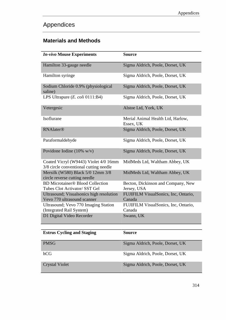
Appendices
314
Appendices
Materials and Methods
In-vivo Mouse Experiments Source
Hamilton 33-gauge needle Sigma Aldrich, Poole, Dorset, UK
Hamilton syringe Sigma Aldrich, Poole, Dorset, UK
Sodium Chloride 0.9% (physiological
saline)
Sigma Aldrich, Poole, Dorset, UK
LPS Ultrapure (E. coli 0111:B4) Sigma Aldrich, Poole, Dorset, UK
Vetergesic Alstoe Ltd, York, UK
Isoflurane Merial Animal Health Ltd, Harlow,
Essex, UK
RNAlater® Sigma Aldrich, Poole, Dorset, UK
Paraformaldehyde Sigma Aldrich, Poole, Dorset, UK
Povidone Iodine (10% w/v) Sigma Aldrich, Poole, Dorset, UK
Coated Vicryl (W9443) Violet 4/0 16mm
3/8 circle conventional cutting needle
MidMeds Ltd, Waltham Abbey, UK
Mersilk (W580) Black 5/0 12mm 3/8
circle reverse cutting needle
MidMeds Ltd, Waltham Abbey, UK
BD Microtainer® Blood Collection
Tubes Clot Activator/ SST Gel
Becton, Dickinson and Company, New
Jersey, USA
Ultrasound; Visualsonics high resolution
Vevo 770 ultrasound scanner
FUJIFILM VisualSonics, Inc, Ontario,
Canada
Ultrasound; Vevo 770 Imaging Station
(Integrated Rail System)
FUJIFILM VisualSonics, Inc, Ontario,
Canada
D1 Digital Video Recorder Swann, UK
Estrus Cycling and Staging Source
PMSG Sigma Aldrich, Poole, Dorset, UK
hCG Sigma Aldrich, Poole, Dorset, UK
Crystal Violet Sigma Aldrich, Poole, Dorset, UK
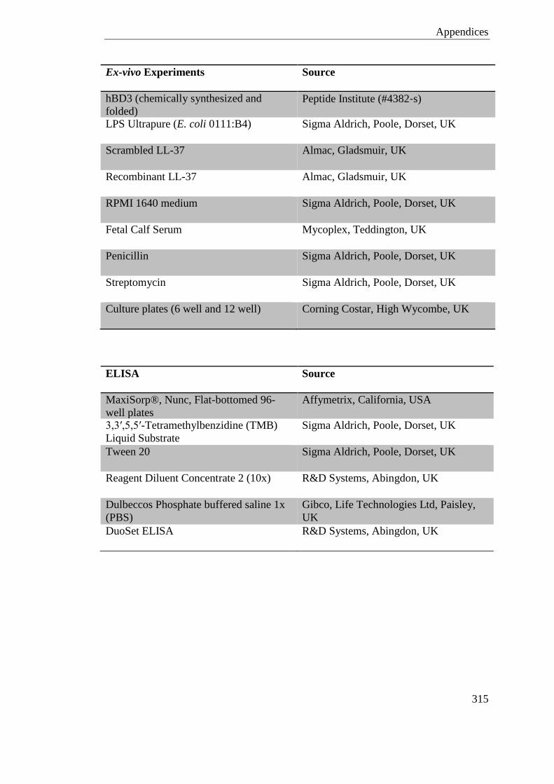
Appendices
315
Ex-vivo Experiments Source
hBD3 (chemically synthesized and
folded) Peptide Institute (#4382‐s)
LPS Ultrapure (E. coli 0111:B4) Sigma Aldrich, Poole, Dorset, UK
Scrambled LL-37 Almac, Gladsmuir, UK
Recombinant LL-37 Almac, Gladsmuir, UK
RPMI 1640 medium Sigma Aldrich, Poole, Dorset, UK
Fetal Calf Serum Mycoplex, Teddington, UK
Penicillin Sigma Aldrich, Poole, Dorset, UK
Streptomycin Sigma Aldrich, Poole, Dorset, UK
Culture plates (6 well and 12 well) Corning Costar, High Wycombe, UK
ELISA Source
MaxiSorp®, Nunc, Flat-bottomed 96-
well plates
Affymetrix, California, USA
3,3′,5,5′-Tetramethylbenzidine (TMB)
Liquid Substrate
Sigma Aldrich, Poole, Dorset, UK
Tween 20 Sigma Aldrich, Poole, Dorset, UK
Reagent Diluent Concentrate 2 (10x) R&D Systems, Abingdon, UK
Dulbeccos Phosphate buffered saline 1x
(PBS)
Gibco, Life Technologies Ltd, Paisley,
UK
DuoSet ELISA R&D Systems, Abingdon, UK
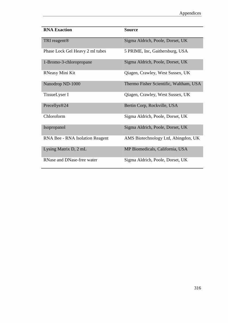
Appendices
316
RNA Exaction Source
TRI reagent® Sigma Aldrich, Poole, Dorset, UK
Phase Lock Gel Heavy 2 ml tubes 5 PRIME, Inc, Gaithersburg, USA
1-Bromo‐3‐chloropropane Sigma Aldrich, Poole, Dorset, UK
RNeasy Mini Kit Qiagen, Crawley, West Sussex, UK
Nanodrop ND‐1000 Thermo Fisher Scientific, Waltham, USA
TissueLyser I Qiagen, Crawley, West Sussex, UK
Precellys®24 Bertin Corp, Rockville, USA
Chloroform Sigma Aldrich, Poole, Dorset, UK
Isopropanol Sigma Aldrich, Poole, Dorset, UK
RNA Bee - RNA Isolation Reagent AMS Biotechnology Ltd, Abingdon, UK
Lysing Matrix D, 2 mL MP Biomedicals, California, USA
RNase and DNase-free water Sigma Aldrich, Poole, Dorset, UK
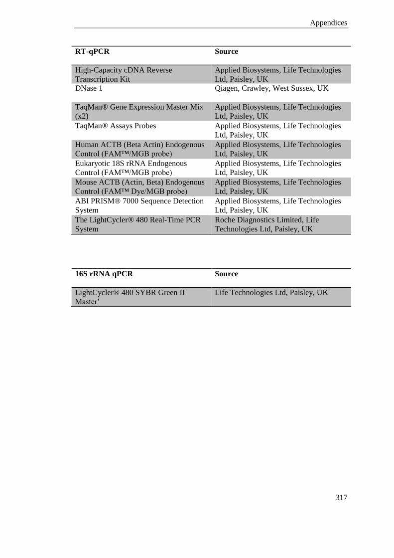
Appendices
317
RT-qPCR Source
High-Capacity cDNA Reverse
Transcription Kit
Applied Biosystems, Life Technologies
Ltd, Paisley, UK DNase 1 Qiagen, Crawley, West Sussex, UK
TaqMan® Gene Expression Master Mix
(x2)
Applied Biosystems, Life Technologies
Ltd, Paisley, UK
TaqMan® Assays Probes Applied Biosystems, Life Technologies
Ltd, Paisley, UK
Human ACTB (Beta Actin) Endogenous
Control (FAM™/MGB probe)
Applied Biosystems, Life Technologies
Ltd, Paisley, UK
Eukaryotic 18S rRNA Endogenous
Control (FAM™/MGB probe)
Applied Biosystems, Life Technologies
Ltd, Paisley, UK
Mouse ACTB (Actin, Beta) Endogenous
Control (FAM™ Dye/MGB probe)
Applied Biosystems, Life Technologies
Ltd, Paisley, UK
ABI PRISM® 7000 Sequence Detection
System
Applied Biosystems, Life Technologies
Ltd, Paisley, UK
The LightCycler® 480 Real-Time PCR
System
Roche Diagnostics Limited, Life
Technologies Ltd, Paisley, UK
16S rRNA qPCR Source
LightCycler® 480 SYBR Green II
Master’
Life Technologies Ltd, Paisley, UK
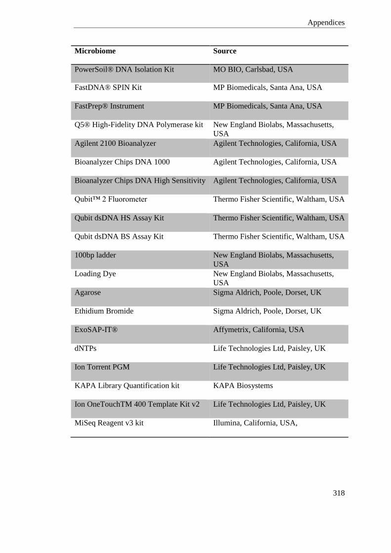
Appendices
318
Microbiome Source
PowerSoil® DNA Isolation Kit MO BIO, Carlsbad, USA
FastDNA® SPIN Kit MP Biomedicals, Santa Ana, USA
FastPrep® Instrument MP Biomedicals, Santa Ana, USA
Q5® High-Fidelity DNA Polymerase kit New England Biolabs, Massachusetts,
USA
Agilent 2100 Bioanalyzer Agilent Technologies, California, USA
Bioanalyzer Chips DNA 1000 Agilent Technologies, California, USA
Bioanalyzer Chips DNA High Sensitivity Agilent Technologies, California, USA
Qubit™ 2 Fluorometer Thermo Fisher Scientific, Waltham, USA
Qubit dsDNA HS Assay Kit Thermo Fisher Scientific, Waltham, USA
Qubit dsDNA BS Assay Kit Thermo Fisher Scientific, Waltham, USA
100bp ladder New England Biolabs, Massachusetts,
USA
Loading Dye New England Biolabs, Massachusetts,
USA
Agarose Sigma Aldrich, Poole, Dorset, UK
Ethidium Bromide Sigma Aldrich, Poole, Dorset, UK
ExoSAP-IT® Affymetrix, California, USA
dNTPs Life Technologies Ltd, Paisley, UK
Ion Torrent PGM Life Technologies Ltd, Paisley, UK
KAPA Library Quantification kit KAPA Biosystems
Ion OneTouchTM 400 Template Kit v2 Life Technologies Ltd, Paisley, UK
MiSeq Reagent v3 kit Illumina, California, USA,
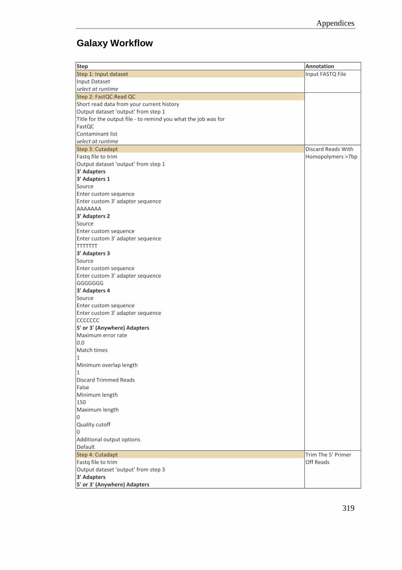
Appendices
319
Galaxy Workflow
Step Annotation
Step 1: Input dataset Input Dataset select at runtime
Input FASTQ File
Step 2: FastQC:Read QC Short read data from your current history Output dataset 'output' from step 1 Title for the output file - to remind you what the job was for FastQC Contaminant list select at runtime
Step 3: Cutadapt Fastq file to trim Output dataset 'output' from step 1 3' Adapters 3' Adapters 1 Source Enter custom sequence Enter custom 3' adapter sequence AAAAAAA 3' Adapters 2 Source Enter custom sequence Enter custom 3' adapter sequence TTTTTTT 3' Adapters 3 Source Enter custom sequence Enter custom 3' adapter sequence GGGGGGG 3' Adapters 4 Source Enter custom sequence Enter custom 3' adapter sequence CCCCCCC 5' or 3' (Anywhere) Adapters Maximum error rate 0.0 Match times 1 Minimum overlap length 1 Discard Trimmed Reads False Minimum length 150 Maximum length 0 Quality cutoff 0 Additional output options Default
Discard Reads With Homopolymers >7bp
Step 4: Cutadapt Fastq file to trim Output dataset 'output' from step 3 3' Adapters 5' or 3' (Anywhere) Adapters
Trim The 5' Primer Off Reads
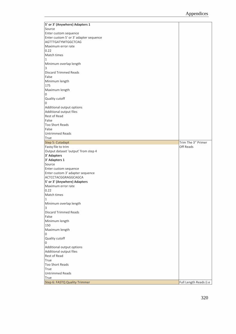
Appendices
320
5' or 3' (Anywhere) Adapters 1 Source Enter custom sequence Enter custom 5' or 3' adapter sequence AGTTTGATYMTGGCTCAG Maximum error rate 0.22 Match times 1 Minimum overlap length 3 Discard Trimmed Reads False Minimum length 175 Maximum length 0 Quality cutoff 0 Additional output options Additional output files Rest of Read False Too Short Reads False Untrimmed Reads True
Step 5: Cutadapt Fastq file to trim Output dataset 'output' from step 4 3' Adapters 3' Adapters 1 Source Enter custom sequence Enter custom 3' adapter sequence ACTCCTACGGRAGGCAGCA 5' or 3' (Anywhere) Adapters Maximum error rate 0.22 Match times 1 Minimum overlap length 3 Discard Trimmed Reads False Minimum length 150 Maximum length 0 Quality cutoff 0 Additional output options Additional output files Rest of Read True Too Short Reads True Untrimmed Reads True
Trim The 3'' Primer Off Reads
Step 6: FASTQ Quality Trimmer Full Length Reads (i.e
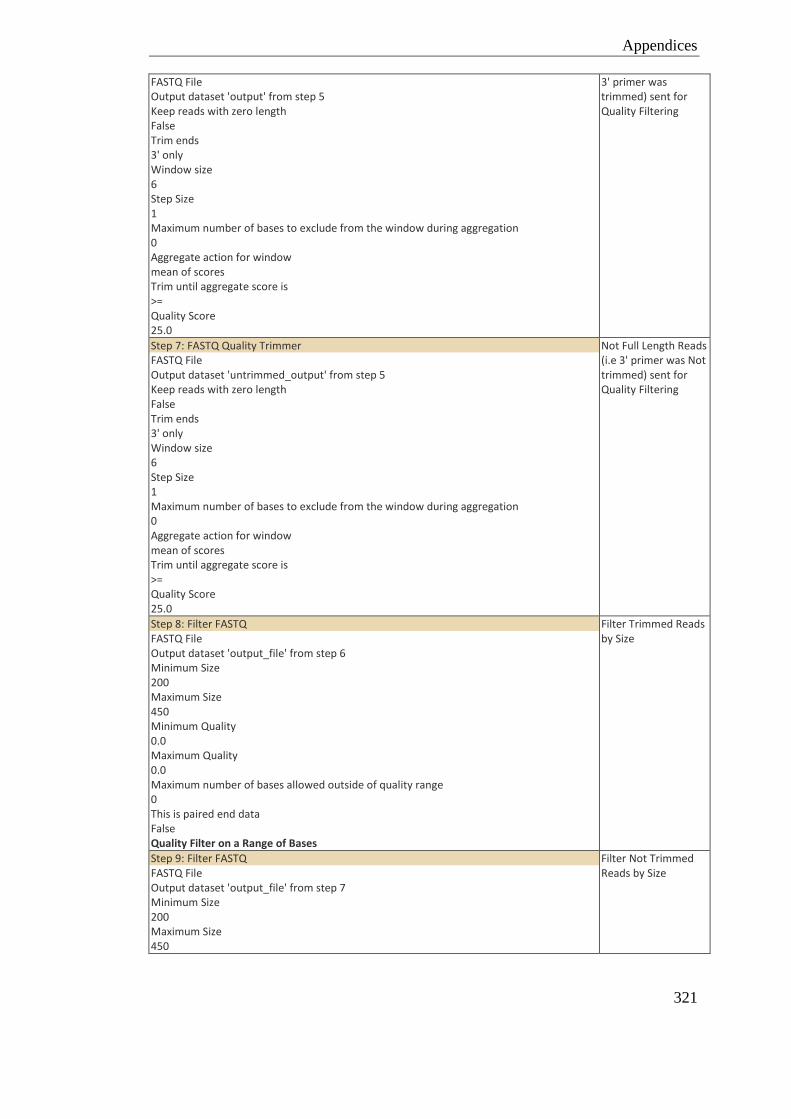
Appendices
321
FASTQ File Output dataset 'output' from step 5 Keep reads with zero length False Trim ends 3' only Window size 6 Step Size 1 Maximum number of bases to exclude from the window during aggregation 0 Aggregate action for window mean of scores Trim until aggregate score is >= Quality Score 25.0
3' primer was trimmed) sent for Quality Filtering
Step 7: FASTQ Quality Trimmer FASTQ File Output dataset 'untrimmed_output' from step 5 Keep reads with zero length False Trim ends 3' only Window size 6 Step Size 1 Maximum number of bases to exclude from the window during aggregation 0 Aggregate action for window mean of scores Trim until aggregate score is >= Quality Score 25.0
Not Full Length Reads (i.e 3' primer was Not trimmed) sent for Quality Filtering
Step 8: Filter FASTQ FASTQ File Output dataset 'output_file' from step 6 Minimum Size 200 Maximum Size 450 Minimum Quality 0.0 Maximum Quality 0.0 Maximum number of bases allowed outside of quality range 0 This is paired end data False Quality Filter on a Range of Bases
Filter Trimmed Reads by Size
Step 9: Filter FASTQ FASTQ File Output dataset 'output_file' from step 7 Minimum Size 200 Maximum Size 450
Filter Not Trimmed Reads by Size
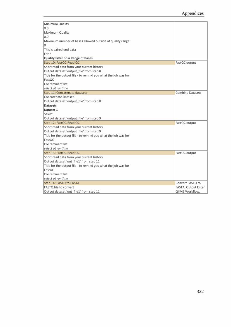
Appendices
322
Minimum Quality 0.0 Maximum Quality 0.0 Maximum number of bases allowed outside of quality range 0 This is paired end data False Quality Filter on a Range of Bases
Step 10: FastQC:Read QC Short read data from your current history Output dataset 'output_file' from step 8 Title for the output file - to remind you what the job was for FastQC Contaminant list select at runtime
FastQC output
Step 11: Concatenate datasets Concatenate Dataset Output dataset 'output_file' from step 8 Datasets Dataset 1 Select Output dataset 'output_file' from step 9
Combine Datasets
Step 12: FastQC:Read QC Short read data from your current history Output dataset 'output_file' from step 9 Title for the output file - to remind you what the job was for FastQC Contaminant list select at runtime
FastQC output
Step 13: FastQC:Read QC Short read data from your current history Output dataset 'out_file1' from step 11 Title for the output file - to remind you what the job was for FastQC Contaminant list select at runtime
FastQC output
Step 14: FASTQ to FASTA FASTQ file to convert Output dataset 'out_file1' from step 11
Convert FASTQ to FASTA. Output Enter QIIME Workflow.
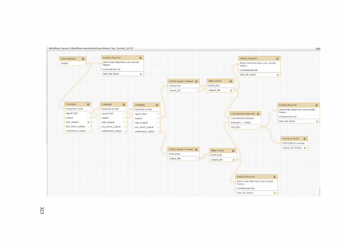
323
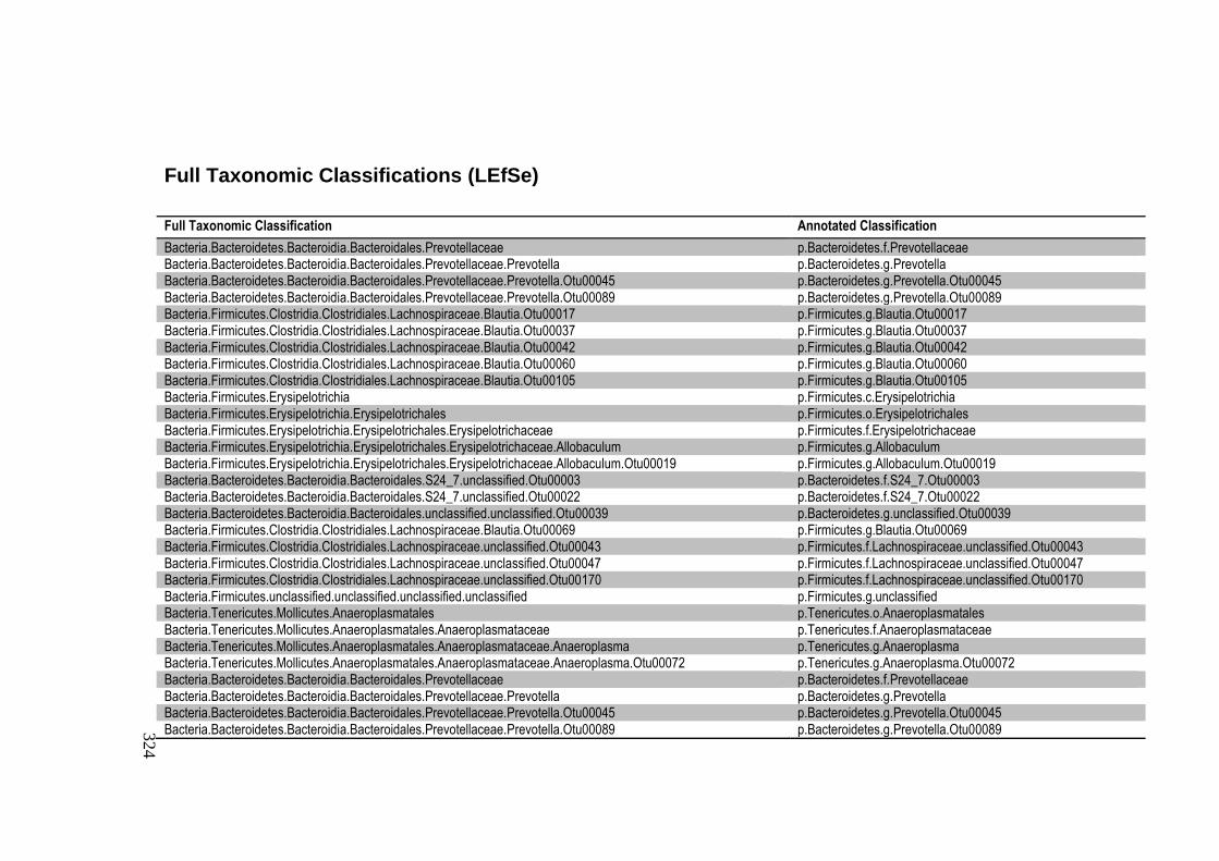
324
Full Taxonomic Classifications (LEfSe)
Full Taxonomic Classification Annotated Classification
Bacteria.Bacteroidetes.Bacteroidia.Bacteroidales.Prevotellaceae p.Bacteroidetes.f.Prevotellaceae Bacteria.Bacteroidetes.Bacteroidia.Bacteroidales.Prevotellaceae.Prevotella p.Bacteroidetes.g.Prevotella Bacteria.Bacteroidetes.Bacteroidia.Bacteroidales.Prevotellaceae.Prevotella.Otu00045 p.Bacteroidetes.g.Prevotella.Otu00045 Bacteria.Bacteroidetes.Bacteroidia.Bacteroidales.Prevotellaceae.Prevotella.Otu00089 p.Bacteroidetes.g.Prevotella.Otu00089 Bacteria.Firmicutes.Clostridia.Clostridiales.Lachnospiraceae.Blautia.Otu00017 p.Firmicutes.g.Blautia.Otu00017 Bacteria.Firmicutes.Clostridia.Clostridiales.Lachnospiraceae.Blautia.Otu00037 p.Firmicutes.g.Blautia.Otu00037 Bacteria.Firmicutes.Clostridia.Clostridiales.Lachnospiraceae.Blautia.Otu00042 p.Firmicutes.g.Blautia.Otu00042 Bacteria.Firmicutes.Clostridia.Clostridiales.Lachnospiraceae.Blautia.Otu00060 p.Firmicutes.g.Blautia.Otu00060 Bacteria.Firmicutes.Clostridia.Clostridiales.Lachnospiraceae.Blautia.Otu00105 p.Firmicutes.g.Blautia.Otu00105 Bacteria.Firmicutes.Erysipelotrichia p.Firmicutes.c.Erysipelotrichia Bacteria.Firmicutes.Erysipelotrichia.Erysipelotrichales p.Firmicutes.o.Erysipelotrichales Bacteria.Firmicutes.Erysipelotrichia.Erysipelotrichales.Erysipelotrichaceae p.Firmicutes.f.Erysipelotrichaceae Bacteria.Firmicutes.Erysipelotrichia.Erysipelotrichales.Erysipelotrichaceae.Allobaculum p.Firmicutes.g.Allobaculum Bacteria.Firmicutes.Erysipelotrichia.Erysipelotrichales.Erysipelotrichaceae.Allobaculum.Otu00019 p.Firmicutes.g.Allobaculum.Otu00019 Bacteria.Bacteroidetes.Bacteroidia.Bacteroidales.S24_7.unclassified.Otu00003 p.Bacteroidetes.f.S24_7.Otu00003 Bacteria.Bacteroidetes.Bacteroidia.Bacteroidales.S24_7.unclassified.Otu00022 p.Bacteroidetes.f.S24_7.Otu00022 Bacteria.Bacteroidetes.Bacteroidia.Bacteroidales.unclassified.unclassified.Otu00039 p.Bacteroidetes.g.unclassified.Otu00039 Bacteria.Firmicutes.Clostridia.Clostridiales.Lachnospiraceae.Blautia.Otu00069 p.Firmicutes.g.Blautia.Otu00069 Bacteria.Firmicutes.Clostridia.Clostridiales.Lachnospiraceae.unclassified.Otu00043 p.Firmicutes.f.Lachnospiraceae.unclassified.Otu00043 Bacteria.Firmicutes.Clostridia.Clostridiales.Lachnospiraceae.unclassified.Otu00047 p.Firmicutes.f.Lachnospiraceae.unclassified.Otu00047 Bacteria.Firmicutes.Clostridia.Clostridiales.Lachnospiraceae.unclassified.Otu00170 p.Firmicutes.f.Lachnospiraceae.unclassified.Otu00170 Bacteria.Firmicutes.unclassified.unclassified.unclassified.unclassified p.Firmicutes.g.unclassified Bacteria.Tenericutes.Mollicutes.Anaeroplasmatales p.Tenericutes.o.Anaeroplasmatales Bacteria.Tenericutes.Mollicutes.Anaeroplasmatales.Anaeroplasmataceae p.Tenericutes.f.Anaeroplasmataceae Bacteria.Tenericutes.Mollicutes.Anaeroplasmatales.Anaeroplasmataceae.Anaeroplasma p.Tenericutes.g.Anaeroplasma Bacteria.Tenericutes.Mollicutes.Anaeroplasmatales.Anaeroplasmataceae.Anaeroplasma.Otu00072 p.Tenericutes.g.Anaeroplasma.Otu00072 Bacteria.Bacteroidetes.Bacteroidia.Bacteroidales.Prevotellaceae p.Bacteroidetes.f.Prevotellaceae Bacteria.Bacteroidetes.Bacteroidia.Bacteroidales.Prevotellaceae.Prevotella p.Bacteroidetes.g.Prevotella Bacteria.Bacteroidetes.Bacteroidia.Bacteroidales.Prevotellaceae.Prevotella.Otu00045 p.Bacteroidetes.g.Prevotella.Otu00045 Bacteria.Bacteroidetes.Bacteroidia.Bacteroidales.Prevotellaceae.Prevotella.Otu00089 p.Bacteroidetes.g.Prevotella.Otu00089

325


![Home [arena-birmingham.academy] … · 2/1/2019 · Our younger students too have sought academic excellence this half term. ... pregnancy and social influences. ... professionals](https://img.pdfslide.net/doc/110x75/601c3251d23c4754432e828d/home-arena-212019-our-younger-students-too-have-sought-academic-excellence.jpg)