Embed Size (px)
Citation preview
HOW BIG IS STREPTOCOCCUS IN YOGURT?
BY: ALEJANDRO ARIEL GARCIA ARRIAGA, COACALCO DE BERRIOZABAL, ESTADO DE MEXICO, MEXICO
INTRODUCTION:
I have been working for a certain time on measuring microscopic samples, four articles about this topic were accepted in Micscape last year :
MEASUREMENTS OF THE MICRO WORLD PART 1: CALIBRATING THE CAMERA
MEASURING WITH A LASER BEAM
MEASURING ACCURATELY WITH REFERENCES.
CALIBRATING A DEDICATED MICROSCOPE CAMERA WITH REFERENCE TO THE LAST LEARNING STEP BEFORE STARTING TO MEASURE THE MICRO WORLD.
These articles show how to calibrate and use a dedicated camera to measure accurately both with a special micro ruler and with references.
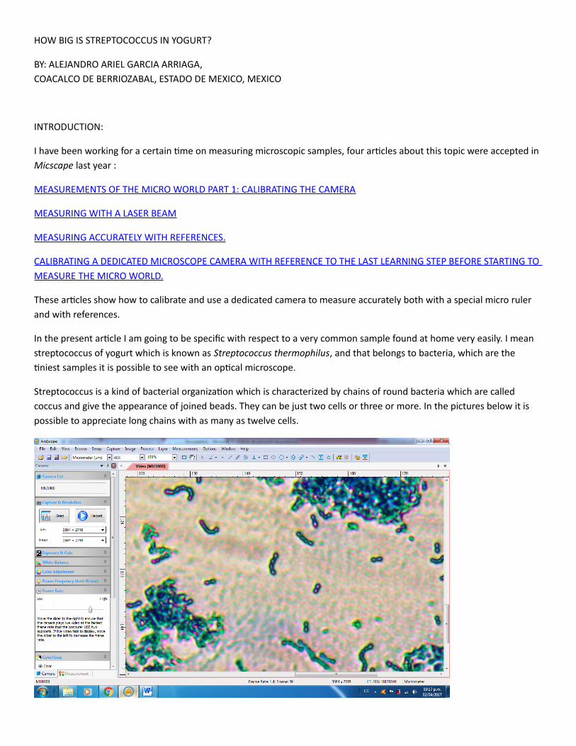
In the present article I am going to be specific with respect to a very common sample found at home very easily. I mean streptococcus of yogurt which is known as Streptococcus thermophilus, and that belongs to bacteria, which are the tiniest samples it is possible to see with an optical microscope.
Streptococcus is a kind of bacterial organization which is characterized by chains of round bacteria which are called coccus and give the appearance of joined beads. They can be just two cells or three or more. In the pictures below it is possible to appreciate long chains with as many as twelve cells.
DEVELOPMENT:
I already had a sample stained for several months with simple staining using methylene blue. This was a sample of a dropof natural yogurt, this is a permanent sample I made to study streptococcus.
NOTE: If the reader does not have a sample, just get some yogurt. Some methylene blue is very easy to find at the local pet shop and prepare a sample, it just takes a few minutes.
For more information about staining a sample read my earlier Micscape article:
SIMPLE POSITIVE STAIN WITH THREE EASY TO FIND DYES - GENTIAN VIOLET, METHYLENE BLUE, DENTAL PLAQUE REVEALER (ERYTHROSINE).
So I decided to measure the streptococcus in this sample.
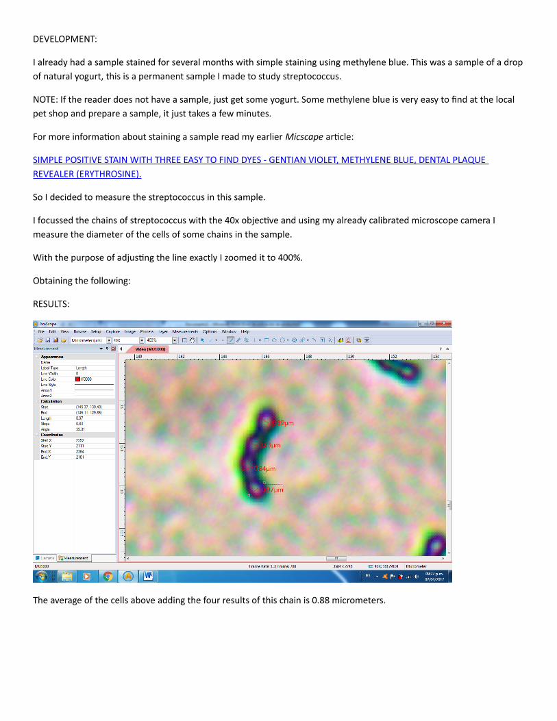
I focussed the chains of streptococcus with the 40x objective and using my already calibrated microscope camera I measure the diameter of the cells of some chains in the sample.
With the purpose of adjusting the line exactly I zoomed it to 400%.
Obtaining the following:
RESULTS:
The average of the cells above adding the four results of this chain is 0.88 micrometers.

The average of the cells of the chain in this picture is 0.82 micrometers.
Now remeasuring with references:
In the article MEASURING ACCURATELY WITH REFERENCES. I presented a table in which I showed the size of a millimeter on the screen of my computer and with the software of my camera, the reader can do the same for his or her own,
These values were with the different microscope objectives and at different zoom settings with the camera.
So from this table it is seen that a millimeter at 40x and 100% zoom is 390 cm so if we follow this statement at 400% zoom as in the picture it would be 1560 cm.
And if we measure with a common rule on the screen of the computer we have the following results:
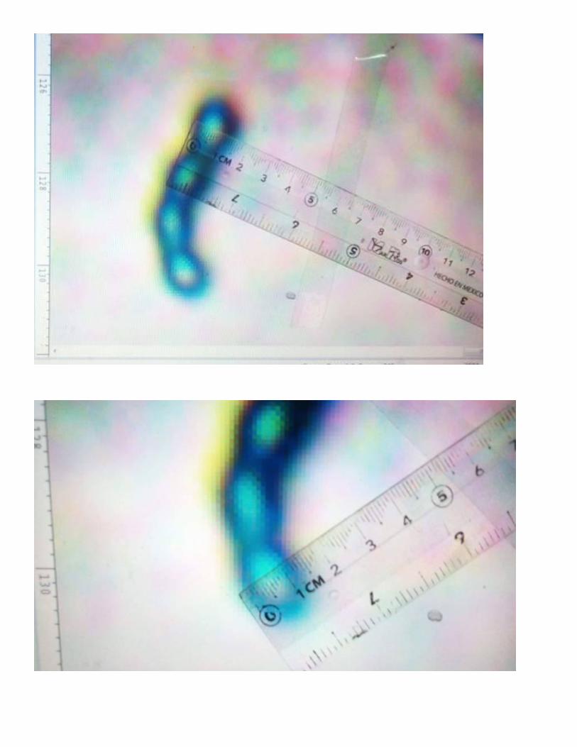
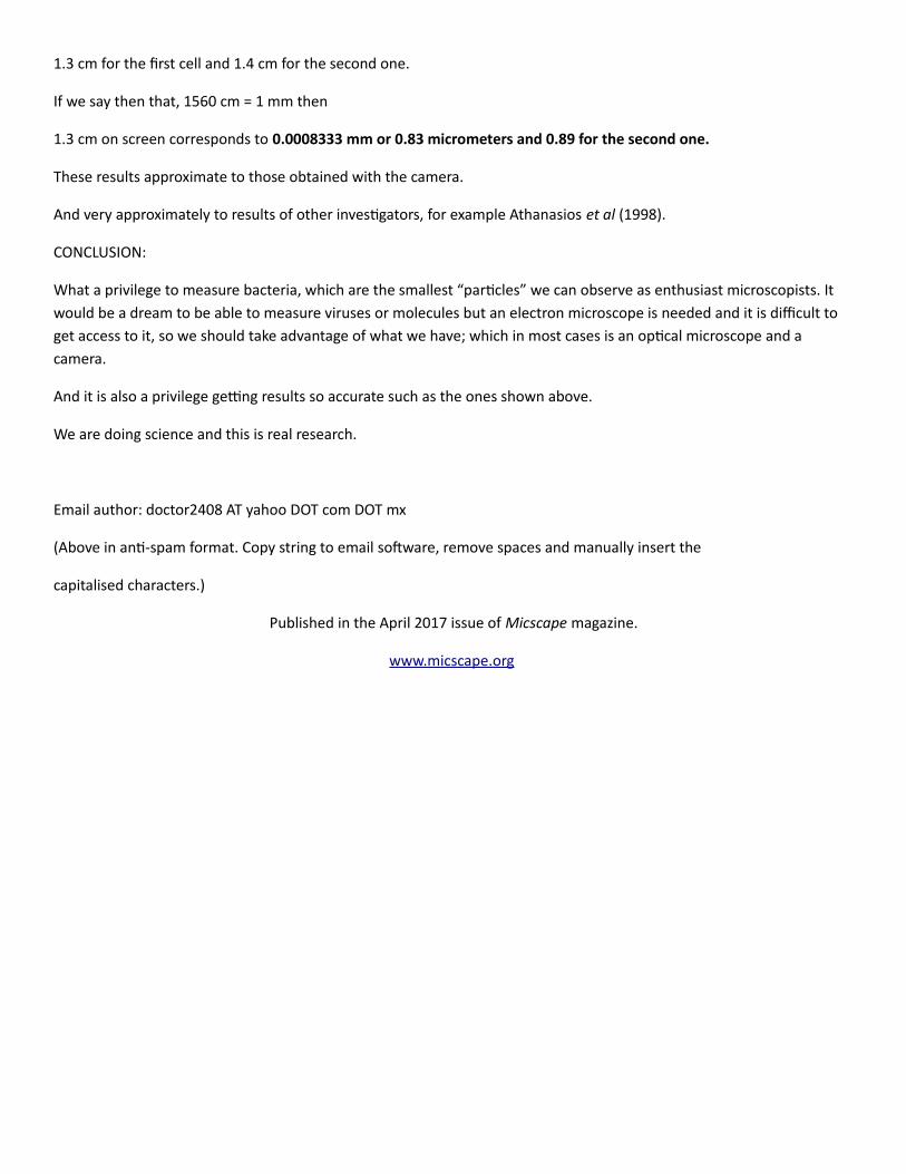
1.3 cm for the first cell and 1.4 cm for the second one.
If we say then that, 1560 cm = 1 mm then
1.3 cm on screen corresponds to 0.0008333 mm or 0.83 micrometers and 0.89 for the second one.
These results approximate to those obtained with the camera.
And very approximately to results of other investigators, for example Athanasios et al (1998).
CONCLUSION:
What a privilege to measure bacteria, which are the smallest “particles” we can observe as enthusiast microscopists. It would be a dream to be able to measure viruses or molecules but an electron microscope is needed and it is difficult to get access to it, so we should take advantage of what we have; which in most cases is an optical microscope and a camera.
And it is also a privilege getting results so accurate such as the ones shown above.
We are doing science and this is real research.
Email author: doctor2408 AT yahoo DOT com DOT mx
(Above in anti-spam format. Copy string to email software, remove spaces and manually insert the
capitalised characters.)
Published in the April 2017 issue of Micscape magazine.
www.micscape.org


![Cronicon · foods such as yogurt contains live beneficial bacteria of Lactobacillus bulgaricus and Streptococcus thermophilus [18]. In dietary supple-ments probiotics are available](https://img.pdfslide.net/doc/110x75/5fa69a779751cf5d8e373fc1/cronicon-foods-such-as-yogurt-contains-live-beneficial-bacteria-of-lactobacillus.jpg)