Embed Size (px)
Citation preview

How do cells maintain structure, connections &
organize activities?

Proteins!
• Ultimately responsible for each of these activities.
• Proteins provide structure, allow movement & mediate interactions

Some plasma membrane proteins
Transporta) Passiveb) Active
Enzymes
Initiating enzyme cascades
Intercellular junctions
Cell-cell recognition
Attach to cytoskeleton;Motor proteins

Tight junctions staple neighboring cells exposed to chemical stress
Gap junctions allow rapid communication & sharing between neighbors
Desmosomes bind neighboring cells exposed to mechanical stress

Extracellular environment • Space between cells• Extracellular matrix: sticky fluid
derived from plasma– nutrients for cells - glycoprotein, salts,
amino acids, etc.
– Cellular wastes – CO2, lactic acid, etc.
• Cells must exchange nutrients & wastes with the environment.

Cell membranes areselectively permeable
• Some compounds pass uninhibited through membrane (passive diffusion), some require assistance from membrane proteins (facilitated diffusion), and some require assistance AND energy expenditure (active transport)
1. Diffusion– Passive diffusion– Carrier or channel-mediated (facilitated) diffusion
2. Active Transport– Pumps, bulk transport
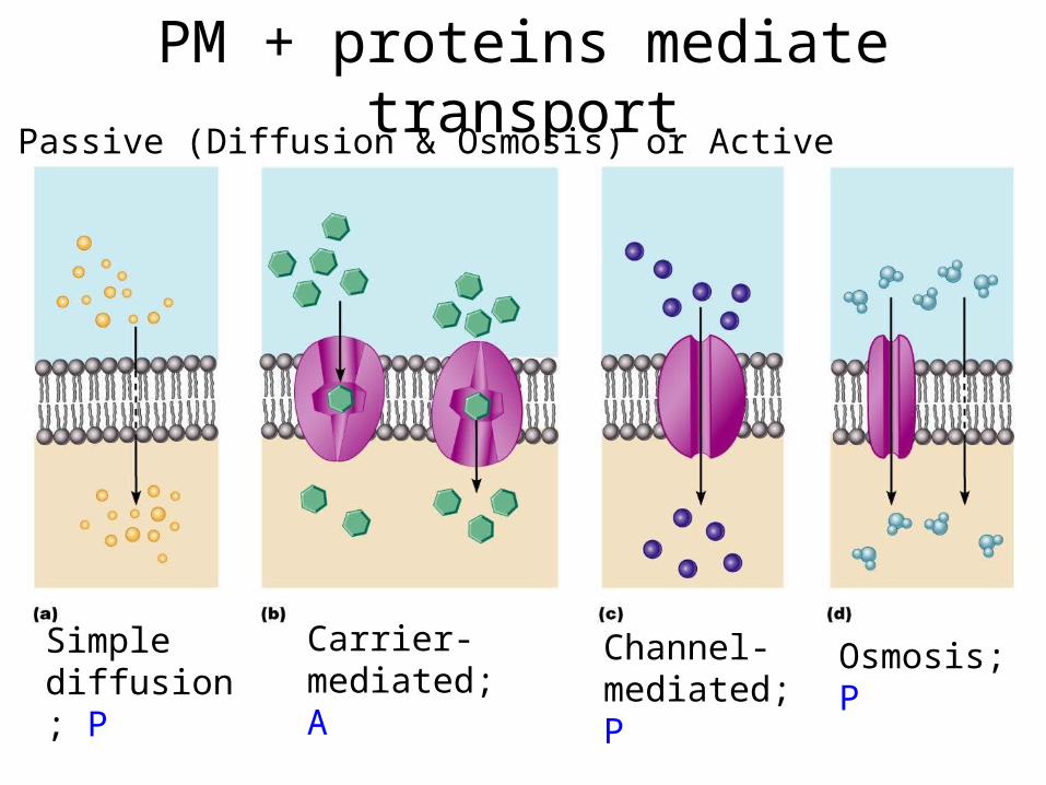
PM + proteins mediate transport
Passive (Diffusion & Osmosis) or Active
Simple diffusion; P
Osmosis; P
Channel-mediated; P
Carrier- mediated; A

What determines whether transport is passive or
active?
What determines rate of transport?

First, terminology• Solvent: The predominant liquid or
gas in a solution• Solute: The stuff that is dissolved in a
solution• Diffusion: The net movement of
solute from a higher to a lower concentration (Concentration gradient), until equilibrium is achieved. Uses intrinsic Kinetic Energy (KE).

Passive diffusion
• Kinetic energy causes particles to move
• Diffusion occurs due to random collisions between these energized particles

Osmosis + Diffusion• Both are happening all the time
across cell membranes
• Osmosis (H20) occurs RAPIDLY, diffusion (solutes) occurs SLOWLY
• H20 moves into cells with high solute concentration and out of cells with low solute concentration

Cytoskeleton• Cytoskeleton = cell
skeleton• All cells contain
structural filaments:– Microfilaments– Intermediate
filaments– Microtubules– Thick filaments
(muscle cells)
• Made of proteins

Microfilaments• Actin strands• Primarily in periphery of
cell• Functions:
– Anchor cytoskeleton to integral proteins of cell membrane
– Interact with myosin to promote cell shortening (Ex: muscle cells)

Microvilli• Microfilaments
(actin)• Increase SA of cell
– maximizes absorptive surface (Ex: intestinal walls)
• No movement

Intermediate filaments• (7-11nm)• Most durable
cytoskeletal fiber• Located throughout
cell; High # in superficial layers of skin
• Functions– Provides shape to cell– Stabilize (encase)
organelles

Thick filaments
• Only found in muscle cells, interact with actin to form a contraction

Microtubules• tubulin protein subunits;
ALL cells contain these
• Microtubular array centered near the nucleus (@ centrosome)
• Functions– Cell shape & rigidity– Anchor organelles; RR tracks
for organelle movement– Forms spindle apparatus– Forms centrioles, basal
bodies, parts of flagella

Centrioles & Basal bodies• Centrioles
– Form anchors of spindle apparatus
– Anchor is independent of spindle apparatus
• Basal bodies– Anchors flagella & cilia
to a cell– Anchor is an extension
of flagella & cilia
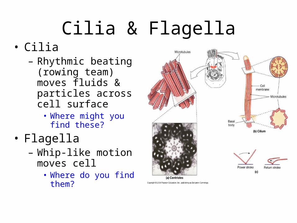
Cilia & Flagella• Cilia
– Rhythmic beating (rowing team) moves fluids & particles across cell surface
• Where might you find these?
• Flagella– Whip-like motion
moves cell• Where do you find
them?

Movement
• Dynein arms (red) anchored to microtubule
• Grab adjacent microtubule and “walk” along
• Produces bending• Show “flagella & cilia”