Embed Size (px)
Citation preview
How I treat
How I treat Waldenstrom macroglobulinemiaSteven P. Treon1
1Bing Center for Waldenstrom’s Macroglobulinemia, Dana-Farber Cancer Institute, Harvard Medical School, Boston, MA
Waldenstrom macroglobulinemia (WM) isa distinct B-cell disorder resulting fromthe accumulation, predominantly in thebone marrow, of clonally related IgM-secreting lymphoplasmacytic cells. Ge-netic factors play an important role, with20% of patients demonstrating a familialpredisposition. Asymptomatic patientsshould be observed. Patients with adisease-related hemoglobin level lessthan 10 g/L, platelet count less than100 � 109/L, bulky adenopathy or organo-
megaly, symptomatic hyperviscosity, pe-ripheral neuropathy, amyloidosis, cryo-globulinemia, cold-agglutinin disease, orevidence of disease transformationshould be considered for therapy. Plasma-pheresisshouldbeconsidered forsymptom-atic hyperviscosity and for prophylaxis inpatients in whom rituximab therapy is con-templated. The use of rituximab as mono-therapy or in combination with cyclophos-phamide, nucleoside analog, bortezomib, orthalidomide-based regimens can be consid-
ered for the first-line therapy of WM andshould take into account specific treatmentgoals, future autologous stem cell transplan-tation eligibility, and long-term risks of sec-ondary malignancies. In the salvage setting,the reuse or use of an alternative frontlineregimen can be considered as well as bor-tezomib, alemtuzumab, and stem cell trans-plantation. Newer agents, such as benda-mustine and everolimus, can also beconsidered in the treatment of WM. (Blood.2009;114:2375-2385)
Introduction
Waldenstrom macroglobulinemia (WM) is a distinct B-celldisorder resulting from the accumulation, predominantly in thebone marrow, of clonally related lymphoplasmacytic cells,which secrete a monoclonal IgM protein.1 This condition isconsidered to correspond to the lymphoplasmacytic lymphoma(LPL) as defined by the Revised European American Lymphomaand World Health Organization classification systems.2,3 Mostcases of LPL are WM, with less than 5% of cases made up ofIgA, IgG, and nonsecreting LPL.
Clinical features
The clinical and laboratory findings for 356 newly diagnosedpatients who presented to our institution are depicted in Table 1.Unlike most indolent lymphomas, splenomegaly and lymphadenopa-thy are present in only a minority of patients (� 15%). Themorbidity associated with WM is typically mediated by tissueinfiltration by neoplastic cells, the physicochemical and immuno-logic properties of the monoclonal IgM, or both. As shown in Table2, the monoclonal IgM can produce clinical manifestations throughseveral distinct mechanisms, including an effect on serum viscos-ity, mediation of autoantibody activity, interactions with otherproteins, precipitation on cooling, and tissue deposition.4-6
Diagnostic workup
History taking
There is a strong familial predisposition in WM7-9; therefore, agood family history is important. Although the identification ofsuch familiarity does not at this time influence treatment decisions,it may spawn a discussion in families with multiple cases of WM or
related B-cell disorders to participate in familial studies aimed atidentifying genetic predispositions to WM, which are currentlyunder way at the Dana-Farber Cancer Institute and the NationalInstitutes of Health. Exposure to hepatitis C has been implicated insome, but not all, studies, and evaluation of risks/exposure isimportant, particularly among patients who have type II (mixed)cryoglobulinemia.10-12 A thorough review of systems is veryimportant in the workup of WM patients, given the vast array ofpresenting symptoms, and may well impact on treatment consider-ations. A review of systems checklist, which we use at ourinstitution, along with their implications for the care of WMpatients is presented in Table 3 and can be used in the workup ofWM patients.
Laboratory studies
To establish the diagnosis of WM, it is necessary to demonstratean IgM monoclonal protein, along with histologic evidence ofinfiltration of the bone marrow by lymphoplasmacytic cells.1
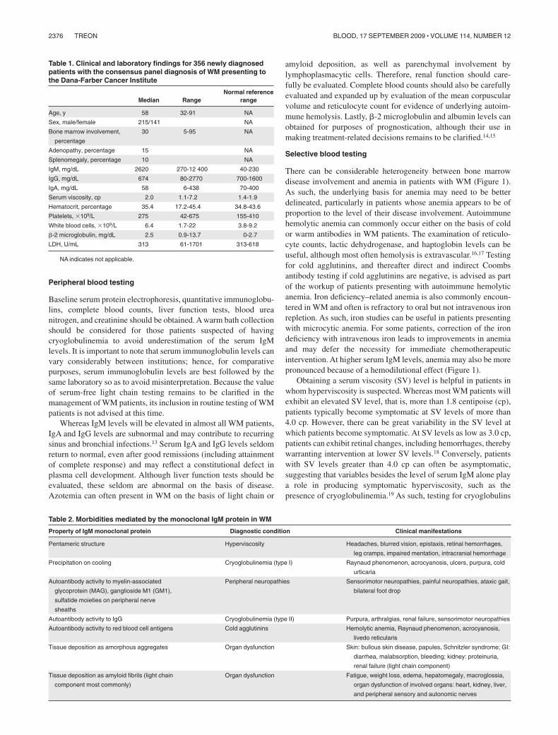
There is no minimal serum IgM level, nor a minimal percentageof bone marrow infiltration to establish the diagnosis of WMbecause patients can be symptomatic and in need of treatmenteven at low levels of IgM (� 1000 mg/dL) or bone marrowinvolvement. Indeed, there can be great heterogeneity amongpatients between their respective serum IgM levels and bonemarrow involvement (Figure 1). In general, for an individualpatient, serum IgM levels tend to show good correlation withdisease burden. There are some exceptions to this point, whichusually emerge in patients with cryoglobulinemia, as well asthose patients undergoing treatment with certain biologic agents(ie, rituximab, bortezomib). The assessment of response forpatients undergoing treatment with biologic agents is furtherdiscussed in “Response assessment.”
Submitted May 16, 2009; accepted July 6, 2009. Prepublished online as BloodFirst Edition paper, July 17, 2009; DOI 10.1182/blood-2009-05-174359.
© 2009 by The American Society of Hematology
2375BLOOD, 17 SEPTEMBER 2009 � VOLUME 114, NUMBER 12
Peripheral blood testing
Baseline serum protein electrophoresis, quantitative immunoglobu-lins, complete blood counts, liver function tests, blood ureanitrogen, and creatinine should be obtained. A warm bath collectionshould be considered for those patients suspected of havingcryoglobulinemia to avoid underestimation of the serum IgMlevels. It is important to note that serum immunoglobulin levels canvary considerably between institutions; hence, for comparativepurposes, serum immunoglobulin levels are best followed by thesame laboratory so as to avoid misinterpretation. Because the valueof serum-free light chain testing remains to be clarified in themanagement of WM patients, its inclusion in routine testing of WMpatients is not advised at this time.
Whereas IgM levels will be elevated in almost all WM patients,IgA and IgG levels are subnormal and may contribute to recurringsinus and bronchial infections.13 Serum IgA and IgG levels seldomreturn to normal, even after good remissions (including attainmentof complete response) and may reflect a constitutional defect inplasma cell development. Although liver function tests should beevaluated, these seldom are abnormal on the basis of disease.Azotemia can often present in WM on the basis of light chain or
amyloid deposition, as well as parenchymal involvement bylymphoplasmacytic cells. Therefore, renal function should care-fully be evaluated. Complete blood counts should also be carefullyevaluated and expanded up by evaluation of the mean corpuscularvolume and reticulocyte count for evidence of underlying autoim-mune hemolysis. Lastly, �-2 microglobulin and albumin levels canobtained for purposes of prognostication, although their use inmaking treatment-related decisions remains to be clarified.14,15
Selective blood testing
There can be considerable heterogeneity between bone marrowdisease involvement and anemia in patients with WM (Figure 1).As such, the underlying basis for anemia may need to be betterdelineated, particularly in patients whose anemia appears to be ofproportion to the level of their disease involvement. Autoimmunehemolytic anemia can commonly occur either on the basis of coldor warm antibodies in WM patients. The examination of reticulo-cyte counts, lactic dehydrogenase, and haptoglobin levels can beuseful, although most often hemolysis is extravascular.16,17 Testingfor cold agglutinins, and thereafter direct and indirect Coombsantibody testing if cold agglutinins are negative, is advised as partof the workup of patients presenting with autoimmune hemolyticanemia. Iron deficiency–related anemia is also commonly encoun-tered in WM and often is refractory to oral but not intravenous ironrepletion. As such, iron studies can be useful in patients presentingwith microcytic anemia. For some patients, correction of the irondeficiency with intravenous iron leads to improvements in anemiaand may defer the necessity for immediate chemotherapeuticintervention. At higher serum IgM levels, anemia may also be morepronounced because of a hemodilutional effect (Figure 1).
Obtaining a serum viscosity (SV) level is helpful in patients inwhom hyperviscosity is suspected. Whereas most WM patients willexhibit an elevated SV level, that is, more than 1.8 centipoise (cp),patients typically become symptomatic at SV levels of more than4.0 cp. However, there can be great variability in the SV level atwhich patients become symptomatic. At SV levels as low as 3.0 cp,patients can exhibit retinal changes, including hemorrhages, therebywarranting intervention at lower SV levels.18 Conversely, patientswith SV levels greater than 4.0 cp can often be asymptomatic,suggesting that variables besides the level of serum IgM alone playa role in producing symptomatic hyperviscosity, such as thepresence of cryoglobulinemia.19 As such, testing for cryoglobulins
Table 1. Clinical and laboratory findings for 356 newly diagnosedpatients with the consensus panel diagnosis of WM presenting tothe Dana-Farber Cancer Institute
Median RangeNormal reference
range
Age, y 58 32-91 NA
Sex, male/female 215/141 NA
Bone marrow involvement,
percentage
30 5-95 NA
Adenopathy, percentage 15 NA
Splenomegaly, percentage 10 NA
IgM, mg/dL 2620 270-12 400 40-230
IgG, mg/dL 674 80-2770 700-1600
IgA, mg/dL 58 6-438 70-400
Serum viscosity, cp 2.0 1.1-7.2 1.4-1.9
Hematocrit, percentage 35.4 17.2-45.4 34.8-43.6
Platelets, �109/L 275 42-675 155-410
White blood cells, �109/L 6.4 1.7-22 3.8-9.2
�-2 microglobulin, mg/dL 2.5 0.9-13.7 0-2.7
LDH, U/mL 313 61-1701 313-618
NA indicates not applicable.
Table 2. Morbidities mediated by the monoclonal IgM protein in WM
Property of IgM monoclonal protein Diagnostic condition Clinical manifestations
Pentameric structure Hyperviscosity Headaches, blurred vision, epistaxis, retinal hemorrhages,
leg cramps, impaired mentation, intracranial hemorrhage
Precipitation on cooling Cryoglobulinemia (type I) Raynaud phenomenon, acrocyanosis, ulcers, purpura, cold
urticaria
Autoantibody activity to myelin-associated
glycoprotein (MAG), ganglioside M1 (GM1),
sulfatide moieties on peripheral nerve
sheaths
Peripheral neuropathies Sensorimotor neuropathies, painful neuropathies, ataxic gait,
bilateral foot drop
Autoantibody activity to IgG Cryoglobulinemia (type II) Purpura, arthralgias, renal failure, sensorimotor neuropathies
Autoantibody activity to red blood cell antigens Cold agglutinins Hemolytic anemia, Raynaud phenomenon, acrocyanosis,
livedo reticularis
Tissue deposition as amorphous aggregates Organ dysfunction Skin: bullous skin disease, papules, Schnitzler syndrome; GI:
diarrhea, malabsorption, bleeding; kidney: proteinuria,
renal failure (light chain component)
Tissue deposition as amyloid fibrils (light chain
component most commonly)
Organ dysfunction Fatigue, weight loss, edema, hepatomegaly, macroglossia,
organ dysfunction of involved organs: heart, kidney, liver,
and peripheral sensory and autonomic nerves
2376 TREON BLOOD, 17 SEPTEMBER 2009 � VOLUME 114, NUMBER 12
should be considered in patients with symptomatic hyperviscositywho display relatively low serum IgM and SV levels.
Peripheral neuropathy is an important morbidity in patients withWM, with up to 20% to 25% of patients demonstrating disease-related peripheral neuropathy, which most often is sensory innature.20,21 In patients suspected of having IgM-related peripheralneuropathy, the evaluation of anti–myelin-associated glycoprotein,anti-ganglioside M1, and anti-sulfatide IgM antibodies is appropri-ate. Although the presence of one of these antibodies may supportthe diagnosis of IgM-related neuropathy, their absence should notexclude the diagnosis because other myelin-associated antigensmay be targeted that are not clinically evaluable at this time.Amyloidosis should also be considered in patients presenting witha peripheral neuropathy, and a fat pad biopsy with Congo redstaining obtained.22 Electromyography may be helpful and oftenshows a demyelinating neuropathy. A sural nerve biopsy should beavoided because of frequent neuropathic complications. In rarecircumstances where a myopathy may be suspected on the basis ofWM, the investigation for antidecorin IgM antibodies can beconsidered.23
Bone marrow evaluation
The bone marrow is almost always involved in WM; and as such, abone marrow biopsy and aspiration should be obtained. Central to
the diagnosis of WM is the demonstration of bone marrowinfiltration by a lymphoplasmacytic cell population manifested bysmall lymphocytes with evidence of plasmacytoid/plasma celldifferentiation.1 The pattern of bone marrow infiltration may bediffuse, interstitial, or nodular and is usually intertrabecular.A solely paratrabecular pattern of infiltration is unusual and shouldraise the possibility of follicular lymphoma.1 The bone marrowinfiltration should be supported by immunophenotypic studies(flow cytometry and/or immunohistochemistry) showing the follow-ing profile: sIgM�CD19�CD20�CD22�CD79�.24,25 Up to 20% ofcases may express CD5, CD10, or CD23.26 In such cases, careshould be taken to satisfactorily exclude chronic lymphocyticleukemia and mantle cell lymphoma.1 An increased number of mastcells, usually in association with the lymphoid aggregates, iscommonly found in WM, and their presence may help in differenti-ating WM from other B-cell lymphomas.2,3
Cytogenetic studies
Multiple studies have been published on cytogenetic findings inWM and have demonstrated a great variety of numerical andstructural chromosomal abnormalities. Chromosome 6q deletionsencompassing 6q21-25 have been observed in up to half of WMpatients, and at a comparable frequency among patients with andwithout a familial history.7,27,28 Several candidate tumor suppressor
Table 3. A review of systems and their implications in the workup of patients with WM
Symptom Implications Action
Energy level/changes in activities of
daily life
Anemia, fatigue without anemia Evaluate for anemia, underlying etiology, including iron
deficiency, hemolytic anemia (warm and cold antibodies);
consider amyloidosis; exclude other medical causes of
anemia
Constitutional symptoms Tumor-related fever, chills, night sweats
Recurrent sinus and bronchial infections Chronic sinusitis, usually on the basis of IgA and
IgG hypogammaglobulinemia
Antibiotic support, if refractory to multiple antibiotic courses,
hospitalizations, or life-threatening, strongly consider
intravenous immunoglobulin replacement
Headaches, blurry vision or visual loss,
confusional episodes, epistaxis
Hyperviscosity Funduscopic examination for hyperviscosity-related
changes, obtain serum IgM and viscosity levels; consider
emergency plasmapheresis for symptomatic
hyperviscosity; strongly consider in patients with serum
viscosity � 4.0 cp given high risk of hyperviscosity-
related events
Easy bruising, bleeding diathesis Thrombocytopenia, acquired von Willebrand
disorder
Complete blood count, evaluate for immune
thrombocytopenia or hypersplenism if indicated; consider
evaluation for von Willebrand disorder; consider
amyloidosis
Progressive symmetric numbness,
tingling, burning, pain in feet and
hands; unsteady gait, deficits in motor
function
IgM-related neuropathy or myopathy;
amyloidosis
Obtain anti-MAG, anti-GM1, anti-sulfatidyl IgM antibody
studies; if myopathy is present, consider obtaining
antidecorin antibodies; consider obtaining fat pad biopsy
and stain for amyloid; consider EMG studies
Raynaud-like symptoms, acrocyanosis,
ulcerations on extremities
Cryoglobulinemia, cold agglutinemia Obtain cryoglobulins, cold agglutinins, in patients suspected
of having cryoglobulins, all studies, including quantitative
immunoglobulins, should be obtained in a warm bath to
avoid cryoprecipitation and false lowering of serum IgM
levels
Diarrhea, gastrointestinal cramping Malabsorption, secondary to amyloidosis, IgM
deposition, tumor involvement; rarely,
autonomic neuropathy on basis of
autoantibody or amyloidosis
Endoscopy to evaluate small bowel, biopsy to evaluate for
amyloidosis, IgM deposition, tumor involvement
Hearing loss Hyperviscosity, sensorineural hearing loss,
amyloid or tumor deposition, thrombus
formation
Consider evaluation for anti-Hu and anti-hsp 70 antibodies,
MRI to assess for amyloidoma, tumor deposition;
evaluate for hyperviscosity syndrome (as earlier in Table
3); assess for IgM antiphospholipid antibodies
Thrombotic events Antiphospholipid antibody syndrome Assess for IgM antiphospholipid antibodies
Urticaria, papules, dermatitis Schnitzler syndrome (nonpruritic urticaria), IgM
or tumor cell infiltration, amyloid deposition
Skin biopsy, histologic examination for tumor cell infiltration,
stain for IgM, Congo red for amyloid
WALDENSTROM MACROGLOBULINEMIA 2377BLOOD, 17 SEPTEMBER 2009 � VOLUME 114, NUMBER 12
genes in this region are under study, including BLIMP-1.29 Despitean earlier study suggesting prognostic significance to 6q deletionsin WM, a more recent study did not confirm such significance.27,30
As such, routine cytogenetic testing is not advised at this time. Anexception, however, is the use of cytogenetics to clarify thediagnosis of WM from suspected cases of IgM myeloma. In thelatter, IgH switch region rearrangements (14q32 translocations) area predominant feature, whereas these are typically absent in WM.31
Imaging studies
CT scans of the chest, abdomen, and pelvis should be obtained attime of diagnosis to properly stage the patient.32 Up to 20% of WMpatients may have extramedullary disease, and CT scans offer anopportunity to assess for adenopathy, splenomegaly, and for otherextramedullary disease sites. Follow-up CT scans are necessaryonly for those patients with baseline extramedullary disease (orwho are later suspected of having extramedullary disease), and maybe used to assess disease progression, as well as response. There isno role for routine magnetic resonance imaging; as well, there isnot a routine role for positron emission tomography scanningunless disease transformation is suspected.
Ophthalmologic examination
Patients with WM often exhibit retinal changes resulting fromhyperviscosity-related changes that occur as a consequence ofelevated IgM levels. Retinal findings associated with hyperviscos-ity can include peripheral dot-and-blot-like hemorrhages, dilatedretinal veins, central hemorrhages, tortuous blood vessels, venous“sausaging,” and/or optic disc edema. In one study, retinal changeswere observed at serum IgM and viscosity levels as low as3000 mg/dL and 2.4 cp, respectively.18 Importantly, plasmaphere-sis can lead to prompt resolution of hyperviscosity-related retinal
changes.33 Examination of the retina may therefore be useful inidentifying the symptomatic threshold of serum viscosity levels inpatients with WM and may be used as an important gauge for theeffectiveness of both plasmapheresis and chemotherapy. In ourpractice, we typically recommend a baseline ophthalmologicexamination in those WM patients whose serum IgM levels aremore than 3000 mg/dL.
Treatment approaches to WM
Management of the asymptomatic or smoldering WM patient
Patients with a disease-related hemoglobin level less than 10 g/dL,platelet count less than 100 � 109/L, bulky adenopathy or organo-megaly, symptomatic hyperviscosity, moderate to severe or advanc-ing peripheral neuropathy on the basis of disease, symptomaticamyloidosis, cryoglobulinemia, or cold-agglutinin disease shouldbe considered for therapy.14 Initiation of therapy should not bebased on serum monoclonal protein levels per se, and asymptom-atic patients should be observed. Asymptomatic patients with a low�-2 microglobulin (� 3 g/dL) and a hemoglobin level of more than12 g/dL may have an indolent course and not require therapy for along period of time, even when their monoclonal protein exceeds3000 mg/dL. As such, the identification of the asymptomaticpatient is important, and close observation (ie, every few months)rather than therapy is appropriate for these patients.
Management of the symptomatic WM
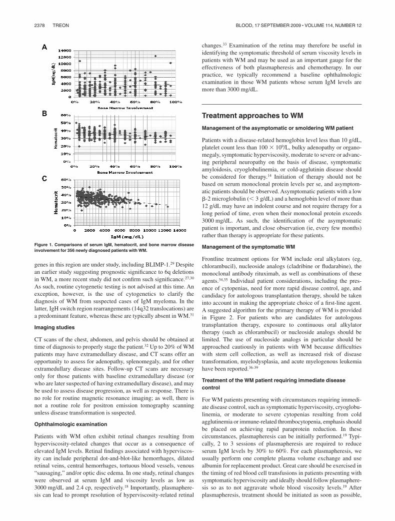
Frontline treatment options for WM include oral alkylators (eg,chlorambucil), nucleoside analogs (cladribine or fludarabine), themonoclonal antibody rituximab, as well as combinations of theseagents.34,35 Individual patient considerations, including the pres-ence of cytopenias, need for more rapid disease control, age, andcandidacy for autologous transplantation therapy, should be takeninto account in making the appropriate choice of a first-line agent.A suggested algorithm for the primary therapy of WM is providedin Figure 2. For patients who are candidates for autologoustransplantation therapy, exposure to continuous oral alkylatortherapy (such as chlorambucil) or nucleoside analogs should belimited. The use of nucleoside analogs in particular should beapproached cautiously in patients with WM because difficultieswith stem cell collection, as well as increased risk of diseasetransformation, myelodysplasia, and acute myelogenous leukemiahave been reported.36-39
Treatment of the WM patient requiring immediate diseasecontrol
For WM patients presenting with circumstances requiring immedi-ate disease control, such as symptomatic hyperviscosity, cryoglobu-linemia, or moderate to severe cytopenias resulting from coldagglutinemia or immune-related thrombocytopenia, emphasis shouldbe placed on achieving rapid paraprotein reduction. In thesecircumstances, plasmapheresis can be initially performed.19 Typi-cally, 2 to 3 sessions of plasmapheresis are required to reduceserum IgM levels by 30% to 60%. For each plasmapheresis, weusually perform one complete plasma volume exchange and usealbumin for replacement product. Great care should be exercised inthe timing of red blood cell transfusions in patients presenting withsymptomatic hyperviscosity and ideally should follow plasmaphere-sis so as to not aggravate whole blood viscosity levels.19 Afterplasmapheresis, treatment should be initiated as soon as possible,
Figure 1. Comparisons of serum IgM, hematocrit, and bone marrow diseaseinvolvement for 356 newly diagnosed patients with WM.
2378 TREON BLOOD, 17 SEPTEMBER 2009 � VOLUME 114, NUMBER 12

as IgM levels will steadily begin to rise and return to baseline levelsin 4 to 5 weeks.19 For patients requiring immediate disease control,the use of bortezomib-based therapy, such as bortezomib, dexameth-asone, and rituximab (BDR) is preferable so as to achieve morerapid disease control.40 The time to at least a minimum response inWM patients treated with BDR was 1.1 months in a Waldenstrom’sMacroglobulinemia Clinical Trials Group (WMCTG) study, whereasthe overall response rate with BDR was 96%, with 22% of patientsachieving a complete response. With a median follow-up of2 years, 80% of patients remained free of disease progression,including all patients achieving a very good partial response orbetter in this study. Herpes zoster prophylaxis should be institutedwith BDR therapy using an oral antiviral agent, such as acyclovir,famvir, or valcyclovir, and maintained throughout the course ofBDR, and thereafter for at least 6 months. A close watch for thedevelopment of bortezomib-related neuropathy should be main-tained on BDR. Treatment-related peripheral neuropathy, whichoccurred at a grade 3 level in 30% of patients, was reversible inmost patients, who benefited by interim support with pregabalin.A rituximab-related flare with BDR was observed in only 9% ofpatients, which may reflect the ability of bortezomib to suppressIgM production independent of tumor cell killing.41,42 As analternative to the twice-a-week schedule of bortezomib used withBDR, the use of once-a-week bortezomib at a higher dose (ie,1.6 mg/m2) may be considered with rituximab and appears in onestudy to be associated with lower risk of neuropathy.43,44 Diseasecontrol may lag, and the incidence of rituximab-related IgM flaremay be higher (20%) with once-a-week versus twice-a-weekadministration of bortezomib.
As an alternative to bortezomib-based therapy, a cyclophosphamide-based rituximab-containing regimen can be considered in patientsyounger than 70 years45-48; a nucleoside analog in combinationwith rituximab can also be considered in patients 70 years of age orolder or in younger patients where the use of bortezomib- orcyclophosphamide-based therapy may not be an option.49-51 In arecent update of the Southwest Oncology Group–directed Inter-group Trial S9003, the 10-year event-free survival to single-agentfludarabine was 20%.52 By multivariate analysis, patients withlower levels of �-2 microglobulin (� 3 mg/L) demonstrated signifi-
cantly better event-free survival in this series. It is unclear whetherthe inclusion of cyclophosphamide to a nucleoside analog–containing rituximab regimen (such as fludarabine, cyclosphospha-mide, rituximab) in WM extends response, and its addition maycontribute to additional toxicity.53 The overall response rates withcyclophosphamide or nucleoside analog-based rituximab-containingregimens is 70% to 80%, with complete response attainment inapproximately 10% of patients. Cytokine support following Ameri-can Society of Clinical Oncology guidelines should be consideredwith either cyclophosphamide or nucleoside analog-based therapy.
There has been considerable debate on the value of includingdoxorubicin and vincristine to cyclophosphamide-based therapy inWM. Dimopoulos et al46 reported that the combination of ritux-imab, cyclophosphamide, and dexamethasone (R-CD) led to over-all and complete responses in 78% and 7% of WM patients,respectively, and a 2-year progression-free survival of 80%, whichappear on par with those results achieved in a comparablepopulation of untreated WM who received cyclophosphamide,doxorubicin, vincristine, and prednisone with rituximab (CHOP-R).45,47 Ioakimidis et al48 compared the activity and toxicityassociated with CHOP-R, cyclophosphamide, vincristine, andprednisone with rituximab (CVP-R), and cyclophosphamide andprednisone with rituximab (CP-R) in patients with WM. The use ofCP-R was associated with analogous response rates to CVP-R andCHOP-R in the frontline treatment of WM, whereas treatment-related complications, including febrile neutropenia, hospitaliza-tions, and vincristine-related neuropathy, were less. As such, CP-Ror R-CD may be preferable to CVP-R or CHOP-R in themanagement of WM.
An important consideration in the treatment of WM patients,particularly those with high IgM levels, is the potential for arituximab-mediated IgM flare, which may lead to symptomatichyperviscosity, as well as worsening of IgM-related neuropathy,cryoglobulinemia, and other IgM-related complications.54-60 Theoccurrence of an IgM flare is quite common after rituximab therapyin WM patients, with a 40% to 50% occurrence rate whenrituximab is used as monotherapy.54,55 In combination therapy, the
Figure 2. Guide to the primary therapy of WM. HVindicates hyperviscosity; cp, centipoise; BDR, bor-tezomib, dexamethasone, rituximab; CPR, cyclophospha-mide, prednisone, rituximab; RCD, rituximab, cyclophos-phamide, dexamethasone; VR, bortezomib, rituximab;FR, fludarabine, rituximab; and R, rituximab. (1) Becauseof potential risk of stem cell damage and/or secondarymalignancies, may consider as an alternative option ifother treatment choices are either unavailable or inappro-priate for a particular patient. (2) Consider an attenuatedschedule for fludarabine administration in patients withmore indolent disease presentation. (3) Avoid as mono-therapy in patients with hyperviscosity and with Fc�RIIIA-158 F/F polymorphism. For rituximab-based therapies,consider maintenance rituximab in responding patients.Clinical trials should be considered for patients wheneverpossible.
WALDENSTROM MACROGLOBULINEMIA 2379BLOOD, 17 SEPTEMBER 2009 � VOLUME 114, NUMBER 12
occurrence of the rituximab-mediated IgM flare can vary consider-ably and appear dependent on both the regimen used as well as thesequencing of rituximab administration.40,43,48,61-63
Because of concern over a rituximab-related IgM flare aggravat-ing serum viscosity levels or IgM-related morbidity, the omissionof rituximab can also be considered for the first 1 or 2 cycles oftreatment. Serum IgM levels should be closely monitored (at leastweekly) during the time patients are receiving rituximab-basedtherapy. The IgM flare may last for several weeks, and evenmonths, and does not per se herald treatment failure.54,62 The use ofrituximab is best avoided as single-agent therapy in patients withhigh IgM levels because in 2 studies considerably lower responserates were observed in those patients with higher serum IgM levels(� 4000 mg/dL).64,65
Treatment of the WM patient requiring nonimmediate diseasecontrol
For those WM patients presenting with disease not requiringimmediate disease control, several options can be considered andshould ideally take into account specific disease-controlling objec-tives. The use of CP-R or R-CD may be considered and may beparticularly preferable in younger transplantation-eligible patients.The overall reported response rates with CP-R or R-CD are 70% to80%, and the 2-year progression-free survival rates with theseregimens is 70%.46,48 In patients older than 70 years, nucleosideanalog-based therapy may also be considered.49-51 In a recent studyby the WMCTG, the overall response rate to fludarabine withrituximab was 96%, and the median progression-free survival was51.2 months; in patients achieving a very good partial response, themedian progression-free survival in this series was in excess of88 months.50 Because of the significant and prolonged myelosup-pression observed in the WMCTG study, which used 6 cycles offludarabine, each with 5-day courses at 25 mg/m2, a more con-densed course (ie, 4 days of fludarabine at 25 mg/m2 per cycle for4 cycles) may be preferable in more indolent patients.50 The use ofthalidomide in combination with rituximab (TR) also represents analternative choice in the management of WM patients not requiringimmediate disease control, and is associated with an overallresponse rate of 70%, and a median progression-free survival of3 years.61 TR may be particularly applicable to those WM patientspresenting with significant myelosuppression. Peripheral neuropa-thy (� grade 2) was seen in 40% of WM patients treated with TR,in whom doses of 200 to 400 mg daily were used.62 Lower doses ofthalidomide (ie, 100 mg daily) may be more appropriate in WMpatients given the increased propensity for WM patients to developtreatment-related neuropathy. The use of lenalidomide has beenexplored in WM and was associated in one study with an acute dropin hematocrit and hospitalizations of several patients resulting fromaggravated anemia and related complications.63 Despite dosereductions, lenalidomide-related anemia persisted in many pa-tients. As such, the use of lenalidomide should be avoided in WMpatients. The use of rituximab as a single agent can also beconsidered in select patients with WM, such as those presentingwith a low tumor burden, mild to moderate cytopenias resultingfrom bone marrow involvement or autoimmune-related destruction(ie, cold agglutinemia, immune mediated thrombocytopenia), orIgM-related neuropathy (see “Treatment of peripheral neuropa-thy”). Overall response rates with 4 weekly infusions of rituximabare 20% to 30%,64-66 whereas the use of extended-dose rituximab(ie, 4 weekly infusions followed by 4 more weekly infusions atweek 12) has been associated with higher overall response rates(40%–50%).67,68 Polymorphisms in position 158 of the CD16
(Fc�RIIIA-158) receptor have been shown in WM and relatedindolent lymphomas to predict response.69 In WM patients, a 4-foldhigher rate of response has been reported among WM patientsbearing the V/V or V/F polymorphism versus F/F. Testing for theFc�RIIIA-158 polymorphism was recently cleared by the US Foodand Drug Administration and may help in predicting response tosingle-agent rituximab. In WM patients with the Fc�RIIIA-158 F/Fpolymorphism, clinicians can consider alternatives to single-agentrituximab treatment, such as the use of rituximab in combinationtherapy or the use of a non–rituximab-based therapy.
Treatment of peripheral neuropathy
The treatment of IgM-related neuropathy is usually rituximabbased, with improvements in sensory function accompanyingreduction in antineuronal antibody titers observed in severalstudies, including a recent placebo-controlled trial.70-72 The use ofsingle-agent rituximab can be considered in patients with mild,progressive neuropathy. In patients with moderate to severeIgM-related neuropathy, or where the course of the IgM neuropathyappears aggressive, the use of CP-R or R-CD may be preferable toachieve more robust paraprotein reductions. There is debate on therole of novel agents, such as bortezomib or thalidomide, incombination with rituximab for the treatment of IgM-relatedneuropathy because these agents are associated with treatment-related neuropathy. Despite these concerns, improvements inIgM-related neuropathy have been observed in patients receivingrituximab with either bortezomib or thalidomide.40,62 The risk/benefit of treatment-related neuropathy versus control of IgM-related neuropathy needs to be considered, and the use of eitherbortezomib or thalidomide is better considered as a salvagemeasure for those patients not responding to CP-R or R-CD. Insuch cases, an attenuated schedule or dosing of these agents shouldbe considered to minimize treatment-related neuropathy.
Maintenance therapy in WM
There is considerable debate on the use of maintenance rituximabin WM patients. In our practice, we typically use maintenancerituximab in those patients who have responded to rituximab-containing regimens. Although there have been no prospective,randomized trials addressing the role of maintenance rituximab inWM patients per se, the lessons learned in related indolentlymphomas appear applicable to the care of WM patients whoseresponse characteristics to rituximab therapy closely parallel thoseattained in patients with other indolent B-cell lymphomas. More-over, in 2 studies examining the role of extended rituximab in WMpatients, improvements in the overall response rate and possiblyprogression-free survival were observed versus earlier studiesexamining standard 4 weekly rituximab infusions.67,68 As withother indolent B-cell lymphomas, the exact schedule and length ofmaintenance therapy with rituximab in WM patients remain to beclarified. In our practice, we typically administer one singleinfusion of rituximab (at 375 mg/m2) every 3 months for 2 yearsbased on the schedule reported by van Oers et al.73 In somepatients, the IgM flare can occur in the maintenance phase ofrituximab administration and can be mistaken for progression. Forthis reason, a bone marrow biopsy should be obtained afterinduction therapy and can be repeated if there is ambiguity aboutwhether the patient is experiencing an IgM flare related to
2380 TREON BLOOD, 17 SEPTEMBER 2009 � VOLUME 114, NUMBER 12
rituximab or whether disease progression is occurring duringmaintenance therapy.
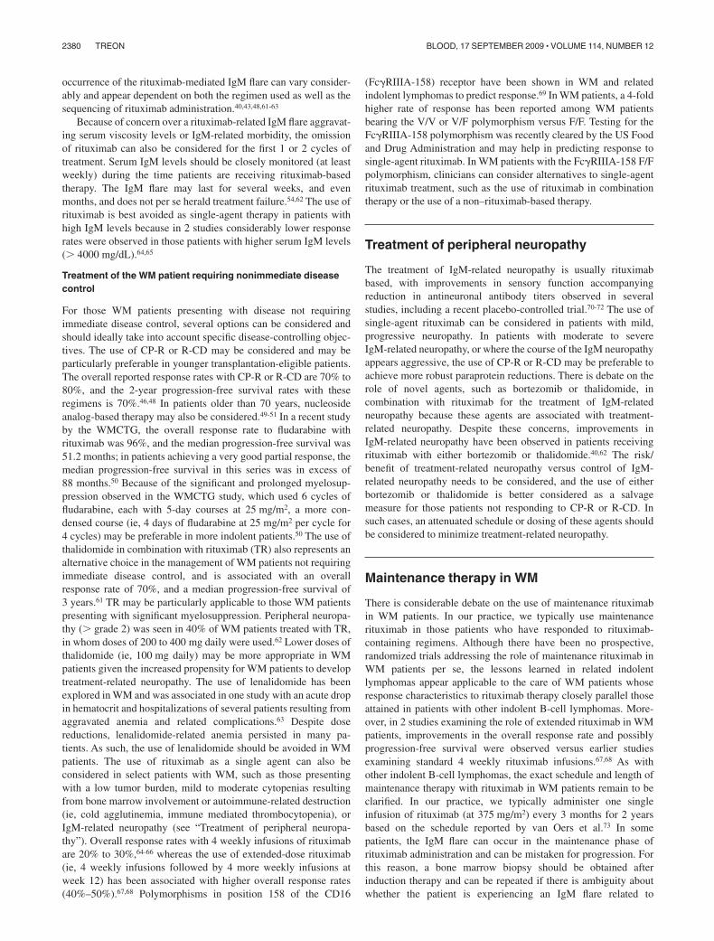
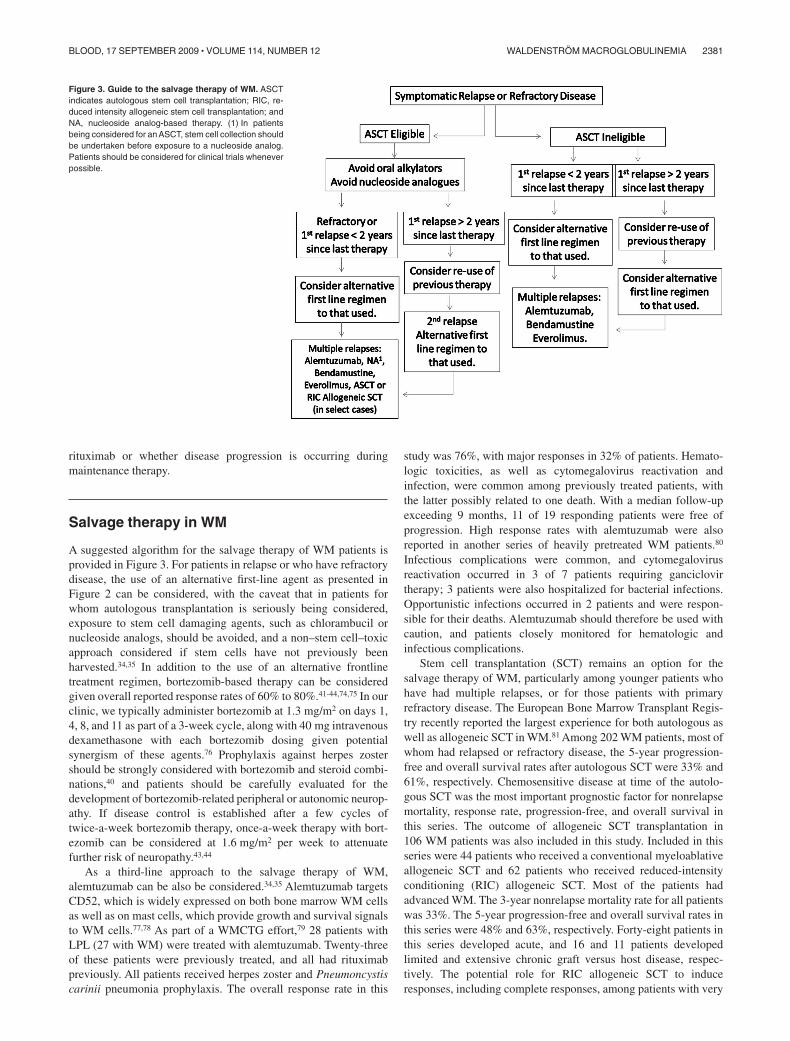
Salvage therapy in WM
A suggested algorithm for the salvage therapy of WM patients isprovided in Figure 3. For patients in relapse or who have refractorydisease, the use of an alternative first-line agent as presented inFigure 2 can be considered, with the caveat that in patients forwhom autologous transplantation is seriously being considered,exposure to stem cell damaging agents, such as chlorambucil ornucleoside analogs, should be avoided, and a non–stem cell–toxicapproach considered if stem cells have not previously beenharvested.34,35 In addition to the use of an alternative frontlinetreatment regimen, bortezomib-based therapy can be consideredgiven overall reported response rates of 60% to 80%.41-44,74,75 In ourclinic, we typically administer bortezomib at 1.3 mg/m2 on days 1,4, 8, and 11 as part of a 3-week cycle, along with 40 mg intravenousdexamethasone with each bortezomib dosing given potentialsynergism of these agents.76 Prophylaxis against herpes zostershould be strongly considered with bortezomib and steroid combi-nations,40 and patients should be carefully evaluated for thedevelopment of bortezomib-related peripheral or autonomic neurop-athy. If disease control is established after a few cycles oftwice-a-week bortezomib therapy, once-a-week therapy with bort-ezomib can be considered at 1.6 mg/m2 per week to attenuatefurther risk of neuropathy.43,44
As a third-line approach to the salvage therapy of WM,alemtuzumab can be also be considered.34,35 Alemtuzumab targetsCD52, which is widely expressed on both bone marrow WM cellsas well as on mast cells, which provide growth and survival signalsto WM cells.77,78 As part of a WMCTG effort,79 28 patients withLPL (27 with WM) were treated with alemtuzumab. Twenty-threeof these patients were previously treated, and all had rituximabpreviously. All patients received herpes zoster and Pneumoncystiscarinii pneumonia prophylaxis. The overall response rate in this
study was 76%, with major responses in 32% of patients. Hemato-logic toxicities, as well as cytomegalovirus reactivation andinfection, were common among previously treated patients, withthe latter possibly related to one death. With a median follow-upexceeding 9 months, 11 of 19 responding patients were free ofprogression. High response rates with alemtuzumab were alsoreported in another series of heavily pretreated WM patients.80
Infectious complications were common, and cytomegalovirusreactivation occurred in 3 of 7 patients requiring ganciclovirtherapy; 3 patients were also hospitalized for bacterial infections.Opportunistic infections occurred in 2 patients and were respon-sible for their deaths. Alemtuzumab should therefore be used withcaution, and patients closely monitored for hematologic andinfectious complications.
Stem cell transplantation (SCT) remains an option for thesalvage therapy of WM, particularly among younger patients whohave had multiple relapses, or for those patients with primaryrefractory disease. The European Bone Marrow Transplant Regis-try recently reported the largest experience for both autologous aswell as allogeneic SCT in WM.81 Among 202 WM patients, most ofwhom had relapsed or refractory disease, the 5-year progression-free and overall survival rates after autologous SCT were 33% and61%, respectively. Chemosensitive disease at time of the autolo-gous SCT was the most important prognostic factor for nonrelapsemortality, response rate, progression-free, and overall survival inthis series. The outcome of allogeneic SCT transplantation in106 WM patients was also included in this study. Included in thisseries were 44 patients who received a conventional myeloablativeallogeneic SCT and 62 patients who received reduced-intensityconditioning (RIC) allogeneic SCT. Most of the patients hadadvanced WM. The 3-year nonrelapse mortality rate for all patientswas 33%. The 5-year progression-free and overall survival rates inthis series were 48% and 63%, respectively. Forty-eight patients inthis series developed acute, and 16 and 11 patients developedlimited and extensive chronic graft versus host disease, respec-tively. The potential role for RIC allogeneic SCT to induceresponses, including complete responses, among patients with very
Figure 3. Guide to the salvage therapy of WM. ASCTindicates autologous stem cell transplantation; RIC, re-duced intensity allogeneic stem cell transplantation; andNA, nucleoside analog-based therapy. (1) In patientsbeing considered for an ASCT, stem cell collection shouldbe undertaken before exposure to a nucleoside analog.Patients should be considered for clinical trials wheneverpossible.
WALDENSTROM MACROGLOBULINEMIA 2381BLOOD, 17 SEPTEMBER 2009 � VOLUME 114, NUMBER 12
advanced WM has also been reported by Maloney,82 who observed6 complete, 1 near complete, and 4 partial responses among13 patients. The median number of prior therapies was 5, and theoverall and progression-free survival was 60% in this series. Day100 nonrelapse mortality was 8%, with 54% of patients experienc-ing at least grade 2 GVHD.
Autologous as well as RIC allogeneic SCT may therefore beconsidered as appropriate salvage modalities for relapsed orrefractory WM patients, although the risks and benefits of thesemodalities should be carefully weighed against other availabletreatment options.34,35 Conversely, myeloablative allogeneic SCTrepresents a high-risk option given the reported transplantation-related mortality, and should only be considered in the context of aclinical trial. Although, in general, we opt in our clinic to deferautologous stem cell transplantation (ASCT) or RIC allogeneicSCT as a salvage modality for WM patients who have had multiplerelapses, or refractory disease, the use of ASCT can be consideredas a consolidation strategy for patients presenting with amyloid-related organ dysfunction.83
Additional options for therapy of WM
The Study Group for Lymphomas recently examined the activity ofbendamustine plus rituximab (BR) versus CHOP-R in a largecohort of previously untreated patients with indolent non-Hodgkinlymphoma.84 Included in this study were 42 patients with WM,40 of whom were available for response assessment.85 The overallresponse rate with BR in this study was similar to CHOP-R (96%vs 94%, respectively). With a median follow-up of 26 months,progressive disease was documented in 2 of 23 patients treated withBR, whereas 7 of 17 patients treated with CHOP-R progressed. BRwas associated with a lower incidence of grade 3 or 4 neutropenia,infectious complications, and alopecia in this study. These resultssuggest that BR may be a preferable option to CHOP-R in thefrontline therapy of WM. Studies addressing the role of bendamus-tine in combination with other active agents, and as a salvagetherapy for indolent NHL patients are currently under way.
Everolimus (RAD001) is an oral inhibitor of the mTORpathway, which recently was approved by the US Food and DrugAdministration for the treatment of renal cell carcinoma. Geneexpression profiling and quantitative reverse-transcribed polymer-ase chain reaction analysis of WM tumor cells shows activation ofthe Akt-mTOR-p70 pathway, and inhibition of this pathway leadsto apoptosis in primary WM cells, as well as WM cell lines.86,87
A phase 2 study of everolimus was recently reported in 50 patientswith relapsed/refractory WM, who had a median number of priortherapies of 3.88 Patients in this study received everolimus at 10 mgdaily, with dose reduction permitted to 5 mg per day for toxicity.The overall response rate in this study was 70%, with 44% ofpatients achieving a major response and 28% of patients achieved aminor response. At 1 year, 67% of patients remain progression free.Tolerance to therapy in this series was good, and a clinical trialexamining the activity of everolimus in previously untreatedpatients with WM has recently been initiated by the WMCTG.
Finally, as has been emphasized by the Consensus Panels on theTreatment of WM,34,35 patients should be considered wheneverpossible for participation in a clinical trial given the paucity ofreported clinical trials in WM. Several novel clinical trials,including novel combination strategies with rituximab, bor-tezomib, bendamustine, as well as novel signal inhibitors, protea-some inhibitors, epigenetic modifiers, and immunomodulators
have been initiated or are contemplated. Details on several thesestudies as well as other WM-related clinical trials can be found atwww.clinicaltrials.gov.
Response assessment
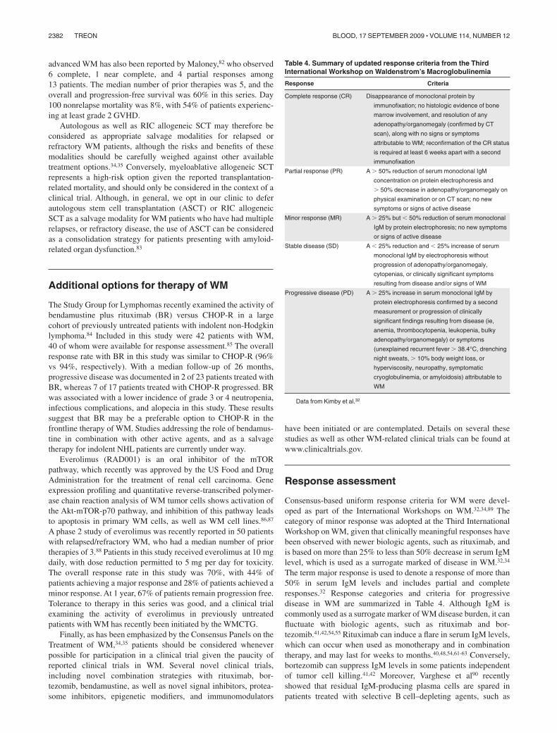
Consensus-based uniform response criteria for WM were devel-oped as part of the International Workshops on WM.32,34,89 Thecategory of minor response was adopted at the Third InternationalWorkshop on WM, given that clinically meaningful responses havebeen observed with newer biologic agents, such as rituximab, andis based on more than 25% to less than 50% decrease in serum IgMlevel, which is used as a surrogate marked of disease in WM.32,34
The term major response is used to denote a response of more than50% in serum IgM levels and includes partial and completeresponses.32 Response categories and criteria for progressivedisease in WM are summarized in Table 4. Although IgM iscommonly used as a surrogate marker of WM disease burden, it canfluctuate with biologic agents, such as rituximab and bor-tezomib.41,42,54,55 Rituximab can induce a flare in serum IgM levels,which can occur when used as monotherapy and in combinationtherapy, and may last for weeks to months.40,48,54,61-63 Conversely,bortezomib can suppress IgM levels in some patients independentof tumor cell killing.41,42 Moreover, Varghese et al90 recentlyshowed that residual IgM-producing plasma cells are spared inpatients treated with selective B cell–depleting agents, such as
Table 4. Summary of updated response criteria from the ThirdInternational Workshop on Waldenstrom’s Macroglobulinemia
Response Criteria
Complete response (CR) Disappearance of monoclonal protein by
immunofixation; no histologic evidence of bone
marrow involvement, and resolution of any
adenopathy/organomegaly (confirmed by CT
scan), along with no signs or symptoms
attributable to WM; reconfirmation of the CR status
is required at least 6 weeks apart with a second
immunofixation
Partial response (PR) A � 50% reduction of serum monoclonal IgM
concentration on protein electrophoresis and
� 50% decrease in adenopathy/organomegaly on
physical examination or on CT scan; no new
symptoms or signs of active disease
Minor response (MR) A � 25% but � 50% reduction of serum monoclonal
IgM by protein electrophoresis; no new symptoms
or signs of active disease
Stable disease (SD) A � 25% reduction and � 25% increase of serum
monoclonal IgM by electrophoresis without
progression of adenopathy/organomegaly,
cytopenias, or clinically significant symptoms
resulting from disease and/or signs of WM
Progressive disease (PD) A � 25% increase in serum monoclonal IgM by
protein electrophoresis confirmed by a second
measurement or progression of clinically
significant findings resulting from disease (ie,
anemia, thrombocytopenia, leukopenia, bulky
adenopathy/organomegaly) or symptoms
(unexplained recurrent fever � 38.4°C, drenching
night sweats, � 10% body weight loss, or
hyperviscosity, neuropathy, symptomatic
cryoglobulinemia, or amyloidosis) attributable to
WM
Data from Kimby et al.32
2382 TREON BLOOD, 17 SEPTEMBER 2009 � VOLUME 114, NUMBER 12
rituximab and alemtuzumab, and may therefore skew responseassessment. Therefore, in circumstances where serum IgM levelsmay appear out of clinical context, a bone marrow biopsy should beconsidered to clarify the patient’s underlying disease burden.Soluble CD27 may serve as an alternative surrogate marker in WMand appears to remain a faithful marker of disease in patientsexperiencing a rituximab-related IgM flare, as well asplasmapheresis.91,92
Acknowledgments
The author thanks the staff of the Bing Center for WaldenstromMacroglobulinemia and, in particular, Robert J. Manning, Christo-pher J. Patterson, and Lefkothea Ioakimidis for the data collectionused in this manuscript.
This work was supported by the Peter and Helen Bing Fund forWaldenstrom Macroglobulinemia, the Linda and Edward Nelson
Fund, the Bailey Family Fund for Waldenstrom Macroglobuline-mia, and the International Waldenstrom MacroglobulinemiaFoundation.
Authorship
Contribution: S.P.T. reviewed data and prepared the article.Conflict-of-interest disclosure: The author has received research
support, honoraria and/or consultation fees in connection withproducts discussed in this article from Berlex Oncology Inc,Biogen IDEC Inc, Celgene Corporation, Genentech BioOncologyInc, Millenium Pharmaceuticals Inc, the Takeda Company, andPGX Health Inc.
Correspondence: Steven P. Treon, Bing Center for Walden-strom’s Macroglobulinemia, Dana-Farber Cancer Institute, M548,44 Binney St, Boston, MA 02115; e-mail: [email protected].
References
1. Owen RG, Treon SP, Al-Katib A, et al. Clinico-pathological definition of Waldenstrom’s macro-globulinemia: Consensus Panel Recommenda-tions from the Second International Workshop onWaldenstrom’s macroglobulinemia. Semin Oncol.2003;30(2):110-115.
2. Harris NL, Jaffe ES, Stein H, et al. A revisedEuropean-American classification of lymphoidneoplasms: a proposal from the InternationalLymphoma Study Group. Blood. 1994;84(5):1361-1392.
3. Harris NL, Jaffe ES, Diebold J, et al. The WorldHealth Organization classification of neoplasticdiseases of the hematopoietic and lymphoid tis-sues: Report of the Clinical Advisory Committeemeeting, Airlie House, Virginia, November, 1997.J Clin Oncol. 1999;155(1):257-265.
4. Merlini G, Farhangi M, Osserman EF. Monoclonalimmunoglobulins with antibody activity in my-eloma, macroglobulinemia and related plasmacell dyscrasias. Semin Oncol. 1986;13(3):350-365.
5. Farhangi M, Merlini G. The clinical implications ofmonoclonal immunoglobulins. Semin Oncol.1986;13(3):366-379.
6. Marmont AM, Merlini G. Monoclonal autoimmu-nity in hematology. Haematologica. 1991;76(6):449-459.
7. Treon SP, Hunter ZR, Aggarwal A, et al. Charac-terization of familial Waldenstrom’s macroglobu-linemia. Ann Oncol. 2006;17(3):488-494.
8. McMaster ML, Csako G, Giambarresi TR, et al.Long-term evaluation of three multiple-case Wal-denstrom macroglobulinemia families. Clin Can-cer Res. 2007;13(17):5063-5069.
9. Kristinsson SY, Bjorkholm M, Goldin LR, et al.Risk of lymphoproliferative disorders among first-degree relatives of lymphoplasmacytic lym-phoma/Waldenstrom macroglobulinemia patients:a population-based study in Sweden. Blood.2008;112(8):3052-3056.
10. Santini GF, Crovatto M, Modolo ML, et al. Wal-denstrom macroglobulinemia: a role of HCV in-fection? Blood. 1993;82(9):2932.
11. Silvestri F, Barillari G, Fanin R, et al. Risk ofhepatitis C virus infection, Waldenstrom’s macro-globulinemia, and monoclonal gammopathies.Blood. 1996;88(3):1125-1126.
12. Leleu X, O’Connor K, Ho A, et al. Hepatitis C viralinfection is not associated with Waldenstrom’smacroglobulinemia. Am J Hematol 2006. Am JHematol. 2007;82(1):83-84.
13. Treon SP, Hunter Z, Ciccarelli BT, et al. IgA andIgG hypogammaglobulinemia is a constitutivefeature in most Waldenstrom’s macroglobuline-mia patients and may be related to mutations as-sociated with common variable immunodeficiencydisorder (CVID). Blood. 2008;112(11):Abstract3749.
14. Kyle RA, Treon SP, Alexanian R, et al. Prognosticmarkers and criteria to initiate therapy in Walden-strom’s macroglobulinemia: Consensus PanelRecommendations from the Second InternationalWorkshop on Waldenstrom’s Macroglobulinemia.Semin Oncol. 2003;30(2):116-120.
15. Morel P, Duhamel A, Gobbi P, et al. Internationalprognostic scoring system for Waldenstrom mac-roglobulinemia. Blood. 2009;113(18):4163-4170.
16. Pruzanski W, Shumak KH. Biologic activity ofcold-reacting autoantibodies (first of two parts).N Engl J Med. 1977;297(11):538-542.
17. Pruzanski W, Shumak KH. Biologic activity ofcold-reacting autoantibodies (second of twoparts). N Engl J Med. 1977;297(10):583-589.
18. Menke MN, Feke GT, McMeel JW, Branagan A,Hunter Z, Treon SP. Hyperviscosity-related reti-nopathy in Waldenstrom’s macroglobulinemia.Arch Ophthalmol. 2006;124(11):1601-1606.
19. Menke MN, Treon SP. Hyperviscosity syndrome.In: Sekeres MA, Kalaycio ME, Bolwell BJ, eds.Clinical Malignant Hematology. New York, NY:McGraw-Hill; 2007:937-941.
20. Merlini G, Baldini L, Broglia C, et al. Prognosticfactors in symptomatic Waldenstrom’s macro-globulinemia. Semin Oncol. 2003;30(2):211-215.
21. Nobile-Orazio E, Marmiroli P, Baldini L, et al. Pe-ripheral neuropathy in macroglobulinemia: inci-dence and antigen-specificity of M proteins. Neu-rology. 1987;37(9):1506-1514.
22. Garces-Sanchez M, Dyck PJ, Kyle RA, et al. Anti-bodies to myelin-associated glycoprotein (anti-MAG) in IgM amyloidosis may influence expres-sion of neuropathy in rare patients. MuscleNerve. 2008;37(4):490-495.
23. Al-Lozi MT, Pestronk A, Choski R. A skeletalmuscle-specific form of decorin is a target antigenfor a serum IgM M-protein in a patient with aproximal myopathy. Neurology. 1997;49(6):1650-1654.
24. Owen RG, Barrans SL, Richards SJ, et al. Wal-denstrom macroglobulinemia: development ofdiagnostic criteria and identification of prognosticfactors. Am J Clin Pathol. 2001;116(3):420-428.
25. San Miguel JF, Vidriales MB, Ocio E, et al. Immu-nophenotypic analysis of Waldenstrom’s macro-globulinemia. Semin Oncol. 2003;30(2):187-195.
26. Hunter ZR, Branagan AR, Manning R, et al. CD5,CD10, CD23 expression in Waldenstrom’s mac-roglobulinemia. Clin Lymphoma. 2005;5(4):246-249.
27. Schop RF, Kuehl WM, Van Wier SA, et al. Wal-denstrom macroglobulinemia neoplastic cells lackimmunoglobulin heavy chain locus translocationsbut have frequent 6q deletions. Blood. 2002;100(8):2996-3001.
28. Ocio EM, Schop RF, Gonzalez B, et al. 6q dele-tion in Waldenstrom’s macroglobulinemia is asso-ciated with features of adverse prognosis. Br JHaematol. 2007;136(1):80-86.
29. Leleu X, Hunter ZR, Xu L, et al. Expression ofregulatory genes for lymphoplasmacytic cell dif-ferentiation in Waldenstrom macroglobulinemia.Br J Haematol. 2009;145(1):59-63.
30. Chang H, Qi C, Trieu Y, et al. Prognostic rel-evance of 6q deletion in Waldenstrom’s macro-globulinemia. Proceedings of the 5th InternationalWorkshop on Waldenstrom’s macroglobulinemia,Stockholm, Sweden, 2008 [Abstract 125].
31. Avet-Loiseau H, Garand R, Lode L, Robillard N,Bataille R. 14q32 translocations discriminate IgMmultiple myeloma from Waldenstrom’s macro-globulinemia. Semin Oncol. 2003;30(2):153-155.
32. Kimby E, Treon SP, Anagnostopoulos A, et al.Update on recommendations for assessing re-sponse from the Third International Workshop onWaldenstrom’s Macroglobulinemia. Clin Lym-phoma Myeloma. 2006;6(5):380-383.
33. Menke MN, Feke GT, McMeel JW, Treon SP.Ophthalmologic techniques to assess the severityof hyperviscosity syndrome and the effect of plas-mapheresis in patients with Waldenstrom’s mac-roglobulinemia. Clin Lymphoma Myeloma. 2009;9(1):100-103.
34. Treon SP, Gertz MA, Dimopoulos MA, et al. Up-date on treatment recommendations from theThird International Workshop on Waldenstrom’sMacroglobulinemia. Blood. 2006;107(9):3442-3446.
35. Dimopoulos MA, Gertz MA, Kastritis E, et al. Up-date on treatment recommendations from theFourth International Workshop on Waldenstrom’sMacroglobulinemia. J Clin Oncol. 2009;27(1):120-126.
36. Thomas S, Hosing C, Delasalle KB, et al. Suc-cess rates of autologous stem cell collection inpatients with Waldenstrom’s macroglobulinemia.Proceedings of the 5th International Workshop onWaldenstrom’s macroglobulinemia, Stockholm,Sweden, 2008 [Abstract].
WALDENSTROM MACROGLOBULINEMIA 2383BLOOD, 17 SEPTEMBER 2009 � VOLUME 114, NUMBER 12
37. Leleu XP, Manning R, Soumerai JD, et al. In-creased incidence of transformation and myelo-dysplasia/acute leukemia in patients with Wal-denstrom macroglobulinemia treated withnucleoside analogs. J Clin Oncol. 2009;27(2):250-255.
38. Leleu X, Tamburini J, Roccaro A, et al. Balancingrisk versus benefit in the treatment of Walden-strom’s macroglobulinemia patients with nucleo-side analogue based therapy. Clin LymphomaMyeloma. 2009;9(1):71-73.
39. Rakkhit R, Delasalle KB, Gavino MB, et al. Inci-dence of transformation to large cell lymphomaand to second malignancies in symptomatic pa-tients with Waldenstrom’s macroglobulinemia(WM) treated with cladribine (2-CdA) combinationinduction. Blood. 2008;112(11): Abstract 3065.
40. Treon SP, Ioakimidis L, Soumerai JD, et al. Pri-mary therapy of Waldenstrom macroglobulinemiawith bortezomib, dexamethasone and rituximab:WMCTG clinical trial 05-180. J Clin Oncol. 2009;27(23):3830-3835.
41. Treon SP, Hunter ZR, Matous J, et al. Multicenterclinical trial of bortezomib in relapsed/refractoryWaldenstrom’s macroglobulinemia: results ofWMCTG Trial 03-248. Clin Cancer Res. 2007;13(11):3320-3325.
42. Strauss SJ, Maharaj L, Hoare S, et al. Bort-ezomib therapy in patients with relapsed or re-fractory lymphoma: potential correlation of in vitrosensitivity and tumor necrosis factor alpha re-sponse with clinical activity. J Clin Oncol. 2006;24(13):2105-2112.
43. Ghobrial IM, Matous J, Padmanabhan S, et al.Phase II trial of combination of bortezomib andrituximab in relapsed and/or refractory Walden-strom’s macroglobulinemia. Blood. 2008;112(11):Abstract 832.
44. Agathocleous A, Rule S, Johnson P. Preliminaryresults of a phase I/II study of weekly or twiceweekly bortezomib in combination with rituximabin patients with follicular lymphoma, mantle celllymphoma, and Waldenstrom’s macroglobuline-mia. Blood. 2007;110(11):Abstract 2559.
45. Treon SP, Hunter Z, Branagan A. CHOP plus rit-uximab therapy in Waldenstrom’s macroglobu-linemia. Clin Lymphoma Myeloma. 2005;5(4):273-277.
46. Dimopoulos MA, Anagnostopoulos A, KyrtsonisMC, et al. Primary treatment of Waldenstrom’smacroglobulinemia with dexamethasone, ritux-imab and cyclophosphamide. J Clin Oncol. 2007;25(22):3344-3349.
47. Buske C, Hoster E, Dreyling MH, et al. The addi-tion of rituximab to front-line therapy with CHOP(R-CHOP) results in a higher response rate andlonger time to treatment failure in patients withlymphoplasmacytic lymphoma: results of a ran-domized trial of the German Low-Grade Lym-phoma Study Group (GLSG). Leukemia. 2009;23(1):153-161.
48. Ioakimidis L, Patterson CJ, Hunter ZR, et al.Comparative outcomes following CP-R, CVP-Rand CHOP-R in Waldenstrom’s macroglobuline-mia. Clin Lymphoma Myeloma. 2009;9(1):62-66.
49. Weber DM, Dimopoulos MA, Delasalle K, et al.2-Chlorodeoxyadenosine alone and in combina-tion for previously untreated Waldenstrom’s mac-roglobulinemia. Semin Oncol. 2003;30(2):243-247.
50. Treon SP, Branagan AR, Ioakimidis L, et al. Longterm outcomes to fludarabine and rituximab inWaldenstrom’s macroglobulinemia. Blood. 2009;113(16):3673-3678.
51. Tam CS, Wolf MM, Westerman D, et al. Fludara-bine combination therapy is highly effective infirst-line and salvage treatment of patients withWaldenstrom’s macroglobulinemia. Clin Lym-phoma Myeloma. 2005;6(2):136-139.
52. Dhodapkar MV, Hoering A, Gertz MA, et al. Long-term survival in Waldenstrom macroglobulinemia:
10-year follow-up of Southwest Oncology Group-directed intergroup trial S9003. Blood. 2009;113(4):793-796.
53. Tedeschi A, Alamos SM, Ricci F, et al.Fludarabine-based combination therapies forWaldenstrom’s macroglobulinemia. Clin Lym-phoma Myeloma. 2009;9(1):67-70.
54. Treon SP, Branagan AR, Hunter Z, Santos D,Tournhilac O, Anderson KC. Paradoxical in-creases in serum IgM and viscosity levels follow-ing rituximab in Waldenstrom’s macroglobuline-mia. Ann Oncol. 2004;15(10):1481-1483.
55. Ghobrial IM, Fonseca R, Greipp PR, et al. Initialimmunoglobulin M “flare” after rituximab therapyin patients with Waldenstrom macroglobulinemia:an Eastern Cooperative Oncology Group Study.Cancer. 2004;101(11):2593-2598.
56. Ghobrial IM, Uslan DZ, Call TG, Witzig TE, GertzMA. Initial increase in the cryoglobulin level afterrituximab therapy for type II cryoglobulinemiasecondary to Waldenstrom macroglobulinemiadoes not indicate failure of response. Am J He-matol. 2004;77(4):329-330.
57. Noronha V, Fynan TM, Duffy T. Flare in neuropa-thy following rituximab therapy for Waldenstrom’smacroglobulinemia. J Clin Oncol. 2006;24(1):E3.
58. Broglio L, Lauria G. Worsening after rituximabtreatment in anti-MAG neuropathy. Muscle Nerve.2005;32(3):378-379.
59. Kilidireas C, Anagnostopoulos A, Karandreas N,et al. Rituximab therapy in monoclonal IgM-related neuropathies. Leuk Lymphoma. 2006;47(5):859-864.
60. Izzedine H, Bourry E, Amrouche L, et al. Immuno-globulin M ‘flare’ after rituximab-associated acutetubular necrosis in Waldenstrom’s macroglobu-linemia. Int J Hematol. 2009;89(2):218-222.
61. Nichols GL, Savage DG. Timing of rituximab/flu-darabine in Waldenstrom’s macroglobulinemiamay avert hyperviscosity. Blood. 2004;104(11):Abstract 4612.
62. Treon SP, Soumerai JD, Branagan AR, et al. Tha-lidomide and rituximab in Waldenstrom’s macro-globulinemia. Blood. 2008;112(12):4452-4457.
63. Treon SP, Soumerai JD, Branagan AR, et al. Le-nalidomide and rituximab in Waldenstrom’s mac-roglobulinemia. Clin Cancer Res. 2009;15(1):355-360.
64. Foran JM, Rohatiner AZ, Cunningham D, et al.European phase II study of rituximab (chimericanti-CD20 monoclonal antibody) for patients withnewly diagnosed mantle-cell lymphoma and pre-viously treated mantle-cell lymphoma, immunocy-toma, and small B-cell lymphocytic lymphoma.J Clin Oncol. 2000;18(2):317-324.
65. Treon SP, Agus DB, Link B, et al. CD20-directedantibody-mediated immunotherapy induces re-sponses and facilitates hematologic recovery inpatients with Waldenstrom’s macroglobulinemia.J Immunother. 2001;24(3):272-279.
66. Gertz MA, Rue M, Blood E, et al. Multicenterphase 2 trial of rituximab for Waldenstrom macro-globulinemia (WM): an Eastern Cooperative On-cology Group Study (E3A98). Leuk Lymphoma.2004;45(10):2047-2055.
67. Dimopoulos MA, Zervas C, Zomas A, et al. Treat-ment of Waldenstrom’s macroglobulinemia withrituximab. J Clin Oncol. 2002;20(9):2327-2333.
68. Treon SP, Emmanouilides C, Kimby E, et al. Ex-tended rituximab therapy in Waldenstrom’s mac-roglobulinemia. Ann Oncol. 2005;16(1):132-138.
69. Treon SP, Hansen M, Branagan AR, et al. Poly-morphisms in Fc�RIIIA (CD16) receptor expres-sion are associated with clinical responses to rit-uximab in Waldenstrom’s macroglobulinemia.J Clin Oncol. 2005;23(3):474-481.
70. Pestronk A, Florence J, Miller T, et al. Treatmentof IgM antibody associated polyneuropathies us-ing rituximab. J Neurol Neurosurg Psychiatry.2003;74(4):485-489.
71. Benedetti L, Briani C, Grandis M, et al. Predictorsof response to rituximab in patients with neuropa-thy and anti-myelin associated glycoprotein im-munoglobulin M. J Peripher Nerv Syst. 2007;12(2):102-107.
72. Dalakas MC, Rakocevic G, Salajegheh MD, et al.Placebo-controlled trial of rituximab in IgManti-myelin-associated glycoprotein antibody de-myelinating neuropathy. Ann Neurol. 2009;65(3):286-293.
73. van Oers MH, Klasa R, Marcus RE, et al. Ritux-imab maintenance improves clinical outcome ofrelapsed/resistant follicular non-Hodgkin lym-phoma in patients both with and without rituximabduring induction: results of a prospective random-ized phase 3 intergroup trial. Blood. 2006;108(10):3295-3301.
74. Chen CI, Kouroukis CT, White D, et al. Bort-ezomib is active in patients with untreated or re-lapsed Waldenstrom’s macroglobulinemia: aphase II study of the National Cancer Institute ofCanada Clinical Trials Group. J Clin Oncol. 2007;25(12):1570-1575.
75. Dimopoulos MA, Anagnostopoulos A, KyrtsonisMC, et al. Treatment of relapsed or refractoryWaldenstrom’s macroglobulinemia with bort-ezomib. Haematologica. 2005;90(12):1655-1657.
76. Jagannath S, Richardson PG, Barlogie B, et al.Bortezomib in combination with dexamethasonefor the treatment of patients with relapsed and/orrefractory multiple myeloma with less than opti-mal response to bortezomib alone. Haemato-logica. 2006;91(7):929-934.
77. Treon SP, Kelliher A, Keele B, et al. Expression ofserotherapy target antigens in Waldenstrom’smacroglobulinemia: therapeutic applications andconsiderations. Semin Oncol. 2003;30(2):248-252.
78. Santos DD, Hatjiharissi E, Tournilhac O, et al.CD52 is expressed on human mast cells and is apotential therapeutic target in Waldenstrom’smacroglobulinemia and mast cell disorders. ClinLymphoma Myeloma. 2006;6(6):478-483.
79. Hunter ZR, Boxer M, Kahl B, et al. Phase II studyof alemtuzumab in lymphoplasmacytic lym-phoma: results of WMCTG trial 02-079. Proc AmSoc Clin Oncol. 2006;24(33):Abstract 7523.
80. Owen RG, Rawstron AC, Osterborg A, et al. Ac-tivity of alemtuzumab in relapsed/refractory Wal-denstrom’s macroglobulinemia. Blood. 2003;102(11):644a.
81. Kyriakou H, on behalf of the Lymphoma WorkingParty of the European Group for Blood and BoneMarrow Transplantation. Haematopoietic stemcell transplantation for Waldenstrom’s macro-globulinemia. Proceedings of the 5th InternationalWorkshop on Waldenstrom’s macroglobulinemia,Stockholm, Sweden, 2008 [Abstract 146].
82. Maloney D. Evidence for GVWM following mini-allo in Waldenstrom’s macroglobulinemia. Pro-ceedings of the 5th International Workshop onWaldenstrom’s macroglobulinemia, Stockholm,Sweden, 2008 [Abstract 147].
83. Gertz MA, Hayman SR, Buadi FK. Transplanta-tion for IgM amyloidosis and IgM myeloma. ClinLymphoma Myeloma. 2009;9(1):77-79.
84. Rummel MJ, von Gruenhagen U, Niederle N, etal. Bendamustine plus rituximab versus CHOPplus rituximab in the firstline treatment of patientswith follicular, indolent and mantle cell lympho-mas: results of a randomized phase III study ofthe Study Group Indolent Lymphomas (StiL).Blood. 2008;112(11):Abstract 2596.
85. Rummel MJ, von Gruenhagen U, Niederle N, etal. Bendamustine plus rituximab versus CHOPplus rituximab in the first-line treatment of pa-tients with Waldenstrom’s macroglobulinemia:first interim results of a randomized phase IIIstudy of the Studygroup Indolent Lymphomas(StiL). Proceedings of the 5th International Work-shop on Waldenstrom’s macroglobulinemia,Stockholm, Sweden, 2008 [Abstract 139].
2384 TREON BLOOD, 17 SEPTEMBER 2009 � VOLUME 114, NUMBER 12
86. Hatjiharissi E, Mitsiades CS, Ciccarelli B, et al.Comprehensive molecular characterization ofmalignant and microenvironmental cells inWaldenstrom’s macroglobulinemia by gene ex-pression profiling. Blood. 2007;110(11):Abstract3174.
87. Leleu X, Jia X, Runnels J, et al. The Akt pathwayregulates survival and homing in Waldenstrommacroglobulinemia. Blood. 2007;110(11):4417-4426.
88. Ghobrial IM, Chuma S, Sam A, et al. Phase II trialof the mTOR inhibitor RAD001 in relapsed and/or
refractory Waldenstrom macroglobulinemia: theDana Farber Cancer Institute Experience. Blood.2008;112(11):Abstract 1011.
89. Weber D, Treon SP, Emmanouilides C, et al. Uni-form response criteria in Waldenstrom’s macro-globulinemia: consensus panel recommendationsfrom the Second International Workshop on Wal-denstrom’s Macroglobulinemia. Semin Oncol.2003;30(2):127-131.
90. Varghese AM, Rawstron AC, Ashcroft J, et al. As-sessment of bone marrow response in Walden-
trom’s macroglobulinemia. Clin Lymphoma My-eloma. 2009;9(1):53-55.
91. Ho A, Leleu X, Hatjiharissi E, et al. CD27-CD70 inter-actions in the pathogenesis of Waldenstrom’s macro-globulinemia. Blood. 2008;112(12):4683-4689.
92. Ciccarelli BT, Yang G, Hatjiharissi E, et al. SolubleCD27 is a faithful marker of disease burden andis unaffected by the rituximab induced IgM flare,as well as plasmapheresis in patients with Wal-denstrom’s macroglobulinemia. Clin LymphomaMyeloma. 2009;9(1):56-58.
WALDENSTROM MACROGLOBULINEMIA 2385BLOOD, 17 SEPTEMBER 2009 � VOLUME 114, NUMBER 12