Embed Size (px)
Citation preview

p
An Overview of Brain StructureBrain TerminologyThe Brain’s Surface FeaturesThe Brain’s Internal FeaturesMicroscopic Inspection: Cells and FibersFocus on Disorders: Meningitis and
EncephalitisFocus on Disorders: Stroke
A Closer Look at NeuroanatomyThe Cranial Nervous SystemThe Spinal Nervous SystemThe Internal Nervous SystemFocus on Disorders: Magendie, Bell, and Bell’s
Palsy
The Functional Organization of the BrainPrinciple 1: The Sequence of Brain Processing
Is “In Integrate Out”Principle 2: Sensory and Motor Divisions Exist
Throughout the Nervous SystemPrinciple 3: The Brain’s Circuits Are CrossedPrinciple 4: The Brain Is Both Symmetrical and
AsymmetricalPrinciple 5: The Nervous System Works
Through Excitation and InhibitionPrinciple 6: The Central Nervous System Has
Multiple Levels of FunctionPrinciple 7: Brain Systems Are Organized Both
Hierarchically and in ParallelPrinciple 8: Functions in the Brain Are Both
Localized and Distributed
36 ■
How Is the Brain Organized?
C H A P T E R
2
A. Klehr / Stone Images
Micrograph: Carolina Biological Supply Co. / Phototake
p
W hen buying a new car, people first inspect the
outside carefully, admiring the flawless finish
and perhaps even kicking the tires. Then they
open the hood and examine the engine, the part of the car
responsible for most of its behavior—and misbehavior.
This means gazing at a maze of tubes, wires, boxes, and
fluid reservoirs. All most of us can do is gaze, because
what we see simply makes no sense, except in the most
general way. We know that the engine burns gasoline to
make the car move and somehow generates electricity to
run the radio and lights. But this tells us nothing about
what all the engine’s many parts do. What we need is in-
formation about how such a system works.
In many ways, examining a brain for the first time is
similar to looking under the hood of a car. We have a vague
sense of what the brain does but no sense of how the parts
that we see accomplish these tasks. We may not even be
able to identify many of the parts. In fact, at first glance the
outside of a brain may look more like a mass of folded
tubes divided down the middle than like a structure with
many interconnected pieces. See what you can make of the
human brain in Figure 2-1. Can you say anything about
how it works? At least a car engine has parts with regular
shapes that are recognizably similar in different engines.
This is not true of mammals’ brains, as shown in Figure 2-2.
When we compare the brain of a cat with that of a human,
for example, we see that there is an enormous difference
not just in overall size, but in the relative sizes of parts and
in structure. In fact, some parts present in one are totally ab-
sent in the other. What is it that all these parts do that makes
one animal stalk mice and another read textbooks?
To make matters worse, even for trained research sci-
entists, the arrangement of the brain’s parts does not just
seem random, it really is haphazard. The challenge that we
face in learning about the brain is to identify some regulari-
ties in its organization and to establish a set of principles
that can help us understand how the nervous system
works. After decades of investigation, we now have a good
idea of how the nervous system functions, at least in a gen-
eral way. That knowledge is the subject of this chapter. But
before we turn our attention to the operation manual for
the brain and the rest of the nervous system, let us examine
what the brain is designed to do. Knowing the brain’s func-
tions will make it easier to grasp the rules of how it works.
Perhaps the simplest statement of the brain’s functions
is that it produces behavior, as seen in Chapter 1. There is
more to this statement than is immediately apparent, how-
ever. In order for the brain to produce behavior, it must
have information about the world, such as information
about the objects around us—their size, shape, move-
ment, and so forth. Without such information, the brain
cannot know how to orient and direct the body to produce
an appropriate response. This is especially true when the
response needed is some complex behavior, such as
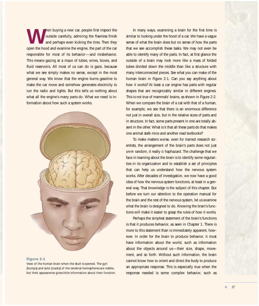
Figure 2-1 View of the human brain when the skull is opened. The gyri(bumps) and sulci (cracks) of the cerebral hemispheres are visible,but their appearence gives little information about their function.
■ 37

catching a ball. To perform complex behaviors, the ner-
vous system has organs designed to receive information
from the world and convert this information into biologi-
cal activity that produces subjective experiences of reality.
The brain thus produces what we believe is reality in order
for us to move. These subjective experiences of reality are
essential to carrying out any complex task.
This view of the brain’s primary purpose may seem ab-
stract to you, but it is central to understanding how the
brain functions. Consider the task of answering a telephone.
The brain directs the body to pick up the receiver when the
nervous system responds to vibrating molecules of air by
creating the subjective experience of a ring. We perceive
this sound and react to it as if it actually existed, when in
fact the sound is merely a fabrication of the brain. That fab-
rication is produced by a chain reaction that takes place
when vibrating air molecules hit the eardrum. In the ab-
sence of the nervous system, especially the brain, there is
no such thing as sound. Rather, there is only the movement
of air molecules.
The subjective nature of the experiences that the brain
creates can be better understood by comparing the realities
of two different kinds of animals. You are probably aware
that dogs perceive sounds that humans do not. This differ-
ence in perception does not mean that a dog’s nervous sys-
tem is better than ours or that our hearing is poorer. Rather,
a dog brain simply creates a different world from that of
our brain. Neither subjective experience is “right.” The dif-
ference in experience is merely due to two different sys-
Monkey Human
Brainstem
Cat
CerebellumCerebrum
BrainstemOlfactorybulb
Cerebellum
Cerebrum
Brainstem Cerebellum
Cerebrum
Cerebellum
Rat
Cerebrum
BrainstemOlfactory bulb
Figure 2-2
Inspection of the outside features of the brains of a cat, rat, monkey,and human shows them to differ dramatically in size and in generalappearance. The rat brain is smooth, whereas the other brains havefurrows in the cerebral cortex. The pattern of furrows differsconsiderably in the human, the monkey, and the cat. The cat brainand, to some extent, the monkey brain have long folds that appearto run much of the length of the brain, whereas the human brainhas a more diffuse pattern. The cerebellum is wrinkled in all speciesand is located above the brainstem. The brainstem is the route bywhich information enters and exits the brain. The olfactory bulb,which controls the perception of smells, is relatively larger in cats andrats but is not visible in monkeys and humans, because it is small andlies under the brain.Photos courtesy of Wally Welker, University of Wisconsin ComparativeMammalian Brain Collection.
p
38 ■ CHAPTER 2

tems for processing physical stimuli. The same differences
exist in visual perceptions. Dogs see very little color,
whereas our world is rich with color because our brains
create a different reality from that of a dog’s brain. Such dif-
ferences in subjective realities exist for good reason: they
allow different animals to exploit different features of their
environments. Dogs use their hearing to detect the move-
ments of mice in the grass, whereas early humans probably
used color for such tasks as identifying ripe fruit in trees.
Evolution, then, equipped each species with a view of the
world that would help it survive.
These examples show how a brain’s sensory experi-
ences help guide an organism’s behavior. For this link be-
tween sensory processing and behavior to be made, the
brain must also have a system for accumulating, integrating,
and using knowledge. Whenever the brain collects sensory
information, it is essentially creating knowledge about the
world, knowledge that can be used to produce more effec-
tive behaviors. The knowledge currently being created in
one sensory domain can be compared both with past
knowledge and with knowledge gathered in other domains.
We can now identify the brain’s three primary functions:
1. to produce behavior;
2. to create a sensory reality; and
3. to create knowledge that integrates information from
different times and sensory domains and to use that
knowledge to guide behavior.
Each of the brain’s three functions requires specific ma-
chinery. The brain must have systems to create the sensory
world, systems to produce behavior, and systems to inte-
grate the two.
In this chapter, we consider the basic structures and
functions of those systems. First, we identify the compo-
nents of the nervous system. Then we look at what those
components do. Finally, we look at how the parts work to-
gether and at some general principles of brain function.
Many of the ideas introduced in this chapter are devel-
oped throughout the rest of the book, so you may want to
return to this chapter often to reconsider the basic princi-
ples as new topics are introduced.
AN OVERVIEW OF BRAIN STRUCTUREThe place to start our overview of the brain’s structure is to “open the hood” by open-ing the skull and looking at the brain snug in its home. Figure 2-1 shows a brainviewed from this perspective. The features that you see are part of what is called thebrain’s “gross anatomy,” not because they are ugly, but because they constitute a broadoverview. Zooming in on the brain’s microscopic cells and fibers is largely reserved forChapter 3, although this section ends with a brief introduction of some terms usedfor these tiny structures. Those terms are just a few of a great many new terms thatyou will encounter in this book, which is why we deal with brain terminology in gen-eral before moving on to a look at the brain itself. Because many of the words in thischapter will seem foreign to you, they will be accompanied by a pronunciation guideat their first appearance.
Brain TerminologyThere are hundreds, even thousands of brain regions, making the task of masteringbrain terminology seem daunting. To make matters worse, many structures have sev-eral names, and many terms are often used interchangeably. This peculiar nomencla-ture arose because research on brain and behavior has spanned several centuries.When the first anatomists began to examine the brain with the primitive tools of theirtime, they made many erroneous assumptions about how the brain works, and thenames that they chose for brain regions are often manifestations of those errors. For
HOW IS THE BRAIN ORGANIZED? ■ 39
p
Link to an index listing the roots ofneuroanatomical terms atwww.worthpublishers.com/kolb/chapter2.

instance, they named one region of the brain the gyrus fornicatus because theythought it had a role in sexual function. In fact, most of this region has nothing to dowith sexual function. Another area was named the red nucleus because it appears red-dish in fresh tissue. This name denotes nothing of the area’s potential functions,which turn out to be the control of limb movements.
As time went on, the assumptions and tools of brain research changed, but thenaming continued to be haphazard and inconsistent. Early investigators named struc-tures after themselves or objects or ideas. They used different languages, especiallyLatin, Greek, and English. More recently, investigators have often used numbers orletters, but even this system lacks coherence because the numbers may be Arabic orRoman numerals and are often used in combination with letters, which may be eitherGreek or Latin. When we look at current brain terminology, then, we see a mixture ofall these naming systems.
Despite this sometimes confusing variety, many names do include informationabout a structure’s location in the brain. Table 2-1 summarizes these location-relatedterms, and Figure 2-3 shows how they relate to body locations. Structures found onthe top of the brain or on the top of some structure within the brain are dorsal. Struc-
40 ■ CHAPTER 2
p
Figure 2-3
Anatomical terms are used to describeanatomical locations. (A) Anatomicaldirections relative to the head and brain.Because a human is upright, the termsposterior and caudal (both meaning“tail”) refer to a slightly differentorientation for the human headcompared with the head of a four-legged animal. (B) Anatomical directionsrelative to the body.
Figure 2-4
An afferent nerve carries informationinto the brain, and an efferent nervetakes information out of the brain andcontrols movement of a muscle.
(A)
(B)
Meaning “above,” sometimes referred to as superior
Meaning “tail,” sometimes referred to as caudal
Meaning “below” or “belly,” sometimes referred to as inferior
Meaning “middle”
Meaning “front,” sometimes referred to as frontal or rostral
Posterior
Anterior Lateral
Medial
Anterior Posterior
Vent
ral
Dors
alVe
ntra
lDo
rsal
Anterior Posterior
Meaning “side”Do
rsal
Dors
al
Vent
ral
Vent
ral
This afferent nerve carries information from sensory receptors in skinto the brain.
Sensory endings
This efferent nerve carries informationfrom the brain to the neurons controlling leg muscle, causing a response.

tures located toward the bottom of the brain or one ofits parts are ventral. Structures found toward the mid-dle of the brain are medial, whereas those located to-ward the side are lateral. Structures located toward thefront of the brain are anterior, whereas those locatedtoward the back of the brain are posterior. Sometimesthe terms rostral and caudal are used instead of ante-rior and posterior, respectively. And, occasionally, theterms superior and inferior are used to refer to struc-tures that are located dorsally or ventrally (these termsdo not label structures according to their importance).It is also common to combine terms. For example, astructure may be described as dorsolateral, whichmeans that it is located “up and to the side.”
You should also learn two terms that describe thedirection of information flowing to and from cells inthe brain. Afferent refers to information coming intothe brain or a part of the brain, whereas efferent refersto information leaving the brain or one of its parts,meaning that efferent refers to brain signals that trig-ger some response (Figure 2-4). These words are verysimilar, but there is an easy way to keep them straight.The letter “a” in afferent comes alphabetically before the “e” in efferent, and sensory in-formation must come into the brain before an outward-flowing signal can trigger a re-sponse. Therefore, afferent means “incoming” and efferent means “outgoing.”
The Brain’s Surface FeaturesReturning to the brain in the open skull, you are now ready to examine its structuresmore closely. The first thing to notice is that the brain is covered by a tough materialknown as the meninges [men in jeez (the accented syllable is in boldface type)], whichis a three-layered structure, as illustrated in Figure 2-5. The outer layer is known as thedura mater (from Latin, meaning “hard mother”). It is a tough double layer of fibroustissue enclosing the brain in a kind of loose sack. The middle layer is the arachnoidlayer (from Greek, meaning “like a spider’s web”). It is a very thin sheet of delicate
HOW IS THE BRAIN ORGANIZED? ■ 41
p
Table 2-1 Orientation Terms for the Brain
Term Meaning with respect to the nervous system
Anterior Located near or toward the front or the head
Caudal Located near or toward the tail
Dorsal On or toward the back or, in reference to brain nuclei, locatedabove
Frontal “Of the front“ or, in reference to brain sections, a viewing orientation from the front
Inferior Located below
Lateral Toward the side of the body
Medial Toward the middle; sometimes written as mesial
Posterior Located near or toward the tail
Rostral ”Toward the beak”; located toward the front
Sagittal Parallel to the length (from front to back) of the skull; used inreference to a plane
Superior Located above
Ventral On or toward the belly or side of the animal in which the bellyis located or, in reference to brain nuclei, located below
Figure 2-5
The brain is covered by thick coveringsknown as the meninges and is cushionedby a fluid known as the cerebrospinalfluid (CSF).
On the CD, visit the module on theCentral Nervous System to better visual-ize the various planes of the brain.
Skull
Dura mater
MeningesArachnoidlayer
Subarachnoid space (filled with CSF)Brain
Pia mater

p
42 ■ CHAPTER 2
Figure 2-6
In these views of the human brain (fromthe top, bottom, side, and middle), thelocations of the frontal, parietal,occipital, and temporal lobes of thecerebral hemispheres are shown, as arethe cerebellum and the three major sulci (the central sulcus, lateral fissure, andlongitudinal fissure) of the cerebralhemispheres.Photos courtesy of Yakolev Collection/AFIP.
Lateral view
Medial view
Frontallobe
Central sulcus
Central sulcus
Cerebellum
Temporallobe
Lateralfissure
Parietallobe
Occipitallobe
Frontallobe
Temporallobe Brainstem
Parietallobe
Occipitallobe
Dorsal view
Frontallobe
Longitudinalfissure
Parietallobe
Central sulcus
Occipitallobe
Ventral view
Frontallobe
Temporal lobeCerebellum
BrainstemCranial nerves

connective tissue that follows the brain’s contours. The inner layer is the pia mater (fromLatin, meaning “soft mother”). It is a moderately tough membrane of connective-tissue fibers that cling to the surface of the brain. Between the arachnoid and piamater is a fluid, known as cerebrospinal fluid (CSF), which is a colorless solution ofsodium chloride and other salts. It provides a cushion so that the brain can move orexpand slightly without pressing on the skull. (Meningitis is an infection of themeninges. Its symptoms are described in “Meningitis and Encephalitis” on page 46.)
If we remove the meninges, we can now remove the brain from the skull and ex-amine its various parts. As we look at the brain from the top or the side, it appears tohave two major parts, each wrinkly in appearance. The larger part is the cerebrum [sa ree brum], which consists of two cerebral hemispheres, the left and the right, andthe smaller part is the cerebellum [sair a bell um]. Both the cerebrum and the cere-bellum are visible in the brains shown in Figure 2-2. Each of these structures is wrin-kled in large-brained animals because its outer surface is made of a relatively thinsheet of tissue, the cortex, that has been pushed together to make it fit into the skull.To see why the cortex is wrinkled, force a piece of writing paper, 81�2 by 11 inches, intoa cup. The only way is to crinkle the paper up into a ball. Essentially the same crinkling-up has been done to the cortex of the cerebrum and the cerebellum. Like a crinkledpiece of paper, much of the cortex is invisible from the surface. All we can see fromthe surface are bumps and cracks. The bumps are known as gyri [jye rye; singular:gyrus ( jye russ)], whereas the cracks are known as sulci [sul sigh; singular: sulcus (sul kus)]. Some of the sulci are very deep and so are often called fissures. The twobest-known fissures are the longitudinal fissure and the lateral fissure, both of whichare shown in Figure 2-6, along with the central sulcus.
If we now look at the bottom of the brain, we see something completely different.The cerebrum is still the wrinkled part, but now there is also a whitish structure downthe middle with little tubes attached. This middle structure is known as the brain-stem, and the little tubes are cranial nerves that run to and from the head.
One final gross feature is obvious: the brain appears to be covered in blood ves-sels. As in other parts of the body, the brain receives blood through arteries and sendsit back through veins to the kidneys and lungs for cleaning and oxygenation. The ar-teries come up the neck and then wrap around the outside of the brainstem, cere-brum, and cerebellum, finally piercing the brain’s surface to get to its inner regions.Figure 2-7 shows the three major arteries that feed blood to the cerebrum — namely,the anterior, middle, and posterior cerebral arteries. Because the brain is very sensitiveto loss of blood, a blockage or break in a cerebral artery is likely to lead to the death ofthe affected region, a condition known as a stroke (see “Stroke” on page 48). Becausethe three cerebral arteries service different parts of the brain, strokes disrupt differentbrain functions, depending on the artery affected.
HOW IS THE BRAIN ORGANIZED? ■ 43
p
Anterior cerebralartery
Middle cerebral artery
Posterior cerebralartery
Figure 2-7
Each of the three major arteries of thecerebral hemispheres— the anterior,middle, and posterior—provides bloodto a different region of the cerebrum.
Plug in the CD to examine, locate,and rotate the parts of the brain in the sec-tion on the subdivisions of the CNS in themodule on the Central Nervous System.
Cerebrum. The major structure of theforebrain, consisting of two equal hemi-spheres (left and right).
Cerebellum. Major structure of the hind-brain specialized for motor coordination;in large-brained animals, it may also havea role in the coordination of other mentalprocesses.
Brainstem. Central structures of thebrain including the hindbrain, midbrain,thalamus, and hypothalamus.
Cranial nerve. One of a set of nervesthat control sensory and motor functionsof the head; includes senses of smell, vi-sion, audition, taste, and touch on theface and head.

The Brain’s Internal FeaturesThe simplest way to examine what is inside something — be it an engine, a pear, or abrain — is to cut it in half. The orientation in which we cut makes a difference in whatwe see, however. Consider what happens when we slice through a pear held in differ-ent orientations. If we cut a pear from side to side, we cut across the core; whereas, ifwe cut it from top to bottom, we cut parallel to the core. Our impression of what theinside of a pear looks like is clearly influenced by the way in which we slice it. Thesame is true of the brain.
We can begin by cutting the brain in half, slicing it downward through the mid-dle. The result is shown in Figure 2-8. This view of the brain is known as a frontal sec-tion because we can now see the inside of the brain from the front.
Several features of the brain’s interior are immediately apparent. First, it containsfour cavities, known as ventricles [ven trik uls], which are shown in Figure 2-9. Cellsthat line the ventricles make the cerebrospinal fluid that fills them. The ventricles areconnected, so CSF flows from the two lateral ventricles to the ventricles that lie on thebrain’s midline, eventually flowing into the space between the lower layers of themeninges as well as into the spinal-cord canal. Although the function of the ventriclesis not well understood, they are thought to play an important role in maintaining thebrain. The CSF may allow certain compounds access to the brain, and it probablyhelps the brain excrete metabolic wastes. Very likely, too, the CSF produced in theventricles acts as a kind of shock absorber. The CSF surrounds the brain; so, if there isa blow to the head, this fluid cushions the movement of the brain within the skull.
A second feature apparent in our frontal section of the brain is that the brain’s in-terior is not homogeneous. There are both light and dark regions. These light anddark regions may not seem as distinct as the different parts of a car’s engine, but nev-ertheless they represent different components. The light regions, called white matter,are mostly fibers with fatty coverings. The fatty coverings produce the white appear-ance, much as fat droplets in milk make it appear white. The dark regions, called graymatter because of their gray-brown color, are areas where capillary blood vessels andcell bodies predominate. Some regions of the brain have a mottled gray and white, ornetlike, appearance. These regions, which have both cell bodies and fibers mixed to-gether, are called reticular matter (from the Latin word rete, meaning “net”).
Another way to cut the brain is from front to back. The result is a side view, calleda sagittal [sadj i tal] section. If we make our cut down the brain’s midline, we dividethe cerebrum into its two hemispheres. Figure 2-10 shows such a sagittal section.
44 ■ CHAPTER 2
p
Lateralventricles
Gray matter
Temporallobe
Lateralsulcus
White matter
Corpuscallosum
(A) (B) (C)
Figure 2-8
This frontal section through the brainshows the internal features. The brain is(A) cut and then (B) viewed at a slightangle. This section displays regions thatare relatively white and gray. The whiteareas are largely composed of fibers,whereas the gray areas are composed ofcell bodies. The large bundle of fibersjoining the two areas is the corpuscallosum. Each ventricle is a fluid-filledtube.
White matter. Those areas of the ner-vous system rich in axons, leading to awhite appearance.
Gray matter. Those areas of the nervoussystem composed predominantly of cellbodies, leading to a gray appearance.
Reticular matter. Area composed of in-termixed cell bodies and axons that pro-duce a mottled gray and white, or netlike,appearance.
Look at the CD to examine a three-dimensional model of the ventricular sys-tem in the section on subcortical struc-tures in the module on the CentralNervous System.
Gla
uber
man
/ Ph
oto
Rese
arch
ers

One feature seen from this viewing angle is a long band of white matter thatruns much of the length of the cerebral hemispheres. This band is called the corpus callosum [ka loh sum]. The corpus callosum contains about 200 millionfibers that join the two hemispheres and allow communication between them. It isalso clear in Figure 2-10 that the cortex covers the cerebral hemispheres above thecorpus callosum, whereas below the corpus callosum are vari-ous internal structures of the brain. Owing to their location below the cortex, these structures are known as subcortical regions.
We can see the internal structures of the brain in much moredetail by coloring them with special stains. For example, if we usea dye that selectively stains cell bodies, we can see that the distri-bution of cells within the gray matter is not homogeneous, asshown in Figure 2-11. In particular, it becomes apparent that thecerebral cortex is composed of layers, each of which containssimilarly staining cells. Furthermore, subcortical regions are nowseen to be composed of clusters, known as nuclei, of similarlystained cells. Although layers and nuclei are very different in ap-pearance, they both form functional units within the brain.Whether a particular brain region has layers or nuclei is largelyan accident of evolution.
If you were to compare the two sides of the brain in sagittalsection, you would be struck by their symmetry. The brain, infact, has two of nearly every structure, one on each side. The fewstructures that are one of a kind are found along the brain’s mid-line. Examples are the third and fourth ventricles and the pinealgland, mentioned in Chapter 1 in reference to Descartes’s theoryabout how the brain works.
HOW IS THE BRAIN ORGANIZED? ■ 45
p
Lateralventricles
Thirdventricle
Fourthventricle
Figure 2-9
There are two lateral cerebral ventricles,one in each hemisphere, and a third andfourth cerebral ventricle, each of whichlies in the midline of the brain.
Figure 2-10
In this sagittal section through the brain,the brain is (A) cut and then (B) viewedfrom the side. This particular plane of cutseparates the hemispheres, allowing aview of the midline structures of thebrain, including the corpus callosum,which connects the two hemispheres.You can see the subcortical structures(ventricle, brainstem, and cerebellum)that lie below the corpus callosum.
(A) (B)
Cortex
Brainstem
Corpus callosum
Ventricle
Cerebellum
Plane of cut
Subcortical regions. All of the regionsof the brain that are located beneath theneocortex; the term is usually used to dis-tinguish regions of the brain that controlbasic functions from those regions con-trolling cognitive functions, which are mediated by the neocortex.
Microscopic Inspection: Cells and FibersAlthough the parts of a car engine are all large enough to be seen with the naked eye,the fundamental units of the brain — its cells — are so small that they can be viewedonly with the aid of a microscope. By using a microscope, we quickly discover that the

p
46 ■ CHAPTER 2
Figure 2-11
When brain sections are stained, variousregions become clearly demarcated.These brain sections from the lefthemisphere of a monkey (midline is tothe left in each photograph) are stainedwith (A) a selective cell-body stain,known as a Nissl stain, and (B) a selectivefiber stain, staining for myelin. It isimmediately apparent that the twostains reveal a very different picture ofthe brain. (C and D) Higher-powermicrographs through the Nissl- andmyelin-stained sections show differentcortical regions. Notice the difference inappearance.
A large number of harmful organisms can invade the linings,
or meninges, of the brain, particularly the pia mater and the
arachnoid layer, as well as the cerebrospinal fluid between
them. Such infections are called meningitis. One symptom
is inflammation, which, because the skull is solid, places
pressure on the brain. This pressure often leads to delirium
and, if the infection progresses, to drowsiness, stupor, and
even coma.
Meningitis usually begins with severe headache and a
stiff neck (known as cervical rigidity). Head retraction is an
extreme form of cervical rigidity. Convulsions are a common
symptom in children. They indicate that the brain also is af-
fected by the inflammation.
Infection of the brain itself is called encephalitis. There
are many forms of encephalitis, some of which have great
historical significance. In World War I, a form of encephali-
tis referred to as sleeping sickness (encephalitis lethargica)
reached epidemic proportions. The first symptoms were dis-
turbances of sleep. People slept all day and became wake-
ful, even excited, at night. Subsequently, they showed symp-
toms of Parkinson’s disease, characterized by severe tremors
and difficulty in controlling body movements. Many were
completely unable to make any voluntary movements, such
as walking or even combing their hair. (These patients were
immortalized in the movie Awakenings.) The cause of these
symptoms is death of the brain nucleus known as the sub-
stantia nigra (black substance). Other forms of encephalitis
may have different effects on the brain. For example, Ras-
mussen’s encephalitis attacks one cerebral hemisphere in
children. In most cases, the only effective treatment is a radi-
cal one: removal of the entire affected hemisphere. Surpris-
ingly, some young children who lose a hemisphere adapt
rather well. They may even complete college, literally with
half a brain. But, unfortunately, retardation is a more com-
mon outcome of hemispherectomy after encephalitis.
Meningitis and Encephalitis
Focus on Disorders
In this photograph of the right hemisphere of a braininfected with meningitis, there is pus visible over the surfaceof the brain.
Biop
hoto
Ass
ocia
tes
/ Sci
ence
Sou
rce
/ Pho
to R
esea
rche
rs
(A) (B)
(C) (D)

p
HOW IS THE BRAIN ORGANIZED? ■ 47
brain has two main types of cells: neurons and glia, illustrated in Fig-ure 2-12. There are about 80 billion neurons and 100 billion glia in a hu-man brain. Neurons are the cells that carry out the brain’s major func-tions, whereas glia play a supporting role to aid and modulate the neurons’ activities. Both neurons and glia come in many forms, each de-termined by the work done by particular cells. We return to neurons andglia in Chapter 3.
A key feature of neurons is that they are connected to one another by fibers known as axons. When axons run along together, much like the wires that run from a car engine to the dashboard, they form a nerve tract. By convention, the term tract is usually used to refer to collections of nerve fibers found within the brain (or within the brainand spinal cord), whereas bundles of fibers located outside these cen-tral structures are typically referred to simply as nerves. Thus, the pathway from the eye to the brain is known as the optic nerve, whereasthe pathway from the cerebral cortex to the spinal cord is known as thecorticospinal tract.
In ReviewWe began this chapter by looking at the brain as we would a car engine. Inside theskull and under the meninges, we find two main structures: the cerebral hemispheresand the cerebellum. Both have many gyri and sulci covering their surfaces. At the base of the brain, we see the brainstem, of which the cerebellum is a part. Cuttingopen the brain, we observe the fluid-filled ventricles, the corpus callosum that con-nects the two hemispheres, and the cortex and subcortical regions below it. We alsosee that brain tissue is of three basic types: white matter, gray matter, and reticular matter. The question is how this jumble of parts produces behaviors as complex ashuman thought.
Several axons running together are a nerve (when outside the brain) or a tract (when inside the brain).
Neuron 1
Neuron 2Axon
Cell body Terminal
Neuron(pyramidal cell)
Glial cell(astrocyte)
Figure 2-12
These examples of a prototypical neuronand glial cell show that both the neuronand the astrocyte have branchesemanating from the cell body. Thisbranching organization increases thesurface area of the cell membrane. Theneuron is a pyramidal cell, so calledbecause the cell body is shapedsomewhat like a pyramid; the glial cell isan astrocyte, so called because of its star-shaped appearance.Photos: left, CNRI/ Science Photo Library;right, N. Kedesha/ Science Photo Library.

A CLOSER LOOK AT NEUROANATOMYWhen we look at the parts of a car engine, we can make some pretty good guessesabout what each part does. For example, we can guess that the battery must provideelectrical power to run the radio and lights, and, because batteries need to be charged,we can infer that there must be some mechanism for charging them. The same ap-proach can be taken to deduce the functions of the parts of the brain. For example, wecan guess that the part of the brain connected to the optic nerve coming from an eyemust have something to do with vision. Similarly, we can guess that brain structuresconnected to the auditory nerve coming from an ear must have something to do withhearing. With these simple observations, we can begin to understand how the brain isorganized. The real test of inferences about the brain is analysis of actual brain func-tion. Nevertheless, the place to start is with brain anatomy.
48 ■ CHAPTER 2
p
Stroke is the sudden appearance of neurological symptoms
when severe interruption of blood flow to the brain kills
brain cells. Stroke is a serious and common illness that oc-
curs approximately every minute in the United States. This
rate of occurrence produces about 500,000 new stroke vic-
tims in the United States every year.
The effects of stroke are illustrated by the case of Mr.
Anderson, an electrical engineer who was 45 years old at
the time of his stroke. He and his wife had three children
and a comfortable middle-class life style. One Saturday af-
ternoon in early 1998, Mr. Anderson was at a movie theater
with his children when he suddenly collapsed. He was
rushed to the hospital, where he was diagnosed with a mas-
sive stroke of the middle cerebral artery of his left hemi-
sphere. A year after his stroke, Mr. Anderson was still unable
to speak, although he could understand simple conversa-
tions. He had severe difficulties in moving his right leg,
which required him to use a walker. Because he could not
move the fingers of his right hand, he had difficulty in feed-
ing himself. It is unlikely that Mr. Anderson will ever return
to his old job or be able to drive or to get around on his
own. In addition, the lost income and stroke-related medical
bills have had a significant effect on the Andersons’ standard
of living.
The prognosis for many other stroke victims is equally
grave. For every ten people who have a stroke, two die, six
have varying degrees of disability, and two achieve some
neurological recovery but still have a diminished quality of
life. The survivors risk suffering further strokes, with the an-
nual rate of recurrence being about 10 percent a year.
Strokes, then, have significant consequences, both for the
people who have them and for their families. Most stroke
survivors require help to perform everyday tasks. Their care-
givers are usually female, either a wife or a daughter, and
most of them must give up work or other activities to care
for the stroke victims. One year after a stroke occurs, half
the caregivers develop an emotional illness, primarily de-
pression or anxiety or both.
The hopeful news regarding stroke is that it may now be
treatable with a new drug called tissue plasminogen activa-
tor (t-PA). The results of clinical trials showed that, when
patients were given t-PA within 3 hours of a stroke, the
number who made a nearly complete recovery increased by
32 percent, compared with those who were given a placebo
(Chiu et al., 1998). In addition, impairments were reduced
in the remaining patients who survived the stroke. Other
drugs offering an even better outcome will likely become
avail-able in the future. These drugs may extend the 3-hour
window for administering treatment after a stroke. But, even
with the best and fastest medical attention, most stroke vic-
tims will still suffer some residual motor, sensory, or cogni-
tive deficit.
Stroke
Focus on Disorders

One traditional way of categorizing the parts of the nervous system is to groupthem into two major divisions: the central nervous system (CNS) and the peripheralnervous system (PNS). These two major divisions were introduced in Chapter 1. TheCNS consists of the brain and the spinal cord, and the PNS encompasses everythingelse. This way of dividing up the nervous system is shown in Figure 2-13B.
The CNS–PNS distinction, however, is based more on anatomy than on function.It is not very helpful for investigating how the nervous system actually works. A betterapproach for a functional analysis is shown in Figure 2-13A, which depicts three ma-jor divisions: the cranial, the spinal, and the internal nervous systems. The cranialnervous system includes the brain and its connections to parts of the head, such as tothe eyes and ears. Because this system can control the other two systems, it can regu-late all of behavior. The spinal nervous system includes the spinal cord and its con-nections to and from the body’s muscles, as well as its connections from the joints andthe skin. This system produces movements of the body (excluding movements of thehead and face). It also receives incoming sensory information about such things astouch on the body’s surface and the position and movement of limbs. Finally, the in-ternal nervous system (also called the autonomic nervous system, discussed in Chap-ter 1) controls the body’s internal organs and is composed of two subdivisions: thesympathetic and the parasympathetic. In the following sections, we explore theanatomy of the human nervous system by using this three-division approach. We be-gin with the master control center: the cranial nervous system.
The Cranial Nervous SystemThe cranial nervous system includes the brain and all of the nerves that connect thebrain to the muscles and sensory organs of the head. There are literally thousands ofparts to the cranial nervous system. Learning the name of a particular part is pointlesswithout also learning something about its function. In this section, therefore, we fo-cus on the names and functions of the major components of the cranial nervous sys-tem. We divide this system into the three subdivisions outlined in Table 2-2: the cra-nial nerves, the brainstem, and the forebrain.
These three subdivisions introduce a concept known as levels of function. Thisconcept means that something is organized into functional levels, with newer levelspartly replicating the work of older ones. A simple example is learning to read. When
HOW IS THE BRAIN ORGANIZED? ■ 49
p
Spinalnervous system
(A)
Nervoussystem
Brain Cranialnerves
Peripheralnerves
Sympatheticnervous system
Parasympatheticnervous system
Parasympatheticnervous system
Sympatheticnervous system
Spinalcord
Cranialnervous system
Internalnervous system
(B)
Nervoussystem
Brain Spinalcord
Somaticnervoussystem
Centralnervous system
Peripheralnervous system
Autonomicnervoussystem
Figure 2-13
In a conceptualization of the nervoussystem, the nervous system can be (A) divided into three gross divisions:cranial, spinal, and internal. Each ofthese divisions can in turn be subdividedinto smaller component parts. In a moretraditional division of the nervoussystem, it is (B) divided into the centraland peripheral nervous systems. Again,each division is subdivided into smallercomponents. The diagram in (A) is basedon a practical functional distinction,whereas the diagram in (B) is based on a purely anatomical distinction. Theinternal nervous system (A) is equivalentto the autonomic nervous system (B).The peripheral nerves (A) are equivalentto the somatic nervous system (B).

you began to read in grade 1, you learned simple wordsand sentences. Then, as you progressed to higher levels,you mastered new, more challenging words and longer,more complicated sentences, but you still retained thesimpler skills that you had learned before. Much later,you encountered Shakespeare, with a complexity andsubtlety of language unimagined in grade school. Eachnew level of training added new abilities that overlappedand built on previously acquired skills. Yet all the levelsdealt with reading. In much the same way, the brain hasfunctional levels that overlap each other in purpose butallow for a growing complexity of behavior. For in-stance, the brain has functional levels that control move-ments. With the addition of each new level, the com-plexity of movements becomes increasingly refined. Wereturn to this concept of levels of function at the end ofthis chapter.
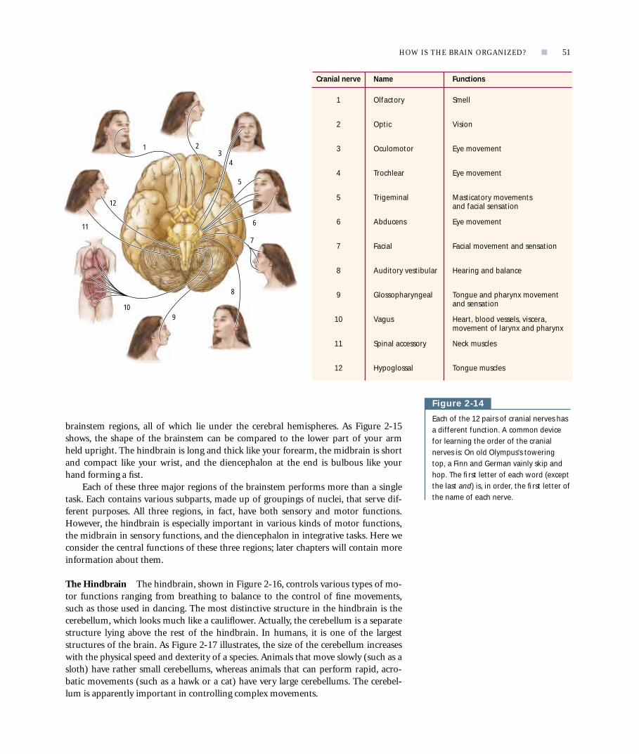
THE CRANIAL NERVESThe cranial nerves are all the nerves that link the brain to various parts of the head, asillustrated in Figure 2-14, as well as to the internal organs. Cranial nerves can have ei-ther afferent functions, such as inputs to the brain from the eyes, ears, mouth, andnose, or efferent functions, such as control of the facial muscles, tongue, and eyes.There are 12 pairs of cranial nerves. One set of 12 controls the left side of the head,whereas the other set controls the head’s right side. This arrangement makes sense forinnervating duplicated parts of the head (such as the eyes), but it is not so clear whyseparate nerves should control the right and left sides of a singular structure (such asthe tongue). Yet this is how the cranial nerves work. If you have ever received novo-caine for dental work, you know that usually just one side of your tongue becomesanesthetized because the dentist injects the drug into only one side of your mouth.The rest of the skin and muscles on each side of the head are similarly controlled bycranial nerves located on that side.
We consider many of the cranial nerves in some detail later when we deal withtopics such as vision and hearing. For now, you simply need to know that cranialnerves form part of the cranial nervous system, providing inputs to the brain fromthe head’s sensory organs and muscles and controlling head and face movements.
THE BRAINSTEMIt is now time to look at the brain itself, starting at its base with the region called thebrainstem. The brainstem begins where the spinal cord enters the skull and extendsupward to the lower areas of the forebrain. The brainstem receives afferent nervesfrom all of the body’s senses, and it sends efferent nerves to control all of the body’smovements except the most complex movements of the fingers and toes. The brain-stem, then, both produces movements and creates a sensory world. In some animals,such as frogs, the entire brain is largely equivalent to the brainstem of mammals orbirds. With this kind of brain, frogs get along quite well, indicating that the brainstemmust be a relatively sophisticated piece of machinery. If we had only a brainstem, wewould still be able to create a world, but it would be a far simpler world, more likewhat a frog experiences.
The brainstem can be divided into three regions: the hindbrain, the midbrain,and the diencephalon [dye en seff a lon], which is also sometimes called the “betweenbrain” because it borders upper parts of the brain. Figure 2-15 illustrates these three
50 ■ CHAPTER 2
p
Table 2-2 Anatomical Divisions of the Cranial Nervous System
Anatomical divisions Functional divisions Principal structures
Forebrain Forebrain Cerebral cortex
Basal ganglia
Limbic system
Brainstem Diencephalon Thalamus
Hypothalamus
Midbrain Tectum
Tegmentum
Hindbrain Cerebellum
Pons
Medulla oblongata
Reticular formation
Cranial nerves Cranial nerves 12 cranial nerves
On the CD, visit the module on theCentral Nervous System for a detailed,three-dimensional view of the brainstem.
Hindbrain. The embryonic part of thebrain that contains the brainstem andcerebellum; thought to coordinate move-ments and support movements of walkingand posture.
Midbrain. The middle part of the embry-onic brain, which, in the adult, containscircuits for hearing and seeing as well aswalking.
Diencephalon. The part of the brain thatcontains the hypothalamus, thalamus, andepithalamus; thought to coordinate manybasic instinctual behaviors, including tem-perature regulation, sexual behavior, andeating.
brainstem regions, all of which lie under the cerebral hemispheres. As Figure 2-15shows, the shape of the brainstem can be compared to the lower part of your armheld upright. The hindbrain is long and thick like your forearm, the midbrain is shortand compact like your wrist, and the diencephalon at the end is bulbous like yourhand forming a fist.
Each of these three major regions of the brainstem performs more than a singletask. Each contains various subparts, made up of groupings of nuclei, that serve dif-ferent purposes. All three regions, in fact, have both sensory and motor functions.However, the hindbrain is especially important in various kinds of motor functions,the midbrain in sensory functions, and the diencephalon in integrative tasks. Here weconsider the central functions of these three regions; later chapters will contain moreinformation about them.
The Hindbrain The hindbrain, shown in Figure 2-16, controls various types of mo-tor functions ranging from breathing to balance to the control of fine movements,such as those used in dancing. The most distinctive structure in the hindbrain is thecerebellum, which looks much like a cauliflower. Actually, the cerebellum is a separatestructure lying above the rest of the hindbrain. In humans, it is one of the largeststructures of the brain. As Figure 2-17 illustrates, the size of the cerebellum increaseswith the physical speed and dexterity of a species. Animals that move slowly (such as asloth) have rather small cerebellums, whereas animals that can perform rapid, acro-batic movements (such as a hawk or a cat) have very large cerebellums. The cerebel-lum is apparently important in controlling complex movements.
HOW IS THE BRAIN ORGANIZED? ■ 51
p
Figure 2-14
Each of the 12 pairs of cranial nerves hasa different function. A common devicefor learning the order of the cranialnerves is: On old Olympus’s towering top, a Finn and German vainly skip andhop. The first letter of each word (exceptthe last and) is, in order, the first letter ofthe name of each nerve.
1 23
4
5
7
8
910
11
12
6
Cranial nerve Name Functions
1 Olfactory Smell
2 Optic Vision
Oculomotor3 Eye movement
Trochlear Eye movement4
Trigeminal Masticatory movementsand facial sensation
5
Abducens Eye movement6
Facial Facial movement and sensation7
Auditory vestibular Hearing and balance8
Glossopharyngeal Tongue and pharynx movementand sensation
9
Vagus Heart, blood vessels, viscera,movement of larynx and pharynx
10
Spinal accessory Neck muscles11
Hypoglossal Tongue muscles12

As we look below the cerebellum at the rest of the hindbrain, we find that it iscomposed of three subparts: the reticular formation, the pons, and the medulla. Thereticular formation is a mixture of neurons and nerve fibers that gives this structurethe mottled appearance from which its name comes (the term reticular, as stated ear-lier, comes from the Latin word for “net”). We can visualize the reticular formation asbeing formed by a stack of poker chips lying on its side. Each chip has a special func-tion in stimulating the forebrain, such as in awakening from sleep. Not surprisingly,
52 ■ CHAPTER 2
p
Figure 2-16
The principal structures of the hindbrainare the cerebellum, pons, medulla, andreticular formation.
Figure 2-15
This medial view of the brain (upper left)shows the brainstem and its relation tothe cerebral hemisphere. The shapes and relative sizes of the parts of thebrainstem can be imagined to be like ahand and forearm, with thediencephalon being the fist, themidbrain being the wrist, and thehindbrain being the forearm.
Diencephalon
Hindbrain
Cerebellum
Midbrain
Fist (analogous to diencephalon)
Wrist(analogousto midbrain)
Forearm(analogousto hindbrain)
CerebellumMedulla
Reticularformation
Pons
Reticular formation. A part of the mid-brain in which nuclei and fiber pathwaysare mixed, producing a netlike appear-ance; associated with sleep–wake behav-ior and behavioral arousal.

the reticular formation is sometimes also called the reticular activating system. Theother two structures in the hindbrain, the pons and medulla, contain substructuresthat control many vital movements of the body. The pons has nuclei that receive in-puts from the cerebellum and provide a key bridge (the word pons means “bridge”)between the cerebellum and the rest of the brain. The medulla has several nuclei thatcontrol such vital functions as the regulation of breathing and the cardiovascular sys-tem. For this reason, a blow to the back of the head can kill you — your breathingstops if the control centers in the hindbrain are injured.
The Midbrain In the midbrain, shown in Figure 2-18, a specialized structureknown as the tectum receives a massive amount of information from the eyes andears. The tectum consists of two principal parts, the superior and inferior colliculi[kuh lik yew lee], which have visual and auditory functions, respectively. The opticnerve sends a large bundle of nerve fibers to the superior colliculus, whereas the infe-rior colliculus receives much of its input from auditory pathways. But the colliculi
HOW IS THE BRAIN ORGANIZED? ■ 53
p
Figure 2-17
(A) The cerebellum is necessary for fine,coordinated movements such as flightand landing in birds and prey catching incats. Like the sloth, animals that haveslow movements have relatively smallercerebellums. (B) Like the cerebrum, thecerebellum has a cortex, which has grayand white matter, and subcortical nuclei,as shown in this gross structure of thecerebellum.
Figure 2-18
The major structures of the midbrain arethe tectum and tegmentum. The tectum ismade up of the superior colliculus, whichreceives visual input, and the inferiorcolliculus, which receives auditory input.
Three-toed sloth
LeopardHawk
(A) (B)
Gray matter(cerebellar cortex)
White matter(cerebellar cortex)
Subcorticalnuclei
Tegmentum
Cerebellum
Superior colliculus (receives visual input)
TectumInferior colliculus (receives auditory input)
Tectum. The roof, or area above the ven-tricle, of the midbrain; its functions aresensory.

function not only to process sensory information. They also produce movements re-lated to sensory inputs, such as turning your head to see the source of a sound. Thisorienting behavior is not as simple as it may seem. To produce it, the auditory and vi-sual systems must share some sort of common “map” of the external world so that theears can tell the eyes where to look. If the auditory and visual systems had differentmaps, it would be impossible to use the two systems together. In fact, the colliculi alsohave a tactile map. After all, if you want to look at the source of an itch on your leg,your visual and tactile systems need a common representation of where that place is.
Lying below the tectum is the tegmentum. The tegmentum is not a single struc-ture but is composed of many nuclei, largely with movement-related functions. It hasseveral nuclei that control eye movements, the so-called red nucleus, controlling limbmovements, and the substantia nigra, connected to the forebrain; both the substantianigra and the forebrain are especially important in initiating movements.
The Diencephalon The diencephalon, shown in Figure 2-19, has more structuresthan the hindbrain and midbrain have, owing to its role in both motor and sensoryfunctions, as well as their integration. The two principal structures of the diencephalonare the hypothalamus and the thalamus. The hypothalamus is composed of about 22small nuclei, as well as fiber systems that pass through it. Attached to the base of thehypothalamus is the pituitary gland. Although comprising only about 0.3 percent ofthe brain’s weight, the hypothalamus takes part in nearly all aspects of behavior, in-cluding feeding, sexual behavior, sleeping, temperature regulation, emotional behavior,hormone function, and movement. The hypothalamus is organized more or less simi-larly in different mammals, largely because the control of feeding, temperature, and soon, is carried out similarly. But there are sex differences in the structures of some partsof the hypothalamus, which are probably due to differences between males and females
54 ■ CHAPTER 2
p
Tegmentum. The floor, or area belowthe ventricle, of the midbrain; has motorfunctions.
Hypothalamus. A part of the dien-cephalon that contains many nuclei asso-ciated with temperature regulation, eatingand drinking, and sexual behavior.
Thalamus. A part of the diencephalonthrough which all of the sensory systemprojects to reach the neocortex and thenprojects, through one region of the neo-cortex, to relay messages to another re-gion of the forebrain.
Figure 2-19
The diencephalon is composed of thethalamus, hypothalamus, and associatedpituitary gland. The hypothalamus andpituitary are at the base of the brain andlie above the roof of the mouth. Thepituitary gland lies adjacent to the opticchiasm, which is the place where the leftand right optic nerves (originating fromthe eyes) cross over. The hypothalamus iscomposed of many nuclei, each withdistinctly different functions. Thethalamus lies above the hypothalamus.The connections of only three of thethalamic nuclei are shown, but everythalamic nucleus connects to a discreteregion of cortex.
Medial geniculate nucleus
Optic nerve
Diencephalon Thalamus
Hypothalamus and pituitary gland Auditory nerve
Lateral geniculatenucleus
Dorsomedialnucleus(connects tofrontal lobe)
Hypothalamus
Pituitarystalk
Pituitarygland
Plug in the CD to examine the hypo-thalamus and the thalamus in threedimensions in the module on the CentralNervous System in the subsection on sub-cortical structures.

in activities such as sexual behavior and parenting. A criticalfunction of the hypothalamus is to control the body’s pro-duction of various hormones, which is accomplished by in-teractions with the pituitary gland.
The other principal structure of the diencephalon is thethalamus, which is much larger than the hypothalamus. Likethe hypothalamus, the thalamus contains about 20 nuclei, al-though the nuclei in the thalamus are much larger than thosein the hypothalamus. Perhaps the most distinctive function ofthe thalamus is to act as a kind of gateway for sensory infor-mation traveling to the cerebral cortex. All of the sensory sys-tems send inputs to the thalamus, which then relays this in-formation to the cortex. The optic nerve, for example, sendsinformation through a large bundle of fibers to a region ofthe thalamus known as the lateral geniculate nucleus. In turn,the lateral geniculate nucleus processes some of this informa-tion and then sends it to the visual region of the cortex. Anal-ogous regions of the thalamus receive auditory and tactile in-formation, which is subsequently relayed to the respectiveauditory and tactile cortical regions. Some thalamic regionsare not sensory in function. These regions have motor func-tions or perform some sort of integrative task. An example ofa region with an integrative function is the dorsomedial thala-mic nucleus. It has connections to most of the frontal lobe ofthe cortex. We return to the thalamic sensory nuclei in Chapters 8 through 10, where weexamine how sensory information is processed. Other thalamic regions are consideredin Chapters 11 and 13, where we explore motivation and memory.
THE FOREBRAINThe forebrain, shown in Figure 2-20, is the largest region of the mammalian brain. Itsthree principal structures are the cortex, the limbic system, and the basal ganglia.Extending our analogy between the brainstem and your forearm, imagine that the“fist” of the brainstem (the diencephalon) is thrust inside a watermelon. The water-melon represents the forebrain, with the rind being the cortex and the fruit inside be-ing the limbic system and the basal ganglia. By varying the size of the watermelon, wecan vary the size of the brain. In a sense, this is what evolution has done. The fore-brain varies considerably in size across species.
The three principal structures of the forebrain are the largest parts of the mam-malian brain, and each has multiple functions. To summarize these functions briefly,the cortex regulates mental activities such as perception and planning, the basal gan-glia control movement, and the limbic system regulates emotions and behaviors thatrequire memory. Because we encounter each of these structures in detail later in thisbook, they are only briefly presented here.
The Cortex There are actually two types of cortex. The first type, called neocortex,has six layers of gray matter on top of a layer of white matter. The neocortex is the tis-sue that is visible when we view the brain from the top or the side, as in two of theviews in Figure 2-6. This cortex is unique to mammals, and its primary function is tocreate a perceptual world. The second type of cortex, sometimes called limbic cortex,has three or four layers of gray matter on top of a layer of white matter. This tissue isnot easily observed on the outside surface of the human brain, except for where itforms the cingulate cortex, which lies just above the corpus callosum (see Figure 2-24).
HOW IS THE BRAIN ORGANIZED? ■ 55
p
Forebrain. The most anterior part of theembryonic brain; contains the basal gan-glia and the neocortex and is thereforethought to coordinate advanced cognitivefunctions such as thinking, planning, andlanguage.
Cortex (neocortex). Newest layer ofthe forebrain, forming the outer layer or“new bark” and composed of about sixlayers.
Limbic system. Consists of structuresthat lie between the neocortex and thebrainstem and form a hypothetical func-tional system that controls affective be-havior and certain forms of memory; in-cludes the cingulate cortex andhippocampus.
Basal ganglia. A group of structures inthe forebrain that are located just beneaththe neocortex and have connections tothe thalamus and to the midbrain; thoughtto have motor functions that coordinatethe movements of the limbs and the body.
Basal ganglia(caudate nucleus,putamen, globuspallidus)
Cerebralcortex
Amygdala
Hippocampus
Figure 2-20
The major structures of the forebraininclude the cerebral cortex, the basalganglia, and the limbic system of whichthe amygdala and hippocampus areshown.

The limbic cortex is more primitive than the neocortex. It is found in the brains ofother animals in addition to mammals, especially in birds and reptiles. This cortex isthought to play a role in controlling motivational states. Although anatomical andfunctional differences exist between the neocortex and the limbic cortex, the distinc-tions are not critical for most discussions in this book. Therefore, we will usually referto both types of tissue simply as cortex.
Measured by volume, the cortex makes up most of the forebrain, comprising 80 percent of the brain overall. It is the brain region that has expanded the most dur-ing mammalian evolution. The human neocortex has an area as large as 2500 squarecentimeters but a thickness of only 1.5 to 3.0 millimeters. This area is equivalent toabout four pages of this book. (In contrast, a chimpanzee has a cortical area equiva-lent to about one page.) The pattern of sulci and gyri formed by the folding of thecortex varies across species. Some species, such as rats, have no sulci or gyri, whereascarnivores have gyri that form a longitudinal pattern (look back at Figure 2-2). In hu-mans, the sulci and gyri form a more diffuse pattern.
As Figure 2-6 shows, the human cortex consists of two nearly symmetrical hemi-spheres, the left and the right, which are separated by the longitudinal fissure. Eachhemisphere is subdivided into the four lobes introduced in Chapter 1: the frontal, thetemporal, the parietal, and the occipital. These names correspond to the skull bonesoverlying each hemisphere. Unfortunately, there is little relation between bone loca-tion and brain function. As a result, the lobes of the cortex are rather arbitrarily de-fined regions that include many different functional zones.
Fissures and sulci often establish the boundaries of cortical lobes. For instance, inhumans, the central sulcus and lateral fissure form the boundaries of each frontallobe. They also form the boundaries of each parietal [pa rye i tul] lobe, but in thiscase the lobes lie behind the central sulcus, not in front of it (refer again to Figure 2-6). The lateral fissure demarcates each temporal [tem por ul] lobe as well, formingits dorsal (top) boundary. The occipital [ok sip i tul] lobes are not so clearly separatedfrom the parietal and temporal lobes, because there is no large fissure to mark theirboundaries. Traditionally, the occipital lobes are defined on the basis of otheranatomical features, which are presented in Chapter 8.
The layers of the cortex have several distinct characteristics. First, different layershave different cell types, as shown in Figure 2-11. Second, the density of the cells varies,ranging from virtually no cells in layer I (the top layer) to very dense cell packing inlayer IV. Third, there are other differences in appearance related to the functions of cor-tical layers in different regions. These visible differences led neuroanatomists of theearly twentieth century to make maps of the cortex, like the one in Figure 2-21A.
56 ■ CHAPTER 2
p
Look at the CD for a three-dimensionalmodel of the cortex along with photographs of cortical sections in themodule on the Central Nervous System.
Click onto the Web site to see how thecortex looks in other animals at www.worthpublishers.com/kolb/chapter2.
(A) (B) Touch (3–1–2)
Vision (17)Hearing (41)
12
34
56
78
9
10
11
19
17
1819
20
21
22
37
42
4046
45 44
47
43
39
18
Figure 2-21
(A) In his cytoarchitectonic map of thecortex, Brodmann defined areas by theorganization and characteristics of thecells. A few numbers are missing fromthe original sources, including 12through 16 and 48 through 51. (B) Thisschematic map shows the regions associ-ated with the simplest sensory percep-tions of touch, vision, and audition. Aswe shall see, the areas of the cortex pro-cessing sensory information are fargreater than these basic areas.
LongitudinalfissureLeft
hemisphere
Righthemisphere
Lefthemisphere
Righthemisphere
Frontallobe
Temporallobe
Parietallobe
Occipitallobe

Because these maps are based on cell characteristics, the subject ofcytology, they are called cytoarchitectonic maps. As the early neu-roanatomists suspected, the characteristics of cells in a particularregion of the cortex are related to that region’s function. For exam-ple, sensory regions of the parietal lobe, shown in red in Figure 2-22, have a distinct layer IV, whereas motor regions of the frontallobe, shown in blue in the same illustration, have a more distinc-tive layer V. Layer IV is an afferent layer, whereas layer V is an effer-ent one. It makes sense that a sensory region would have a large in-put layer, whereas a motor region would have a large output layer.Finally, there are chemical differences in the cells in different re-gions of the cortex. These differences can be revealed by coloringcortical tissue with stains that have affinities for specific chemicals.Some regions are rich in one chemical, whereas others are rich inanother. These differences are presumably related to functionalspecialization of different areas of the cortex.
There is one significant difference between the organizationof the cortex and the organization of other parts of the brain. Un-like most brain structures that connect to only selective brain re-gions, the cortex is connected to virtually all other parts of thebrain. The cortex, in other words, is the ultimate meddler. It takespart in everything. This fact not only makes it difficult to identifyspecific functions of the cortex, but also complicates our study ofthe rest of the brain because the cortex’s role in other brain re-gions must always be considered.
To illustrate, consider your perception of clouds. Undoubt-edly, you have gazed up at clouds on a summer’s day and imagined that they look likefamiliar shapes. You see in them galleons, elephants, faces, and countless other objects.Although a cloud does not really look exactly like an elephant, you can concoct an im-age of one if you impose your cortex’s imagination on the sensory inputs. This kind ofcortical activity is known as top-down processing because the top level of the nervoussystem, the cortex, is influencing how information is processed in lower regions—inthis case the midbrain and hindbrain. The cortex influences many things besides theperception of objects. It influences our cravings for foods, our lust for things (or peo-ple), and how we interpret the meaning of abstract concepts such as words. The cortexis the ultimate creator of our reality, and one reason that it serves this function is that itis so well connected.
The Basal Ganglia The basal ganglia are a collection of nuclei that lie within theforebrain just below the white matter of the cortex. The three principal structures ofthe basal ganglia, shown in Figure 2-23 on page 58, are the caudate nucleus, the puta-men, and the globus pallidus. Together with the thalamus and two closely associatedstructures, the substantia nigra and subthalamic nucleus, the basal ganglia form a sys-tem that functions primarily to control certain aspects of movement.
We can observe the functions of the basal ganglia by analyzing the behavior ofpeople who have one of the many diseases that interfere with the normal functioningof these nuclei. For instance, people afflicted with Parkinson’s disease, one of the mostcommon disorders of movement in the elderly, take short, shuffling steps, have bentposture, and often require a walker to get around. Many have an almost continualtremor of the hands and sometimes of the head as well. (We return to this disorder inChapter 10.) Another example of a disorder of the basal ganglia is Tourette’s syn-drome, characterized by various forms of tics, involuntary noises (including curse
HOW IS THE BRAIN ORGANIZED? ■ 57
p
Visit the CD for a three-dimensionalmodel of the basal ganglia in the sectionon subcortical structures in the moduleon the Central Nervous System.
Motor cortex Sensory cortex
I
Input of sensoryinformation
Output to otherparts of brain
Integrativefunctions
II
III
IV
V
VI
I
II
III
IV
V
VI
Motor cortex Sensory cortex
Figure 2-22
As this comparison of cortical layers inthe sensory and motor cortices shows,layer IV is relatively thick in the sensorycortex and relatively thin in the motorcortex. Afferents go to layer IV (from thethalamus) as well as to layers II and III.Efferents go to other parts of the cortexand to the motor structures.

words and animal sounds), and odd, involuntary movements of the body, especiallyof the face and head. Neither Parkinson’s disease nor Tourette’s syndrome is a disor-der of producing movements, as in paralysis. Rather they are disorders of controllingmovements. The basal ganglia, therefore, must play a role in the control and coordi-nation of movement patterns, not in activating the muscles.
The Limbic System In the 1930s, psychiatry was dominated by the theories of Sig-mund Freud, who emphasized sexuality and emotion in understanding human be-havior. At the time, regions controlling these behaviors had not been identified in thebrain, but there was a group of brain structures, collectively called the limbic system,that as yet had no known function. It was a simple step to thinking that perhaps thelimbic system played a central role in sexuality and emotion. One sign that this hy-pothesis might be right came from James Papez, who discovered that people with ra-bies had infections of limbic structures, and one of the symptoms of rabies is emo-tional blunting. We now know that such a simple view of the limbic system isinaccurate. In fact, the limbic system is not a unitary system at all, and, although somelimbic structures have roles in emotion and sexual behaviors, limbic structures serveother functions, too, including memory.
58 ■ CHAPTER 2
p
Figure 2-23
This frontal section of the cerebralhemispheres shows the basal gangliarelative to the surrounding structures.Two associated structures, the substantianigra and subthalamic nucleus, also areillustrated.
Figure 2-24
This medial view of the right hemisphereillustrates the principal structures of thelimbic system, including the cingulatecortex, the hippocampus, and theamygdala.
Caudatenucleus
Thalamus
Lateral ventricle
Corpus callosum
Putamen Basalganglia
Basalganglia
Globuspallidus
Subthalamicnucleus
Substantianigra
Amygdala
Cingulate cortex(limbic cortex)
Temporallobe
Hippocampus(buried in temporal lobe)

The principal structures of the limbic system are shown in Figure 2-24. They in-clude the amygdala [a mig da la], the hippocampus, and the cingulate cortex, which liesin the cingulate gyrus. Removal of the amygdala produces truly startling changes inemotional behavior. For example, a cat with the amygdala removed will wanderthrough a colony of monkeys, completely undisturbed by their hooting and threats.No self-respecting cat would normally be caught anywhere near such bedlam. Thehippocampus, the cingulate cortex, and associated structures have roles in certainmemory functions, as well as in the control of navigation in space.
The Olfactory System At the very front of the brain are the olfactory bulbs, the or-gans responsible for our sense of smell. The olfactory system is unique among thesenses, as Figure 2-25 shows, because it is almost entirely a forebrain structure. Unlikethe other sensory systems, which send most of their inputs from the sensory receptorsto the midbrain and thalamus, the olfactory bulb sends most of its inputs to a special-ized region of the cortex lying on the bottom of the brain. This region is known as thepyriform cortex. Compared with the olfactory bulbs of animals such as rats and dogs,which depend more heavily on the sense of smell than we do, the human olfactorybulb is relatively small. Because the sense of smell tends to be less important in hu-mans than the senses of vision, hearing, and touch, we will not consider the olfactorysystem in any detail.
The Spinal Nervous SystemAlthough producing movements of the body is one of the functions of the brain, it isultimately the spinal nervous system that controls these movements. To understandhow important the spinal nervous system is, think of the old saying “running aroundlike a chicken with its head cut off.” This saying refers to the spinal nervous system atwork. When a chicken’s head is lopped off to provide dinner for the farmer’s family,the chicken is still capable of running around the barnyard until it collapses from lossof blood. The chicken accomplishes this feat with its spinal nervous system, becausethat system can act independently of the brain.
You can demonstrate movement controlled by the spinal nervous system in yourown body by tapping your patellar tendon, just below your kneecap (the patella), asshown in Figure 2-26 on page 60. Your lower leg kicks out and, try as you might, it isvery hard to prevent the movement from occurring. Your brain, in other words, hastrouble inhibiting the spinal nervous system reaction. This type of automatic move-ment is known as a spinal reflex, a topic we return to in Chapter 10.
The spinal nervous system is composed of both the spinal cord, which lies insidethe bony spinal column, and the nerves running to and from the skin, joints, andmuscles. As Figure 2-27 shows (see page 60), the spinal column is made up of a seriesof small bones called vertebrae that are categorized into five groups: the cervical, tho-racic, lumbar, sacral, and coccygeal. You can think of each vertebra (the singular of ver-tebrae) within these five groups as a very short segment of the spinal column. Thespinal cord within each vertebra functions as that segment’s “minibrain.”
This arrangement of having so many minibrains within the spinal column mayseem a bit odd, but it has a long evolutionary history. Think of a simple animal, suchas a worm, which evolved long before humans did. A worm’s body is a tube dividedinto segments. Within that tube is another tube, this one of neurons, which also issegmented. Each of the worm’s nervous system segments receives fibers from sensoryreceptors in the part of the body adjacent to it, and that nervous system segmentsends fibers back to the muscles in that body part. Each segment, therefore, works
HOW IS THE BRAIN ORGANIZED? ■ 59
p
Figure 2-25
The olfactory bulb lies at the base of thehuman brain and is connected toreceptor cells that lie in the nasal cavity.Although relatively small in humans, theolfactory bulb is larger in animals, suchas rats or cats, that rely more heavily onthe sense of smell.
Pyriformcortex
To pyriformcortex
Sensory input from nose
Olfactory bulb

relatively independently, although fibers interconnect the segments and coordinatetheir activities. As vertebrates evolved a spinal column, this segmental organizationwas maintained. The vertebrae correspond to the segments of the worm’s nerve tube,and the nerves running in and out of a vertebra’s section of the spinal cord send in-formation to and from muscles and sensory receptors at that particular level of thebody.
A complication arises in animals that have limbs. The limbs may originate at onesegment level, but they extend past other segments of the spinal column. Your shoul-ders, for example, may begin at C3 (cervical segment 3), but your arms hang downwell past the sacral segments. So, unlike the worm, which has nerve-tube segmentsthat connect to body segments directly adjacent to them, human body segments ap-pear to be in a strange patchwork pattern, as shown in Figure 2-27B.
Regardless of their complex pattern, however, the segments of our bodies stillcorrespond to segments of the spinal cord. Each of these body segments is called adermatome (meaning “skin cut”). A dermatome has both a sensory nerve, whichsends information from the skin, joints, and muscles to the spinal cord, and a motornerve, which controls the movements of the muscles in that particular segment of thebody. These sensory and motor nerves are known as peripheral nerves, and they arefunctionally equivalent to the cranial nerves of the head. Whereas the cranial nervesreceive information from sensory receptors in the eyes, ears, facial skin, and so forth,the peripheral nerves receive information from sensory receptors in the rest of the
60 ■ CHAPTER 2
p
Figure 2-27
(A) The spinal column showing the vertebrae is illustrated in this saggitalview. There are five segments: cervical(C), thoracic (T) , lumbar (L), sacral (S),and coccygeal. (B) Each spinal segmentcorresponds to a region of body surface(dermatome) that is identified by thesegment number (examples are C5 and L2).
Figure 2-26
In the knee-jerk reflex (also known as the patellar reflex), when the patellartendon is struck lightly, the lower legflexes out “reflexively.”
(A) (B)
C8
C3C4C5C6C7
T1T2T3T4T5T6T7T8T9T10T11T12L1L2L3L4L5S1
S1
S2
S2
S3S4S5
C2
C7
L5L5
Thoracicnerves
Lumbarnerves
Coccygealsegment
Sacralnerves
Cervicalnerves
Spinal cordDermatomes
Vertebrae (spinal column)
C1C2C3C4C5C6C7C8T1T2T3T4T5T6T7T8T9
T10T11T12
L1
L2
L3
L4
L5
S1
S2
S3
S4
S5Dermatome. Area of the skin suppliedwith afferent nerve fibers by a singlespinal-cord dorsal root.
Patellartendon

body. Similarly, whereas the cranial nerves move the muscles of the eyes, tongue, andface, the peripheral nerves move the muscles of the limbs and trunk. Like the cranialnervous system, the spinal nervous system is also two sided. The left side of the spinalcord controls the left side of the body, and the right side of the spinal cord controlsthe body’s right side.
You are now ready to look inside the spinal cord to see how it is structured.Figure 2-28 shows a cross section of it. Look first at the fibers entering the spinalcord’s dorsal side (in the body of a normally upright animal such as a human, thedorsal side means the back, as illustrated in Figure 2-3). These dorsal fibers carry in-formation from the body’s sensory receptors. The fibers collect together as they entera spinal-cord segment, and this collection of fibers is called a dorsal root. Fibers leav-ing the spinal cord’s ventral side (ventral here means the front) carry informationfrom the spinal cord to the muscles. They, too, bundle together as they exit the spinalcord and so form a ventral root. As you can see in the cross section at the top of thedrawing in Figure 2-28, the outer part of the spinal cord consists of white matter, ortracts. These tracts are arranged so that, with few exceptions, the dorsal tracts are sen-sory and the ventral tracts are motor. The inner part of the cord, which has a butterflyshape, is gray matter. It is composed largely of cell bodies.
HOW IS THE BRAIN ORGANIZED? ■ 61
p
Figure 2-28
The spinal cord runs inside the vertebralcolumn. Part of the internal nervoussystem (the sympathetic nerve chain) liesoutside the spinal column. The graymatter is made up largely of cell bodies,whereas the white matter is made up offiber tracts that ascend and descend toand from the brain, respectively. Notethat dorsal is in front in this diagram sothat you can imagine that the skin andmuscles of the back have been removedto allow this view of the spinal cord andvertebral column. Below, a photo showsthe exposed spinal column.
Sympatheticchain
Spinalnerve
White matter
Gray matter
Ventral root(motor nerves)
Dorsal root(sensory nerves)
Layers of dura mater
Vertebra
Ventral
Dorsal
Dorsal fibers carry information from body to spinal cord.
Ventral fibers carry information from spinal cord to muscles.
Gray matter consists mostly of cell bodies.
White matter is arranged in dorsal tracts and ventral tracts.
Go to the CD and find the spinal cordarea of the Central Nervous System mod-ule. There you can see various sections ofthe spinal cord as well as a rotatablemodel of a spinal cord.
Bass
ett/
Visu
als
Unl
imit
ed

The observation that the dorsal side of the spinal cord is sensory and the ventralside is motor is known as the law of Bell and Magendie, one of the nervous system’svery few laws. The Bell and Magendie law, combined with an understanding of thespinal cord’s segmental organization, enables neurologists to make quite accurate in-ferences about the location of spinal-cord damage or disease on the basis of changesin sensation or movement that patients experience. For instance, if a person experi-ences numbness in the fingers of the left hand but can still move the hand fairly nor-mally, one or more of the dorsal nerves in spinal-cord segments C7 and C8 must bedamaged. In contrast, if sensation in the hand is normal but the person cannot movethe fingers, the ventral roots of the same segments must be damaged. The topic of di-agnosing spinal-cord injury or disease is further discussed in “Magendie, Bell, andBell’s Palsy.”
So far we have emphasized the segmental organization of the spinal cord, but thespinal cord must also somehow coordinate inputs and outputs across different seg-ments. For example, many body movements require the coordination of muscles thatare controlled by different segments, just as many sensory experiences require the co-ordination of sensory inputs to different parts of the spinal cord. How is this coordi-nation of spinal-cord activities accomplished? The answer is that the spinal-cord seg-ments are interconnected in such a way that adjacent segments can operate togetherto form rather complex coordinated movements.
The integration of spinal-cord activities does not require the brain’s participa-tion, which is why the headless chicken can run around in a reasonably coordinatedway. Still, there must be a close working relation between the brain (and therefore thecranial nervous system) and the spinal nervous system. Otherwise, how could we con-sciously plan and execute our voluntary actions? Somehow information must be re-layed back and forth between the cranial and the spinal systems. Examples of thissharing of information are numerous. For instance, tactile information from sensorynerves in the skin travels not just to the spinal cord, but also to the cerebral cortexthrough the thalamus. Similarly, the cerebral cortex and other brain structures cancontrol movements because of their connections to the ventral roots of the spinalcord. So, even though the cranial and spinal nervous systems can function indepen-dently, the two are intimately connected in their functions.
The Internal Nervous SystemThe internal nervous system (which, as mentioned earlier, is another term for the au-tonomic nervous system) is a hidden partner in controlling behavior. Even withoutour conscious awareness, it stays on the job to keep the heart beating, the liver releas-ing glucose, the pupils of the eyes adjusting to light, and so forth. Without the internalnervous system, life would quickly cease, because the internal nervous system is incharge of regulating all the body’s internal organs and glands. Although it is possibleto learn to exert some conscious control over some of the internal nervous system’sactivities, such conscious interference is unnecessary. One important reason is thatthe internal nervous system must keep working during sleep, a time when consciousawareness is off-duty.
The internal nervous system is composed of two opposing subsystems: the sym-pathetic and the parasympathetic. These subsystems work in opposition to eachother, with the sympathetic acting to arouse the body for action and the parasympa-thetic acting to quiet down the body. For example, the sympathetic system stimulatesthe heart to beat faster and inhibits digestion, whereas the parasympathetic systemslows the heartbeat and stimulates digestion.
62 ■ CHAPTER 2
p

The internal nervous system is connected to the rest of the nervous system, espe-cially to the spinal nervous system. In fact, activation of the sympathetic system startsin the thoracic and lumbar spinal-cord regions. But the spinal nerves do not directlycontrol the target organs. Rather, the spinal cord is connected to autonomic controlcenters, which are collections of cells called ganglia. It is the ganglia that actually con-trol the organs. The sympathetic system ganglia are located near the spinal cord,forming a chain that runs parallel to the cord, as illustrated in Figure 2-29 on page 64.The parasympathetic system also is connected to the spinal cord — in this case, to thesacral region. But an even larger part of it derives from three cranial nerves: the vagusnerve, which controls most of the internal organs, and the facial and oculomotornerves, which control salivation and pupil dilation, respectively. In contrast with thesympathetic system, the parasympathetic system connects with ganglia that are nearthe target organs, as shown in Figure 2-29.
HOW IS THE BRAIN ORGANIZED? ■ 63
p
François Magendie, a volatile and committed French experi-
mental physiologist, reported in a three-page paper in 1822
that he had succeeded in cutting the dorsal and ventral roots
of puppies, animals in which the roots are sufficiently segre-
gated to allow such surgery. Magendie found that cutting the
dorsal roots caused loss of sensation, whereas cutting the
ventral roots caused loss of movement. Eleven years earlier,
however, a Scotsman named Charles Bell also had proposed
functions for these nerve roots based on anatomical infor-
mation and the results of somewhat inconclusive experi-
ments on rabbits. Although Bell’s findings were not identical
with Magendie’s, they were similar enough to ignite a con-
troversy. Bell hotly disputed Magendie’s claim to the discov-
ery of dorsal and ventral root functions. As a result, the prin-
ciple of sensory and motor segregation in the nervous
system has been given both researchers’ names: the law of
Bell and Magendie.
Magendie’s conclusive experiment on puppies was con-
sidered extremely important because it enabled neurologists
for the first time to localize nervous system damage from the
symptoms that a patient displays. Bell went on to describe
an example of such localized motor-nerve dysfunction,
which still bears his name—Bell’s palsy. Bell’s palsy is a fa-
cial paralysis that occurs when the motor part of the facial
nerve on one side of the head becomes inflamed (see the
accompanying photograph). The onset of Bell’s palsy is typi-
cally sudden. Often the stricken person wakes up in the
morning and is shocked to discover that the face is para-
lyzed on one side. He or she cannot open the mouth on that
side of the head or completely close the eye on that side.
Most people fully recover from Bell’s palsy, although it may
take several months. But, in rare instances, such as that of
Jean Chretien, the Prime Minister of Canada, paralysis of the
mouth is permanent.
Magendie, Bell, and Bell’s Palsy
Focus on Disorders A young man suffering from Bell’s palsy, a paralysis of the
facial nerve that causes weakness over one side of the face.He was photographed during an involuntary tic (a nervousreaction) that affects the right side of the face, causing hisright eye to close tightly.
Dr.
P. M
araz
zi /
Scie
nce
Phot
o Li
brar
y/
Phot
o Re
sear
cher
s

p
In ReviewWe began the chapter by taking a general look at the anatomy of the nervous system andthen, in this section, we started to make some guesses about what different nervous sys-tem structures might do. Traditional discussions of the nervous system distinguishbetween the central nervous system, which consists of the brain and spinal cord, and theperipheral nervous system, which encompasses everything else. An alternative catego-rization is based more on function and divides the nervous system into the cranial ner-vous system (which includes the brain and its connections to parts of the head), the
64 ■ CHAPTER 2
Figure 2-29
This diagram shows the pathways of theinternal nervous system, also called theautonomic nervous system. The internalnervous system consists of two parts, thesympathetic and the parasympathetic,that exert opposing effects on theorgans that they innervate. All of theautonomic fibers “stop” en route fromthe spinal cord to their target organs. Forthe sympathetic system, the fibers “stop”near the spinal cord in the celiacganglion. For the parasympatheticsystem, the fibers “stop” on individualparasympathetic ganglia that are foundnear the target organs.
Cervical
Thoracic
Lumbar
Sacral
Cervical
Oculomotornerve
Facial nerve
Vagusnerve
Thoracic
Lumbar
Sacral
Sympathetic divisionStimulation: “fight or flight”
Parasympathetic divisionInhibitory: “rest and digest”
Liver
PancreasPancreas
Sympatheticchainganglion
Stomach
IntestinesIntestines
Rectum
Contracts pupil
Inhibi
tssa
liv
ation
Inhibi
tssa
liv
ation
Relaxes airway
Inhibitsdige
stion
esoculgsetalumitS
esaeler
Contracts bladder
Cons
tricts
blood
vessels
Dilates bloodvessels
Stimulates ejaculation Stimulates erection
Stimulates bladder
Accelerates heartbeat Slows heartbeat
Constricts airway
Stimulates salivation
Dilates pupil Contracts pupil
Kidney
Sympatheticprevertebral ganglia
Genitals
Adrenal gland
Stimulates digestion
Gall bladder
Cranial Cranial
Liver
Lungs
Heart
Secretion ofadrenalin

spinal nervous system (which includes the spinal cord and its connections to and fromthe body’s muscles, joints, and skin), and the internal nervous system (which controls thebody’s internal organs). Each of these sections of the nervous system can be subdividedinto large subsections that are functionally distinct, such as the forebrain and hindbrainof the cranial nervous system. Similarly, within each of the subsections, we find morefunctional subregions, such as the limbic system and the basal ganglia of the forebrain.Finally, each of these functional systems can be further divided into areas that have theirown unique functions, such as the caudate nucleus, putamen, and globus pallidus of thebasal ganglia. The process of learning the anatomy of the nervous system is to work fromthe general to the more specific in each part of the nervous system and, in each case, toremember to associate structure with function.
THE FUNCTIONAL ORGANIZATION OF THE BRAINKnowing the parts of a car engine is the place to start in understanding how an en-gine works. But knowing the parts, unfortunately, is not enough. You also need someprinciples concerning how the parts work together. For example, even though youknow which part the carburetor is, you will not understand its function until yougrasp the principle of air and fuel mixing and igniting in the cylinders. Similarly, al-though you now know the basic parts of the nervous system, you need some generalprinciples to help you understand how these different parts work together. Table 2-3lists eight such principles, which form the basis for many discussions later in thisbook. You should spend the time needed to understand these principles fully beforemoving on to the next chapters.
Principle 1: The Sequence of Brain Processing Is “In Integrate Out”The parts of the brain make a great many connections with one another. Recall, forexample, the meddling cerebral cortex that appears to be connected to everything.This connectivity of the brain is the key to its functioning. The points of connection,known as synapses, allow cells in different brain regions to influence one another.Chapter 5 explains the organization of the synapse. The key point here is that most
HOW IS THE BRAIN ORGANIZED? ■ 65
p
Table 2-3 Principles of Brain Functioning
1. Information is processed in a sequence of ”in integrate out.“
2. There is a functional division between sensory and motor throughout the nervous system.
3. Inputs and outputs to the brain are crossed.
4. There is both symmetry and asymmetry in brain anatomy and function.
5. The nervous system operates by a juxtaposition of excitation and inhibition.
6. The nervous system has multiple levels of function.
7. The nervous system operates as a series of systems arranged both in parallel and hierarchically.
8. Functions in the brain are both localized in specific regions and distributed.

neurons have afferent (incoming) connections with tens or sometimes hundreds ofthousands of other neurons, as well as efferent (outgoing) connections to many othercells.
Figure 2-30 shows how these multiple connections enable neurons to integrate in-formation, creating new information. The inputs to a neuron at any given moment are“summed up,” and the neuron sends out signals to other neurons that incorporate thissummation. The summation is more than just a matter of adding up equally weightedinputs. Some inputs have a greater influence than others on the receiving neuron, andthe simultaneous occurrence of certain inputs may have effects that far exceed theirsimple sum. The summation of information, then, allows that information to betransformed in some way before being passed on to other neurons. This transforma-tion makes the summation process partly one of creating new information.
Figure 2-31 gives a simple example of the creation of new information in thebrain. This example begins with receptor cells that are located in the eyes’ retinas andare maximally responsive to light of a particular wavelength: red, green, or blue. Nowimagine a brain with neurons that receive inputs from one or more of these color-sensitive receptors. A neuron can receive inputs from only one receptor type, fromtwo receptor types, or from all three receptor types. A neuron receiving input fromgreen-type receptor cells would “know” only about green and would forward onlygreen information. In contrast, a neuron receiving input from both green- and red-type receptors would “know” about two colors and would forward a very differentmessage, as would a neuron receiving input from all three receptor types. Both neu-rons with more than one kind of input would sum the information that they get. In asense, they would create new information that did not previously exist. Such a neuron
66 ■ CHAPTER 2
p
For an animation of how neuronsintegrate information, go to the sectionon neural integration in the CD moduleon Neural Communication.
Figure 2-30
The neuron and the brain are devices forgathering, integrating, and sendinginformation. At the cellular level, each ofneurons 1 through 5 sends some messageto neuron 6, which essentially now“knows” what neurons 1 through 5signaled. This “knowledge” is a form ofintegration. The output from neuron 6 issent to neurons 7 and 8, whose activity isaffected by the “knowledge” of neuron6. Similarly, three neural areas, Athrough C, send their signals to area X,which can combine (integrate) theinformation from areas A through C. Theintegrated information is then sent toareas Y and Z. A similar concept can beapplied at the level of the brain as well.The world provides information to thebrain, which produces behavior.
Source Inputs Integration
Molecularlevel
Outputs
N1 = neuron 1
N4
N5
N1
N2
N3 N6
N7
N8
Nucleilevel
AreaA
AreaB
AreaX
AreaY
AreaZArea
C
Brainlevel World Brain Behaviors

might know that an object is both green and red, whereas the in-puts coming from each receptor contain information about onecolor only. This creation of new information is what is meant bythe “integration operation” of the brain.
The same principle holds for the functioning of a nucleuswithin the brain or of a layer of brain tissue, as illustrated in Fig-ure 2-30. In regard to a nucleus, the inputs to each neuron in a nu-cleus are not identical, so there are internal connections betweenthe neurons. These internal connections are known as intrinsicconnections. Several areas of the nucleus might each send differ-ent information to another area, which integrates that informa-tion and sends a combined message along to several other areas.This process is much like the summation of information in a sin-gle neuron, but, in this case, the summation takes place in a collec-tion of neurons. As a result of the summation, the output of all ofthe cells in the nucleus is changed. Once again, there is integration.
The logical extension of this discussion is to view the entire brain as an organ thatreceives inputs, creates information, and expresses thoughts about the world, as illus-trated in Figure 2-30. To the animal whose brain is engaged in this process, the cre-ation of information from inputs represents reality. The bigger the brain, the morecomplex the reality that can be created and, subsequently, the more complex thethoughts that can be expressed. The emergence of thought that enables consciousnessmay be the brain’s ultimate form of integration.
Principle 2: Sensory and Motor Divisions ExistThroughout the Nervous SystemYou learned earlier that the spinal cord has separate sensory and motor structures, de-scribed in the Bell and Magendie law. This segregation of sensory and motor func-tions exists throughout the nervous system. However, distinctions between motor andsensory functions become more subtle in places such as the forebrain.
SENSORY AND MOTOR DIVISIONS AT THE PERIPHERYThe peripheral nerves going to and from the spinal cord can be divided into sensoryand motor parts, as shown in Figure 2-28. In addition, the cranial nerves, too, havesensory and motor functions, although they are arranged somewhat differently fromthose in the peripheral nerves. Some of the cranial nerves are exclusively sensory;some are exclusively motor; and some have two parts, one sensory and one motor,much like the sensory and motor branches of peripheral nerves.
SENSORY AND MOTOR DIVISIONS IN THE BRAINThe hindbrain and midbrain are essentially extensions of the spinal cord; they devel-oped as simple animals evolved a brain at the anterior end of the body. It makes sense,therefore, that, in these lower brainstem regions, there should be a division betweenstructures having sensory functions and those having motor functions, with sensorystructures located dorsally and motor ones ventrally. Recall that an important func-tion of the midbrain is to orient the body to stimuli. This orientation requires bothsensory input and motor output. The midbrain’s colliculi, which are located dorsally,are the sensory component, whereas the tegmentum, which is ventral (below the colli-culi), is a motor structure that plays a role in controlling various types of movements,including orienting ones. Both these midbrain structures are illustrated in Figure 2-18.
HOW IS THE BRAIN ORGANIZED? ■ 67
p
B
Color in the world
Neuronin brain
Receptorcell
G
R
B
B G
B G R
Figure 2-31
Input and output cells differ with respectto their knowledge about the world.Receptor cells B (blue), G (green), and R(red) each code information about onehue in the world. Neurons B, B/G, andB/G/R receive this information and alter itto send a new message that weunderstand to be color.

Distinct sensory and motor nuclei are present in the thalamus, too, although theyare no longer located dorsally. Because all sensory information reaches the forebrainthrough the thalamus, it is not surprising to find separate nuclei associated with vi-sion, hearing, and touch. Separate thalamic nuclei also control movements. Other nu-clei have neither sensory nor motor functions. They have connections to cortical ar-eas, such as the frontal lobe, that perform more integrative tasks.
Finally, sensory and motor functions are divided in the cortex as well. This divi-sion exists in two ways. First, there are separate sensory and motor cortical regions.Some primarily process a particular sensory input, such as vision, hearing, or touch.Others control detailed movements of discrete body parts, such as the fingers. Second,the entire cortex can be viewed as being organized around the sensory and motor dis-tinction. For instance, layer IV of the cortex always receives sensory inputs, whereaslayers V and VI always send motor outputs, as shown in Figure 2-22. Layers I, II, andIII are integrative, providing a connection between sensory and motor operations.
Principle 3: The Brain’s Circuits Are CrossedA most peculiar organizational feature of the brain is that its inputs and outputs are“crossed.” Each of its halves receives sensory stimulation from the opposite (called con-tralateral) side of the body and controls muscles on the opposite side as well. Exam-ples of this crossed organization are shown in Figure 2-32. Crossed organization ex-plains why people with strokes in the left cerebral hemisphere may have difficulty insensing stimulation to the right side of the body or in moving body parts on the rightside. The opposite is true of people with strokes in the right cerebral hemisphere.
In humans, who have forward-facing eyes, the visual system is crossed in a morecomplex fashion than in animals with eyes on the sides of their head. This complexityis required because, if two eyes are facing forward instead of sideward, they inevitablysee much the same thing, except on the far sides of the field of vision. The problemwith this arrangement is that, to see an object, information about it must go to thesame place in the brain. Duplicate information cannot be sent to two different places.Figure 2-32 (right) shows how the brain solves this problem by dividing each eye’s vi-sual field into a left half and a right half. The information that either eye receives fromthe left half of its visual field is sent to the right side of the brain, and the informationthat either eye receives from the right half of its visual field is sent to the left side ofthe brain. The visual system is still crossed, but in a more complicated way than aresystems for other parts of the body.
One difficulty with a crossed nervous system is that the two sides of the worldmust be joined together somehow. This joining is accomplished by having numerousconnections between the left and right sides of the brain. The most prominent con-necting cable is the corpus callosum, shown in Figure 2-8. As stated earlier, the corpuscallosum connects the left and right cerebral hemispheres with about 200 million fibers.
Principle 4: The Brain Is Both Symmetrical andAsymmetricalYou know that the brain has two halves, the left and the right hemispheres, whichlook like mirror images of each other. Although these two halves are symmetrical inmany ways, they also have some asymmetrical features. This asymmetrical organiza-tion is essential for certain tasks. Consider speaking. If a language zone existed in bothhemispheres, each connected to one side of the mouth, we would have the strangeability to talk out of both sides of the mouth at once. This would not make talking
68 ■ CHAPTER 2
p
For an animation and illustration ofhow the visual system is crossed, go tothe section on the optic chiasm in theVisual System module on the CD.

easy, to say the least. One solution is to locate control of the mouth on one side of thebrain only. Organizing the brain in this way would allow us to speak with a singlevoice. A similar problem arises in controlling the body’s movement in space. Wewould not want the left and the right hemispheres each trying to take us to a differentplace. Again, the problem can be solved if a single brain area controls this sort of spa-tial processing.
In fact, processes such as language and spatial navigation are localized on onlyone side of the brain. Language is usually on the left side, and spatial functions areusually on the right. This asymmetrical organization is not unique to humans. Birdshave developed a solution to producing song by locating the control of singing in onehemisphere. Curiously, like human language, bird song also is usually located on thebrain’s left side. Why this similarity exists is not completely clear. But the key point isthat, in many species, the brain has both symmetrical and asymmetrical organization.
Principle 5: The Nervous System Works Through Excitation and InhibitionImagine that, while you are reading this page, the telephone rings. You stop reading,get up, walk to the telephone, pick it up, and talk to a friend who convinces you thatgoing to a movie would be more fun than reading. To carry out this series of actions,not only must your brain produce certain behaviors, it must also stop other behav-iors. When you were walking and talking, for example, you were not engaged in read-ing. To walk and talk, you had to first stop reading. Producing behavior, then, requiresboth initiating some actions and inhibiting others.
In talking about the nervous system, we refer to the initiation of an activity as ex-citation and the stoppage of an activity as inhibition. The juxtaposition of excitationand inhibition is a key principle of nervous system functioning. Recall, for example,that the sympathetic and parasympathetic systems produce opposite actions. The sym-pathetic system excites the heart, whereas the parasympathetic inhibits it. This generalprinciple of excitation and inhibition is central to how the nervous system works.
HOW IS THE BRAIN ORGANIZED? ■ 69
p
Contralateralside of cortex
Contralateralside of cortex
Right visual fieldLeft visual fieldRight visual fieldLeft visual field
Motor
SensoryMotor
Sensory
Motor
Sensory
Contralateralside of body Contralateral
side of body
Fixation point
Figure 2-32
(Left) This schematic representation of arat’s brain from a dorsal view shows theprojection of visual and somatosensoryinput to contralateral (opposite-side)areas of the cortex and the crossedprojection of the motor cortex to thecontralateral side of the body. The eyesof the rat are laterally placed, such thatmost of the input from each eye travelsto the opposite hemisphere. (Right) Inthe human head, the two eyes arefrontally placed. As a result, the visualinput is split in two so that input fromthe right side of the world as seen byboth eyes goes to the left hemisphereand input from the left side of the worldas seen by both eyes goes to the righthemisphere. The somatosensory input ofboth rats and humans is completelycrossed, so that information coming fromthe right paw or hand goes to the lefthemisphere. Note that although thediagram as drawn shows activity as goingdirectly to and from the brain, there areconnectors en route.
The same principle can be seen in the activity of individual neurons. Neurons canpass on information to other neurons either by being active or by being silent. That is,they can be “on” or “off.” Some neurons in the brain function primarily to excite otherneurons, whereas others function to inhibit other neurons. These excitatory and in-hibitory effects are produced by various chemicals that turn the neurons on or off.
Just as individual neurons can act to excite or inhibit other neurons, brain nuclei(or layers) can do the same to other nuclei (or layers). These actions are especially ob-vious in the motor systems. Inhibiting reading and initiating walking to the telephone,for example, result from the on and off actions of specific motor-system nuclei.
Now imagine that one of the nuclei that normally inhibits some type of move-ment is injured. The injury will result in an inability to inhibit that particular re-sponse. This symptom can be seen in people with frontal-lobe injury. Such people areoften unable to inhibit behaviors, such as talking at inappropriate times or using cer-tain words, such as curse words. In contrast, people with injury to the speech zones ofthe left hemisphere may be unable to talk at all, because the injury is in an area thatnormally initiates the behavior of speech. These contrasting symptoms lead to an im-portant conclusion: brain injury can produce either a loss of behavior or a release ofbehavior. Behavior is lost when the damage prevents excitatory instructions; behavioris released when the damage prevents inhibitory instructions.
Principle 6: The Central Nervous System HasMultiple Levels of FunctionYou have seen that similar sensory and motor functions are carried out in variousparts of the brain and spinal cord. But why are multiple areas with overlapping func-tions needed? After all, it would seem simpler to put all the controls for a certainfunction in a single place. Why bother with duplication?
The answer is that the mammalian brain is a product of evolution in which itchanged according to the “descent with modification” rule. As the brain evolved, newareas were added, but old ones were retained. As a result, there was a problem of whereto put the new regions. The simplest solution was to add them on top of the existingbrain. We can see this solution in the evolution from primitive vertebrates to amphib-ians to mammals. For instance, primitive vertebrates, such as fish, make only whole-body movements to swim, movements that are controlled by the spinal cord and hind-brain. Amphibians developed legs and corresponding neural control areas in thebrainstem. Mammals later developed new capacities with their limbs, such as indepen-dent limb movements and fine digit movements. These movements, too, required newcontrol areas, which were added in the forebrain. We therefore find three distinct areasof motor control in mammals: the spinal cord, the brainstem, and the forebrain.
At the beginning of the twentieth century, John Hughlings-Jackson suggested thatthe addition of new brain structures in the course of evolution could be viewed asadding new levels of nervous system control. The lowest level is the spinal cord, thenext level is the brainstem, and the highest level is the forebrain. These levels are notautonomous, however. To move the arms, the brainstem must use circuits in thespinal cord. Similarly, to make independent movements of the arms and fingers, suchas in tying a shoelace, the cortex must use circuits in both the brainstem and thespinal cord. Each new level offers a refinement and elaboration of the motor controlprovided by one or more lower levels.
We can observe the operation of functional levels in the behavior of people withbrain injuries. Someone whose spinal cord is disconnected from the brain cannot vol-untarily move a limb because the brain has no way to control the movement. But the
70 ■ CHAPTER 2
p
For more about the evolution of thebrain, go to the Web site for a connectionto the National Museum of Health andMedicine for photographs of brains fromall sorts of creatures and discussion ofbrain evolution.

limb can still move automatically to withdraw from a noxious stimulus because thecircuits for moving the muscles are still intact in the spinal cord. Similarly, if the fore-brain is not functioning but the brainstem is still connected to the spinal cord, a per-son can still move, but the movements are relatively simple: there is limited limb useand no digit control.
The principle of multiple levels of function can also be applied to the cortex inmammals, which evolved by adding new areas, mostly sensory-processing ones. Thenewer areas essentially added new levels of control that provide more and more ab-stract analysis of inputs. Consider the recognition of an object, such as a car. The sim-plest level of analysis recognizes the features of this object such as its size, shape, andcolor. A higher level of analysis recognizes this object as a car. And an even higherlevel of analysis recognizes it as Susan’s car with a dent in the fender. Probably thehighest levels are cortical regions that substitute one or more words for the object(Honda Civic, for instance) and can think about the car in its absence.
When we consider the brain as a structure composed of multiple levels of func-tion, it is clear that these levels must be extensively interconnected to integrate theirprocessing and create unified perceptions or movements. The nature of this connec-tivity in the brain leads to the next principle of brain function: the brain has bothparallel and hierarchical circuitry.
Principle 7: Brain Systems Are Organized BothHierarchically and in ParallelThe brain and spinal cord are two semiautonomous nervous systems organized intofunctional levels, and, even within a single level, more than one area may take part ina given function. How then, with these different systems and levels, do we eventuallyobtain a unified conscious experience? Why, when we look at Susan’s car, do we nothave the sense that one part of the brain is processing features such as shape while an-other part is processing color? Or why, when we tie our shoelaces, are we not awarethat different levels of motor control are at work to move our arms and fingers andcoordinate their actions? These questions are part of what is called the “binding prob-lem.” It focuses on how the brain ties together its various activities into a whole. Thesolution to the problem must somehow be related to the ways in which the parts ofthe nervous system are connected.
There are two alternative possibilities for wiring the nervous system: serial or par-allel circuits. A serial circuit hooks up in a series all the regions concerned with a givenfunction. Consider Susan’s car again. In a serial system, the information from the eyeswould go first to a region (or regions) that performs the simplest analysis — for exam-ple, the detection of specific properties, such as color and shape. This informationwould then be passed on to another region that sums up the information and identi-fies a car. The information would next proceed to another region that compares thiscar with stored images and identifies it as Susan’s car. Notice how the perceptualprocess entails the flow of information sequentially through the circuit. Because theregions in the chain range from simple to complex, this serial type of system is usuallycalled a simple hierarchical model. It is illustrated in Figure 2-33A on page 72.
In the 1960s and 1970s, neuropsychologists proposed models of hierarchical orga-nization to help in understanding how the brain might produce complex behaviors.One difficulty with such models, however, is that functionally related structures in thebrain are not always linked serially. Although the brain has many serial connections,many expected connections are missing. For example, within the visual system, onegroup of cortical areas is not connected with what appears to be a parallel group of areas.
HOW IS THE BRAIN ORGANIZED? ■ 71
p

One solution to this problem is to imagine multiple hierarchical systems that operate in parallel to one another but are also interconnected. Figure 2-33B illustratesthe flow of information in such a distributed hierarchical model. If you trace the infor-mation flow from the primary area to levels 2, 3, and 4, you can see that there are par-allel pathways. These multiple parallel pathways are also connected to each other. How-ever, the connections are more selective than those that exist in a purely serial circuit.
The visual system provides a good example of such parallel hierarchical pathways.Once again we return to Susan’s car—this time to one of its doors. As we look at the cardoor, one set of visual pathways processes information about its nature, such as its colorand shape, while another set of pathways processes information about door-relatedmovements, such as those required to open the door. It may surprise you to learn thatthese two systems are independent of each other, with no connections between them.Yet your perception when you pull the door open is not one of two different representa-
tions—the door’s size, shape, and color, on the one hand, and the opening move-ments, on the other. When you open the door, you have the impression of unity in
your conscious experience. Interestingly, the brain is organized into multiple par-allel pathways in all of its subsystems. Yet our conscious experiences are always
unified. We will return to this conundrum (and the binding problem) at the end of this book. For now, keep in mind that your common-sense im-pressions of how the brain works may not always be right.
Principle 8: Functions in the Brain Are BothLocalized and DistributedIn our consideration of brain organization, we have so far assumed that functions canbe localized in specific parts of the brain. This assumption makes intuitive sense, butit turns out to be controversial. One of the great debates in the history of brain re-search has been about what aspects of different functions are actually localized in spe-cific brain regions.
Perhaps the fundamental problem is that of defining a function. Consider language,for example. Language includes the comprehension of spoken words, written words,signed words (as in American Sign Language), and even touched words (as in Braille).Language also includes processes of producing words, both orally and in writing, as wellas constructing whole linguistic compositions, such as stories, poems, songs, and essays.Because the function that we call language has many aspects, it is not surprising thatthey reside in widely separated areas of the brain. We see evidence of this widespreaddistribution in language-related brain injuries. People with injuries in different loca-tions may selectively lose the abilities to produce words, understand words, read words,write words, and so forth. Specific language-related abilities, therefore, are found in spe-cific locations, but language itself is distributed throughout a wide region of the brain.
Memory provides another example of this same pattern. Memories can be ex-tremely rich in detail and can include sensory material, feelings, words, and muchmore. Like language, then, memory is not located in just one brain region. Rather, it isdistributed throughout a vast area of the brain.
Because many functions are both localized and distributed in the brain, damage to asmall brain region produces only focal symptoms. Massive brain damage is required tocompletely remove some function. For instance, a relatively small injury could impairsome aspect of language functioning, but it would take a very widespread injury to com-pletely remove all language abilities. In fact, one of the characteristics of dementing dis-eases, such as Alzheimer’s, is that people can have widespread deterioration of the cortexyet maintain remarkably normal language functions until late stages of the disease.
72 ■ CHAPTER 2
p
(A)
Primary
Secondary
Tertiary
(B)
Level 4
Level 3
Level 2
Primary
Level 3 Level 3
Level 4
Level 2
Level 4 Level 4
Figure 2-33
(A) This simple serial hierarchical modelof cortical processing is similar to thatfirst proposed by A. R. Luria. In Luria’sscheme, information was conceptualizedas being organized into three levels:primary, secondary, and tertiary. (B) InDaniel Felleman and David van Essen’sdistributed hierarchical model, there aremultiple levels in each of severalprocessing streams. Areas at each levelare interconnected with one another.

In ReviewKnowing the parts of a car engine or a brain, and some general notions of what theymight do, was only the beginning. The next step was to learn some principles concerninghow the parts work together. We have identified eight such principles that will allow usto proceed to a closer look, in the chapters that follow, at how the brain produces behav-ior. You will benefit from reviewing each of these principles with an eye toward under-standing the general concept being addressed, rather than simply memorizing the state-ment itself. For example, as you think about the principle stating that the nervous systemworks through excitation and inhibition, think about the balance created by this princi-ple’s application and what this balance means for the functioning brain. What wouldhappen, say, if you could not inhibit behavior? When we encounter the rules that governthe operation of neurons in the next four chapters, you will want to revisit theinhibition–excitation principle and again think about how it can relate to the way inwhich individual cells work to produce behavior.
SUMMARY1. What are some of the larger external and internal features of the brain? Under the
tough, protective meninges that covers the brain lie its two major structures —the larger cerebrum, divided into two hemispheres, and the smaller cerebellum.Both structures are covered with gyri (bumps) and sulci (cracks), some of whichare so deep that they are called fissures. At the base of the brain, where it joins thespinal cord, the brainstem is visible, as are the cranial nerves that run to the head.Cutting the brain in half reveals the fluid-filled ventricles inside it, as well as thewhite matter, gray matter, and reticular matter that make up its tissue. Also ap-parent is the corpus callosum, which joins the two hemispheres and the subcorti-cal regions below the cerebral cortex.
2. How can the nervous system be divided for functional analysis? The nervous systemis composed of subsystems that function semiautonomously. These subsystemsare the cranial, spinal, and internal nervous systems. The cranial nervous systemincludes the brain and the cranial nerves, which link the brain to various parts ofthe head. The spinal nervous system consists of the spinal cord and the peripheralnerves that enter and leave it, going to and from muscles, skin, and joints in thebody. The internal nervous system, which controls the body’s internal organs, isalso called the autonomic nervous system. It has two parts, the sympathetic andparasympathetic divisions. They work in opposition to each other, one arousingthe body for action and the other calming the body down.
3. What are the basic structures and functions of the lower part of the brain, called thebrainstem? The brainstem consists of three regions: the hindbrain, the midbrain, andthe diencephalon. The cerebellum is important in controlling complex movements.Three other structures are the reticular formation (which serves to activate the fore-brain), the pons (which provides a bridge from the cerebellum to the rest of thebrain), and the medulla (which controls such vital functions as breathing). In themidbrain, the tectum processes information from the eyes and ears and producesmovements related to these sensory inputs. Below it lies the tegmentum, which con-sists of many nuclei, largely with movement-related functions. Finally, the dien-cephalon consists of two main structures: the thalamus and the hypothalamus.
HOW IS THE BRAIN ORGANIZED? ■ 73
p

4. What are some important structures in the upper part of the brain, called the fore-brain? The forebrain is the largest region of the brain. Its outer surface is the cor-tex. Sulci, especially deep ones called fissures, form the boundaries of the fourlobes on each cerebral hemisphere: the frontal, the parietal, the temporal, and theoccipital. The cells of the cortex form distinctive layers based on their specializedfunctions. Interconnections exist between these layers and virtually all other partsof the brain. These extensive connections to other brain regions are essential tothe cortex with its directing role in top-down processing. The basal ganglia, lyingjust below the white matter of the cortex, are another important part of the fore-brain that primarily play a role in movement. Also important in the forebrain isthe limbic system. The part of the limbic system called the amygdala regulatesemotional behavior, whereas the hippocampus and the cingulate cortex both haveroles in memory and in navigating the body in space.
5. How does the spinal nervous system work? The spinal nervous system has sensoryinput from the skin, muscles, and joints of the body. It also has efferent connec-tions to the skeletal muscles, which makes it responsible for controlling the body’smovements. The spinal cord functions as a kind of minibrain for the nerves thatenter and leave a particular spinal segment. Each segment works relatively inde-pendently, although fibers interconnect them and coordinate their activities. Ac-cording to the law of Bell and Magendie, nerves entering a segment’s dorsal sidecarry information from sensory receptors in the body, whereas nerves leaving asegment’s ventral side carry information to the muscles.
6. What are some basic principles related to the functional organization of the brain?One principle is that the sequence of processing within the brain is “in inte-grate out,” in which the term integrate refers to the creation of new informa-tion as cells, nuclei, and brain layers sum the inputs that they receive from differ-ent sources. A second principle is that sensory and motor functions are separatedthroughout the nervous system, not just in the spinal system but in the brain aswell. A third principle is that the organization of the brain is crossed, meaningthat the right hemisphere is connected to the left side of the body, whereas the lefthemisphere is connected to the body’s right side. Fourth is the principle that thebrain, though largely symmetrical, also has asymmetrical organization appropri-ate for certain tasks, and fifth is the principle that the nervous system worksthrough a combination of excitatory and inhibitory signals. The sixth principle isthat the nervous system has multiple levels of function. Tasks are often duplicatedin these multiple levels, which range from older, more primitive ones to higherlevels that evolved more recently. Two final principles are that brain circuits areorganized both hierarchically and in parallel and that functions are both localizedand distributed in the brain.
74 ■ CHAPTER 2
p
basal ganglia, p. 55brainstem, p. 43cerebellum, p. 43cerebrum, p. 43cortex (neocortex), p. 55cranial nerve, p. 43dermatome, p. 60
diencephalon, p. 50forebrain, p. 55gray matter, p. 44hindbrain, p. 50hypothalamus, p. 54limbic system, p. 55midbrain, p. 50
reticular formation, p. 52reticular matter, p. 44subcortical regions, p. 45tectum, p. 53tegmentum, p. 54thalamus, p. 54white matter, p. 44
KEY TERMS

REVIEW QUESTIONS1. What are the three primary functions of the brain?
2. What features of the brain are visible from the outside?
3. Contrast the anatomical and functional divisions of the nervous system.
4. Expand the Bell and Magendie law to include the entire nervous system.
5. In what sense is the nervous system crossed?
6. In what sense is the activity of the nervous system a summation of excitatory andinhibitory processes?
7. What does it mean to say that the nervous system is organized into levels?
FOR FURTHER THOUGHT In the course of studying the effects of the removal of the entire cerebral cortex on thebehavior of dogs, Franz Goltz noticed that the dogs were still able to walk, smell, bark,sleep, withdraw from pain, and eat. He concluded that functions must not be local-ized in the brain, reasoning that only widely distributed functions could explain howthe dogs still performed all these behaviors despite so much lost brain tissue. On thebasis of the principles introduced in this chapter, how would you explain why thedogs behaved so normally in spite of having lost about one-quarter of the brain?
RECOMMENDED READINGDiamond, M. C., Scheibel, A. B., & Elson, L. M. (1985). The human brain coloring book. New
York: Barnes & Noble. Although a coloring book might seem an odd way to learn neu-roanatomy, many students find this book to be a painless way to study the relations be-tween brain structures.
Jerison, H. J. (1991). Brain size and the evolution of mind. New York: American Museum ofNatural History. What is the mind and why do we have language? These questions andmany more are discussed by the leading expert in brain evolution. This monograph is afascinating introduction to the issues surrounding why the brain grew larger in the pri-mate evolutionary branch and what advantage a large brain might confer in creating aricher sensory world.
Heimer, L. (1995). The human brain and spinal cord: Functional neuroanatomy and dissectionguide (2nd ed.). New York: Springer Verlag. If coloring books aren’t your thing, thenHeimer’s dissection guide to the human brain will provide a more traditional, and so-phisticated, guide to human brain anatomy.
Luria, A. R. (1973). The working brain. Harmondsworth, England: Penguin. Luria was a Rus-sian neurologist who studied thousands of patients over a long career. He wrote a seriesof books outlining how the human brain functions, of which The Working Brain is themost accessible. In fact, this book is really the first human neuropsychology book. Al-though many of the details of Luria’s ideas are now outdated, his general framework forhow the brain is organized is substantially correct.
Zeki, S. (1993). A vision of the brain. London: Blackwell Scientific. Humans are visual crea-tures. Zeki’s book uses the visual system as a way of introducing the reader to how thebrain is organized. It is entertaining and introduces the reader to Zeki’s ideas about howthe brain functions.
HOW IS THE BRAIN ORGANIZED? ■ 75
p
neuroscience interact ive
There are many resources available for
expanding your learning on line:
■ www.worthpublishers.com/kolb/chapter2Try some self-tests to reinforce your
mastery of the material in Chapter 2.
Look at some of the news updates re-
flecting current research on the brain.
You’ll also be able to link to some other
sites which will reinforce what you’ve
learned.
■ www.neurophys.wisc.edu/brainLink to brain atlases that include pho-
tographs, stained sections, and movie
clips from humans, monkeys, and even
a dolphin. Note the differences among
the brains (the increasing complexity as
you move from mouse to human) as
well as the similarities (try to find brain
nuclei that are the same across species).
■ www.williamcalvin.com/1990s/1998SciAmer.htmRead an article about the theory of
brain evolution from Scientific Ameri-
can.
On your CD-ROM you’ll be able to
quiz yourself on your comprehension
of Chapter 2. An entire module devoted
to the Central Nervous System includes
a rotatable, three-dimensional brain
and many sections of the brain you can
move through with the click of a
mouse. In addition, the Research
Methods module includes various CT
and MRI images of the brain, including
a video clip of a coronal MRI scan.