Embed Size (px)
Citation preview

200
mm
How to read printouts IOLMaster 700

Index
General Information......................IOL Power Calculation
a. Haigis Suite............................b. Multiformula.........................
Analysis Report..............................Biometry Data................................Reference Image............................
2
34567
Patient
Date of birth GenderPatient ID
Smith Carol1/20/1955 Female151769901
Doctor OperatorJohn Doe John Doe
IOLMaster 700 Version 1.14 Report dated 1/21/2015 5:08 PM created by Doe, John was printed. Page 1 of 7
Z E
I S S
| T
e m
p l
a t e
V
e r s
i o
n 0
.1
05/2
012
| -
C o
p y
r i g
h t
2 0
1 2
A
l l
r i g
h t
s r
e s
e r
v e
d
Date of calibration test: 1/19/2015 by: Surgeon Result: OKDate of measurement: 1/21/2015 n: 1.3375 CVD: 12.00 mmTarget refraction: plano Formula:
right leftAL: 22.30 mm (SD = 4 µm)
ACD: 2.99 mm (SD = 5 µm)LT: 3.96 mm (SD = 23 µm)
R: 7.76 mmR1: 7.83 mm @ 156ºR2: 7.69 mm @ 66º WTW: 11.9 mm
Δ D: -0.75 dpt @ 156º Visual acuity: ---SIA: +0.00 dpt @ 0º LVC: -Ref: ---LS: Phakic; VS: Vitreous body
AL: 22.19 mm (SD = 6 µm)ACD: 2.98 mm (SD = 7 µm)
LT: 3.94 mm (SD = 11 µm)R: 7.72 mm
R1: 7.81 mm @ 40ºR2: 7.63 mm @ 130º WTW: 11.9 mm
Δ D: -1.03 dpt @ 40º Visual acuity: ---SIA: +0.00 dpt @ 0º LVC: -Ref: ---LS: Phakic; VS: Vitreous body
Comment
A0: A1: A2:+1.309 +0.400 +0.100
IOL (D) Ref (D)+26.50+26.00
+25.00+24.50
-0.56-0.20
+0.52+0.87
A0: A1: A2:+0.978 +0.400 +0.100
IOL (D) Ref (D)+26.00+25.50
+24.50+24.00
-0.79-0.42
+0.32+0.69
A0: A1: A2:+1.309 +0.400 +0.100
IOL (D) Ref (D)+27.00+26.50
+25.50+25.00
-0.84-0.47
+0.25+0.61
A0: A1: A2:+0.978 +0.400 +0.100
IOL (D) Ref (D)+26.00+25.50
+24.50+24.00
-0.70-0.33
+0.41+0.77
A0: A1: A2:+0.647 +0.400 +0.100
IOL IOL IOL Ref Ref Ref RefSE Cyl Axis SE Sph Cyl Axis
+25.00+24.50
+23.50+23.00
+0.75+0.75
+0.75+0.75
66º66º
66º66º
-0.62-0.24
+0.51+0.88
-0.54-0.16
+0.60+0.97
-0.17-0.17
-0.17-0.17
156º156º
156º156º
A0: A1: A2:+0.647 +0.400 +0.100
IOL IOL IOL Ref Ref Ref RefSE Cyl Axis SE Sph Cyl Axis
+25.50+25.00
+24.00+23.50
+1.50+1.50
+1.50+1.50
130º130º
130º130º
-0.93-0.54
+0.22+0.59
-0.86-0.48
+0.28+0.65
-0.13-0.13
-0.12-0.12
130º130º
130º130º
Space for Clinic Details
2How to read printoutsIOLMaster 700
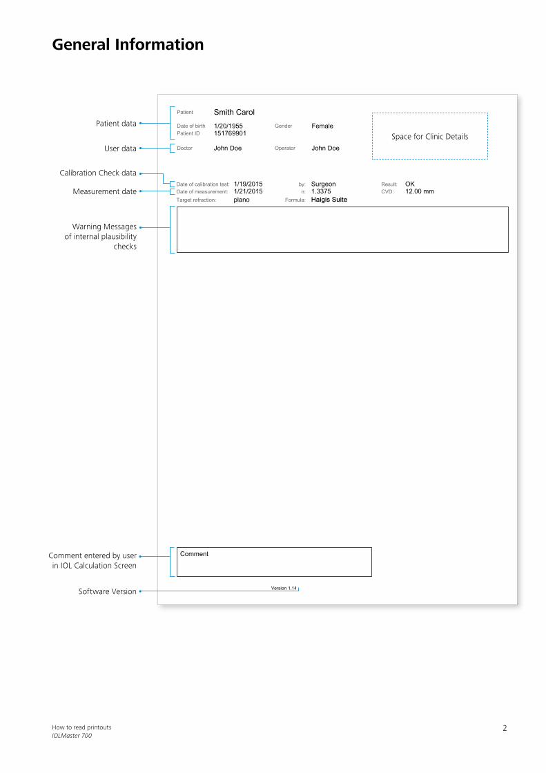
General Information
Patient data
User data
Warning Messages of internal plausibility
checks
Comment entered by user in IOL Calculation Screen
Software Version
Calibration Check data
Measurement date

Patient
Date of birth GenderPatient ID
Smith Carol1/20/1955 Female151769901
Doctor OperatorJohn Doe John Doe
IOLMaster 700 Version 1.14 Report dated 1/21/2015 5:08 PM created by Doe, John was printed. Page 1 of 7
Z E
I S S
| T
e m
p l
a t e
V
e r s
i o
n 0
.1
05/2
012
| -
C o
p y
r i g
h t
2 0
1 2
A
l l
r i g
h t
s r
e s
e r
v e
d
Date of calibration test: 1/19/2015 by: Surgeon Result: OKDate of measurement: 1/21/2015 n: 1.3375 CVD: 12.00 mmTarget refraction: plano Formula:
right leftAL: 22.30 mm (SD = 4 µm)
ACD: 2.99 mm (SD = 5 µm)LT: 3.96 mm (SD = 23 µm)
R: 7.76 mmR1: 7.83 mm @ 156ºR2: 7.69 mm @ 66º WTW: 11.9 mm
Δ D: -0.75 dpt @ 156º Visual acuity: ---SIA: +0.00 dpt @ 0º LVC: -Ref: ---LS: Phakic; VS: Vitreous body
AL: 22.19 mm (SD = 6 µm)ACD: 2.98 mm (SD = 7 µm)
LT: 3.94 mm (SD = 11 µm)R: 7.72 mm
R1: 7.81 mm @ 40ºR2: 7.63 mm @ 130º WTW: 11.9 mm
Δ D: -1.03 dpt @ 40º Visual acuity: ---SIA: +0.00 dpt @ 0º LVC: -Ref: ---LS: Phakic; VS: Vitreous body
Comment
A0: A1: A2:+1.309 +0.400 +0.100
IOL (D) Ref (D)+26.50+26.00
+25.00+24.50
-0.56-0.20
+0.52+0.87
A0: A1: A2:+0.978 +0.400 +0.100
IOL (D) Ref (D)+26.00+25.50
+24.50+24.00
-0.79-0.42
+0.32+0.69
A0: A1: A2:+1.309 +0.400 +0.100
IOL (D) Ref (D)+27.00+26.50
+25.50+25.00
-0.84-0.47
+0.25+0.61
A0: A1: A2:+0.978 +0.400 +0.100
IOL (D) Ref (D)+26.00+25.50
+24.50+24.00
-0.70-0.33
+0.41+0.77
A0: A1: A2:+0.647 +0.400 +0.100
IOL IOL IOL Ref Ref Ref RefSE Cyl Axis SE Sph Cyl Axis
+25.00+24.50
+23.50+23.00
+0.75+0.75
+0.75+0.75
66º66º
66º66º
-0.62-0.24
+0.51+0.88
-0.54-0.16
+0.60+0.97
-0.17-0.17
-0.17-0.17
156º156º
156º156º
A0: A1: A2:+0.647 +0.400 +0.100
IOL IOL IOL Ref Ref Ref RefSE Cyl Axis SE Sph Cyl Axis
+25.50+25.00
+24.00+23.50
+1.50+1.50
+1.50+1.50
130º130º
130º130º
-0.93-0.54
+0.22+0.59
-0.86-0.48
+0.28+0.65
-0.13-0.13
-0.12-0.12
130º130º
130º130º
Patient
Date of birth GenderPatient ID
validierung christian6/4/1955 Female1513338687
Doctor OperatorDoctor Doctor Goeschwitzer Strasse 51-52
www.meditec.zeiss.de
IOLMaster 700 Version 1.50 Report dated 6/4/2015 09:42 created by Smith, John was printed. Page 1 of 4
Z E
I S S
| T
e m
p l
a t e
V
e r s
i o
n 0
.1
05/2
012
| -
C o
p y
r i g
h t
2 0
1 2
A
l l
r i g
h t
s r
e s
e r
v e
d
Date of calibration test: 6/3/2015 by: Doctor Result: OKDate of measurement: 6/4/2015 n: 1.3375 CVD: 12.00 mmFormula:
This IOL calculation contains values that were edited manually.
right left(!) Indicates an uncertain measurement value.(*) Indicates that this value has been edited manually.--- Indicates a measurement failure.
AL: 22.32 mm (SD = 4 µm)ACD: 3.02 mm (SD = 6 µm)
LT: 3.94 mm (SD = 14 µm)R: 7.80 mm (*)
R1: 7.91 mm @ 141ºR2: 7.70 mm @ 51º
Δ D: -1.19 D @ 141º WTW: 11.7 mm (!)Ref: --- VA: ---Target ref.: -0.50 D SIA: +0.00 D @ 0º
LS: Phakic; VS: Vitreous body;Ref. surgery: Untreated; LVC mode: -
AL: 21.21 mm (*)ACD: 3.01 mm (SD = 6 µm)
LT: 3.92 mm (SD = 12 µm)R: 7.75 mm
R1: 7.82 mm @ 38ºR2: 7.67 mm @ 128º
Δ D: -0.87 D @ 38º WTW: 11.8 mmRef: --- VA: ---Target ref.: -0.50 D SIA: +0.00 D @ 0º
LS: Phakic; VS: Vitreous body;Ref. surgery: Untreated; LVC mode: -
Comment
A0: A1: A2:+1.170 +0.400 +0.100
IOL (D) Ref (D)+27.50+27.00
+26.00+25.50
-1.36-0.98
-0.24+0.13
A0: A1: A2:+1.170 +0.400 +0.100
IOL (D) Ref (D)+31.50+31.00
+30.00+29.50
-1.17-0.79
-0.05+0.31
A0: A1: A2:+1.040 +0.400 +0.100
IOL IOL IOL Ref Ref Ref RefSE Cyl Axis SE Sph Cyl Axis
+27.25+26.75
+25.75+25.25
+1.50+1.50
+1.50+1.50
51º51º
51º51º
-1.43-1.04
-0.29+0.08
-1.40-1.01
-0.26+0.11
-0.06-0.06
-0.07-0.07
141º141º
141º141º
A0: A1: A2:+1.040 +0.400 +0.100
IOL IOL IOL Ref Ref Ref RefSE Cyl Axis SE Sph Cyl Axis--- ---
--- ---
--- ---
--- ---
--- ---
--- ---
--- ---
--- ---
--- ---
--- ---
--- ---
--- ---
--- ---
--- ---
3How to read printoutsIOLMaster 700
IOL Power Calculation – Haigis Suite
Target refraction
IOL power calculation formula
Refractive index
The Haigis Suite is a combination of four formulas: Haigis, Haigis-L for post LASIK cases, Haigis-T for toric IOL calculation, Haigis-TL for toric IOL calculation for post LASIK cases. Depending on the lenses selected (i.e. toric and or non-toric IOLS) Haigs and Haigis-T are applied. For toric IOLs, Haigis-T will be applied and for spheric IOLs Haigis will be used. Both toric and non-toric IOLs can be calculated at any time and simultaneously.
Composite Values: AL, ACD, LT with Standard Deviation (SD)
Spheric IOL
IOL constant(s) of relevant formula
Composite Value: K Flat, K Steep, Cylinder
IOL power caclulation results with recommendation
highlighted
IOL power caclulation results including add. values:
Spherical Equivalent (SE), Cylinder (Cyl), Axis, refraction
information for SE, Sphere (Sph), Cylinder and Axis with recommendation highlighted
Toric IOL
Manifest refraction
Eye status: Lens state (LS), Vitreous state (VS)
Corneal Vertex Distance in mm (CVD)
White-to-white measurement
Edited value, marked with an asterix
Surgically induced astigmatism
Corneal status: Laser Vision Correction (LVC)

Patient
Date of birth GenderPatient ID
Smith Carol1/20/1955 Female151769901
Doctor OperatorJohn Doe John Doe
IOLMaster 700 Version 1.14 Report dated 1/26/2015 3:52 PM created by Doe, John was printed. Page 1 of 6
Z E
I S S
| T
e m
p l
a t e
V
e r s
i o
n 0
.1
05/2
012
| -
C o
p y
r i g
h t
2 0
1 2
A
l l
r i g
h t
s r
e s
e r
v e
d
Date of calibration test: 1/19/2015 by: Surgeon Result: OKDate of measurement: 1/21/2015 n: 1.3375 CVD: 12.00 mmTarget refraction: plano IOL:
right leftAL: 22.30 mm (SD = 4 µm)
ACD: 2.99 mm (SD = 5 µm)LT: 3.96 mm (SD = 23 µm)
R: 7.76 mmR1: 7.83 mm @ 156ºR2: 7.69 mm @ 66º WTW: 11.9 mm
Δ D: -0.75 dpt @ 156º Visual acuity: ---SIA: --- LVC: ---Ref: ---LS: Phakic; VS: Vitreous body
AL: 22.19 mm (SD = 6 µm)ACD: 2.98 mm (SD = 7 µm)
LT: 3.94 mm (SD = 11 µm)R: 7.72 mm
R1: 7.81 mm @ 40ºR2: 7.63 mm @ 130º WTW: 11.9 mm
Δ D: -1.03 dpt @ 40º Visual acuity: ---SIA: --- LVC: ---Ref: ---LS: Phakic; VS: Vitreous body
Comment
A0: A1: A2:+1.309 +0.400 +0.100
IOL (D) Ref (D)+26.50+26.00
+25.00+24.50
-0.56-0.20
+0.52+0.87
pACD: +5.52
IOL (D) Ref (D)+26.50+26.00
+25.00+24.50
-0.63-0.28
+0.41+0.75
A0: A1: A2:+1.309 +0.400 +0.100
IOL (D) Ref (D)+27.00+26.50
+25.50+25.00
-0.84-0.47
+0.25+0.61
pACD: +5.52
IOL (D) Ref (D)+26.50+26.00
+25.00+24.50
-0.55-0.20
+0.49+0.83
ACD: +5.549
IOL (D) Ref (D)+26.50+26.00
+25.00+24.50
-0.59-0.24
+0.45+0.79
A-Const: 119.00
IOL (D) Ref (D)+26.50+26.00
+25.00+24.50
-0.89-0.53
+0.18+0.52
ACD: +5.549
IOL (D) Ref (D)+27.00+26.50
+25.50+25.00
-0.84-0.49
+0.21+0.55
A-Const: 119.00
IOL (D) Ref (D)+26.50+26.00
+25.00+24.50
-0.79-0.43
+0.27+0.62
4How to read printoutsIOLMaster 700
IOL Power Calculation – Multiformula
Name of the IOL power calculation formula
IOL constant(s) of relevant formula respectively
Selected IOL
IOL power calculation formula
White-to-white measurement
Corneal status: Laser Vision Correction (LVC)
Surgically induced astigmatism
Manifest refraction
Eye status: Lens state (LS), Vitreous state (VS)
Target refraction
Refractive index
Composite Value: K Flat, K Steep, Cylinder
Composite Values: AL, ACD, LT with Standard Deviation
(SD)
Corneal Vertex Distance in mm (CVD)
IOL power caclulation results with recommendation
highlighted
Comparing up to four different IOL power calculation formulae for one IOL Model.

Patient
Date of birth GenderPatient ID
Smith Carol1/20/1955 Female151769901
Doctor OperatorJohn Doe John Doe
IOLMaster 700 Version 1.14 Report dated 1/26/2015 3:52 PM created by Doe, John was printed. Page 2 of 6
Z E
I S S
| T
e m
p l
a t e
V
e r s
i o
n 0
.1
05/2
012
| -
C o
p y
r i g
h t
2 0
1 2
A
l l
r i g
h t
s r
e s
e r
v e
d
Date of calibration test: 1/19/2015 by: Surgeon Result: OKDate of measurement: 1/21/2015
right
(SD = 4 µm)(SD = 5 µm)(SD = 23 µm)
n: 1.3375(SD = 6 µm)(SD = 13 µm)(SD = 3 µm)
(SD = 3 µm) WTW: 11.9 mm Ix: +0.4 mm Iy: +0.2 mmP: 4.7 mm Px: +0.4 mm Py: +0.2 mm
Comment
5How to read printoutsIOLMaster 700
Analysis Report
Enhanced or single B-Scan image in 0° (birds view of
horizontal scan)
Composite Values: AL, ACD, LT with Standard Deviation
(SD)
Composite value: Central Corneal Thickness with
Standard Deviation
First of 15 keratometry raw images
White-to-white image Enhanced or single fixation check image ~ 1mm horizontal retina scan
White-to-white & pupil diameter
Offsets corneal vertex to Iris center (lx, ly) and pupil center (Px, Py) in X and Y
direction
Composite K, Flat Meridian, Steep Meridian, Cylinder

Patient
Date of birth GenderPatient ID
Smith Carol1/20/1955 Female151769901
Doctor OperatorJohn Doe John Doe
IOLMaster 700 Version 1.14 Report dated 1/26/2015 3:52 PM created by Doe, John was printed. Page 2 of 6
Z E
I S S
| T
e m
p l
a t e
V
e r s
i o
n 0
.1
05/2
012
| -
C o
p y
r i g
h t
2 0
1 2
A
l l
r i g
h t
s r
e s
e r
v e
d
Date of calibration test: 1/19/2015 by: Surgeon Result: OKDate of measurement: 1/21/2015
right
(SD = 4 µm)(SD = 5 µm)(SD = 23 µm)
n: 1.3375(SD = 6 µm)(SD = 13 µm)(SD = 3 µm)
(SD = 3 µm) WTW: 11.9 mm Ix: +0.4 mm Iy: +0.2 mmP: 4.7 mm Px: +0.4 mm Py: +0.2 mm
Comment
Patient
Date of birth GenderPatient ID
Smith Carol1/20/1955 Female151769901
Doctor OperatorJohn Doe John Doe
IOLMaster 700 Version 1.14 Report dated 1/26/2015 3:52 PM created by Doe, John was printed. Page 4 of 6
Z E
I S S
| T
e m
p l
a t e
V
e r s
i o
n 0
.1
05/2
012
| -
C o
p y
r i g
h t
2 0
1 2
A
l l
r i g
h t
s r
e s
e r
v e
d
Date of calibration test: 1/19/2015 by: Surgeon Result: OKDate of measurement: 1/21/2015
right left
LS: Phakic; VS: Vitreous body LS: Phakic; VS: Vitreous body
(SD = 4 µm)(SD = 5 µm)(SD = 23 µm)
(SD = 6 µm)(SD = 7 µm)(SD = 11 µm)
AL ACD LT22.30 mm22.30 mm22.30 mm22.29 mm22.30 mm22.30 mm
2.99 mm2.99 mm2.99 mm2.99 mm2.98 mm2.99 mm
3.97 mm4.00 mm3.95 mm3.96 mm3.96 mm3.96 mm
AL ACD LT22.18 mm22.19 mm22.19 mm22.19 mm22.18 mm22.19 mm
2.98 mm2.99 mm2.99 mm2.98 mm2.99 mm2.98 mm
3.95 mm3.94 mm3.93 mm3.94 mm3.93 mm3.95 mm
n: 1.3375(SD = 6 µm)(SD = 13 µm)(SD = 3 µm)
R: 7.75 mm SE: 43.52 dptΔ D: -0.67 dpt @ 155ºR: 7.76 mm SE: 43.50 dptΔ D: -0.75 dpt @ 155ºR: 7.77 mm SE: 43.46 dptΔ D: -0.82 dpt @ 158º
(SD = 3 µm)(SD = 5 µm)(SD = 5 µm)
R: 7.72 mm SE: 43.72 dptΔ D: -1.01 dpt @ 40ºR: 7.72 mm SE: 43.73 dptΔ D: -1.01 dpt @ 40ºR: 7.72 mm SE: 43.73 dptΔ D: -1.07 dpt @ 39º
(SD = 3 µm)542 µm 543 µm 542 µm540 µm 542 µm 543 µm
(SD = 4 µm)544 µm 549 µm 543 µm542 µm 551 µm 551 µm
WTW: 11.9 mm Ix: +0.4 mm Iy: +0.2 mmP: 4.7 mm Px: +0.4 mm Py: +0.2 mm
WTW: 11.9 mm Ix: -0.5 mm Iy: +0.2 mmP: 4.8 mm Px: -0.5 mm Py: +0.1 mm
Comment
6How to read printoutsIOLMaster 700
Biometry Data
Eye status
Refractive Index
Composite valueCentral Corneal Thickness
with Standard Deviation
Composite Values: AL, ACD, LT with Standard Deviation
(SD)
Average Values: AL, ACD, LT of 3 scans each in 6
meridians
Composite K, Flat Meridian,Steep Meridian, Cylinder
Single value
White-to-white diameter (WTW), Pupil diameter (P)
3 Average Ks of 5 single measurements each
Status Reference Image:“Image Stored” or “No image”
Offsets corneal vertex to Iris center (lx, ly) and pupil center (Px, Py) in X and Y
direction

Patient
Date of birth GenderPatient ID
Smith Carol1/20/1955 Female151769901
Doctor OperatorJohn Doe John Doe
IOLMaster 700 Version 1.14 Report dated 1/26/2015 3:52 PM created by Doe, John was printed. Page 5 of 6
Z E
I S S
| T
e m
p l
a t e
V
e r s
i o
n 0
.1
05/2
012
| -
C o
p y
r i g
h t
2 0
1 2
A
l l
r i g
h t
s r
e s
e r
v e
d
Date of calibration test: 1/19/2015 by: Surgeon Result: OKDate of measurement: 1/21/2015 n: 1.3375 CVD: 12.00 mm
rightAL: 22.30 mm (SD = 4 µm)
ACD: 2.99 mm (SD = 5 µm)LT: 3.96 mm (SD = 23 µm)
Visual acuity: ---LS: Phakic; VS: Vitreous body
R: 7.76 mmR1: 7.83 mm @ 156ºR2: 7.69 mm @ 66º
Δ D: -0.75 dpt @ 156ºWTW: 11.9 mm
Ref: ---
Comment
7How to read printoutsIOLMaster 700
Reference Image
Refractive index
Corneal Vertex Distance in mm (CVD)
Composite Values AL, ACD, LT with Standard Deviation
(SD)Composite K, Flat / Steep
Meridian, Cylinder
Eye status: Lens state (LS), Vitreous state (VS)
White-to-white diameter & manifest refraction
Reference Image for markerless toric IOL alignment including
limbus diameter & steep keratometry axis overlay

Carl Zeiss Meditec AGGoeschwitzer Strasse 51–5207745 JenaGermanywww.zeiss.com/iolmaster700
EN_3
2_20
0_00
09I
Int
erna
tiona
l ver
sion
: Not
for s
ale
in th
e Un
ited
Stat
es
Not
all
prod
ucts
, ser
vice
s or
off
ers
are
appr
oved
or o
ffer
ed in
eve
ry m
arke
t and
app
rove
d la
belin
g an
d in
stru
ctio
ns m
ay v
ary
from
one
cou
ntry
to a
noth
er. T
he c
onte
nts
of th
e br
ochu
re m
ay d
iffer
from
the
curr
ent s
tatu
s of
app
rova
l of t
he p
rodu
ct in
you
r cou
ntry
. Ple
ase
cont
act o
urre
gion
al re
pres
enta
tive
for m
ore
info
rmat
ion.
Sub
ject
to c
hang
e in
des
ign
and
scop
e of
del
iver
y an
d as
a re
sult
of o
ngoi
ng te
chni
cal d
evel
opm
ent.
IOLM
aste
r, FO
RUM
, LUM
ERA
and
CALL
ISTO
eye
are
eith
er tr
adem
arks
or r
egis
tere
d tr
adem
arks
of C
arl Z
eiss
Med
itec
AG.
© C
arl Z
eiss
Med
itec
AG, 2
015.
All
copy
right
s re
serv
ed.

IOLMaster 700 – Quick Guide
Please note: This document is a preliminary version and does not replace the user manual which is delivered together with the device. Final version is to be released soon.
WARNING - GENERAL HAZARDS
These quick instructions are intended as an overview. Instructions for the safe operation of the instrument are to be found in the user manual and online help.
Always perform measurements in the correct sequence! All device measurements must be performed before undertaking any measurements involving direct contact with the eye.
Calibration test
• Always check the calibration daily before carrying out measurements on patients!
• Tap the Start button to start the first of three calibration test steps. The test eye in the chin rest compartment will rise up automatically.
• Now carry out the measurements on the test eye. Follow the instructions on the screen. The overview shows the individual steps of the calibration test and their status
• If the Calibration test was passed, press the button ”next” and the application will be started.
• If the Calibration test was failed, repeat the calibration test by pressing the button “repeat”.
New Patient
• New patient data can be entered by tapping the Add button. The following information is required: Last name, first name, Gender (Gender drop-down box), Date of birth (Open the Date selection box and select the patient's day, month and year of birth. Confirm with OK.), and Refractive Surgery.
• It is important that the correct lens state and vitreous body state are selected as this cannot be changed after the measurement has been done. If changes are necessary, the measurement needs to be repeated.
• An existing patient dataset from the patient list on the left side of the screen can also be selected.
• Input field for entering a search string Parts of the patient name, ID or date of birth may be entered for the search.
• With the buttons “Analysis”, “IOL Calculation” and “Measurement” the according dialogs screens are opened.
Measurement Coarse alignment
Fine alignment
Fixation
• Coarse alignment: The measuring head is coarsely adjusted on the pupil center. Usually the measurement head is moved towards the eye from a position further away of the eye. The reflective pattern of the six LED test marks must be in the center of the cross hair and focused. A WTW image is captured here to detect the iris edge. Instruct the patient to look steadily at the fixation point in the center and ensure that the device head is aligned as accurately as possible.
• Fine Alignment : For fine adjustment, the reflective pattern from 18 LED test marks is focused by moving the measurement head closer to the eye. In addition to the live image, the vertical (right of the central live image) and horizontal (on the bottom of the central live image) OCT scan is represented. In each image, an adjustment area is proposed (shown in green). If the alignment is correct, a yellow dot is displayed within the green rectangle of the adjustment area in the horizontal and vertical OCT scan image. Measurement is triggered automatically or manually depending on the selection made earlier. In this measurement step also the reference image or sclera-image is captured. To capture a good sclera image, it is important that the patient opens his/her eyes wide, the lighting in the room is not too bright and the vessels in the patient's eye can be recognized (no sclera image is captured if vessels cannot be recognized). In the Measurement dialog area of Advanced settings you can enter the value of corneal astigmatism which determines a clinical indication. If this value is reached, a sclera image is automatically triggered.
• Fixation: The Fixation follows the same principle as the fine alignment. The difference is that only 6 LED test marks need to be focused.
• Measure other eye / repeat Measurement: You can repeat the current measurement (button “repeat measurement”) or measure the other eye after moving the measuring head to the other eye using the joystick. After finishing the measurement the Quality-Check is complete.

IOLMaster 700 – Quick Guide
Carl Zeiss Meditec
IOL.7405
IOLMaster 700 © Carl Zeiss Meditec AG, Jena
Änderungen vorbehalten
Quality-Check
Successful measurement: A green quality indicator shows a technically perfect measurement
Warning for uncertain measurement: A yellow quality indicator will signal to the user that he/she should verify the corresponding measurement marks thoroughly
Measurement failed: A red quality indicator means that no measurement results could be detected
The quality check helps to evaluate the quality of the measurement. From the application point of view, the user should check whether the measured values are correct and plausible. This can be done by using the analysis window. In addition the Fixation of the patient should be checked. Good fixation is shown by a depression in the center (right picture), the so-called foveal pit. If the foveal pit can be easily recognized, the patient fixated well during measurement. If it cannot be recognized (left picture), the patient has not correctly fixated. In this case, measurement should be repeated and the patient be instructed to fixate on the fixation light in the device. If the patient cannot fixate during several measurements the user should verify if there may be another problem with the patient which requires special examination.
Analysis
The following images are displayed in the analysis dialog separately for the right and left eye: - Biometry-Scan - Keratometry-Scan - WTW-Image - Sclera- Image - Fixation Check. They should be checked for artifacts in B-Scans, WTW and sclera images which are out of focus or deformed or missing spots in keratometry. The measurement values for the right eye are shown on the left side (OD) and the measurement values for the left eye on the right side (OS) of the screen. Measurement values should be checked thoroughly for plausibility. Warning messages should be read with caution.
IOL Calculation
• For each eye, the IOL calculation is done separately. For better orientation, the measurement data of the selected eye are displayed on a white background, the data of the other eye are greyed out. Additionally, the selected eye is shown in bold and green letters in the upper right corner of the display.
• Tap the measurement data area on the right or left side of the screen to alternate between the eye or use the
button . Beneath the measurement data, the lens state and refractive surgery history is shown for each eye.
• Tapping and select the calculation formulae to be applied.
• IOL calculation is started by tapping . To be able to print or export to FORUM, always an IOL calculation has to be done.
• By tapping Finish/ Print / Export (view can vary according to individual setting), reports will be printed / exported and the application will jump to the Patient Manager where other patients can be opened and / or additional measurements can be done.
• The measurement values can be edited by tapping the appropriate field. Edited values are marked with an *. If the IOL calculation window is closed, the entries will be canceled. When re-starting the IOL calculation, the measured values are displayed in these fields.
Shutdown
Tap the X (Close) button to open a menu with options for logging out the current user or switching the device off after finishing all measurements.
Please share your experience with the IOLMaster 700 with us!
• www.zeiss.com/cataract-community
• www.ocusoft.de/ulib/