Embed Size (px)
Citation preview
Muscle biopsy: what and why andwhen?
Jon Walters,1 Atik Baborie2
ABSTRACTSkeletal muscle biopsy remains an importantinvestigative tool in the diagnosis of a variety ofmuscle disorders. Traditionally, someone witha limb-girdle muscle weakness, myopathicchanges on electrophysiology and raised serumcreatine kinase (CK) would have a muscle biopsy.However, we are living through a geneticsrevolution, and so do all such patients still needa biopsy? When should we undertake a musclebiopsy in patients with a distal, scapuloperoneal orother patterns of muscle weakness? When shouldpatients with myositis, rhabdomyolysis, myalgia,hyperCKaemia or a drug-related myopathy havea muscle biopsy? What does normal musclehistology look like and what changes occur inneurogenic and myopathic disorders? As withKipling’s six honest serving men, we hope that byaddressing these issues we can all become moreconfident about when to request a muscle biopsyand develop clearer insights into musclepathology.
INTRODUCTIONBefore consideringwhen youmight requesta muscle biopsy, it is worth consideringhowa biopsy is done andwhat can be learntfrom it.Having anunderstanding ofmusclepathology enables us to communicate withpathology colleagues and, when results areavailable, to translate muscle biopsy resultsfor our patients’ benefit. We already recog-nise how such a similar investment helpsour discussions with neuroradiologycolleagues.
What is a muscle biopsy?Muscle biopsy can be either open orclosed (needle or punch) and both havetheir champions. A closed biopsy has theadvantages of being more cosmeticallyappealing, minimally invasive and poten-tially yielding more than one specimen.The small sample size, however, createschallenges with handling and orientation;if pathology is patchy it may be missed.An open biopsy is more invasive, but
the operator can see the muscle, avoid
tendinous insertions and can obtain anadequate specimen for biochemical stu-dies, which is potentially important inmitochondrial disease, as we shall see.The person taking the biopsy needs ade-quate experience, skill and interest. Inadult practice, open biopsy is generallyfavoured.An open biopsy is undertaken in a sterile
operating theatre. The skin and subcuta-neous tissue are infiltrated with a localanaesthetic, avoiding the muscle. The inci-sion is 4–6 cm along the long axis of thelimb. The skin, subcutaneous fat and fasciaoverlying the muscle are dissected, and thebiopsy is taken with muscle fibre fasciclesrunning longitudinally. Complications arerare but include infection, delayed haema-toma formation, muscle herniation throughfascial defect and wound dehiscence.The biopsy, usually about 15×10×5mm
in size, is laid on a non-absorbent card ina dry specimen container. The principle isto ‘snap freeze’ the muscle in isopentanecooled by liquid nitrogen (to approxi-mately −160°C). This renders the samplein as life-like a state as possible withoutusing a chemical fixative. ‘Snap freezing’allows the tissue to be cut for diagnosticinvestigations and prevents the damagecaused by formalin fixation or ice crystalformation (slow freezing) (contrast figures1 with 2).
Which muscles should be biopsied?Select a muscle that is weak but can over-come gravity. Very weak muscles showmarked loss of muscle fibres with fatty orconnective tissue replacement and revealno remnant of the underlying disease pro-cess: an ‘end-stage’ biopsy. Avoid musclesthat have been injured, injected orsampled by neurophysiology within 6months, as these situations can result infinding misleading inflammation.A limited number of muscles are biop-
sied: in the upper limb, biceps brachii ordeltoid; in the lower limb, quadriceps
1Neurology, Morriston Hospital,Swansea, UK2Department of CellularPathology, University Hospital ofWales, Cardiff, UK
Correspondence toDr Jon Walters, Neurology,Morriston Hospital, SwanseaSA6 6NL, UK; [email protected]
Accepted 30 April 2020
© Author(s) (or theiremployer(s)) 2020. Re-usepermitted under CC BY-NC.No commercial re-use. Seerights and permissions.Published by BMJ.
To cite: Walters J, BaborieA. Pract Neurol2020;20:385–395.
Walters J, Baborie A. Pract Neurol 2020;20:385–395. doi:10.1136/practneurol-2019-002465 385
HOW TO UNDERSTAND IT on O
ctober 18, 2021 by guest. Protected by copyright.
http://pn.bmj.com
/P
ract Neurol: first published as 10.1136/practneurol-2019-002465 on 5 June 2020. D
ownloaded from
femoris (vastus lateralis or rectus femoris) or gastro-cnemius. The pathologist needs to know what thesemuscles look like when healthy. Fibre-type composi-tion and the normal range of fibre size will varybetween muscles, depending upon their functionalrole. If contemplating using another muscle, consultwith pathology colleagues.Timely communication with the person undertaking
the biopsy and pathologist is key, including clinicaldetails and our diagnostic suspicions.
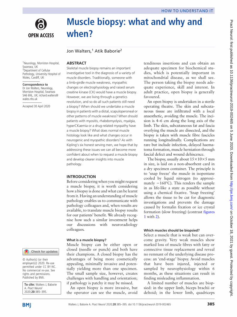
What does normal muscle look like?A transverse section of normal muscle reveals fibresof roughly the same size, each comprising hundredsof myofibrils. The intermyofibrillar network sitsbetween the myofibrils, composed of sarcoplasm,mitochondria and sarcoplasmic reticulum with the
transverse tubular system. Nuclei occupy the periph-ery of the fibres (figure 1).Each fibre is roughly polygonal transversely and
packed snuggly alongside its neighbours, with theendomysium squeezed between adjacent fibres.Perimysium binds groups of fibres together, and thefascicles are collected together within the musclebelly, wrapped by the epimysium.
What changes do you see in denervated muscle?Muscle fibres that have lost their nerve supply atrophyand become small angulated remnants, squeezed betweentheir normal-sized, innervated neighbours. Denervatedmuscle fibres attract nerve twigs from healthy neigh-bours; the nerve twig sprouts and reinnervates the neu-rone-deprived myofibre. The reinnervated fibre assumesthe same identity (type 1 or type 2 fibre) as its neighbour,since the reinnervating anterior horn cell exclusivelyinnervates either type 1 or type 2 fibres. Reinnervatedneighbouring muscle fibres, therefore, assume the sameidentity, and the normalmosaic distribution of fibre typesbecomes distorted into sheets of neighbouring fibres thatare all the same type (fibre-type grouping).
What sort of changes do you see in myopathies?In many myopathies, muscle fibre size is no longeruniform, and instead, there is considerable fibre sizevariation. Large fibres may split, particularly in muscledystrophies. Nuclei, usually situated peripherally, startto migrate centrally.Type 1 and 2 fibres may be selectively involved in
some myopathies and in other health conditions. Type2 fibre atrophy, for instance, typically occurs duringimmobility, corticosteroid use or general ill health.Type 1 atrophy occurs in some congenital myopathies;type 1 fibres tend to be more numerous than type 2 incongenital myopathies.In many myopathies, the intermyofibrillar network
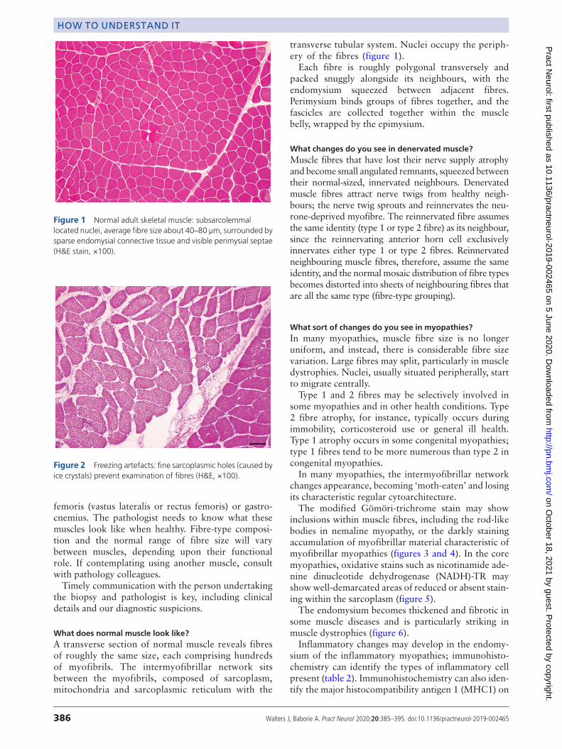
changes appearance, becoming ‘moth-eaten’ and losingits characteristic regular cytoarchitecture.The modified Gömöri-trichrome stain may show
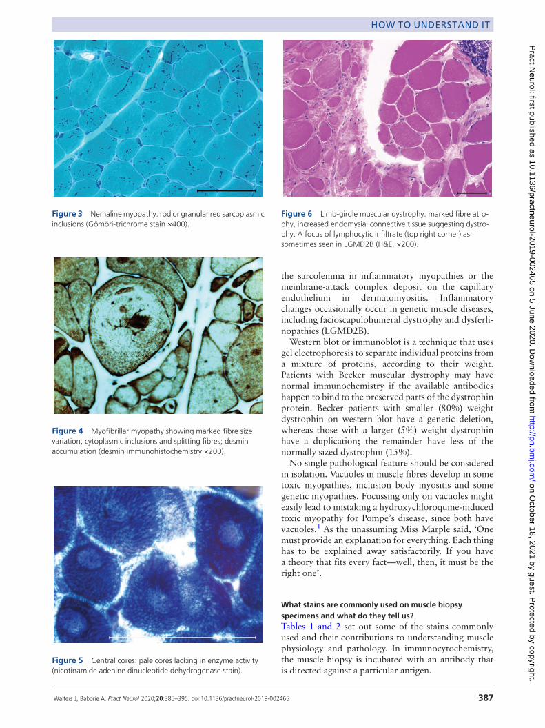
inclusions within muscle fibres, including the rod-likebodies in nemaline myopathy, or the darkly stainingaccumulation of myofibrillar material characteristic ofmyofibrillar myopathies (figures 3 and 4). In the coremyopathies, oxidative stains such as nicotinamide ade-nine dinucleotide dehydrogenase (NADH)-TR mayshowwell-demarcated areas of reduced or absent stain-ing within the sarcoplasm (figure 5).The endomysium becomes thickened and fibrotic in
some muscle diseases and is particularly striking inmuscle dystrophies (figure 6).Inflammatory changes may develop in the endomy-
sium of the inflammatory myopathies; immunohisto-chemistry can identify the types of inflammatory cellpresent (table 2). Immunohistochemistry can also iden-tify the major histocompatibility antigen 1 (MHC1) on
Figure 1 Normal adult skeletal muscle: subsarcolemmallocated nuclei, average fibre size about 40–80 µm, surrounded bysparse endomysial connective tissue and visible perimysial septae(H&E stain, ×100).
Figure 2 Freezing artefacts: fine sarcoplasmic holes (caused byice crystals) prevent examination of fibres (H&E, ×100).
386 Walters J, Baborie A. Pract Neurol 2020;20:385–395. doi:10.1136/practneurol-2019-002465
HOW TO UNDERSTAND IT on O
ctober 18, 2021 by guest. Protected by copyright.
http://pn.bmj.com
/P
ract Neurol: first published as 10.1136/practneurol-2019-002465 on 5 June 2020. D
ownloaded from
the sarcolemma in inflammatory myopathies or themembrane-attack complex deposit on the capillaryendothelium in dermatomyositis. Inflammatorychanges occasionally occur in genetic muscle diseases,including facioscapulohumeral dystrophy and dysferli-nopathies (LGMD2B).Western blot or immunoblot is a technique that uses
gel electrophoresis to separate individual proteins froma mixture of proteins, according to their weight.Patients with Becker muscular dystrophy may havenormal immunochemistry if the available antibodieshappen to bind to the preserved parts of the dystrophinprotein. Becker patients with smaller (80%) weightdystrophin on western blot have a genetic deletion,whereas those with a larger (5%) weight dystrophinhave a duplication; the remainder have less of thenormally sized dystrophin (15%).No single pathological feature should be considered
in isolation. Vacuoles in muscle fibres develop in sometoxic myopathies, inclusion body myositis and somegenetic myopathies. Focussing only on vacuoles mighteasily lead to mistaking a hydroxychloroquine-inducedtoxic myopathy for Pompe’s disease, since both havevacuoles.1 As the unassuming Miss Marple said, ‘Onemust provide an explanation for everything. Each thinghas to be explained away satisfactorily. If you havea theory that fits every fact—well, then, it must be theright one’.
What stains are commonly used on muscle biopsyspecimens and what do they tell us?Tables 1 and 2 set out some of the stains commonlyused and their contributions to understanding musclephysiology and pathology. In immunocytochemistry,the muscle biopsy is incubated with an antibody thatis directed against a particular antigen.
Figure 3 Nemalinemyopathy: rod or granular red sarcoplasmicinclusions (Gömöri-trichrome stain ×400).
Figure 4 Myofibrillar myopathy showing marked fibre sizevariation, cytoplasmic inclusions and splitting fibres; desminaccumulation (desmin immunohistochemistry ×200).
Figure 5 Central cores: pale cores lacking in enzyme activity(nicotinamide adenine dinucleotide dehydrogenase stain).
Figure 6 Limb-girdle muscular dystrophy: marked fibre atro-phy, increased endomysial connective tissue suggesting dystro-phy. A focus of lymphocytic infiltrate (top right corner) assometimes seen in LGMD2B (H&E, ×200).
Walters J, Baborie A. Pract Neurol 2020;20:385–395. doi:10.1136/practneurol-2019-002465 387
HOW TO UNDERSTAND IT on O
ctober 18, 2021 by guest. Protected by copyright.
http://pn.bmj.com
/P
ract Neurol: first published as 10.1136/practneurol-2019-002465 on 5 June 2020. D
ownloaded from
In limb-girdle muscular dystrophies 2A and 2I, noimmunocytochemical stain is invariably abnormal.Furthermore, since proteins interact with each other,there can be secondary immunocytochemical changes.For example, the transmembrane sarcoglycan complexlinks to the ‘C’ region of dystrophin; an abnormality inone sarcoglycan protein may disrupt its fellow sarcogly-cans as well as dystrophin (all for one and one for all).2 3
Electron microscopy often provides further informa-tion, including identifying filamentous inclusions ininclusion body myositis or various abnormalities in
congenital myopathies. Electron microscopy led tothe discovery that mitochondria have their own DNAand that people with mitochondrial diseases have visi-ble paracrystalline ‘parking lot’ inclusions within theirmitochondria.4
When to request a muscle biopsy in a patient withsuspected muscle weakness?Investigating potentially myopathic weakness oftenrequires consideration of a muscle biopsy. The practicalneurologist aims to narrow the differential diagnosisclinically before embarking on investigations. There aremany clues to help to distinguish between an acquiredmyopathy and genetic disorder, see further reading.35–7
Several myopathies may then become recognisable(or almost recognisable) at the bedside, and so thenext diagnostic step might be a genetic or other bloodtest, rather than a muscle biopsy (table 3).Gestalt impressions are vital in endocrine-related
myopathies, confirmed with appropriate biochemicalinvestigations (table 3). A muscle biopsy might still benecessary if there is no improvement despite restoringa normal hormonal milieu.There are six patterns of weakness commonly
referred to in muscle diseases; we look at each in turnand provide some guidance about the role of musclebiopsy in each. For the sake of this discussion, we havecombined distal and scapuloperoneal forms.
Limb-girdle weaknessGenerally, in a patient with a likely genetic cause forlimb-girdle weakness, especially with pseudohypertro-phy and raised serum creatine kinase, we requesta dried blood spot test for α-glucosidase, and genetictesting for dystrophinopathy and LGMD2I, beforea muscle biopsy.5 6 8 The clinical rules distinguishinggenetic and acquired myopathies are robust but fallible,one reason to favour biopsy at this point.However, practice varies, and some neurologists use
next-generation sequencing before muscle biopsy.A muscle biopsy and next-generation sequencing arenot mutually exclusive; patients often need both.Whole-exome sequencing, for instance, may identifylarge numbers of potential pathogenic variants.A muscle biopsy can help to identify an ‘in-depth phe-notype’, steering us towards the culprit gene that isknown to produce the particular muscle pathology.9 10
Distal or scapuloperoneal patterns of weaknessMuscle conditions with distal or scapuloperoneal pat-terns of weakness are also sometimes recognisable at thebedside (table 3).Myofibrillar or rimmed vacuoles are tobe expected, but there may be other less commonpathology; this favours an early biopsy rather thana narrower ‘phenotype-driven’ gene panel approach.For example, Cori–Forbes disease with its characteristicpools of subsarcolemmal glycogen can present with dis-tal weakness.3 7 11
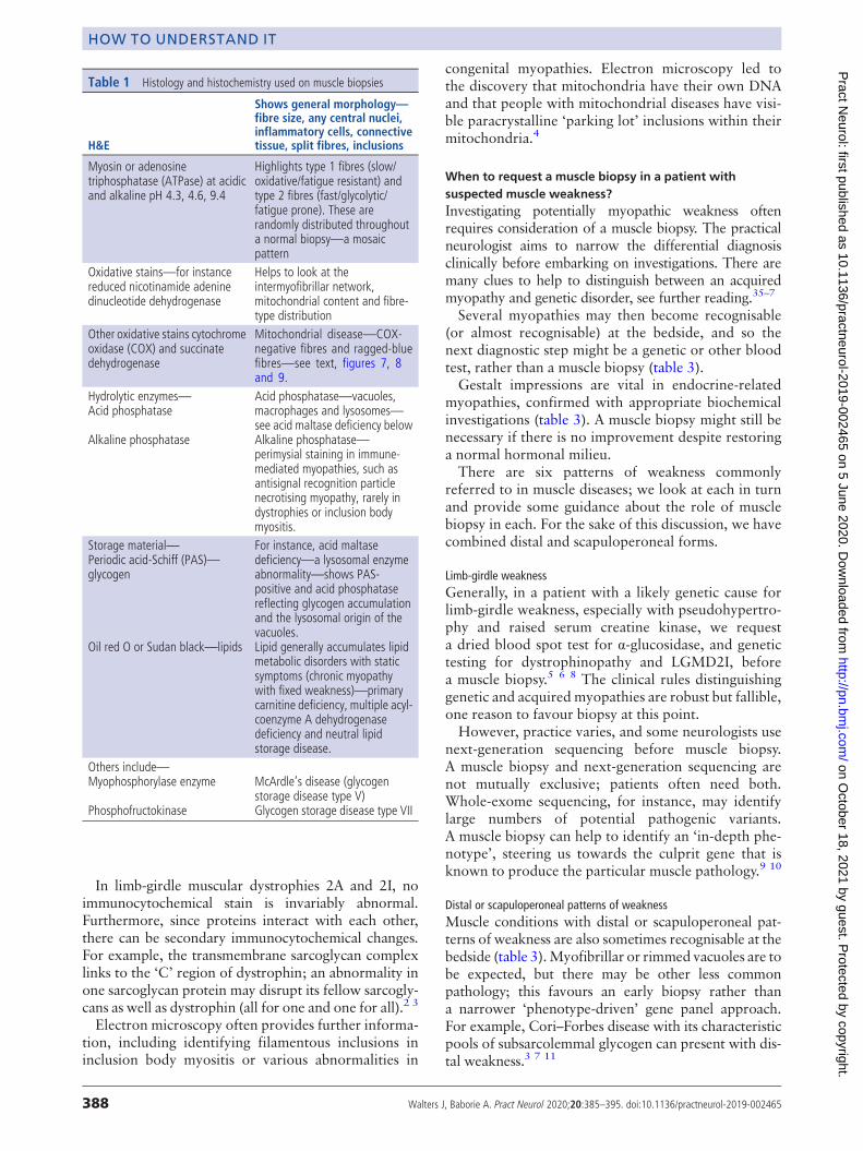
Table 1 Histology and histochemistry used on muscle biopsies
H&E
Shows general morphology—fibre size, any central nuclei,inflammatory cells, connectivetissue, split fibres, inclusions
Myosin or adenosinetriphosphatase (ATPase) at acidicand alkaline pH 4.3, 4.6, 9.4
Highlights type 1 fibres (slow/oxidative/fatigue resistant) andtype 2 fibres (fast/glycolytic/fatigue prone). These arerandomly distributed throughouta normal biopsy—a mosaicpattern
Oxidative stains—for instancereduced nicotinamide adeninedinucleotide dehydrogenase
Helps to look at theintermyofibrillar network,mitochondrial content and fibre-type distribution
Other oxidative stains cytochromeoxidase (COX) and succinatedehydrogenase
Mitochondrial disease—COX-negative fibres and ragged-bluefibres—see text, figures 7, 8and 9.
Hydrolytic enzymes—Acid phosphatase
Alkaline phosphatase
Acid phosphatase—vacuoles,macrophages and lysosomes—see acid maltase deficiency belowAlkaline phosphatase—perimysial staining in immune-mediated myopathies, such asantisignal recognition particlenecrotising myopathy, rarely indystrophies or inclusion bodymyositis.
Storage material—Periodic acid-Schiff (PAS)—glycogen
Oil red O or Sudan black—lipids
For instance, acid maltasedeficiency—a lysosomal enzymeabnormality—shows PAS-positive and acid phosphatasereflecting glycogen accumulationand the lysosomal origin of thevacuoles.Lipid generally accumulates lipidmetabolic disorders with staticsymptoms (chronic myopathywith fixed weakness)—primarycarnitine deficiency, multiple acyl-coenzyme A dehydrogenasedeficiency and neutral lipidstorage disease.
Others include—Myophosphorylase enzyme
Phosphofructokinase
McArdle’s disease (glycogenstorage disease type V)Glycogen storage disease type VII
388 Walters J, Baborie A. Pract Neurol 2020;20:385–395. doi:10.1136/practneurol-2019-002465
HOW TO UNDERSTAND IT on O
ctober 18, 2021 by guest. Protected by copyright.
http://pn.bmj.com
/P
ract Neurol: first published as 10.1136/practneurol-2019-002465 on 5 June 2020. D
ownloaded from
Distal arm and proximal leg weaknessThis pattern raises suspicion of inclusion body myositis,although myotonic dystrophy type 2 (DM2) can havea similar distribution of weakness. We have a low thresh-old for testing for DM2, particularly if there are familialcataracts.We recommend a biopsy to confirm inclusion body
myositis and to exclude granulomatous myositis, whichhas a similar distribution of weakness but, unlike inclu-sion body myositis, may respond to immunosuppression.
Light microscopy in inclusion body myositis shouldshow endomyseal inflammation, cytotoxic T cells par-tially invading non-necrotic muscle fibres and rimmedvacuoles. All three features are not always present;rimmed vacuoles may be a late feature. There are oftenmitochondrial abnormalities including COX-negativefibres or ragged-red fibres (see below). Electron micro-scopy shows 15 nm tubulofilamentous inclusions in thecytoplasm or nucleus, and immunohistochemistry mayidentify ‘Alzheimer-characteristic proteins’ such as
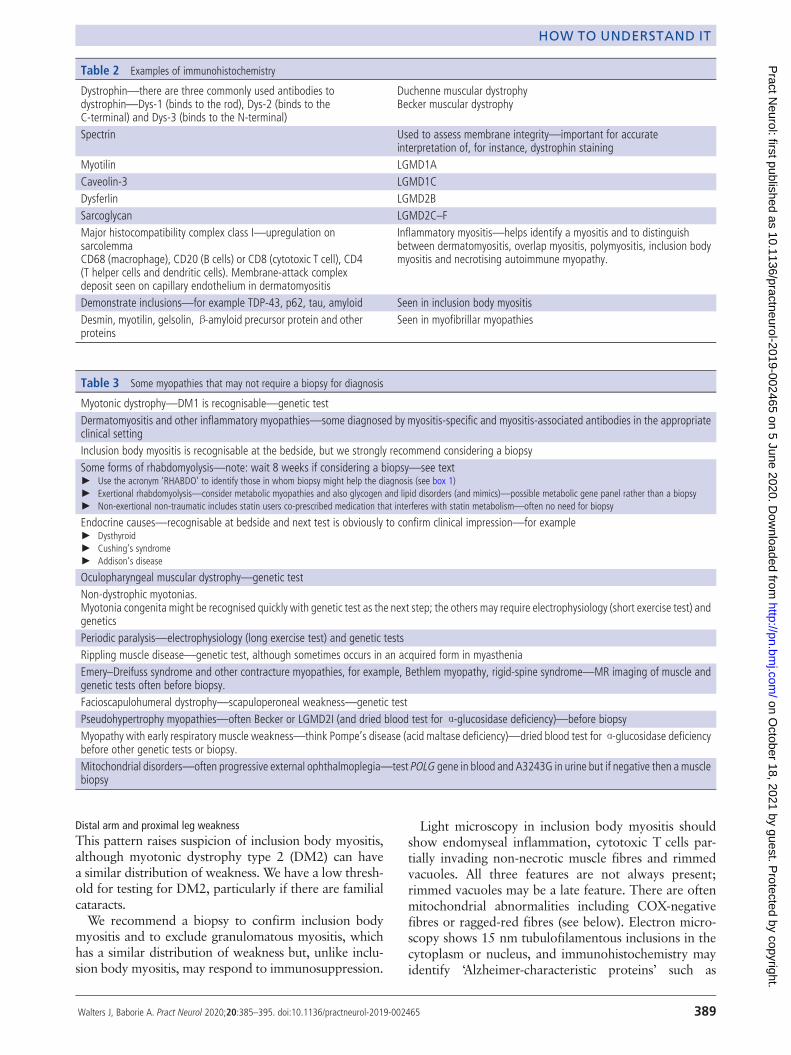
Table 2 Examples of immunohistochemistry
Dystrophin—there are three commonly used antibodies todystrophin—Dys-1 (binds to the rod), Dys-2 (binds to theC-terminal) and Dys-3 (binds to the N-terminal)
Duchenne muscular dystrophyBecker muscular dystrophy
Spectrin Used to assess membrane integrity—important for accurateinterpretation of, for instance, dystrophin staining
Myotilin LGMD1ACaveolin-3 LGMD1CDysferlin LGMD2BSarcoglycan LGMD2C–FMajor histocompatibility complex class I—upregulation onsarcolemmaCD68 (macrophage), CD20 (B cells) or CD8 (cytotoxic T cell), CD4(T helper cells and dendritic cells). Membrane-attack complexdeposit seen on capillary endothelium in dermatomyositis
Inflammatory myositis—helps identify a myositis and to distinguishbetween dermatomyositis, overlap myositis, polymyositis, inclusion bodymyositis and necrotising autoimmune myopathy.
Demonstrate inclusions—for example TDP-43, p62, tau, amyloid Seen in inclusion body myositisDesmin, myotilin, gelsolin,β-amyloid precursor protein and otherproteins
Seen in myofibrillar myopathies
Table 3 Some myopathies that may not require a biopsy for diagnosis
Myotonic dystrophy—DM1 is recognisable—genetic testDermatomyositis and other inflammatory myopathies—some diagnosed by myositis-specific and myositis-associated antibodies in the appropriateclinical settingInclusion body myositis is recognisable at the bedside, but we strongly recommend considering a biopsySome forms of rhabdomyolysis—note: wait 8 weeks if considering a biopsy—see text► Use the acronym ‘RHABDO’ to identify those in whom biopsy might help the diagnosis (see box 1)► Exertional rhabdomyolysis—consider metabolic myopathies and also glycogen and lipid disorders (and mimics)—possible metabolic gene panel rather than a biopsy► Non-exertional non-traumatic includes statin users co-prescribed medication that interferes with statin metabolism—often no need for biopsy
Endocrine causes—recognisable at bedside and next test is obviously to confirm clinical impression—for example► Dysthyroid► Cushing’s syndrome► Addison’s disease
Oculopharyngeal muscular dystrophy—genetic testNon-dystrophic myotonias.Myotonia congenita might be recognised quickly with genetic test as the next step; the others may require electrophysiology (short exercise test) andgeneticsPeriodic paralysis—electrophysiology (long exercise test) and genetic testsRippling muscle disease—genetic test, although sometimes occurs in an acquired form in myastheniaEmery–Dreifuss syndrome and other contracture myopathies, for example, Bethlem myopathy, rigid-spine syndrome—MR imaging of muscle andgenetic tests often before biopsy.Facioscapulohumeral dystrophy—scapuloperoneal weakness—genetic testPseudohypertrophy myopathies—often Becker or LGMD2I (and dried blood test for α-glucosidase deficiency)—before biopsyMyopathy with early respiratory muscle weakness—think Pompe’s disease (acid maltase deficiency)—dried blood test forα-glucosidase deficiencybefore other genetic tests or biopsy.Mitochondrial disorders—often progressive external ophthalmoplegia—test POLG gene in blood and A3243G in urine but if negative then a musclebiopsy
Walters J, Baborie A. Pract Neurol 2020;20:385–395. doi:10.1136/practneurol-2019-002465 389
HOW TO UNDERSTAND IT on O
ctober 18, 2021 by guest. Protected by copyright.
http://pn.bmj.com
/P
ract Neurol: first published as 10.1136/practneurol-2019-002465 on 5 June 2020. D
ownloaded from
hyperphosphorylated tau. Finding partial invasion andmitochondrial abnormalities is particularly supportive ofa clinical diagnosis of inclusion body myositis.12
Progressive external ophthalmoplegia and/or ptosisIt is worth considering myasthenia, dysthyroid eye dis-ease or oculopharyngeal muscular dystrophy, which donot require a muscle biopsy. Mitochondrial disordersthen form the majority of the remaining patients.We look for two common genetic causes of mitochon-
drial disease, the A3243Gmutation in the mitochondrialgenome (mtDNA) in a urine sample and POLG, a nucleargene encoding the catalytic subunit of DNA polymerasegamma, detectable in leucocyte DNA. Polymerasegamma is one of several nuclear genes required to main-tain healthy mtDNA; POLG mutations cause secondarymtDNA mutations that accumulate with age.7
If these are negative, we undertake a muscle biopsyfrom quadriceps or deltoid; levator palpebrae or orbi-cularis oculi biopsies are potentially rich in pathology,but limb muscles seem to be adequate.Cytochrome c oxidase (COX, complex IV) histo-
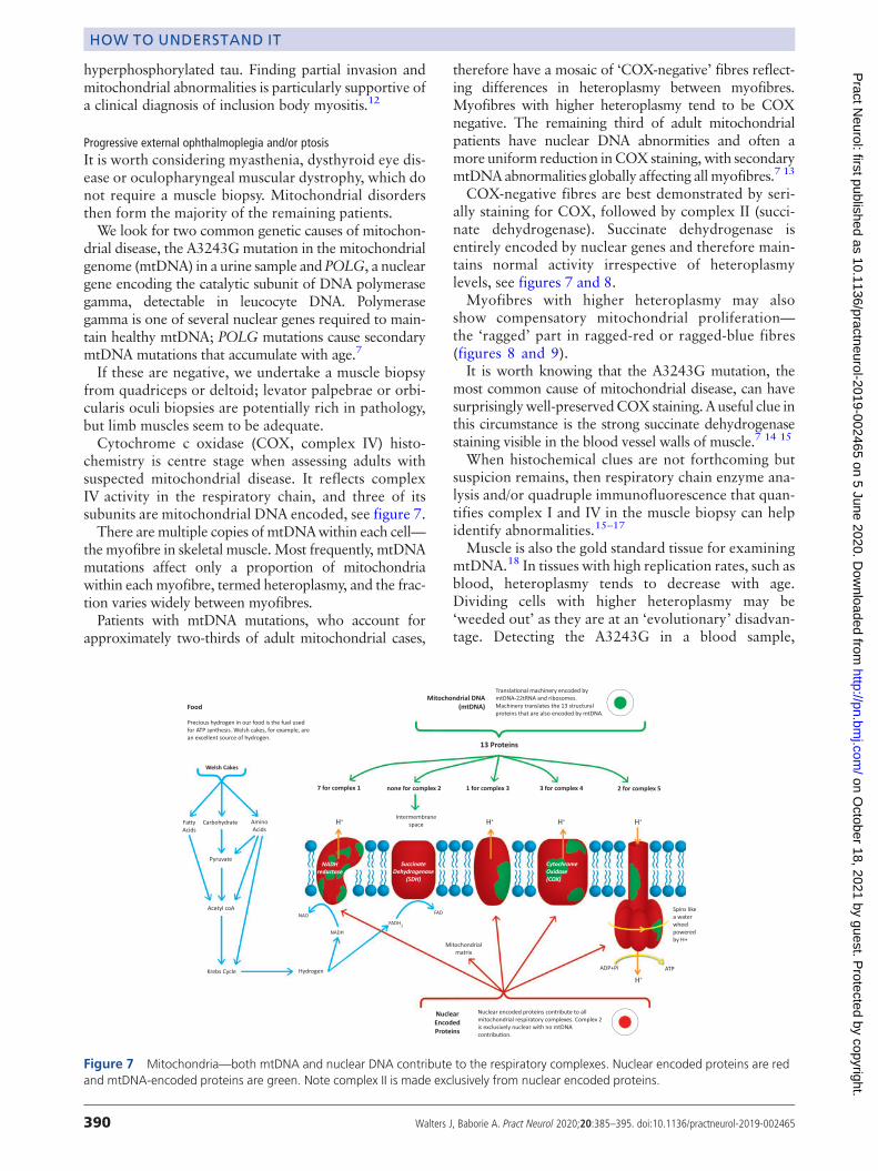
chemistry is centre stage when assessing adults withsuspected mitochondrial disease. It reflects complexIV activity in the respiratory chain, and three of itssubunits are mitochondrial DNA encoded, see figure 7.There aremultiple copies of mtDNAwithin each cell—
themyofibre in skeletal muscle.Most frequently, mtDNAmutations affect only a proportion of mitochondriawithin eachmyofibre, termed heteroplasmy, and the frac-tion varies widely between myofibres.Patients with mtDNA mutations, who account for
approximately two-thirds of adult mitochondrial cases,
therefore have a mosaic of ‘COX-negative’ fibres reflect-ing differences in heteroplasmy between myofibres.Myofibres with higher heteroplasmy tend to be COXnegative. The remaining third of adult mitochondrialpatients have nuclear DNA abnormities and often amore uniform reduction inCOXstaining,with secondarymtDNAabnormalities globally affecting allmyofibres.7 13
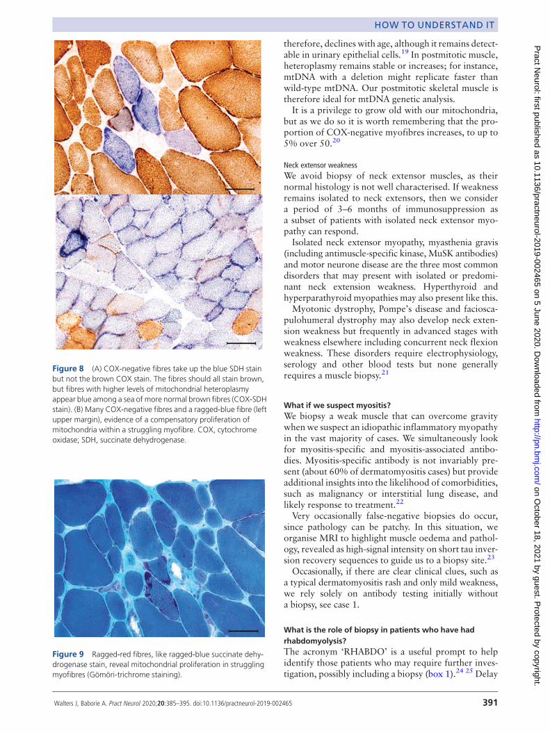
COX-negative fibres are best demonstrated by seri-ally staining for COX, followed by complex II (succi-nate dehydrogenase). Succinate dehydrogenase isentirely encoded by nuclear genes and therefore main-tains normal activity irrespective of heteroplasmylevels, see figures 7 and 8.Myofibres with higher heteroplasmy may also
show compensatory mitochondrial proliferation—the ‘ragged’ part in ragged-red or ragged-blue fibres(figures 8 and 9).It is worth knowing that the A3243G mutation, the
most common cause of mitochondrial disease, can havesurprisinglywell-preservedCOXstaining. Auseful clue inthis circumstance is the strong succinate dehydrogenasestaining visible in the blood vessel walls of muscle.7 14 15
When histochemical clues are not forthcoming butsuspicion remains, then respiratory chain enzyme ana-lysis and/or quadruple immunofluorescence that quan-tifies complex I and IV in the muscle biopsy can helpidentify abnormalities.15–17
Muscle is also the gold standard tissue for examiningmtDNA.18 In tissues with high replication rates, such asblood, heteroplasmy tends to decrease with age.Dividing cells with higher heteroplasmy may be‘weeded out’ as they are at an ‘evolutionary’ disadvan-tage. Detecting the A3243G in a blood sample,
Figure 7 Mitochondria—both mtDNA and nuclear DNA contribute to the respiratory complexes. Nuclear encoded proteins are redand mtDNA-encoded proteins are green. Note complex II is made exclusively from nuclear encoded proteins.
390 Walters J, Baborie A. Pract Neurol 2020;20:385–395. doi:10.1136/practneurol-2019-002465
HOW TO UNDERSTAND IT on O
ctober 18, 2021 by guest. Protected by copyright.
http://pn.bmj.com
/P
ract Neurol: first published as 10.1136/practneurol-2019-002465 on 5 June 2020. D
ownloaded from
therefore, declines with age, although it remains detect-able in urinary epithelial cells.19 In postmitotic muscle,heteroplasmy remains stable or increases; for instance,mtDNA with a deletion might replicate faster thanwild-type mtDNA. Our postmitotic skeletal muscle istherefore ideal for mtDNA genetic analysis.It is a privilege to grow old with our mitochondria,
but as we do so it is worth remembering that the pro-portion of COX-negative myofibres increases, to up to5% over 50.20
Neck extensor weaknessWe avoid biopsy of neck extensor muscles, as theirnormal histology is not well characterised. If weaknessremains isolated to neck extensors, then we considera period of 3–6 months of immunosuppression asa subset of patients with isolated neck extensor myo-pathy can respond.Isolated neck extensor myopathy, myasthenia gravis
(including antimuscle-specific kinase, MuSK antibodies)and motor neurone disease are the three most commondisorders that may present with isolated or predomi-nant neck extension weakness. Hyperthyroid andhyperparathyroid myopathies may also present like this.Myotonic dystrophy, Pompe’s disease and faciosca-
pulohumeral dystrophy may also develop neck exten-sion weakness but frequently in advanced stages withweakness elsewhere including concurrent neck flexionweakness. These disorders require electrophysiology,serology and other blood tests but none generallyrequires a muscle biopsy.21
What if we suspect myositis?We biopsy a weak muscle that can overcome gravitywhenwe suspect an idiopathic inflammatory myopathyin the vast majority of cases. We simultaneously lookfor myositis-specific and myositis-associated antibo-dies. Myositis-specific antibody is not invariably pre-sent (about 60% of dermatomyositis cases) but provideadditional insights into the likelihood of comorbidities,such as malignancy or interstitial lung disease, andlikely response to treatment.22
Very occasionally false-negative biopsies do occur,since pathology can be patchy. In this situation, weorganise MRI to highlight muscle oedema and pathol-ogy, revealed as high-signal intensity on short tau inver-sion recovery sequences to guide us to a biopsy site.23
Occasionally, if there are clear clinical clues, such asa typical dermatomyositis rash and only mild weakness,we rely solely on antibody testing initially withouta biopsy, see case 1.
What is the role of biopsy in patients who have hadrhabdomyolysis?The acronym ‘RHABDO’ is a useful prompt to helpidentify those patients who may require further inves-tigation, possibly including a biopsy (box 1).24 25 Delay
Figure 8 (A) COX-negative fibres take up the blue SDH stainbut not the brown COX stain. The fibres should all stain brown,but fibres with higher levels of mitochondrial heteroplasmyappear blue among a sea of more normal brown fibres (COX-SDHstain). (B) Many COX-negative fibres and a ragged-blue fibre (leftupper margin), evidence of a compensatory proliferation ofmitochondria within a struggling myofibre. COX, cytochromeoxidase; SDH, succinate dehydrogenase.
Figure 9 Ragged-red fibres, like ragged-blue succinate dehy-drogenase stain, reveal mitochondrial proliferation in strugglingmyofibres (Gömöri-trichrome staining).
Walters J, Baborie A. Pract Neurol 2020;20:385–395. doi:10.1136/practneurol-2019-002465 391
HOW TO UNDERSTAND IT on O
ctober 18, 2021 by guest. Protected by copyright.
http://pn.bmj.com
/P
ract Neurol: first published as 10.1136/practneurol-2019-002465 on 5 June 2020. D
ownloaded from
the muscle biopsy for about 8 weeks following rhabdo-myolysis, otherwise the overwhelming destruction willdwarf and obscure other diagnostic features.The causes of rhabdomyolysis are categorised as trau-
matic, non-traumatic exertional and non-traumaticnon-exertional.The non-traumatic exertional group includes the
metabolic myopathies. Their workup often includesa gene panel for rhabdomyolysis and metabolic disor-ders—their clinical features are described elsewhere (seefurther reading).24 26 Some myopathies that cause non-traumatic exertional rhabdomyolysis are not included insuch a panel, for example, muscle dystrophies (case 2),ryanodine receptor myopathies and mitochondrialdisorders.27–29 Clinicians should seek relevant clinicalclues when reviewing patients following their crisis. Theindications for biopsy in these circumstances will thenbe similar to those when suspecting a dystrophy ora mitochondrial disorder (outlined above).Rhabdomyolysis precipitated in someone taking sta-
tins when given medication that interferes with statinbreakdown does not require a biopsy. However,a biopsy may be appropriate for someone taking statinswho develops a suspected immune-mediated necrotis-ing myopathy; such a complication can occur monthsto years after starting statins (see further reading).
What is the role of biopsy in patients with hyperCKaemia?If a patient’s serum CK has been high, then repeat thetest after refraining from exercise for a week.Interpretation requires flexibility; the heavyweightchampion of the world will probably have a serumCK beyond the upper limit of normal.30
Drugs, endocrine, metabolic problems as well asmacro CK can all raise the serum CK concentration.After considering these, many neurologists then pro-ceed to electrophysiology and a possible biopsy. Next-generation sequencing can play a role, but once again,biopsy is often required for a ‘deep phenotype’ to helpinterpret the genetic tests.9
A diagnosis is more likely if electrophysiology showsmyopathic changes, if the patient is young (<25 years),or if the serum CK is over three times the upper limit of
normal. We tend to consider performing a musclebiopsy when a patient has one or more of these.31 Wewarn patients that the chances of a diagnosis are onlyabout 1 in 4. These diagnoses may include metabolicmyopathies, dystrophinopathies, limb-girdle musculardystrophies and inflammatory myopathies.32 33
When should we consider a biopsy for a potentialdrug-related muscle complication?Table 4 summarises some of the medications that haveprovoked muscle problems that we have encounteredrecently. We consider a muscle biopsy if there is noimprovement 3–6 months after drug withdrawal, butsooner if the patientworsens.34–36Wemight also considera biopsy if there are several competing narratives. Forexample, consider a patient with a renal transplant andunderlying connective tissue disorder who develops agradually progressive proximal weakness. Is it the corti-costeroid, the colchicine or the statin? Could it be amyositis?Muscle biopsy can help; vacuolar changes with-out inflammation would, for example, incriminatecolchicine.
What is the role of biopsy in patients with myalgia?Muscle pain is a near-universal experience. Forinstance, it accompanies infections ranging from chi-kungunya (meaning ‘to become contorted’ inKimakonde) through to humdrum colds and ‘influenza.Clinical criteria define conditions like fibromyalgia
and chronic fatigue. They are ‘bread and butter’ for ourprimary care colleagues and expertlymanaged by them.The following might prompt further investigations:
Table 4 Iatrogenic myopathies witnessed in muscle clinic over the lastfew years
Drugs and myalgiaStatins—with or without raised serum creatine kinaseCalcium channel blockersCorticosteroid and selective serotonin reuptake inhibitor (SSRI)withdrawalNitrofurantoin and ciprofloxacinAromatase inhibitors—medical treatment for breast cancerColchicineDrugs and inflammatory myositisStatinsImmune checkpoint inhibitors—immune-related adverse events(irAE)Drugs and rhabdomyolysisHeroin, cocaine, alcohol and amphetamineStatinsOtherAcute quadriplegic myopathy (critical care myopathy) including onepatient who had a striking external ophthalmoplegiaPolymyositis (chronic graft vs host) following allogeneic bone marrowtransplantation
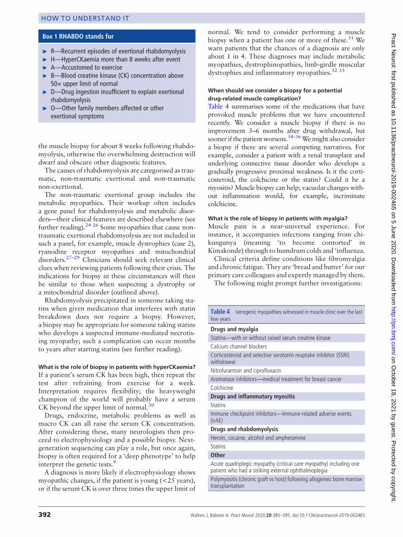
Box 1 RHABDO stands for
► R—Recurrent episodes of exertional rhabdomyolysis► H—HyperCKaemia more than 8 weeks after event► A—Accustomed to exercise► B—Blood creatine kinase (CK) concentration above
50× upper limit of normal► D—Drug ingestion insufficient to explain exertional
rhabdomyolysis► O—Other family members affected or other
exertional symptoms
392 Walters J, Baborie A. Pract Neurol 2020;20:385–395. doi:10.1136/practneurol-2019-002465
HOW TO UNDERSTAND IT on O
ctober 18, 2021 by guest. Protected by copyright.
http://pn.bmj.com
/P
ract Neurol: first published as 10.1136/practneurol-2019-002465 on 5 June 2020. D
ownloaded from
► Weakness. Pain can create the appearance ofweakness, oftenwith a sudden reduction in effort. Watching a person walk-ing on their toes or heels, hopping, or walking up-stairs ismore revealing than just a couch-based assessment.37
► Myopathic electrophysiology► HyperCKaemia (>2 to 3 times)► Exercise-induced pain (including a second wind)► Hypertrophy or muscle atrophyMyotonic dystrophy type 2 may present almost
exclusively with myalgia, and similarly, Parkinsonismis easily overlooked in this context.We explain to patients that the chance of diagnosis is
low, perhaps 1 in 20 biopsies, and that non-specificabnormalities are common.38–41 An excellent article onmuscle biopsy remarked, ‘If the clinician cannot findsomething wrong it is highly unlikely that the testswill!’.42
Avariety of pathologies may emerge, including meta-bolic myopathies, congenital myopathies, neurogenicdisorders, mitochondrial disorders, myositis and mus-cular dystrophies.41 43
What is the role of biopsy in patientswith exercise-inducedmuscle pain?Common causes for exercise-induced myalgia includeneurogenic or vascular claudication; amyloid myopa-thy can rarely cause claudication.44
The celebrated ‘lactate burn’ is well known, beingfamiliar to genuine athletes and also a beloved exhorta-tion roared out by commentators and armchair athletes asthe home straight beckons. However, patients withMcArdle’s disease suffer exercise-induced pain but pro-duce no lactate; lactate probably does not fully deserve itsepithet.This group includes the metabolic myopathies: their
clinical features and workup are described elsewhere.26
Remember that the metabolic gene panel is not exhaus-tive; if the panel is negative but clinical suspicionremains then a muscle biopsy may reveal, for example,a mutation in cytochrome b, the only mtDNA-encodedgene in complex III.45
Other exercise-induced muscle phenomena, such asmyotonia, often require electrophysiology and genetictests rather than biopsy. Muscle biopsy has no role insuspected skeletal muscle channelopathy, for instance,and the diagnosis is genetic.
Some other potential indications for a muscle biopsyPatients with suspected peripheral nerve vasculitisshould have combined nerve and muscle biopsy sincethe chances of positive histology increase from about50% to 70% compared with a nerve biopsy alone.46
A generous biopsy from an asymptomatic muscle mayreveal non-caseating granulomas, in a challenging caseof possible neurosarcoidosis.47 Early skin or musclebiopsy may help in the frequently elusive diagnosis ofintravascular lymphoma.48
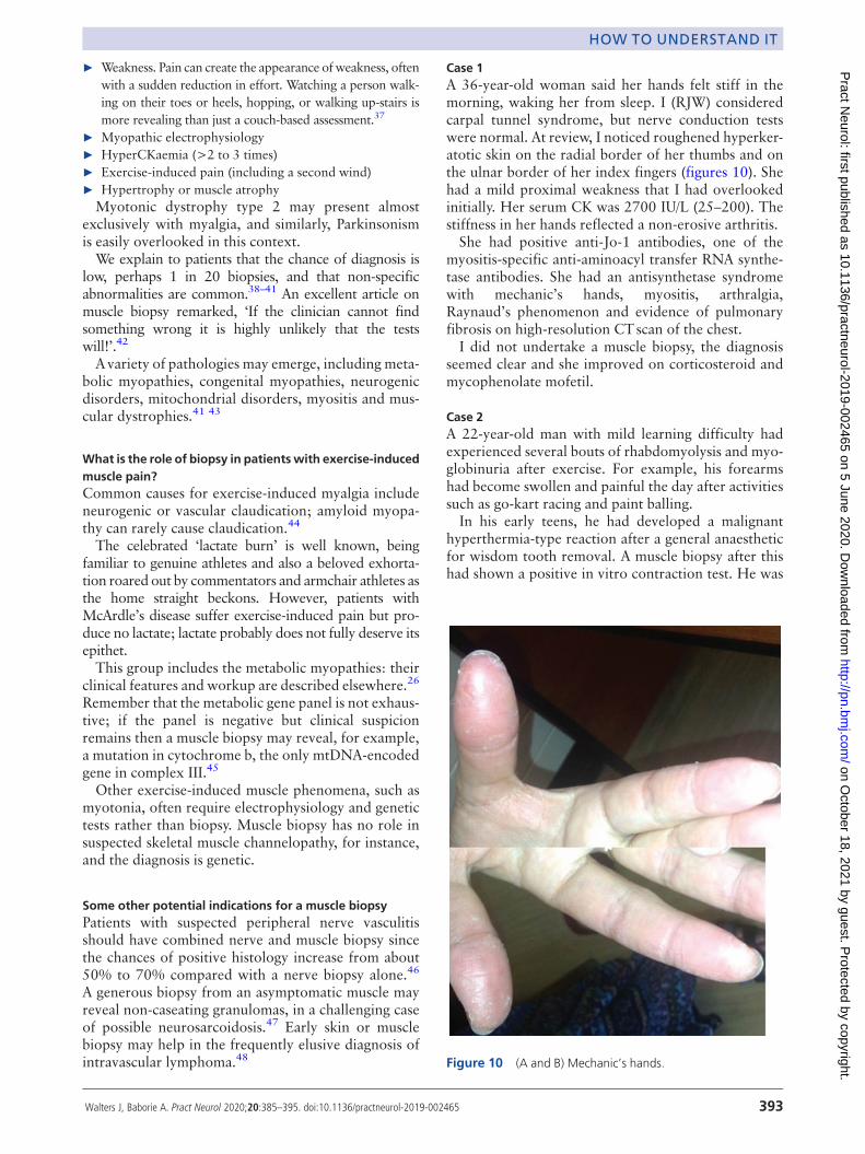
Case 1A 36-year-old woman said her hands felt stiff in themorning, waking her from sleep. I (RJW) consideredcarpal tunnel syndrome, but nerve conduction testswere normal. At review, I noticed roughened hyperker-atotic skin on the radial border of her thumbs and onthe ulnar border of her index fingers (figures 10). Shehad a mild proximal weakness that I had overlookedinitially. Her serum CK was 2700 IU/L (25–200). Thestiffness in her hands reflected a non-erosive arthritis.She had positive anti-Jo-1 antibodies, one of the
myositis-specific anti-aminoacyl transfer RNA synthe-tase antibodies. She had an antisynthetase syndromewith mechanic’s hands, myositis, arthralgia,Raynaud’s phenomenon and evidence of pulmonaryfibrosis on high-resolution CTscan of the chest.I did not undertake a muscle biopsy, the diagnosis
seemed clear and she improved on corticosteroid andmycophenolate mofetil.
Case 2A 22-year-old man with mild learning difficulty hadexperienced several bouts of rhabdomyolysis and myo-globinuria after exercise. For example, his forearmshad become swollen and painful the day after activitiessuch as go-kart racing and paint balling.In his early teens, he had developed a malignant
hyperthermia-type reaction after a general anaestheticfor wisdom tooth removal. A muscle biopsy after thishad shown a positive in vitro contraction test. He was
Figure 10 (A and B) Mechanic’s hands.
Walters J, Baborie A. Pract Neurol 2020;20:385–395. doi:10.1136/practneurol-2019-002465 393
HOW TO UNDERSTAND IT on O
ctober 18, 2021 by guest. Protected by copyright.
http://pn.bmj.com
/P
ract Neurol: first published as 10.1136/practneurol-2019-002465 on 5 June 2020. D
ownloaded from
diagnosed with malignant hyperthermia. The biopsywas not analysed further and he had no genetic tests.We considered a ryanodine mutation, given his
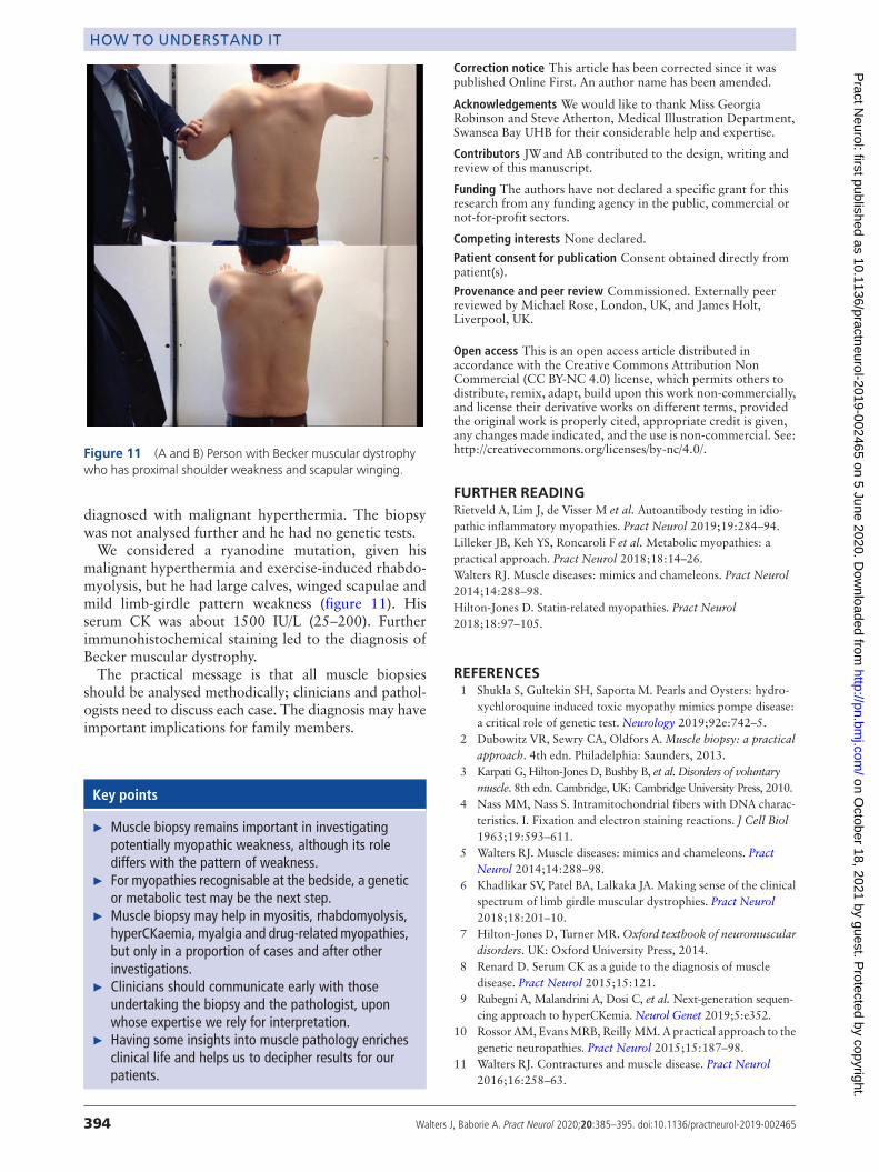
malignant hyperthermia and exercise-induced rhabdo-myolysis, but he had large calves, winged scapulae andmild limb-girdle pattern weakness (figure 11). Hisserum CK was about 1500 IU/L (25–200). Furtherimmunohistochemical staining led to the diagnosis ofBecker muscular dystrophy.The practical message is that all muscle biopsies
should be analysed methodically; clinicians and pathol-ogists need to discuss each case. The diagnosis may haveimportant implications for family members.
Correction notice This article has been corrected since it waspublished Online First. An author name has been amended.
Acknowledgements We would like to thank Miss GeorgiaRobinson and Steve Atherton, Medical Illustration Department,Swansea Bay UHB for their considerable help and expertise.
Contributors JWand AB contributed to the design, writing andreview of this manuscript.
Funding The authors have not declared a specific grant for thisresearch from any funding agency in the public, commercial ornot-for-profit sectors.
Competing interests None declared.
Patient consent for publication Consent obtained directly frompatient(s).
Provenance and peer review Commissioned. Externally peerreviewed by Michael Rose, London, UK, and James Holt,Liverpool, UK.
Open access This is an open access article distributed inaccordance with the Creative Commons Attribution NonCommercial (CC BY-NC 4.0) license, which permits others todistribute, remix, adapt, build upon this work non-commercially,and license their derivative works on different terms, providedthe original work is properly cited, appropriate credit is given,any changes made indicated, and the use is non-commercial. See:http://creativecommons.org/licenses/by-nc/4.0/.
FURTHER READINGRietveld A, Lim J, de Visser M et al. Autoantibody testing in idio-pathic inflammatory myopathies. Pract Neurol 2019;19:284–94.Lilleker JB, Keh YS, Roncaroli F et al. Metabolic myopathies: apractical approach. Pract Neurol 2018;18:14–26.Walters RJ. Muscle diseases: mimics and chameleons. Pract Neurol2014;14:288–98.Hilton-Jones D. Statin-related myopathies. Pract Neurol2018;18:97–105.
REFERENCES1 Shukla S, Gultekin SH, Saporta M. Pearls and Oysters: hydro-
xychloroquine induced toxic myopathy mimics pompe disease:a critical role of genetic test. Neurology 2019;92e:742–5.
2 Dubowitz VR, Sewry CA, Oldfors A. Muscle biopsy: a practicalapproach. 4th edn. Philadelphia: Saunders, 2013.
3 Karpati G, Hilton-Jones D, Bushby B, et al. Disorders of voluntarymuscle. 8th edn. Cambridge, UK: Cambridge University Press, 2010.
4 Nass MM, Nass S. Intramitochondrial fibers with DNA charac-teristics. I. Fixation and electron staining reactions. J Cell Biol1963;19:593–611.
5 Walters RJ. Muscle diseases: mimics and chameleons. PractNeurol 2014;14:288–98.
6 Khadlikar SV, Patel BA, Lalkaka JA. Making sense of the clinicalspectrum of limb girdle muscular dystrophies. Pract Neurol2018;18:201–10.
7 Hilton-Jones D, Turner MR.Oxford textbook of neuromusculardisorders. UK: Oxford University Press, 2014.
8 Renard D. Serum CK as a guide to the diagnosis of muscledisease. Pract Neurol 2015;15:121.
9 Rubegni A, Malandrini A, Dosi C, et al. Next-generation sequen-cing approach to hyperCKemia. Neurol Genet 2019;5:e352.
10 Rossor AM, EvansMRB, ReillyMM. A practical approach to thegenetic neuropathies. Pract Neurol 2015;15:187–98.
11 Walters RJ. Contractures and muscle disease. Pract Neurol2016;16:258–63.
Figure 11 (A and B) Person with Becker muscular dystrophywho has proximal shoulder weakness and scapular winging.
Key points
► Muscle biopsy remains important in investigatingpotentially myopathic weakness, although its rolediffers with the pattern of weakness.
► For myopathies recognisable at the bedside, a geneticor metabolic test may be the next step.
► Muscle biopsy may help in myositis, rhabdomyolysis,hyperCKaemia, myalgia and drug-related myopathies,but only in a proportion of cases and after otherinvestigations.
► Clinicians should communicate early with thoseundertaking the biopsy and the pathologist, uponwhose expertise we rely for interpretation.
► Having some insights into muscle pathology enrichesclinical life and helps us to decipher results for ourpatients.
394 Walters J, Baborie A. Pract Neurol 2020;20:385–395. doi:10.1136/practneurol-2019-002465
HOW TO UNDERSTAND IT on O
ctober 18, 2021 by guest. Protected by copyright.
http://pn.bmj.com
/P
ract Neurol: first published as 10.1136/practneurol-2019-002465 on 5 June 2020. D
ownloaded from
12 Brady S, Squier W, Hilton-Jones D. Clinical assessment deter-mines the diagnosis of inclusion body myositis independently ofpathological features. J Neurol Neurosurg Psychiatry2013;84:1240–6.
13 Gorman GS, Schaefer AM, Ng Y, et al. Prevalence of nuclear andmitochondrial DNA mutations related to adult mitochondrialdisease. Ann Neurol 2015;77:752–9.
14 Chinnery PF. Could it be mitochondrial? When and how toinvestigate. Pract Neurol 2006;6:90–101.
15 Chinnery PF. The mitochondrion and its disorders. Pract Neurol2003;3:100–5.
16 Rocha MC, Grady JP, Grunewald A, et al. A novel immuno-fluorescent assay to investigate oxidative phosphorylation defi-ciency in mitochondrial myopathy: understanding mechanismsan improving diagnosis. Sci Rep 2015;15:15037.
17 Rahman S, HannaMG.Diagnosis and therapy in neuromusculardisorders: diagnosis and new treatments in mitochondrial dis-eases. J Neurol Neurosurg Psychiatry 2009;80:943–53.
18 Stewart JB, Chinnery PF. The dynamics of mitochondrial DNAheteroplasmy: implications for human health and disease. NatRev Genet 2015;16:530–42.
19 Rahman S, Poulton J, Marchington D, et al.Decrease of 3243 A−G mtDNA mutation from blood in MELAS syndrome: alongitudinal study. Am J Hum Genet 2001;68:238–40.
20 Harding AE. Growing old: the most common mitochondrialdisease of all? Nat Genet 1992;2:251–2.
21 Cauchi M, Marsh E. A practical approach to the patient pre-senting with dropped head. Pract Neurol 2016;16:445–51.
22 Rietveld A, Lim J, de Visser M, et al. Autoantibody testing in idio-pathic inflammatory myopathies. Pract Neurol 2019;19:284–94.
23 Van De Viekkert J, MaasM, Hoogendijk JE, et al.CombiningMRIand muscle biopsy improves diagnostic accuracy in subacute-onsetidiopathic inflammatory myopathy.Muscle Nerve 2015;51:253–8.
24 Fernandes PM, Davenport RJ. How to do it: investigate exer-tional rhabdomyolysis (or not). Pract Neurol 2019;19:43–8.
25 Scalco RS, Snoeck M, Quinlivan R, et al. Exertional rhabdomyo-lysis: physiological response or manifestation of an underlyingmyopathy? BMJ Open Sport Exerc Med 2016;2:e000151–15.
26 Lilleker JB, Keh YS, Roncaroli F, et al.Metabolic myopathies: apractical approach. Pract Neurol 2018;18:14–26.
27 Muldoon S, Deuster P, Voelkel M, et al. Exertional heat illness,exertional rhabdomyolysis, and malignant hyperthermia: isthere a link? Curr Sports Med Rep 2008;7:74–80.
28 Nicolau S, Liewluck T, Tracy JA, et al.Congenital myopathies inthe adult neuromuscular clinic. Diagnostic challenges and pit-falls. Neurol Genet 2019;5:e341.
29 Chinnery PF, Johnson MA, Taylor RW, et al. A novel mito-chondrial tRNA phenylalanine mutation presenting with acuterhabdomyolysis. Ann Neurol 2004;41:408–10.
30 Silvestri NJ, Wolfe GI. Asymptomatic/pauci-symptomatic creatinekinase elevations (hyperckemia).Muscle Nerve 2013;47:805–15.
31 Kyriakides T, Angelini C, Scaefer J, et al. EFNS guidelines on thediagnostic approach to pauci or asymptomatic hyperCKemia.Eur J Neurol 2010;17:767–73.
32 Prelle A, Tancredi L, Sciacco M, et al. Retrospective study of alarge population of patients with asymptomatic or minimallysymptomatic raised serum creatine kinase level. J Neurol2002;249:305–11.
33 Fernandez C, de Paula AM, Figarella-Branger D, et al.Diagnostic evaluation of clinically normal subjects with chronichyperCKemia. Neurology 2006;66:1585–7.
34 Hilton-Jones D. Statin-related myopathies. Pract Neurol2018;18:97–105.
35 Parker BA, Capizzi JA, Grimaldi AS, et al. Effect of statins onskeletal muscle function. Circulation 2013;127:96–103.
36 Mastaglia FL. Drug induced myopathies. Pract Neurol2006;6:4–13.
37 Petty R. Evaluating muscle symptoms. J Neurol NeurosurgPsychiatry 2003;74:ii38–42.
38 Kyriakides T, Angelini C, Schaefer J, et al. EFNS review on therole of muscle biopsy in the investigation of myalgia. Eur JNeurol 2013;20:997–1005.
39 Kissel JT. Muscle biopsy in patients with myalgia: still a painfuldecision. Neurology 2007;68:170–1.
40 Echaniz-Laguna A, Chanson J-B. Electromyography and musclebiopsy in chronic isolated myalgia: a prospective study. MuscleNerve 2016;54:321–4.
41 Filosto M, Tonin P, Vattemi G, et al. The role of musclebiopsy in investigating isolated muscle pain. Neurology2007;68:181–6.
42 Hall G. Muscle biopsy. Pract Neurol 2001;1:113–18.43 Te Riele MG, Schreuder TH, van Alfen N, et al. The yield of
diagnostic work-up of patients presenting with myalgia, exerciseintolerance, or fatigue: a prospective observational study.Neuromuscul Disord 2017;27:243–50.
44 GertzMA, Kyle RA.Myopathy in primary systemic amyloidosis.J Neurol Neurosurg Psychiatry 1996;60:655–60.
45 Andreu AL, Hanna MG, Reichmann H, et al. Exercise intoler-ance due to mutations in the cytochrome b gene of mitochon-drial DNA. N Engl J Med 1999;341:1037–44.
46 Collins MP, Mendell JR, Periquet MI, et al. Superficial peronealnerve/peroneus brevis muscle biopsy in vasculitic neuropathy.Neurology 2000;55:636–43.
47 Joseph FG, ScoldingNJ. Sarcoidosis of the nervous system. PractNeurol 2007;7:234–44.
48 Tahsili-Fahadan P, Rashidi A, Cimino PJ, et al. Neurologicmanifestations of intravascular large B−cell lymphoma. NeurolClin Pract 2016;6:55–60.
Walters J, Baborie A. Pract Neurol 2020;20:385–395. doi:10.1136/practneurol-2019-002465 395
HOW TO UNDERSTAND IT on O
ctober 18, 2021 by guest. Protected by copyright.
http://pn.bmj.com
/P
ract Neurol: first published as 10.1136/practneurol-2019-002465 on 5 June 2020. D
ownloaded from