Embed Size (px)
Citation preview
Human High Temperature Requirement Serine Protease A1(HTRA1) Degrades Tau Protein Aggregates*□S
Received for publication, October 27, 2011, and in revised form, April 24, 2012 Published, JBC Papers in Press, April 25, 2012, DOI 10.1074/jbc.M111.316232
Annette Tennstaedt,a1 Simon Popsel,a1 Linda Truebestein,a Patrick Hauske,a Anke Brockmann,a2 Nina Schmidt,a
Inga Irle,a Barbara Sacca,b Christof M. Niemeyer,b Roland Brandt,c Hanna Ksiezak-Reding,d Anca Laura Tirniceriu,e
Rupert Egensperger,e Alfonso Baldi,f Leif Dehmelt,g Markus Kaiser,a Robert Huber,a,h,i,j Tim Clausen,k
and Michael Ehrmanna,j3
From the aCentre for Medical Biotechnology, Faculty of Biology, University Duisburg-Essen, Universitaetsstrasse, 45141 Essen, Germany,the bFakultat Chemie, Biologisch-Chemische Mikrostrukturtechnik, Technische Universitat Dortmund, Otto-Hahn-Strasse 6,44227 Dortmund, Germany, the cFachbereich Biologie/Chemie, University Osnabrueck, D-49076 Osnabrueck, Germany, thedDepartment of Neurology, Mount Sinai School of Medicine, One Gustave L. Levy Place, New York, New York 10029, the eCenter forNeuropathology and Prion Research, Ludwig Maximilians University of Munich, Feodor-Lynen-Strasse 23, 81377 Munich, Germany,the fDepartment of Biochemistry and Biophysics, Section of Pathology, the Second University of Naples, 80100 Naples, Italy, thegMax-Planck-Institute of Molecular Physiology, Otto-Hahn-Strasse 11, 44227 Dortmund, Germany, the hDepartment for ChemicalBiology, Technische Universitat Dortmund University, Otto-Hahn-Strasse 6, 44227 Dortmund, Germany, the iMax-Planck-Institutfur Biochemie, Am Klopferspitz 18A, 82152 Martinsried, Germany, the jSchool of Biosciences, Cardiff University, Cardiff CF10 3US,United Kingdom, and the kResearch Institute of Molecular Pathology, Dr. Bohr-Gasse 7, A-1030 Vienna, Austria
Background: Protein quality control proteases degrade damaged proteins and protein fragments.Results: The human serine protease HTRA1 degrades tau aggregates and is induced by its substrates.Conclusion: A member of the widely conserved HtrA family is involved in protein quality control in mammalian cells.Significance: HTRA1 might function as a tau protease in vivo.
Protective proteases are key elements of protein qualitycontrol pathways that are up-regulated, for example, undervarious protein folding stresses. These proteases areemployed to prevent the accumulation and aggregation ofmisfolded proteins that can impose severe damage to cells.The high temperature requirement A (HtrA) family of serineproteases has evolved to perform important aspects of ATP-independent protein quality control. So far, however, noHtrA protease is known that degrades protein aggregates.Weshow here that human HTRA1 degrades aggregated andfibrillar tau, a protein that is critically involved in variousneurological disorders. Neuronal cells and patient brainsaccumulate less tau, neurofibrillary tangles, and neuriticplaques, respectively, when HTRA1 is expressed at elevatedlevels. Furthermore, HTRA1 mRNA and HTRA1 activity areup-regulated in response to elevated tau concentrations.These data suggest that HTRA1 is performing regulated pro-teolysis during protein quality control, the implications ofwhich are discussed.
HumanHTRA1 belongs to the widely conserved high tem-perature requirement A (HtrA)4 family of homo-oligomericand ATP-independent serine proteases (1, 2). HtrAs of pro-and eukaryotes are implicated in protein quality control.They can act as key stress sensors and regulators of unfoldedprotein response signaling pathways and can mediate therepair and assembly or the removal of damaged, fragmented,and mislocalized proteins (3–9). Defining features of HtrAproteases are their homo-oligomeric architecture and thepresence of C-terminal PDZ domains that can be involved insubstrate processing, sensing of misfolded proteins, mediat-ing allosteric and cooperative regulation of the proteolyticactivity, and in the switch between various oligomeric states(2).Among the four human HTRAs, HTRA1–4, the ubiqui-
tously expressedHTRA1 consists of a signal sequence for secre-tion, partial insulin-like growth factor-binding protein-7domain, serine protease domain resembling classic serine pro-teases such as trypsin and one C-terminal PDZ domain. LikeEscherichia coli DegP, a prototypic HtrA protease involved inprotein quality control, HTRA1 is activated by oligomerization.Substrate binding triggers the switch between the resting andthe active conformations and between various oligomeric states(10, 11).HTRA1 has at least three cellular localizations and a multi-
tude of functions. Extracytoplasmic HTRA1 is involved in thehomeostasis of the extracellular matrix, and elastin, fibulin 5,
* Brain tissue was obtained from the German Brain Bank “Brain-Net,” which is supported by the Federal Ministry of Education andResearch.
□S This article contains supplemental Figs. S1–S4, Experimental Procedures,and additional references.
1 Both authors contributed equally to this work.2 Supported by grants from Second University of Naples and Futura-onlus.3 Supported by Deutsche Forschungsgemeinschaft, Alzheimer’s Research
Trust, the Fonds der Chemischen Industrie, and a grant from the Inno-vation, Science, Research, and Technology Ministry of Nordrhein-West-falen, Germany. To whom correspondence should be addressed. Tel.:49-201-183-2949; E-mail: [email protected].
4 The abbreviations used are: HtrA, high temperature requirement A; AD,Alzheimer disease; AFM, atomic force microscopy; Bis-Tris, bis(2-hydroxy-ethyl)iminotris(hydroxymethyl)methane; PHP, pseudohyperphosphory-lated; pNA, p-nitroaniline; qRT-PCR, quantitative RT-PCR; ThT, thioflavin T.
THE JOURNAL OF BIOLOGICAL CHEMISTRY VOL. 287, NO. 25, pp. 20931–20941, June 15, 2012© 2012 by The American Society for Biochemistry and Molecular Biology, Inc. Published in the U.S.A.
JUNE 15, 2012 • VOLUME 287 • NUMBER 25 JOURNAL OF BIOLOGICAL CHEMISTRY 20931
by guest on February 16, 2018http://w
ww
.jbc.org/D
ownloaded from
nidogen 2, fibronectin, fibromodulin, aggrecan, and decorinhave been identified as substrates (12–17). IntracellularHTRA1 was localized to microtubules and to the nucleus (18,19). Cytoplasmic HTRA1 has been implicated in the degrada-tion of tuberin thereby modulating cell growth and prolifera-tion (20). Furthermore, microtubule-associated HTRA1degrades tubulins thereby inhibiting cell migration (18, 21).Consequently, HTRA1 has been implicated in several severepathologies including cancer, age-related macular degenera-tion, Alzheimer disease (AD), arthritis, and familial ischemiccerebral small vessel disease (12, 13, 22–26). In many of thesediseases, protein fragments or aggregates are either causativefor disease or are disease-modifying factors that are producedor degraded by HTRA1.Recent studies suggest that substrate specificity and process-
ing of individual HtrA proteases can differ significantly.Whereas bacterial DegS is a regulatory protease that cleaves itssingle substrate at one defined position, other HtrAs such asE. coli DegP digest a great many of un- or misfolded proteinsinto small peptides (8, 9, 27, 28). However, these and otherstudies suggested that HtrA proteases do not degrade proteinaggregates. This model was supported, for example, by the pre-cise understanding of the proteolytic mechanism of DegP,requiring concurrent binding of substrates to its PDZ domain 1and the active site for both activation and proteolysis (28–30).However, the recent elucidation of crystal structures of HTRA1and complementing mechanistic studies indicating that prote-olysis and activation of HTRA1 occur in a PDZ domain-inde-pendent manner prompted us to address the question ofwhether human HTRA1 is able to use protein aggregates assubstrates (10).To testwhetherHTRA1degrades protein aggregatesweused
tau as a model substrate because tau is, like HTRA1, associatedwith microtubules. Furthermore, tau is of important clinicalrelevance, and its aggregation is widely studied. It is thereforewell established that the tau protein can aggregate into intra-cellular neurofibrillary tangles that are specific pathologicalfeatures of AD and other tauopathies. Normal tau, which isabundant in axons, is thought to regulate microtubule dynam-ics. Interaction of tau with microtubules is mediated by itsmicrotubule binding domain consisting of three or four repeatssharing the consensus sequence VXSKXGSXXN(L/I)XHX-PGGG. Posttranslational modifications, such as phosphoryla-tion, can decrease the affinity of tau to microtubules leading todissociation and polymerization into straight or paired helicalfilaments, ribbons, and other conformations. Progressive inter-molecular interactions of fibrils composed of paired helical fil-aments of tau lead to the formation of neurofibrillary tangles. Acore domain of paired helical filaments consists of three or fourrepeats of tau involved in stable tau-tau interactions (Fig. 1A).Within this core domain, one short stretch, the self- assemblyregion VQI(I/V)XK, seems of critical importance. Proteolyticprocessing of tau stimulates the assembly of tau into fibrils. Suchassembly can capture further full-length tau proteins for process-ing leading to progressive growth of the toxic protein aggregateseventually causing cell death (for review see Refs. 31, 32).
EXPERIMENTAL PROCEDURES
Plasmids
Bacterial expression vectors of human taus and the GFP-tauconstruct pEGFP-C3–3Rtau were described earlier (33, 34).Expression plasmid pHTRA1 was generated by PCR and sub-cloning of HTRA1 into pBABE (35). HTRA1-mCherry con-structs were derivatives of pmCherry-N1 (Clontech). Expres-sion of WT-tau, Ala-tau, and pseudohyperphosphorylated(PHP)-tau was done as described (36).
Purification of HTRA1, HTRA1�PDZ Domain, WT-Tau, Ala-Tau,and PHP-Tau
For all in vitro protease assays, HTRA1 and HTRA1�PDZdomain were purified and used as described (24) except that anadditional hydroxyapatite column (Bio-Rad) was added.Recombinant tau variants were isolated by boiling of clearedbacterial cell extracts (lysis buffer: 33 mM Tris-HCl, pH 8, 100mM KCl) in the water bath for 30 min. Tau remains soluble,whereas the precipitated bacterial proteins were cleared fromthe lysate by centrifugation (35,000 � g, 40 min). For furtherpurification of the 3R tau variants (0N3R, 352 amino acids), thesupernatant was incubated with ammonium sulfate at 30% sat-uration for 30min, 4 °C, followed by centrifugation at 20,000�g, 30 min. Tau was precipitated from the supernatant with40% saturated ammonium sulfate. Purified tau was sus-pended in 80 mM PIPES/KOH, pH 6.8, 1 mM EGTA, 1 mM
MgCl2. For the isolation of insoluble and soluble fractions oftau, protein reconstituted from ammonium sulfate pelletswas incubated at 25 °C for 20 min to ensure complete dis-solving of precipitated tau. Ultracentrifugation (100,000 � g,for 1 h) was used to separate insoluble pellet (3RP) and sol-uble fractions (3RS). The insoluble protein was resuspendedfrom the resulting pellet and the protein concentrationdetermined by Bradford quantitation.
Purification of 441-Amino Acid WT-Tau Protein
The 4R WT isoform of human tau (2N4R, 441 amino acids)used for heparin-induced fibrillization was purified in the sameway as the 3R variants described above except that instead ofammonium sulfate precipitation the boiled lysatewas subjectedto hydroxyapatite chromatography (Bio-Rad). 4R tau waseluted with an NaCl gradient in 100 mM HEPES, 10 mM KPO4,2 mM DTT, pH 7.6. Subsequently, monomeric and dimeric 4Rtau was isolated using size exclusion chromatography (Super-dex HiLoad 200 26/60; GE Healthcare).
Heparin-induced 4R Tau Fibrillization, Degradation of 4R TauFibrils
The in vitro formation of PHF-like tau filaments was per-formed as described (37). Briefly, 20�M4R tauwas incubated at55 °C, 10min in aggregation buffer (100mM sodiumacetate, pH7.0, 2 mM DTT) before addition of 50 �M heparin (Sigma-Al-drich) and incubation at 37 °C, 1,000 rpm, for the time pointsindicated. Proteolytic digests of the tau aggregates were per-formed as described above except for the following modifica-tions. A 5-fold molar excess of tau over the protease was usedbased on themolecular mass of monomeric tau, 5 mM reducing
HTRA1 Degrades Protein Aggregates
20932 JOURNAL OF BIOLOGICAL CHEMISTRY VOLUME 287 • NUMBER 25 • JUNE 15, 2012
by guest on February 16, 2018http://w
ww
.jbc.org/D
ownloaded from
agent Tris(2-carboxyethyl)phosphine was added to the reac-tions and 50 mM NaH2PO4, pH 8, was used for proteolysis byHTRA1.
Atomic Force Microscopy (AFM)
Tau protein samples (1 �M tau in PBS, pH 7.5) were depos-ited on a freshly cleaved mica surface (Plano GmbH) withoutany previous treatment and adsorbed for 3 min at room tem-perature. After addition of 15 �l of 1� TAEM (40 mM Tris, 20mM acetic acid, 2 mM EDTA, 12.5 mM Mg acetate, pH 8.0), thesample was scanned in tapping mode using a MultiModeTMmicroscope (Veeco Metrology, Santa Barbara, CA) equippedwith a Nanoscope IV controller. 0.58 N/m force constant can-tilevers with sharpened pyramidal tips (SNL-10 tips, VeecoMetrology) were used for scanning. After engagement, the tap-
ping amplitude set point was typically less than 1 volt, and thescan rates ranged between 1 and 2 Hz. Multiple AFM imageswere recorded from different locations of the mica surface toensure reproducibility of the results. All images were analyzedwith the Nanoscope 6.14R1 and ImageJ software (38).
Thioflavin T (ThT) Fluorescence
To characterize the aggregation state of tau, 10-�l duplicatesof the samples at a tau concentration of 20�Mwere added to 90�l of 12 �M ThT (Sigma-Aldrich) in 50 mM glycine, pH 8.5,using black clear-bottom 96-well microtiter plates. After incu-bation at 37 °C, 900 rpm, the fluorescence was measured with aSpectraMax M5 Microplate Reader (Molecular Devices). Foremission spectra, the excitation wavelength was kept constantat 440 nm with emission wavelengths ranging from 450 to 520
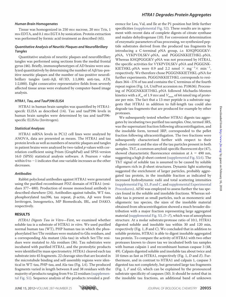
FIGURE 1. Proteolysis of tau by HTRA1, calpain 1, and caspase 3. A, HTRA1 cleaves tau at multiple sites in its core domain which is relevant for aggregation.WT-, PHP-, and Ala-tau were completely digested by HTRA1. Proteolytic products were identified by mass spectrometry. Forward slashes indicate cleavage sitesthat were identical in the three tau variants analyzed. For simplicity, we show cleavage sites detected in tau Ala239–Val399, the region that is implicated inaggregation. The sequence of the synthetic substrate PGGGNKKIETHKL-pNA is underlined. B and C, HTRA1 degrades tau. Purified soluble (B) and insoluble (C)tau derivatives were incubated with or without HTRA1. Samples were removed at the time points indicated and subjected to SDS-PAGE and visualized by silverstaining. D–G, degradation of tau by calpain 1 (D and E) and caspase 3 (F and G) was carried out as described in B and C (see “Experimental Procedures”),respectively. The band marked by an asterisk present in all insoluble fractions of tau (C, E, and G) is a contaminant and therefore not degraded by either protease.
HTRA1 Degrades Protein Aggregates
JUNE 15, 2012 • VOLUME 287 • NUMBER 25 JOURNAL OF BIOLOGICAL CHEMISTRY 20933
by guest on February 16, 2018http://w
ww
.jbc.org/D
ownloaded from
nm in 5-nm intervals. Single measurements were performed atan excitation and emission wavelength of 440 and 480 nm,respectively. The cutoff filter was set to 450 nm in all cases.
Protease Assays and Mass Spectrometry
Protease assays were performed at 37 °C in 50 mM Tris-HCl,pH 8.0. For mass spectrometry, 1 �g of HTRA1 was incubatedwith 10 �g ofWT-tau, Ala-tau, or PHP-tau in a final volume of300 �l in 50 mM Tris-HCl, pH 8.0. After 4 h at 37 °C, sampleswere centrifuged for 30 min, and the supernatants containingtau fragments were precipitated with acetone. MALDI-TOFmass spectra were obtained as described (24). Samples wereloaded onto 10% SDS-PAGE and analyzed byWestern blottingusing a specific antibody against the tau396 phosphorylationsite.HTRA1 Assay—The proteolytic activity of human recombi-
nant HTRA1s was measured using PGGGNKKIETHKL-p-nitroaniline (pNA) (tau fragment (Pro363–Leu376)) and VFN-TLPMMGKASPV-pNA as substrates that were synthesizedfollowing standard procedures (10, 39, 40). 1.35 �M HTRA1swere incubated with 500 �M respective substrate and the con-centrations of WT- or PHP-tau indicated in 50 mM Tris-HCl,pH 8, at 37 °C. The release of nitroaniline was monitored con-tinuously by measuring the absorption at � � 405 nm everyminute for 90 min. The reactions were performed in 96-wellmicroplates using a SpectraMax M5 Microplate Reader. Forcalculation of the specific activity, a time segment of linearincrease in absorption was used to quantify the turnover of thesubstrate by employing the specific molar absorption coeffi-cient of nitroaniline, 8,800 M�1� cm1.Caspase 3 Assay—The proteolytic activity of human recom-
binant caspase 3 was assayed using 200 �M Ac-DEVD-pNA(AAT Bioquest) as a substrate and 50 mM HEPES, pH 7.4, 100mM NaCl, 0.1% CHAPS, 1 mM EDTA, 10% glycerol, 10 mM
DTT, as the reaction buffer.Fluorometric Calpain Assay—Human calpain 1 (Calbi-
ochem) activity was determined at 23 °C using Suc-LLVY-AMC (Sigma) as a substrate in 50 mM Tris-HCl, 100 mM NaCl,2 mM CaCl2, 1 mM DTT, pH 7.5. Substrate and calpain 1 con-centrations were 200 �M and 0.52 �M, respectively. Fluores-cence was monitored continuously by performing single mea-surements every minute for 90 min with an excitationwavelength of 380 nm and an emission wavelength of 450 nmusing a SpectraMaxM5Microplate Reader, and the cutoff filterwas set to 435 nm. From the initial linear increase in fluores-cence, the concentration of free AMC was quantified using anAMC standard curve generated. The specific activity of calpain1 was calculated as the ratio of substrate turnover and amountof enzyme.Proteolysis of Insoluble and Soluble Tau Fractions—Purified
WT-3R tau samples were centrifuged at 100,000 � g, 4 °C, for1 h to separate the soluble and insoluble fractions usingan Optima MAX-XP Benchtop Ultracentrifuge (BeckmanCoulter). The pellets containing the insoluble tau protein wereresuspended with 50 mM Tris-HCl, pH 8. The protein sampleswere diluted to the assay concentration with the respectiveassay buffers, whichwere 50mMTris-HCl, pH8, forHTRA1, 50mMTris-HCl, pH7.5, 100mMNaCl, 2mMCaCl2, 1mMDTT for
calpain 1 and 50 mM HEPES, pH 7.4, 100 mM NaCl, 0.1%CHAPS, 1 mM EDTA, 10% glycerol, 10 mM DTT for caspase 3.Before addition of the protease, tau was diluted in assay bufferto the final concentration, which was for calpain 1 and HTRA120 ng/�l and for caspase 3 1.8 ng/�l. After incubation of the tausolutions at the assay temperature for 2 min, protease wasadded at the molar ratio of protease to substrate of 1:10. Thesamples were incubated at 37 °C (HTRA1 and caspase 3) or23 °C (calpain 1) with agitation, aliquots were taken at the timepoints indicated. Aliquots were mixed with SDS loading dyeand a final concentration of 40 mM reducing agent TCEP andimmediately frozen with liquid N2. Prior to SDS-PAGE usingNovexNuPage 10%Bis-Tris gels (Invitrogen) andMES runningbuffer, the samples were heat-treated at 75 °C for 10 min. Pro-tein bands were visualized by silver staining.
Transfection
Stable transfection and transduction of PC12 cells with tauconstructs or HTRA1 were carried out as described (36, 41).
Microscopy
To analyze the localization of HTRA1 in U373 cells, cellswere transiently transfected. Cellswere plated in a 35-mmpoly-D-lysine-coated glass-bottom dish (MatTek) at a density of 105
cells/plate. 24 h after plating, cells were transfected by Jet-Pei(Polyplus) either with one plasmid or for co-localization studieswith two plasmids. 24 h after transfection cells were analyzed ina LeicaTCS SL (SP5) laser confocalmicroscope, and LeicaCon-focal Software was used for imaging. Images were taken usingthe HCX PL APO �63 oil objective lens.
Quantitative RT-PCR (qRT-PCR)
qRT-PCR was carried out as described (42) using the follow-ing primers: ratHtrA1 forward, CCTTTTTGATGACATCAC-TGACATC and ratHtrA1 reverse, GATGTAATCTCCGGAG-CATATATC; rat �-actin forward, GATTACTGCTCTGGCT-CCTAG and rat �-actin reverse, ACTCATCGTACTCCTGC-TTGC; human tau forward, CCATGCCAGACCTGAAGAATand tau reverse, TGCTCAGGTCAACTGGTTTG. The house-keeping gene �-actin was used to normalize the results.
Human Brain Tissue
Brain tissue was obtained from the German Brain Bank“Brain-Net” and collected at the Institute of Neuropathology,University HospitalMuenster. Prior to autopsies, consent frompatients’ families was obtained to use samples for research. Inall cases (AD brain and controls) staging of AD-related neuro-fibrillary pathology according to Braak was performed (43, 44).All control cases have Braak stages I or II (absence of neurofi-brillary tangles in the frontal cortex); all AD cases have Braakstages V or VI (abundant neurofibrillary tangles in the frontalcortex). 100 mg of tissue was taken from frozen postmortemsamples of the frontal gray matter of 29 AD cases (mean age76.2 years; range 63–88; mean postmortem time 23.8 h, range5–48 h) and 24 sex-matched controls (mean age 71.3 years;range 59–92; mean postmortem time 19.3 h, range 5–43 h).
HTRA1 Degrades Protein Aggregates
20934 JOURNAL OF BIOLOGICAL CHEMISTRY VOLUME 287 • NUMBER 25 • JUNE 15, 2012
by guest on February 16, 2018http://w
ww
.jbc.org/D
ownloaded from
Human Brain Homogenates
Tissue was homogenized in 250 mM sucrose, 20 mM Tris, 1mMEDTA, and 0.1mMEGTAby sonication. Protein extractionwas performed by formic acid treatment as described (45).
Quantitative Analysis of Neuritic Plaques and NeurofibrillaryTangles
Quantitative analysis of neuritic plaques and neurofibrillarytangles was performed using sections from the medial frontalgyrus (46). Briefly, immunophenotypes of AD brains were ana-lyzed quantitatively by determining the number of A�/tau-pos-itive neuritic plaques and the number of tau-positive neurofi-brillary tangles (anti-A�, 6F/3D, 1:1,000; anti-tau, AT8,1:2,000). Eight consecutive representative fields from severelyaffected tissue areas were evaluated by computer-based imageanalysis.
HTRA1, Tau, and TauP396 ELISA
HTRA1 in human brain samples was quantified by HTRA1-specifc ELISA as described (14). Tau and tauP396 levels inhuman brain samples were determined by tau and tauP396-specific ELISAs (Invitrogen).
Statistical Analyses
HTRA1 mRNA levels in PC12 cell lines were analyzed byANOVA, data are presented as means. The HTRA1 and tauprotein levels as well as numbers of neuritic plaques and tanglesin patient brains were analyzed by two-tailed p values with cor-relation coefficient using Prism (GraphPad) and SPSS version16.0 (SPSS) statistical analysis software. A Pearson r valuewithin 0 to�1 indicates that one variable increases as the otherdecreases.
Antibodies
Rabbit polyclonal antibodies against HTRA1 were generatedusing the purified recombinant PDZ domain of HTRA1 (resi-dues 377–480). Production of mouse monoclonal antibody isdescribed elsewhere (24). Antibodies against tubulin, WT-tau,phosphorylated tau396, tau repeat, �-actin, A� were fromInvitrogen, Innogenetics, MP Biomedicals, IBL, and DAKO,respectively.
RESULTS
HTRA1 Digests Tau in Vitro—First, we examined whethersoluble tau is a substrate of HTRA1 in vitro. We used purifiednormal human tau (WT), PHP human tau in which the phos-phorylated Ser/Thr residues weremutated to Glu residues, anda corresponding Ala mutant (Ala-tau) in which Ser/Thr resi-dues were mutated to Ala residues (36). Tau substrates wereincubated with purified HTRA1, and the proteolytic productswere identified bymass spectrometry. HTRA1 cleaved each tausubstrate into 45 fragments. 22 cleavage sites that are located inthe microtubule binding and self-assembly regions were iden-tical inWT-tau, PHP-tau, and Ala-tau (Fig. 1A). The producedfragments varied in length between 8 and 38 residues with themajority of products ranging from9 to 22 residues (supplemen-tal Fig. S1). Sequence analysis of the products revealed a pref-
erence for Leu, Val, and Ile at the P1 position but little furtherspecificities (supplemental Fig. S2). These results are in agree-ment with recent data of complete digests of citrate synthaseand malate dehydrogenase (10). For convenient determinationof enzymatic parameters of tau processing, we synthesized pep-tide substrates derived from the produced tau fragments byintroducing a C-terminal pNA group, i.e. KHQPGGGKV-pNA, VYKPVDLSKV-pNA, and PGGGNKKIETHKL-pNA.Whereas KHQPGGGKV-pNA was not processed by HTRA1,the specific activities for VYKPVDLSKV-pNA and PGGGNK-KIETHKL-pNA were 0.9 and 2.2 nmol � mg�1� min�1,respectively.We therefore chose PGGGNKKIETHKL-pNA forfurther experiments. PGGGNKKIETHKL corresponds to resi-dues 364–376 of tau and contains the C terminus of the fourthrepeat region (Fig. 1A, UniProt accession no. P10636). Process-ing of PGGGNKKIETHKL-pNA followed Michaelis-Mentenkinetics with a Km of 1.9 mM and Vmax of 64 nmol/mg of prote-ase per min. The fact that a 13-mer peptide is a substrate sug-gests that HTRA1 in addition to full-length tau could alsodegrade tau fragments that are produced for example by otherproteases.We subsequently tested whether HTRA1 digests tau aggre-
gates by incubating two purified tau samples. One, termed 3RS,was the supernatant fraction following ultracentrifugation, andthe insoluble form, termed 3RP, corresponded to the pelletfraction following ultracentrifugation. The two fractions weresubsequently characterized further with respect to their�-sheet content and the size of the tau particles present in bothsamples. ThT, a common amyloid-specific fluorescent dye (47),showed characteristic fluorescence emission at � � 490 nm,suggesting a high�-sheet content (supplemental Fig. S3A). TheThT signal of soluble tau is assumed to be caused by solubleoligomers rich in �-sheet structures. Dynamic light scatteringsuggested the enrichment of larger particles, probably aggre-gated tau protein, in the insoluble fraction as indicated byincreased hydrodynamic radii and total scattering intensities(supplemental Fig. S3,B andC, and supplemental ExperimentalProcedures). AFM was employed to assess further the tau spe-cies found in the soluble and insoluble fractions. Whereas sol-uble tau is present as small particles, such as monomeric andoligomeric tau species, the sizes of the insoluble materialobtained from ultracentrifugation showed a much broader dis-tribution with a major fraction representing large aggregatedmaterial (supplemental Fig. S3,D–F), which was of amorphousstructure. At a molar substrate:protease ratio of 10:1, HTRA1digested soluble and insoluble tau within 30 and 120 min,respectively (Fig. 1, B and C). We concluded that in addition tosoluble proteins, HTRA1 is able to digest insoluble aggregatedtau protein. To compare the activity of HTRA1 with two otherproteases known to cleave tau we incubated both tau sampleswith human calpain 1 and recombinant human caspase 3 (48,49). Calpain digested soluble and insoluble tau about twice and10 times as fast as HTRA1, respectively (Fig. 1, D and E). Fur-thermore, and in contrast to HTRA1 and calpain 1, caspase 3digested tau not completely, but generated large tau fragments(Fig. 1, F and G), which can be explained by the pronouncedsubstrate specificity of caspases (50). It should be noted that inthe insoluble tau fractions, an additional band of unknown
HTRA1 Degrades Protein Aggregates
JUNE 15, 2012 • VOLUME 287 • NUMBER 25 JOURNAL OF BIOLOGICAL CHEMISTRY 20935
by guest on February 16, 2018http://w
ww
.jbc.org/D
ownloaded from
identity migrates slightly above the tau monomer. This proteinis not digested by either protease.AsAFM images of the tau aggregates described above did not
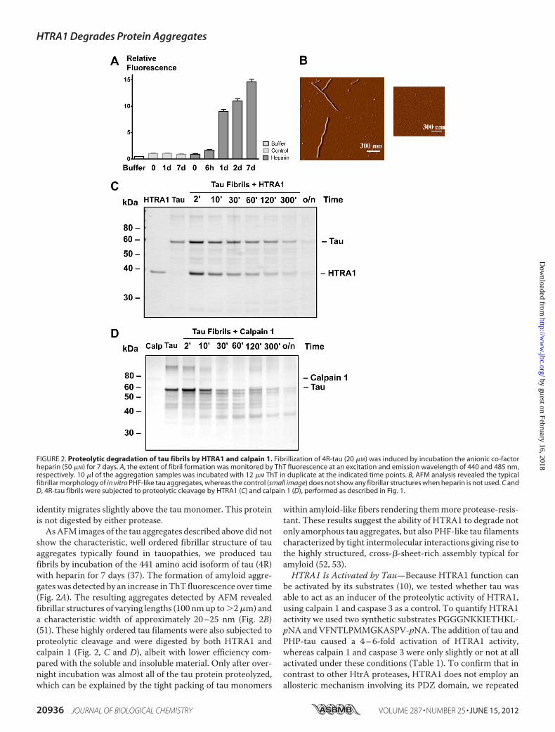
show the characteristic, well ordered fibrillar structure of tauaggregates typically found in tauopathies, we produced taufibrils by incubation of the 441 amino acid isoform of tau (4R)with heparin for 7 days (37). The formation of amyloid aggre-gateswas detected by an increase inThT fluorescence over time(Fig. 2A). The resulting aggregates detected by AFM revealedfibrillar structures of varying lengths (100 nmup to�2�m) anda characteristic width of approximately 20–25 nm (Fig. 2B)(51). These highly ordered tau filaments were also subjected toproteolytic cleavage and were digested by both HTRA1 andcalpain 1 (Fig. 2, C and D), albeit with lower efficiency com-pared with the soluble and insoluble material. Only after over-night incubation was almost all of the tau protein proteolyzed,which can be explained by the tight packing of tau monomers
within amyloid-like fibers rendering themmore protease-resis-tant. These results suggest the ability of HTRA1 to degrade notonly amorphous tau aggregates, but also PHF-like tau filamentscharacterized by tight intermolecular interactions giving rise tothe highly structured, cross-�-sheet-rich assembly typical foramyloid (52, 53).HTRA1 Is Activated by Tau—Because HTRA1 function can
be activated by its substrates (10), we tested whether tau wasable to act as an inducer of the proteolytic activity of HTRA1,using calpain 1 and caspase 3 as a control. To quantify HTRA1activity we used two synthetic substrates PGGGNKKIETHKL-pNA andVFNTLPMMGKASPV-pNA. The addition of tau andPHP-tau caused a 4–6-fold activation of HTRA1 activity,whereas calpain 1 and caspase 3 were only slightly or not at allactivated under these conditions (Table 1). To confirm that incontrast to other HtrA proteases, HTRA1 does not employ anallosteric mechanism involving its PDZ domain, we repeated
FIGURE 2. Proteolytic degradation of tau fibrils by HTRA1 and calpain 1. Fibrillization of 4R-tau (20 �M) was induced by incubation the anionic co-factorheparin (50 �M) for 7 days. A, the extent of fibril formation was monitored by ThT fluorescence at an excitation and emission wavelength of 440 and 485 nm,respectively. 10 �l of the aggregation samples was incubated with 12 �M ThT in duplicate at the indicated time points. B, AFM analysis revealed the typicalfibrillar morphology of in vitro PHF-like tau aggregates, whereas the control (small image) does not show any fibrillar structures when heparin is not used. C andD, 4R-tau fibrils were subjected to proteolytic cleavage by HTRA1 (C) and calpain 1 (D), performed as described in Fig. 1.
HTRA1 Degrades Protein Aggregates
20936 JOURNAL OF BIOLOGICAL CHEMISTRY VOLUME 287 • NUMBER 25 • JUNE 15, 2012
by guest on February 16, 2018http://w
ww
.jbc.org/D
ownloaded from
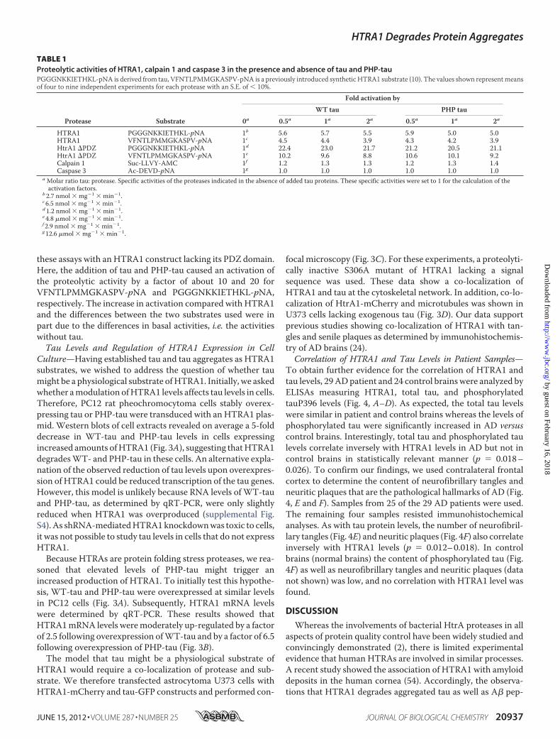
these assays with anHTRA1 construct lacking its PDZ domain.Here, the addition of tau and PHP-tau caused an activation ofthe proteolytic activity by a factor of about 10 and 20 forVFNTLPMMGKASPV-pNA and PGGGNKKIETHKL-pNA,respectively. The increase in activation compared with HTRA1and the differences between the two substrates used were inpart due to the differences in basal activities, i.e. the activitieswithout tau.Tau Levels and Regulation of HTRA1 Expression in Cell
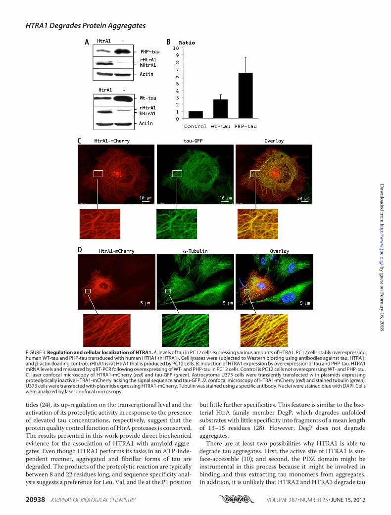
Culture—Having established tau and tau aggregates as HTRA1substrates, we wished to address the question of whether taumight be a physiological substrate ofHTRA1. Initially, we askedwhether amodulation ofHTRA1 levels affects tau levels in cells.Therefore, PC12 rat pheochromocytoma cells stably overex-pressing tau or PHP-tau were transduced with an HTRA1 plas-mid. Western blots of cell extracts revealed on average a 5-folddecrease in WT-tau and PHP-tau levels in cells expressingincreased amounts ofHTRA1 (Fig. 3A), suggesting thatHTRA1degradesWT- and PHP-tau in these cells. An alternative expla-nation of the observed reduction of tau levels upon overexpres-sion of HTRA1 could be reduced transcription of the tau genes.However, this model is unlikely because RNA levels ofWT-tauand PHP-tau, as determined by qRT-PCR, were only slightlyreduced when HTRA1 was overproduced (supplemental Fig.S4). As shRNA-mediatedHTRA1knockdownwas toxic to cells,it was not possible to study tau levels in cells that do not expressHTRA1.Because HTRAs are protein folding stress proteases, we rea-
soned that elevated levels of PHP-tau might trigger anincreased production of HTRA1. To initially test this hypothe-sis, WT-tau and PHP-tau were overexpressed at similar levelsin PC12 cells (Fig. 3A). Subsequently, HTRA1 mRNA levelswere determined by qRT-PCR. These results showed thatHTRA1mRNA levels weremoderately up-regulated by a factorof 2.5 following overexpression ofWT-tau and by a factor of 6.5following overexpression of PHP-tau (Fig. 3B).The model that tau might be a physiological substrate of
HTRA1 would require a co-localization of protease and sub-strate. We therefore transfected astrocytoma U373 cells withHTRA1-mCherry and tau-GFP constructs and performed con-
focal microscopy (Fig. 3C). For these experiments, a proteolyti-cally inactive S306A mutant of HTRA1 lacking a signalsequence was used. These data show a co-localization ofHTRA1 and tau at the cytoskeletal network. In addition, co-lo-calization of HtrA1-mCherry and microtubules was shown inU373 cells lacking exogenous tau (Fig. 3D). Our data supportprevious studies showing co-localization of HTRA1 with tan-gles and senile plaques as determined by immunohistochemis-try of AD brains (24).Correlation of HTRA1 and Tau Levels in Patient Samples—
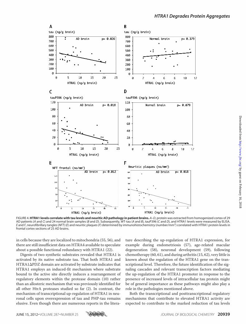
To obtain further evidence for the correlation of HTRA1 andtau levels, 29ADpatient and 24 control brainswere analyzed byELISAs measuring HTRA1, total tau, and phosphorylatedtauP396 levels (Fig. 4, A–D). As expected, the total tau levelswere similar in patient and control brains whereas the levels ofphosphorylated tau were significantly increased in AD versuscontrol brains. Interestingly, total tau and phosphorylated taulevels correlate inversely with HTRA1 levels in AD but not incontrol brains in statistically relevant manner (p � 0.018–0.026). To confirm our findings, we used contralateral frontalcortex to determine the content of neurofibrillary tangles andneuritic plaques that are the pathological hallmarks of AD (Fig.4, E and F). Samples from 25 of the 29 AD patients were used.The remaining four samples resisted immunohistochemicalanalyses. As with tau protein levels, the number of neurofibril-lary tangles (Fig. 4E) and neuritic plaques (Fig. 4F) also correlateinversely with HTRA1 levels (p � 0.012–0.018). In controlbrains (normal brains) the content of phosphorylated tau (Fig.4F) as well as neurofibrillary tangles and neuritic plaques (datanot shown) was low, and no correlation with HTRA1 level wasfound.
DISCUSSION
Whereas the involvements of bacterial HtrA proteases in allaspects of protein quality control have been widely studied andconvincingly demonstrated (2), there is limited experimentalevidence that human HTRAs are involved in similar processes.A recent study showed the association of HTRA1 with amyloiddeposits in the human cornea (54). Accordingly, the observa-tions that HTRA1 degrades aggregated tau as well as A� pep-
TABLE 1Proteolytic activities of HTRA1, calpain 1 and caspase 3 in the presence and absence of tau and PHP-tauPGGGNKKIETHKL-pNA is derived from tau, VFNTLPMMGKASPV-pNA is a previously introduced synthetic HTRA1 substrate (10). The values shown representmeansof four to nine independent experiments for each protease with an S.E. of � 10%.
Protease Substrate
Fold activation by
WT tau PHP tau
0a 0.5a 1a 2a 0.5a 1a 2a
HTRA1 PGGGNKKIETHKL-pNA 1b 5.6 5.7 5.5 5.9 5.0 5.0HTRA1 VFNTLPMMGKASPV-pNA 1c 4.5 4.4 3.9 4.3 4.2 3.9HtrA1 �PDZ PGGGNKKIETHKL-pNA 1d 22.4 23.0 21.7 21.2 20.5 21.1HtrA1 �PDZ VFNTLPMMGKASPV-pNA 1e 10.2 9.6 8.8 10.6 10.1 9.2Calpain 1 Suc-LLVY-AMC 1f 1.2 1.3 1.3 1.2 1.3 1.4Caspase 3 Ac-DEVD-pNA 1g 1.0 1.0 1.0 1.0 1.0 1.0
a Molar ratio tau: protease. Specific activities of the proteases indicated in the absence of added tau proteins. These specific activities were set to 1 for the calculation of theactivation factors.
b2.7 nmol � mg�1 � min�1.c6.5 nmol � mg�1 � min�1.d1.2 nmol � mg�1 � min�1.e4.8 �mol � mg�1 � min�1.f2.9 nmol � mg�1 � min�1.g12.6 �mol � mg�1 � min�1.
HTRA1 Degrades Protein Aggregates
JUNE 15, 2012 • VOLUME 287 • NUMBER 25 JOURNAL OF BIOLOGICAL CHEMISTRY 20937
by guest on February 16, 2018http://w
ww
.jbc.org/D
ownloaded from
tides (24), its up-regulation on the transcriptional level and theactivation of its proteolytic activity in response to the presenceof elevated tau concentrations, respectively, suggest that theprotein quality control function ofHtrA proteases is conserved.The results presented in this work provide direct biochemicalevidence for the association of HTRA1 with amyloid aggre-gates. Even though HTRA1 performs its tasks in an ATP-inde-pendent manner, aggregated and fibrillar forms of tau aredegraded. The products of the proteolytic reaction are typicallybetween 8 and 22 residues long, and sequence specificity anal-ysis suggests a preference for Leu, Val, and Ile at the P1 position
but little further specificities. This feature is similar to the bac-terial HtrA family member DegP, which degrades unfoldedsubstrates with little specificity into fragments of amean lengthof 13–15 residues (28). However, DegP does not degradeaggregates.There are at least two possibilities why HTRA1 is able to
degrade tau aggregates. First, the active site of HTRA1 is sur-face-accessible (10); and second, the PDZ domain might beinstrumental in this process because it might be involved inbinding and thus extracting tau monomers from aggregates.In addition, it is unlikely that HTRA2 and HTRA3 degrade tau
FIGURE 3. Regulation and cellular localization of HTRA1. A, levels of tau in PC12 cells expressing various amounts of HTRA1. PC12 cells stably overexpressinghuman WT-tau and PHP-tau transduced with human HTRA1 (hHTRA1). Cell lysates were subjected to Western blotting using antibodies against tau, HTRA1,and �-actin (loading control). rHtrA1 is rat HtrA1 that is produced by PC12 cells. B, induction of HTRA1 expression by overexpression of tau and PHP-tau. HTRA1mRNA levels and measured by qRT-PCR following overexpressing of WT- and PHP-tau in PC12 cells. Control is PC12 cells not overexpressing WT- and PHP-tau.C, laser confocal microscopy of HTRA1-mCherry (red) and tau-GFP (green). Astrocytoma U373 cells were transiently transfected with plasmids expressingproteolytically inactive HTRA1-mCherry lacking the signal sequence and tau-GFP. D, confocal microscopy of HTRA1-mCherry (red) and stained tubulin (green).U373 cells were transfected with plasmids expressing HTRA1-mCherry. Tubulin was stained using a specific antibody. Nuclei were stained blue with DAPI. Cellswere analyzed by laser confocal microscopy.
HTRA1 Degrades Protein Aggregates
20938 JOURNAL OF BIOLOGICAL CHEMISTRY VOLUME 287 • NUMBER 25 • JUNE 15, 2012
by guest on February 16, 2018http://w
ww
.jbc.org/D
ownloaded from
in cells because they are localized to mitochondria (55, 56), andthere are still insufficient data onHTRA4 available to speculateabout a possible functional redundancy with HTRA1 (22).Digests of two synthetic substrates revealed that HTRA1 is
activated by its native substrate tau. That both HTRA1 andHTRA1�PDZ domain are activated by substrate indicates thatHTRA1 employs an induced-fit mechanism where substratebound to the active site directly induces a rearrangement ofregulatory elements within the protease domain (10) ratherthan an allosteric mechanism that was previously identified forall other HtrA proteases studied so far (2). In contrast, themechanism of transcriptional up-regulation of HTRA1 in neu-ronal cells upon overexpression of tau and PHP-tau remainselusive. Even though there are numerous reports in the litera-
ture describing the up-regulation of HTRA1 expression, forexample during endometriosis (57), age-related maculardegeneration (58), neuronal development (59), followingchemotherapy (60, 61), and during arthritis (15, 62), very little isknown about the regulation of the HTRA1 gene on the tran-scriptional level. Therefore, the future identification of the sig-naling cascades and relevant transcription factors mediatingthe up-regulation of the HTRA1 promoter in response to thepresence of increased levels of intracellular tau protein mightbe of general importance as these pathways might also play arole in the pathologies mentioned above.Both the transcriptional and posttranscriptional regulatory
mechanisms that contribute to elevated HTRA1 activity areexpected to contribute to the marked reduction of tau levels
FIGURE 4. HTRA1 levels correlate with tau levels and neuritic AD pathology in patient brains. A–D, protein was extracted from homogenized cortex of 29AD patients (A and C) and 24 normal brain samples (B and D). Subsequently, WT-tau (A and B), tauP396 (C and D), and HTRA1 levels were measured by ELISA.E and F, neurofibrillary tangles (NFT) (E) and neuritic plaques (F) determined by immunohistochemistry (number/mm2) correlated with HTRA1 protein levels infrontal cortex sections of 25 AD brains.
HTRA1 Degrades Protein Aggregates
JUNE 15, 2012 • VOLUME 287 • NUMBER 25 JOURNAL OF BIOLOGICAL CHEMISTRY 20939
by guest on February 16, 2018http://w
ww
.jbc.org/D
ownloaded from
upon overexpression of HTRA1 in neuronal PC12 cells. Fur-thermore, a significant inverse correlation between HTRA1and tau levels is observed in AD patient brains. These datasuggest that HTRA1 might function as a tau protease in vivo.Unfolded protein response systems may represent one strat-
egy of how organisms prevent early onset of protein foldingdiseases. Because these diseases are more prevalent in agedindividuals it is conceivable that the various stress responsesystems are overwhelmed by additional tasks. In case of AD,additional substrates produced by aged tissues could competewith tau or A� for clearance by stress response factors leadingto the accumulation and ultimately aggregation of toxic proteinfragments. The serine proteaseHTRA1 represents a viable can-didate for a disease-modifying factor that might contribute,perhaps in concert with other proteases such as calpain, tomaintaining A� and tau levels low. This notion is supported byprevious work suggesting that HTRA1 is involved in the �-am-yloid pathway by performing alternative processing of variousamyloid precursor protein fragments, i.e. Cys99, A�42, andA�40. In line with this hypothesis, accumulation of A� wasobserved in astrocytoma cell culture supernatants followingchemical inhibition of HTRA1 and by co-localization ofHTRA1 with �-amyloid deposits in human brain samples (24).The recently reported association of HTRA1with corneal amy-loid deposits of TGFBI (transforming growth factor �-inducedgene), however, raises both the possibility of the generation ofamyloidogenic fragments and the clearance of amyloid aggre-gates through proteolysis by HTRA1 (54). Moreover, recentevidence from studying adult macular degeneration suggeststhat it is the increased activity of HTRA1, resulting from itsoverexpression that causes disease symptoms (12, 13). Thesedata illustrate conflicting findings with regard to the toxicity ofprotein fragments, their aggregation, as well as the functionalrole of proteolytic processing by protein quality control factors.Addressing these contrasting models in future studies is likelyan important step for our general understanding of the under-lying mechanisms of protein folding diseases.
REFERENCES1. Clausen, T., Southan, C., and Ehrmann, M. (2002) The HtrA family of
proteases: implications for protein composition and cell fate.Mol. Cell 10,443–455
2. Clausen, T., Kaiser, M., Huber, R., and Ehrmann, M. (2011) HTRA pro-teases: regulated proteolysis in protein quality control.Nat. Rev. Mol. CellBiol. 12, 152–162
3. Kley, J., Schmidt, B., Boyanov, B., Stolt-Bergner, P. C., Kirk, R., Ehrmann,M., Knopf, R. R., Naveh, L., Adam, Z., and Clausen, T. (2011) Structuraladaptation of the plant proteaseDeg1 to repair photosystem II during lightexposure. Nat. Struct. Mol. Biol. 18, 728–731
4. Sawa, J., Malet, H., Krojer, T., Canellas, F., Ehrmann, M., and Clausen, T.(2011)Molecular adaptation of the DegQ protease to exert protein qualitycontrol in the bacterial cell envelope. J. Biol. Chem. 286, 30680–30690
5. Merdanovic, M., Clausen, T., Kaiser, M., Huber, R., and Ehrmann, M.(2011) Protein quality control in the bacterial periplasm. Annu. Rev. Mi-crobiol. 65, 149–168
6. Isaac, D. D., Pinkner, J. S., Hultgren, S. J., and Silhavy, T. J. (2005) Theextracytoplasmic adaptor protein CpxP is degraded with substrate byDegP. Proc. Natl. Acad. Sci. U.S.A. 102, 17775–17779
7. Ehrmann, M., and Clausen, T. (2004) Proteolysis as a regulatory mecha-nism. Annu. Rev. Genet. 38, 709–724
8. Wilken, C., Kitzing, K., Kurzbauer, R., Ehrmann, M., and Clausen, T.
(2004) Crystal structure of the DegS stress sensor: how a PDZ domainrecognizes misfolded protein and activates a protease. Cell 117, 483–494
9. Spiess, C., Beil, A., and Ehrmann, M. (1999) A temperature-dependentswitch from chaperone to protease in a widely conserved heat shock pro-tein. Cell 97, 339–347
10. Truebestein, L., Tennstaedt, A., Monig, T., Krojer, T., Canellas, F., Kaiser,M., Clausen, T., and Ehrmann, M. (2011) Substrate-induced remodelingof the active site regulates human HTRA1 activity. Nat. Struct. Mol. Biol.18, 386–388
11. Krojer, T., Sawa, J., Schafer, E., Saibil, H. R., Ehrmann, M., and Clausen, T.(2008) Structural basis for the regulated protease and chaperone functionof DegP. Nature 453, 885–890
12. Jones, A., Kumar, S., Zhang, N., Tong, Z., Yang, J. H., Watt, C., Anderson,J., Amrita, Fillerup, H.,McCloskey,M., Luo, L., Yang, Z., Ambati, B.,Marc,R., Oka, C., Zhang, K., and Fu, Y. (2011) Increased expression of multi-functional serine protease, HTRA1, in retinal pigment epithelium inducespolypoidal choroidal vasculopathy in mice. Proc. Natl. Acad. Sci. U.S.A.108, 14578–14583
13. Vierkotten, S., Muether, P. S., and Fauser, S. (2011) Overexpression ofHTRA1 leads to ultrastructural changes in the elastic layer of Bruch’smembrane via cleavage of extracellular matrix components. PLoS ONE 6,e22959
14. Grau, S., Richards, P. J., Kerr, B., Hughes, C., Caterson, B., Williams, A. S.,Junker, U., Jones, S. A., Clausen, T., and Ehrmann, M. (2006) The role ofhuman HtrA1 in arthritic disease. J. Biol. Chem. 281, 6124–6129
15. Tsuchiya, A., Yano,M., Tocharus, J., Kojima, H., Fukumoto,M., Kawaichi,M., and Oka, C. (2005) Expression of mouse HtrA1 serine protease innormal bone and cartilage and its up-regulation in joint cartilage damagedby experimental arthritis. Bone 37, 323–336
16. Chamberland, A., Wang, E., Jones, A. R., Collins-Racie, L. A., LaVallie,E. R., Huang, Y., Liu, L., Morris, E. A., Flannery, C. R., and Yang, Z. (2009)Identification of a novel HtrA1-susceptible cleavage site in human aggre-can: evidence for the involvement ofHtrA1 in aggrecan proteolysis in vivo.J. Biol. Chem. 284, 27352–27359
17. Hadfield, K. D., Rock, C. F., Inkson, C. A., Dallas, S. L., Sudre, L., Wallis,G. A., Boot-Handford, R. P., and Canfield, A. E. (2008) HtrA1 inhibitsmineral deposition by osteoblasts: requirement for the protease and PDZdomains. J. Biol. Chem. 283, 5928–5938
18. Chien, J., Ota, T., Aletti, G., Shridhar, R., Boccellino, M., Quagliuolo, L.,Baldi, A., and Shridhar, V. (2009) Serine protease HtrA1 associates withmicrotubules and inhibits cell migration.Mol. Cell. Biol. 29, 4177–4187
19. Clawson, G. A., Bui, V., Xin, P.,Wang, N., and Pan,W. (2008) Intracellularlocalization of the tumor suppressor HtrA1/Prss11 and its associationwith HPV16 E6 and E7 proteins. J. Cell. Biochem. 105, 81–88
20. Campioni, M., Severino, A., Manente, L., Tuduce, I. L., Toldo, S., Caraglia,M., Crispi, S., Ehrmann, M., He, X., Maguire, J., De Falco, M., De Luca, A.,Shridhar, V., and Baldi, A. (2010) The serine protease HtrA1 specificallyinteracts and degrades the tuberous sclerosis complex 2 protein. Mol.Cancer Res. 8, 1248–1260
21. Chien, J., He, X., and Shridhar, V. (2009) Identification of tubulins assubstrates of serine protease HtrA1 by mixture-based oriented peptidelibrary screening. J. Cell. Biochem. 107, 253–263
22. Chien, J., Campioni, M., Shridhar, V., and Baldi, A. (2009) HtrA serineproteases as potential therapeutic targets in cancer. Curr. Cancer DrugTargets 9, 451–468
23. Yang, Z., Camp, N. J., Sun, H., Tong, Z., Gibbs, D., Cameron, D. J., Chen,H., Zhao, Y., Pearson, E., Li, X., Chien, J., Dewan, A., Harmon, J., Bernstein,P. S., Shridhar, V., Zabriskie, N. A., Hoh, J., Howes, K., and Zhang, K.(2006) A variant of theHTRA1 gene increases susceptibility to age-relatedmacular degeneration. Science 314, 992–993
24. Grau, S., Baldi, A., Bussani, R., Tian, X., Stefanescu, R., Przybylski, M.,Richards, P., Jones, S. A., Shridhar, V., Clausen, T., and Ehrmann, M.(2005) Implications of the serine protease HtrA1 in amyloid precursorprotein processing. Proc. Natl. Acad. Sci. U.S.A. 102, 6021–6026
25. Milner, J. M., Patel, A., and Rowan, A. D. (2008) Emerging roles of serineproteinases in tissue turnover in arthritis.Arthritis Rheum. 58, 3644–3656
26. Hara, K., Shiga, A., Fukutake, T., Nozaki, H., Miyashita, A., Yokoseki, A.,Kawata, H., Koyama, A., Arima, K., Takahashi, T., Ikeda, M., Shiota, H.,
HTRA1 Degrades Protein Aggregates
20940 JOURNAL OF BIOLOGICAL CHEMISTRY VOLUME 287 • NUMBER 25 • JUNE 15, 2012
by guest on February 16, 2018http://w
ww
.jbc.org/D
ownloaded from
Tamura,M., Shimoe, Y., Hirayama,M., Arisato, T., Yanagawa, S., Tanaka,A., Nakano, I., Ikeda, S., Yoshida, Y., Yamamoto, T., Ikeuchi, T., Kuwano,R.,Nishizawa,M., Tsuji, S., andOnodera,O. (2009)Association ofHTRA1mutations and familial ischemic cerebral small-vessel disease. N. Engl.J. Med. 360, 1729–1739
27. Hasenbein, S., Meltzer, M., Hauske, P., Kaiser, M., Huber, R., Clausen, T.,and Ehrmann, M. (2010) Conversion of a regulatory into a degradativeprotease. J. Mol. Biol. 397, 957–966
28. Krojer, T., Pangerl, K., Kurt, J., Sawa, J., Stingl, C., Mechtler, K., Huber, R.,Ehrmann, M., and Clausen, T. (2008) Interplay of PDZ and protease do-main of DegP ensures efficient elimination of misfolded proteins. Proc.Natl. Acad. Sci. U.S.A. 105, 7702–7707
29. Krojer, T., Sawa, J., Huber, R., and Clausen, T. (2010) HtrA proteases havea conserved activation mechanism that can be triggered by distinct mo-lecular cues. Nat. Struct. Mol. Biol. 17, 844–852
30. Merdanovic, M., Mamant, N., Meltzer, M., Poepsel, S., Auckenthaler, A.,Melgaard, R., Hauske, P., Nagel-Steger, L., Clarke, A. R., Kaiser,M., Huber,R., and Ehrmann, M. (2010) Determinants of structural and functionalplasticity of a widely conserved protease chaperone complex. Nat. Struct.Mol. Biol. 17, 837–843
31. Morris, M., Maeda, S., Vossel, K., andMucke, L. (2011) Themany faces oftau. Neuron 70, 410–426
32. Brandt, R., Hundelt, M., and Shahani, N. (2005) Tau alteration and neu-ronal degeneration in tauopathies:mechanisms andmodels.Biochim. Bio-phys. Acta 1739, 331–354
33. Eidenmuller, J., Fath, T., Hellwig, A., Reed, J., Sontag, E., and Brandt, R.(2000) Structural and functional implications of tau hyperphosphoryla-tion: information fromphosphorylation-mimickingmutated tau proteins.Biochemistry 39, 13166–13175
34. Roger, B., Al-Bassam, J., Dehmelt, L.,Milligan, R. A., andHalpain, S. (2004)MAP2c, but not tau, binds and bundles F-actin via itsmicrotubule bindingdomain. Curr. Biol. 14, 363–371
35. Morgenstern, J. P., and Land, H. (1990) Advancedmammalian gene trans-fer: high titre retroviral vectors with multiple drug selection markers anda complementary helper-free packaging cell line. Nucleic Acids Res. 18,3587–3596
36. Fath, T., Eidenmuller, J., and Brandt, R. (2002) Tau-mediated cytotoxicityin a pseudohyperphosphorylation model of Alzheimer’s disease. J. Neuro-sci. 22, 9733–9741
37. Li, W., and Lee, V. M. (2006) Characterization of two VQIXXKmotifs fortau fibrillization in vitro. Biochemistry 45, 15692–15701
38. Abramoff, M. D., Magalhaes, P. J., and Ram, S. J. (2004) Image Processingwith ImageJ. Biophotonics Int. 11, 36–42
39. Hauske, P., Meltzer, M., Ottmann, C., Krojer, T., Clausen, T., Ehrmann,M., and Kaiser, M. (2009) Selectivity profiling of DegP substrates andinhibitors. Bioorg. Med. Chem. 17, 2920–2924
40. Meltzer, M., Hasenbein, S., Hauske, P., Kucz, N., Merdanovic, M., Grau, S.,Beil, A., Jones, D., Krojer, T., Clausen, T., Ehrmann,M., andKaiser,M. (2008)Allosteric activation of HtrA protease DegP by stress signals during bacterialprotein quality control.Angew. Chem. Int. Ed. Engl. 47, 1332–1334
41. Xia, S. J., Rajput, P., Strzelecki, D. M., and Barr, F. G. (2007) Analysis ofgenetic events that modulate the oncogenic and growth suppressive ac-tivities of the PAX3-FKHR fusion oncoprotein. Lab. Invest. 87, 318–325
42. Severino,A.,Campioni,M., Straino, S., Salloum,F.N., Schmidt,N.,Herbrand,U., Frede,S.,Toietta,G.,DiRocco,G.,Bussani,R., Silvestri, F., Piro,M.,Liuzzo,G.,Biasucci, L.M.,Mellone,P., Feroce, F.,Capogrossi,M.,Baldi, F., Fandrey, J.,Ehrmann,M., Crea, F., Abbate, A., and Baldi, A. (2007) Identification of pro-tein disulfide isomerase as a cardiomyocyte survival factor in ischemic car-diomyopathy. J. Am. Coll. Cardiol. 50, 1029–1037
43. Mirra, S. S., Heyman, A., McKeel, D., Sumi, S. M., Crain, B. J., Brownlee,L. M., Vogel, F. S., Hughes, J. P., van Belle, G., and Berg, L. (1991) TheConsortium to Establish a Registry forAlzheimer’s Disease (CERAD). PartII. Standardization of the neuropathologic assessment of Alzheimer’s dis-ease. Neurology 41, 479–486
44. Braak, H., Alafuzoff, I., Arzberger, T., Kretzschmar, H., andDel Tredici, K.(2006) Staging of Alzheimer disease-associated neurofibrillary pathologyusing paraffin sections and immunocytochemistry. Acta Neuropathol.112, 389–404
45. Yang, L. S., Gordon-Krajcer, W., and Ksiezak-Reding, H. (1997) Tau re-leased from paired helical filaments with formic acid or guanidine is sus-ceptible to calpain-mediated proteolysis. J. Neurochem. 69, 1548–1558
46. Egensperger, R., Kosel, S., von Eitzen, U., and Graeber, M. B. (1998) Mi-croglial activation in Alzheimer disease: association with APOE genotype.Brain Pathol. 8, 439–447
47. Friedhoff, P., Schneider, A., Mandelkow, E. M., andMandelkow, E. (1998)Rapid assembly of Alzheimer-like paired helical filaments from microtu-bule-associated protein tau monitored by fluorescence in solution. Bio-chemistry 37, 10223–10230
48. Rissman, R. A., Poon,W.W., Blurton-Jones, M., Oddo, S., Torp, R., Vitek,M. P., LaFerla, F. M., Rohn, T. T., and Cotman, C. W. (2004) Caspase-cleavage of tau is an early event in Alzheimer disease tangle pathology.J. Clin. Invest. 114, 121–130
49. Mercken,M., Grynspan, F., andNixon, R. A. (1995) Differential sensitivityto proteolysis by brain calpain of adult human tau, fetal human tau andPHF-tau. FEBS Lett. 368, 10–14
50. Talanian, R. V., Quinlan, C., Trautz, S., Hackett, M. C., Mankovich, J. A.,Banach, D., Ghayur, T., Brady, K. D., and Wong, W. W. (1997) Substratespecificities of caspase family proteases. J. Biol. Chem. 272, 9677–9682
51. Crowther, R. A. (1991) Straight and paired helical filaments in Alzheimerdisease have a common structural unit. Proc. Natl. Acad. Sci. U.S.A. 88,2288–2292
52. Berriman, J., Serpell, L. C., Oberg, K. A., Fink, A. L., Goedert, M., andCrowther, R. A. (2003) Tau filaments from human brain and from in vitroassembly of recombinant protein show cross-� structure. Proc. Natl.Acad. Sci. U.S.A. 100, 9034–9038
53. Barghorn, S., Davies, P., and Mandelkow, E. (2004) Tau paired helicalfilaments fromAlzheimer’s disease brain and assembled in vitro are basedon �-structure in the core domain. Biochemistry 43, 1694–1703
54. Karring, H., Runager, K., Thøgersen, I. B., Klintworth, G. K., Højrup, P.,and Enghild, J. J. (2012)Composition and proteolytic processing of cornealdeposits associated with mutations in the TGFBI gene. Exp. Eye Res. 96,163–170
55. Suzuki, Y., Imai, Y., Nakayama, H., Takahashi, K., Takio, K., and Taka-hashi, R. (2001) A serine protease, HtrA2, is released from the mitochon-dria and interacts with XIAP, inducing cell death.Mol. Cell 8, 613–621
56. Beleford, D., Rattan, R., Chien, J., and Shridhar, V. (2010) High tempera-ture requirement A3 (HtrA3) promotes etoposide- and cisplatin-inducedcytotoxicity in lung cancer cell lines. J. Biol. Chem. 285, 12011–12027
57. Dentillo, D. B., Meola, J., Rosa e Silva, J. C., Giuliatti, S., Silva, W. A., Jr.,Ferriani, R. A., andMartelli, L. (2010)Deregulation of LOXL1 andHTRA1gene expression in endometriosis. Reprod. Sci. 17, 1016–1023
58. Yang, Z., Tong, Z., Chen, Y., Zeng, J., Lu, F., Sun, X., Zhao, C., Wang, K.,Davey, L., Chen, H., London, N., Muramatsu, D., Salasar, F., Carmona, R.,Kasuga, D., Wang, X., Bedell, M., Dixie, M., Zhao, P., Yang, R., Gibbs, D.,Liu, X., Li, Y., Li, C., Campochiaro, B., Constantine, R., Zack, D. J., Cam-pochiaro, P., Fu, Y., Li, D. Y., Katsanis, N., and Zhang, K. (2010) Geneticand functional dissection of HTRA1 and LOC387715 in age-related mac-ular degeneration. PLoS Genet. 6, e1000836
59. Launay, S., Maubert, E., Lebeurrier, N., Tennstaedt, A., Campioni, M.,Docagne, F., Gabriel, C., Dauphinot, L., Potier, M. C., Ehrmann, M., Baldi,A., and Vivien, D. (2008) HtrA1-dependent proteolysis of TGF-� controlsboth neuronal maturation and developmental survival. Cell Death Differ.15, 1408–1416
60. Chien, J., Aletti, G., Baldi, A., Catalano, V.,Muretto, P., Keeney, G. L., Kalli,K. R., Staub, J., Ehrmann, M., Cliby, W. A., Lee, Y. K., Bible, K. C., Hart-mann, L. C., Kaufmann, S. H., and Shridhar, V. (2006) Serine proteaseHtrA1modulates chemotherapy-induced cytotoxicity. J. Clin. Invest. 116,1994–2004
61. Spugnini, E. P., Cardillo, I., Verdina, A., Crispi, S., Saviozzi, S., Calogero, R.,Nebbioso, A., Altucci, L., Cortese, G., Galati, R., Chien, J., Shridhar, V.,Vincenzi, B., Citro, G., Cognetti, F., Sacchi, A., and Baldi, A. (2006) Piroxi-cam and cisplatin in a mouse model of peritoneal mesothelioma. Clin.Cancer Res. 12, 6133–6143
62. Polur, I., Lee, P. L., Servais, J. M., Xu, L., and Li, Y. (2010) Role of HTRA1,a serine protease, in the progression of articular cartilage degeneration.Histol. Histopathol. 25, 599–608
HTRA1 Degrades Protein Aggregates
JUNE 15, 2012 • VOLUME 287 • NUMBER 25 JOURNAL OF BIOLOGICAL CHEMISTRY 20941
by guest on February 16, 2018http://w
ww
.jbc.org/D
ownloaded from
Baldi, Leif Dehmelt, Markus Kaiser, Robert Huber, Tim Clausen and Michael EhrmannBrandt, Hanna Ksiezak-Reding, Anca Laura Tirniceriu, Rupert Egensperger, AlfonsoBrockmann, Nina Schmidt, Inga Irle, Barbara Sacca, Christof M. Niemeyer, Roland
Annette Tennstaedt, Simon Pöpsel, Linda Truebestein, Patrick Hauske, AnkeTau Protein Aggregates
Human High Temperature Requirement Serine Protease A1 (HTRA1) Degrades
doi: 10.1074/jbc.M111.316232 originally published online April 25, 20122012, 287:20931-20941.J. Biol. Chem.
10.1074/jbc.M111.316232Access the most updated version of this article at doi:
Alerts:
When a correction for this article is posted•
When this article is cited•
to choose from all of JBC's e-mail alertsClick here
Supplemental material:
http://www.jbc.org/content/suppl/2012/04/25/M111.316232.DC1
http://www.jbc.org/content/287/25/20931.full.html#ref-list-1
This article cites 62 references, 18 of which can be accessed free at
by guest on February 16, 2018http://w
ww
.jbc.org/D
ownloaded from