Embed Size (px)
Citation preview
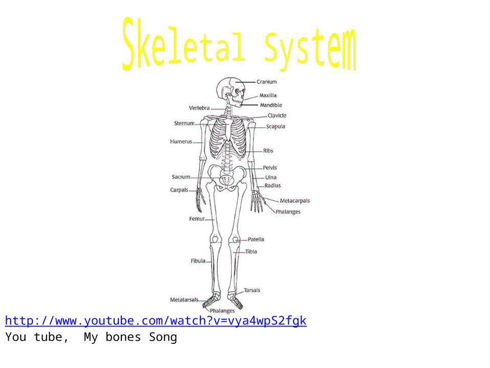
http://www.youtube.com/watch?v=vya4wpS2fgkYou tube, My bones Song
Chapter 5: The Skeletal System
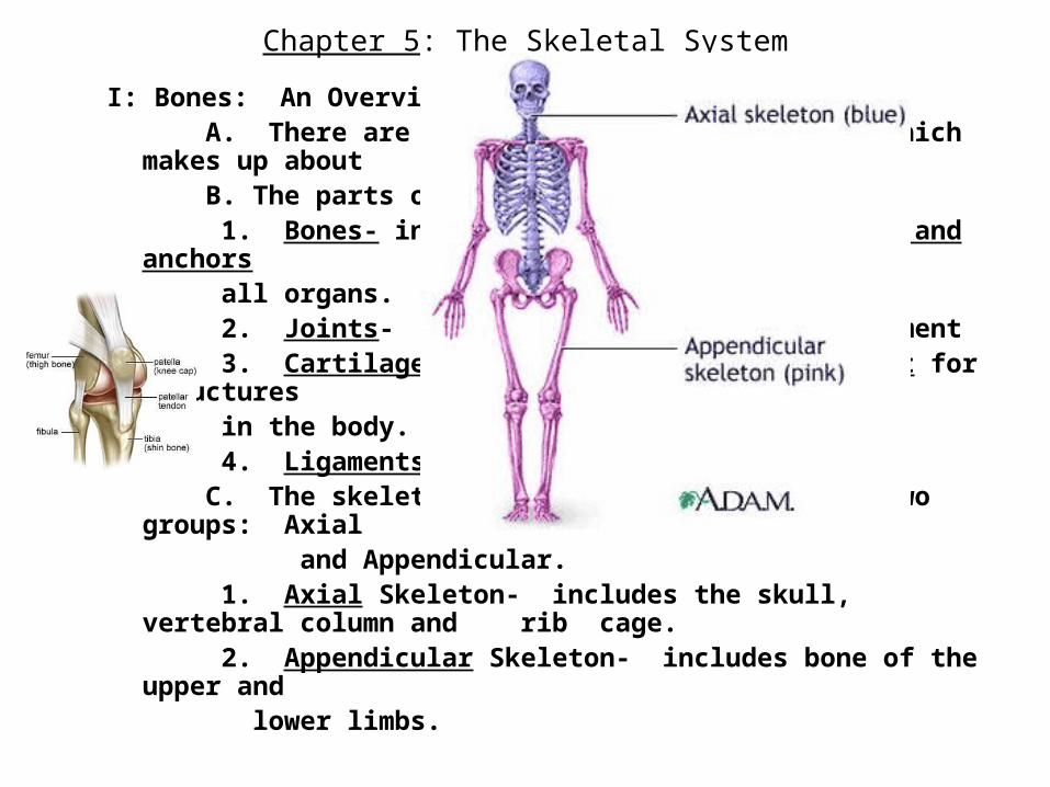
I: Bones: An Overview A. There are 206 bones in the human body, which makes up about 20% of our body mass. B. The parts of the skeletal system include:
1. Bones- internal framework that supports and anchors all organs. 2. Joints- allow for flexibility and movement 3. Cartilages- provides additional support for structures in the body. 4. Ligaments- connect bone to bone.
C. The skeletal system is subdivided into two groups: Axial and Appendicular.
1. Axial Skeleton- includes the skull, vertebral column and rib cage.
2. Appendicular Skeleton- includes bone of the upper and
lower limbs.
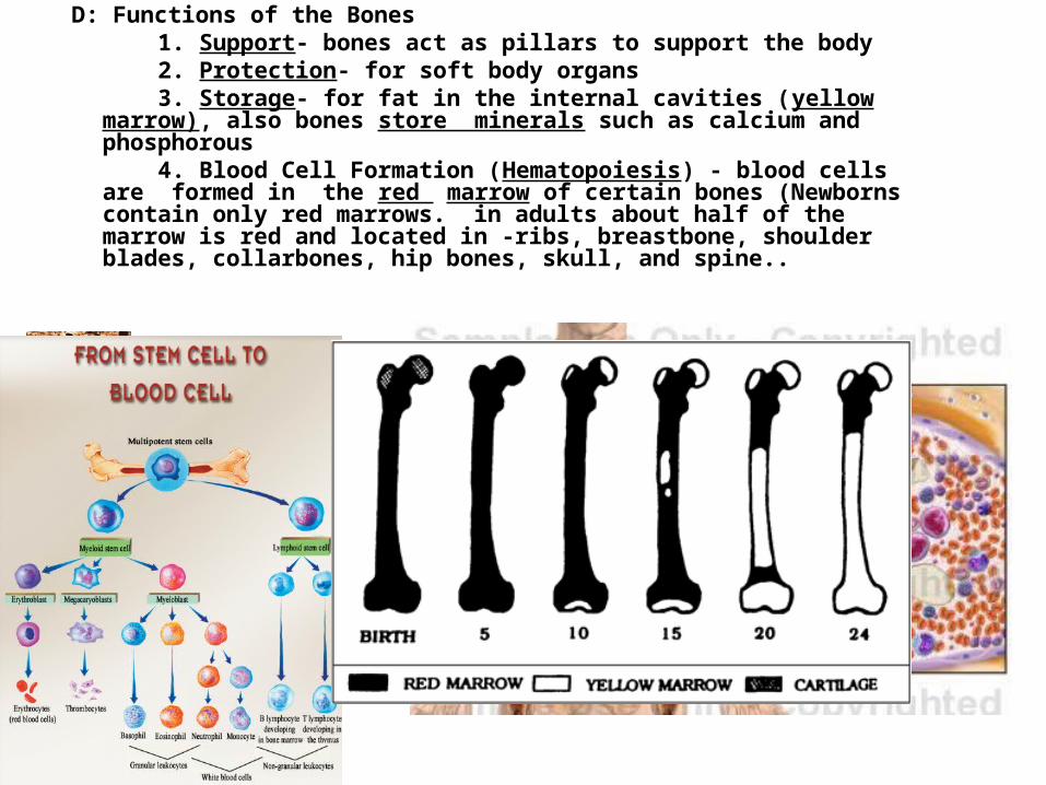
D: Functions of the Bones 1. Support- bones act as pillars to support the body 2. Protection- for soft body organs 3. Storage- for fat in the internal cavities (yellow marrow), also bones store minerals such as calcium and phosphorous 4. Blood Cell Formation (Hematopoiesis) - blood cells are formed in the red marrow of certain bones (Newborns contain only red marrows. in adults about half of the marrow is red and located in -ribs, breastbone, shoulder blades, collarbones, hip bones, skull, and spine..
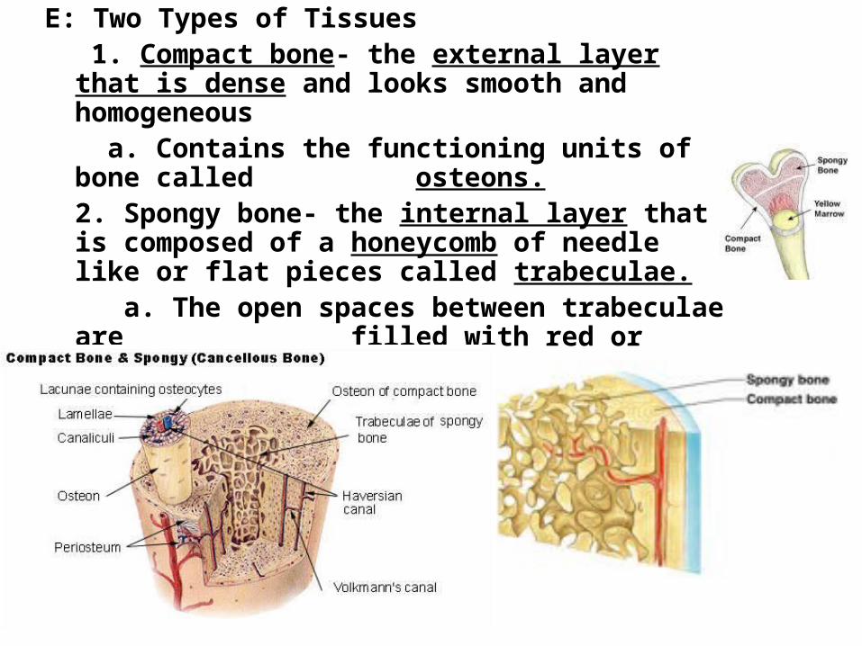
E: Two Types of Tissues 1. Compact bone- the external layer that is dense and looks smooth and homogeneous
a. Contains the functioning units of bone called osteons.
2. Spongy bone- the internal layer that is composed of a honeycomb of needle like or flat pieces called trabeculae.
a. The open spaces between trabeculae are filled with red or yellow bone
marrow
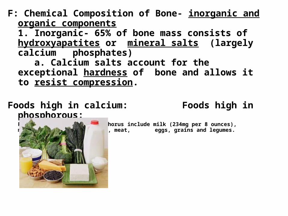
F: Chemical Composition of Bone- inorganic and organic components
1. Inorganic- 65% of bone mass consists of hydroxyapatites or mineral salts (largely calcium phosphates)
a. Calcium salts account for the exceptional hardness of bone and allows it to resist compression.
Foods high in calcium: Foods high in phosphorous:Foods that are high in phosphorus include milk (234mg per 8 ounces), milk products, poultry, fish, meat, eggs, grains and legumes.
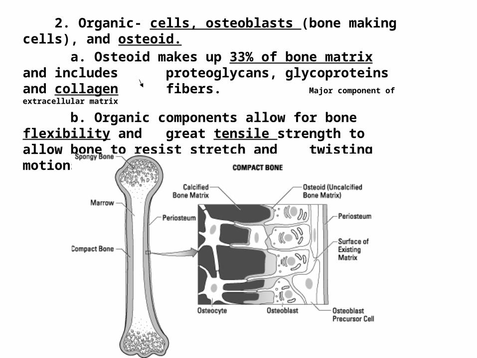
2. Organic- cells, osteoblasts (bone making cells), and osteoid.a. Osteoid makes up 33% of bone matrix and includes proteoglycans, glycoproteins and collagen fibers.Major component of extracellular matrix
b. Organic components allow for bone flexibility and great tensile strength to allow bone to resist stretch and twisting motions.
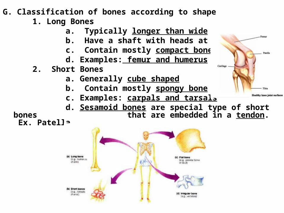
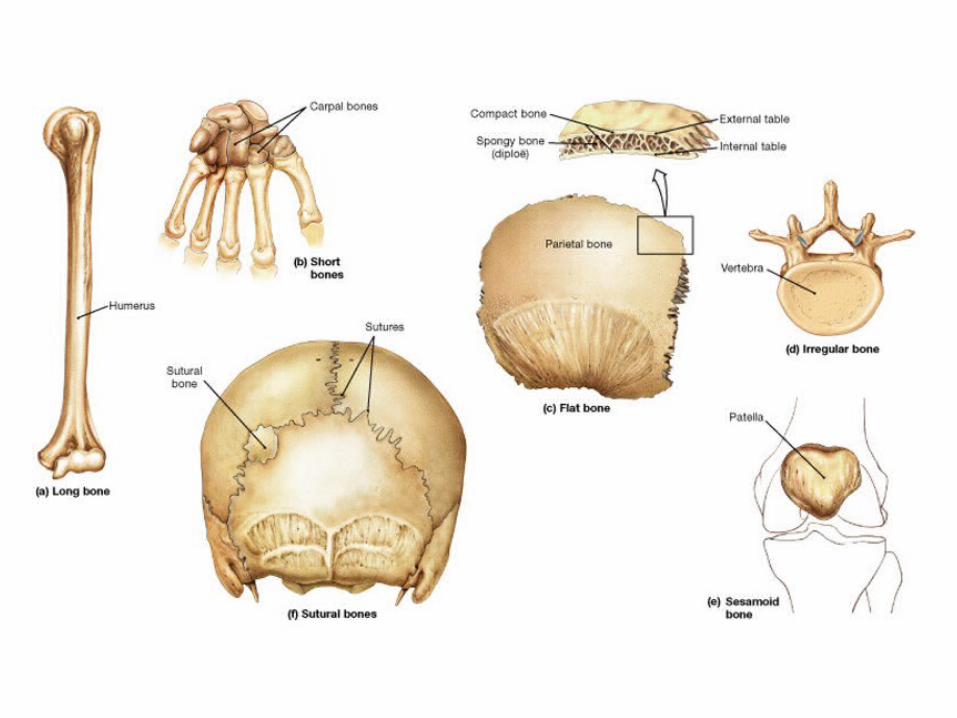
G. Classification of bones according to shape 1. Long Bones
a. Typically longer than wide b. Have a shaft with heads at both ends c. Contain mostly compact bone d. Examples: femur and humerus
2. Short Bones a. Generally cube shaped b. Contain mostly spongy bone c. Examples: carpals and tarsals d. Sesamoid bones are special type of short bones that are embedded in a tendon. Ex. Patella
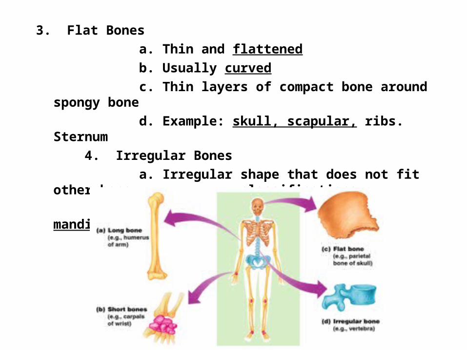
3. Flat Bones a. Thin and flattened b. Usually curved c. Thin layers of compact bone around spongy bone d. Example: skull, scapular, ribs. Sternum
4. Irregular Bones a. Irregular shape that does not fit other bone classifications b. Example: vertebrae , sacrum, mandible
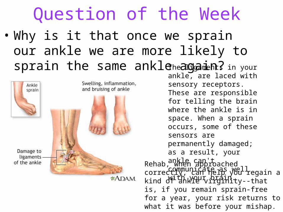
Question of the Week• Why is it that once we sprain our ankle we are
more likely to sprain the same ankle again?The ligaments in your ankle, are laced with sensory receptors. These are responsible for telling the brain where the ankle is in space. When a sprain occurs, some of these sensors are permanently damaged; as a result, your ankle can't communicate as well with your brain.
Rehab, when approached correctly, can help you regain a kind of ankle virginity--that is, if you remain sprain-free for a year, your risk returns to what it was before your mishap.
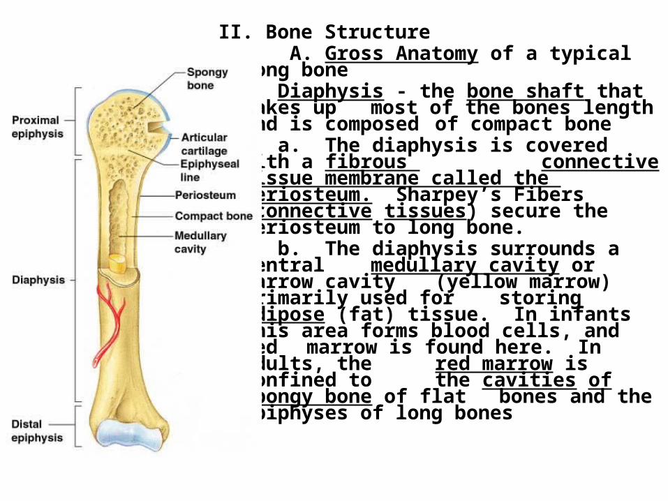
II. Bone Structure A. Gross Anatomy of a typical long bone
1. Diaphysis - the bone shaft that makes up most of the bones length and is composed of compact bone
a. The diaphysis is covered with a fibrous connective tissue membrane called the periosteum. Sharpey’s Fibers
(connective tissues) secure the periosteum to long bone.
b. The diaphysis surrounds a central medullary cavity or marrow cavity (yellow marrow) primarily used for storing adipose (fat) tissue. In infants this area forms blood cells, and red marrow is found here. In adults, the red marrow is confined to the cavities of spongy bone of flat bones and the epiphyses of long bones
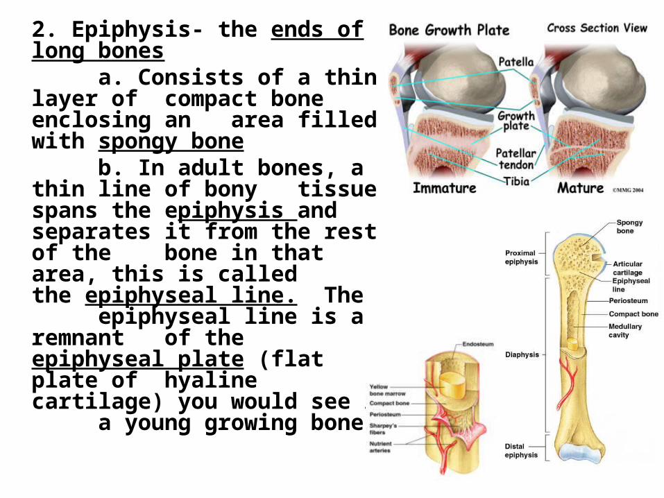
2. Epiphysis- the ends of long bones a. Consists of a thin layer of compact bone enclosing an area filled with spongy bone b. In adult bones, a thin line of bony
tissue spans the epiphysis and separates it from the rest of the bone in that area, this is called the epiphyseal line. The
epiphyseal line is a remnant of the epiphyseal plate (flat plate of hyaline cartilage) you would see in a young growing bone.
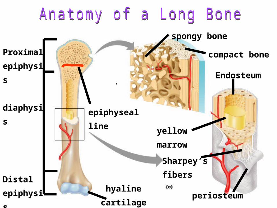
Distal
epiphysi
s
Proximal
epiphysis
diaphysis
yellow
marrow
epiphyseal
line
periosteum
compact
bone
spongy bone
Endosteu
m
hyaline
cartilage
Sharpey’s
fibers
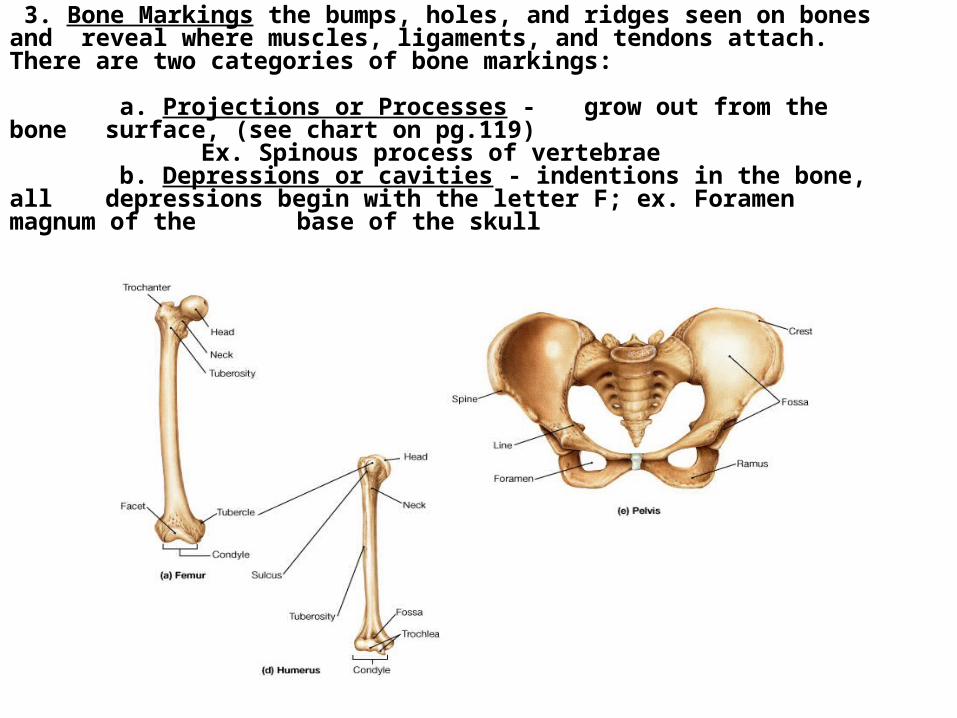
3. Bone Markings the bumps, holes, and ridges seen on bones and reveal where muscles, ligaments, and tendons attach. There are two categories of bone markings:
a. Projections or Processes - grow out from the bone surface, (see chart on pg.119)
Ex. Spinous process of vertebrae b. Depressions or cavities - indentions in the bone, all
depressions begin with the letter F; ex. Foramen magnum of the base of the skull
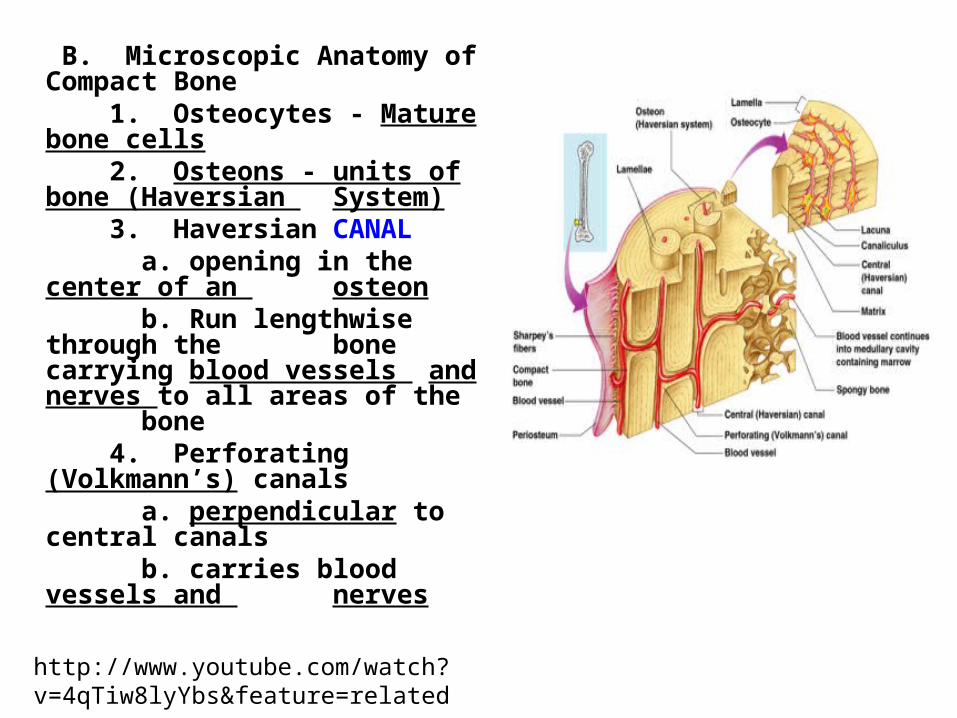
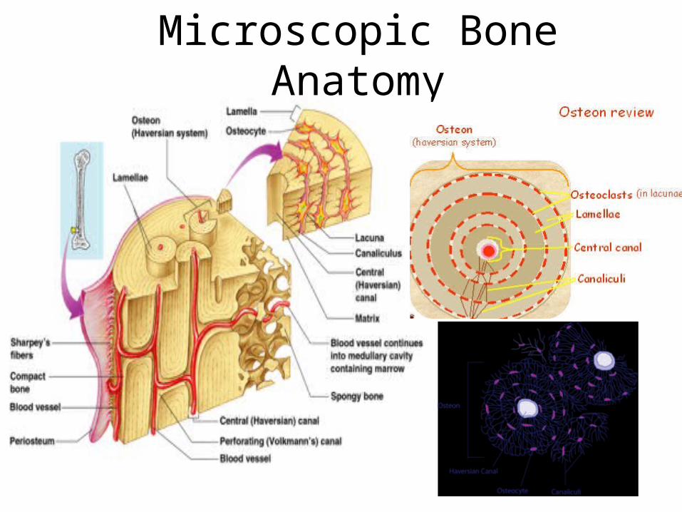
B. Microscopic Anatomy of Compact Bone 1. Osteocytes - Mature bone cells 2. Osteons - units of bone (Haversian
System) 3. Haversian CANAL
a. opening in the center of an osteonb. Run lengthwise through the bone carrying blood vessels and nerves to all areas of the bone
4. Perforating (Volkmann’s) canals a. perpendicular to central canals
b. carries blood vessels and nerves
http://www.youtube.com/watch?v=4qTiw8lyYbs&feature=related
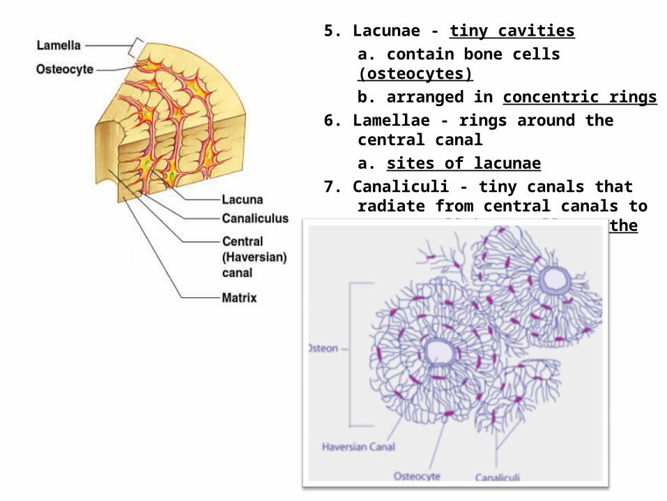
5. Lacunae - tiny cavitiesa. contain bone cells (osteocytes)b. arranged in concentric rings
6. Lamellae - rings around the central canala. sites of lacunae
7. Canaliculi - tiny canals that radiate from central canals to connect all bone cells to the nutrient supply
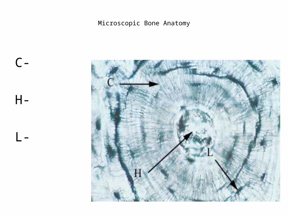
Microscopic Bone Anatomy
C-
H-
L-
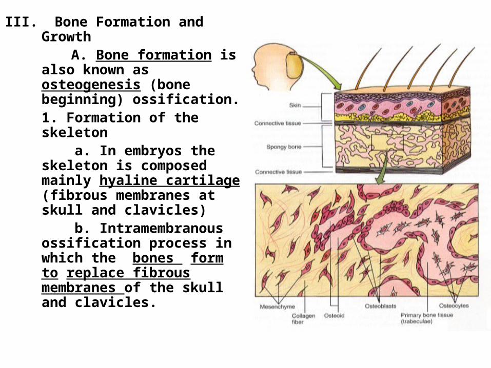
III. Bone Formation and Growth A. Bone formation is also
known as osteogenesis (bone beginning) ossification.1. Formation of the skeleton
a. In embryos the skeleton is composed mainly hyaline cartilage (fibrous membranes at skull and clavicles)
b. Intramembranous ossification process in which the bones form to replace fibrous membranes of the skull and clavicles.
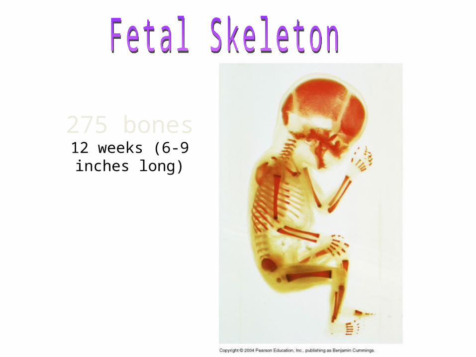
275 bones12 weeks (6-9 inches
long)
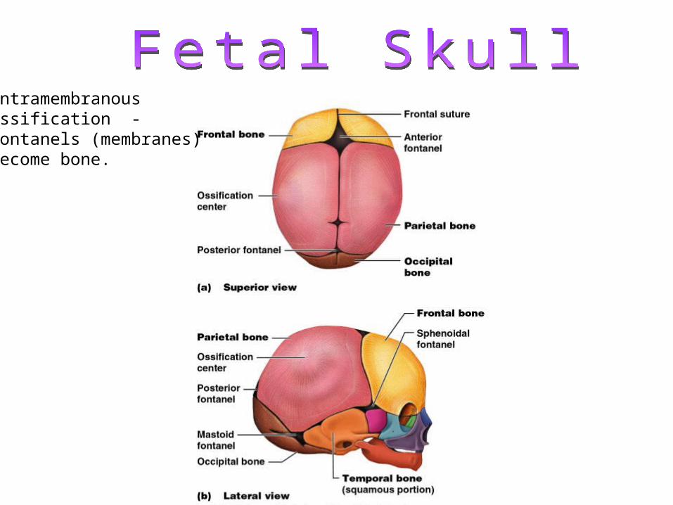
IntramembranousOssification -Fontanels (membranes)become bone.
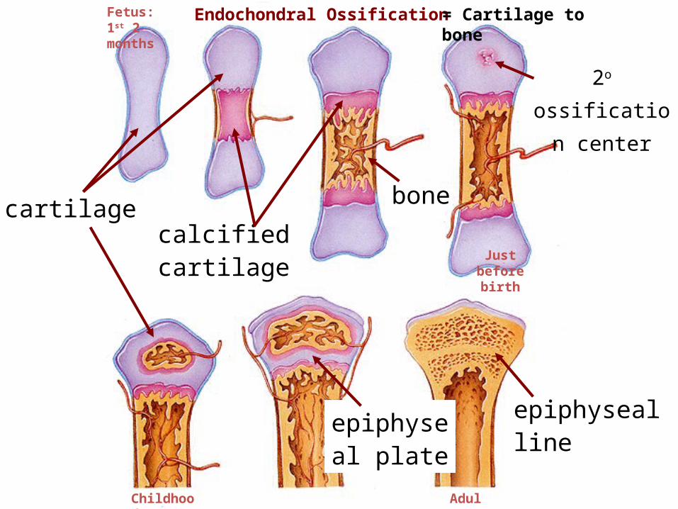
cartilagecalcified cartilage
bone
epiphyseal plate
epiphyseal line
Endochondral Ossification
2o
ossification
center
Fetus: 1st 2 months
Adult
Childhood
Just before birth
= Cartilage to bone
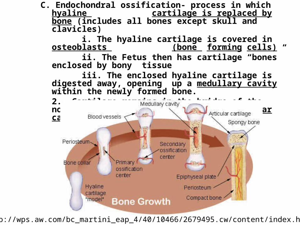
C. Endochondral ossification- process in which hyaline cartilage is replaced by bone (includes all bones except skull and clavicles)
i. The hyaline cartilage is covered in osteoblasts (bone forming cells)ii. The Fetus then has cartilage “bones” enclosed by bony tissueiii. The enclosed hyaline cartilage is digested away,
opening up a medullary cavity within the newly formed bone.2. Cartilage remains in the bridge of the nose, parts of the ribs, joints (articular cartilage), and epiphyseal plates.
http://wps.aw.com/bc_martini_eap_4/40/10466/2679495.cw/content/index.html
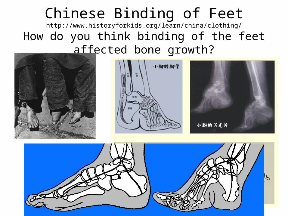
Chinese Binding of Feethttp://www.historyforkids.org/learn/china/clothing/
How do you think binding of the feet affected bone growth?
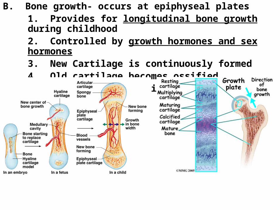
B. Bone growth- occurs at epiphyseal plates1. Provides for longitudinal bone growth during childhood2. Controlled by growth hormones and sex hormones3. New Cartilage is continuously formed4. Old cartilage becomes ossified
a. Cartilage is broken down and replaced by bone
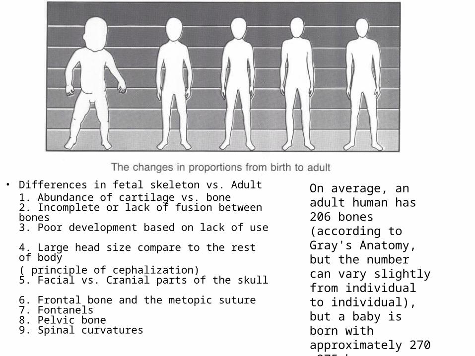
• Differences in fetal skeleton vs. Adult1. Abundance of cartilage vs. bone 2. Incomplete or lack of fusion between bones
3. Poor development based on lack of use 4. Large head size compare to the rest of body
( principle of cephalization) 5. Facial vs. Cranial parts of the skull 6. Frontal bone and the metopic suture 7. Fontanels 8. Pelvic bone 9. Spinal curvatures
On average, an adult human has 206 bones (according to Gray's Anatomy, but the number can vary slightly from individual to individual), but a baby is born with approximately 270 -275 bones.
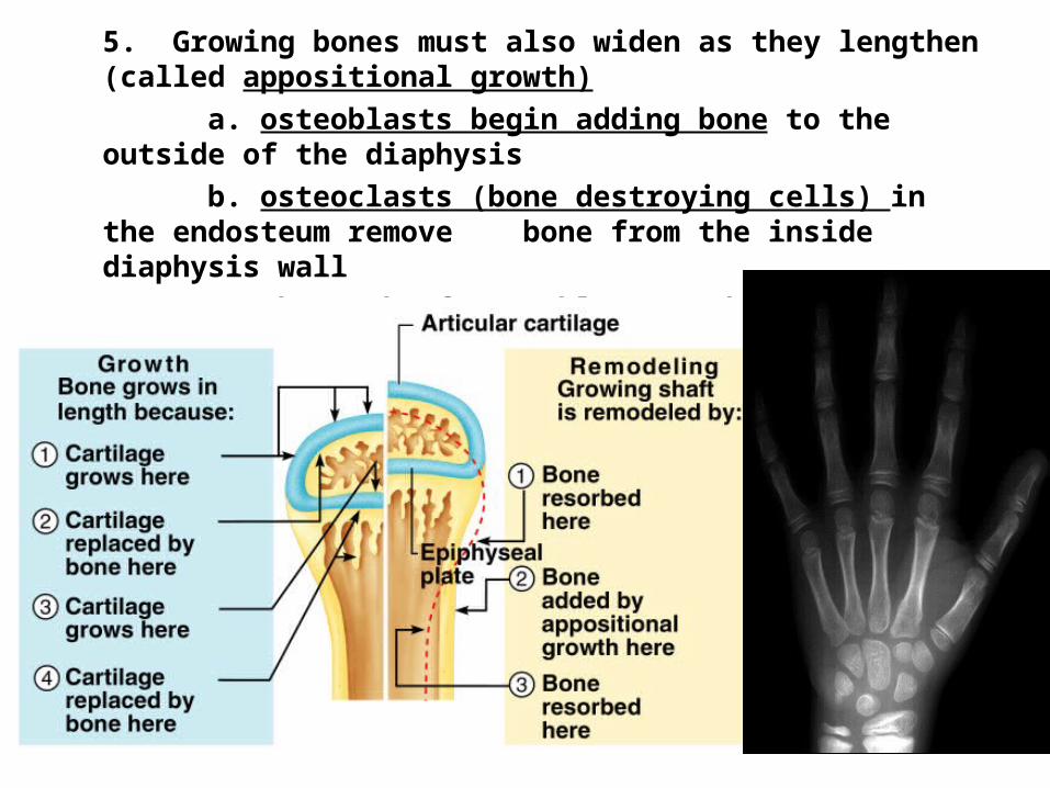
5. Growing bones must also widen as they lengthen (called appositional growth)
a. osteoblasts begin adding bone to the outside of the diaphysisb. osteoclasts (bone destroying cells) in the endosteum remove bone from the inside diaphysis wallc. The work of osteoblasts and osteoclasts occur at almost the
same rate allowing the bone to expand and widen
Body Ratios and ProportionsExplain the changes to our skeletal system throughout our lifetime (osteogenesis to bone growth to changes in old age
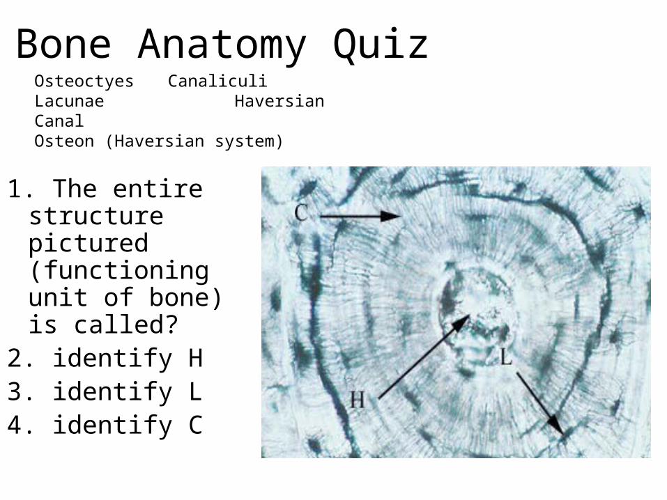
Bone Anatomy Quiz
1. The entire structure pictured (functioning unit of bone) is called?
2. identify H3. identify L4. identify C
Osteoctyes CanaliculiLacunae Haversian CanalOsteon (Haversian system)
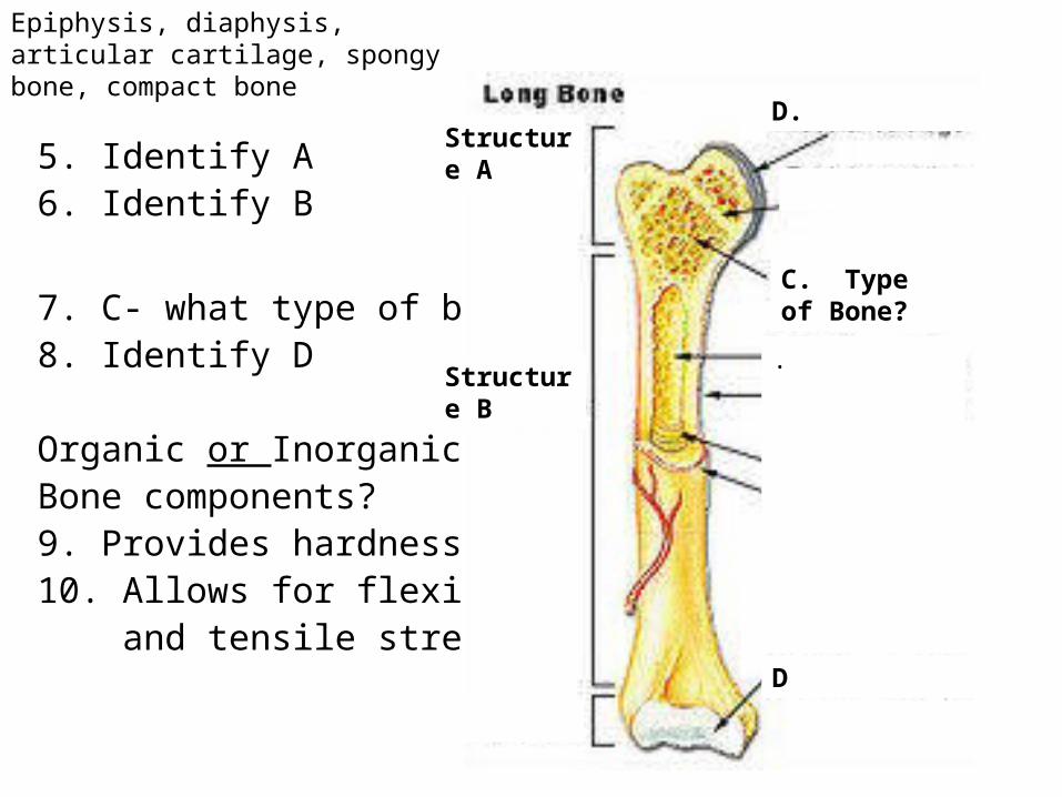
5. Identify A6. Identify B
7. C- what type of bone?8. Identify D
Organic or InorganicBone components?9. Provides hardness10. Allows for flexibility and tensile strength
Structure A
Structure B
C. Type of Bone?
D
D.
.
Epiphysis, diaphysis, articular cartilage, spongy bone, compact bone
11. Growth in bones occurs at _________plates.12.Function of red marrow?13.Function of yellow marrow?14.Which term does not belong?
Lamellae----- canaliculi-----circulation----osteoblasts
How much calcium is in that?• http://www.fitsugar.com/How-Much-Calcium-
182622
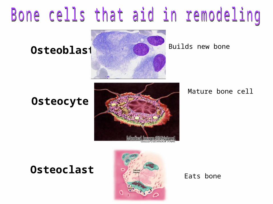
Osteoblast
Osteocyte
OsteoclastEats bone
Builds new bone
Mature bone cell
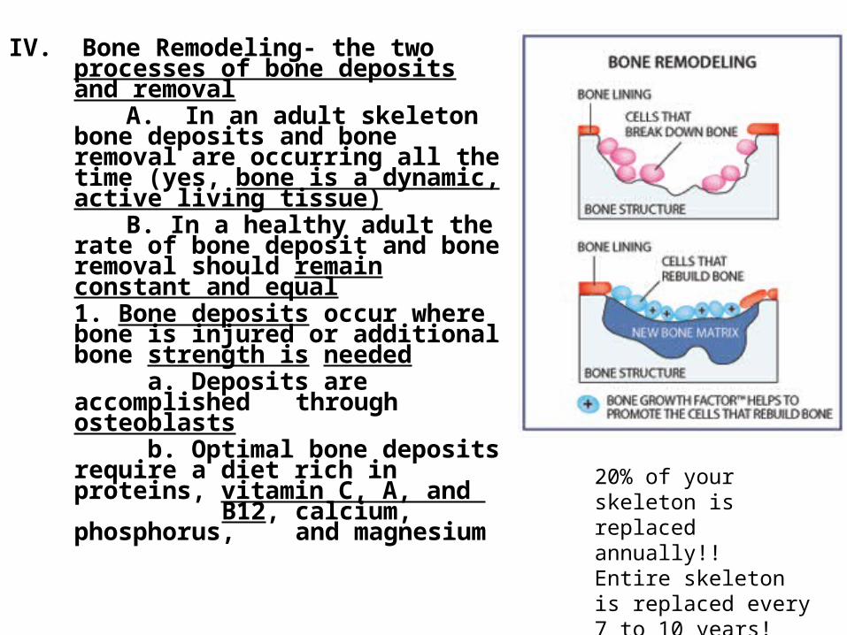
IV. Bone Remodeling- the two processes of bone deposits and removal
A. In an adult skeleton bone deposits and bone removal are occurring all the time (yes, bone is a dynamic, active living tissue)
B. In a healthy adult the rate of bone deposit and bone removal should remain constant and equal1. Bone deposits occur where bone is injured or additional bone strength is needed
a. Deposits are accomplished through osteoblasts
b. Optimal bone deposits require a diet
rich in proteins, vitamin C, A, and B12, calcium, phosphorus,
and magnesium
20% of your skeleton is replaced annually!! Entire skeleton is replaced every 7 to 10 years!
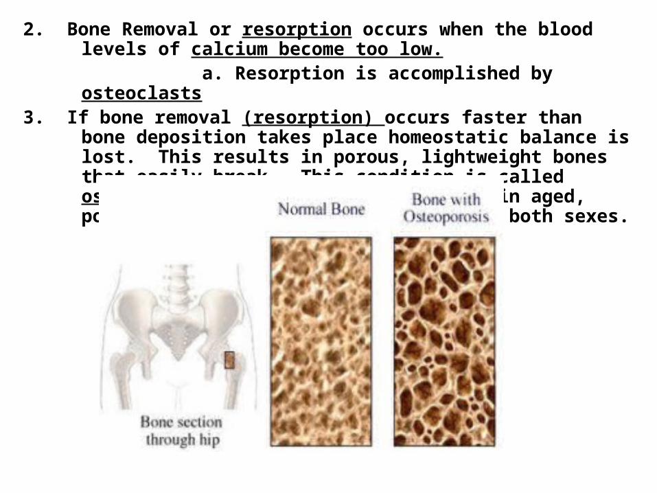
2. Bone Removal or resorption occurs when the blood levels of calcium become too low.
a. Resorption is accomplished by osteoclasts3. If bone removal (resorption) occurs faster than bone deposition takes place
homeostatic balance is lost. This results in porous, lightweight bones that easily break. This condition is called osteoporosis. This occurs most often in aged, postmenopausal women, but can occur in both sexes.
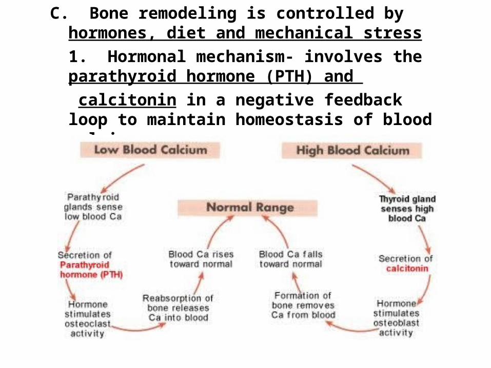
C. Bone remodeling is controlled by hormones, diet and mechanical stress1. Hormonal mechanism- involves the parathyroid hormone (PTH) and calcitonin in a negative feedback loop to maintain homeostasis of blood calcium.
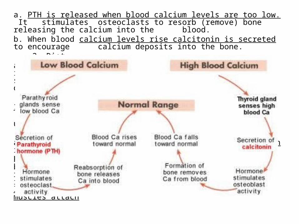
a. PTH is released when blood calcium levels are too low. It stimulates osteoclasts to resorb (remove) bone releasing the calcium into the blood.
b. When blood calcium levels rise calcitonin is secreted to encourage calcium deposits into the bone. 2. Diet
a. Calcium is necessary for transmission of nerve impulses, muscle contraction,blood coagulation, secretion by gland and nerve cells, and cell division
i. Your daily recommend levels of calcium is 1200-1400 mg.
ii. Calcium is absorbed by the intestines under the control of vitamin D metabolites
3. Mechanical Stressa. This set of controls serves the needs of the skeleton itself- keeping
bones strong where stressors are present.b. Wolff’s Law - holds that bones grow or remodel in response to the
forces or demands placed on it. Bone is laid down where bone is needed- for example large bony projections occur where heavy, active muscles attach
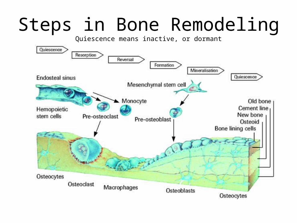
Steps in Bone RemodelingQuiescence means inactive, or dormant
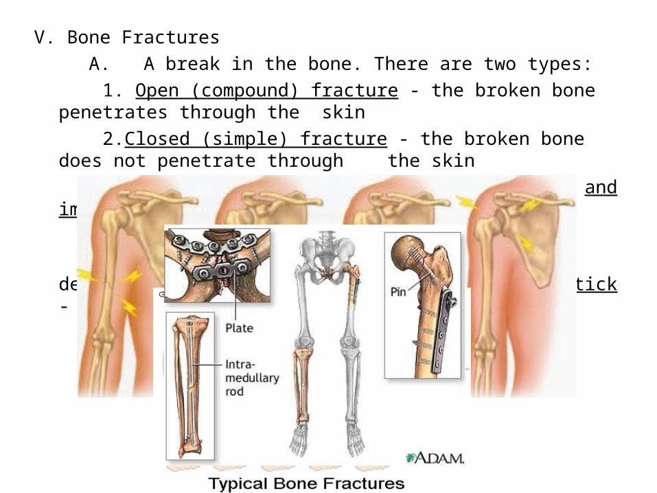
V. Bone Fractures A. A break in the bone. There are two types:
1. Open (compound) fracture - the broken bone penetrates through the skin
2.Closed (simple) fracture - the broken bone does not penetrate through the skin
B. Bone fractures are treated by reduction and immobilization 1. Realignment of the bone.
C. Common types - comminuted , compression, depression, compacted, spiral and green stick - see book for pictures
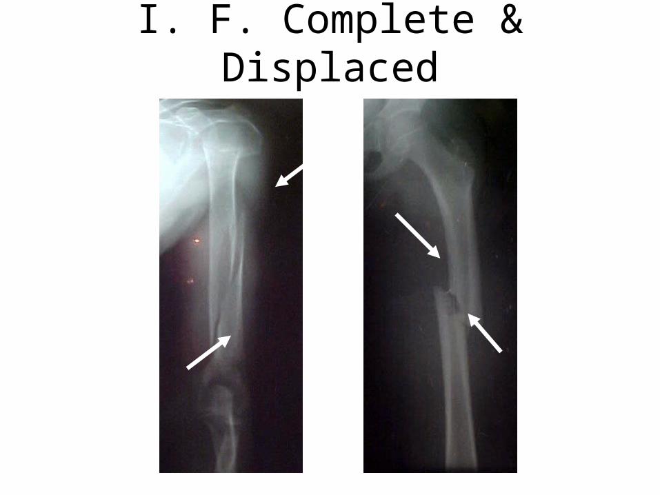
I. F. Complete & Displaced
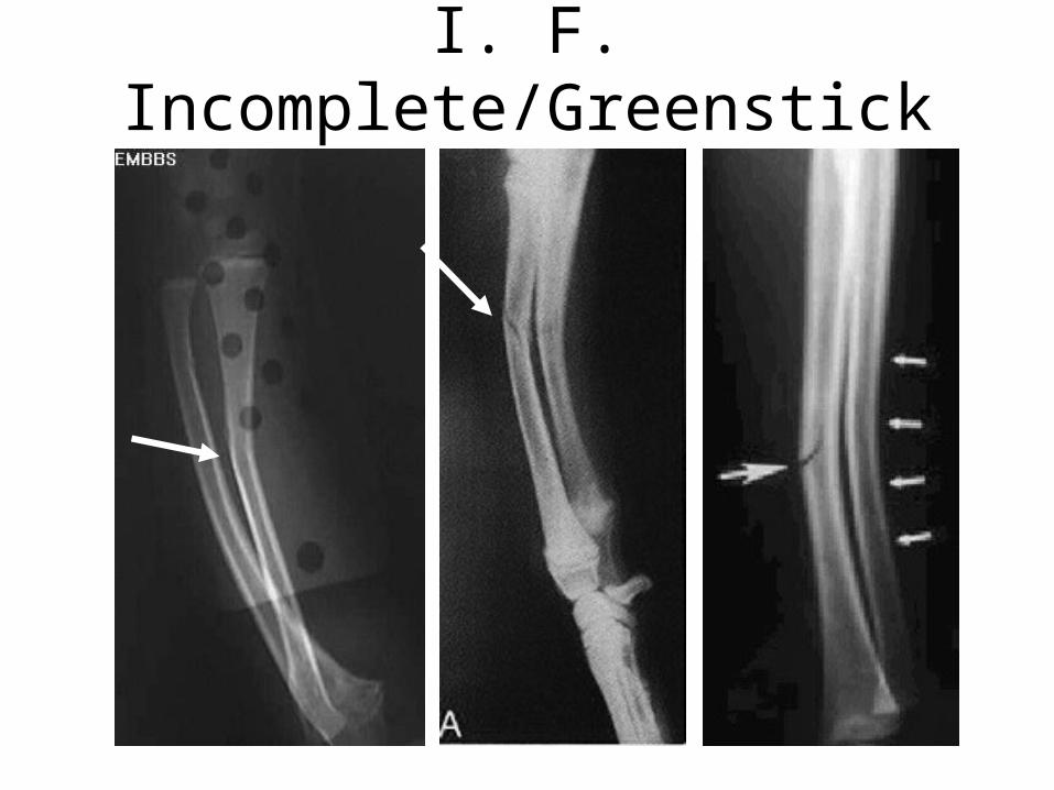
I. F. Incomplete/Greenstick
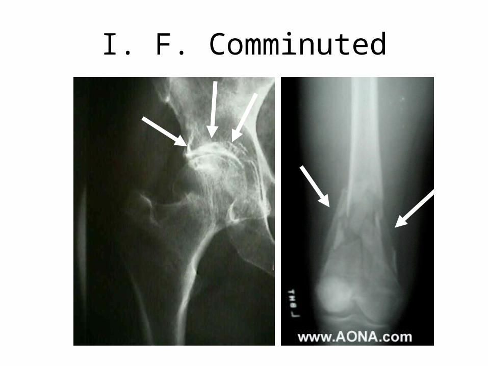
I. F. Comminuted
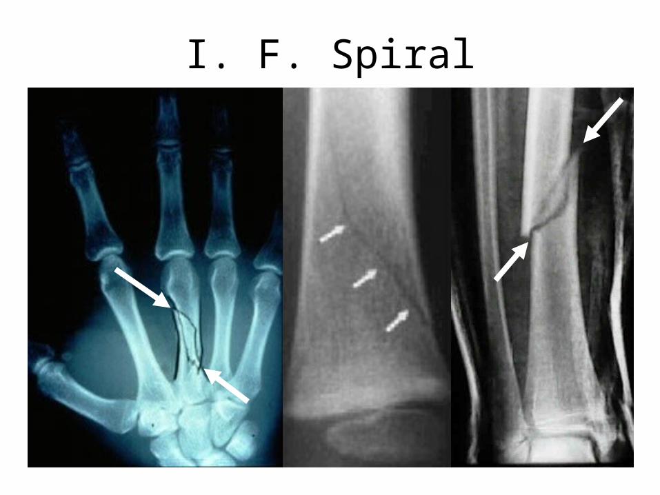
I. F. Spiral
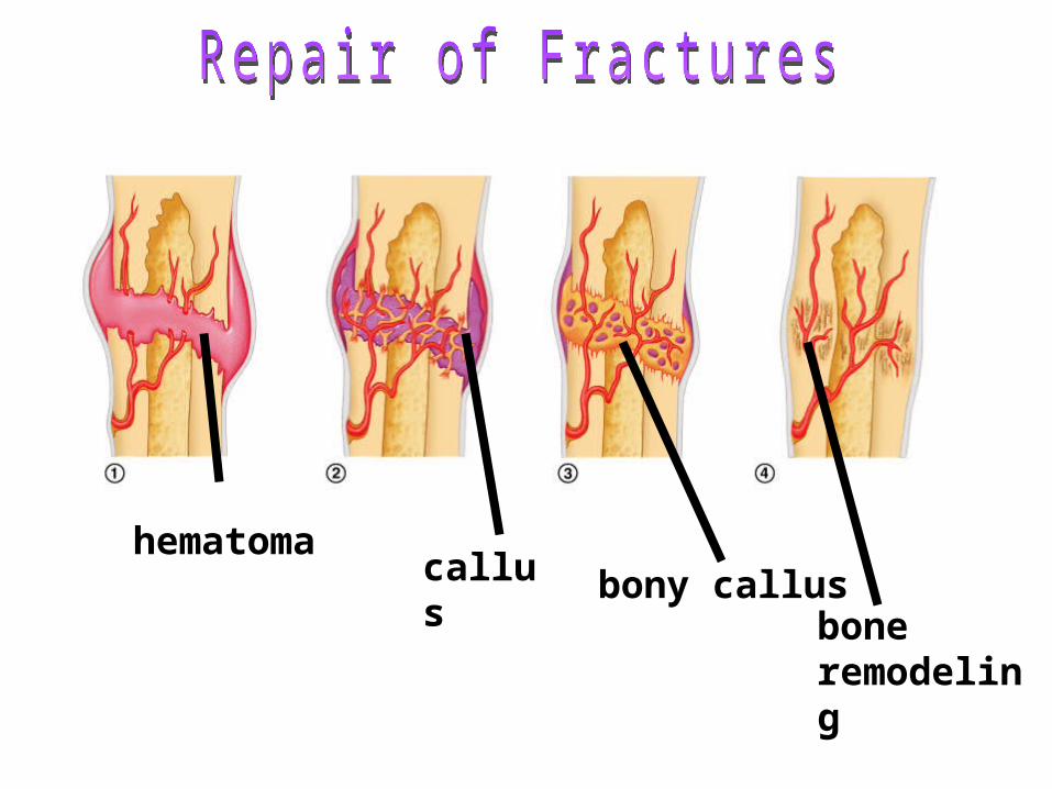
hematomacallus bony callus
bone remodeling
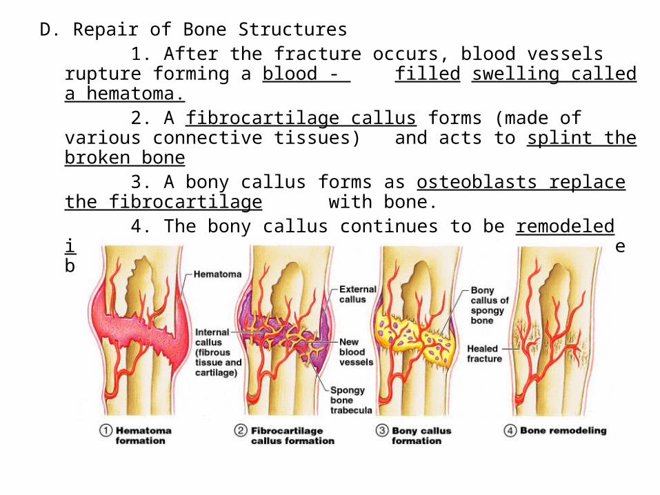
D. Repair of Bone Structures1. After the fracture occurs, blood vessels rupture forming a blood - filled swelling called a hematoma.2. A fibrocartilage callus forms (made of various connective tissues) and acts to splint the broken bone3. A bony callus forms as osteoblasts replace the fibrocartilage with bone.4. The bony callus continues to be remodeled in response to
mechanical stress placed upon the bone.
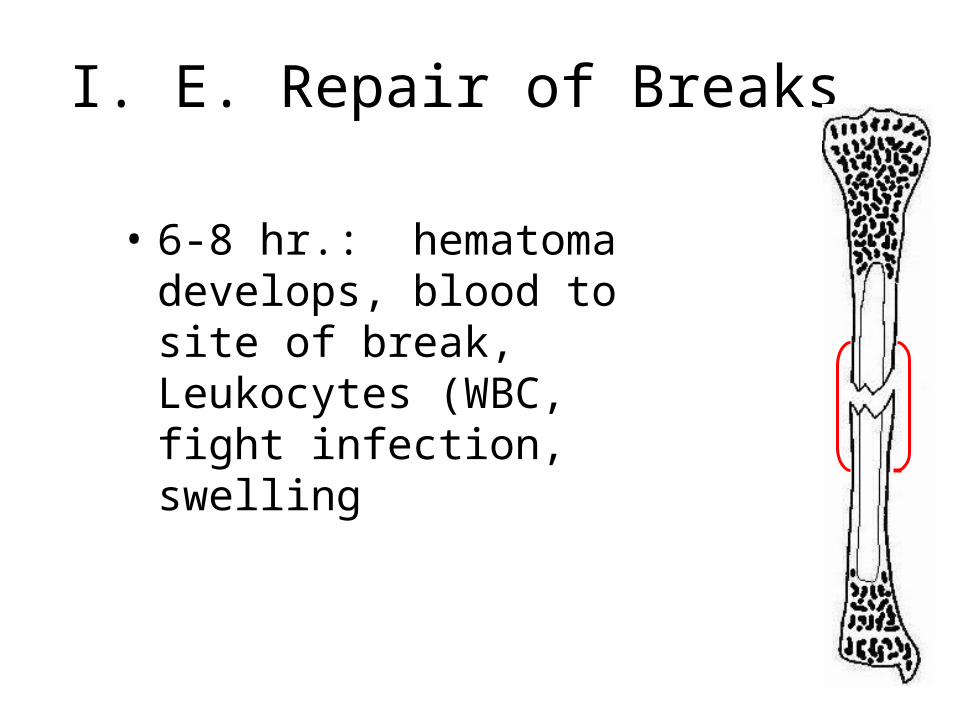
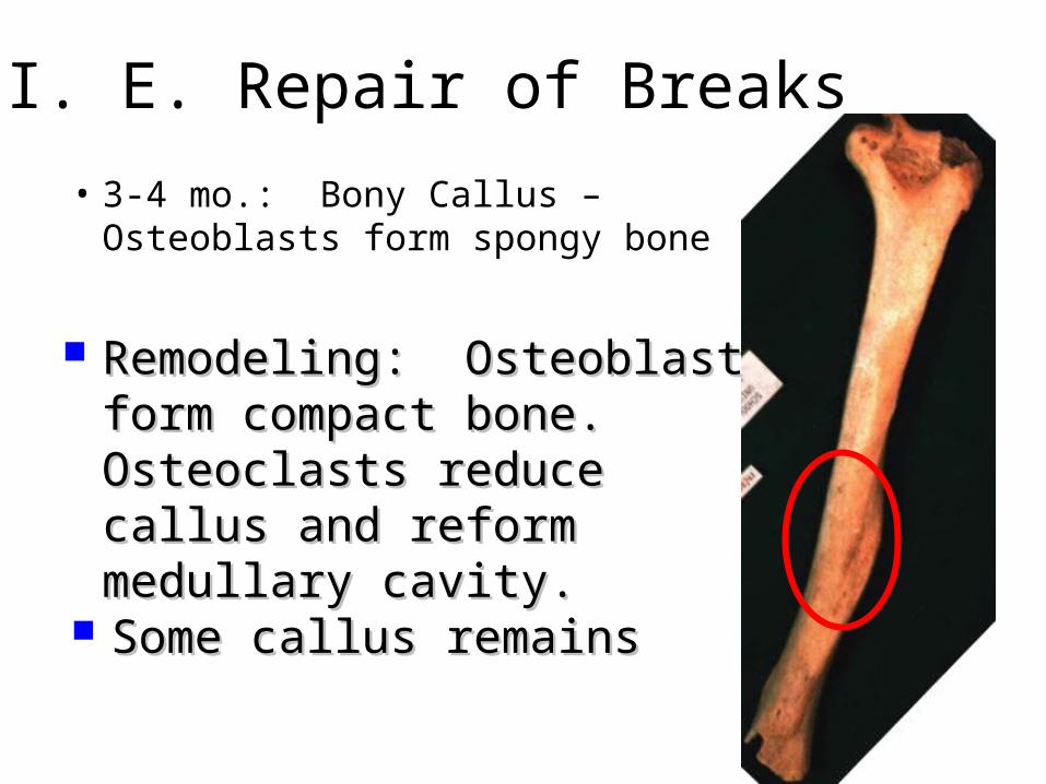
I. E. Repair of Breaks
• 6-8 hr.: hematoma develops, blood to site of break, Leukocytes (WBC, fight infection, swelling
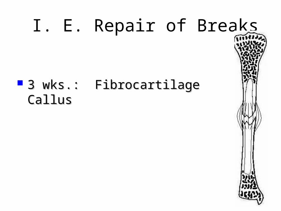
I. E. Repair of Breaks
3 wks.: Fibrocartilage Callus3 wks.: Fibrocartilage Callus
I. E. Repair of Breaks
• 3-4 mo.: Bony Callus – Osteoblasts form spongy bone
Remodeling: Osteoblasts Remodeling: Osteoblasts form compact bone. form compact bone. Osteoclasts reduce callus Osteoclasts reduce callus and reform medullary and reform medullary cavity.cavity.
Some callus remainsSome callus remains
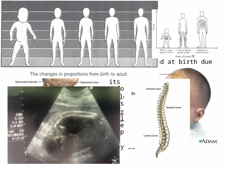
IV. Developmental Aspects of the Skeleton A. At birth, some fontanels are still present in the skull, Fontanels allow for
brain growth and ease birth passage. The growth of the cranium is related to brain growth and the increase in size of the facial skeleton follows tooth development and the respiratory passages.
B. The vertebral column is C-shaped at birth due to curvatures present in the thoracic and lumbar vertebrae (similar to a four-legged animal). When the baby begins to lift its head and walk the spine changes into its S-shaped form.
C. During youth the skeleton changes not only in size but in body proportions as well. At birth the head and trunk are one and half times as long as the lower limbs. The lower limbs then begin to grow much more rapidly than the trunk. During puberty the female pelvis widens to prepare for childbearing. Once the adult height is reached the skeleton changes very little until late middle age.
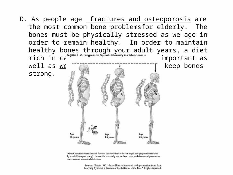
D. As people age fractures and osteoporosis are the most common bone problemsfor elderly. The bones must be physically stressed as we age in order to remain healthy. In order to maintain healthy bones through your adult years, a diet rich in calcium and Vitamin D are important as well as weight bearing exercise to keep bones strong.
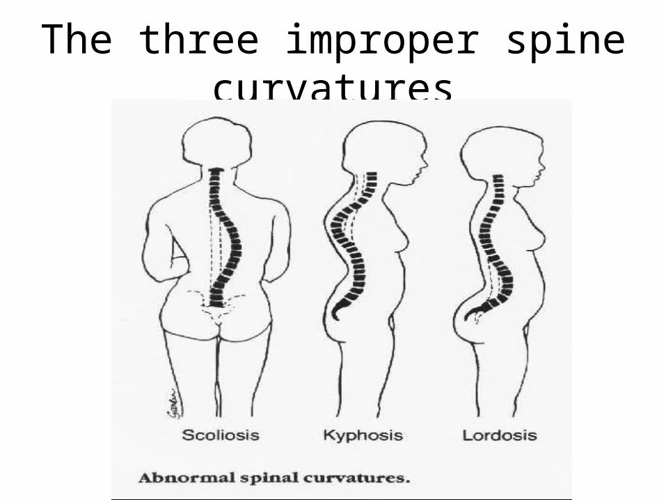
The three improper spine curvatures
biomedical engineer:
• A person who blends traditional engineering techniques with the biological sciences and medicine to improve the quality of human health and life.
• Biomedical engineers design medical devices and implants, artificial body parts, surgical and diagnostic tools, and medical treatment methods.
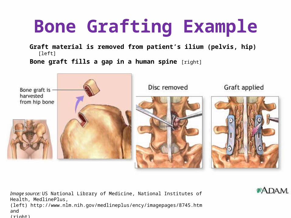
Bone Grafting ExampleGraft material is removed from patient’s ilium (pelvis, hip) [left]
Bone graft fills a gap in a human spine [right]
Image source: US National Library of Medicine, National Institutes of Health, MedlinePlus, (left) http://www.nlm.nih.gov/medlineplus/ency/imagepages/8745.htm and (right) http://www.nlm.nih.gov/medlineplus/ency/presentations/100136_4.htm
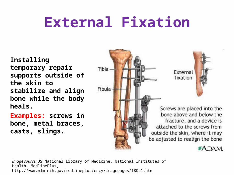
External Fixation
Installing temporary repair supports outside of the skin to stabilize and align bone while the body heals. Examples: screws in bone, metal braces, casts, slings.
Image source: US National Library of Medicine, National Institutes of Health, MedlinePlus, http://www.nlm.nih.gov/medlineplus/ency/imagepages/18021.htm