Embed Size (px)
Citation preview

Hughston Health AlertHughston Health Alert6262 Veterans Parkway, PO Box 9517, Columbus, GA 31908-9517 • www.hughston.com/hha
Extended (straight) knee
Flexed (bent) knee
Femur(thighbone)
Patella(kneecap)
Medial meniscus
MCL(medial
collateral ligament)
Tibia(shinbone)
Fibula
LCL(lateral collateral ligament)
Lateral meniscus
Articular cartilage
PCL (posterior cruciate ligament)
Torn ACL(anteriorcruciate
ligament)
Fig. 1. Knee anatomy and ACL injury.
Patella(kneecap)
The Hughston Foundation, Inc. ©2014
In Perspective: Anterior Cruciate Ligament Tears
In 1992, Dr. Jack C. Hughston (1917-2004), one of the world’s most respected authorities on knee ligament surgery, shared some of his thoughts regarding injuries to the ACL.
“You tore your anterior cruciate ligament.” On hearing your physician speak those words, you are filled with a sense of dread. You envision the end of your athletic life, even recreational sports.
Today, a torn ACL (Fig. 1) has almost become a household word. Through friends, newspapers, television, sports magazines, and even our physicians, we are inundated with the hype that the knee joint will deteriorate and become arthritic if the ACL is not operated on as soon as possible.
You have been convinced that to save your knee you must have an operation immediately to repair the ligament. Your surgery is scheduled for the following day. You are scared.
But there is an old truism in orthopaedic surgery that says, “no knee is so bad that it can’t be made worse by operating on it.”
For many years, torn ACLs were treated as an emergency and were operated on immediately, even before the initial pain and swelling of the injury subsided.
The trauma of the injury, plus the trauma of the operation added insult to injury, often resulting in the formation of scar tissue. Sometimes the results were stiff, painful knees without normal motion or function. The result was knees that caused the patient disability, even with simple walking.
Over the years, I have had to try to correct these problem knees. Those that were less stiff often responded to concentrated rehabilitation exercises and regained acceptable functional status. Others, however, required surgical release, and some had to have multiple operations to remove scar tissue to loosen the joint.
Inside...• Rotator Cuff Disease
• Bunions and Lesser Toe Deformities
• Tendon Injuries of the Hand
FOR A HEALTHIER LIFESTYLE
VOLUME 26, NUMBER 4 - FALL 2014

Fig. 2. Examination performed to test medial (inside) ligament damage.
Internal view of the bones, that make up the knee, during the examination.
Femur(thighbone)
Patella(kneecap)
Tibia(shinbone)
The Hughston Foundation, Inc. ©2014
The Hughston Foundation, Inc. ©2014
Fig. 3. Examination performed to test if the ACL is normal.
When the ACL is normal, the tibia does not shift during exam.
Tibia
Normal ACL
Femur
The Hughston Foundation, Inc. ©2014
Reprinted from the Hugshton Health Alert Volume 4, Number 4, Fall 1992.
Isn’t there an alternative to this all too common scene? Another treatment approach that isn’t as frightful? The answer is a definite “YES”! In many instances, nonsurgical treatment is successful. In other cases, surgery is necessary. Immediate surgery may be needed to repair other ligaments and torn menisci in the knee. But if only the ACL is torn, immediate repair is not necessary.
If the knee is shown to be loose during the physical exam, then most likely other ligaments in addition to the ACL have been damaged (Fig. 2). These other ligaments, when torn badly enough, may need to be surgically repaired within the first week. After they have been repaired and after a good rehabilitation program of six or more months, the decision to reconstruct the ACL can be made. If, at this time, there is a functional need for a ligament replacement, the operation can be done without the risk of subsequent stiffness and disability that can result from emergency repairs of the ACL.
If only the ACL is torn (Fig. 3), it may be difficult for your physician to confirm any looseness or instability of the knee joint by physical exam. I have seen cases where the ACL tear was only diagnosed by MRI or some other form of imaging study, and based on those findings, the patients were advised to have immediate surgery to repair the ligament. Be wary of this sort of advice. If your knee is not loose enough for your physician to be able to physically demonstrate the instability to you, then you should get a second opinion before having surgery.
In other cases, when the ACL and other ligaments are damaged and there is significant joint instability, surgery can be planned when the pain and swelling have subsided and knee motion has returned to almost normal. This usually occurs six weeks or more after the injury, in the meantime, you will have been performing prescribed daily rehabilitation exercises and using crutches to aid with your walking. When the knee is re-examined, there will be less discomfort and your physician will be able to perform a better evaluation. If the looseness is severe enough, an ACL reconstruction can be performed and the chance of complications is less than with an emergency operation.
The important thing to remember in all of this is that you don’t need to be frightened that your knee will be ruined forever if the torn ACL is not repaired immediately. On the contrary, a torn ACL by itself is not a reason for emergency surgery. Rather, it is time for calm, conservative management and appropriate follow-up. If this does not seem to be the approach your physician is taking, don’t hesitate to get a second opinion.
Jack C. Hughston, MD(1917-2004)
2 FOR A HEALTHIER LIFESTYLE

Rotator Cuff Disease A SPECTRUM OF PROBLEMS
The rotator cuff is a collection of 4 tendons that attach a group of 4 muscles to the head of the humerus, or top of the upper arm bone, and surrounds the shoulder joint. The rotator cuff holds the humeral head in place and stabilizes the shoulder during movement. The 4 muscles are the subscapularis, supraspinatus, infraspinatus, and teres minor (Fig. 1).
Imagine a ball and socket joint, such as the hip joint. With a deep socket, the ball remains relatively secure in the socket as the ball rotates, which makes it difficult to dislocate. Unfortunately, the shoulder joint functions more like a golf ball on a tee, making it more likely to dislocate. If you have ever tried to play golf in a strong wind, you know that the ball can fall off the tee. Similarly, if the shoulder muscles have become unstable, the head of the humerus, can slip off the glenoid (socket cavity), like the golf ball falls off the golf tee.
Symptoms and severity of rotator cuff problemsSymptoms can be quite different for patients who have
rotator cuff problems. For some patients, a rotator cuff injury may not be painful at all. It is a strange phenomenon that is not completely understood, which is why studies have been completed on individuals who function without pain or shoulder dysfunction, yet have a rotator cuff tear. Rotator cuffs can degenerate with age, but they do not necessarily become painful. In fact, in a study published in the Journal of Shoulder and Elbow Surgery, 50% of a group of people 80 years old and older had rotator cuff tears but had no idea they had the tears.
However, patients who have rotator cuff tears often do experience pain. Pain can be felt throughout the shoulder, but, for the most part, it tends to start in the back of the shoulder and radiates down deep to the shoulder’s deltoid muscle. It can stay toward the back or move toward the side of the shoulder. Activities that require you to stretch your arm out, reaching to the side, or reaching overhead can become difficult. The motions can be painful or impossible depending on the level of function of the rotator cuff.
Types of rotator cuff problemsA spectrum of problems—ranging from early and not
severe to complete loss of shoulder function with lots of pain—can affect patients with rotator cuff disease. Subacromial bursitis is the least severe form of rotator cuff dysfunction. The subacromial bursa is a thin sac of fluid positioned over the rotator cuff that reduces friction and cushions the tendons as they pass beneath the acromion. As the rotator cuff motion becomes dysfunctional, often
The Hughston Foundation, Inc. ©2014
The Hughston Foundation, Inc. ©2014
through abnormal motion of the shoulder blade or weak rotator cuff muscles, the subacromial bursa can become irritated and inflamed (Fig. 2). If you have subacromial bursitis, your doctor can prescribe physical therapy to correct the underlying problem of abnormal shoulder motion or weak rotator cuff muscles without the need for surgery or medication. Once the motion abnormality is corrected, the inflammation often improves on its own.
The next level of severity, rotator cuff tendinitis, involves irritation and inflammation of the rotator cuff itself and can often be diagnosed by using magnetic resonance imaging (MRI, a scan that shows the bones, muscles, tendons, and ligaments). Rotator cuff tendinitis can be a precursor to a rotator cuff tear, but it does not necessarily mean that you need surgery. Your doctor may feel you have a good chance at relieving your pain with nonsurgical treatments, such as physical therapy, anti-inflammatory medications, and corticosteroid injections.
A partial-thickness rotator cuff tear occurs when the
Humerus(arm bone)
Humerus(arm bone)
Clavicle (collar bone)Acromion
AcromionScapula
Fig. 1. Shoulder anatomy.
Subscapularis
Supraspinatus
Infraspinatus
Teres minor
Front view
Back view
FOR A HEALTHIER LIFESTYLE 3

rotator cuff tendon has pulled away from where it attaches to the bone, called the rotator cuff footprint (Fig. 3). With a partial tear, a portion of the tendon remains attached to the footprint and you often have full ability to move your arm, although movement can be painful. You can have a partial-thickness rotator cuff tear and function well; however, if it becomes too painful to bear and you are unable to use your shoulder, surgery often becomes necessary.
A full-thickness rotator cuff tear involves the entire tendon of at least 1 of the rotator cuff muscles (Fig. 4). If the tendon is completely detached from the footprint, the shoulder can be tender and painful to move. You can experience weakness and not be able to move your arms over your head or reach out to the side. Your physician will examine your shoulder and may lift your arm above your head. If you cannot hold your arm in place, it can be a sign that you have a full-thickness rotator cuff tear. During surgical repair of the rotator cuff, the tendon is reattached to its normal position. After surgery, physical therapy helps you regain your normal shoulder function.
Rotator cuff arthropathy, which involves the combination of a rotator cuff tear and arthritis, is the most severe form of rotator cuff disease. Often, the rotator cuff becomes completely nonfunctional and no longer supports the normal motion of the humeral head in the glenoid. Over time, the loss of normal motion can lead to arthritis, which can become progressively worse until the cartilage in the shoulder has completely eroded away. If your rotator cuff becomes nonfunctional, you are no longer a candidate for rotator cuff repair; instead, you may need shoulder replacement surgery to restore function.
Muscle and tendon degeneration due to aging can be a factor in rotator cuff disease. You can also injure your rotator cuff by falling on an outstretched arm or develop an injury over time doing repetitive activities at work or while playing a sport. If you are experiencing shoulder pain or have difficulty raising your arm above your head or out to your side, you should consider seeing your physician to help clarify the underlying problem. Early evaluation and treatment can reduce the severity of the injury and can reduce the time it takes you to restore function.
Christopher Van Hofwegen, MD Bellingham, Washington
Clavicle
Humerus(arm bone) The Hughston Foundation,
Inc. ©2014
The Hughston Foundation, Inc. ©2014
The Hughston Foundation, Inc. ©2014
Fig. 3. Partial-thickness rotator cuff tear.
Fig. 4. Complete rotator cuff tear.
Humerus(cross section)
Fig. 2. Subacromial bursitis.
Clavicle
Acromion
Subacromial bursa
Scapula
Deltoidmuscle
4 FOR A HEALTHIER LIFESTYLE

Toe deformities can have a negative impact on your life, even if you’re not on your feet for long periods of time. Toe deformity can make normal everyday activities difficult, including walking or participating in physical activities. A shoe that typically fits your foot can become uncomfortable to wear with a toe deformity. Special shoes with a wide, deep, or padded toe box can accommodate feet with toe deformities.
Foot and toe problems more often affect women who wear tight, narrow shoes or high heels, but men are prone to toe deformities, as well. Shoes that are too tight can cause irritating corns or calluses and lead to more serious and disabling problems, such as bunions in the big toe or mallet toe, hammer toe, and claw toe in the smaller toes. Problems that affect the toes are not limited to shoe wear. Toe deformities may be congenital (from birth) or resulting from an injury after a sprain or fracture. Diseases, such as diabetes or alcoholism may cause muscle or nerve damage in the foot leading to deformity.
BunionsA bunion is perhaps the most common and well-
known toe deformity. A bunion occurs when the metatarsophalangeal joint (MTP, the joint that connects the big toe to the foot) develops a swollen, painful bulge in the joint. Often, the hallux, or big toe angles away from the bunion and points toward the smaller toes in what’s called the valgus position. The condition, hallux valgus, worsens as the joint capsule on the inside of the toe stretches out and the tendons (tissue connecting muscle to bones) that pass over the big toe shift and pull the toe in an abnormal direction (Fig. 1).
Often, bunion pain can be treated without surgery by relieving the pressure points. A change in activity, such as wearing shoes with a wider toe box, using a shoe insert to support the foot, or a bunion splint can be worn for pain relief. When simple remedies fail, there are surgical options to consider.
Depending on the severity of the deformity and the presence of arthritis in the toe joint, there are several surgical procedures used to treat a bunion. The big toe can be realigned to a straighter position to reduce the turning out of the metatarsal, which causes the bunion to develop. If the toe joint does not have arthritis, the surgeon can remove the bulging portion of the bone and balance the soft tissue to maintain the toe in a straighter position. With advanced arthritis, fusion (bones fused together permanently) of the arthritic joint can keep the toe in a normal position. In cases of severe bunion deformity, the metatarsal is realigned by cutting the bone and stabilizing it with pins or screws. Most surgical treatments can be completed as an outpatient procedure.
Bunions & Lesser Toe Deformities
After surgery, protective shoe wear, splints, and crutches might be required while the bones and soft tissues heal.
Lesser toe deformitiesThe lesser, or small toes are formed by 3 small bones—
the distal, middle and proximal phalanges. The joint between the distal and middle phalanges is the distal interphalangeal (DIP) joint, and the joint between the middle and proximal phalanges is the proximal interphalangeal (PIP) joint. The joint that connects the toe to the foot at the metatarsal bone is called the metatarsophalangeal (MTP) joint. The 3 major types of small toe deformities—mallet toe, hammer toe, and claw toe—can result from improper shoe wear, trauma, genetic abnormalities, inflammatory arthritis, and neuromuscular and metabolic diseases.
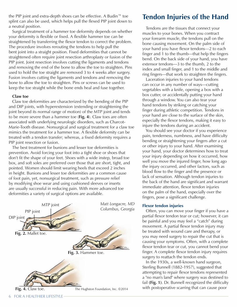
Mallet toeMallet toe is characterized by a flexion (bending)
deformity of the tip of the toe at the DIP joint, while the PIP and MTP joints remain straight (Fig. 2). A tight flexor tendon or a ruptured extensor tendon can cause this deformity. Often, a callus forms at the tip of the toe as the toe is driven into the sole of the shoe. Nonsurgical treatment focuses on relieving pressure on the toe through various cushioned inserts or roomier shoes. Surgical treatment options include cutting the tight flexor tendon and transferring the flexor tendon to the top of the toe to make it an extensor tendon, resection of the DIP joint, or fusion of the joint.
Hammer toeHammer toe is distinguished by a flexion, or bending
deformity at the PIP joint (Fig. 3). Often, there is an extension, or straightening of the DIP and MTP joints which gives the deformity a hammer shape. With a hammer toe, a callus often forms at the PIP joint, as the joint rubs the top of the shoe. Nonsurgical treatments using cushioned sleeves or foam cutouts to relieve pressure on
NormalMetatarsalphalangeal (MTP) joint
Metatarsal
Narrow toe box can cause pressure on the joint
The Hughston Foundation, Inc. ©2014
Fig. 1. Bunion.
FOR A HEALTHIER LIFESTYLE 5
Tendon Injuries of the HandTendons are the tissues that connect your
muscles to your bones. When you contract your forearm muscle, the tendons pull on the bone causing movement. On the palm side of your hand you have flexor tendons—2 to each finger and 1 to the thumb—that help the fingers bend. On the back side of your hand, you have extensor tendons—3 to the thumb, 2 to the index and small finger, and 1 to the middle and ring fingers—that work to straighten the fingers.
Laceration injuries to your hand tendons can occur in any number of ways—cutting vegetables with a knife, opening a box with a box cutter, or accidentally putting your hand through a window. You can also tear your hand tendons by striking or catching your finger during athletic competition. Tendons in your hand are close to the surface of the skin, especially the flexor tendons, making it easy to injure the tendons during an accident.
You should see your doctor if you experience pain, tenderness, numbness, and have difficulty bending or straightening your fingers after a cut or other injury to your hand. After examining your hand, your doctor determines how to treat your injury depending on how it occurred, how well you move the injured finger, how long ago the injury occurred, and other factors, such as blood flow to the finger and the presence or lack of sensation. Although tendon injuries to the back of the hand are significant and warrant immediate attention, flexor tendon injuries on the palm of the hand, especially over the fingers, pose a significant challenge.
Flexor tendon injuriesOften, you can move your finger if you have a
partial flexor tendon tear or cut; however, it can be painful and you may feel a “catch” during movement. A partial flexor tendon injury may be treated with wound care and therapy, or you may need surgery to repair the cut that is causing your symptoms. Often, with a complete flexor tendon tear or cut, you cannot bend your finger. A complete flexor tendon injury requires surgery to reattach the tendon ends.
In the 1930s, a well-known hand surgeon, Sterling Bunnell (1882-1957), suggested that attempting to repair flexor tendons represented a "no man's land" where surgery was destined to fail (Fig. 1). Dr. Bunnell recognized the difficulty with postoperative scarring that can cause poor The Hughston Foundation, Inc. ©2014
Fig. 3. Hammer toe.
Fig. 4. Claw toe.
Fig. 2. Mallet toe.
DIP jointPIP joint
MTP joint
the PIP joint and extra-depth shoes can be effective. A Budin™ toe splint can also be used, which helps pull the flexed PIP joint down to a neutral position.
Surgical treatment of a hammer toe deformity depends on whether your deformity is flexible or fixed. A flexible hammer toe can be straightened by transferring the flexor tendon to correct the problem. The procedure involves rerouting the tendons to help pull the bent joint into a straight position. Fixed deformities that cannot be straightened often require joint resection arthroplasty or fusion of the PIP joint. Joint resection involves cutting the ligaments and tendons and removing the end of the bone to allow the toe to straighten. Pins used to hold the toe straight are removed 3 to 4 weeks after surgery. Fusion involves cutting the ligaments and tendons and removing the bone to allow the toe to straighten. Pins or screws can be used to keep the toe straight while the bone ends heal and fuse together.
Claw toeClaw toe deformities are characterized by the bending of the PIP
and DIP joints, with hyperextension (extending or straightening the joint beyond its normal range of motion) of the MTP joint and tends to be more severe than a hammer toe (Fig. 4). Claw toes are often associated with underlying neurologic disorders, such as Charcot-Marie-Tooth disease. Nonsurgical and surgical treatment for a claw toe mimics the treatment for a hammer toe. A flexible deformity can be treated with a tendon transfer; whereas, a fixed deformity requires a PIP joint resection or fusion.
The best treatment for bunions and lesser toe deformities is prevention. Avoid forcing your foot into a tight shoe or shoes that don’t fit the shape of your feet. Shoes with a wide instep, broad toe box, and soft soles are preferred over those that are short, tight, and pointed. Women should limit wearing heels that exceed 2 inches in height. Bunions and lesser toe deformities are a common cause of foot pain, yet, nonsurgical treatment, such as pressure relief by modifying shoe wear and using cushioned sleeves or inserts are usually successful in reducing pain. With more advanced toe deformities a variety of surgical options are available.
Matt Longacre, MDColumbus, Georgia
6 FOR A HEALTHIER LIFESTYLE

The Hughston Foundation, Inc. ©2014
The Hughston Foundation, Inc. ©2014
DIP joint
Rupture of extensor tendon.
results and require additional surgery. When you bend or straighten your fingers, the flexor tendons slide through a synovial sheath, or tunnel that holds the tendons close to the bone. If postoperative scarring occurs, it can make the tendon’s movement through the tight-fitting tunnel difficult. Often, an additional surgery is needed to remove the scar tissue and make movement through the synovial sheath smoother. There have been many advances in both surgical technique and in postoperative therapy since Dr. Bunnell's revelation; however, a secondary surgery can be necessary to remove scar tissue.
Extensor tendon injuriesPartial tendon tears to the back of your hand often allow
the finger to straighten, but can cause pain during motion. Wound care, wearing a splint, and physical therapy are effective nonsurgical treatments for partial extensor cuts or tears. After the tendon heals, you can begin physical therapy to help regain full range of motion.
Two common extensor tendon injuries—mallet finger and boutonnière deformity—result from a cut or tear of the extensor tendon. Mallet finger often occurs from jamming the finger causing a droop at the finger tip as the tendon tears or pulls away from the bone (Fig. 2). Often, a piece of bone can be pulled off with the tendon. Boutonnière deformity causes the finger to bend down at the middle joint of the finger. These injuries often respond well to wearing a splint for 6 to 8 weeks. However, if you do not wear the splint as instructed by your doctor mallet finger and boutonnière deformity can become permanent. If the splinting is unsuccessful or if the injury is associated with a fracture or joint dislocation, your doctor may recommend surgery.
Tendons that are cut or torn from the bone can be repaired surgically and an internal splint, such as a pin, can be used to stabilize the bone during healing. Because there are no synovial sheaths for the tendons to slide
through at the back of the hand, postoperative outcomes are often good. After surgery, you will wear a splint to limit movement for 3 to 6 weeks while the tendon heals.
Physical therapyA surgically repaired tendon requires a strict
postoperative therapy plan and hard work from you and your hand therapist. After surgery, you should wear a splint and follow the guidelines for early postoperative activity to avoid stretching or pulling the tendon repair apart. You can be taught exercises to help prevent the repaired tendon from attaching to surrounding tissue, which can reduce your range of motion. You will be given guidelines to follow for your therapy sessions several times a week and home exercises to do several times a day. At 4 to 6 weeks after surgery, the therapy regimen increases to include exercises to improve range of motion and gain strength. After 3 months, the tendon should be strong enough to resume normal use.
Flexor and extensor tendon injuries are often caused by an accident; therefore, there are no specific prevention techniques, except to say that you should follow safety precautions while using tools and devices that can injure your hands. It is important to remember that preinjury range of motion might never return; however, you increase your chances of a good outcome if you get to your doctor promptly for treatment and follow your rehabilitation therapy guidelines.
David H. MacDonald, DOColumbus, Georgia
Fig. 1. No man's land.
Fig. 2. Mallet finger.
FOR A HEALTHIER LIFESTYLE 7

EditorThomas N. Bernard, Jr., MD
Managing EditorDennise Brogdon
Art DirectorBelinda J. Klein, MA
Illustrator & LayoutTiffany C. Davis, MS
Editorial BoardChamp L. Baker III, MDMark A. Baker, PT, CEOCarol M. Binns, MA William C. Etchison, MSAndy J. Grubbs, Jr., MEd, ATC Rob Hopkins, PT, SCS Cholly P. MintonSteve Young, PT
The Hughston Health Alert is a quarterly publication of The Hughston Foundation, Inc. The Founda-tion’s mission is to help people of all ages attain the highest possible standards of musculoskeletal health, fitness, and athletic prowess. Information in the Hughston Health Alert reflects the experience and training of physicians at The Hughston Clinic, P.C., of physical therapists and athletic trainers at Hughston Rehabilitation, of physicians who trained as residents and fellows under the auspices of The Hughston Foundation, Inc., and of research scientists and other professional staff at The Hughston Foundation, Inc. The information in the Hughston Health Alert is intended to supplement the advice of your personal phy-sician and should not be relied on for the treatment of an individual’s specific medical problems.
Special written permission is required to reproduce, by any manner, in whole or in part, the material herein contained.
Send inquiries to Medical Writing, The Hughston Foundation, Inc., P.O. Box 9517, 6262 Veterans Parkway, Columbus GA 31908-9517 USA.
Copyright 2014, The Hughston Foundation, Inc. ISSN# 1070-7778www.hughston.com
6262 Veterans ParkwayP.O. Box 9517
Columbus GA 31908-9517Appointments:706-324-6661
1-800-331-2910
Hughston Health AlertThe Hughston Foundation, Inc.6262 Veterans Parkway P.O. Box 9517 Columbus, Georgia 31908-9517
4401 River Chase DrivePhenix City, AL 36867Phone: 334-732-3000
Fax: 334-732-3020
Locations:
SCAN ME for more Hughston Health Alert articles.
2002-2013
NONPROFIT ORGUS POSTAGE
PAIDCOLUMBUS GAPERMIT NO 99
You may receive the Hughston Health Alert via e-mail by registering at: www.hughston.com/health-alert-sign-up.aspx
Locations:
Champ L. Baker Jr., MD - Arthroscopy & Sports Medicine
Champ L. Baker III, MD - Arthroscopy & Sports Medicine
Thomas N. Bernard Jr., MD - Orthopaedic Spine Surgery
Jared A. Brummel, DO - Sports Medicine & General Orthopaedics
J. Kenneth Burkus, MD - Orthopaedic Spine Surgery
Kevin J. Collins, MD - General Orthopaedics & Sports Medicine
Norman L. Donati Jr., MD - General Orthopaedics, Foot & Ankle
John D. Dorchak, MD - Orthopaedic Spine Surgery
Jason M. Evans, MD - Orthopaedic Trauma
Patrick J. Fernicola, MD - Shoulder, Knee, Total Joint Replacement
Fred Flandry, MD, FACS - Trauma, Arthroscopy & Sports Medicine
John C. P. Floyd, MD, FACS - Orthopaedic Traumatologist
Ryan M. Geringer, DO - General Orthopaedics & Sports Medicine
Garland K. Gudger, MD - General Orthopaedics & Sports Medicine
Robert M. Harris, MD - Director of Orthopaedic Trauma
Albany • Auburn • Columbus • Cordele • Dothan • Gwinnett • LaGrange • Moultrie • Thomaston • Thomasville • Phenix City • Valdosta • Vidalia • Waycross
Locations:
J. Matthew Heaton, MD - General Orthopaedics & Sports Medicine
Kurt E. Jacobson, MD, FACS - Knee, Sports Medicine & General Orthopaedics
Cameron Kersey, MD - Radiology
Philip J. Kregor, MD - Orthopaedic Trauma
David H, MacDonald, DO - Hand & Upper Extremities, Arthroscopic Surgery
James E. McGrory, MD - Orthopaedic Spine Surgery & Total Joint Replacement
William Min, MD, MS, MBA - Orthopaedic Traumatologist
Jesse L. Pace, DO - Arthroscopic Surgery, General Orthopaedics & Sports Medicine
Douglas W. Pahl, MD - Orthopaedic Spine Surgery
Randall J. Ruark, MD - General Orthopaedics, Hip & Knee Reconstruction
David C. Rehak, MD - Hand, Wrist & Upper Extremities
Carlton G. Savory, MD, FACS - Hip, Knee, Total Joint Replacement
Rourke M. Stay MD - Radiology
Michael M. Tucker Jr., MD - Knee, Shoulder, Foot, Ankle & Sports Medicine
John I. Waldrop, MD - General Orthopaedics, Total Joint Replacement
Bruce H. Ziran, MD - Director of Orthopaedic Trauma
YESTERDAY. TODAY. TOMORROW.
HUGHSTON DIFFERENCE
THE
It’s our privilege to serve you