Embed Size (px)
Citation preview
HUMAN ANATOMY AND PHYSIOLOGY
The Skeletal System NotesChapter 5
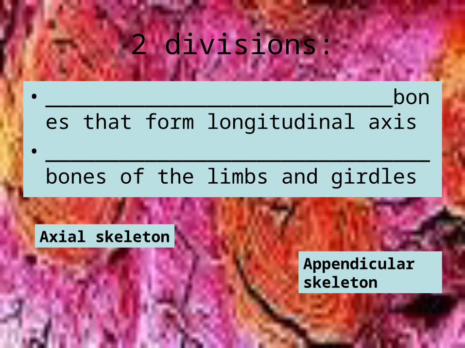
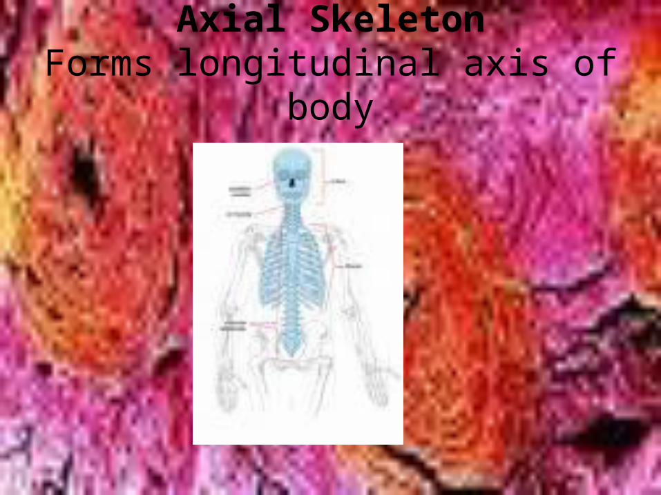
2 divisions:
• ____________________________bones that form longitudinal axis
• _______________________________bones of the limbs and girdles
Axial skeleton
Appendicular skeleton
• The system includes joints ,cartilage and ligaments.
I. Bones:An Overview
• Functions-besides giving body shape and Form:
– 1)Support-supports body and cradles soft organs– 2)Protection-protect soft organs-eg. Protects
brain/vertebrae-spine and ribs for organs of the thoracic cavity
Functions cont’d
• 3)Movement-Skeletal muscles attach to bones by _______________and bones work as levers
tendons
Functions cont’d
– 4)Storage-» ______in internal cavities of bones» Storehouse for minerals-esp.Ca and P….A small
amount of Ca must be in blood at all times to reach the nerve tissue for transmission,so muscles contract and help clot blood.Ca in bones as salts go to provide Ca 2+ ions for blood
» Hormones control the movement of Ca to and from blood according to body need…..too much Ca can be a problem
5)Blood Cell Formation or ________________________ in the marrow cavities of certain bones
Fat
hematopoiesis

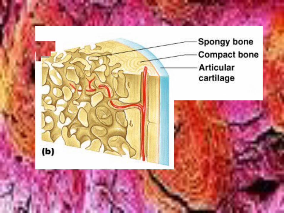
B.Classification of Bones*(all 206 of them)
– There are 2 main types of osseous tissue
» ____________________________dense and looks smooth and homogeneous
• ___________________________ made of needlelike
pieces of bone and lots of open space
Compact boneSpongy bone
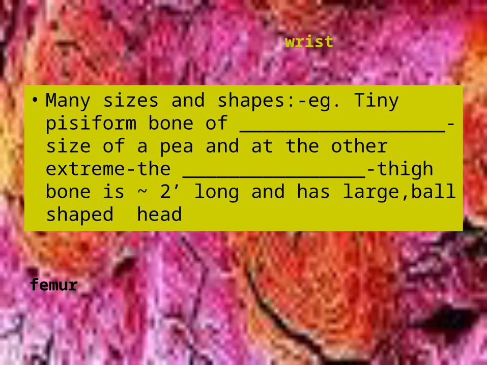
• Many sizes and shapes:-eg. Tiny pisiform bone of __________________-size of a pea and at the other extreme-the ________________-thigh bone is ~ 2’ long and has large,ball shaped head
wrist
femur
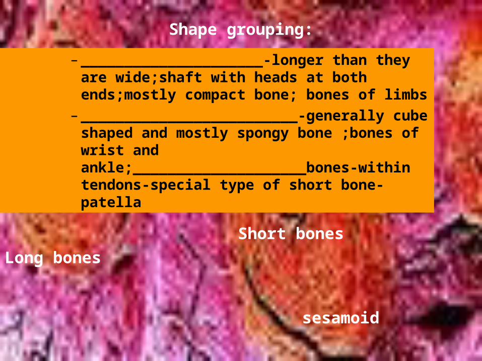
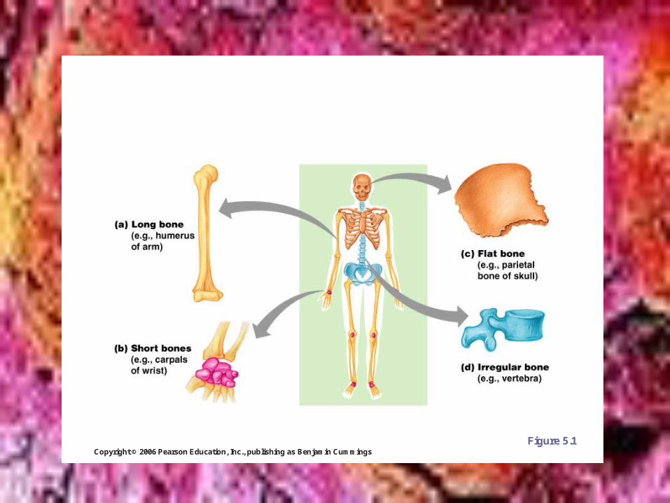
Shape grouping:
– _____________________-longer than they are wide;shaft with heads at both ends;mostly compact bone; bones of limbs
– _________________________-generally cube shaped and mostly spongy bone ;bones of wrist and ankle;____________________bones-within tendons-special type of short bone-patella
Long bones
Short bones
sesamoid
Shape cont’d
– ____________________-thin,flattened and usually curved-2 thin layers of compact bone sandwiching spongy bone-skull,ribs,sternum
– __________________________don’t fit other categories-vertebrae and hip
Flat bones Irregular bones
Copyright © 2006 Pearson Education, Inc., publishing as Benjamin Cummings
Classification of Bones on the Basis of Shape
Figure 5.1
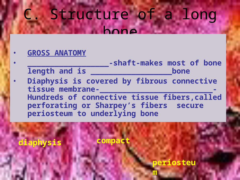
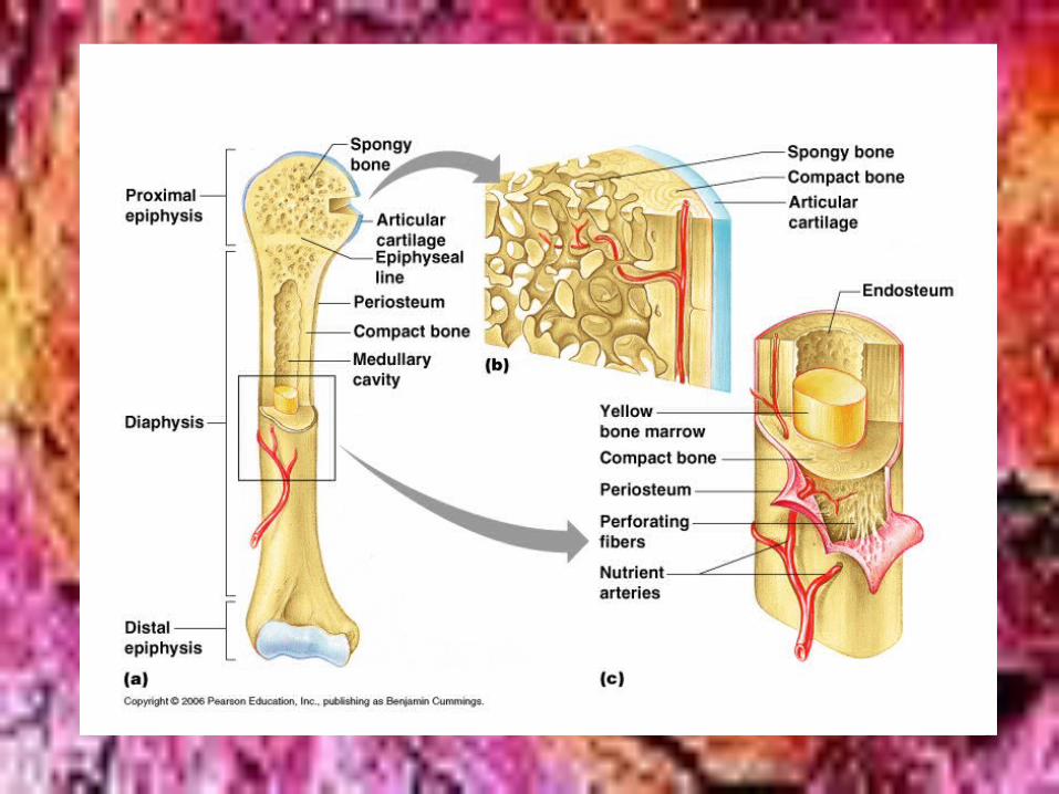
C. Structure of a long bone
• GROSS ANATOMY• __________________-shaft-makes most of bone
length and is __________________bone• Diaphysis is covered by fibrous connective tissue
membrane-_________________________-Hundreds of connective tissue fibers,called perforating or Sharpey’s fibers secure periosteum to underlying bone
diaphysis compact
periosteum
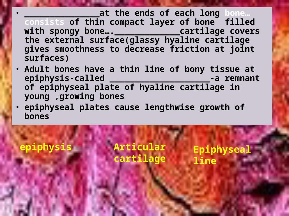
• _______________at the ends of each long bone…consists of thin compact layer of bone filled with spongy bone…._____________cartilage covers the external surface(glassy hyaline cartilage gives smoothness to decrease friction at joint surfaces)
• Adult bones have a thin line of bony tissue at epiphysis-called ____________________-a remnant of epiphyseal plate of hyaline cartilage in young ,growing bones
• epiphyseal plates cause lengthwise growth of bones
epiphysis Articular cartilage
Epiphyseal line
• At end of puberty,hormones inhibit long bone growth and the epiphyseal plate is completetly replaced w/bone
• Cavity of shaft stores adipose tissue--________________________,or medullary cavity …In infants this area forms RBC’s-red marrow is there as well
• For adults ___________________is in cavities of spongy bone of flat bones and epiphyses of some long bones—Note:Areas of red marrow are more limited in adults—to places such as sternum
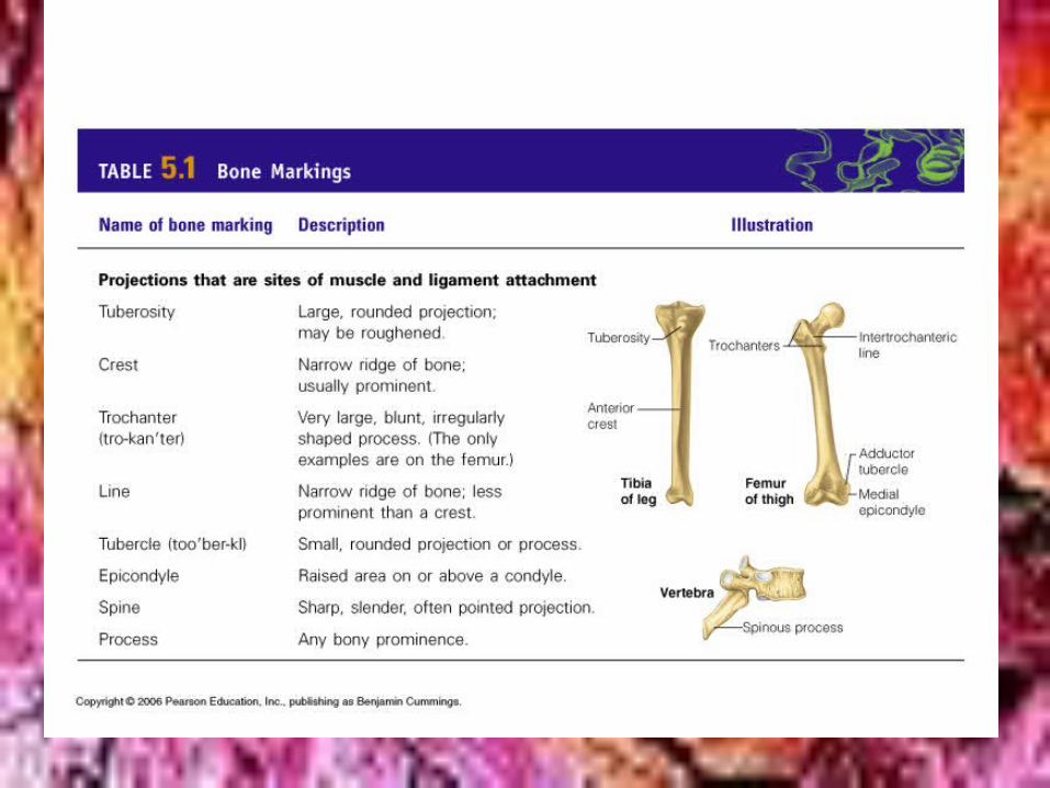
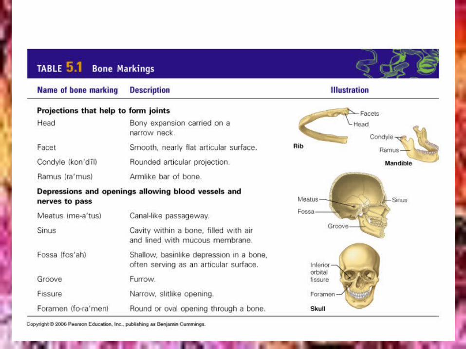
• Surfaces have bumps holes and ridges=__________________________-show where muscles,tendons,and ligaments were attached and where blood vessels and nerves pass
Yellow marrow Red marrowBone markings
Bone markings
– 1.projections or processes• -grow out from bone surface---goes w/terms
beginning w/T• 2.depressions or
________________________-indentations in bone---goes w/terms starting w/F(except facet
cavities
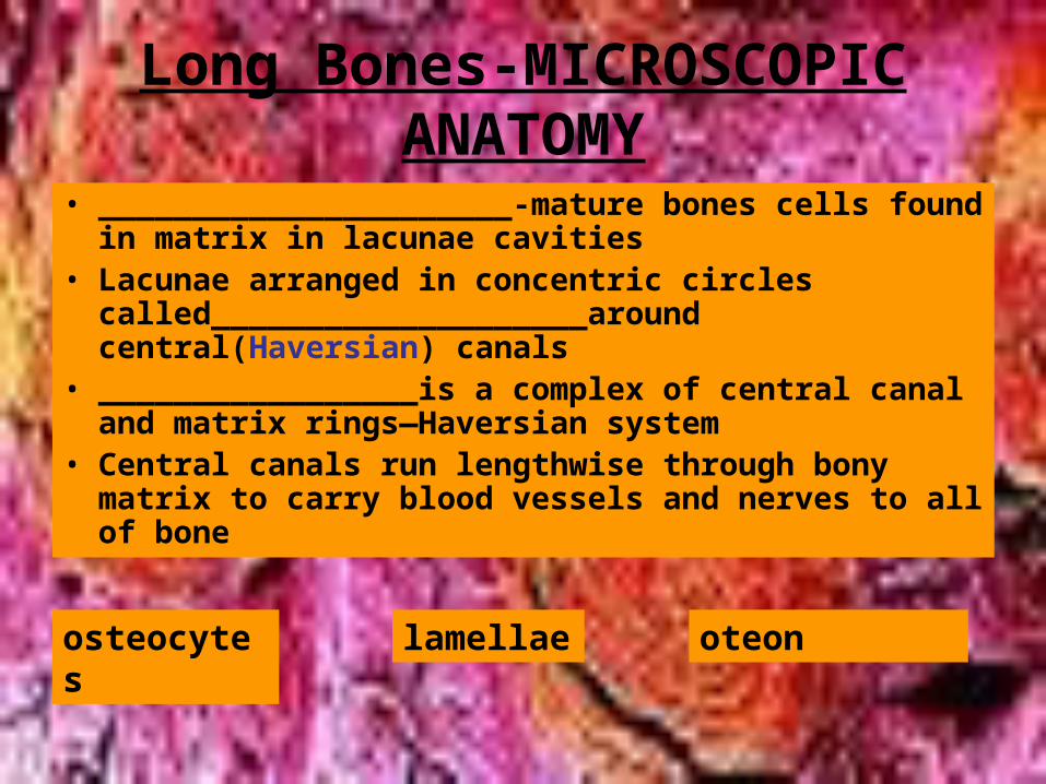
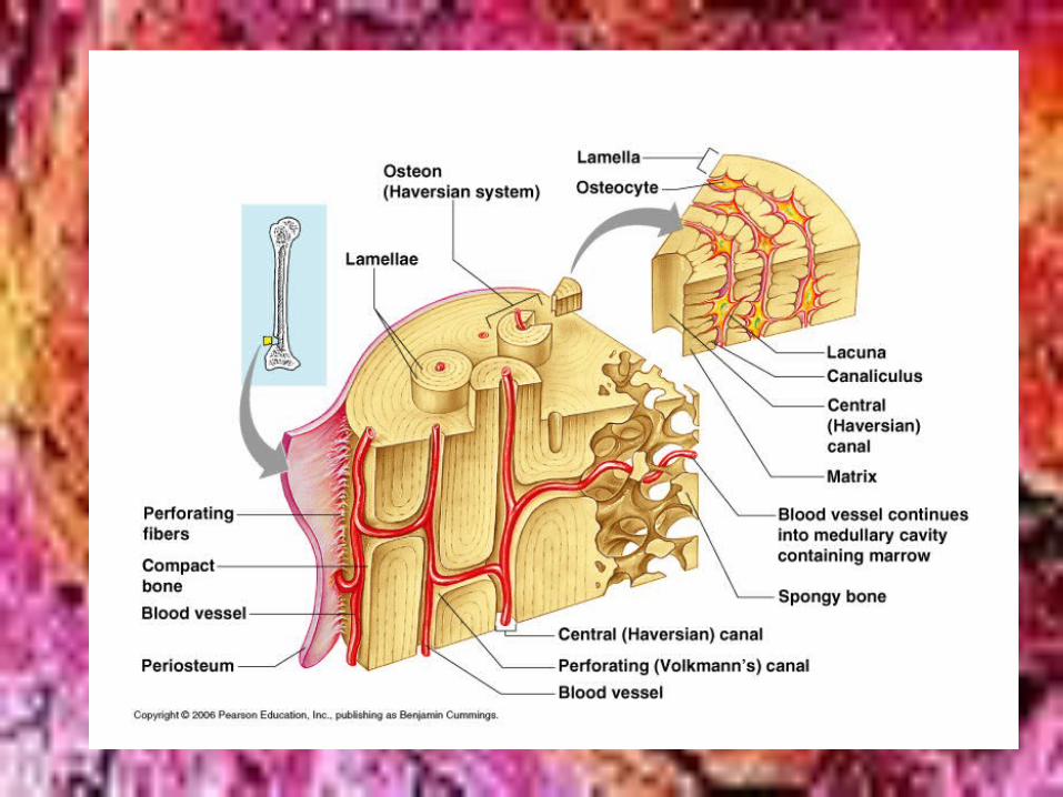
Long Bones-MICROSCOPIC ANATOMY
• ______________________-mature bones cells found in matrix in lacunae cavities
• Lacunae arranged in concentric circles called____________________around central(Haversian) canals
• _________________is a complex of central canal and matrix rings—Haversian system
• Central canals run lengthwise through bony matrix to carry blood vessels and nerves to all of bone
osteocytes lamellae oteon
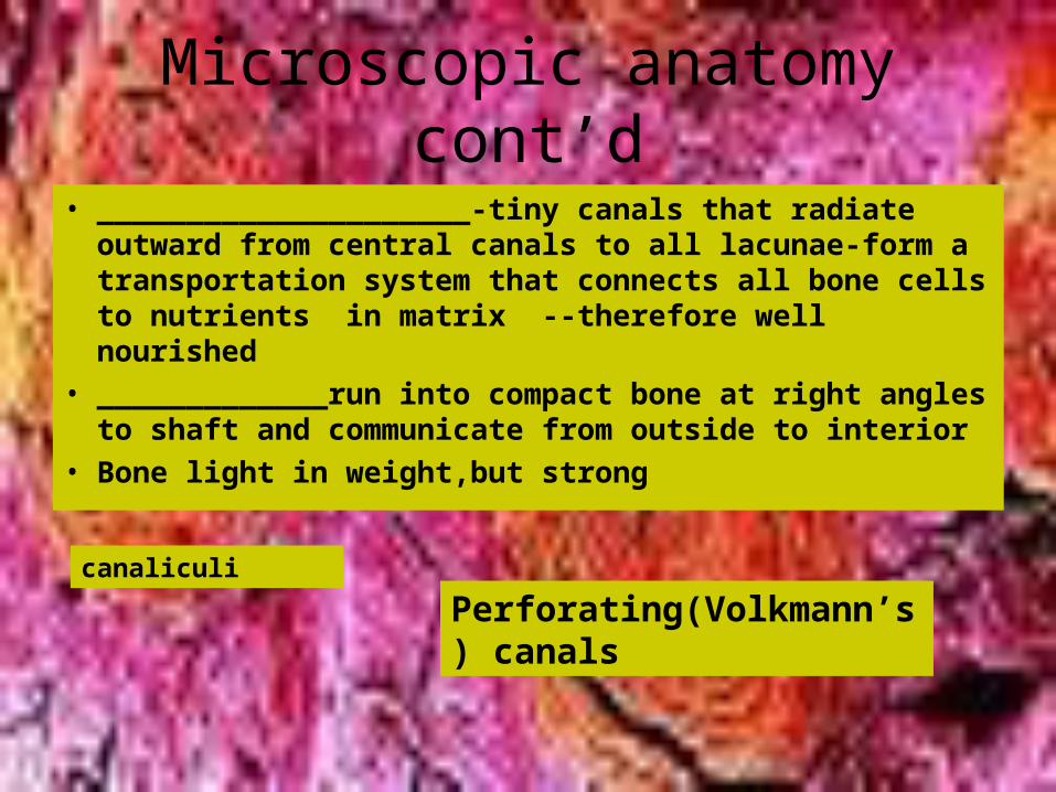
Microscopic anatomy cont’d
• _____________________-tiny canals that radiate outward from central canals to all lacunae-form a transportation system that connects all bone cells to nutrients in matrix --therefore well nourished
• _____________run into compact bone at right angles to shaft and communicate from outside to interior
• Bone light in weight,but strong
canaliculi
Perforating(Volkmann’s) canals
Bone Formation,Growth and Remodeling
• 2 most strong and supportive tissue-bone and cartilage
• Except for flat bones,most bones develop using hyaline cartilage as model
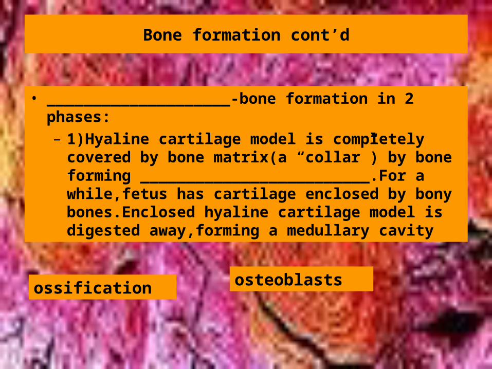
Bone formation cont’d
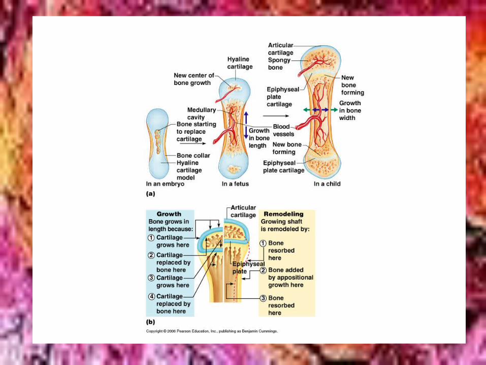
• ____________________-bone formation in 2 phases:– 1)Hyaline cartilage model is completely covered
by bone matrix(a “collar”) by bone forming _________________________.For a while,fetus has cartilage enclosed by bony bones.Enclosed hyaline cartilage model is digested away,forming a medullary cavity
ossification osteoblasts
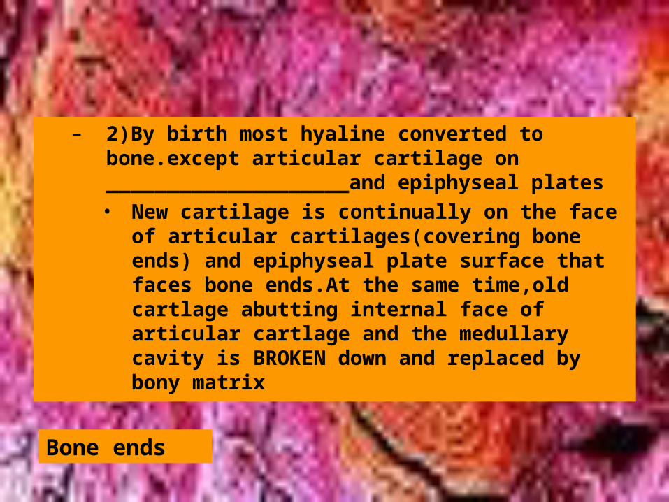
– 2)By birth most hyaline converted to bone.except articular cartilage on ____________________and epiphyseal plates• New cartilage is continually on the face of
articular cartilages(covering bone ends) and epiphyseal plate surface that faces bone ends.At the same time,old cartlage abutting internal face of articular cartlage and the medullary cavity is BROKEN down and replaced by bony matrix
Bone ends
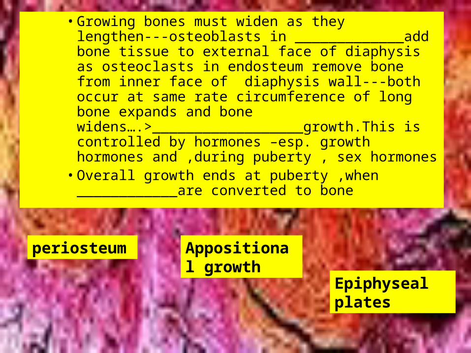
• Growing bones must widen as they lengthen---osteoblasts in _____________add bone tissue to external face of diaphysis as osteoclasts in endosteum remove bone from inner face of diaphysis wall---both occur at same rate circumference of long bone expands and bone widens….>__________________growth.This is controlled by hormones –esp. growth hormones and ,during puberty , sex hormones
• Overall growth ends at puberty ,when ____________are converted to bone
periosteum Appositional growth
Epiphyseal plates
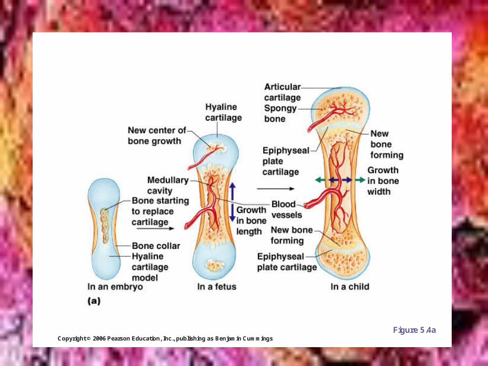
Copyright © 2006 Pearson Education, Inc., publishing as Benjamin Cummings
Long Bone Formation and Growth
Figure 5.4a
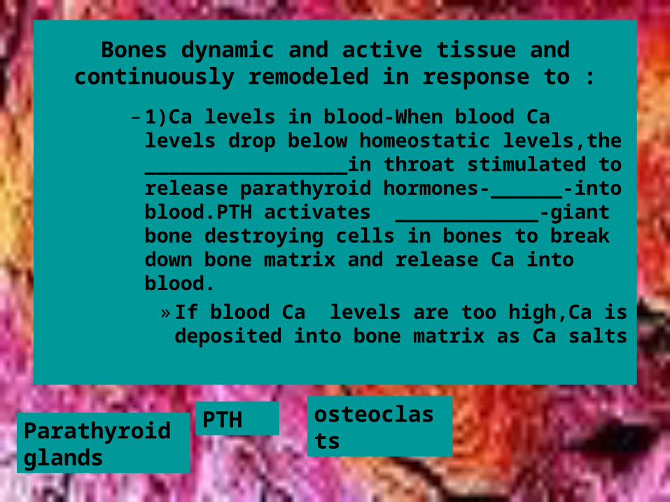
Bones dynamic and active tissue and continuously remodeled in response to :
– 1)Ca levels in blood-When blood Ca levels drop below homeostatic levels,the _________________in throat stimulated to release parathyroid hormones-______-into blood.PTH activates ____________-giant bone destroying cells in bones to break down bone matrix and release Ca into blood.
» If blood Ca levels are too high,Ca is deposited into bone matrix as Ca salts
Parathyroid glands
PTH osteoclasts
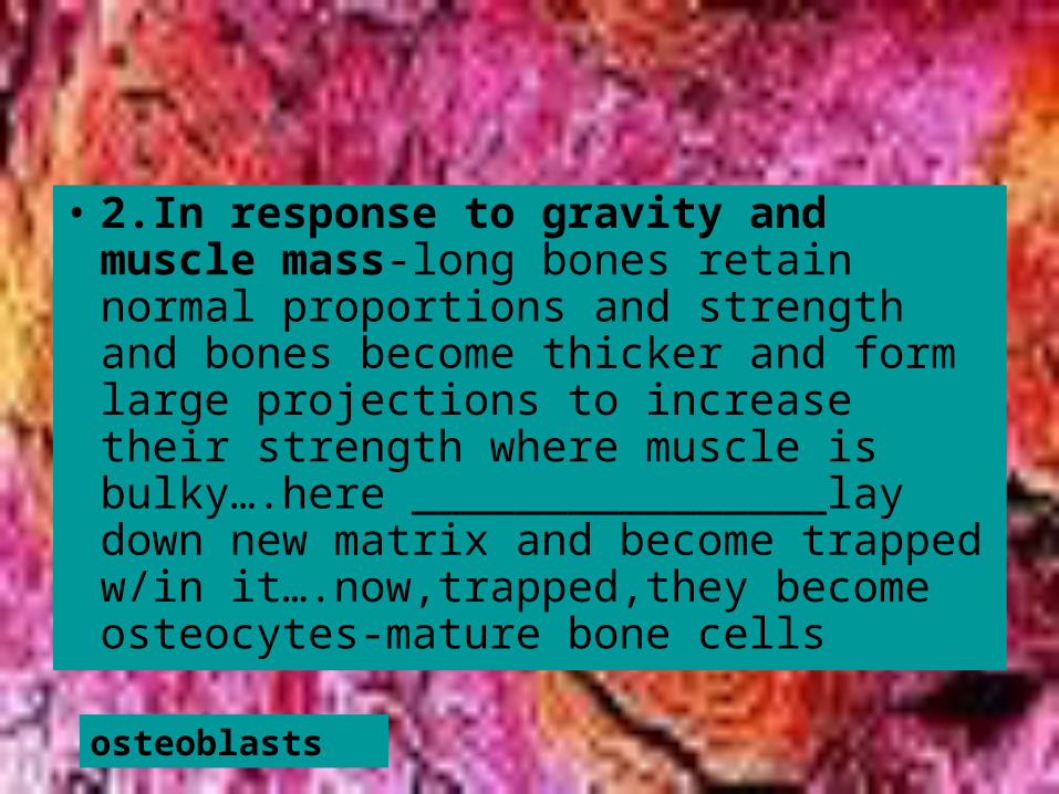
• 2.In response to gravity and muscle mass-long bones retain normal proportions and strength and bones become thicker and form large projections to increase their strength where muscle is bulky….here ________________lay down new matrix and become trapped w/in it….now,trapped,they become osteocytes-mature bone cells
osteoblasts
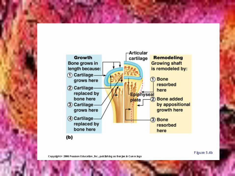
Copyright © 2006 Pearson Education, Inc., publishing as Benjamin Cummings
Long Bone Formation and Growth
Figure 5.4b
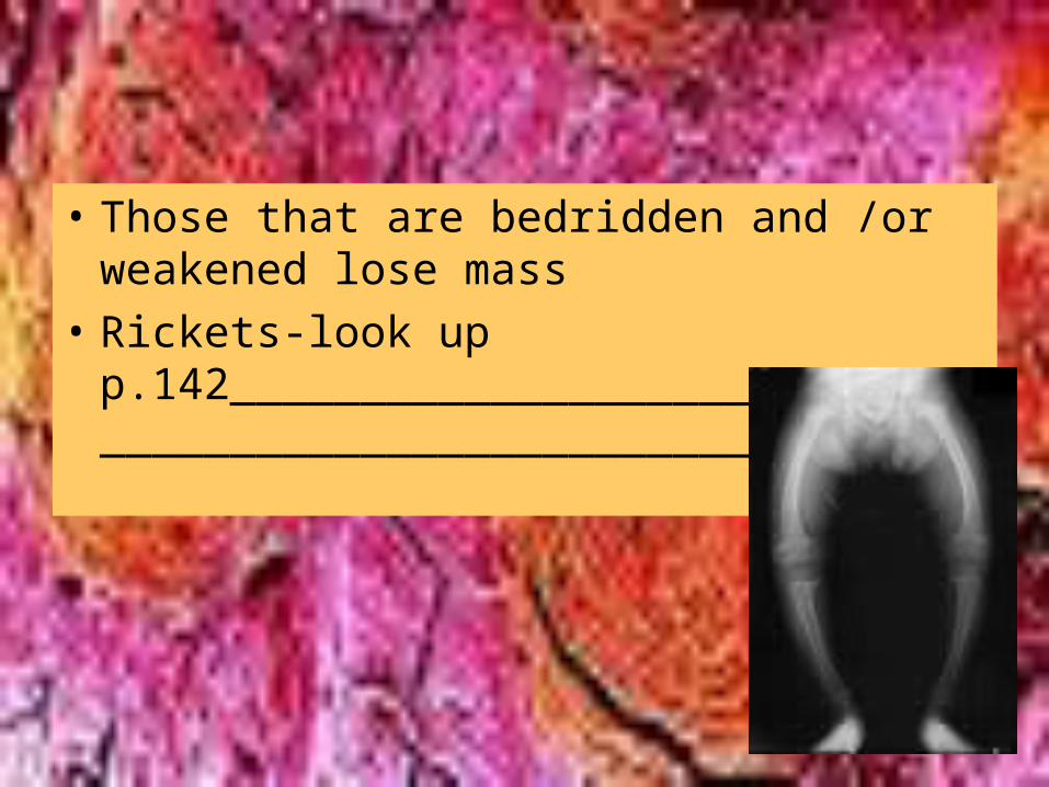
• Those that are bedridden and /or weakened lose mass
• Rickets-look up p.142______________________________________________________________
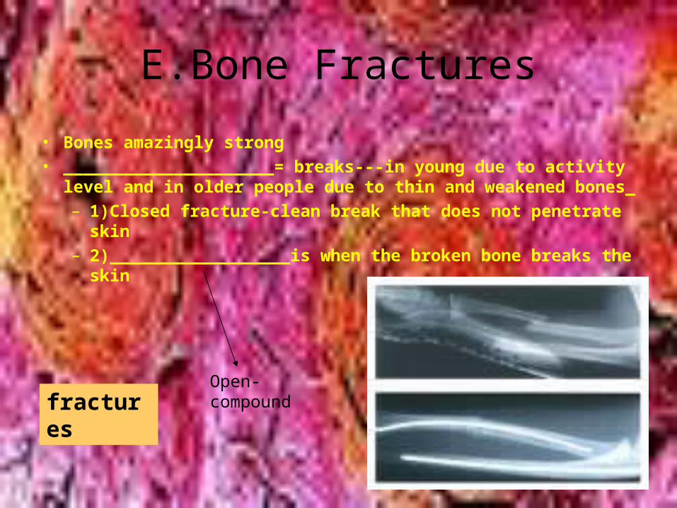
E.Bone Fractures
• Bones amazingly strong• _____________________= breaks---in young due to activity
level and in older people due to thin and weakened bones_– 1)Closed fracture-clean break that does not penetrate skin– 2)__________________is when the broken bone breaks the
skin
fracturesOpen-compound
Copyright © 2006 Pearson Education, Inc., publishing as Benjamin Cummings
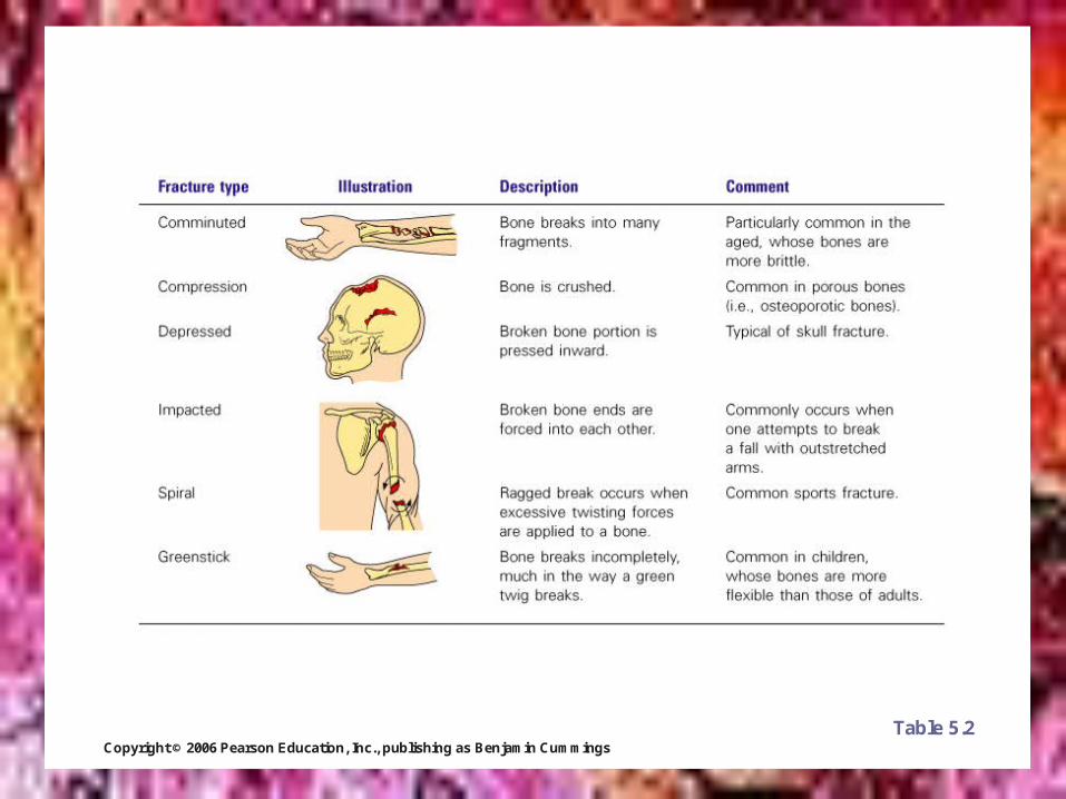
Common Types of Fractures
Table 5.2
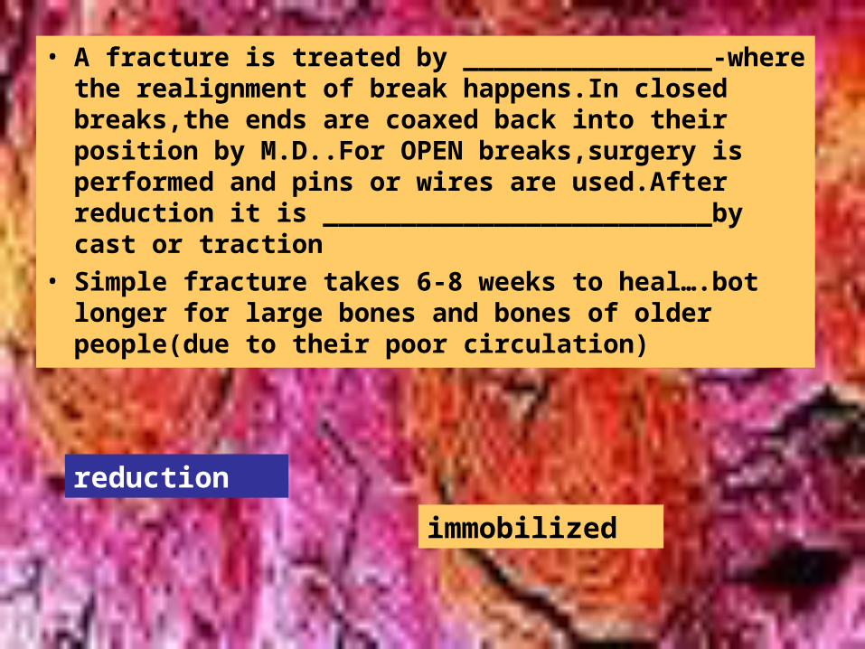
• A fracture is treated by ________________-where the realignment of break happens.In closed breaks,the ends are coaxed back into their position by M.D..For OPEN breaks,surgery is performed and pins or wires are used.After reduction it is _________________________by cast or traction
• Simple fracture takes 6-8 weeks to heal….bot longer for large bones and bones of older people(due to their poor circulation)
reduction
immobilized
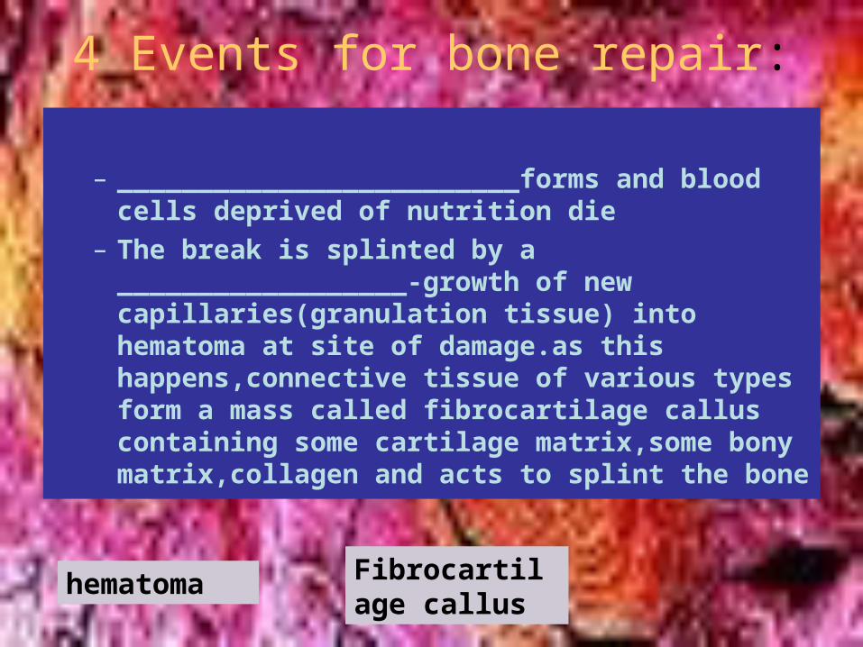
4 Events for bone repair:
– _________________________forms and blood cells deprived of nutrition die
– The break is splinted by a __________________-growth of new capillaries(granulation tissue) into hematoma at site of damage.as this happens,connective tissue of various types form a mass called fibrocartilage callus containing some cartilage matrix,some bony matrix,collagen and acts to splint the bone
hematoma Fibrocartilage callus
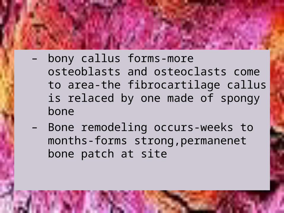
– bony callus forms-more osteoblasts and osteoclasts come to area-the fibrocartilage callus is relaced by one made of spongy bone
– Bone remodeling occurs-weeks to months-forms strong,permanenet bone patch at site
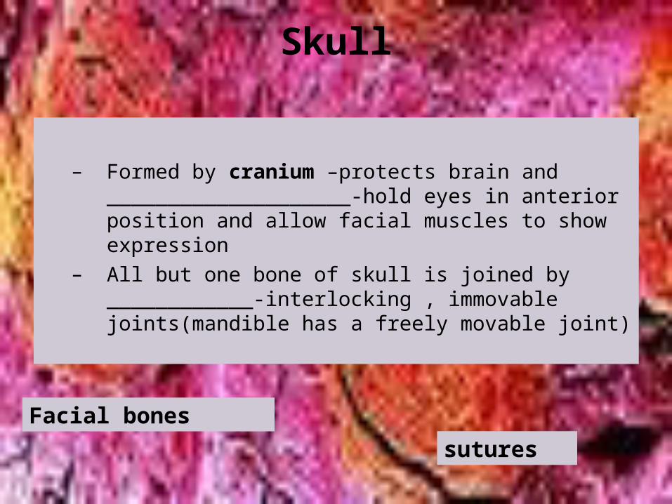
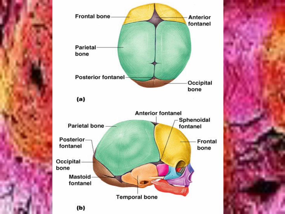
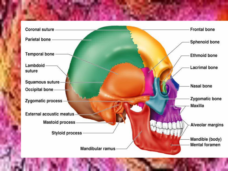
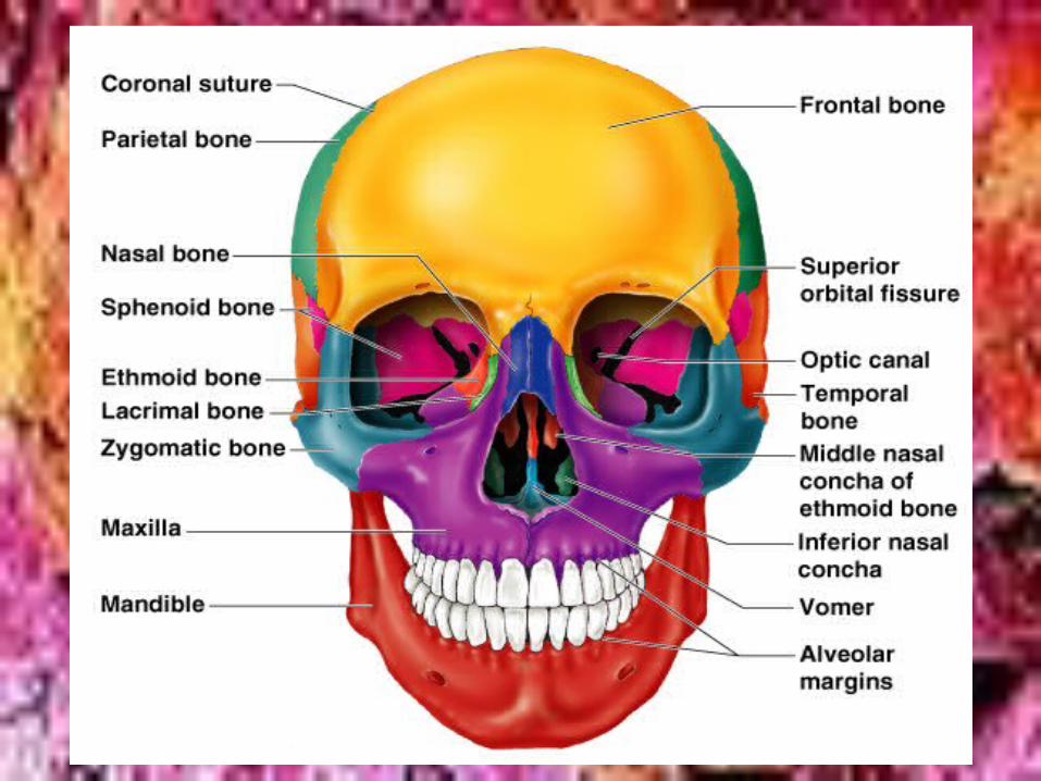
Skull
– Formed by cranium –protects brain and ____________________-hold eyes in anterior position and allow facial muscles to show expression
– All but one bone of skull is joined by ____________-interlocking , immovable joints(mandible has a freely movable joint)
Facial bones
sutures
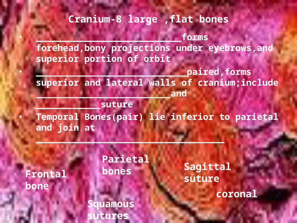
Cranium-8 large ,flat bones
• ___________________________forms forehead,bony projections under eyebrows,and superior portion of orbit
• ____________________________paired,forms superior and lateral walls of cranium;include _________________________and ____________suture
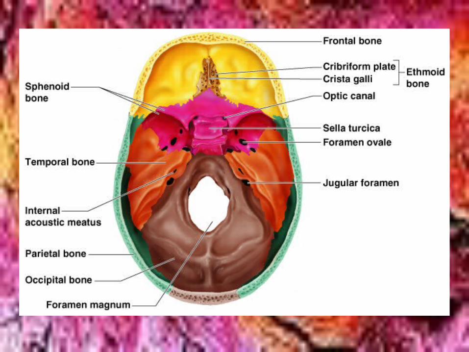
• Temporal Bones(pair) lie inferior to parietal and join at ___________________________________
Frontal bone
Parietal bones Sagittal
suture
coronalSquamous sutures
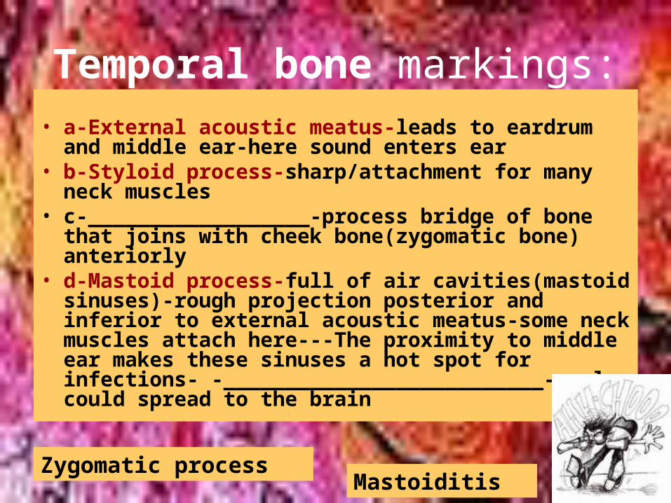
Temporal bone markings:
• a-External acoustic meatus-leads to eardrum and middle ear-here sound enters ear
• b-Styloid process-sharp/attachment for many neck muscles
• c-__________________-process bridge of bone that joins with cheek bone(zygomatic bone) anteriorly
• d-Mastoid process-full of air cavities(mastoid sinuses)-rough projection posterior and inferior to external acoustic meatus-some neck muscles attach here---The proximity to middle ear makes these sinuses a hot spot for infections- -__________________________---also could spread to the brain
Zygomatic processMastoiditis
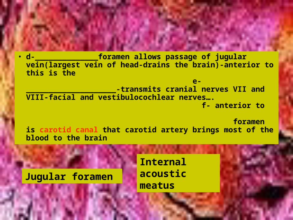
• d-______________foramen allows passage of jugular vein(largest vein of head-drains the brain)-anterior to this is the e-____________________-transmits cranial nerves VII and VIII-facial and vestibulocochlear nerves…. f- anterior to foramen is carotid canal that carotid artery brings most of the blood to the brain
Jugular foramen
Internal acoustic meatus
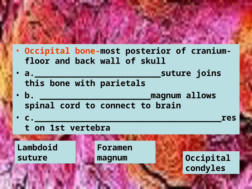
• Occipital bone-most posterior of cranium-floor and back wall of skull
• a._________________________suture joins this bone with parietals
• b. ______________________magnum allows spinal cord to connect to brain
• c._____________________________________rest on 1st vertebra
Lambdoid suture
Foramen magnum Occipital
condyles
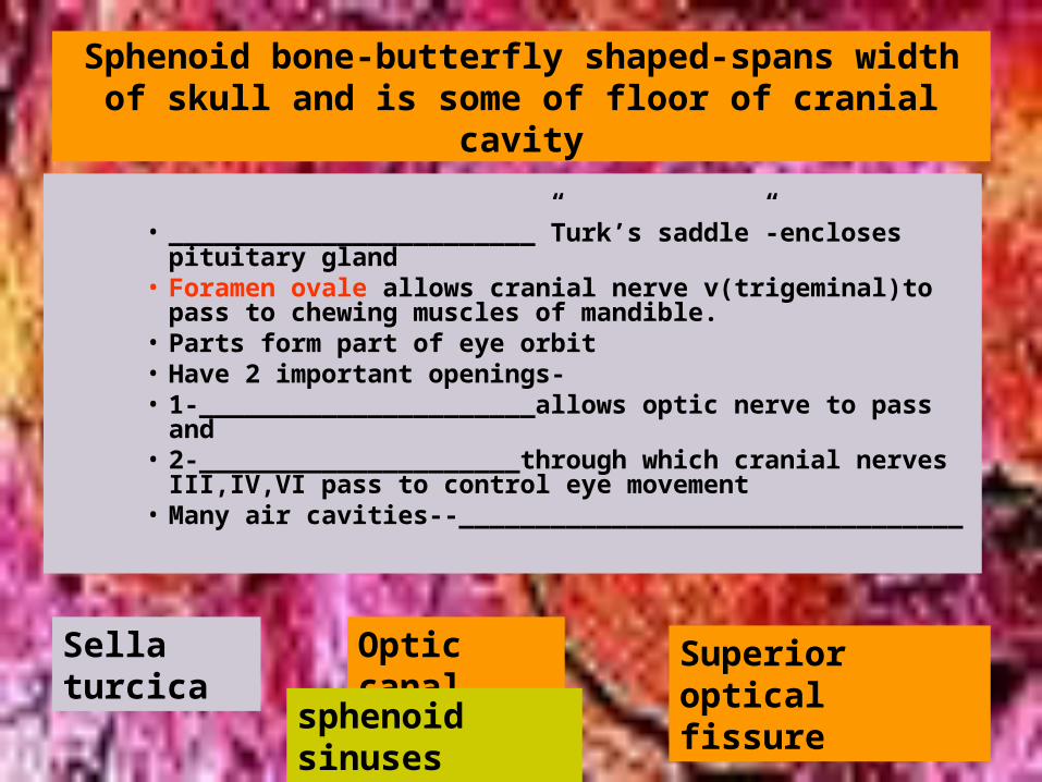
Sphenoid bone-butterfly shaped-spans width of skull and is some of floor of cranial cavity
• ________________________”Turk’s saddle”-encloses pituitary gland
• Foramen ovale allows cranial nerve v(trigeminal)to pass to chewing muscles of mandible.
• Parts form part of eye orbit• Have 2 important openings-• 1-______________________allows optic nerve to pass
and• 2-_____________________through which cranial nerves
III,IV,VI pass to control eye movement• Many air cavities--_________________________________
Sella turcica
Optic canal Superior optical fissure
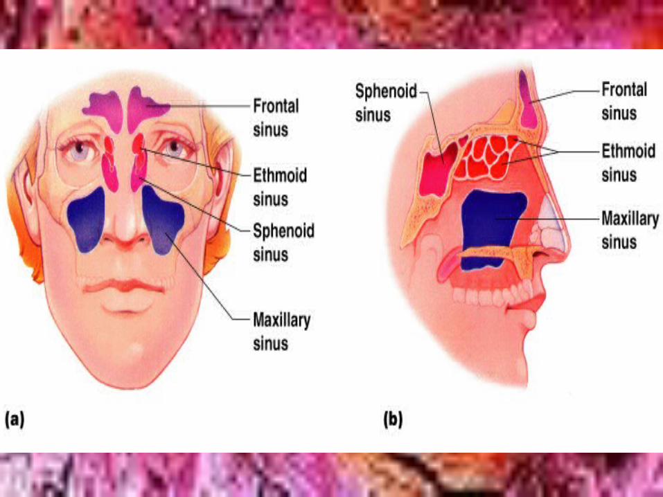
sphenoid sinuses
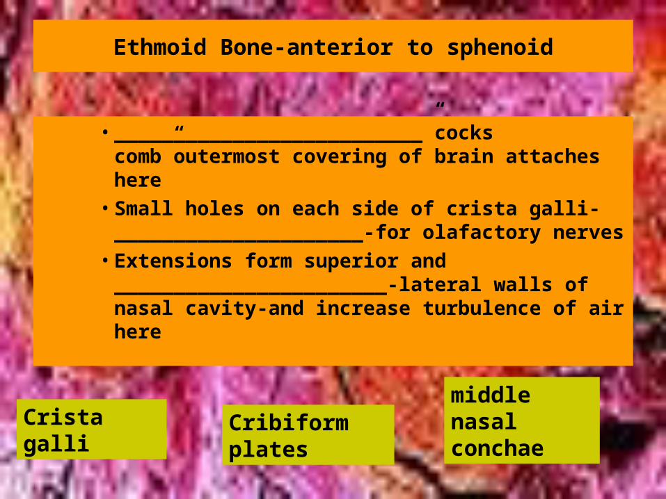
Ethmoid Bone-anterior to sphenoid
• __________________________”cocks comb”outermost covering of brain attaches here
• Small holes on each side of crista galli-_____________________-for olafactory nerves
• Extensions form superior and _______________________-lateral walls of nasal cavity-and increase turbulence of air here
Crista galli Cribiform plates
middle nasal conchae
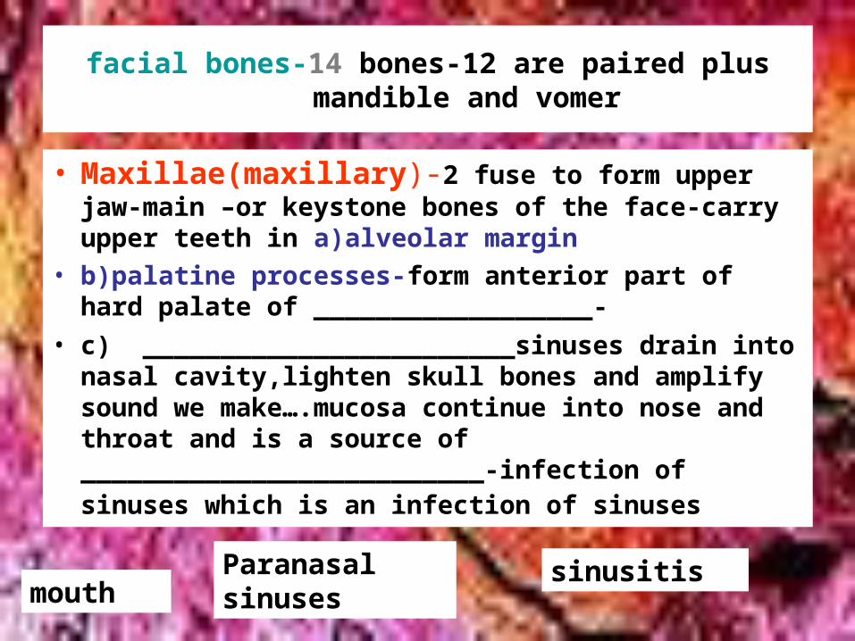
facial bones-14 bones-12 are paired plus mandible and vomer
• Maxillae(maxillary)-2 fuse to form upper jaw-main –or keystone bones of the face-carry upper teeth in a)alveolar margin
• b)palatine processes-form anterior part of hard palate of __________________-
• c) ________________________sinuses drain into nasal cavity,lighten skull bones and amplify sound we make….mucosa continue into nose and throat and is a source of __________________________-infection of sinuses which is an infection of sinuses
mouthParanasal sinuses
sinusitis
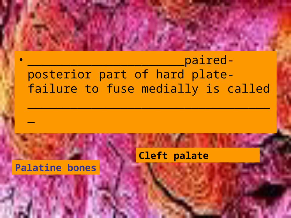
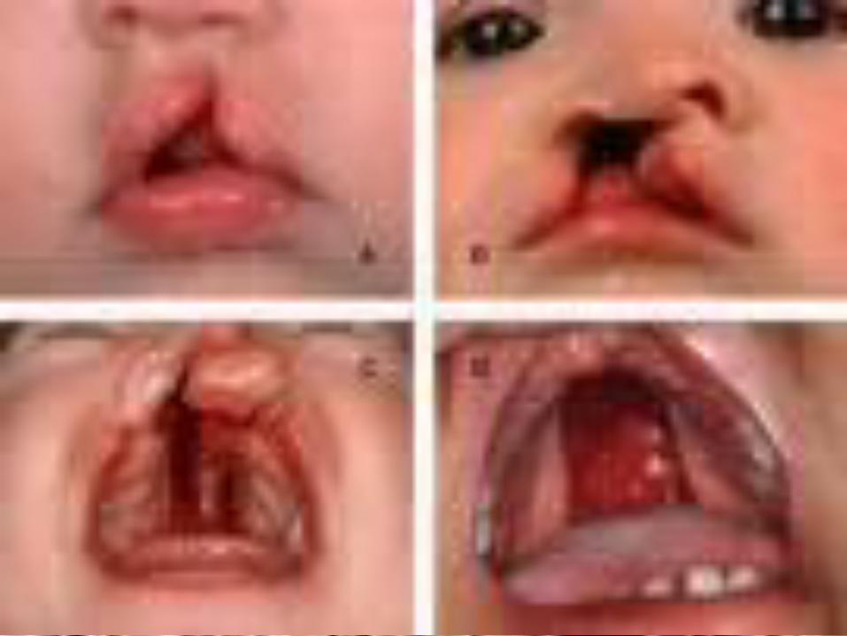
• ______________________paired-posterior part of hard plate-failure to fuse medially is called ___________________________________
Palatine bonesCleft palate
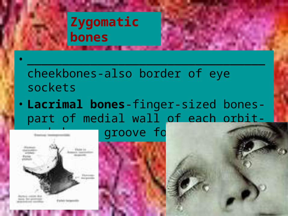
• __________________________________cheekbones-also border of eye sockets
• Lacrimal bones-finger-sized bones-part of medial wall of each orbit-each has a groove for tears
Zygomatic bones
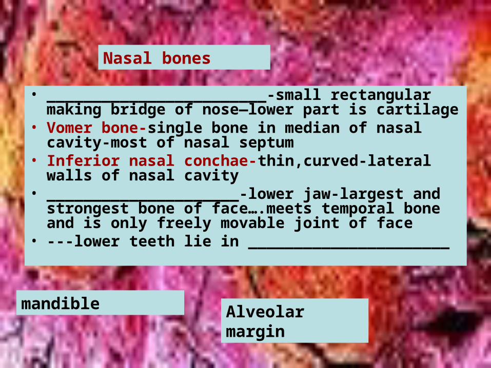
• ________________________-small rectangular making bridge of nose—lower part is cartilage
• Vomer bone-single bone in median of nasal cavity-most of nasal septum
• Inferior nasal conchae-thin,curved-lateral walls of nasal cavity
• _____________________-lower jaw-largest and strongest bone of face….meets temporal bone and is only freely movable joint of face
• ---lower teeth lie in ______________________
Nasal bones
mandible Alveolar margin
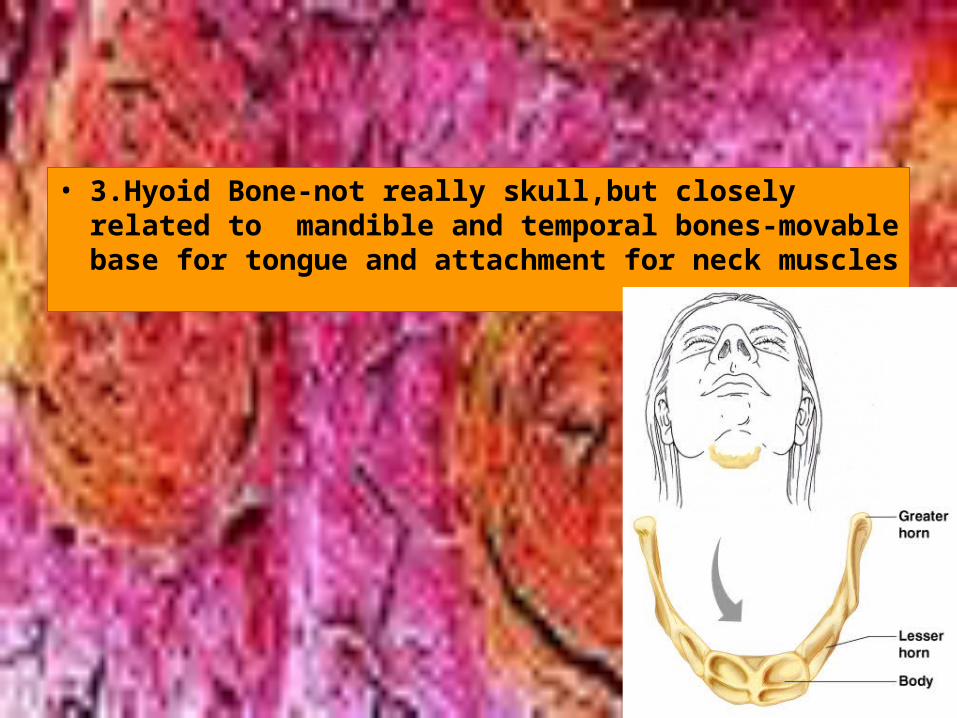
• 3.Hyoid Bone-not really skull,but closely related to mandible and temporal bones-movable base for tongue and attachment for neck muscles
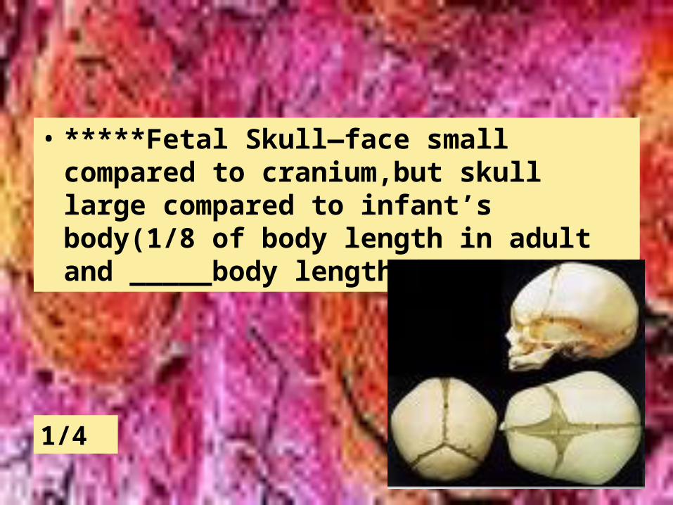
• *****Fetal Skull—face small compared to cranium,but skull large compared to infant’s body(1/8 of body length in adult and _____body length in infant
1/4
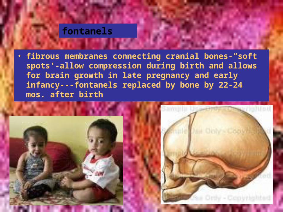
• fibrous membranes connecting cranial bones-“soft spots’-allow compression during birth and allows for brain growth in late pregnancy and early infancy---fontanels replaced by bone by 22-24 mos. after birth
fontanels
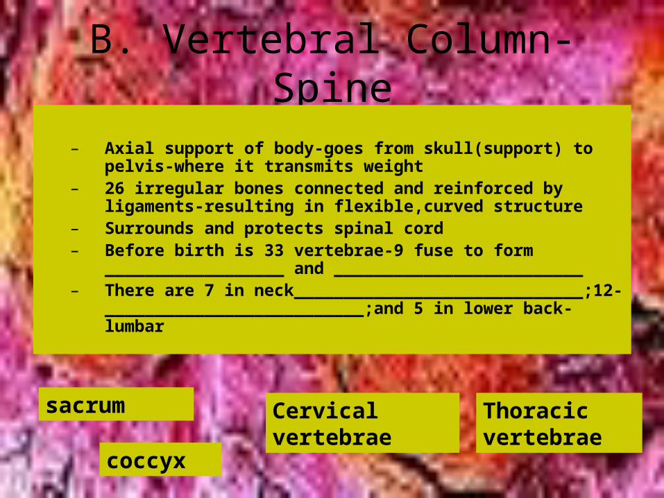
B. Vertebral Column-Spine
– Axial support of body-goes from skull(support) to pelvis-where it transmits weight
– 26 irregular bones connected and reinforced by ligaments-resulting in flexible,curved structure
– Surrounds and protects spinal cord– Before birth is 33 vertebrae-9 fuse to form
__________________ and _________________________– There are 7 in neck_____________________________;12-
__________________________;and 5 in lower back-lumbar
sacrum
coccyx
Cervical vertebrae
Thoracic vertebrae
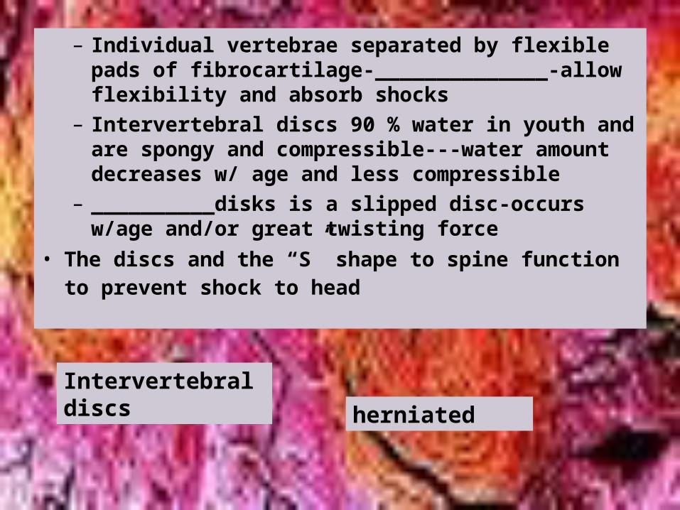
– Individual vertebrae separated by flexible pads of fibrocartilage-______________-allow flexibility and absorb shocks
– Intervertebral discs 90 % water in youth and are spongy and compressible---water amount decreases w/ age and less compressible
– __________disks is a slipped disc-occurs w/age and/or great twisting force
• The discs and the “S” shape to spine function to prevent shock to head
Intervertebral discs herniated
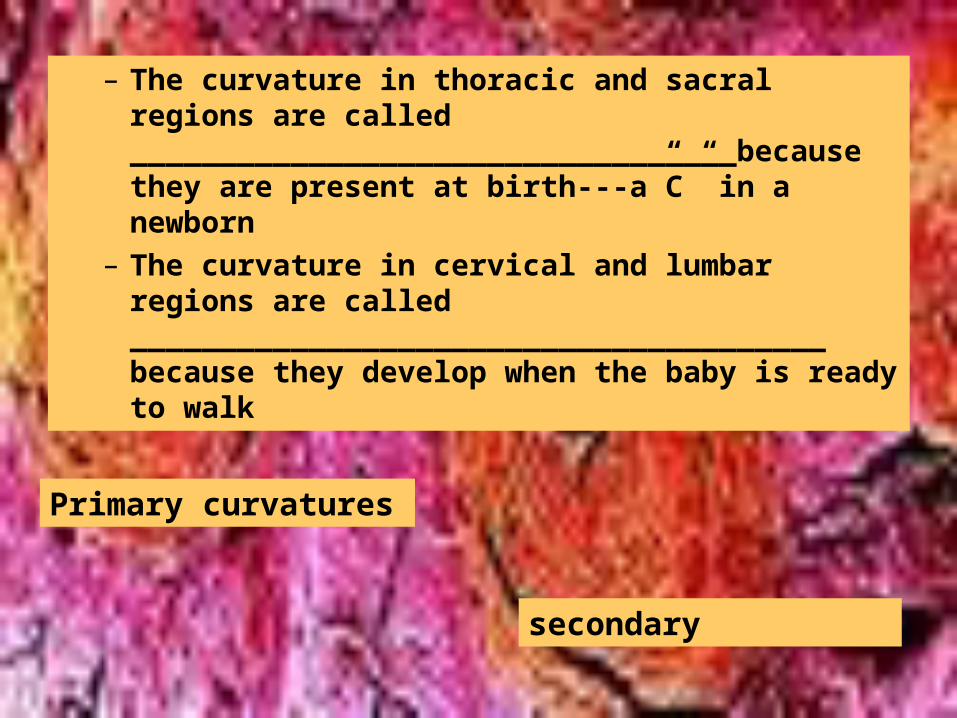
– The curvature in thoracic and sacral regions are called __________________________________because they are present at birth---a”C” in a newborn
– The curvature in cervical and lumbar regions are called _______________________________________ because they develop when the baby is ready to walk
Primary curvatures
secondary
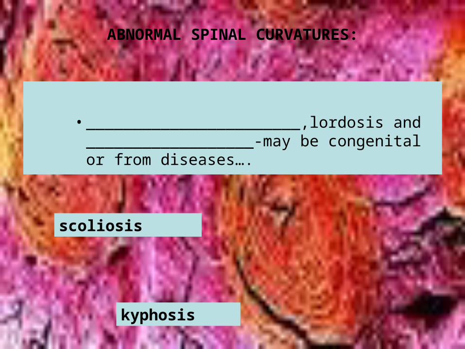
ABNORMAL SPINAL CURVATURES:
• _______________________,lordosis and __________________-may be congenital or from diseases….
scoliosis
kyphosis
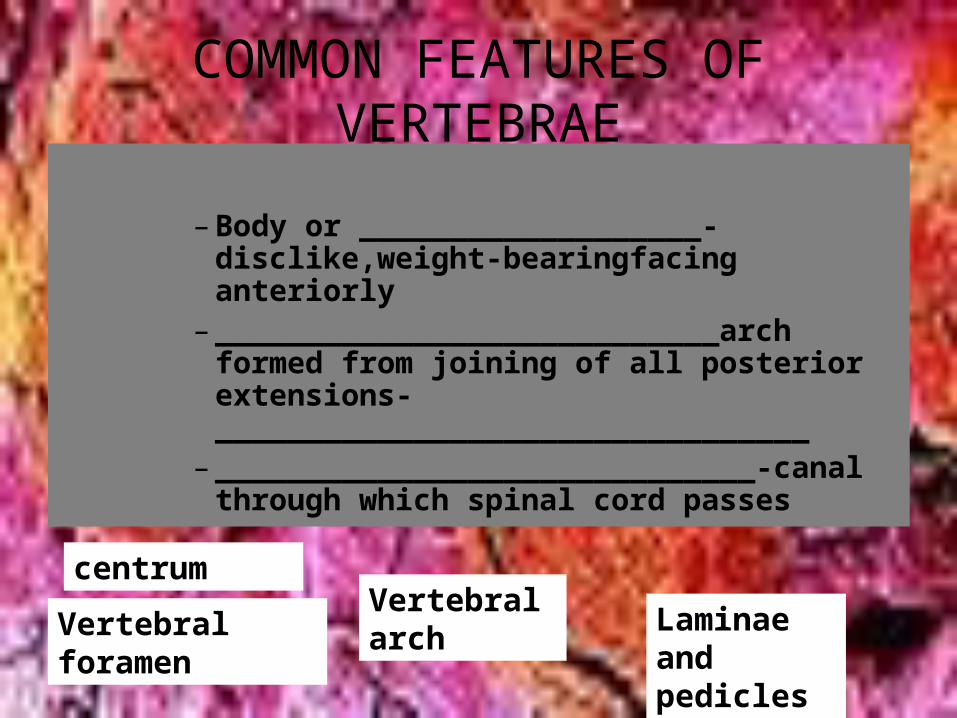
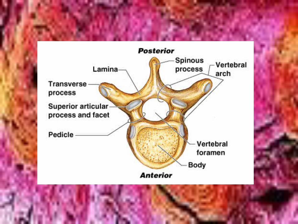
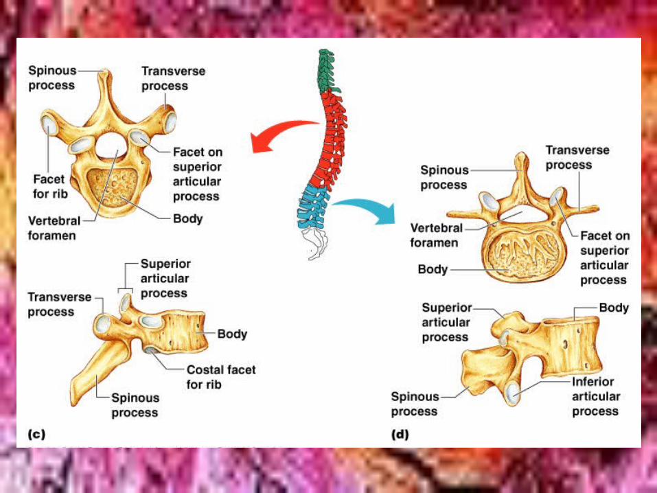
COMMON FEATURES OF VERTEBRAE
– Body or ___________________-disclike,weight-bearingfacing anteriorly
– ____________________________arch formed from joining of all posterior extensions-_________________________________
– ______________________________-canal through which spinal cord passes
centrumVertebral arch
Laminae and pedicles
Vertebral foramen
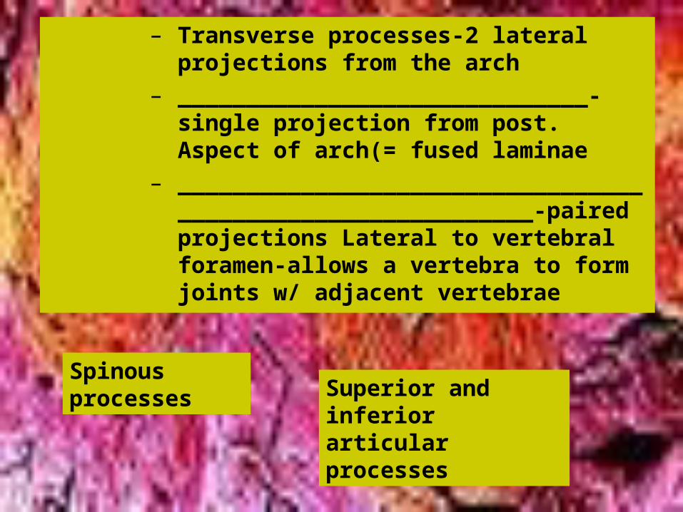
– Transverse processes-2 lateral projections from the arch
– ______________________________-single projection from post. Aspect of arch(= fused laminae
– ____________________________________________________________-paired projections Lateral to vertebral foramen-allows a vertebra to form joints w/ adjacent vertebrae
Spinous processes Superior and inferior
articular processes
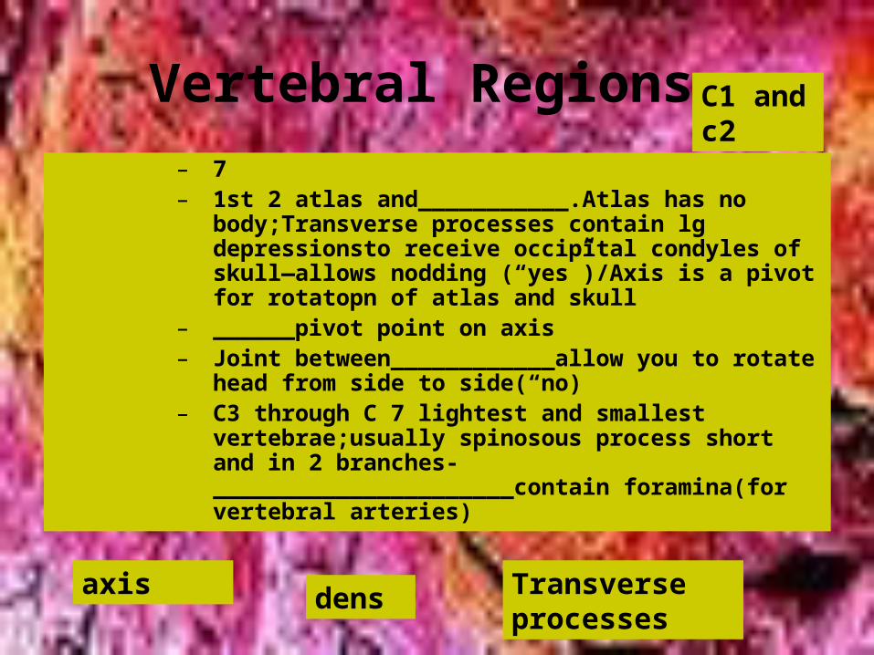
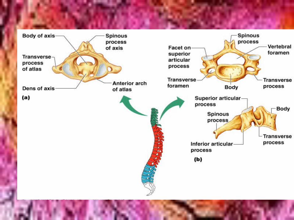
Vertebral Regions
– 7– 1st 2 atlas and___________.Atlas has no
body;Transverse processes contain lg depressionsto receive occipital condyles of skull—allows nodding (“yes”)/Axis is a pivot for rotatopn of atlas and skull
– ______pivot point on axis– Joint between____________allow you to rotate
head from side to side(“no)– C3 through C 7 lightest and smallest
vertebrae;usually spinosous process short and in 2 branches-______________________contain foramina(for vertebral arteries)
axis dens Transverse processes
C1 and c2
THORACIC-
• 12(T1-T12)-all typical• Only vertebrae to articulate with ribs• Body somewhat heart –shaped and has 2
costal facets(articulating surfaces0 on ea. Side receiving heads of robs
• Transverse processes articulate w/knoblike tubercles of ribs
• Spinosous process long
Lumbar(L1-L5)
» Massive blocklike bodies;short spinous processes (“moose head”)
» Sturdiest vertebrae because receives most stress
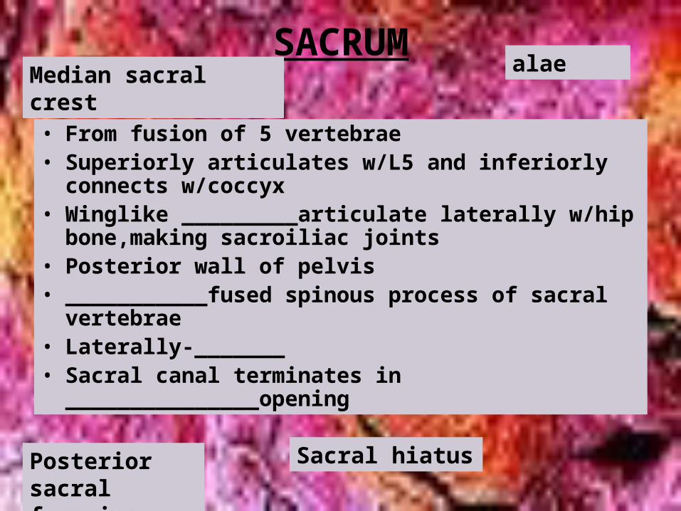
SACRUM
• From fusion of 5 vertebrae• Superiorly articulates w/L5 and inferiorly connects
w/coccyx• Winglike _________articulate laterally w/hip
bone,making sacroiliac joints• Posterior wall of pelvis• ___________fused spinous process of sacral
vertebrae• Laterally-_______• Sacral canal terminates in _______________opening
alaeMedian sacral crest
Posterior sacral foramina
Sacral hiatus
COCCYX:
• Fusion of 3-5 tiny vertebrae
• Tailbone

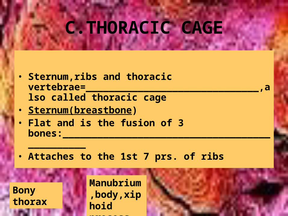
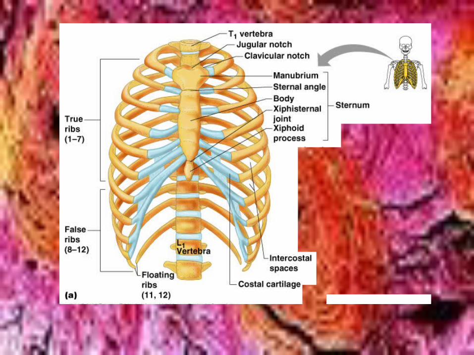
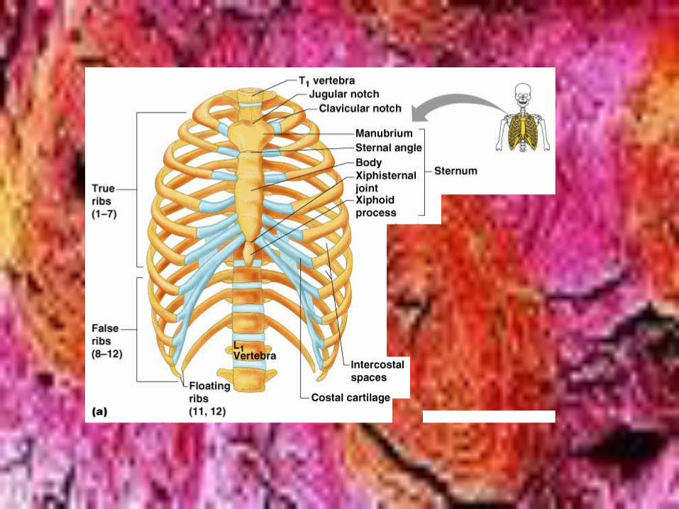
C.THORACIC CAGE
• Sternum,ribs and thoracic vertebrae=______________________________,also called thoracic cage
• Sternum(breastbone)• Flat and is the fusion of 3
bones:______________________________________________
• Attaches to the 1st 7 prs. of ribs
Bony thorax
Manubrium,body,xiphoid process
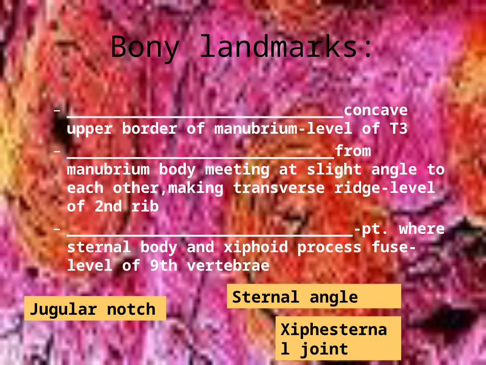
Bony landmarks:
– ______________________________concave upper border of manubrium-level of T3
– _____________________________from manubrium body meeting at slight angle to each other,making transverse ridge-level of 2nd rib
– _______________________________-pt. where sternal body and xiphoid process fuse-level of 9th vertebrae
Jugular notchSternal angle
Xiphesternal joint
• _____________________________taking marrow sample at sternum for diagnoses of blood diseases
Sternal puncture
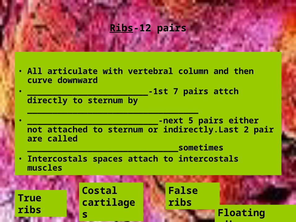
Ribs-12 pairs
• All articulate with vertebral column and then curve downward
• ________________________-1st 7 pairs attch directly to sternum by __________________________________
• __________________________-next 5 pairs either not attached to sternum or indirectly.Last 2 pair are called ______________________________sometimes
• Intercostals spaces attach to intercostals muscles
True ribsCostal cartilages
False ribs
Floating ribs
• III.Appendicular Skeleton
• 126 bones of limbs and pectoral or pelvic girdle(which attach limbs to axial skeleton)
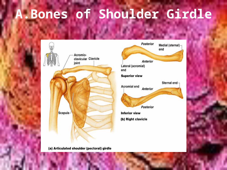
A.Bones of Shoulder Girdle
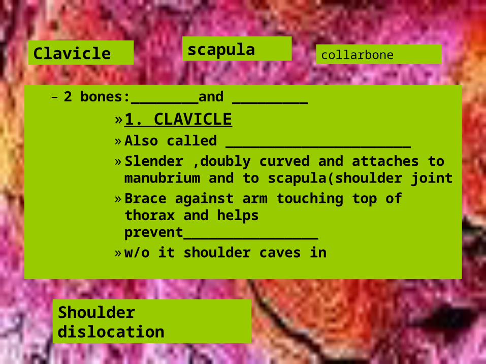
– 2 bones:________and _________
»1. CLAVICLE» Also called ______________________» Slender ,doubly curved and attaches to
manubrium and to scapula(shoulder joint» Brace against arm touching top of thorax
and helps prevent________________» w/o it shoulder caves in
Clavicle scapula collarbone
Shoulder dislocation
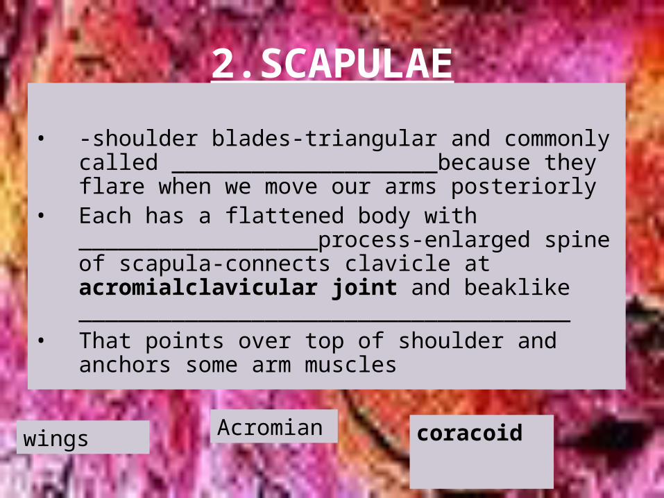
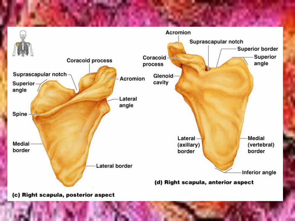
2.SCAPULAE
• -shoulder blades-triangular and commonly called ____________________because they flare when we move our arms posteriorly
• Each has a flattened body with __________________process-enlarged spine of scapula-connects clavicle at acromialclavicular joint and beaklike _____________________________________
• That points over top of shoulder and anchors some arm muscles
wings Acromian coracoid
• ____________________________-serves as nerve passageway
• Scapula loosely held by trunk muscles• Scapula has 3 borders-_________________________
and 3 angles:superior,inferior and lateral• ___________________-shallow socket receives head of
arm bone-in lateral angle
Suprascapular notch
Superior,medial(vere-bral) and lateral(axiallary)
Glenoid cavity
Shoulder girdle is light and allows upper limb free movement due to 3 factors:
– each shoulder girdle attaches at one point to axial skeleton-________________________________
– loose attachment of scapula allows it to slide back and forth v. thorax
– ___________________________is shallow and shoulder joint is poorly reinforced by ligaments
• *****drawback to so much flexibility is _________________
Sternoclavicular joint Glenoid cavity
dislocation
B.Bones of the Upper Limbs-30 bones-arms,forearms and hands
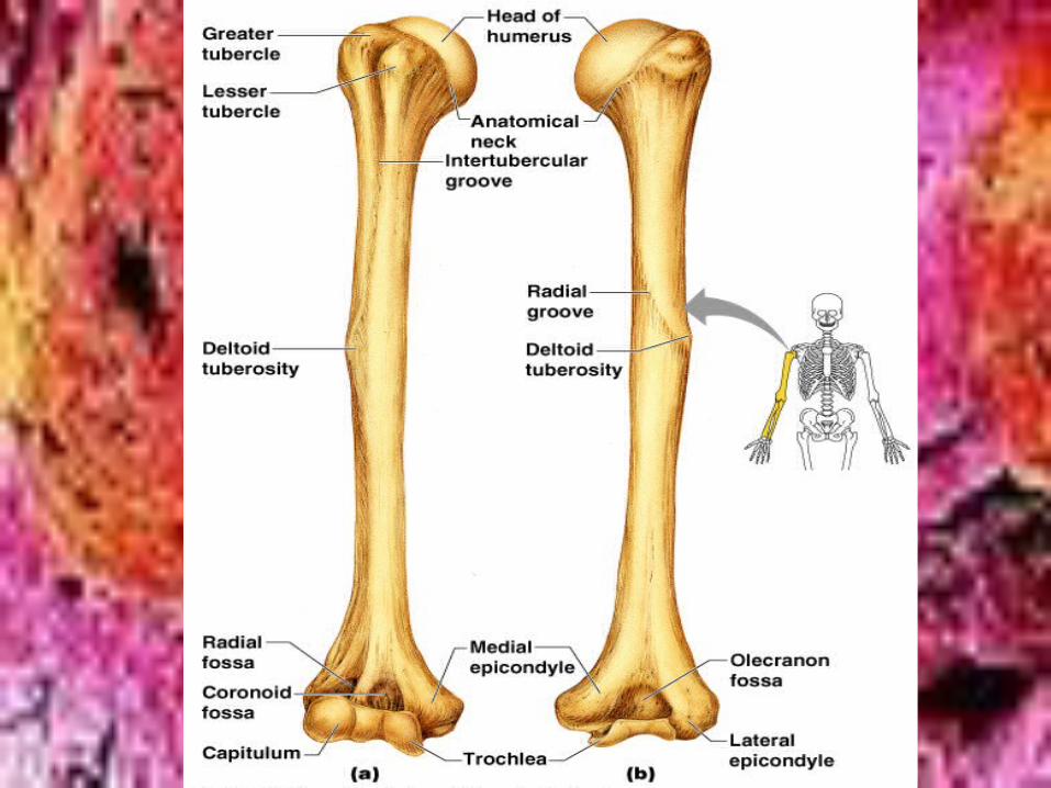
• ARM• Humerus-head fits into glenoid
cavity,_________________________;2 bony projections separated by intertubercular sulcus and ______________________-sites of muscle attachment;also ____________________________-most frequently fractured part of bone;___________________________roughened area in mid-shaft-deltoid muscle attaches
Anatomical neck
Greater and lesser tubercles
Surgical neck Deltoid tuberosity
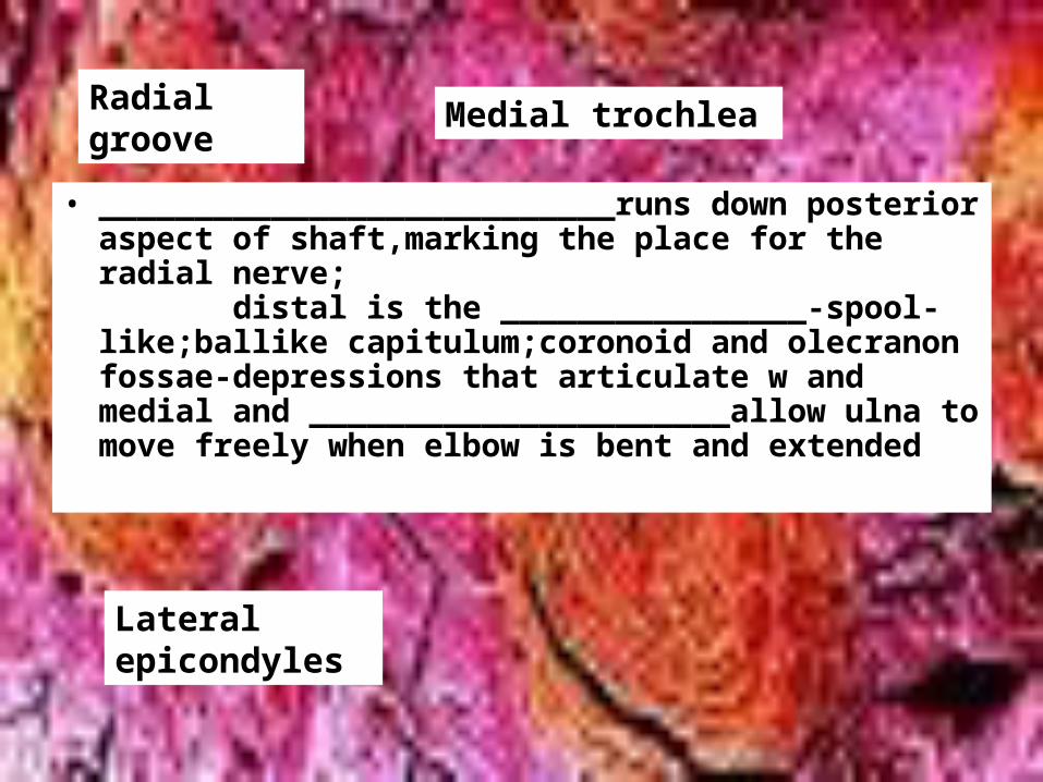
• ___________________________runs down posterior aspect of shaft,marking the place for the radial nerve; distal is the ________________-spool-like;ballike capitulum;coronoid and olecranon fossae-depressions that articulate w and medial and ______________________allow ulna to move freely when elbow is bent and extended
Radial groove
Medial trochlea
Lateral epicondyles
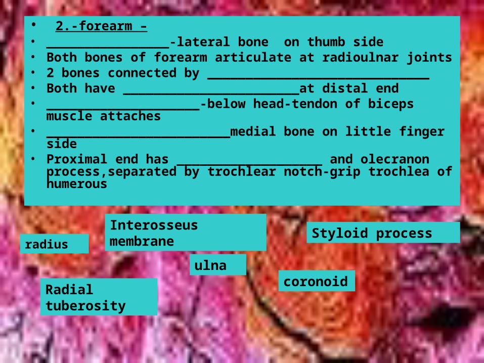
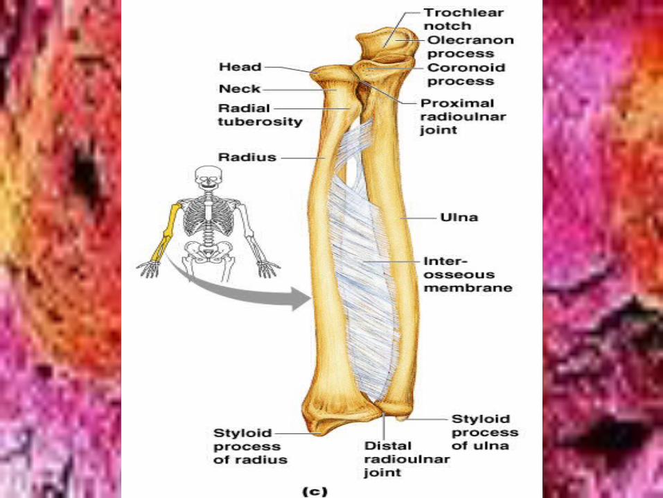
• 2.-forearm –• ________________-lateral bone on thumb side• Both bones of forearm articulate at radioulnar joints• 2 bones connected by _____________________________• Both have _______________________at distal end• ____________________-below head-tendon of biceps muscle
attaches• ________________________medial bone on little finger side• Proximal end has ___________________ and olecranon
process,separated by trochlear notch-grip trochlea of humerous
radius
Interosseus membraneStyloid process
Radial tuberosity
ulnacoronoid
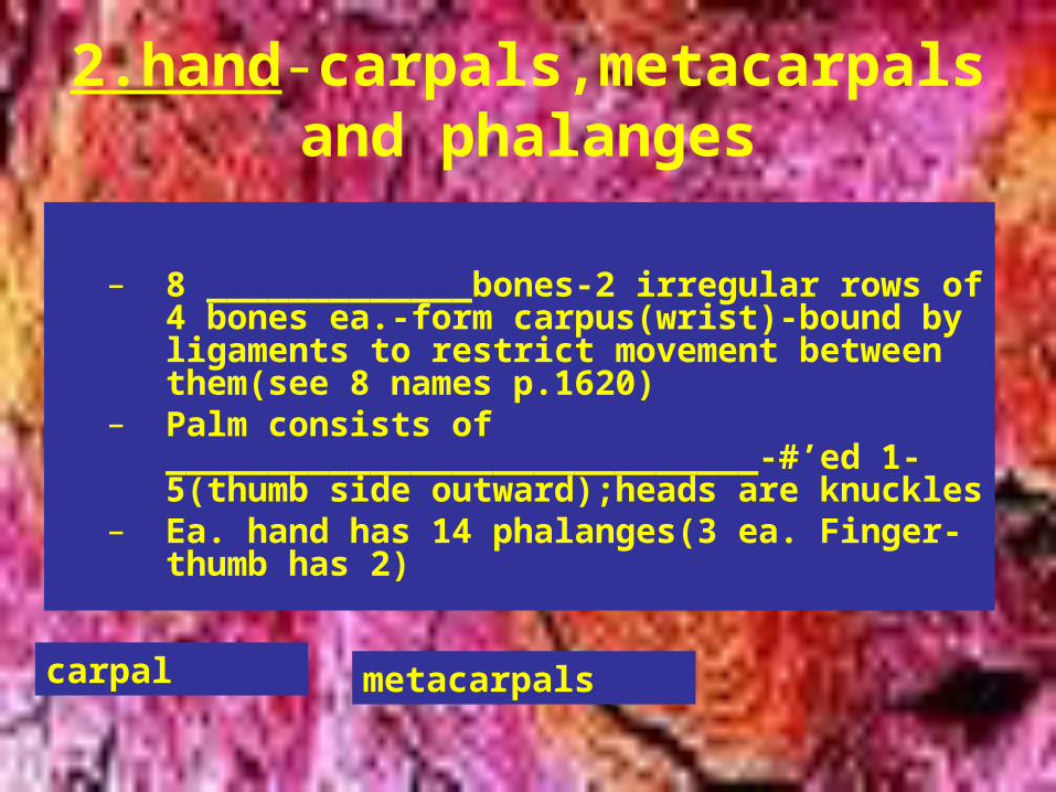
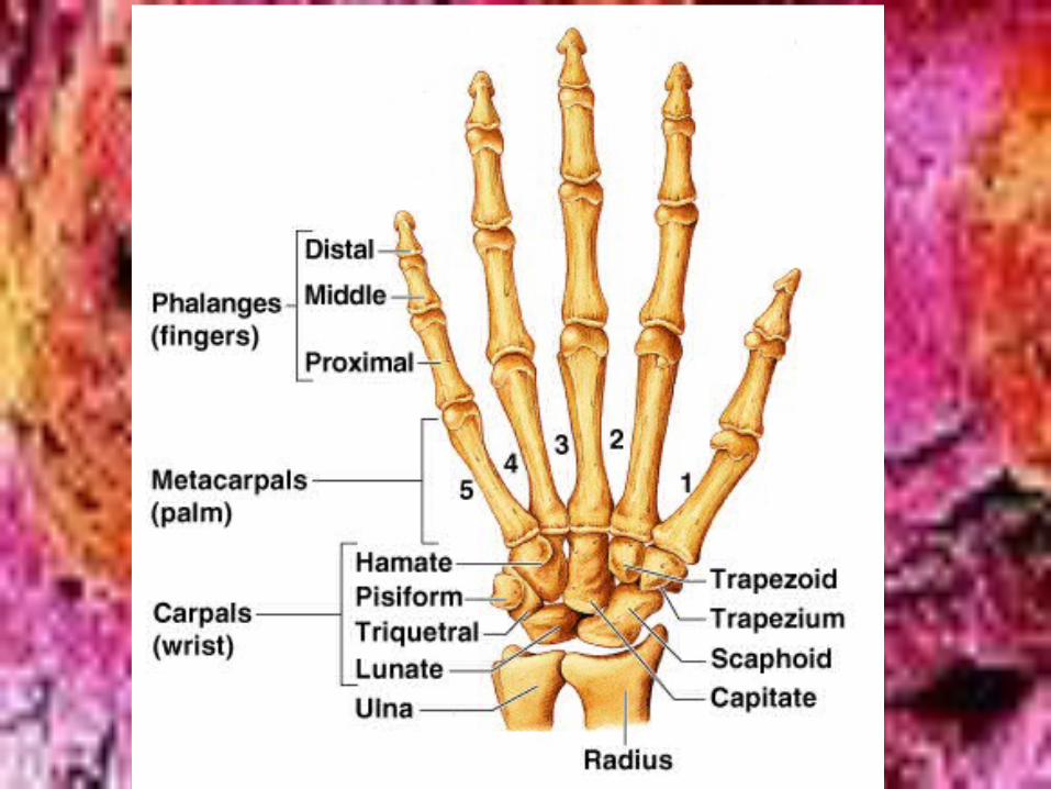
2.hand-carpals,metacarpals and phalanges
– 8 _____________bones-2 irregular rows of 4 bones ea.-form carpus(wrist)-bound by ligaments to restrict movement between them(see 8 names p.1620)
– Palm consists of _____________________________-#’ed 1-5(thumb side outward);heads are knuckles
– Ea. hand has 14 phalanges(3 ea. Finger-thumb has 2)
carpal metacarpals
C.bones of the Pelvic Girdle-
• formed by 2 coxal(_____________________)-hip bones;form bony pelvis w/sacrum and coccyx
• bony pelvis=2 coxal bones,sacrum and coccyx;whereas pelvic girdle=2coxal bones
• large and heavy and attached securely to ___________________________
• sockets deep and securely attached by ligaments• most important function is
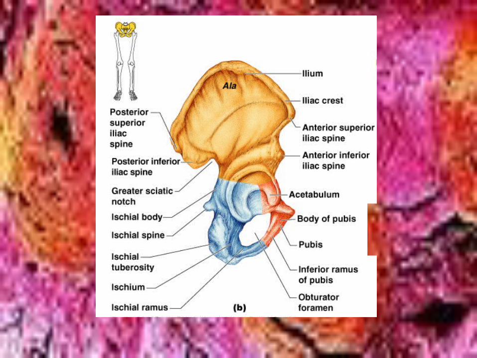
____________________________ and protect organs• each hip bone formed by fusion of 3
bones:____________________
Ossa coxae Axial skeleton
Bearing weight Ilium,ischium and pubis
• ilium -connects posteriorly w/ ____________________joint-forms most of hip bone;alae are winglike portions of ilia-upper portion= _____________________ and ends at anterior superior iliac spine and posteriorly w/posterior iliac spine
Sacroiliac joint Iliac crest
• ischium-_______________________-inferior of coxal bone;__________________________roughened area receiving weight when sitting;ischial spine-superior to tuberosity-THIS NARROWS THE OUTLET OF PELVIS FOR PASSAGE OF BABY IN CHILDBIRTH !
• ----____________________________allows blood vessels and lg sciatic nerve to pass from pelvis posteriorly into thigh---injections must stay clear of this area
Sit-down bone Ischial tuberosity
Greater sciatic notch
• _______________________most anterior of coxal;__________________________________-opening that allows blood vessels and nerves to pass to ant. Thigh;each pubis joins to form cartilaginous joint-__________________________________
• Ilium,ischium and pubis fuse at deep socket:__________________________________-“vinegar cup”-receives head of thigh bone
pubis Obturator foramen
Pubic symphysis acetabulum
• Bony pelvis divided into false and true pelvis-which must large enough to allow passage of infants head in women for childbirth-_______________________measured by obgyn
Dimensions of cavity
• Individual pelvic measurements vary,but stark difference in male and female
– Female inlet larger and more circular– Fem. pelvis as a whole more shallow and
bones lighter and thinner– Fem ilia flare more laterally– Her sacrum shorter and less curved– Her ischial spines shorter and farther apart-
thus outlet larger
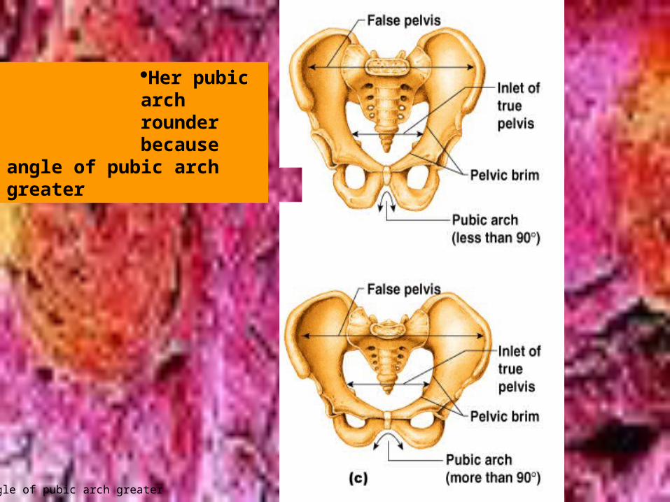
Her pubic arch rounder because
angle of pubic arch greater
angle of pubic arch greater
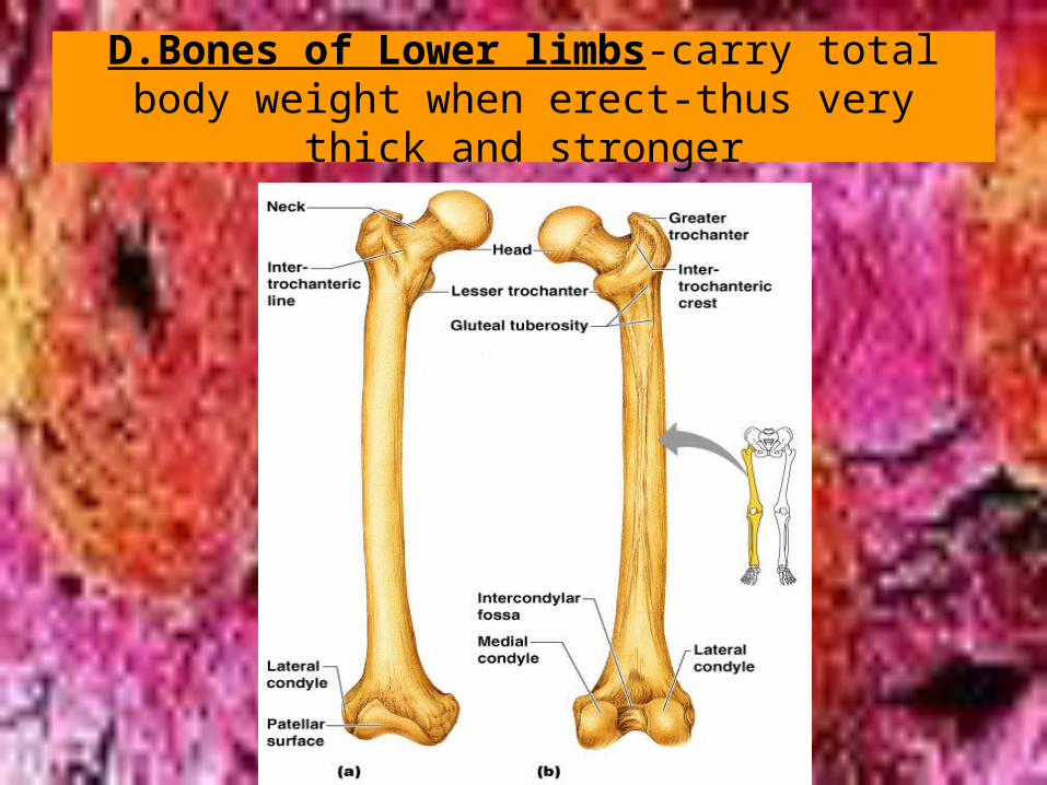
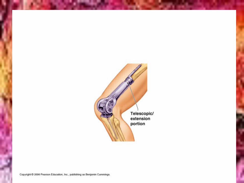
D.Bones of Lower limbs-carry total body weight when erect-thus very thick and stronger
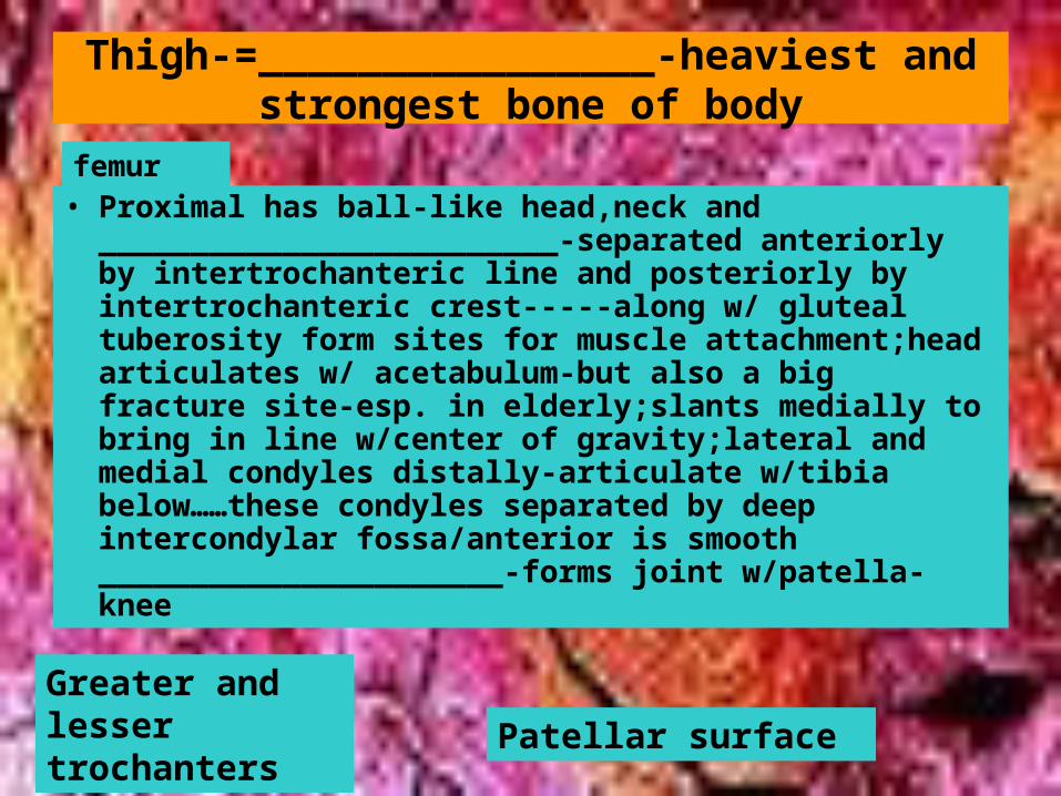
Thigh-=________________-heaviest and strongest bone of body
• Proximal has ball-like head,neck and _________________________-separated anteriorly by intertrochanteric line and posteriorly by intertrochanteric crest-----along w/ gluteal tuberosity form sites for muscle attachment;head articulates w/ acetabulum-but also a big fracture site-esp. in elderly;slants medially to bring in line w/center of gravity;lateral and medial condyles distally-articulate w/tibia below……these condyles separated by deep intercondylar fossa/anterior is smooth ______________________-forms joint w/patella-knee
Greater and lesser trochanters
Patellar surface
femur
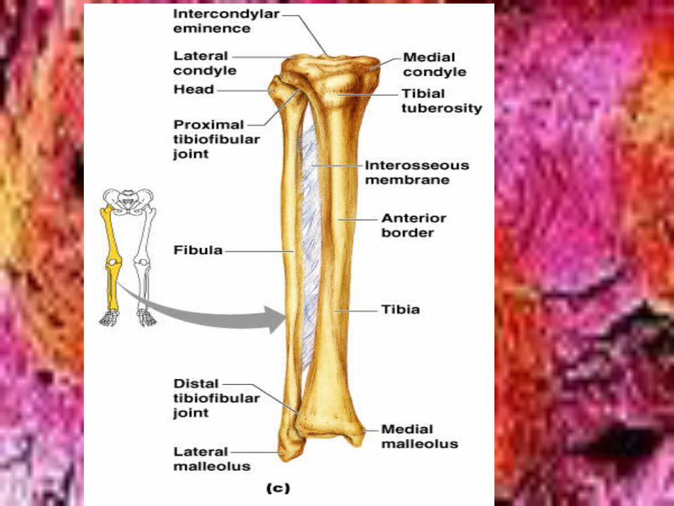
Leg
» -tibia and fibula connected by _______________________-tibia is shin bone-condyles proximally and articulate w/distal femur to make ________joint-patellar ligament attaches to _____________________-distally_____________________forms inner bulge of ankle-ant. tibia is sharp-anterior border for muscles
» ________________-lies alongside tibia and joints proximally and distally-NO PART Of KNEE JOINT-lateral malleolus outer part of ankle
Interosseus membrane
knee
Tibial tuberosity
Medial malleolus
fibula
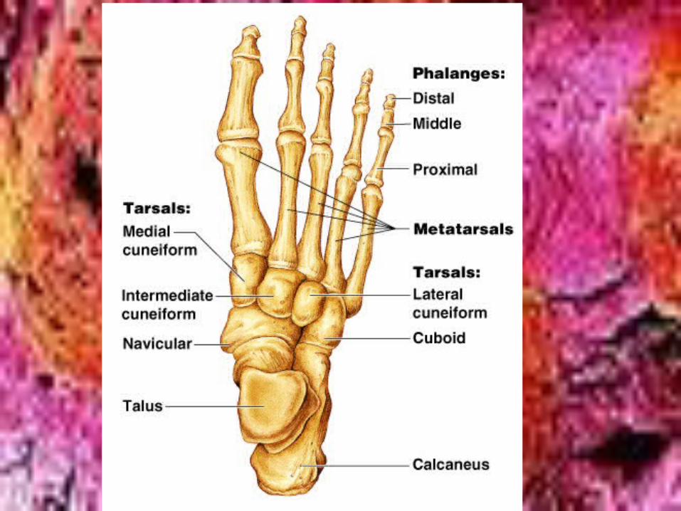
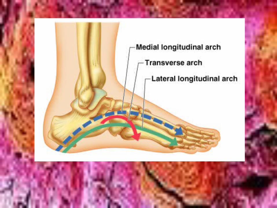
FOOT
» Tarsals,metatarsals and phalanges» 2 most important functions__________» Tarsus-post. 1/2 of foot-7 tarsal bones» ________________-heelbone» ______________lies between tibia and
calcaneus» 5 metatarsals and 14 phalanges-each
toe has 3 phalanges and big toe has 2» Form 3 strong arches-bound by
ligaments and tendons(for muscles)-fallen arches or flat feet occur
Support weight and act as lever
calcaneus
talus
• IV.Joints= articulations
• *****Except for hyoid,all bones form joints with at least 1 other bone
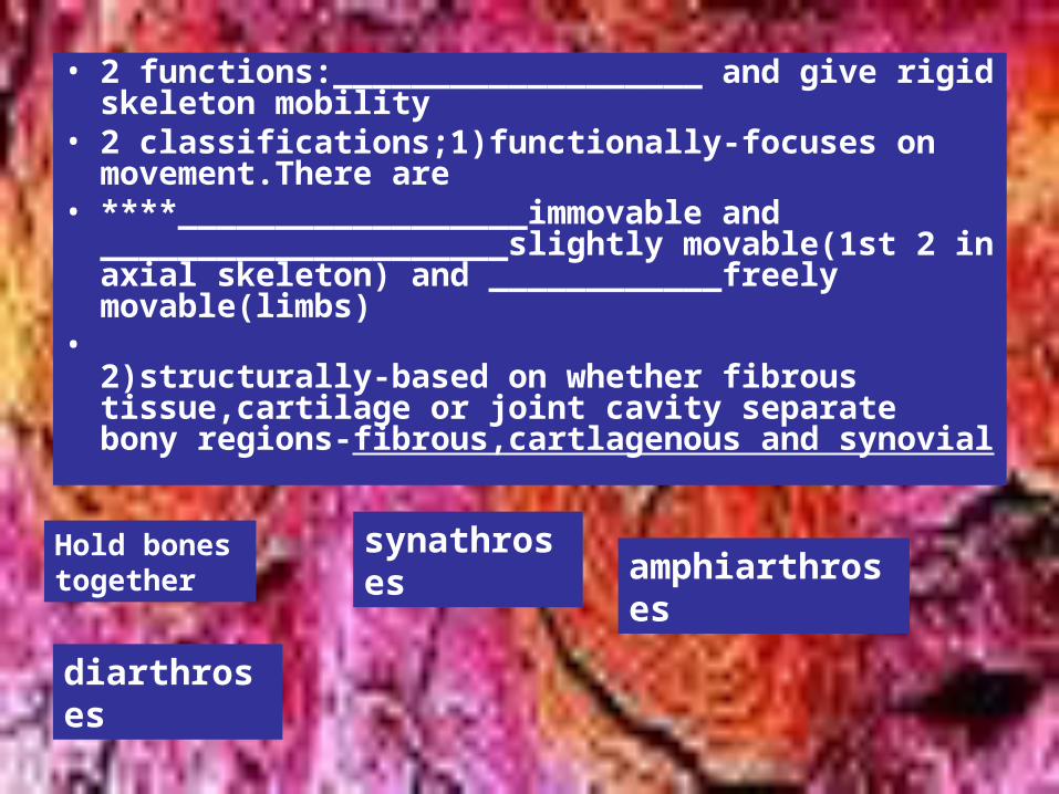
• 2 functions:___________________ and give rigid skeleton mobility
• 2 classifications;1)functionally-focuses on movement.There are
• ****__________________immovable and _____________________slightly movable(1st 2 in axial skeleton) and ____________freely movable(limbs)
• 2)structurally-based on whether fibrous tissue,cartilage or joint cavity separate bony regions-fibrous,cartlagenous and synovial
Hold bones together
synathrosesamphiarthroses
diarthroses
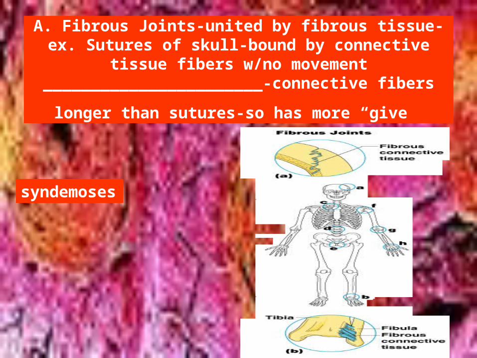
A. Fibrous Joints-united by fibrous tissue-ex. Sutures of skull-bound by connective tissue fibers w/no
movement_______________________-connective fibers longer
than sutures-so has more “give
syndemoses
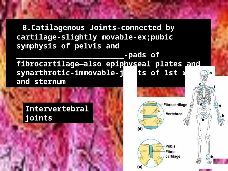
• B.Catilagenous Joints-connected by cartilage-slightly movable-ex;pubic symphysis of pelvis and ________________________-pads of fibrocartilage—also epiphyseal plates and synarthrotic-immovable-joints of 1st ribs and sternum
Intervertebral joints
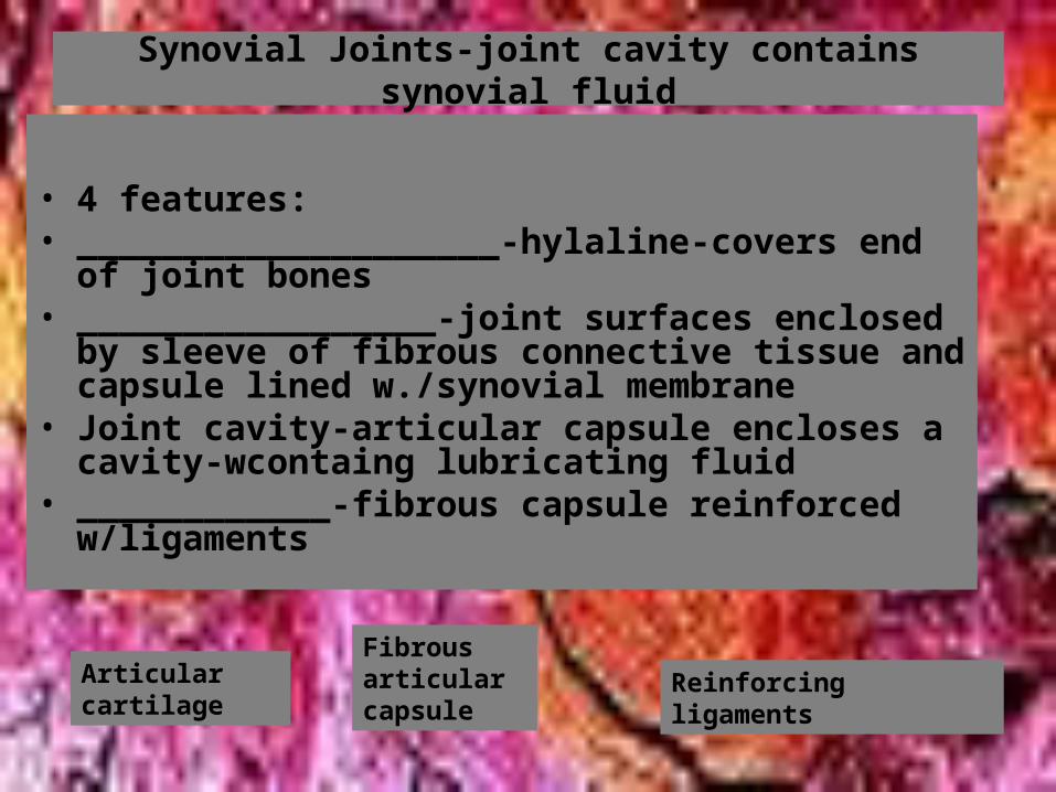
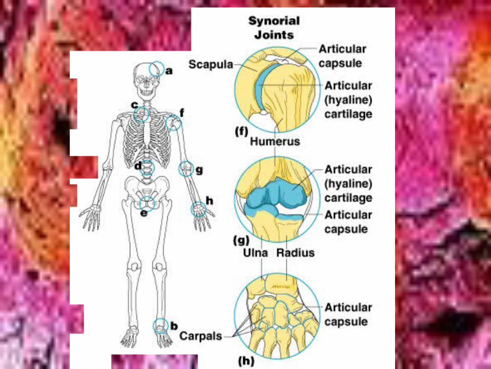
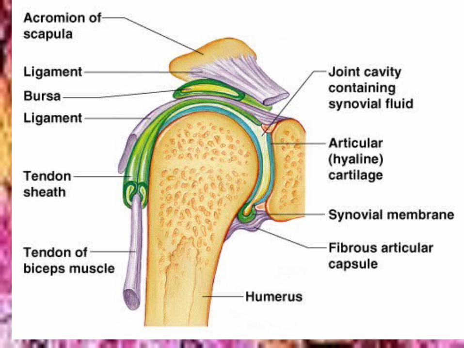
Synovial Joints-joint cavity contains synovial fluid
• 4 features:• ____________________-hylaline-covers end of joint
bones• _________________-joint surfaces enclosed by
sleeve of fibrous connective tissue and capsule lined w./synovial membrane
• Joint cavity-articular capsule encloses a cavity-wcontaing lubricating fluid
• ____________-fibrous capsule reinforced w/ligaments
Articular cartilage
Fibrous articular capsule
Reinforcing ligaments
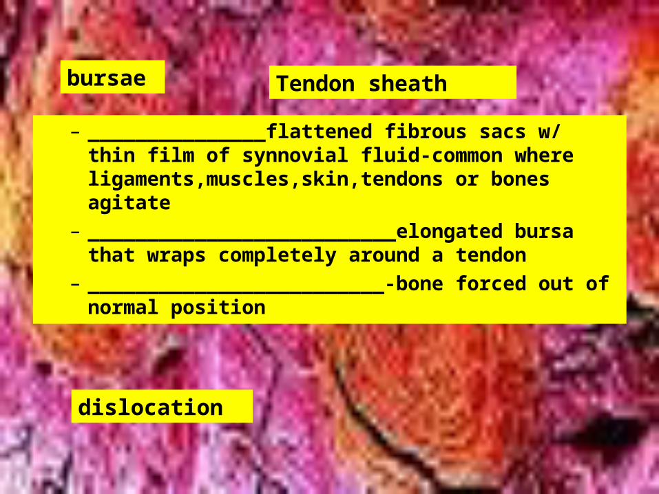
– _______________flattened fibrous sacs w/ thin film of synnovial fluid-common where ligaments,muscles,skin,tendons or bones agitate
– __________________________elongated bursa that wraps completely around a tendon
– _________________________-bone forced out of normal position
bursae Tendon sheath
dislocation
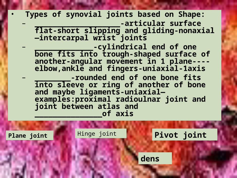
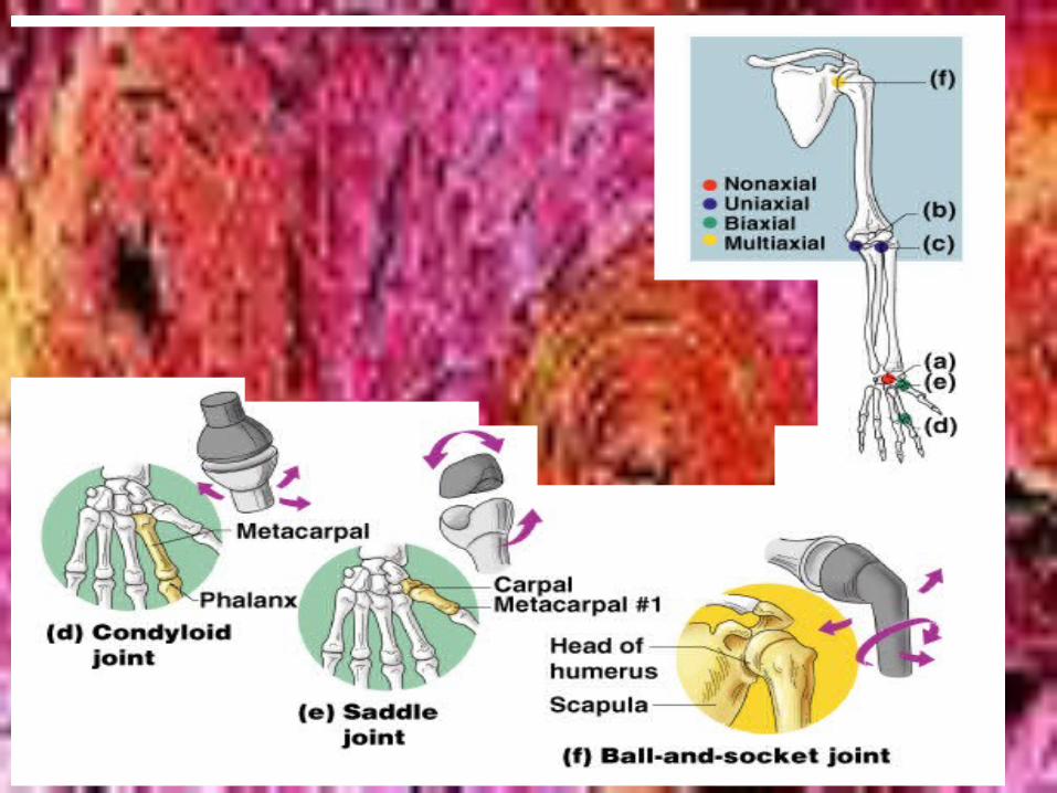
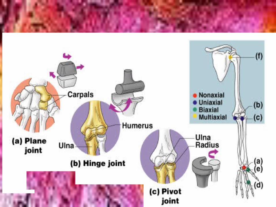
• Types of synovial joints based on Shape:– ___________________-articular surface flat-
short slipping and gliding-nonaxial—intercarpal wrist joints
– _____________-cylindrical end of one bone fits into trough-shaped surface of another-angular movement in 1 plane----elbow,ankle and fingers-uniaxial-1axis
– ________-rounded end of one bone fits into sleeve or ring of another of bone and maybe ligaments-uniaxial—examples:proximal radioulnar joint and joint between atlas and _______________of axis
Plane joint Hinge joint Pivot joint
dens

– ____________________________”knuckle-like”—egg-shaped surface into oval cavity-side to side or back and forth-can’t rotate around long axis-biaxial-knuckle joints
– Saddle joints-convex and concave area on both surfaces-thumb-twiddling thumbs
– ____________________________head of one bone in round socket of another-multiaxial-shoulder and hips
Condyloid joint
Ball-and-socket joint
• __________________________-inflammation of bursae or synovial membrane
• ________________________-ligaments or tendons damaged by stretching-or torn-heal slowly due to poor blood supply
bursitis sprain
• __________________> 100 different inflammatory or dgenerative joint diseases---possibly bacterial invasion
• ______________(OA)-most common arthritis-chronic and degenerative ,typically afftects aged-affects articular cartilage-bone thickens and bone spurs grow on margin of joint-make crunching-____________-noise----affects fingers,C and L spine and knees and hips;slow and irreversible,but rarely crippling;can be treated symptomatically for pain and inflammation….possible treatments(?)-capsaicin or glucosamine sulfate
arthritis osteoarthritis
crepitus
• ____________________________(RA)-a chronic inflammatory disorder-usually begins 40-50,but there is a juvenile form;3x as many women as men-many joints affected and usually symmetrically;course varies.It is _________________________-destroy own tissue-trigger unknown;begins w/ inflammation of synovial membranes and fluid accumulates and destroy tissue;PANNUS-abnormal tissue clings to joint and erodes articular cartilage;scar tissue forms and ossifies and bone ends become firmly fused________________________________-not all cases reach this stage.Treatment includes immunosuppressant drugs and symptomatic treatment
Rhematoid arthritis
autoimmune
ankylosis
• ________________________-uric acid(normal waste of nucleic acid metabolism) accumulates in blood and may deposit as crystals in joints-extreme pain….more common in men,usually after 30-untreated bones fuse and joint is immobilized….several drugs prevent acute gout-colchicine,ibuprofen and dietary recommendations
gout
V.Developmental aspects
• Young fetus is _________________________ and fibrous membranes
• Bone growth along epiphyseal plates as one matures
• Changes from”C” spine to “S” spine• __________________________-bone thinning
disease-
Hyaline cartilage osteoporosis
• READ THIS SECTION AND TAKE ADDITIONAL NOTES