Embed Size (px)
Citation preview
Human and Animal Prostate Cancer Similarities and Differences: Models to
Study This DiseaseElisabete Nascimento-Gonçalves1,2; Bruno Colaço2,3; Rita Ferreira4; Paula Alexandra Oliveira1,2*
1Department of Veterinary Sciences, University of Trás-os-Montes and Alto Douro (UTAD), Vila Real, Portugal.
2Center for the Research and Technology of Agro-Environmental and Biological Sciences (CITAB), UTAD, Vila Real,
Portugal.
3Department of Zootechnics, University of Trás-os-Montes and Alto Douro (UTAD), Vila Real, Portugal; 4Organic
Chemistry, Natural Products and Food Stuffs (QOPNA), Aveiro, Portugal.
*Correspondence to: Paula Alexandra Oliveira, Department of Veterinary Sciences, University of Trás-os-Montes and
Alto Douro (UTAD), Vila Real, Portugal.
Email: [email protected]
Chapter 1
Prostate Cancer
Abstract
Prostate cancer is the second most common cancer in men, is associated with high morbidity and mortality rates. The study of prostate cancer biopathology and the discovery of new therapies is very important to reduce these numbers and increase patients’ quality of life. Animal models performed with laboratory animals or studying spontaneous tumours in company animals, allow us to study the mechanisms underlying the onset and development of cancer and to explore different therapeutic approaches. For a better extrapolation of the results of animals studies to the human clinical practise, it is very important understand the differences and similarities between them.
Keywords: Models; Prostate; Cancer
2
ww
w.openaccessebooks.comProstate Cancer
Ale
xand
ra O
livei
ra P
1. Introduction
Worldwide, prostate cancer is the second most common cancer and the fifth leading cause of oncological death among men [1]. There were approximately 1.3 million new cases and 358 989 deaths estimated in 2018 [2]. Although the causes of the prostate cancer are not fully understood, many risk factors have been considered, such as age, race, family history, diet, hormone exposure and inflammation [3,4]. Steroid hormones, particularly androgens play an important role in human prostate cancer. Prostate cancer is dependent on androgen receptor activation for growth and survival, but the precise mechanism underlying this process is not clear [4–6]. Adenocarcinomas represent more than 95% of prostate cancers and arises from the prostate gland epithelial cells. Other type of prostate cancer includes sarcomas, small cell carcinomas, neuroendocrine tumours and transitional cell carcinomas [7]. Intraepithelial neoplasia (PIN), that is characterized as an intra-luminal proliferation of epithelium exhibiting varying degrees of malignant criteria, is considered a precursor of prostate cancer [8]. Prostate glands with extensive PIN also have more multifocal carcinomas and these developed preferentially in the peripheral zone of the gland [9]. Proliferative inflammatory atrophy (PIA) is also a pre-neoplastic disorder, that is caused by tissue damage, by an initiating inflammatory insult [8] and are often found in close proximity to foci of PIN and carcinoma, and commonly contain somatic mutations that are present in PIN lesions and carcinoma [10]. Prostate cancer metastasizes manly to bone [11], but also to lymph nodes, lungs, liver and brain [12].
Over the years, animal models have been used to study several diseases, including cancer, and contribute to understand many aspects involved in disease progression and for the discovery and development of new pharmacology and non-pharmacology therapies and preventive strategies [13,14]. An ideal animal model should be simple, not expensive and could mimic the Human disease as much as possible.
In the present book chapter, we summarized the features of animal’s models, namely non-human primates, dogs, cats, mice and rats to study prostate cancer, in relation to the prostate anatomy, epidemiology and etiology of prostate cancer, and compared the similarities and differences with Human prostate cancer.
2. Human Prostate
The man prostate is a walnut-size gland located behind the inferior border of the pubic symphysis and pubic arch and anterior to the rectal ampulla and it is just one tubule-alveolar gland [7,15]. It is composed of a base, attached to the neck of the bladder, an apex, on the superior surface of the urogenital diaphragm that makes contact between the medial surface of the levator any muscles, anterior, posterior and inferior-lateral surfaces [16].
3
Prostate Cancer
The prostate contains distinct regions with different functions which are differentiated by their histology pattern and anatomic landmarks. Peripheral zone (PZ) represents approximately 70% of the glandular prostate and most of the adenocarcinomas arise in PZ as well chronic prostatitis, inflammation and post inflammatory atrophy [7]. Central zone (CZ) represents approximately 25% of prostate volume and is located at the base of the prostate between the peripheral and transition zones. The transitional zone (TZ) represents only 5% of the glandular prostate volume and is the most common site for benign hyperplasia lesions and less commonly adenocarcinoma [7,17]. The periurethral gland or anterior fibromuscular stroma (AFMS) represents less than 1% of the glandular prostate and fills the space between PZ and pre-prostatic urethra [18].
Prostate cancer is an age-related disease, with the majority of the cases diagnosed in men between 60 and 70 years [2,3,19]. Early prostate cancer detected by screening usually has asymptomatic. In advanced stage, the symptoms included weak or interrupted urine flow, the inability to urinate or difficulty to start and stop urine flow, the need to urinate frequently, blood in urine and pain during urination [19,20]. These symptoms may also be due to the benign prostatic hyperplasia, so it’s is important confirm prostate cancer through other methods, like biopsy [21]. Prostate-specific antigen (PSA) is and androgen-regulated serine protein and is produced by the secretory epithelial cells in the ducts and acini of the prostate [18] and is the most widely used serum marker for prostate cancer [3,22,23]. However, it is not specific for prostate cancer and levels can be elevated in other prostate disorders like benign prostatic hyperplasia (BHP) [22]. To categorize prostate biopsy the pathologists use a grading system known as the Gleason Scoring system [15] based upon the architecture differentiation of the carcinomas cells in hematoxylin-eosin stained tissue section. These system helps in the determination of the aggressiveness of prostate cancer and in choosing the most appropriate treatment options and the most suitable animal models to study this disease[8].
3. Animal Models of Prostate Carcinogenesis
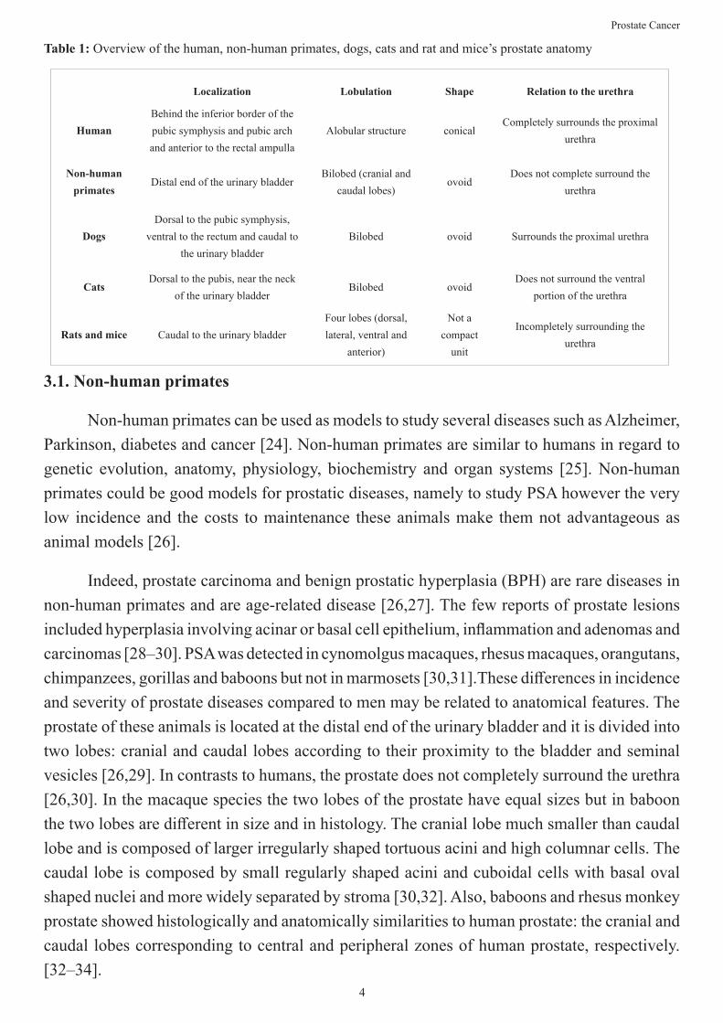
There are several animal models to study prostate cancer. In this section we focused in non-human primates, dogs, cats, mice and rats’ models. There are significant anatomical differences between the prostate of the different animal models (see Table 1), but also similarities. It is important to understand them when choosing the most suitable animal model.
4
Prostate Cancer
3.1. Non-human primates
Non-human primates can be used as models to study several diseases such as Alzheimer, Parkinson, diabetes and cancer [24]. Non-human primates are similar to humans in regard to genetic evolution, anatomy, physiology, biochemistry and organ systems [25]. Non-human primates could be good models for prostatic diseases, namely to study PSA however the very low incidence and the costs to maintenance these animals make them not advantageous as animal models [26].
Indeed, prostate carcinoma and benign prostatic hyperplasia (BPH) are rare diseases in non-human primates and are age-related disease [26,27]. The few reports of prostate lesions included hyperplasia involving acinar or basal cell epithelium, inflammation and adenomas and carcinomas [28–30]. PSA was detected in cynomolgus macaques, rhesus macaques, orangutans, chimpanzees, gorillas and baboons but not in marmosets [30,31].These differences in incidence and severity of prostate diseases compared to men may be related to anatomical features. The prostate of these animals is located at the distal end of the urinary bladder and it is divided into two lobes: cranial and caudal lobes according to their proximity to the bladder and seminal vesicles [26,29]. In contrasts to humans, the prostate does not completely surround the urethra [26,30]. In the macaque species the two lobes of the prostate have equal sizes but in baboon the two lobes are different in size and in histology. The cranial lobe much smaller than caudal lobe and is composed of larger irregularly shaped tortuous acini and high columnar cells. The caudal lobe is composed by small regularly shaped acini and cuboidal cells with basal oval shaped nuclei and more widely separated by stroma [30,32]. Also, baboons and rhesus monkey prostate showed histologically and anatomically similarities to human prostate: the cranial and caudal lobes corresponding to central and peripheral zones of human prostate, respectively. [32–34].
Table 1: Overview of the human, non-human primates, dogs, cats and rat and mice’s prostate anatomy
Localization Lobulation Shape Relation to the urethra
HumanBehind the inferior border of the pubic symphysis and pubic arch and anterior to the rectal ampulla
Alobular structure conicalCompletely surrounds the proximal
urethra
Non-human primates
Distal end of the urinary bladderBilobed (cranial and
caudal lobes)ovoid
Does not complete surround the urethra
DogsDorsal to the pubic symphysis,
ventral to the rectum and caudal to the urinary bladder
Bilobed ovoid Surrounds the proximal urethra
CatsDorsal to the pubis, near the neck
of the urinary bladderBilobed ovoid
Does not surround the ventral portion of the urethra
Rats and mice Caudal to the urinary bladderFour lobes (dorsal, lateral, ventral and
anterior)
Not a compact
unit
Incompletely surrounding the urethra
5
Prostate Cancer
3.2. Cats and dogs
Dogs’ prostate is bilobed and ovoid-shaped and surrounding the neck of the urinary bladder and the proximal urethra [35]. The dorsal surface of the prostate is separated from the ventral surface of the rectum by the two layers of the fold of peritoneum bounding the rectogenital fold [36]. The ventral surface is covered by a layer of retroperitoneal fat. The prostate has a prominent median septum which separate the right and left lobes, and each lobe is divided into lobules by capsular trabeculae. The prostate gland is recovered by a fibromuscular capsule (36,37).It is located dorsal to the pubic symphysis, ventral to the rectum and caudal to the bladder, and is the unique accessory gland in the dog [37].
In cats, prostate located near the neck of the urinary bladder and in contrast to dogs, prostate does not encircle the urethra, just covers only the top and sides [38–41]. As in the dogs, cat prostate is bilobed [38].
Dogs, like humans, develop spontaneous prostate carcinomas more frequently in older animals, with an average age at diagnosis of 10 years [26,36]. Besides, also benign prostatic hyperplasia (BHP) and high-grade prostatic intraepithelial neoplasia (HGPIN) spontaneously developed in intact adult male dogs [42]. In dogs, BHP affects the prostate gland in a diffuse pattern [35], it can be severe and cause others clinical disease, including dorsal compression of the colon and dyschezia, but in contrast to men, does not appear to be a preneoplastic condition [42]. Histologically, there are two patterns depending on dogs’ age [43]. In dogs up to 4 years age, BHP is referred to as diffuse glandular with an intra-alveolar increase in papillary proliferation of prostatic secretory epithelium. Dogs with 6 years of age and older display diffuse glandular BHP along with an increased stromal component [35]. HGPIN had cytological similarities with man, including cell crowding, loss of polarity and nuclear and nucleolar enlargement, basal cell disruption, proliferative index and micro vessel density [44,45]. The prevalence seems to be influenced by aging and testicular androgens [45], however the presence of HGPIN and the risk of development prostate cancer remains unknown [35,42]. The precise location of origin of the carcinomas is not yet know, but may originate from prostatic ductular epithelium adjacent to the periurethral zone, prostatic acinar epithelium or urothelium lining the prostatic urethra, e.g. transitional cell carcinoma [35,36,42].
Another difference between men and dog’s prostate cancer pathogenesis is the role of androgens. As previously mentioned, men prostate cancer is androgen dependent for growth and survival, but in dogs it’s different. In dogs, normal prostatic tissue express androgen receptor and they are important for normal function. Dogs with prostate cancer have no expression of the androgen receptor[35]. Also, the castration does not decrease the risk cancer development, on the contrary, several studies showed that castration may be associated with an increased risk of cancer [46-48]. On the one hand castration alters the prostate stromal component
6
Prostate Cancer
from primarily actin-positive smooth muscle cells to vimentin-positive mesenchymal cells which could promote the development of prostate cancer in castrated dogs [49]. On the other hand, castrated dogs have a higher average life expectancy than intact dogs and have higher predisposition to develop carcinomas [35]. So, the tumor growth is not androgen dependent [26] and the androgen receptor does not seem to play a central role in the pathogenesis of canine prostate cancer development [42].The common type of canine prostate cancer are adenocarcinomas, but the majority of the tumors showed several morphologic changes that included two or more types of differentiation in the same tissue section, like glandular, urothelial, squamoid or sarcomatoid [36]. Histologically, carcinomas show an intra-alveolar pattern, but also contain similar patterns to transitional cell carcinoma [35]. Like humans, prostate cancer in dogs metastasizes to bone, with osteoblastic and osteolytic lesions and formation of new woven bone [35,50-53].
Beyond the ethical issues, the long latency period, the high cost to maintain into laboratory, long gestation period and difficulty of genetic manipulation make the dog an unusual animal model to study prostate cancer [54,55].
The prostate cancer in cats is very rare disease and in the few cases reported the tumors were classified as high-grade carcinomas and adenocarcinomas. The sites of metastasis included lymph node, lungs and pancreas [56]. The development of carcinomas is more frequently in older cat, with average age of eight years [56], and castrated animals [41] and the survival after diagnosis is lower, beyond 3 months [41]. There is no much information about the characteristics of feline prostate cancer, namely risk factors, the role of androgens and the mechanisms of tumor development.
3.3. Rats and mice
The prostate gland of the rats and mice are significantly different from men. The rodents´ prostate consists of four distinct lobes with different histological characteristics and different physiological functions. It is located circumferentially around the urethra, immediately caudal to the urinary bladder. The lobes are classified as ventral, dorsal, lateral and anterior (or coagulating gland) and are named according to their relative position to the urinary bladder [9,17,57]. Spontaneous prostate cancer in wild-type mice are rare [54,57] and in rats also, but the incidence varies with the rats’ strain, are reported 70% of spontaneous rat cancer for ACI/Seg and 8% for Lobund-Wistar [57].
In 1963, was reported the first spontaneous prostate tumor in a 22-month-old Copenhagen male rat [58]. This tumor was classified as a papillary adenocarcinoma that involved the dorsal prostate lobe and were not identified metastases [58]. Pollard, in 1973, reported the development of spontaneous carcinoma in dorso-lateral and anterior prostate in Wistar rats with 26 months of age[59]. Metastatic lesions in lungs, spleen, liver, pancreas and intestine were also identified
7
Prostate Cancer
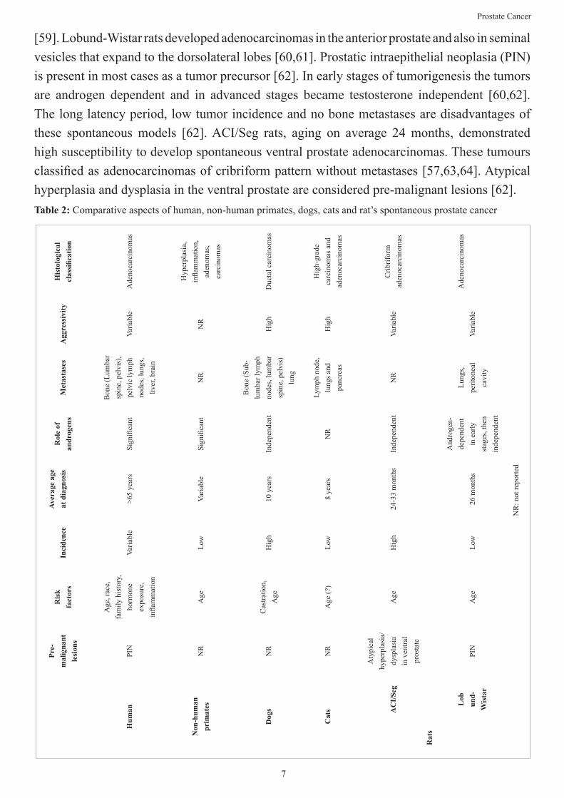
[59]. Lobund-Wistar rats developed adenocarcinomas in the anterior prostate and also in seminal vesicles that expand to the dorsolateral lobes [60,61]. Prostatic intraepithelial neoplasia (PIN) is present in most cases as a tumor precursor [62]. In early stages of tumorigenesis the tumors are androgen dependent and in advanced stages became testosterone independent [60,62]. The long latency period, low tumor incidence and no bone metastases are disadvantages of these spontaneous models [62]. ACI/Seg rats, aging on average 24 months, demonstrated high susceptibility to develop spontaneous ventral prostate adenocarcinomas. These tumours classified as adenocarcinomas of cribriform pattern without metastases [57,63,64]. Atypical hyperplasia and dysplasia in the ventral prostate are considered pre-malignant lesions [62]. Table 2: Comparative aspects of human, non-human primates, dogs, cats and rat’s spontaneous prostate cancer
His
tolo
gica
l cl
assi
ficat
ion
Ade
noca
rcin
omas
Hyp
erpl
asia
, in
flam
mat
ion,
ad
enom
as,
carc
inom
as
Duc
tal c
arci
nom
as
Hig
h-gr
ade
carc
inom
as a
nd
aden
ocar
cino
mas
Crib
rifor
m
aden
ocar
cino
mas
Ade
noca
rcin
omas
NR
: not
repo
rted
Agg
ress
ivity
Varia
ble
NR
Hig
h
Hig
h
Varia
ble
Varia
ble
Met
asta
ses
Bon
e (L
umba
r sp
ine,
pel
vis)
, pe
lvic
lym
ph
node
s, lu
ngs,
liver
, bra
in
NR
Bon
e (S
ub-
lum
bar l
ymph
no
des,
lum
bar
spin
e, p
elvi
s)
lung
Lym
ph n
ode,
lu
ngs a
nd
panc
reas
NR
Lung
s, pe
riton
eal
cavi
ty
Rol
e of
an
drog
ens
Sign
ifica
nt
Sign
ifica
nt
Inde
pend
ent
NR
Inde
pend
ent
And
roge
n-de
pend
ent
in e
arly
st
ages
, the
n in
depe
nden
t
Aver
age
age
at d
iagn
osis
>65
year
s
Varia
ble
10 y
ears
8 ye
ars
24-3
3 m
onth
s
26 m
onth
s
Inci
denc
e
Varia
ble
Low
Hig
h
Low
Hig
h
Low
Ris
k fa
ctor
s
Age
, rac
e,
fam
ily h
isto
ry,
horm
one
expo
sure
, in
flam
mat
ion
Age
Cas
tratio
n,
Age
Age
(?)
Age
Age
Pre-
mal
igna
nt
lesi
ons
PIN
NR
NR
NR
Aty
pica
l hy
perp
lasi
a/dy
spla
sia
in v
entra
l pr
osta
te
PIN
Hum
an
Non
-hum
an
prim
ates
Dog
s
Cat
s AC
I/Se
g
Lob
und-
Wis
tar
Rat
s
8
Prostate Cancer
In contrast to rats, mice are resistant to induction of prostate cancer by chemical carcinogens [57]. Mice are most commonly used as xenograft models and genetically-engineered models [54]. Over the years, many experimental works were performed to discovered chemical compounds to induce tumors and understand how they induce these changes. Carcinogenic agents have the ability to induce tumors in several tissues depending on application site (i.e., prostate, urinary bladder, mammary tissue), absorption site ( i.e., subcutaneous injection, oral administration), organ of metabolism (i.e., liver) and excretory organs (i.e., urethra, urinary bladder) [65]. The metabolic activation of the carcinogenic compound has a specific route. The first step is carcinogenic exposure that can be absorbed in several ways (i.e., cutaneous, injection, oral or inhalator) and depends on the physicochemical properties of the substance. Then, the compound is distributed and suffered biotransformation on liver, kidneys or lungs, occurring its activation. Then may occurred genotoxic mechanisms, such as DNA adducts and chromosome breakages, or non-genotoxic mechanisms, such as inflammation, reactive oxygen species or immunosupression. These mechanisms cause a genomic damage and altered signal transduction which leads to cancer development [66].
4. Chemically-Induced Rat Prostate Cancer Models
Chemically-induced rat models allows the development of tumors in short period of latency, high reproducibility and allow monitoring all carcinogenesis process in a target organ and are also important to evaluate the chemopreventive effects of different agents [66]. However, they have some limitations namely high costs to obtain and maintain the animals, time-consuming and labor-intensive, application of the carcinogen in high doses and during a long-time period, toxicity of the carcinogen compounds to animal and people that handle the animals and the environment and lack of organ specific of the carcinogen compounds [56,67].
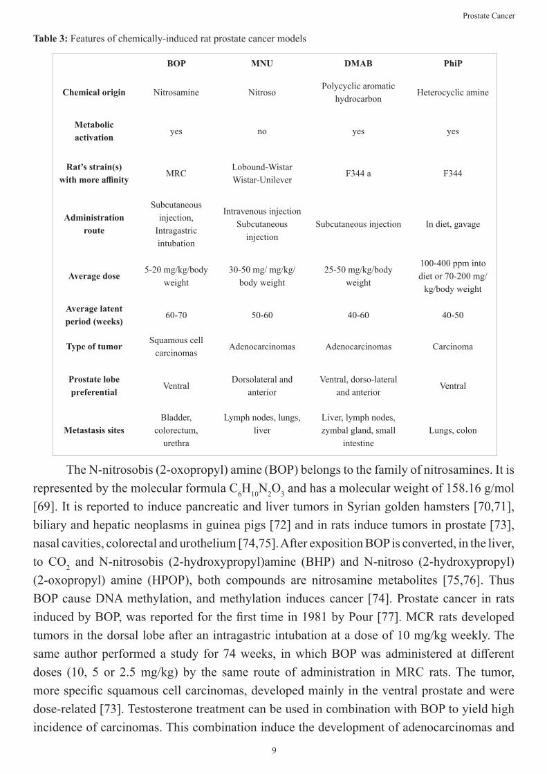
Four chemical compounds have described to induce prostate cancer in laboratory rats: N-nitrosobis (2-oxopropyl) amine (BOP), N-Methyl-N-nitrosourea (MNU), 3,2-dimethyl-4-aminobiphenyl (DMAB) and the 2-amino-1-methyl-6-phenylimidazol[4,5-b]pyridine (PhiP) [68]. The main characteristics of each can be found in Table 3.
9
Prostate Cancer
Table 3: Features of chemically-induced rat prostate cancer models
The N-nitrosobis (2-oxopropyl) amine (BOP) belongs to the family of nitrosamines. It is represented by the molecular formula C6H10N2O3 and has a molecular weight of 158.16 g/mol [69]. It is reported to induce pancreatic and liver tumors in Syrian golden hamsters [70,71], biliary and hepatic neoplasms in guinea pigs [72] and in rats induce tumors in prostate [73], nasal cavities, colorectal and urothelium [74,75]. After exposition BOP is converted, in the liver, to CO2 and N-nitrosobis (2-hydroxypropyl)amine (BHP) and N-nitroso (2-hydroxypropyl) (2-oxopropyl) amine (HPOP), both compounds are nitrosamine metabolites [75,76]. Thus BOP cause DNA methylation, and methylation induces cancer [74]. Prostate cancer in rats induced by BOP, was reported for the first time in 1981 by Pour [77]. MCR rats developed tumors in the dorsal lobe after an intragastric intubation at a dose of 10 mg/kg weekly. The same author performed a study for 74 weeks, in which BOP was administered at different doses (10, 5 or 2.5 mg/kg) by the same route of administration in MRC rats. The tumor, more specific squamous cell carcinomas, developed mainly in the ventral prostate and were dose-related [73]. Testosterone treatment can be used in combination with BOP to yield high incidence of carcinomas. This combination induce the development of adenocarcinomas and
BOP MNU DMAB PhiP
Chemical origin Nitrosamine NitrosoPolycyclic aromatic
hydrocarbonHeterocyclic amine
Metabolic activation
yes no yes yes
Rat’s strain(s) with more affinity
MRCLobound-WistarWistar-Unilever
F344 a F344
Administration route
Subcutaneous injection,
Intragastric intubation
Intravenous injectionSubcutaneous
injectionSubcutaneous injection In diet, gavage
Average dose5-20 mg/kg/body
weight30-50 mg/ mg/kg/
body weight25-50 mg/kg/body
weight
100-400 ppm into diet or 70-200 mg/
kg/body weight
Average latent period (weeks)
60-70 50-60 40-60 40-50
Type of tumorSquamous cell
carcinomasAdenocarcinomas Adenocarcinomas Carcinoma
Prostate lobe preferential
VentralDorsolateral and
anteriorVentral, dorso-lateral
and anteriorVentral
Metastasis sitesBladder,
colorectum, urethra
Lymph nodes, lungs, liver
Liver, lymph nodes, zymbal gland, small
intestineLungs, colon
10
Prostate Cancer
squamous cell carcinomas in dorsolateral and ventral prostate in MRC rats [78]. BOP can be administered by intragastric intubation or subcutaneously and the experimental protocol after carcinogenic administration takes, on average, 60 weeks. However, as BOP mostly induced squamous cell carcinomas and tumors in the ventral lobe prostate, the only lobe that does not have a human homologue, it is not a very adequate animal model to mimic human prostate cancer [63]. The N-methyl-N-nitrosourea (MNU) is a nitroso-compound does not require metabolic activation; it is a direct-acting alkylating agent by methylation of the guanine nucleosides. MNU is represented by molecular formula C2H5N3O2 and has a molecular weight of 103.08 g/mol [65]. This compound is a carcinogenic agent in various animals’ species (rats, hamsters, gerbils, fishes and shrews) and may induce tumors in breast, ovary, uterus, hematopoietic organs, kidney, urinary bladder, liver, intestine, spleen, retina and prostate [65]. Pollard and colleagues developed a method to induced prostate cancer in Lobund-Wistar rats through the administration of MNU associated with hormonal treatment [79,80]. They used 30-40 mg of MNU injected intravenously, followed by a long-term administration of 10-40 mg testosterone propionate via silastic implants. This protocol induced adenocarcinomas and atypical hyperplasia lesions in ventral, dorsolateral and anterior prostate. Metastases can be found in lungs and peritoneal cavity but not in bone [62]. However, others experimental works demonstrated that the majority of the carcinomas originated from the seminal vesicles [81,82]. Bosland developed an animal model that involves stimulation of prostatic cell proliferation by sequential treatment with cyproterone acetate, that inhibit androgen secretion causing atrophy of prostatic epithelial cells, and testosterone propionate [83]. Wistar-Unilever rats are treated with 50 mg of cyproterone acetate, and then receive daily injections of 100 mg/kg body weight testosterone propionate for three consecutive days. One day after the last testosterone injection, rats received a single intravenous injection of MNU (50 mg/kg). The rats were sacrificed 79 weeks after MNU treatment. The tumors developed were classified as adenocarcinomas in the dorsolateral prostate, were invasively growing and metastasize to the lymph nodes and lungs [57,84].
Over the years, this Bosland protocol was improved and now the sequential treatment with an antiandrogen (cyproterone acetate or flutamide), testosterone propionate, MNU and chronic treatment with testosterone is frequently used for prostate cancer induction [63,81]. With more details: the cyproterone acetate or flutamide is given once daily for 2-3 weeks by gavage or by subcutaneous injection, followed by the administration of testosterone propionate (10-100 mg/kg) by subcutaneous injection during three consecutive days after last cyproterone acetate treatment or a single subcutaneous injection of 100 mg/kg on that day. Forty-eight hours later, a single intravenous or intraperitoneal injection of MNU, in doses between 30 and 50 mg/kg, and finally to achieve a high carcinoma incidence, testosterone propionate in silastic implants are placed subcutaneously in interscapular region by surgical approach under general anesthesia. The experimental work should be conducted until 50-60 weeks after MNU
11
Prostate Cancer
injection, to achieve maximal tumor incidence [63,81,85]. The vast majority of the prostate tumors induced were adenorcarcinomas in dorsolateral and anterior prostate, that share some important characteristics with human prostate carcinomas, being this model a suitable model for prostate cancer studies, and nowadays, the most used [82].
The 3,2’-diemthy-4-aminobiphenyl (DMAB is a classical polycyclic aromatic hydrocarbon represented by the molecular formula C14H15N and has a molecular weight of 233.74 g/mol [69]. DMAB have multi-organ tropism, inducing tumors in the colon, urinary bladder, pancreas, prostate, mammary glands, preputial glands, seminal vesicles and Zymbal glands [57,86]. This compound needs to be active in the liver. Then, the metabolites interact with DNA causing transversions in nucleotides, inducing irreversible changes and adducts [86]. Katayama, in 1982, was the first to induce microscopic carcinomas in the ventral prostate of F344 rats by DMAB [87]. The prostate carcinogenicity of DMAB was confirmed, a couple years later, by Shirai and colleagues [88]. They induced prostatic carcinomas in F344 rats by repeated treatment with ethinyl estradiol (75 ppm) added to the basal powdered diet and DMAB (50 mg/kg by subcutaneous injection) for 60 weeks. The treatment with ethinyl estradiol induced a high incidence (up to 85%) of adenocarcinomas in the ventral lobe of the prostate [88]. The carcinogenic potential of DMAB is dose-dependent; a low-dosage of DMAB given over a long period (around 48 weeks) was more effective to induce prostate cancer than a high dosage over a short period of time (10-25 weeks) [89]. There are different susceptibilities between rat strains ( F344 and ACI rats being the most susceptible and Wistar and Sprague-Dawley rats resistant) to DMAB carcinogenesis [90]. Chronic administration of high doses of testosterone incorporated into silastic tubes and implanted into subcutaneous tissue in combination with DMAB can be used to promote tumor development [78]. This combination produced a high incidence of invasively adenocarcinomas in dorsolateral and anterior prostate but not in ventral prostate in F344 rats [91]. The induction of tumours in dorsolateral and anterior prostate and seminal vesicles and the degree of invasiveness depends on the duration and doses of testosterone [91,92]. The tumors are histologically and biologically indistinguishable from those induced by MNU in combination with testosterone [57]. The co-administration of ethinyl estradiol with testosterone propionate increased the yield of carcinomas in the lateral and anterior lobes of the prostate in a dose-related fashion with lobe specificity [93]. In a general way, the induction protocol consists of a subcutaneous injection of 25-50 mg/kg, 10 times at 2-week intervals. Implants of testosterone propionate can be used and should be replaced at 6-week intervals. On average, the studies using DMAB last 60 weeks.
The 2-amino-1-methyl-6-phenylimidazol[4,5-b]pyridine (PhIP) is a heterocyclic amine represented by the molecular formula C13H12N4 and has a molecular weight of 224.27 g/mol [69]. It is present in a variety of meats cooked and fish and induces cancer development in mammary gland, colon and prostate in laboratory rodents [57]. PhIP may be metabolized to
12
Prostate Cancer
biologically active metabolites (N-hydroxy-PhIP and N-acetoxy-PhIP) that form DNA adducts [94]. The metabolic activation involves cytochrome P450-mediated N-hydroxylation [95]. In 1997, Shirai and colleagues, were the first to use PhiP to induce prostate cancer tumors [96]. They subjected F344 rats to PhIP, at a dose of 400 ppm mixed in the diet, for 52 weeks [96]. The adenocarcinomas developed in ventral lobe and were histopathologically equivalent to those caused by DMAB [97]. A long-term pharmacological with testosterone propionate can induce invasive carcinomas in the anterior prostate and seminal vesicles [98]. Overall, the experimental protocols using PhIP consist in administration of this compound mixed into the diet or administered by gavage (70-200 mg/kg).
5. Conclusion
There are many similarities between human and animal prostate cancer, like histology, metastatic process and spontaneous cancer incidence.
Non-human primates are closer phylogenetically to humans than the other animals, and mechanisms underlying the cancer development are expected to be similar. However, in the literature there are few reports of prostate cancer in non-human primates, which can be explained by the difficult access to these animals in their natural habitat, the high cost of captivity for long periods of time, absence of symptoms in captive animals and failure to complete detailed autopsies. Dogs are considered by many researchers the best model to study prostate cancer, due to the development of spontaneous cancer with a higher incidence when compared with other animals and the development of bone metastases. However, they were not very used in experimental studies since they are considered companion animals and for ethical reasons its use is reduced. Mice are mainly used as xenograft models and genetically-engineered models. The reports of spontaneous prostate tumors are rare and mice are resistant to induction of prostate cancer by chemical compounds. Rats also developed spontaneous prostate tumors, although with low incidence and share some similarities with humans, namely the influence of androgens in cancer development. Chemically induced protocols are designed to induce tumors in rat lobes analogues to man counterpart, mainly dorsolateral prostate lobe. However, the occurrence of bone metastases is reduced. Besides this and the anatomical differences, studies found similarities in the molecular mechanisms underlying prostate cancer development in rats and men, making the rat a valid animal model to study prostate cancer development and to evaluate the efficacy of several treatments and preventive measures. Above all, when it is necessary to choose an animal model we must always keep in mind what are the objectives of the work, the amount of samples we need to collect and the type of sample to collect.
6. Acknowledgement
This work was supported by European Investment Funds by FEDER/ COMPETE/
13
Prostate Cancer
POCI - Operational Competitiveness and Internationalization Program, under Project POCI-01-0145-FEDER-016728 and National Funds by FCT - Portuguese Foundation for Science and Technology, under the project project UID/AGR/04033/2019 the project PTDC/DTP-DES/6077/2014 and post-graduation grant SFRH/BD/136747/2018.
7. References
1. Bray F, Ferlay J, Soerjomataram I, Siegel R, Torre L, Jemal A. Global cancer statistics 2018: GLOBOCAN estimates of incidence and mortality worldwide for 36 cancers in 185 countries. CA A J Clin. 2018;00(00):1–31.
2. Globocan. Estimated Incidence, Mortality and Prevalence Worldwide in 2018. [Internet]. International Agency for Research on Cancer (Word Health Organization). 2018.
3. Adjakly M, Ngollo M, Dagdemir A, Judes G, Pajon A, Karsli-Ceppioglu S, et al. Prostate cancer: The main risk and protective factors – Epigenetic modifications. Ann Endocrinol (Paris). 2015;76(1):25–41.
4. Maarten C. B. The role of steroid hormones in prostate carcinogenesis. J Natl Cancer Inst Monogr. 2000;10016(27):39–66.
5. Bosland M. A perspective on the role of estrogen in hormone-induced prostate carcinogenesis. Cancer Lett. 2013;334:28–33.
6. Grossmann M, Cheung AS, Zajac JD. Androgens and prostate cancer; Pathogenesis and deprivation therapy. Best Pract Res Clin Endocrinol Metab. 2013;27(4):603–16.
7. Bhavsar A, Verma S. Anatomic Imaging of the Prostate. Hindawi Publ Corp. 2014;2014.
8. Demarzo AM, Nelson WG, Isaacs WB, Epstein JI. Pathological and molecular aspects of prostate cancer. Lancet. 2003;361:955–64.
9. Roy-Burman P, Wu H, Powell WC, Hagenkord J, Cohen MB. Genetically defined mouse models that mimic natural aspects of human prostate cancer development. Endocr Relat Cancer. 2004;11(2):225–54.
10. Nelson W, De Marzo AM, Isaacs WB. Prostate cancer. N Engl J Med [Internet]. 2003;349:366–81.
11. Hudson BD, Kulp KS, Loots GG. Prostate cancer invasion and metastasis: Insights from mining genomic data. Brief Funct Genomics. 2013;12(5):397–410.
12. Sartor O, Bono J. Metastatic Prostate Cancer. New Engl J. 2018;378:645–57.
13. Steele VE, Lubet RA. The Use of Animal Models for Cancer Chemoprevention Drug Development. Semin Oncol. 2010;37(4):327–38.
14. Fagundes DJ, Taha MO. Modelo animal de doença: critérios de escolha e espécies de animais de uso corrente. Acta Cirúrgica Bras. 2004;19(1):59–65.
15. Dunn MW, Kazer MW. Prostate cancer overview. Semin Oncol Nurs. 2011;27(4):241–50.
16. Lee CH, Akin-Olugbade O, Kirschenbaum A. Overview of Prostate Anatomy, Histology, and Pathology. Endocrinol Metab Clin North Am. 2011;40(3):565–75.
17. Oliveira DSM, Dzinic S, Bonfil AI, Saliganan AD, Sheng S, Bonfil RD. The mouse prostate : a basic anatomical and histological guideline. Bosn J. 2015;16(1):8–13.
18. Sharma M, Gupta S, Dhole B, Kumar A. The Prostate Gland. Basics Hum Androl. 2017;
14
Prostate Cancer
19. Diet , nutrition , physical activity and colorectal cancer. World Cancer Research Fund/American Institute for Cancer Research [Internet]. 2018.
20. Litwin MS, Tan HJ. The diagnosis and treatment of prostate cancer: A review. JAMA - J Am Med Assoc. 2017;317(24):2532–42.
21. Sarkar S, Das S. A Review of Imaging Methods for Prostate Cancer Detection. Biomed Eng Comput Biol. 2016;7 (1).
22. Neal DE, Moon TD, Clejan S, Sarma D. Prostate specific antigen and prostatitis I. Effect of prostatitis on serum psa in the human and nonhuman primate. Prostate. 1992;20(2):105–11.
23. Stamey TA, Hay AR, McNeal JE, Freiha FS, Redwine E. Prostate-specific antigen as a serum marker for adenocarcinoma of the prostate. N Engl J Med. 1987;317(15):909–16.
24. Friedman H, Ator N, Haigwood N, Newsome W, Allen JS, Golos TG, et al. The Critical Role of Nonhuman Primates in Medical Research - White Paper. Pathog Immun. 2017;2(3): 352.
25. Xia H-J, Chen C-S. Progress of non-human primate animal models of cancers. Zool Res. 2011;32(1):70–80.
26. Waters DJ, Sakr WA, Hayden DW, Lang CM, McKinney L, Murphy GP, et al. Workgroup 4: Spontaneous prostate carcinoma in dogs and nonhuman primates. Prostate. 1998; 36(1): 64–7.
27. Mahapokai W, Van Sluijs FJ, Schalken JA. Models for studying benign prostatic hyperplasia. Prostate Cancer Prostatic Dis. 2000;3(1):28–33.
28. McEntee MF, Epstein JI, Syring R, Tierney LA, Strandberg JD. Characterization of prostatic basal cell hyperplasia and neoplasia in aged macaques: Comparative pathology in human and nonhuman primates. Prostate. 1996;29(1):51–9.
29. Lewis RW, Kim JCS, Irani D, Roberts JA. The prostate of the nonhuman primate: Normal anatomy and pathology. Prostate. 1981;2(1):51–70.
30. Mubiru J, Hubbard G, Jr. Dick E, Furman J, Troyer D, Rogers J. Nonhuman primates as models for studies of prostatic specific antigen and prostatic diseases. Prostate. 2008;1(68):1546–54.
31. Karr JF, Kantor JA, Hand PH, Eggensperger DL, Schlom J. The Presence of Prostate-specific Antigen-related Genes in Primates and the Expression of Recombinant Human Prostate-specific Antigen in a Transfected Murine Cell Line. Cancer Res. 1995;55(11):2455–62.
32. Mubiru J, Hubbard G, Jr. Dick E, Butler S, Valente A, Troyer D, et al. A Preliminary Study of the Baboon Prostate Pathophysiology. The Protate. 2007;67:1421–31.
33. Jeyaraj Antony D, UDAYAKUMAR T, Rajalakshmi M, Pal PC, Sharma RS. Effects of Long-Term Administration of Androgens and Estrogen on Rhesus Monkey Prostate : Possible Induction of Benign Prostatic Hyperplasia. J Androl. 2000;21(6):833–41.
34. Blacklock NJ, Bouskill K. The zonal anatomy of the prostate in man and in the Rhesus monkey (Macaca Mulatta). Urol Res. 1977;5(4):163–7.
35. LeRoy BE, Northrup N. Prostate cancer in dogs: Comparative and clinical aspects. Vet J [Internet]. 2009;180(2):149–62.
36. Sun F, Báez-Díaz C, Sánchez-Margallo FM. Canine prostate models in preclinical studies of minimally invasive interventions: part I, canine prostate anatomy and prostate cancer models. Transl Androl Urol. 2017;6(3):538–46.
37. Smith J. Canine prostatic disease : A review of anatomy , pathology , diagnosis , and treatment. Theriogenology. 2008;70:375–83.
15
Prostate Cancer
38. Aspinall V. Reproductive system of the dog and cat Part 2 – the male system. Vet Nurs J. 2011;26(3):89–91.
39. Schaer M, Gaschen FP. Clinical Medicine of the dog and cat. CRC Press; 2016.
40. Kumar MS. Clinical oriented Anantomy of the Dog and Cat. Linus Learning; 2015.
41. Griffin MA, Culp WTN, Rebhun RB. Lower Urinary Tract Neoplasia. Vet Sci. 2018;5(96).
42. Simmons JK, Elshafae SM, Keller ET, Mccauley LK, Rosol TJ. Review of Animal Models of Prostate Cancer Bone Metastasis. Vet Sci. 2014;1(1):16–39.
43. Leav I, Schelling KH, Adams JY, Merk FB, Alroy J. Role of canine basal cells in prostatic post natal development, induction of hyperplasia, sex hormone-stimulated growth; and the ductal origin of carcinoma. Prostate. 2001;47(3):149–63.
44. Waters DJ, Hayden DW, Bell FW, Klausner JS, Qian J, Bostwick DG. Prostatic intraepithelial neoplasia in dogs with spontaneous prostate cancer. Prostate. 1997;30(2):92–7.
45. Waters DJ, Bostwick DG. Prostatic intraepithelial neoplasia occurs spontaneously in the canine prostate. J Urol. 1997;157(2):713–6.
46. Bryan J, Keeler M, Henry C, Bryan M, Hahn A, Caldwell C. A Population Studyof Neutering Status as a Risk Factor for Canine Prostate Cancer. Prostate. 2007;67:1174–81.
47. Teske E, Naan EC, van Dijk EM, Van Garderen E, Schalken JA. Canine prostate carcinoma: epidemiological evidence of an increased risk in castrated dogs. Mol Cell Endocrinol. 2002;197:251–5.
48. Sorenmo KU, Goldschmidt M, Shofer F, Goldkamp C, Ferracone J. Immunohistochemical characterization of canine prostatic carcinoma and correlation with castration status and castration time. Vet Comp Oncol. 2003;1(1):48–56.
49. Shidaifat F, Daradka M, Al-Omari R. Effect of androgen ablation on prostatic cell differentiation in dogs. Endocr Res. 2004;30(3):327–34.
50. Keller JM, Schade GR, Ives K, Cheng X, Rosol TJ, Piert M, et al. A novel canine model for prostate cancer. Prostate. 2013;73(9):952–9.
51. Lamb DJ, Zhang L. Challenges in Prostate Cancer Research : Animal Models for Nutritional Studies of Chemoprevention and Disease Progression. J Nutr. 2005;3009–15.
52. Simmons JK, Supsavhad W, Elshafae SM, Hassan BB, Toribio RE, Rosol TJ. Animal models of bone metastasis. Vet Pathol. 2015;52(5):827–41.
53. Navone NM, Logothetis CJ, Von Eschenbach AC, Troncoso P. Model systems of prostate cancer: Uses and limitations. Cancer Metastasis Rev. 1999;17(4):361–71.
54. Valkenburg K, Williams B. Mouse Models of Prostate Cancer. Prostate Cancer. 2011;2(1):7–13.
55. Pinho SS, Carvalho S, Cabral J, Reis CA, Gärtner F. Canine tumors: A spontaneous animal model of human carcinogenesis. Transl Res. 2012;159(3):165–72.
56. Cekanova M, Rathore K. Animal models and therapeutic molecular targets of cancer: utility and limitations. Drug Des Devel Ther. 2014;8:1911–22.
57. Shirai T, Takahashi S, Cui L, Futakuchi M, Kato K, Tamano S, et al. Experimental prostate carcinogenesis - Rodent models. Mutat Res - Rev Mutat Res. 2000;462(2–3):219–26.
58. Tennant TR, Kim H, Sokoloff M, Rinker-Schaeffer CW. The Dunning model. Prostate. 2000;43(4):295–302.
16
Prostate Cancer
59. Pollard M. Spontaneous prostate adenocarcinomas in aged germfree wistar rats. J Natl Cancer Inst. 1973;51(4):1235–41.
60. Pollard M, Wolter W. Prevention of spontaneous prostate-related cancer in Lobund-Wistar rats by a soy protein isolate/isoflavone diet. Prostate. 2000;45(2):101–5.
61. Pollard M. The Lobund-Wistar rat model of prostate cancer. J Cell Biochem. 1992;37(16):1–4.
62. Lucia MS, Bostwick DG, Bosland M, Cockett AT, Knapp DW, Leav I, et al. Workgroup I: rodent models of prostate cancer. Prostate. 1998;36(1):49–55.
63. Bosland MC. Animal Models for the Study of Prostate Carcinogenesis. 1992;98:89–98.
64. Shain SA, McCullough B, Segaloff A. Spontaneous adenocarcinomas of the ventral prostate of aged A X C rats. J Natl Cancer Inst. 1975;55(1):177–80.
65. Faustino-Rocha AI, Ferreira R, Oliveira PA, Gama A, Ginja M. N-Methyl-N-nitrosourea as a mammary carcinogenic agent. Tumor Biol. 2015;36(12):9095–117.
66. Oliveira P a., Colaço A, Chaves R, Guedes-Pinto H, De-La-Cruz P. LF, Lopes C. Chemical carcinogenesis. An Acad Bras Cienc. 2007;79(4):593–616.
67. Steele VE, Lubet RA, Moon RC. Preclinical Animal Models for the Development of Cancer Chemoprevention Drugs. In: Steele V, Lubet R MR, editor. Cancer chemoprevention. New Jersey: Humana Press; 2005. p. 39–46.
68. Nascimento-Gonçalves E, Faustino-Rocha AI, Seixas F, Ginja M, Colaço B, Ferreira R, et al. Modelling human prostate cancer: Rat models. Life Sci. 2018;203(April):210–24.
69. National Center for Biotechnology Information [Internet]. PubChem Compound Database. [cited 2018 Nov 15].
70. Lawson TA, Gingell R, Nagel D, Hines LA, Ross A. Methylation of hamster DNA by the carcinogen N-Nitroso-Bis (2-oxopropyl) amine. Cancer Lett. 1981;11:251–5.
71. Rao MS, Subbarao V, Scarpelli DG. Effect of N-Nitrosobis (2-Oxopropyl) amine in newborn and suckling hamsters. Br J Cancer. 1980;41:996–9.
72. Rao MS. Development of billiary and hepatic neoplasms in guinea pigs treated with N-Nitrosobis(2-oxopropyl)amine. Cancer Lett. 1978;5:31–4.
73. Pour PM. Prostatic cancer induced in MRC rats by N’-nitrosobis(2-oxopropyl)amine and N-nitrosobis(2-hydroxypropyI)-amine. Carcinogenesis. 1983;4:49–55.
74. Pour PM, Stepan K. Comparative carcinogenicity of N-nitrosobis (2-oxopropyl)- amine and N-nitrosomethyl(2-oxopropyl)amine following subcutaneus or oral administration to rats. Cancer Lett. 1989;45:49–57.
75. Farrelly JG, Saavedra JE, Kupper RJ, Stewart ML. The metabolism of N-nitrosobis(2-oxopropyl)amine by microsomes and hepatocytes from Fischer 344 rats. Carcinogenesis. 1987;8(8):1095–9.
76. Kolar C, Lawson T. The Metabolism of the Pancreas Carcinogen N-nitrosobis ( 2- oxopropyl ) Amine by Hamster Pancreas Duct Epithelial Cell Clones ; Evidence for Different Metabolic Efficiencies and Response to Cytochrome P450 Inducers. J pancreas. 2000;1(1):13–8.
77. Pour PM. A new prostatic cancer model: systemic induction of prostatic cancer in rats by a nitrosamine. Cancer Lett. 1981;13(4):303–8.
78. Shirai T, Yamamoto A, Iwasaki S. Induction of invasive carcinomas of the seminal vesicles and coagulating glands of F344 rats by administration of iV-methyl- nitrosourea or iV-nitrosobis ( 2-oxopropyl ) amine and followed by testosterone propionate with or without high-fat diet. 1991;12(Table II):2169–73.
17
Prostate Cancer
79. Pollard M, Luckert PH. Promotional effects of testosterone and high fat diet on the development of autochthonous prostate cancer in rats. Cancer Lett. 1986;32(2):223–7.
80. Pollard M, Luckert PH. Autochthonous prostate adenocarcinomas in Lobund-Wistar rats: A model system. Prostate. 1987;11:219–27.
81. Bosland M. C. Chemical and hormonal induction of prostate cancer in animal models. Urol Oncol. 1996;2(4):103–10.
82. Bosland MC. Use of Animal Models in Defining Efficacy of Chemoprevention Agents against Prostate Cancer. Eur Urol. 1999;10016(35):459–63.
83. Bosland MC. Promotion by testosterone of N-methyl--nitrosourea-induced prostatic carcinogenesis in rats. Proc Am Assoc Cancer Res. 1989;30.
84. Bosland MC, Prinsen MK, Kroes R. Adenocarcinomas of the prostate induced by N-Nitroso-N-Methylurea in rats preteated with cyproterone acetate and testosterone. Cancer Lett. 1983;18:69–78.
85. Bosland MC. Testosterone treatment is a potent tumor promoter for the rat prostate. Endocrinology. 2014;155(12):4629–33.
86. Shirai T, Tada M, Kojima M, Hasegawa R, Masui T, Ito N. DNA Adducts in Target and Nontarget Tissues of 3,2’ -Dimethyl-4-Aminobiphenyl in Rats. Environ Health Perspect. 1994;102:167–72.
87. Katayana S, Fiala E, Reddy BS, Rivenson A, Silverman J, Williams G, et al. Prostate adenocarcinoma in rats: induction by 3,2’-dimethyl-4-aminobiphenyl. J Natl Cancer Inst. 1982;68:867–73.
88. Shirai T, Fukushima S, Ikawa E, Tagawa Y, Ito N. Induction of Prostate Carcinoma in situ at High-Incidence in F344 Rats By a Combination of 3,2’-Dimethyl-4 Aminobiphenyl and Ethinyl Estradiol. Cancer Res. 1986;46(12):6423–6.