Embed Size (px)
Citation preview
Human Biology Enabling Course –
Module 4
Organ/System Level of Organisation
This document is the property of Endeavour College of Natural Health and contains confidential information of Endeavour
College of Natural Health.
Copyright in the whole and every part of this document belongs to Endeavour College of Natural Health and may not be
used, sold, transferred, adapted or modified or reproduced in whole or in part in any manner or form or in any media, to
any persons other than in agreement with Endeavour College of Natural Health.
This document remains the confidential information of Endeavour College of Natural Health and should not be used for any
other purpose other than that expressly approved by Endeavour College of Natural Health at the time the document was
provided by Endeavour College of Natural Health.
May 2011
Endeavour College of Natural Health Page 2 Enabling Course: Human Biology Module 4
Contents
Contents
Module 4 – Organ/System Level of Organisation
1 Introduction
2 The skeletal system
2.1 Functions of the skeletal system
2.2 Bone tissue
2.3 Axial skeletal system
2.4 Appendicular skeleton
2.5 Activity
3 The muscular system
3.1 Muscular system functions
3.2 Muscle tissues
4 The nervous system
4.1 Nervous system functions
4.2 Nervous system structure
4.3 Components of the nervous system
5 The endocrine system
5.1 Endocrine system functions
5.2 Endocrine system structure
5.3 Important endocrine glands
5.4 Activity
6 References
Endeavour College of Natural Health Page 3 Enabling Course: Human Biology Module 4
Module 4 – Organ/System Level of Organisation
1 Introduction The main aim of this module is to introduce you to the five systems in the human body. As we have previously discussed, the organisational level of the human body starts with chemicals such as atoms/molecules that form cells, cells of the same type form tissues, tissues with a common function form an organ and two or more organs working together create a system. The organ/systems covered in your classes are:
The skeletal system
The muscular system
The nervous system
The endocrine system
The integumentary system
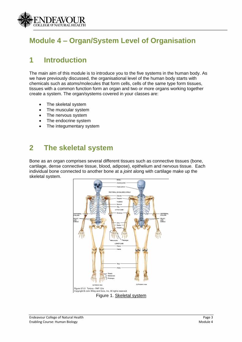
2 The skeletal system Bone as an organ comprises several different tissues such as connective tissues (bone, cartilage, dense connective tissue, blood, adipose), epithelium and nervous tissue. Each individual bone connected to another bone at a joint along with cartilage make up the skeletal system.
Figure 1. Skeletal system

Endeavour College of Natural Health Page 4 Enabling Course: Human Biology Module 4
2.1 Functions of the skeletal system
1. Support for many soft organs of the body. 2. Protection e.g. organs such as the heart and lungs by the ribcage and sternum. 3. Movement via the musculoskeletal system i.e. muscles and bones working
together. 4. Mineral homeostasis e.g. provides the calcium levels in the blood. 5. Blood cell production formed in the bone marrow. 6. Fat storage the storage form of fat is triglyceride and is situated in the bone
marrow.
Figure 2. Note: Spongy bone at the top of the bone and compact bone in the long section
Endeavour College of Natural Health Page 5 Enabling Course: Human Biology Module 4
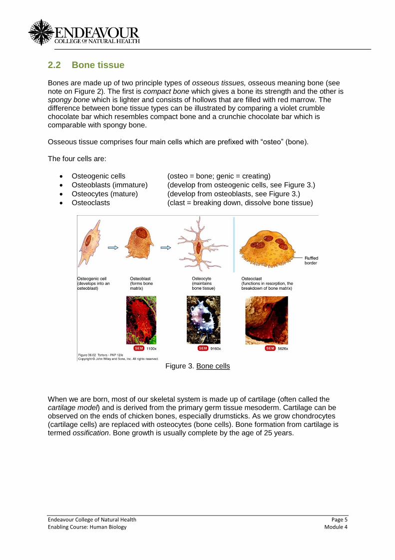
2.2 Bone tissue Bones are made up of two principle types of osseous tissues, osseous meaning bone (see note on Figure 2). The first is compact bone which gives a bone its strength and the other is spongy bone which is lighter and consists of hollows that are filled with red marrow. The difference between bone tissue types can be illustrated by comparing a violet crumble chocolate bar which resembles compact bone and a crunchie chocolate bar which is comparable with spongy bone. Osseous tissue comprises four main cells which are prefixed with “osteo” (bone). The four cells are:
Osteogenic cells (osteo = bone; genic = creating)
Osteoblasts (immature) (develop from osteogenic cells, see Figure 3.)
Osteocytes (mature) (develop from osteoblasts, see Figure 3.)
Osteoclasts (clast = breaking down, dissolve bone tissue)
Figure 3. Bone cells
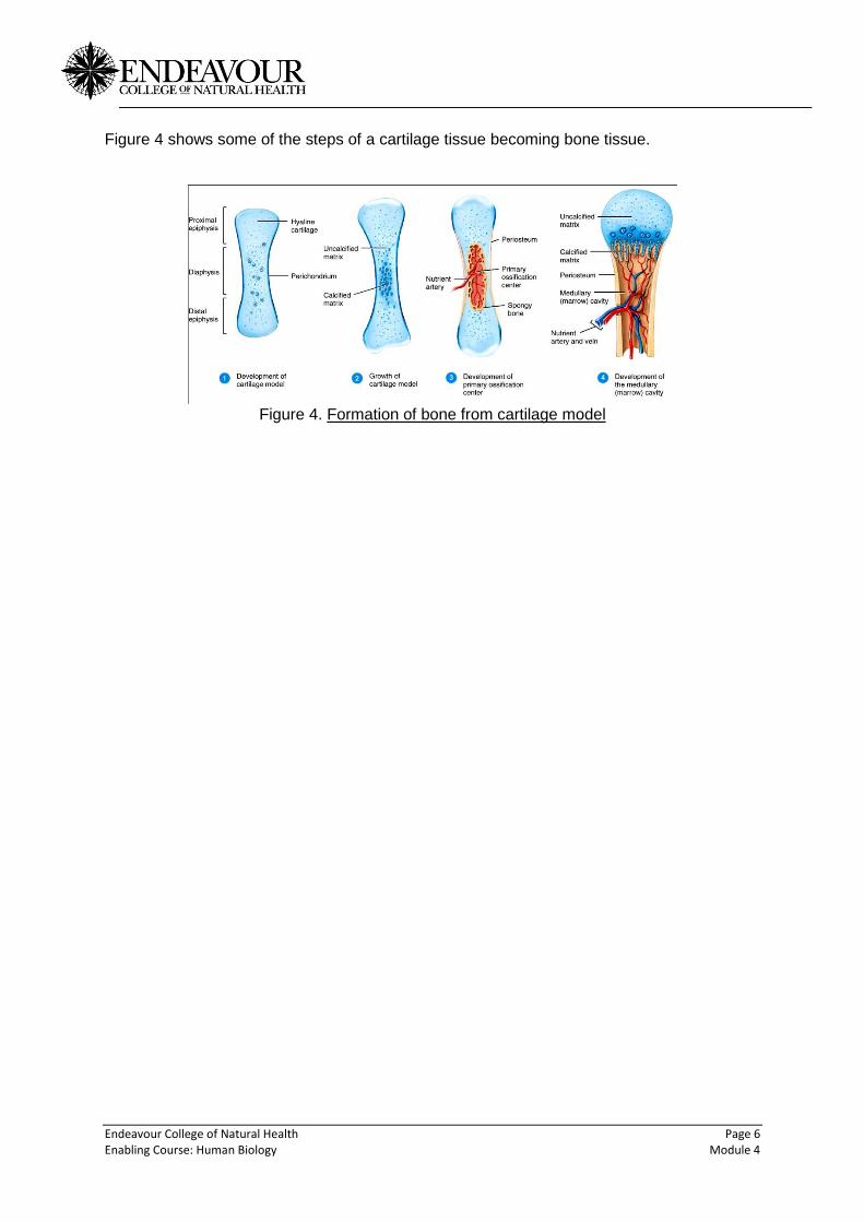
When we are born, most of our skeletal system is made up of cartilage (often called the cartilage model) and is derived from the primary germ tissue mesoderm. Cartilage can be observed on the ends of chicken bones, especially drumsticks. As we grow chondrocytes (cartilage cells) are replaced with osteocytes (bone cells). Bone formation from cartilage is termed ossification. Bone growth is usually complete by the age of 25 years.
Endeavour College of Natural Health Page 6 Enabling Course: Human Biology Module 4
Figure 4 shows some of the steps of a cartilage tissue becoming bone tissue.
Figure 4. Formation of bone from cartilage model
Endeavour College of Natural Health Page 7 Enabling Course: Human Biology Module 4
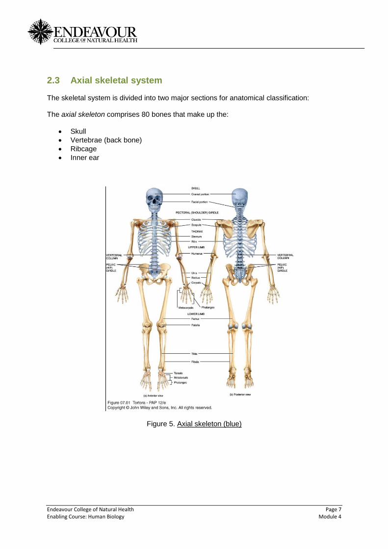
2.3 Axial skeletal system The skeletal system is divided into two major sections for anatomical classification: The axial skeleton comprises 80 bones that make up the:
Skull
Vertebrae (back bone)
Ribcage
Inner ear
Figure 5. Axial skeleton (blue)
Endeavour College of Natural Health Page 8 Enabling Course: Human Biology Module 4
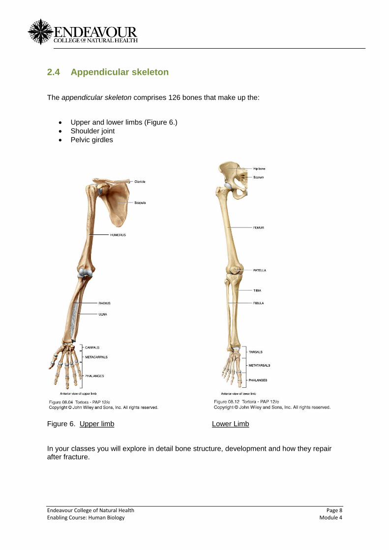
2.4 Appendicular skeleton
The appendicular skeleton comprises 126 bones that make up the:
Upper and lower limbs (Figure 6.)
Shoulder joint
Pelvic girdles
Figure 6. Upper limb Lower Limb In your classes you will explore in detail bone structure, development and how they repair after fracture.
Endeavour College of Natural Health Page 9 Enabling Course: Human Biology Module 4
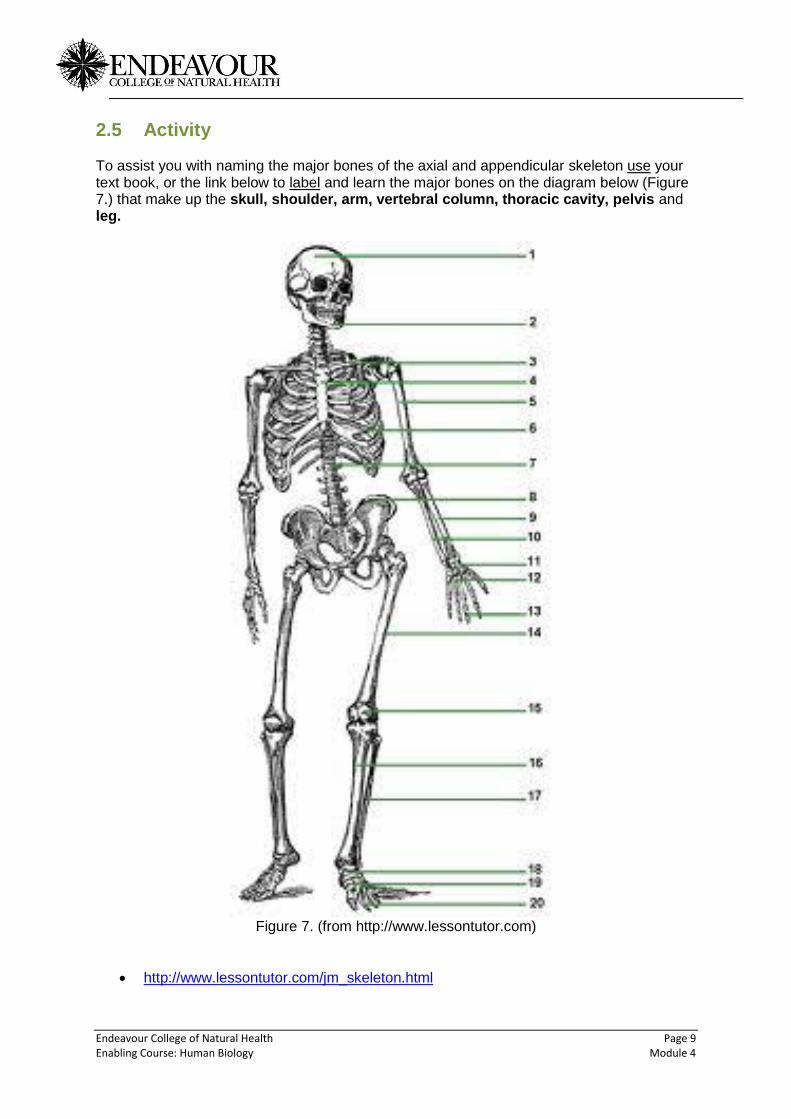
2.5 Activity To assist you with naming the major bones of the axial and appendicular skeleton use your text book, or the link below to label and learn the major bones on the diagram below (Figure 7.) that make up the skull, shoulder, arm, vertebral column, thoracic cavity, pelvis and leg.
Figure 7. (from http://www.lessontutor.com)
http://www.lessontutor.com/jm_skeleton.html
Endeavour College of Natural Health Page 10 Enabling Course: Human Biology Module 4
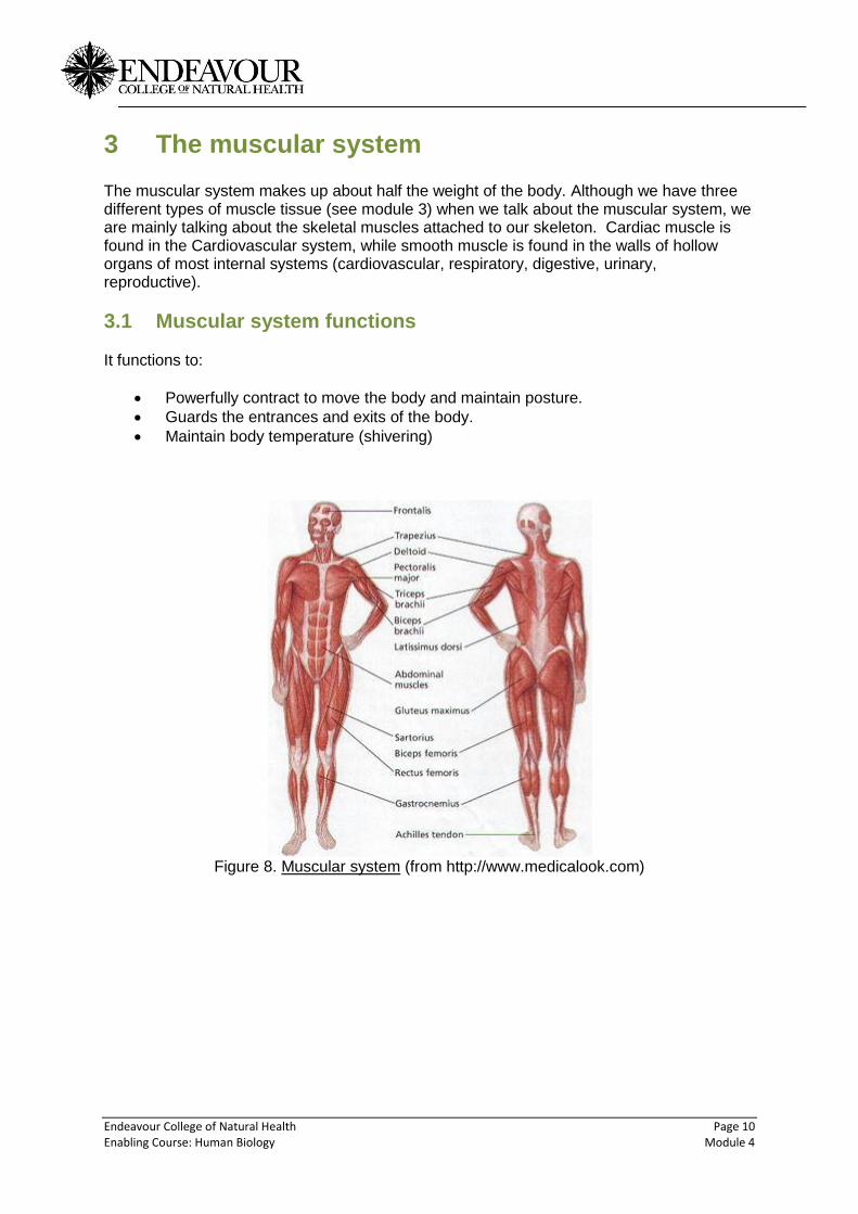
3 The muscular system The muscular system makes up about half the weight of the body. Although we have three different types of muscle tissue (see module 3) when we talk about the muscular system, we are mainly talking about the skeletal muscles attached to our skeleton. Cardiac muscle is found in the Cardiovascular system, while smooth muscle is found in the walls of hollow organs of most internal systems (cardiovascular, respiratory, digestive, urinary, reproductive).
3.1 Muscular system functions It functions to:
Powerfully contract to move the body and maintain posture.
Guards the entrances and exits of the body.
Maintain body temperature (shivering)
Figure 8. Muscular system (from http://www.medicalook.com)
Endeavour College of Natural Health Page 11 Enabling Course: Human Biology Module 4
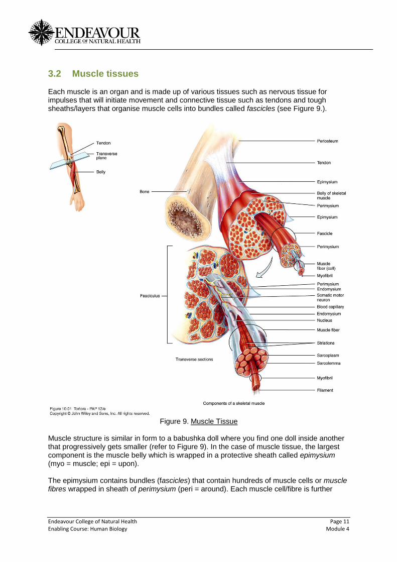
3.2 Muscle tissues Each muscle is an organ and is made up of various tissues such as nervous tissue for impulses that will initiate movement and connective tissue such as tendons and tough sheaths/layers that organise muscle cells into bundles called fascicles (see Figure 9.).
Figure 9. Muscle Tissue
Muscle structure is similar in form to a babushka doll where you find one doll inside another that progressively gets smaller (refer to Figure 9). In the case of muscle tissue, the largest component is the muscle belly which is wrapped in a protective sheath called epimysium (myo = muscle; epi = upon). The epimysium contains bundles (fascicles) that contain hundreds of muscle cells or muscle fibres wrapped in sheath of perimysium (peri = around). Each muscle cell/fibre is further
Endeavour College of Natural Health Page 12 Enabling Course: Human Biology Module 4
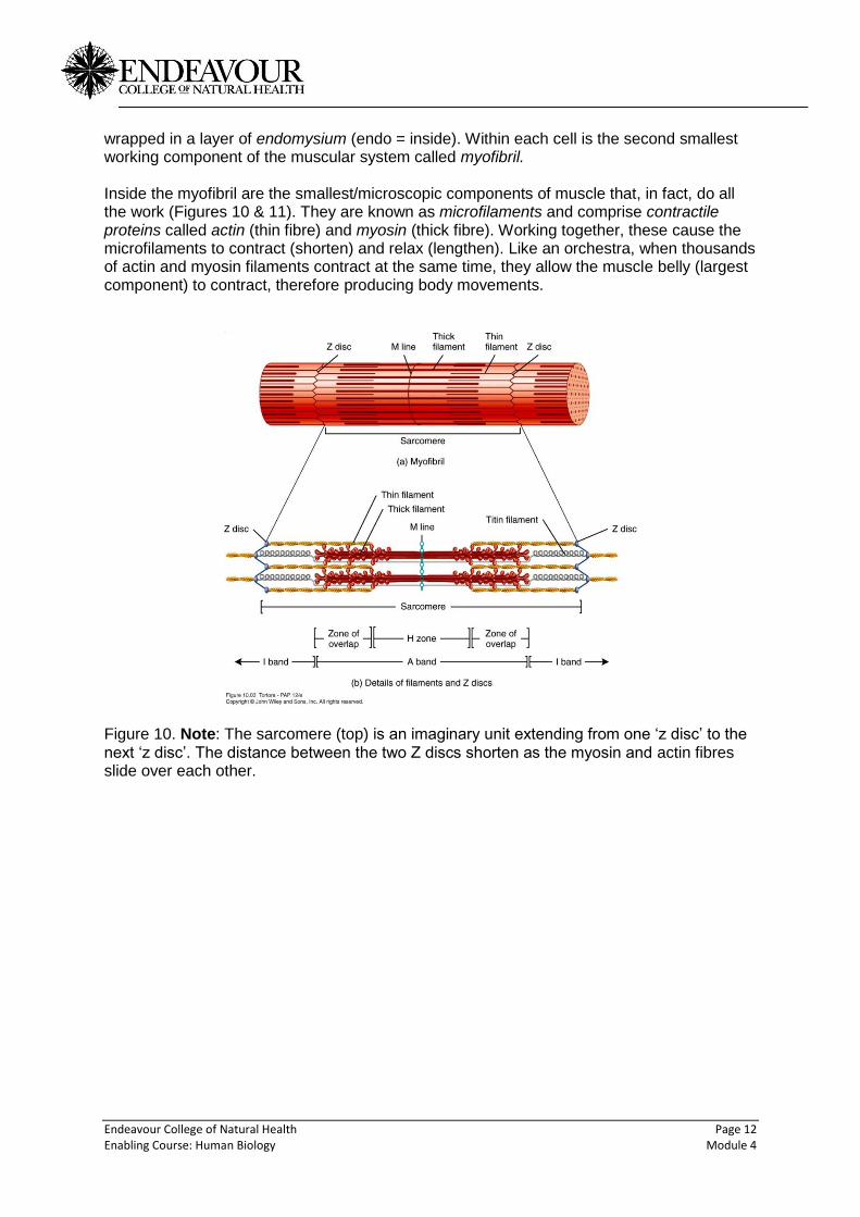
wrapped in a layer of endomysium (endo = inside). Within each cell is the second smallest working component of the muscular system called myofibril. Inside the myofibril are the smallest/microscopic components of muscle that, in fact, do all the work (Figures 10 & 11). They are known as microfilaments and comprise contractile proteins called actin (thin fibre) and myosin (thick fibre). Working together, these cause the microfilaments to contract (shorten) and relax (lengthen). Like an orchestra, when thousands of actin and myosin filaments contract at the same time, they allow the muscle belly (largest component) to contract, therefore producing body movements.
Figure 10. Note: The sarcomere (top) is an imaginary unit extending from one ‘z disc’ to the next ‘z disc’. The distance between the two Z discs shorten as the myosin and actin fibres slide over each other.
Endeavour College of Natural Health Page 13 Enabling Course: Human Biology Module 4
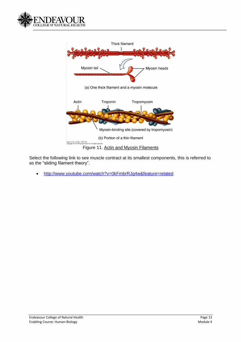
Figure 11. Actin and Myosin Filaments
Select the following link to see muscle contract at its smallest components, this is referred to as the “sliding filament theory”.
http://www.youtube.com/watch?v=0kFmbrRJq4w&feature=related
Endeavour College of Natural Health Page 14 Enabling Course: Human Biology Module 4
4 The nervous system
4.1 Nervous system functions
In a sense the systems covered this semester build a body. For example, so far we have
looked at the skeletal system which can be thought of as the frame of a house. The
muscular system (skeletal) is like the building materials that attach to the frame to form the
house. But to make the house functional to live in you need something to power the systems
required to make it liveable, and that’s where the nervous system plays a major role in the
human body. Without the brain activating electrical/nervous impulses, the muscles and
bones (musculoskeletal system) cannot function.
The nervous system is essentially the command centre to activate most bodily functions. It enables humans to detect and respond to internal and external environments and is a key player in the maintenance of homeostasis.
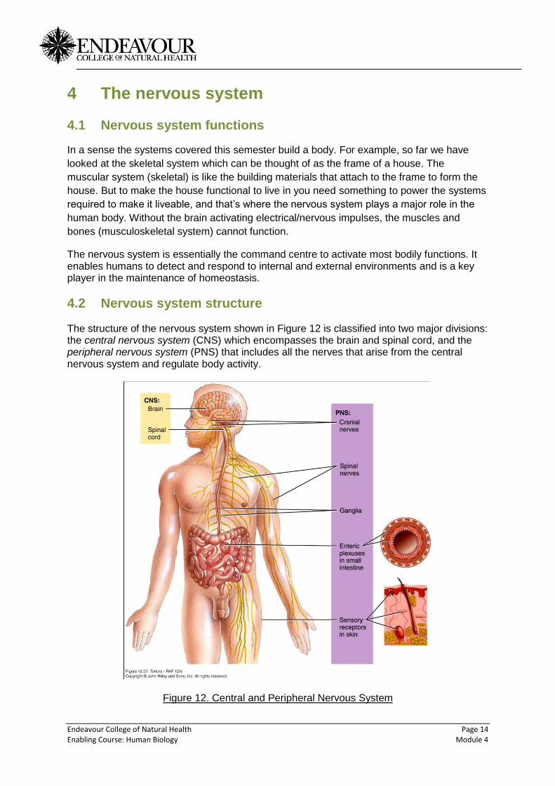
4.2 Nervous system structure The structure of the nervous system shown in Figure 12 is classified into two major divisions: the central nervous system (CNS) which encompasses the brain and spinal cord, and the peripheral nervous system (PNS) that includes all the nerves that arise from the central nervous system and regulate body activity.
Figure 12. Central and Peripheral Nervous System
Endeavour College of Natural Health Page 15 Enabling Course: Human Biology Module 4
When you next lift a cup to your mouth, ponder the amount of nervous activity occurring, for
example information from your eyes is processed by the brain to locate the cup in space.
Nerve impulses are sent to your arm/hand to move it towards the cup, grasp the cup and
move it towards your mouth. The grip on the cup and the weight of it are constantly
monitored to ensure you don’t drop the cup or spill the contents.
Somehow you manage to get the cup to your mouth rather than your ear, as a result of memories encoded in your brain (CNS) from your first experience of lifting cups. Finally, temperature and the sensation of taste of the contents are registered by your special senses such as your tongue, and if you drink enough you will experience a sense of fullness in your stomach which will stop you drinking to prevent making yourself sick.
4.3 Components of the nervous system
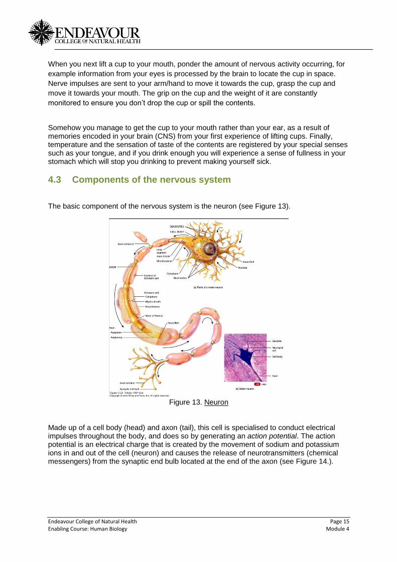
The basic component of the nervous system is the neuron (see Figure 13).
Figure 13. Neuron
Made up of a cell body (head) and axon (tail), this cell is specialised to conduct electrical impulses throughout the body, and does so by generating an action potential. The action potential is an electrical charge that is created by the movement of sodium and potassium ions in and out of the cell (neuron) and causes the release of neurotransmitters (chemical messengers) from the synaptic end bulb located at the end of the axon (see Figure 14.).
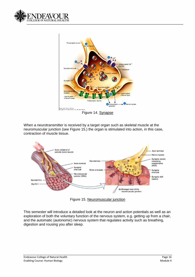
Endeavour College of Natural Health Page 16 Enabling Course: Human Biology Module 4
Figure 14. Synapse
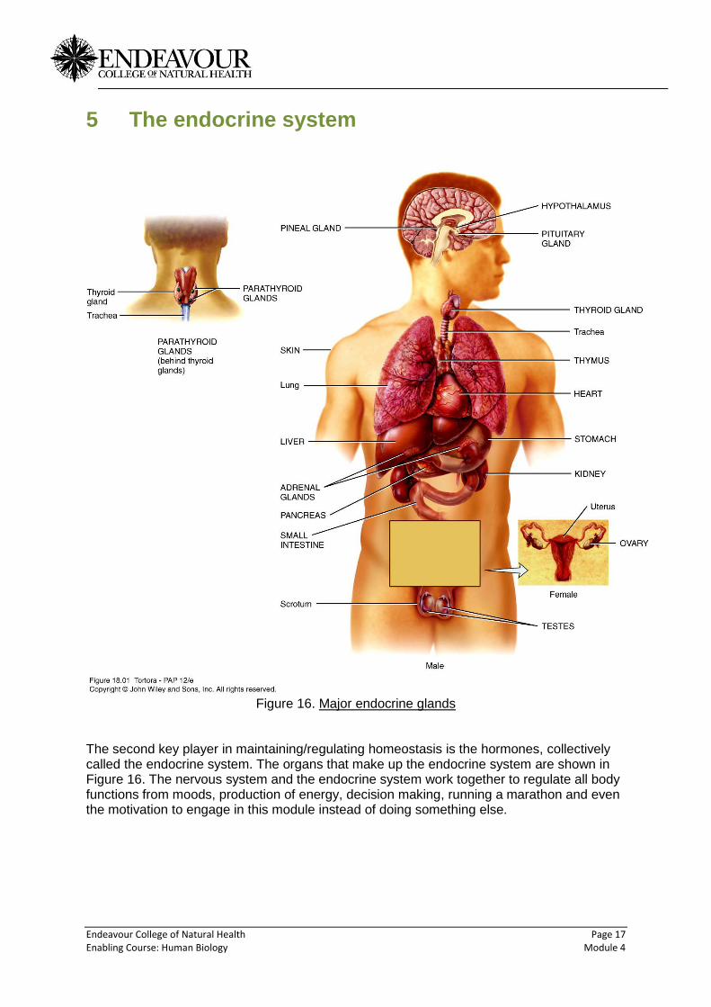
When a neurotransmitter is received by a target organ such as skeletal muscle at the neuromuscular junction (see Figure 15.) the organ is stimulated into action, in this case, contraction of muscle tissue.
Figure 15. Neuromuscular junction This semester will introduce a detailed look at the neuron and action potentials as well as an exploration of both the voluntary function of the nervous system, e.g. getting up from a chair, and the automatic (autonomic) nervous system that regulates activity such as breathing, digestion and rousing you after sleep.
Endeavour College of Natural Health Page 17 Enabling Course: Human Biology Module 4
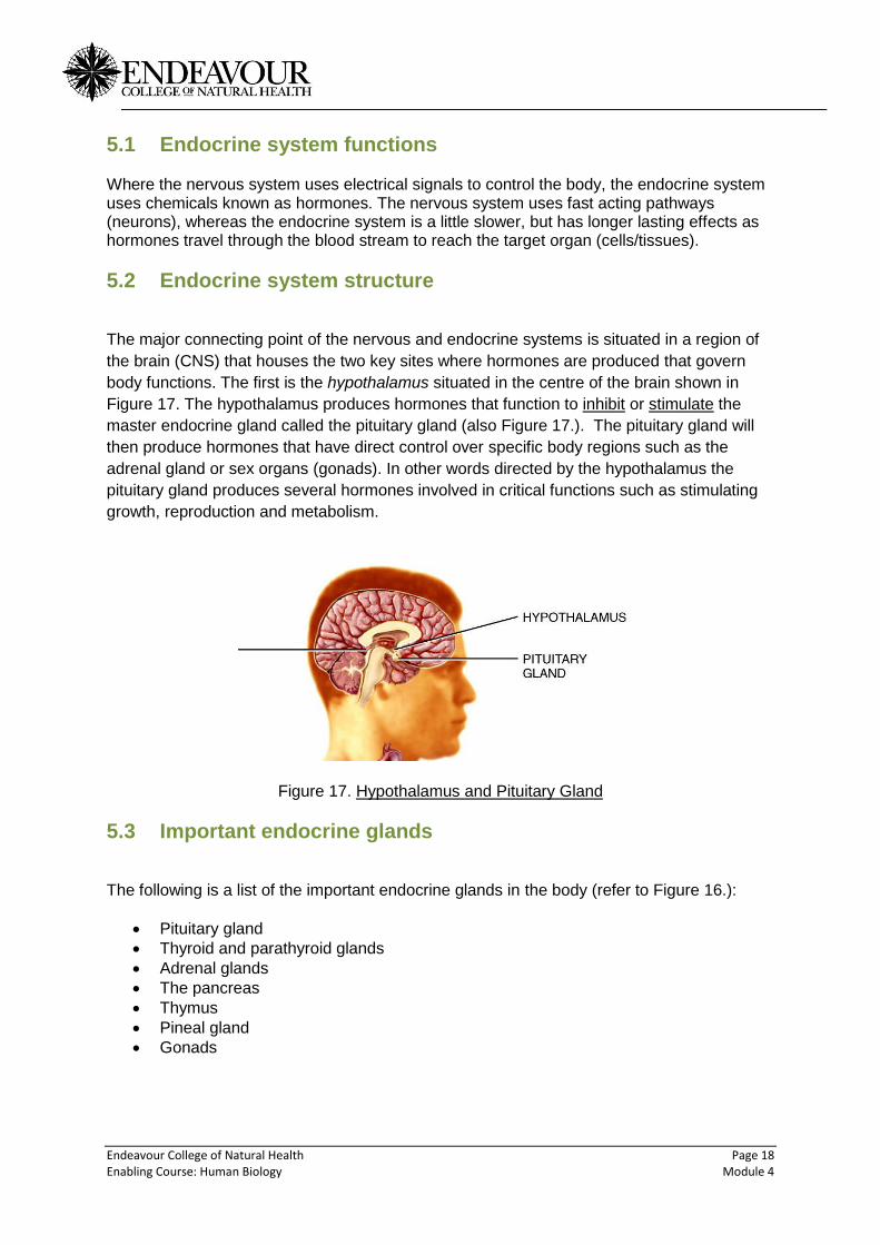
5 The endocrine system
Figure 16. Major endocrine glands
The second key player in maintaining/regulating homeostasis is the hormones, collectively called the endocrine system. The organs that make up the endocrine system are shown in Figure 16. The nervous system and the endocrine system work together to regulate all body functions from moods, production of energy, decision making, running a marathon and even the motivation to engage in this module instead of doing something else.
Endeavour College of Natural Health Page 18 Enabling Course: Human Biology Module 4
5.1 Endocrine system functions Where the nervous system uses electrical signals to control the body, the endocrine system uses chemicals known as hormones. The nervous system uses fast acting pathways (neurons), whereas the endocrine system is a little slower, but has longer lasting effects as hormones travel through the blood stream to reach the target organ (cells/tissues).
5.2 Endocrine system structure
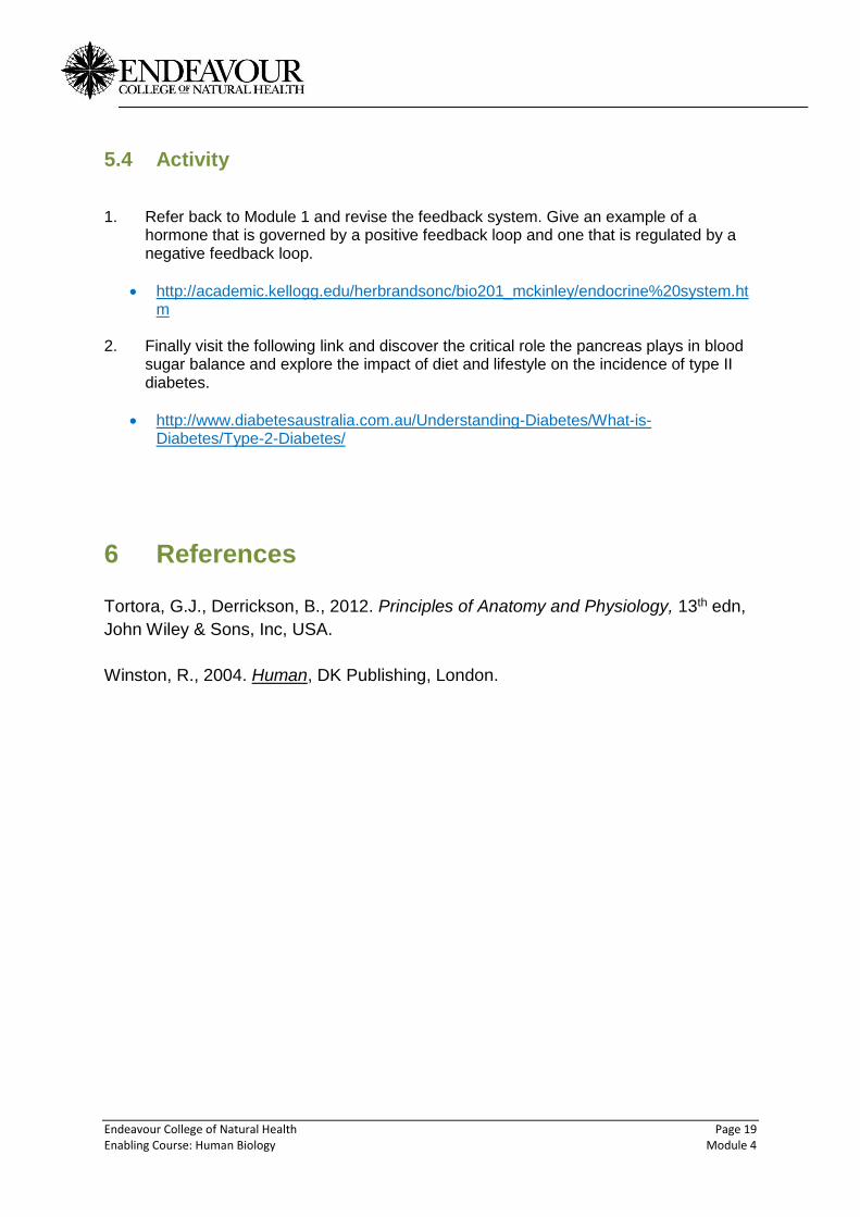
The major connecting point of the nervous and endocrine systems is situated in a region of
the brain (CNS) that houses the two key sites where hormones are produced that govern
body functions. The first is the hypothalamus situated in the centre of the brain shown in
Figure 17. The hypothalamus produces hormones that function to inhibit or stimulate the
master endocrine gland called the pituitary gland (also Figure 17.). The pituitary gland will
then produce hormones that have direct control over specific body regions such as the
adrenal gland or sex organs (gonads). In other words directed by the hypothalamus the
pituitary gland produces several hormones involved in critical functions such as stimulating
growth, reproduction and metabolism.
Figure 17. Hypothalamus and Pituitary Gland
5.3 Important endocrine glands
The following is a list of the important endocrine glands in the body (refer to Figure 16.):
Pituitary gland
Thyroid and parathyroid glands
Adrenal glands
The pancreas
Thymus
Pineal gland
Gonads
Endeavour College of Natural Health Page 19 Enabling Course: Human Biology Module 4
5.4 Activity 1. Refer back to Module 1 and revise the feedback system. Give an example of a
hormone that is governed by a positive feedback loop and one that is regulated by a negative feedback loop.
http://academic.kellogg.edu/herbrandsonc/bio201_mckinley/endocrine%20system.htm
2. Finally visit the following link and discover the critical role the pancreas plays in blood
sugar balance and explore the impact of diet and lifestyle on the incidence of type II diabetes.
http://www.diabetesaustralia.com.au/Understanding-Diabetes/What-is-Diabetes/Type-2-Diabetes/
6 References
Tortora, G.J., Derrickson, B., 2012. Principles of Anatomy and Physiology, 13th edn,
John Wiley & Sons, Inc, USA.
Winston, R., 2004. Human, DK Publishing, London.