Embed Size (px)
Citation preview

Human Bone Magnetic Bead Panel 96-Well Plate Assay Cat. # HBNMAG-51K
MILLIPLEX® MAP
HUMAN BONE MAGNETIC BEAD PANEL 96-Well Plate Assay
HBNMAG-51K
TABLE OF CONTENTS PAGE
Introduction 2
Principle 3
Storage Conditions Upon Receipt 3
Reagents Supplied 4
Materials Required But Not Provided 5
Safety Precautions 5
Technical Guidelines 6
Sample Collection And Storage 7
Preparation of Reagents for Immunoassay 9
Immunoassay Procedure 12
Plate Washing 14
Equipment Settings 14
Quality Controls 16
Assay Characteristics 17
Troubleshooting Guide 19
Replacement Reagents 21
Ordering Information 23
Well Map 24
FOR RESEARCH USE ONLY. NOT FOR USE IN DIAGNOSTIC PROCEDURES.
By purchasing this product, which contains fluorescently labeled microsphere beads authorized by Luminex Corporation (“Luminex”), you, the customer, acquire the right under Luminex’s patent rights, if any, to use this product or any portion of this product, including without limitation the microsphere beads contained herein, only with Luminex’s laser based fluorescent analytical test instrumentation
marketed under the name of Luminex 100® IS, 200TM, HTS, FLEXMAP 3DTM,MAGPIX®.

HBNMAG-51K Rev. 21-MAY-2012 PAGE 2 EMD MILLIPORE
Human Bone Magnetic Bead Panel INTRODUCTION Bone metabolism is the dynamic process of ongoing bone deposition and resorption, controlled by osteoblasts, osteocytes, and osteoclasts. While osteoblasts and osteocytes (osteoblasts surrounded by matrix) are responsible for bone deposition, osteoclasts are responsible for bone resorption. Both are required to maintain bone structure, as well as an adequate supply of calcium. To maintain this metabolic balance these cells rely on complex signaling pathways involving hormones and cytokines to achieve the appropriate rates of growth and differentiation. The disruption of bone metabolism results in such disease as osteoporosis, osteoarthritis, rheumatoid arthritis, chronic kidney disease and bone metastases. EMD Millipore recognizes the need to understand better the role that bone metabolism biomarkers play both in preserving normal bone structure and in the development of disease. Magnetic Beads can make the process of automation and high throughput screening easier with features such as walk-away washing. Advantages even outside automation include:
More flexible plate and plate washer options Improved performance with turbid serum/plasma samples Assay results equivalent to non-magnetic beads Automated washing eliminates technical obstacles (i.e., clogging of wells that contain viscous
samples) which may result during vacuum manifold/manual washing Therefore, the MILLIPLEX® MAP Human Bone Magnetic Bead panel enables you to focus on the therapeutic potential of bone metabolism. Coupled with the Luminex xMAP® platform in a magnetic bead format, you receive the advantage of ideal speed and sensitivity, allowing quantitative multiplex detection of dozens of analytes simultaneously, which can dramatically improve productivity. EMD Millipore’s MILLIPLEX HUMAN BONE Magnetic Bead panel is the most versatile system available for bone metabolism research.
MILLIPLEX MAP offers you the ability to: o Select a 13 plex or o Choose any combination of analytes from our panel of 13 analytes to design a custom
kit that better meets your needs. A convenient “all-in-one” box format gives you the assurance that you will have all the
necessary reagents you need to run your assay. EMD Millipore’s MILLIPLEX MAP Human Bone Magnetic Bead kit is to be used for the simultaneous quantification from the following ACTH, DKK1, IL-6, Insulin, Leptin, TNFOPG, OC, OPN, SOST, IL-1PTH and FGF23.
For Research Use Only. Not for Use in Diagnostic Procedures.
Please read entire protocol before use.
It is important to use same assay incubation conditions throughout your study.
HBNMAG-51K Rev. 21-MAY-2012 PAGE 3 EMD MILLIPORE
PRINCIPLE
MILLIPLEX MAP is based on the Luminex® xMAP® technology — one of the fastest growing and most respected multiplex technologies offering applications throughout the life-sciences and capable of performing a variety of bioassays including immunoassays on the surface of fluorescent-coded magnetic beads known as MagPlexTM-C microspheres.
Luminex® uses proprietary techniques to internally color-code microspheres with two fluorescent dyes. Through precise concentrations of these dyes, 100 distinctly colored bead sets can be created, each of which is coated with a specific capture antibody.
After an analyte from a test sample is captured by the bead, a biotinylated detection antibody is introduced.
The reaction mixture is then incubated with Streptavidin-PE conjugate, the reporter molecule, to complete the reaction on the surface of each microsphere.
The microspheres are allowed to pass rapidly through a laser which excites the internal dyes marking the microsphere set. A second laser excites PE, the fluorescent dye on the reporter molecule.
Finally, high-speed digital-signal processors identify each individual microsphere and quantify the result of its bioassay based on fluorescent reporter signals.
The capability of adding multiple conjugated beads to each sample results in the ability to obtain multiple results from each sample. Open-architecture xMAP® technology enables multiplexing of many types of bioassays reducing time, labor and costs over traditional methods. STORAGE CONDITIONS UPON RECEIPT
Recommended storage for kit components is 2 - 8°C.
For long-term storage, freeze reconstituted standards and controls at -20°C. Avoid multiple (>2) freeze/thaw cycles.
DO NOT FREEZE Antibody-Immobilized Beads, Detection Antibody, and Streptavidin-Phycoerythrin.
HBNMAG-51K Rev. 21-MAY-2012 PAGE 4 EMD MILLIPORE
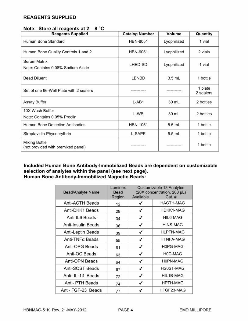
REAGENTS SUPPLIED Note: Store all reagents at 2 – 8 °C
Reagents Supplied Catalog Number Volume Quantity
Human Bone Standard HBN-8051 Lyophilized 1 vial
Human Bone Quality Controls 1 and 2 HBN-6051 Lyophilized 2 vials
Serum Matrix
Note: Contains 0.08% Sodium Azide LHED-SD Lyophilized 1 vial
Bead Diluent LBNBD 3.5 mL 1 bottle
Set of one 96-Well Plate with 2 sealers ----------- ----------- 1 plate
2 sealers
Assay Buffer L-AB1 30 mL 2 bottles
10X Wash Buffer
Note: Contains 0.05% Proclin L-WB 30 mL 2 bottles
Human Bone Detection Antibodies HBN-1051 5.5 mL 1 bottle
Streptavidin-Phycoerythrin L-SAPE 5.5 mL 1 bottle
Mixing Bottle (not provided with premixed panel)
----------- ----------- 1 bottle
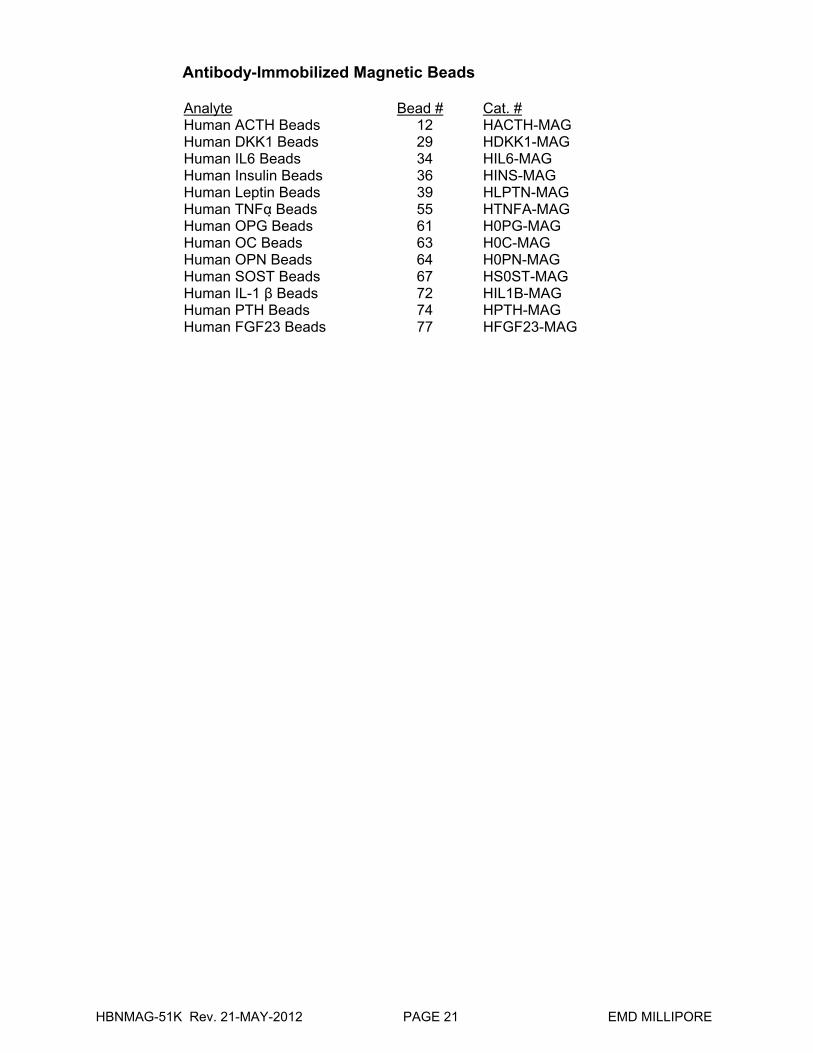
Included Human Bone Antibody-Immobilized Beads are dependent on customizable selection of analytes within the panel (see next page). Human Bone Antibody-Immobilized Magnetic Beads:
Bead/Analyte Name Luminex
Bead Region
Customizable 13 Analytes (20X concentration, 200 µL)
Available Cat. #
Anti-ACTH Beads 12 ✔ HACTH-MAG
Anti-DKK1 Beads 29 ✔ HDKK1-MAG
Anti-IL6 Beads 34 ✔ HIL6-MAG
Anti-Insulin Beads 36 ✔ HINS-MAG
Anti-Leptin Beads 39 ✔ HLPTN-MAG
Anti-TNFα Beads 55 ✔ HTNFA-MAG
Anti-OPG Beads 61 ✔ H0PG-MAG
Anti-OC Beads 63 ✔ H0C-MAG
Anti-OPN Beads 64 ✔ H0PN-MAG
Anti-SOST Beads 67 ✔ HS0ST-MAG
Anti- IL-1β Beads 72 ✔ HIL1B-MAG
Anti- PTH Beads 74 ✔ HPTH-MAG
Anti- FGF-23 Beads 77 ✔ HFGF23-MAG

HBNMAG-51K Rev. 21-MAY-2012 PAGE 5 EMD MILLIPORE
MATERIALS REQUIRED BUT NOT PROVIDED
Reagents
1. Luminex Sheath Fluid (Luminex Catalog #40-50000) or Luminex Drive Fluid (Luminex Catalog # MPXDF-4PK)
Instrumentation / Materials 1. Adjustable Pipettes with Tips capable of delivering 25 μL to 1000 μL 2. Multichannel Pipettes capable of delivering 5 μL to 50 μL or 25 μL to 200 μL 3. Reagent Reservoirs 4. Polypropylene Microfuge Tubes 5. Rubber Bands 6. Aluminum Foil 7. Absorbent Pads 8. Laboratory Vortex Mixer 9. Sonicator (Branson Ultrasonic Cleaner Model #B200 or equivalent) 10. Titer Plate Shaker (Lab-Line Instruments Model #4625 or equivalent) 11. Luminex 200™, HTS, FLEXMAP 3D™, or MAGPIX® with xPONENT software by
Luminex Corporation 12. Automatic Plate washer for magnetic beads (Bio-Tek ELx405, EMD Millipore Catalog
#40-015 or equivalent) or Hand-held Magnetic Separation Block (EMD Millipore Catalog #40-285 or equivalent)
Note: If a plate washer or hand held magnetic separation block for magnetic beads is not available, one can use a microtiter filter plate (EMD Millipore Catalog #MX-PLATE) to run the assay using a Vacuum Filtration Unit (EMD Millipore Vacuum Manifold Catalog #MSVMHTS00 or equivalent with EMD Millipore Vacuum Pump Catalog #WP6111560 or equivalent).
SAFETY PRECAUTIONS
All blood components and biological materials should be handled as potentially hazardous. Follow universal precautions as established by the Centers for Disease Control and Prevention and by the Occupational Safety and Health Administration when handling and disposing of infectious agents.
Sodium Azide or Proclin has been added to some reagents as a preservative. Although the concentrations are low, Sodium Azide and Proclin may react with lead and copper plumbing to form highly explosive metal azides. Dispose of unused contents and waste in accordance with international, federal, state, and local regulations.
HBNMAG-51K Rev. 21-MAY-2012 PAGE 6 EMD MILLIPORE
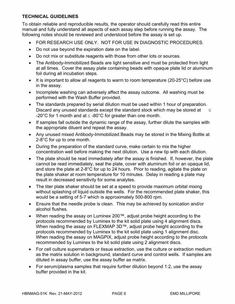
TECHNICAL GUIDELINES
To obtain reliable and reproducible results, the operator should carefully read this entire manual and fully understand all aspects of each assay step before running the assay. The following notes should be reviewed and understood before the assay is set up.
FOR RESEARCH USE ONLY. NOT FOR USE IN DIAGNOSTIC PROCEDURES.
Do not use beyond the expiration date on the label.
Do not mix or substitute reagents with those from other lots or sources.
The Antibody-Immobilized Beads are light sensitive and must be protected from light at all times. Cover the assay plate containing beads with opaque plate lid or aluminum foil during all incubation steps.
It is important to allow all reagents to warm to room temperature (20-25°C) before use in the assay.
Incomplete washing can adversely affect the assay outcome. All washing must be performed with the Wash Buffer provided.
The standards prepared by serial dilution must be used within 1 hour of preparation. Discard any unused standards except the standard stock which may be stored at -20°C for 1 month and at -80°C for greater than one month.
If samples fall outside the dynamic range of the assay, further dilute the samples with the appropriate diluent and repeat the assay.
Any unused mixed Antibody-Immobilized Beads may be stored in the Mixing Bottle at 2-8°C for up to one month.
During the preparation of the standard curve, make certain to mix the higher concentration well before making the next dilution. Use a new tip with each dilution.
The plate should be read immediately after the assay is finished. If, however, the plate cannot be read immediately, seal the plate, cover with aluminum foil or an opaque lid, and store the plate at 2-8°C for up to 24 hours. Prior to reading, agitate the plate on the plate shaker at room temperature for 10 minutes. Delay in reading a plate may result in decreased sensitivity for some analytes.
The titer plate shaker should be set at a speed to provide maximum orbital mixing without splashing of liquid outside the wells. For the recommended plate shaker, this would be a setting of 5-7 which is approximately 500-800 rpm.
Ensure that the needle probe is clean. This may be achieved by sonication and/or alcohol flushes.
When reading the assay on Luminex 200™, adjust probe height according to the protocols recommended by Luminex to the kit solid plate using 4 alignment discs. When reading the assay on FLEXMAP 3D™, adjust probe height according to the protocols recommended by Luminex to the kit solid plate using 1 alignment disc. When reading the assay on MAGPIX, adjust probe height according to the protocols recommended by Luminex to the kit solid plate using 2 alignment discs.
For cell culture supernatants or tissue extraction, use the culture or extraction medium as the matrix solution in background, standard curve and control wells. If samples are diluted in assay buffer, use the assay buffer as matrix.
For serum/plasma samples that require further dilution beyond 1:2, use the assay buffer provided in the kit.
HBNMAG-51K Rev. 21-MAY-2012 PAGE 7 EMD MILLIPORE
For cell/tissue homogenate, the final cell or tissue homogenate should be prepared in a buffer that has a neutral pH, contains minimal detergents or strong denaturing detergents, and has an ionic strength close to physiological concentration. Avoid debris, lipids, and cell/tissue aggregates. Centrifuge samples before use.
Vortex all reagents well before adding to plate.
SAMPLE COLLECTION AND STORAGE
A. Preparation of Serum Samples: Allow the blood to clot for at least 30 minutes before centrifugation for 10
minutes at 1000xg. Remove serum and assay immediately or aliquot and store samples at -20°C.
Osteocalcin is sensitive to freeze/thaw cycles. Avoid multiple (>2) freeze/thaw cycles.
When using frozen samples, it is recommended to thaw the samples completely, mix well by vortexing and centrifuge prior to use in the assay to remove particulates.
Serum samples should be diluted 1:2 in the assay buffer provided in the kit. For example, in a tube, 35 µL of serum may be combined with 35 µL of Assay Buffer. When further dilution beyond 1:2 is required, use assay buffer as the diluent.
B. Preparation of Plasma Samples:
Plasma collection using EDTA as an anti-coagulant is recommended. Centrifuge for 10 minutes at 1000xg within 30 minutes of blood collection. Remove plasma and assay immediately or aliquot and store samples at -20°C.
Osteocalcin is sensitive to freeze/thaw cycles. Avoid multiple (>2) freeze/thaw cycles.
When using frozen samples, it is recommended to thaw the samples completely, mix well by vortexing and centrifuge prior to use in the assay to remove particulates.
Plasma samples should be diluted 1:2 in the assay buffer provided in the kit. For example, in a tube, 35 µL of serum may be combined with 35 µL of Assay Buffer. When further dilution beyond 1:2 is required, use assay buffer as the diluent.
C. Preparation of Tissue Culture Supernatant:
Centrifuge the sample to remove debris and assay immediately or aliquot and store samples at -20°C.
Avoid multiple (>2) freeze/thaw cycles. Tissue culture supernatant may require a dilution with an appropriate control
medium prior to assay. Tissue/cell extracts should be done in neutral buffers containing reagents and conditions that do not interfere with assay performance. Excess concentrations of detergent, salt, denaturants, high or low pH, etc. will negatively affect the assay. Organic solvents should be avoided. The tissue/cell extract samples should be free of particles such as cells or tissue debris.
HBNMAG-51K Rev. 21-MAY-2012 PAGE 8 EMD MILLIPORE
SAMPLE COLLECTION AND STORAGE (continued)
NOTE: A maximum of 25 μL per well of 1:2 freshly diluted serum or plasma can be used.
Tissue culture or other media may also be used.
All samples must be stored in polypropylene tubes. DO NOT STORE SAMPLES IN GLASS.
Avoid debris, lipids and cells when using samples with gross hemolysis or lipemia. Care must be taken when using heparin as an anticoagulant since an excess of
heparin will provide falsely high values. Use no more than 10 IU heparin per mL of blood collected.
PREPARATION OF REAGENTS FOR IMMUNOASSAY
A. Preparation of Antibody-Immobilized Beads
For individual vials of beads, sonicate each antibody-bead vial for 30 seconds; vortex for 1 minute. Add 150 µL from each antibody-bead vial to the Mixing Bottle and bring final volume to 3.0 mL with Bead Diluent. Vortex the mixed beads well. Unused portion may be stored at 2-8°C for up to one month. (Note: Due to the composition of magnetic beads, you may notice a slight color in the bead solution. This does not affect the performance of the beads or the kit.)
Example 1: When using 3 antibody-immobilized beads, add 150 µL from each of the 3 bead sets to the Mixing Bottle. Then add 2.55 mL Bead Diluent.
Example 2: When using 4 antibody-immobilized beads, add 150 µL from each of the 4 bead sets to the Mixing Bottle. Then add 2.40 mL Bead Diluent.
B. Preparation of Quality Controls
Before use, reconstitute Quality Control 1 and Quality Control 2 with 250 µL deionized water. Invert the vial several times to mix and vortex. Allow the vial to sit for 5-10 minutes. Unused portion may be stored at -20°C for up to one month.
C. Preparation of Wash Buffer
Bring the 10X Wash Buffer to room temperature and mix to bring all salts into solution. Dilute 60 mL of 10X Wash Buffer (two bottles) with 540 mL deionized water. Store unused portion at 2-8C for up to one month.
D. Preparation of Serum Matrix
This step is required for serum or plasma samples only.
Add 1.0 mL of Deionized Water and 1.0 mL of Assay Buffer to the bottle containing lyophilized Serum Matrix. Mix well. Allow at least 10 minutes for complete reconstitution. Leftover reconstituted Serum Matrix should be stored at -20C for up to one month.
HBNMAG-51K Rev. 21-MAY-2012 PAGE 9 EMD MILLIPORE
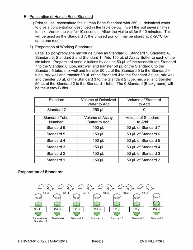
E. Preparation of Human Bone Standard
1.) Prior to use, reconstitute the Human Bone Standard with 250 µL deionized water to give a concentration described in the table below. Invert the vial several times to mix. Vortex the vial for 10 seconds. Allow the vial to sit for 5-10 minutes. This will be used as the Standard 7; the unused portion may be stored at -20C for up to one month.
2). Preparation of Working Standards
Label six polypropylene microfuge tubes as Standard 6, Standard 5, Standard 4, Standard 3, Standard 2 and Standard 1. Add 150 µL of Assay Buffer to each of the six tubes. Prepare 1:4 serial dilutions by adding 50 µL of the reconstituted Standard 7 to the Standard 6 tube, mix well and transfer 50 µL of the Standard 6 to the Standard 5 tube, mix well and transfer 50 µL of the Standard 5 to the Standard 4 tube, mix well and transfer 50 µL of the Standard 4 to the Standard 3 tube, mix well and transfer 50 µL of the Standard 3 to the Standard 2 tube, mix well and transfer 50 µL of the Standard 2 to the Standard 1 tube. The 0 Standard (Background) will be the Assay Buffer.
Standard
Volume of Deionized Water to Add
Volume of Standard to Add
Standard 7 250 µL 0
Standard Tube Number
Volume of Assay Buffer to Add
Volume of Standard to Add
Standard 6 150 µL 50 µL of Standard 7
Standard 5 150 µL 50 µL of Standard 6
Standard 4 150 µL 50 µL of Standard 5
Standard 3 150 µL 50 µL of Standard 4
Standard 2 150 µL 50 µL of Standard 3
Standard 1 150 µL 50 µL of Standard 2
Preparation of Standards
50 µL
150 µL
50 µL50 µL50 µL50 µL 50 µL
Stock 150 µL 150 µL 150 µL 150 µL 150 µL
Reconstituted Standard 7
Standard 6 Standard 5 Standard 4 Standard 3 Standard 2 Standard 1
HBNMAG-51K Rev. 21-MAY-2012 PAGE 10 EMD MILLIPORE
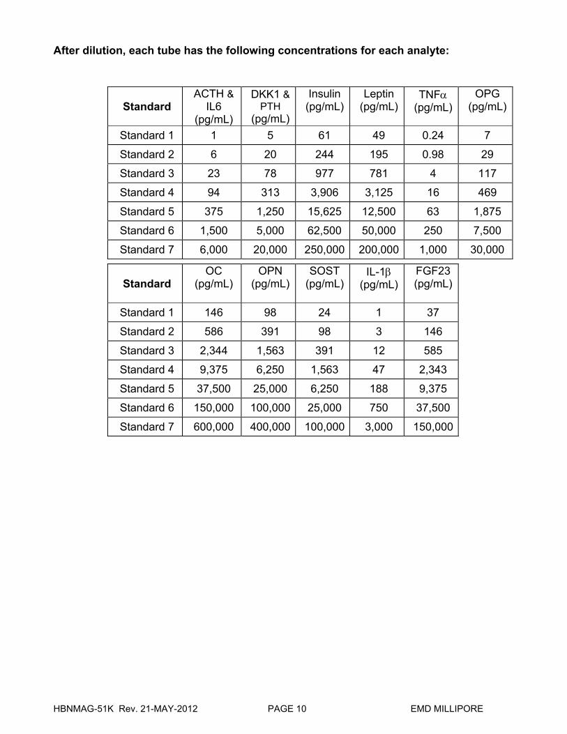
After dilution, each tube has the following concentrations for each analyte:
Standard
ACTH & IL6
(pg/mL)
DKK1 & PTH
(pg/mL)
Insulin (pg/mL)
Leptin (pg/mL)
TNF(pg/mL)
OPG (pg/mL)
Standard 1 1 5 61 49 0.24 7
Standard 2 6 20 244 195 0.98 29
Standard 3 23 78 977 781 4 117
Standard 4 94 313 3,906 3,125 16 469
Standard 5 375 1,250 15,625 12,500 63 1,875
Standard 6 1,500 5,000 62,500 50,000 250 7,500
Standard 7 6,000 20,000 250,000 200,000 1,000 30,000
Standard
OC (pg/mL)
OPN (pg/mL)
SOST (pg/mL)
IL-1(pg/mL)
FGF23(pg/mL)
Standard 1 146 98 24 1 37
Standard 2 586 391 98 3 146
Standard 3 2,344 1,563 391 12 585
Standard 4 9,375 6,250 1,563 47 2,343
Standard 5 37,500 25,000 6,250 188 9,375
Standard 6 150,000 100,000 25,000 750 37,500
Standard 7 600,000 400,000 100,000 3,000 150,000
HBNMAG-51K Rev. 21-MAY-2012 PAGE 11 EMD MILLIPORE
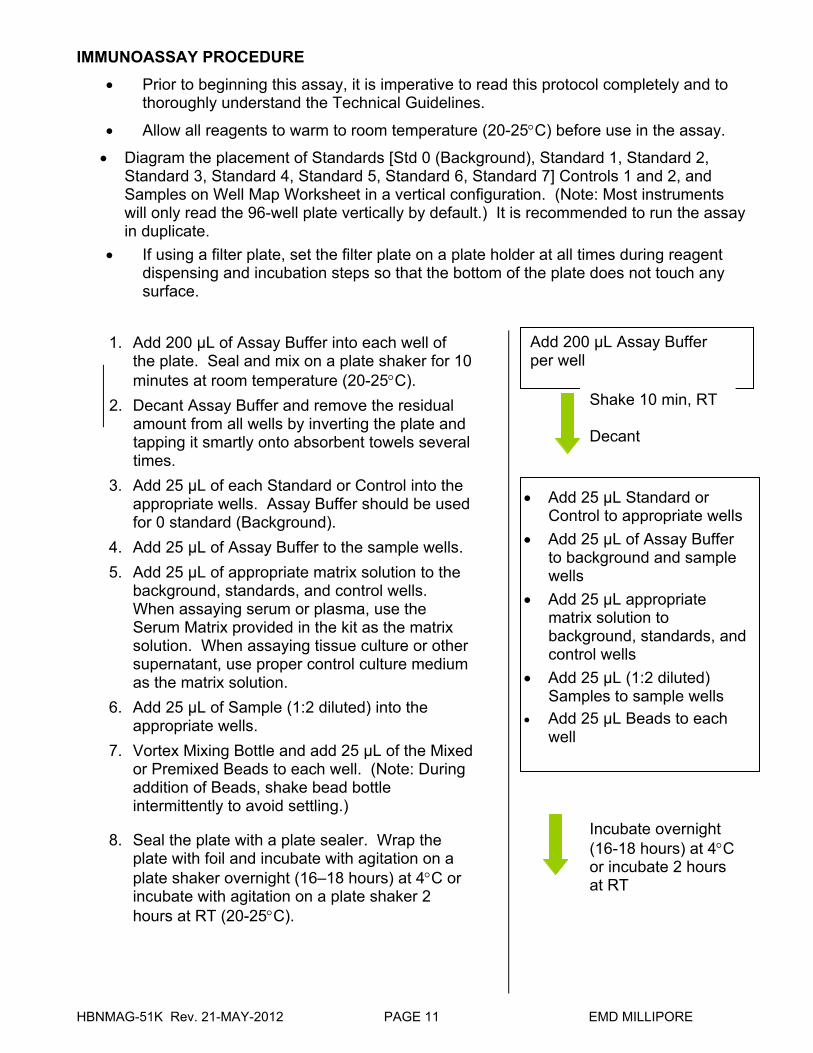
IMMUNOASSAY PROCEDURE
Prior to beginning this assay, it is imperative to read this protocol completely and to thoroughly understand the Technical Guidelines.
Allow all reagents to warm to room temperature (20-25C) before use in the assay.
Diagram the placement of Standards [Std 0 (Background), Standard 1, Standard 2, Standard 3, Standard 4, Standard 5, Standard 6, Standard 7] Controls 1 and 2, and Samples on Well Map Worksheet in a vertical configuration. (Note: Most instruments will only read the 96-well plate vertically by default.) It is recommended to run the assay in duplicate.
If using a filter plate, set the filter plate on a plate holder at all times during reagent dispensing and incubation steps so that the bottom of the plate does not touch any surface.
1. Add 200 µL of Assay Buffer into each well of the plate. Seal and mix on a plate shaker for 10 minutes at room temperature (20-25C).
2. Decant Assay Buffer and remove the residual amount from all wells by inverting the plate and tapping it smartly onto absorbent towels several times.
3. Add 25 µL of each Standard or Control into the appropriate wells. Assay Buffer should be used for 0 standard (Background).
4. Add 25 µL of Assay Buffer to the sample wells.
5. Add 25 µL of appropriate matrix solution to the background, standards, and control wells. When assaying serum or plasma, use the Serum Matrix provided in the kit as the matrix solution. When assaying tissue culture or other supernatant, use proper control culture medium as the matrix solution.
6. Add 25 µL of Sample (1:2 diluted) into the appropriate wells.
7. Vortex Mixing Bottle and add 25 μL of the Mixed or Premixed Beads to each well. (Note: During addition of Beads, shake bead bottle intermittently to avoid settling.)
8. Seal the plate with a plate sealer. Wrap the plate with foil and incubate with agitation on a plate shaker overnight (16–18 hours) at 4C or incubate with agitation on a plate shaker 2 hours at RT (20-25C).
Add 200 µL Assay Buffer per well
Add 25 µL Standard or Control to appropriate wells
Add 25 µL of Assay Buffer to background and sample wells
Add 25 µL appropriate matrix solution to background, standards, and control wells
Add 25 µL (1:2 diluted) Samples to sample wells
Add 25 µL Beads to each well
Shake 10 min, RT Decant
Incubate overnight (16-18 hours) at 4C or incubate 2 hours at RT
HBNMAG-51K Rev. 21-MAY-2012 PAGE 12 EMD MILLIPORE
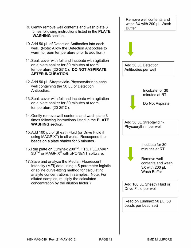
9. Gently remove well contents and wash plate 3 times following instructions listed in the PLATE WASHING section.
10. Add 50 µL of Detection Antibodies into each well. (Note: Allow the Detection Antibodies to warm to room temperature prior to addition.)
11. Seal, cover with foil and incubate with agitation on a plate shaker for 30 minutes at room temperature (20-25C). DO NOT ASPIRATE AFTER INCUBATION.
12. Add 50 µL Streptavidin-Phycoerythrin to each well containing the 50 µL of Detection Antibodies.
13. Seal, cover with foil and incubate with agitation on a plate shaker for 30 minutes at room temperature (20-25C).
14. Gently remove well contents and wash plate 3 times following instructions listed in the PLATE WASHING section.
15. Add 100 µL of Sheath Fluid (or Drive Fluid if using MAGPIX®) to all wells. Resuspend the beads on a plate shaker for 5 minutes.
16. Run plate on Luminex 200TM, HTS, FLEXMAP 3DTM or MAGPIX® with xPONENT software.
17. Save and analyze the Median Fluorescent Intensity (MFI) data using a 5-parameter logistic or spline curve-fitting method for calculating analyte concentrations in samples. Note: For diluted samples, multiply the calculated concentration by the dilution factor.)
Remove well contents and wash 3X with 200 µL Wash Buffer
Add 50 µL Detection Antibodies per well
Incubate for 30 minutes at RT Do Not Aspirate
Add 50 µL Streptavidin-Phycoerythrin per well
Incubate for 30 minutes at RT
Add 100 µL Sheath Fluid or Drive Fluid per well
Read on Luminex 50 µL, 50 beads per bead set)
Remove well contents and wash 3X with 200 µL Wash Buffer
HBNMAG-51K Rev. 21-MAY-2012 PAGE 13 EMD MILLIPORE
PLATE WASHING
1.) Solid Plate If using a solid plate, use either a hand-held magnet or magnetic plate washer.
A.) For hand-held magnet, rest plate on magnet for 60 seconds to allow complete settling of magnetic beads. Remove well contents by gently decanting the plate in an appropriate waste receptacle and gently tapping on absorbent pads to remove residual liquid. Wash plate with 200 uL of Wash Buffer by removing plate from magnet, adding Wash Buffer, shaking for 30 seconds, reattaching to magnet, letting beads settle for 60 seconds and removing well contents as previously described after each wash. Repeat wash steps as recommended in Assay Procedure.
B.) For magnetic plate washer, let plate “soak” on magnet for 60 seconds to allow complete settling of the magnetic beads. Remove well contents by aspiration. Wash plate with 200 µL/well of Wash Buffer, letting beads “soak” for 60 seconds and removing Wash Buffer by aspiration after each wash. Repeat wash steps as recommended in Assay Procedure. Note: If using the recommended plate washer for magnetic beads (Bio-Tek ELx405) follow the appropriate equipment settings outlined in EQUIPMENT SETTINGS.
2.) Filter Plate (EMD Millipore Cat #MX-PLATE)
If using a filter plate, use a vacuum filtration manifold to remove well contents. Wash plate with 200 µL/well of Wash Buffer, removing Wash Buffer by vacuum filtration after each wash. Repeat wash steps as recommended in the Assay Procedure.
EQUIPMENT SETTINGS
Bio-Tek ELx405: The general recommended wash protocol (Link Protocol) is as follows: Soak Program: Wash Program:
Soak → Aspirate→Dispense→Soak→Aspirate→Dispense→Soak→Aspirate
1.) Soak program:
1. Soak duration: 60 sec 2. Shake before soak?: NO
2.) Wash program:
Method:
1. Number of cycles: 3 2. Soak/shake: YES 3. Soak duration: 60 sec 4. Shake before soak: NO 5. Prime after soak: NO
HBNMAG-51K Rev. 21-MAY-2012 PAGE 14 EMD MILLIPORE
EQUIPMENT SETTINGS (continued) Dispense:
1. Dispense volume: 200 µL/well 2. Dispense flow rate: 5 3. Dispense height: 130 (16.51 mm) 4. Horizontal disp pos: 00 (0 mm) 5. Disable Aspirate: YES 6. Bottom Wash first?: NO 7. Prime before start?: NO
Aspiration:
1. Aspirate height: 35 (4.445 mm) 2. Horizontal Asp Pos: 30 (1.372 mm) 3. Aspiration rate: 06 (15.0 mm/sec) 4. Aspiration delay: 0 5. Crosswise Aspir: NO 6. Final Aspir: YES 7. Final Aspir delay: 0 (0 msec)
3.) Link program: (Note: this is the program to use during actual plate washing).
Link together the Soak and Wash programs outlined above. Note: After the final aspiration, there will be approximately 25 μL of residual Wash Buffer in each well. This is expected when using the BioTek Plate washer and this volume does not need to be aspirated from the plate. If using an automatic plate washer other than BioTek ELx405, please refer to the manufacturer’s recommendations for programming instructions. Luminex 200™, HTS, FLEXMAP 3D™ and MAGPIX® with xPONENT software: These specifications are for the Luminex 200™, Luminex HTS, Luminex FLEXMAP 3D™ and Luminex MAGPIX®with xPonent software. Luminex instruments with other software (e.g. MasterPlex, StarStation, LiquiChip, Bio-Plex, LABScan100) would need to follow instrument instructions for gate settings and additional specifications from the vendors for reading Luminex Magnetic Beads. For magnetic bead assays, the Luminex 200™ and HTS instruments must be calibrated with the xPonent 3.1 compatible Calibration Kit (EMD Millipore Cat #40-275) and performance verified with the Performance Verification Kit (EMD Millipore Cat #40-276). The Luminex FLEXMAP 3D™ instrument must be calibrated with the FLEXMAP 3D™ Calibrator Kit (EMD Millipore Cat #40-028) and performance verified with the FLEXMAP 3D™ Performance Verification Kit (EMD Millipore Cat #40-029). The Luminex MAGPIX® instrument must be calibrated with the MAGPIX® Calibration Kit (EMD Millipore Cat #40-049) and performance verified with the MAGPIX® Performance Verification Kit (EMD Millipore Cat #40-050).
HBNMAG-51K Rev. 21-MAY-2012 PAGE 15 EMD MILLIPORE
NOTE: These assays cannot be run on any instruments using Luminex IS 2.3 or Luminex 1.7 software.
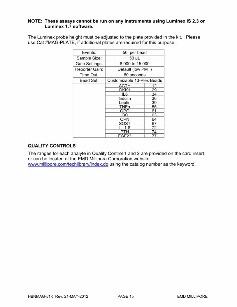
The Luminex probe height must be adjusted to the plate provided in the kit. Please use Cat #MAG-PLATE, if additional plates are required for this purpose.
Events: 50, per bead Sample Size: 50 µL Gate Settings: 8,000 to 15,000 Reporter Gain: Default (low PMT)
Time Out: 60 seconds Bead Set: Customizable 13-Plex Beads
ACTH 12DKK1 29
IL6 34Insulin 36Leptin 39TNFα 55OPG 61OC 63
OPN 64SOST 67IL-1 β 72PTH 74
FGF23 77 QUALITY CONTROLS
The ranges for each analyte in Quality Control 1 and 2 are provided on the card insert or can be located at the EMD Millipore Corporation website www.millipore.com/techlibrary/index.do using the catalog number as the keyword.
HBNMAG-51K Rev. 21-MAY-2012 PAGE 16 EMD MILLIPORE
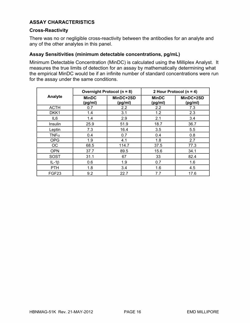
ASSAY CHARACTERISTICS
Cross-Reactivity
There was no or negligible cross-reactivity between the antibodies for an analyte and any of the other analytes in this panel. Assay Sensitivities (minimum detectable concentrations, pg/mL)
Minimum Detectable Concentration (MinDC) is calculated using the Milliplex Analyst. It measures the true limits of detection for an assay by mathematically determining what the empirical MinDC would be if an infinite number of standard concentrations were run for the assay under the same conditions.
Analyte Overnight Protocol (n = 8) 2 Hour Protocol (n = 4)
MinDC (pg/ml)
MinDC+2SD (pg/ml)
MinDC (pg/ml)
MinDC+2SD (pg/ml)
ACTH 0.7 2.2 2.2 7.3 DKK1 1.4 3.1 1.2 2.3
IL6 1.4 2.9 2.1 3.4 Insulin 25.9 51.9 18.7 36.7 Leptin 7.3 16.4 3.5 5.5 TNF 0.4 0.7 0.4 0.8 OPG 1.9 4.1 1.8 2.7 OC 68.5 114.7 37.5 77.3
OPN 37.7 89.5 15.6 34.1 SOST 31.1 67 33 82.4 IL-1 0.6 1.9 0.7 1.6 PTH 1.8 3.4 1.6 4.5
FGF23 9.2 22.7 7.7 17.6
HBNMAG-51K Rev. 21-MAY-2012 PAGE 17 EMD MILLIPORE
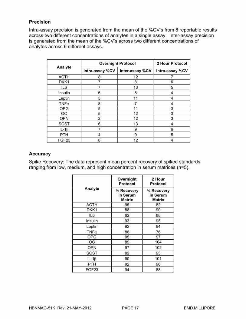
Precision
Intra-assay precision is generated from the mean of the %CV’s from 8 reportable results across two different concentrations of analytes in a single assay. Inter-assay precision is generated from the mean of the %CV’s across two different concentrations of analytes across 6 different assays.
Analyte Overnight Protocol 2 Hour Protocol
Intra-assay %CV Inter-assay %CV Intra-assay %CV
ACTH 8 12 7 DKK1 7 8 6
IL6 7 13 5 Insulin 6 8 4 Leptin 5 11 4 TNF 8 7 4 OPG 5 11 3 OC 5 12 3
OPN 2 12 3 SOST 6 13 4 IL-1 7 9 6 PTH 4 9 5
FGF23 8 12 4
Accuracy
Spike Recovery: The data represent mean percent recovery of spiked standards ranging from low, medium, and high concentration in serum matrices (n=5).
Analyte
Overnight Protocol
2 Hour Protocol
% Recovery in Serum
Matrix
% Recovery in Serum
Matrix ACTH 95 82 DKK1 88 90
IL6 82 88 Insulin 93 95 Leptin 92 94 TNF 86 76 OPG 95 97 OC 89 104
OPN 97 102 SOST 82 95 IL-1 90 101 PTH 92 96
FGF23 94 88
HBNMAG-51K Rev. 21-MAY-2012 PAGE 18 EMD MILLIPORE
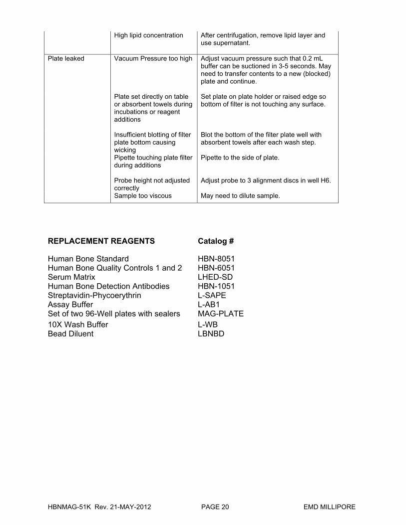
TROUBLESHOOTING GUIDE Problem Probable Cause Solution Insufficient Bead Count
Plate Washer aspirate height set too low
Adjust aspiration height according to manufacturers’ instructions.
Bead mix prepared inappropriately
Sonicate bead vials and vortex just prior to adding to bead mix bottle according to protocol. Agitate bead mix intermittently in reservoir while pipetting this into the plate.
Samples cause interference due to particulate matter or viscosity
See above. Also sample probe may need to be cleaned with Alcohol flush, Back flush and washes; or if needed probe should be removed and sonicated.
Probe height not adjusted correctly When reading the assay on Luminex 200™,
adjust probe height according to the protocols recommended by Luminex to the kit solid plate using 4 alignment discs. When reading the assay on FLEXMAP 3D™, adjust probe height according to the protocols recommended by Luminex to the kit solid plate using 1 alignment disc. When reading the assay on MAGPIX, adjust probe height according to the protocols recommended by Luminex to the kit solid plate using 2 alignment discs.
Background is too high
Background wells were contaminated
Avoid cross-well contamination by using sealer appropriately, and pipeting with Multichannel pipets without touching reagent in plate.
Matrix used has endogenous analyte or interference
Check matrix ingredients for cross reacting components (e.g. interleukin modified tissue culture medium).
Insufficient washes Increase number of washes. Beads not in region or gate
Luminex not calibrated correctly or recently
Calibrate Luminex based on Instrument Manufacturer’s instructions, at least once a week or if temperature has changed by >3oC.
Gate Settings not adjusted correctly
Some Luminex instruments (e.g. Bioplex) require different gate settings than those described in the Kit protocol. Use Instrument default settings.
Wrong bead regions in protocol template
Check kit protocol for correct bead regions or analyte selection.
Incorrect sample type used
Samples containing organic solvents or if highly viscous should be diluted or dialyzed as required.
Instrument not washed or primed
Prime the Luminex 4 times to rid of air bubbles, wash 4 times with sheath fluid or water if there is any remnant alcohol or sanitizing liquid.
HBNMAG-51K Rev. 21-MAY-2012 PAGE 19 EMD MILLIPORE
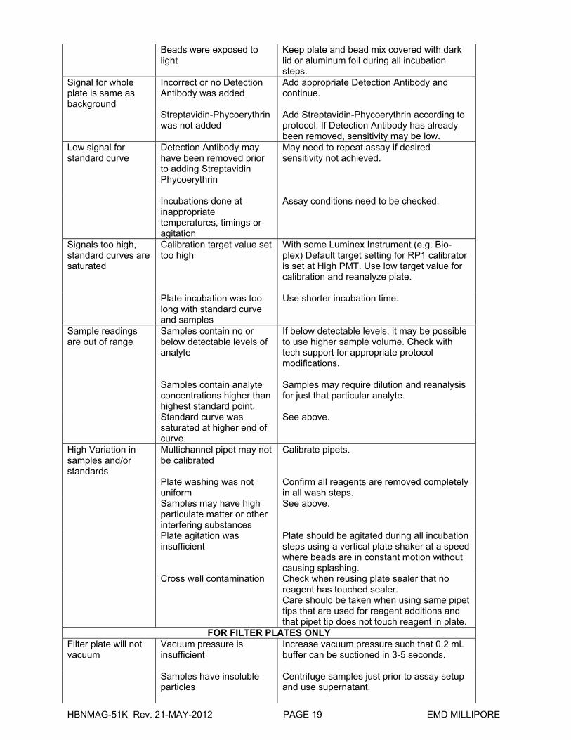
Beads were exposed to light
Keep plate and bead mix covered with dark lid or aluminum foil during all incubation steps.
Signal for whole plate is same as background
Incorrect or no Detection Antibody was added
Add appropriate Detection Antibody and continue.
Streptavidin-Phycoerythrin was not added
Add Streptavidin-Phycoerythrin according to protocol. If Detection Antibody has already been removed, sensitivity may be low.
Low signal for standard curve
Detection Antibody may have been removed prior to adding Streptavidin Phycoerythrin
May need to repeat assay if desired sensitivity not achieved.
Incubations done at inappropriate temperatures, timings or agitation
Assay conditions need to be checked.
Signals too high, standard curves are saturated
Calibration target value set too high
With some Luminex Instrument (e.g. Bio-plex) Default target setting for RP1 calibrator is set at High PMT. Use low target value for calibration and reanalyze plate.
Plate incubation was too long with standard curve and samples
Use shorter incubation time.
Sample readings are out of range
Samples contain no or below detectable levels of analyte
If below detectable levels, it may be possible to use higher sample volume. Check with tech support for appropriate protocol modifications.
Samples contain analyte concentrations higher than highest standard point.
Samples may require dilution and reanalysis for just that particular analyte.
Standard curve was saturated at higher end of curve.
See above.
High Variation in samples and/or standards
Multichannel pipet may not be calibrated
Calibrate pipets.
Plate washing was not uniform
Confirm all reagents are removed completely in all wash steps.
Samples may have high particulate matter or other interfering substances
See above.
Plate agitation was insufficient
Plate should be agitated during all incubation steps using a vertical plate shaker at a speed where beads are in constant motion without causing splashing.
Cross well contamination Check when reusing plate sealer that no reagent has touched sealer.
Care should be taken when using same pipet tips that are used for reagent additions and that pipet tip does not touch reagent in plate.
FOR FILTER PLATES ONLY Filter plate will not vacuum
Vacuum pressure is insufficient
Increase vacuum pressure such that 0.2 mL buffer can be suctioned in 3-5 seconds.
Samples have insoluble particles
Centrifuge samples just prior to assay setup and use supernatant.
HBNMAG-51K Rev. 21-MAY-2012 PAGE 20 EMD MILLIPORE
High lipid concentration After centrifugation, remove lipid layer and use supernatant.
Plate leaked Vacuum Pressure too high Adjust vacuum pressure such that 0.2 mL buffer can be suctioned in 3-5 seconds. May need to transfer contents to a new (blocked) plate and continue.
Plate set directly on table or absorbent towels during incubations or reagent additions
Set plate on plate holder or raised edge so bottom of filter is not touching any surface.
Insufficient blotting of filter plate bottom causing wicking
Blot the bottom of the filter plate well with absorbent towels after each wash step.
Pipette touching plate filter during additions
Pipette to the side of plate.
Probe height not adjusted correctly
Adjust probe to 3 alignment discs in well H6.
Sample too viscous May need to dilute sample.
REPLACEMENT REAGENTS Catalog #
Human Bone Standard HBN-8051 Human Bone Quality Controls 1 and 2 HBN-6051 Serum Matrix LHED-SD Human Bone Detection Antibodies HBN-1051 Streptavidin-Phycoerythrin L-SAPE Assay Buffer L-AB1 Set of two 96-Well plates with sealers MAG-PLATE 10X Wash Buffer L-WB Bead Diluent LBNBD
HBNMAG-51K Rev. 21-MAY-2012 PAGE 21 EMD MILLIPORE
Antibody-Immobilized Magnetic Beads Analyte Bead # Cat. # Human ACTH Beads 12 HACTH-MAG Human DKK1 Beads 29 HDKK1-MAG Human IL6 Beads 34 HIL6-MAG Human Insulin Beads 36 HINS-MAG Human Leptin Beads 39 HLPTN-MAG Human TNFα Beads 55 HTNFA-MAG Human OPG Beads 61 H0PG-MAG Human OC Beads 63 H0C-MAG Human OPN Beads 64 H0PN-MAG Human SOST Beads 67 HS0ST-MAG Human IL-1 β Beads 72 HIL1B-MAG Human PTH Beads 74 HPTH-MAG Human FGF23 Beads 77 HFGF23-MAG
HBNMAG-51K Rev. 21-MAY-2012 PAGE 22 EMD MILLIPORE
ORDERING INFORMATION
To place an order:
To assure the clarity of your custom kit order, please FAX the following information to our customer service department:
Include:
Your name, telephone and/or fax number
Customer account number
Shipping and billing address
Purchase order number
Catalog number and description of product
Quantity of kits
Selection of MILLIPLEX®
Analytes
FAX: (636) 441-8050
Toll-Free US: (800) MILLIPORE (636) 441-8400
Mail Orders: EMD Millipore Corp.
6 Research Park Drive
St. Charles, Missouri 63304 U.S.A.
For International Customers:
To best serve our international customers in placing an order or obtaining additional information about MILLIPLEX MAP products, please contact your multiplex specialist or sales representative or email our European Customer Service at [email protected].
Conditions of Sale
For Research Use Only. Not for Use in Diagnostic Procedures.
Material Safety Data Sheets (MSDS)
Material Safety Data Sheets for EMD Millipore products may be ordered by fax or phone or through our website at www.millipore.com/techlibrary/index.do
Technical Services
For product technical assistance call or write.
Toll-Free US: (781)533-8159
E-mail: [email protected]

HBNMAG-51K Rev. 21-MAY-2012 PAGE 23 EMD MILLIPORE
WELL MAP
1 2 3 4 5 6 7 8 9 10 11 12
A Standard 0
pg/mL (Background)
Standard 4 QC-1
Control
B Standard 0
pg/mL (Background)
Standard 4 QC-1
Control
C Standard 1 Standard 5 QC-2
Control
D Standard 1 Standard 5 QC-2
Control
E Standard 2 Standard 6 Sample
1
F Standard 2 Standard 6 Sample
1
G Standard 3 Standard 7
Sample
2
H Standard 3 Standard 7
Sample
2


![Welcome! [] · 2012-12-05 · Welcome to the fourth number of Fröjel Newsletter. ... beads of lime stone (four made of fossils), one bead of bone/antler (rosary bead), and one of](https://img.pdfslide.net/doc/110x75/5e97a9797b170b1d90430e41/welcome-2012-12-05-welcome-to-the-fourth-number-of-frjel-newsletter-.jpg)