Embed Size (px)
Citation preview
Human cystic echinococcosis, caused by Echinococcus granulosus, and alveolar echinococcosis,
caused by E. multilocularis, are important public health threats in many parts of the world.
Diagnosis of echinococcosis in dogs or other susceptible carnivores relies on the detection of adult
cestodes of the Echinococcus genus or their eggs in their faeces or small intestine. Coproantigen
and copro-DNA assays have proven useful for safe, fast and accurate diagnosis. In intermediate
hosts, diagnosis is dependent on post-mortem detection of the larval cyst form that can infect
almost any organ, particularly the liver and lungs.
Identification of the agent: It was previously accepted that there were five valid species of the
genus Echinococcus:The current view however, informed by biology, epidemiology and particularly
molecular genotyping, recommends the inclusion of at least nine species in the in the genus. All
those species of Echinococcus known to cause cystic echinococcosis in the intermediate host may
be referred to as E. granulosus sensu lato (s.l.), whereas strains G1–G3 (which are closely related)
are now referred to as E. granulosus sensu strictu (s.s.) Larval forms of Echinococcus can usually
be visually detected in organs. Special care has to be taken for a specific diagnosis of
E. granulosus in instances where Taenia hydatigena in sheep is also a problem. Histological
examination may confirm the diagnosis after formalin-fixed material is processed by conventional
staining methods. The presence of a periodic-acid-Schiff positive, acellular laminated layer with or
without an internal cellular, nucleated germinal membrane can be regarded as a specific
characteristic of metacestodes of Echinococcus. Genotyping via polymerase chain reaction (PCR)
is the only method available to confirm the exact species of Echinococcus infecting animals. The
identification of larval E. multilocularis in rodents and other hosts is possible by macroscopic or
microscopic examination and confirmed by DNA detection using the PCR.
The small intestine is required at necropsy for the detection of adult Echinococcus spp. in wild or in
domestic carnivores. Handling infected material needs detailed safety precautions to avoid risk to
the operator of contracting a potentially fatal disease.
Coproantigen or CoproDNA tests: Significant progress is being made in the development of
immunological tests for the diagnosis of intestinal Echinococcus infections by use of coproantigen
detection. The technique has been used successfully in surveys of E. granulosus in dogs and is
currently used in surveys for E. multilocularis in populations of dogs and foxes. Coproantigen
detection is possible in faecal samples collected from dead or living animals or from the
environment. PCR DNA methods for the detection of E. multilocularis and more recently
E. granulosus in definitive hosts have now been established as diagnostic techniques.
Serological tests: Antibodies directed against oncosphere, cyst fluid and protoscolex antigens can
be detected in the serum of infected dogs and sheep, but this approach is presently of limited
practical use as it does not distinguish between current and previous infections. The sensitivity in
cases of low worm burden is poor. Cross-reactivity between Echinococcus and Taenia species also
may occur.
Requirements for vaccines: Progress has been made in the development of an effective vaccine
(Eg95) against infection with the larval stage of E. granulosus in sheep and cattle.
The species within the genus Echinococcus are small tapeworms of carnivores with larval (metacestode) stages
known as hydatids that proliferate asexually in various mammals including humans. Until recently it was accepted that there were five morphologically distinct species in this genus: E. granulosus, E. multilocularis, E. oligarthus, E. vogeli and E. shiquicus. Echinococcus granulosus, formerly regarded as a single species with a high genotypic and phenotypic diversity, is now recognised as an assemblage of cryptic species, which differ considerably in morphology, development, host specificity (including infectivity or pathogenicity for humans). This diversity is reflected in the mitochondrial and nuclear genomes. Based on phenotypic characters and gene sequences, E. granulosus (sensu lato [s.l.]) has now been subdivided into E. granulosus (sensu stricto [s.s.]) (including the formerly identified genotypic variants G1–3), E. felidis (the former ‘lion strain’), Echinococcus equinus (the ‘horse strain’, genotype G4), E. ortleppi (the‘cattle strain’, genotype G5) and E. canadensis. The latter species, as recognised here, shows the highest diversity and is composed of the ‘camel strain’, genotype G6, the ‘pig strain’, genotype G7,and two ‘cervid strains’, genotypes G8 and G10 (Nakao et al., 2013; Romig et al., 2015). Echinococcus granulosus (s.l.) has a global distribution; E. multilocularis occurs in wide areas of the Northern Hemisphere, E. shiquicus is found in China (People’s Rep. of) and E. oligarthus and E. vogeli are confined to
Central and South America. All five of the originally described species are infective to humans causing various forms of echinococcosis although in the most recent taxonomic classification there is no evidence of E. equinus infections in humans. Human cystic echinococcosis, caused by E. granulosus, and alveolar echinococcosis, caused by E. multilocularis, are important public health threats in many parts of the world (WHO/OIE, 2001).
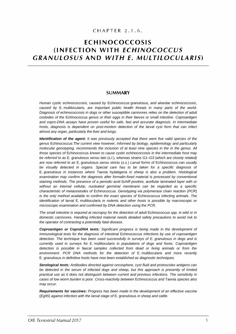
E. granulosus (sensu lato)
E. multilocularis E. oligarthrus
E. vogeli E. shiquicus
Distribution Cosmopolitan Holoarctic region Neotropical region Neotropical region Tibet plateau
Definitive Host Dogs Foxes Wild felids Bush dog Tibetan fox
Intermediate Host Ungulates Microtine rodents Neotropical rodents
Neotropical rodents
Plateau pika
Adult
Body length (mm) 2.0–11.0 1.2–4.5 2.2–2.9 3.9–5.5 1.3–1.7
No. segments 2–7 2–6 3 3 2–3
Length of large hooks (µm)
25.0–49.0 24.9–34.0 43.0–60.0 49.0–57.0 20.0–23.0
Length of small hooks (µm)
17.0–31.0 20.4–31.0 28.0–45.0 30.0–47.0 16.0–17.0
No. testes 25–80 16–35 15–46 50–67 12–20
Position of genital pore
a. Mature segment Near to middle Anterior to middle Anterior to middle Posterior to middle
Near to upper edge
b. Gravid segment Posterior to middle
Anterior to middle Near to middle Posterior to middle
Anterior to middle
Gravid uterus Branching laterally
Sac-like Sac-like Tubular Sac-like
Metacestode Unilocular cysts in viscera
Multilocular cysts in viscera
Polycystic cysts in muscles
Polycystic cysts in viscera
Unilocular cysts in viscera
The parasite is transmitted between the domestic dog and a number of domestic ungulate species. The dog or sheep cycle of E. granulosus (s.s.) is most important. Sylvatic definitive and intermediate hosts also occur, e.g.

wolf or cervid (E. canadensis). The adult varies between 2 and 11 mm in length and usually possesses from two to seven segments, averaging from three to four segments. The penultimate segment is mature, and the genital pore normally opens posterior to the middle in both mature and gravid segments. The last (gravid) segment is usually more than half the length of the entire worm. There are rostellar hooks of various sizes on the protoscolex in two rows. The size of the hooks varies between 25 to 49 µm in the first row, and between 17 and 31 µm in the second row. The gravid uterus has well-developed sacculations.
The larval stage is a fluid-filled bladder or hydatid cyst that is unilocular, although communicating chambers also occur. Growth is expansive, and endogenous daughter cysts may be produced. Individual bladders may reach up to 30 cm in diameter and occur most frequently in liver and lungs, but may develop in other internal organs. The infection with this stage is referred to as cystic echinococcosis.
The cryptic species of E. granulosus (s.l.) in domestic cycles include, dog or sheep in the Mediterranean region, South America (Argentina, Brazil, Chile, Peru and Uruguay), Africa (Ethiopia, Kenya and Sudan), the Middle East and Levant regions, Russia, Central Asia (Kazakhstan, Kyrgyzstan and Uzbekistan), Mongolia, China (People’s Rep. of), Oceania and the United Kingdom; dog or horse in Belgium, Ireland and the United Kingdom; dog or cattle in Belgium, Germany, South Africa and Switzerland; dog or swine in Poland; dog or wolf/reindeer in sub-Arctic regions of Norway, Finland and Alaska; dog or camel in the Middle East, Africa, Central Asia and China (People’s Rep. of).
The parasite is transmitted primarily between wild definitive hosts (e.g. Vulpes vulpes, V. corsac, Alopex lagopus) and small arvicolid rodents (voles and lemmings). The adult varies between 1.2 and 4.5 mm in length and usually possesses from two to six segments, with an average of four to five. The penultimate segment is characteristically mature, and the genital pore is anterior to the midline in both mature and gravid segments. The gravid uterus is sac-like. On the rostellum, the larger hooks of the first row vary in size between 24.9 and 34.0 µm and the smaller hooks of the inner row between 20.4 and 31.0 µm.
The metacestode is a multivesicular structure consisting of conglomerates of small vesicles, usually not exceeding a few millimetres in diameter. Unlike E. granulosus, the larval mass often contains a semisolid rather
than a fluid matrix. It proliferates by exogenous budding, which results in infiltration of tissues. Infection with this stage is commonly referred to as alveolar echinococcosis. There is no clear evidence for distinct strains or genotypes of E. multilocularis, though regional variations at the continental scale have been described (WHO/OIE, 2001).
This zoonotic parasite is found mainly in the Northern Hemisphere, and its life cycle is mainly maintained in wildlife (Kamiya et al., 2007). The sylvatic cycle involves foxes and many species of wild rodents. Coyotes,
raccoon dogs, wolves, wild cats, domestic dogs and cats however, may serve as definitive hosts while pigs, horses, primates and humans can be infected as intermediate hosts (Kamiya et al., 2007). Dogs may also rarely be infected with larval stages producing lesions in internal organs, even simultaneously with adult stages present in the gastro-intestinal tract.
The parasite typically uses neotropical wild felids as definitive hosts (e.g. Felis concolor, F. jaguarundi) and large rodents (e.g. Dasyprocta sp., Cuniculus paca) as intermediate hosts. The adult varies between 2.2 and 2.9 mm in length, and normally possesses three segments, the penultimate of which is mature. The genital pore is anterior to the middle in mature segments and approximately at the middle in gravid segments. The gravid uterus is sac-like.
The metacestode is polycystic and fluid-filled with a tendency to become septate and multichambered. The rostellar hooks of the protoscolex vary in length between 25.9 and 37.9 µm. The hooks are described in more detail in the next section where they are also compared with those of E. vogeli. The single cyst may reach a diameter of approximately 5 cm. Predilection sites are internal organs and muscles. To date, there have only been a few reports of human disease. The parasite appears not to mature in dogs.
The parasite typically uses the South American bush dog (Speothus venaticus) as a wild definitive host, but the domestic dog is susceptible, as are large rodents (e.g. Cuniculus paca) as intermediate hosts. The adult varies between 3.9 and 5.5 mm in length, and usually has three segments, the penultimate of which is mature. The genital pore is situated posterior to the middle in both the mature and gravid segments. The gravid uterus has no
lateral sacculations and is characterised by being relatively long and tubular in form, compared with the other segments, which are sac-like.
The metacestode is similar to that of E. oligarthrus. It has been reported that the two species can be distinguished by comparing differences in the dimensions and proportions of the rostellar hooks on the protoscolex. The hooks of E. oligarthrus vary in length between 25.9 and 37.9 µm (average 33.4 µm) and between 22.6 and 29.5 µm (average 25.45 µm) for large and small hooks, respectively. Those of E. vogeli vary between 19.1 and 43.9 µm (average 41.64 µm) and between 30.4 and 36.5 µm (average 33.6 µm) for the large and small hooks, respectively. Also the hook-guard for E. oligarthrus divides the hook 50:50, compared with 30:70 for E. vogeli.
Echinococcus vogeli is a zoonotic agent with approximately 200 human cases in total reported in South America.
The infection caused by the larval stage of this species is commonly referred to as polycystic echinococcosis.
The parasite was found in the Tibetan fox (Vulpes ferrilata) its definitive host and the plateau pika (Ochotona curzoniae), the intermediate host. In most species of Echinococcus, the gravid segment is connected to a mature
segment; however, a strobila consisting of only two segments (a gravid segment directly attaching to a premature segment) is unique to this species (WHO/OIE, 2001). The adult stage is morphologically similar to E. multilocularis but differs by its smaller hooks, fewer segments, upper position of genital pore in the premature segment and fewer eggs in the gravid segment. It is easily distinguishable from E. granulosus by its shorter
length, branchless gravid uterus and the anterior position of the genital pore in the gravid segment. The adult measures 1.3 to 1.7 mm.
The metacestode is found in the liver and is essentially a unilocular minicyst containing fully developed brood capsules; however, oligovesicular forms have also been observed. It is differentiated from E. granulosus by the
absence of daughter cysts within the fertile cyst (WHO/OIE, 2001).
A detailed description of echinococcosis in humans and animals can be found in the WHO/OIE Manual on echinococcosis (WHO/OIE, 2001).
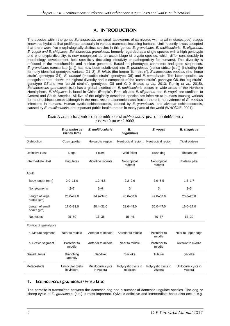
Method
Purpose (metacestode cysts in intermediate hosts)
Population freedom
from infection
Individual animal freedom from
infection prior to movement
Contribute to eradication
policies
Confirmation of clinical
cases
Prevalence of infection – surveillance
Immune status in individual animals or
populations post-vaccination
Agent identification
Parasite Identification/
meat inspection
+ – ++ +++ +++ ++
Antigen detection
– – – – – –
PCR – – – +++ ++ –
Detection of immune response
ELISA – – – – + +
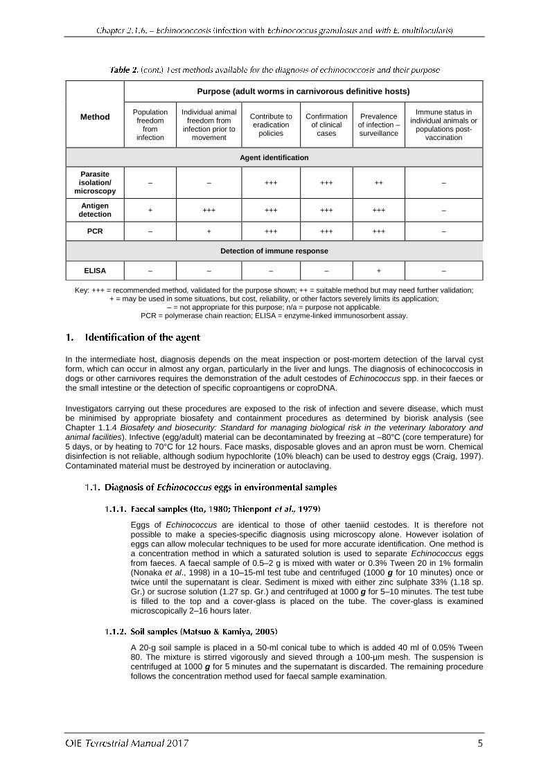
Method
Purpose (adult worms in carnivorous definitive hosts)
Population freedom
from infection
Individual animal freedom from
infection prior to movement
Contribute to eradication
policies
Confirmation of clinical
cases
Prevalence of infection – surveillance
Immune status in individual animals or
populations post-vaccination
Agent identification
Parasite isolation/
microscopy – – +++ +++ ++ –
Antigen detection
+ +++ +++ +++ +++ –
PCR – + +++ +++ +++ –
Detection of immune response
ELISA – – – – + –
Key: +++ = recommended method, validated for the purpose shown; ++ = suitable method but may need further validation; + = may be used in some situations, but cost, reliability, or other factors severely limits its application;
– = not appropriate for this purpose; n/a = purpose not applicable. PCR = polymerase chain reaction; ELISA = enzyme-linked immunosorbent assay.
In the intermediate host, diagnosis depends on the meat inspection or post-mortem detection of the larval cyst form, which can occur in almost any organ, particularly in the liver and lungs. The diagnosis of echinococcosis in dogs or other carnivores requires the demonstration of the adult cestodes of Echinococcus spp. in their faeces or the small intestine or the detection of specific coproantigens or coproDNA.
Investigators carrying out these procedures are exposed to the risk of infection and severe disease, which must be minimised by appropriate biosafety and containment procedures as determined by biorisk analysis (see Chapter 1.1.4 Biosafety and biosecurity: Standard for managing biological risk in the veterinary laboratory and animal facilities). Infective (egg/adult) material can be decontaminated by freezing at –80°C (core temperature) for 5 days, or by heating to 70°C for 12 hours. Face masks, disposable gloves and an apron must be worn. Chemical disinfection is not reliable, although sodium hypochlorite (10% bleach) can be used to destroy eggs (Craig, 1997). Contaminated material must be destroyed by incineration or autoclaving.
Eggs of Echinococcus are identical to those of other taeniid cestodes. It is therefore not possible to make a species-specific diagnosis using microscopy alone. However isolation of eggs can allow molecular techniques to be used for more accurate identification. One method is a concentration method in which a saturated solution is used to separate Echinococcus eggs from faeces. A faecal sample of 0.5–2 g is mixed with water or 0.3% Tween 20 in 1% formalin (Nonaka et al., 1998) in a 10–15-ml test tube and centrifuged (1000 g for 10 minutes) once or twice until the supernatant is clear. Sediment is mixed with either zinc sulphate 33% (1.18 sp. Gr.) or sucrose solution (1.27 sp. Gr.) and centrifuged at 1000 g for 5–10 minutes. The test tube is filled to the top and a cover-glass is placed on the tube. The cover-glass is examined microscopically 2–16 hours later.
A 20-g soil sample is placed in a 50-ml conical tube to which is added 40 ml of 0.05% Tween 80. The mixture is stirred vigorously and sieved through a 100-µm mesh. The suspension is centrifuged at 1000 g for 5 minutes and the supernatant is discarded. The remaining procedure follows the concentration method used for faecal sample examination.
Whereas surveillance for E. granulosus in domestic animals may take place in licensed slaughter houses, that for Echinococcus sp. in wildlife must be done by field surveys. When undertaking surveillance work with E. granulosus in intermediate hosts, it is vitally important that
data are stratified and reported according to the age of animals slaughtered. Prevalence rates are strongly age dependent and reports from abattoirs that may slaughter only young animals will substantially under-represent the true situation. This is because older animals may be heavily infected even when animals have very few larvae.
Hydatid cysts can be observed in many organs, but in large animals, such as sheep and cattle, palpation or incision should be done. Pigs, cattle, sheep and goats may also be infected with larval Taenia hydatigena, and it is sometimes difficult to differentiate between these two parasites when they occur in the liver. In wild animals, such as ruminants and rodents, several other larval cestodes should be considered for differential diagnosis. Please refer to Chapter 2.9.5 Cysticercosis for information on other cestodes found at meat inspection.
i) Suspect parasite material should be removed from the organ by cutting with a scalpel to include the immediate host tissue, and kept in a cool location. (NB hydatid cyst tissue in intact cysts will remain viable for more than 24 hours after death even at ambient temperatures. However viability will be prolonged by storage at 4°C for up to 72 hours. If material cannot be examined within this time, it should be stored in either 10% formol saline for subsequent microscopic examination or in 90% ethanol for subsequent DNA analysis. Ideally a sample of parasite material should be preserved in both media. Parasite tissues that are frozen will not be viable but can be examined morphologically on thawing and subjected to DNA analyses.)
ii) For morphological analysis of cyst contents, fluid should be removed and retained using a syringe. The material inside the cyst should then be washed with saline and the contents examined under the microscope (×4 objective) for the presence of protoscoleces. Note that some hydatid cysts may be sterile and not contain protoscoleces. If no protoscoleces are present, the laminated layer on the inside of the cyst cavity may be observed as a gelatinous structure that can easily be pulled away. Formalin-fixed material can be stained by conventional histological techniques. The presence of a periodic-acid-Schiff (PAS) positive acellular laminated layer, underlying a connective tissue layer, and with or without an internal cellular, nucleated germinal membrane can be regarded as a specific characteristic of the metacestodes of Echinococcus spp.
iii) In all cases exact species identification can only be made through extraction of DNA from ethanol-fixed material (Dinkel et al., 1998) and subsequent genotyping by polymerase chain reaction (PCR). Specific primers for all Echinococcus species and related taeniids are summarised by Roelfsema et al. (2016).
Necropsy is invariably employed in studies of echinococcosis in wildlife and is useful if domestic dogs are humanely culled. It should be emphasised that it is necessary to isolate and identify the adult Echinococcus, because under normal conditions of faecal examination, the eggs of Echinococcus cannot be differentiated from those of Taenia spp. The eggs of E. granulosus and E. multilocularis can now be identified and differentiated from other taeniid eggs by PCR.
The small intestine is removed as soon as possible after death, and tied at both ends. If the material is not frozen or formalin fixed (4–10%), it should be examined quickly, as the parasite can be digested within 24 hours. Formalin does not kill eggs. The fresh intestine is divided into several sections and immersed in 0.9% saline at 37°C for examination. Worms adhering to the intestinal wall may be observed and counted by means of a hand lens (for E. granulosus and E. vogeli). For accurate counts, the unfixed intestine is best divided into four or six sections, opened up and immersed in 0.9% saline at 37°C for 30 minutes to release the parasites. The contents are washed into another container for detailed examination, and the intestinal wall is scraped with a spatula. All material is boiled and washed by sieving to eliminate most of the particulate material and to make it noninfectious. The washed intestinal contents and scrapings
are placed on a black tray, and the worms are counted with the aid of a hand lens or stereoscopic microscope. Echinococcus granulosus is usually found in the first third of the small intestine of dogs and E. multilocularis in the mid/posterior sections. This approach has a greater
than 95% sensitivity, except under low worm burdens where false negative results may occur.
Necropsy is considered to be the most reliable form of diagnosis for E. multilocularis in definitive hosts. It is an inexpensive method for determining the prevalence in a population and the best way to determine worm burden. Carcasses or intestines of definitive hosts for examination should be deep frozen at between –70°C and –80°C for 3–7 days before necropsy to kill any eggs. Eggs of E. multilocularis are resistant to freezing to –50°C.
This technique has been regarded as the ‘gold standard’ for assessing the sensitivity and specificity of other techniques, however the copro-DNA (PCR) test has a greater sensitivity than SCT.
i) The small intestine is incised longitudinally and cut into 20 cm long segments or into 5 pieces of approximately the same length. These pieces are transferred to a glass bottle containing 1 litre physiological saline (0.9% NaCl) solution.
ii) The glass bottle is shaken vigorously for a few seconds and the pieces of intestine are removed. The superficial mucosal layer is stripped by exerting pressure between thumb and forefinger to dislodge attached helminths.
iii) The glass bottle is left for 15 minutes for sedimentation to occur; the supernatant is then decanted. The glass bottle is refilled with physiological saline solution. This procedure is repeated 2–6 times until the supernatant is cleared of coloured particles.
iv) The sediment fraction is examined in small portions of about 5–10 ml in rectangular plastic or Petri dishes with a counting grid (9 × 9 cm) in transmission light under a stereomicroscope at a magnification of ×120.
v) If up to 100 worms are found, the entire sediment fraction is checked; if higher numbers are present, the total worm burden is calculated from the count of one subsample.
i) Deep mucosal scrapings are taken at nearly equal distances from the small intestine using microscope slides (75 × 25 × 1 mm). Five mucosal scrapings from proximal, middle and posterior thirds of the small intestine (total 15) are recommended. Adherent materials are transferred to a square plastic Petri dish.
ii) Scrapings are squashed between slides and examined under a stereoscopic light microscope (×120). Three slides are placed in one plastic dish and examined. Echinococcus multilocularis is usually found in the second half of the small intestine.
i) A plastic vessel (1 litre) that has a plastic screw-on lid with a central hole 6–7 cm in diameter is used. The hole is covered with a high-grade steel mesh (mesh size 500 µm) fixed into the remaining plastic ring with a hot soldering iron. Silicone is applied to seal the edges of the steel mesh.
ii) The longitudinally opened small intestine is transferred to the vessel with all its contents; the vessel is closed with the lid and filed with water.
iii) The vessel is inverted and shaken; the water is decanted. The vessel is refilled with water, and the process is repeated until the decanted water is clear.
iv) The half-filled vessel is opened and the intestines are removed. The intestines are stripped between the thumb and forefinger to dislodge parasites stuck to the mucosa into the vessel.
v) The vessel is closed again, refilled and shaken one last time draining as much water from it as possible.
vi) The remaining sediment is filled into a 1 litre plastic jug and stored at 4°C. For prolonged storage, a 0.9% NaCl solution is added to the sediment to prevent the parasites from shrinking.
vii) For analysis, the materials are placed into small glass Petri dishes and scanned along engraved lines using the stereomicroscope as above.
Intact worms are fragile and for morphological studies are best handled in normal saline with a Pasteur pipette. They are washed free of other material and left for approximately 30 minutes for all movement to cease. After removal of the fluid, cold 5–10% formalin (5°C) or FAA fixative (95% ethanol [80 ml], 37–40% formaldehyde [10 ml], and glacial acetic acid [5 ml]) is added and the worms are left for a further 12 hours. For staining, the worms are washed in water for 15 minutes and transferred to Mayer’s paracarmine (carminic acid [1.0 g], aluminium chloride [0.5 g], calcium chloride [4.0 g], and 70% ethanol [100 ml]) for 12–24 hours. Excess stain is removed by immersion in 0.5–1.0% hydrochloric acid solution for a few seconds. Dehydration is accomplished by serial passage in ascending concentrations of alcohol (41, 50, 70, 85, 95, and 100%) for at least 15 minutes in each, with two changes in 100%. The alcohol is removed by xylol (10 minutes) and cleared with methyl salicylate or creosote. Prior to mounting in any suitable medium such as balsam, picolyte, etc., the specimens should be returned to the xylol for a few minutes. Persons involved in such examinations should receive serological screening for anti-Echinococcus serum-antibodies at least once a year (WHO/OIE, 2001).
Over the past 15 years, methods have been developed with the aim of simplifying and improving epidemiological investigations in final host populations and of allowing diagnosis in living animals. These methods include the detection of coproantigens and PCR DNA detection (see below).
Arecoline has been used to perform surveys of tapeworm infections in dog populations. Its use as a control agent has been superseded by praziquantel.
Adult Echinococcus worms inhabiting the intestine will release both surface or secretory molecules (antigens)and
DNA (usually contained within eggs). Both types of molecules can be detected by assaying faecal samples. The sensitivity of the tests is strongly influenced by the worm burden and stage of maturity.
A specific and sensitive laboratory test for antigen detection in canid faecal samples (coproantigen) has the potential to replace arecoline purgation and is preferable to serology for detection of current infection (Allan et al., 1992; Deplazes et al., 1992). Coproantigen ELISA (enzyme-linked
immunosorbent assay) or coproELISA provides an alternative method for diagnosing canine echinococcosis, and both polyclonal and monoclonal antibodies have been used: directed against either somatic or excretory/secretory (ES) antigens. To create polyclonal antibodies against Echinococcus spp., rabbits were hyperimmunised with Echinococcus antigens, such as adult or
protoscolex ES extracts, or somatic extracts of adult tapeworms. Alternatively, monoclonal antibodies have been produced using donor mice hyperimmunised with E. granulosus somatic or ES antigens (Craig et al., 2015) CoproELISAs are usually genus-specific for Echinococcus spp. (Allan & Craig, 2006), although depending on the endemic region and study aims, coproELISAs have been developed and validated to test for infection with E. multilocularis in foxes and dogs or primarily for E. granulosus. For canine echinococcosis caused by E. granulosus, most authors report reasonable sensitivity (78–100%) and good genus specificity from 85% to greater than 95% as well as a degree of pre-patent detection (Craig et al., 2015). Where cross-reactions occur these generally appear to be caused by infection with Taenia hydatigena, the most common taeniid of dogs, and attempts to improve specificity
by using monoclonal antibodies in coproELISAs have not been able to eliminate this problem. CoproELISA sensitivity broadly correlates with worm burden of E. granulosus, however some low intensity infections (worm burdens <50–100) may give false negatives in coproELISA (Allan & Craig, 2006). Coproantigens can be detected prior to release of eggs by Echinococcus worms, and therefore are not related to egg antigen(s). This has the advantage of detection of prepatent infections. Furthermore, coproantigen levels return to the preinfection baseline within 5 days of anthelminthic treatment of infected dogs (Deplazes et al., 1992). More importantly, it reduces the biohazardous risk of exposure of personnel to potentially infective eggs during purgation or necropsy.
For detection of E. multilocularis infection of foxes, necropsy is time-consuming. Coproantigen testing by ELISA offers a specific practical alternative. Fox faecal samples should be taken at post-mortem from the rectum rather than from the small intestine. Echinococcus coproantigens are also stable in fox
or dog faeces left at 20°C for 1 week and in frozen dog faeces. Coproantigen testing has also been successfully used to evaluate the efficacy of deworming wild foxes infected with E. multilocularis using praziquantel-laced bait, which proved to be a successful combination of eliminating the source of infection.
i) The faecal sample (collected per rectum or from the ground) is mixed with an equal volume of phosphate buffered saline (PBS), pH 7.2, containing 0.3% Tween 20 (PBST), in a capped 5 ml disposable tube. This is shaken vigorously and centrifuged at 2000 g for 20 minutes at room temperature. Faecal supernatants can be tested immediately or stored at –20°C or lower. Supernatants that appear very dark or viscous are still acceptable for use.
ii) A 96-well ELISA microtitre plate is coated with optimal concentration (typically 5 µg per ml) of a protein A purified IgG fraction of rabbit anti-E. granulosus proglottid extract (Allan et al., 1992) in 0.05 M bicarbonate/carbonate buffer, pH 9.6 (100 µl per well). The plate is covered and incubated overnight at 4°C.
iii) The wells are rinsed three times in PBST with 1 minute between washes; 100 µl of the same buffer is added to each well, and the plate is incubated for 1 hour at room temperature.
iv) The PBST is discarded and 50 µl of neat fetal calf serum is added to all wells. This is followed by the addition of 50 µl per well of faecal sample supernatants is added (in duplicate wells). The plate is incubated at room temperature for 1 hour with plastic film to seal the plate.
v) The wells are rinsed as in step iii, but the contents are discarded into a 10% bleach (hypochlorite) solution.
vi) An optimal dilution concentration of around 1 µg/ml of an IgG rabbit anti-E.-granulosus proglottid extract peroxidase conjugate (Allan et al., 1992) in PBST is prepared and 100 µl per well is added to all wells. The plate is incubated for 1 hour at room temperature (22–24°C).
vii) The wells are rinsed as in step iii.
viii) Next, 100 µl per well of tetramethyl benzidene (TMB) substrate is added and the plate is left in the dark for 20 minutes at room temperature (22–24°C).
ix) Absorbance of wells is read at 650 nm. The enzyme-substrate reaction can be stopped by adding 100 µl of 1 M phosphoric acid (H3PO4) to each well. The colour turns from blue to
yellow if positive and is read at 450 nm.
x) Laboratories should establish their own end-point criteria using standard positive and negative samples. Standards can also be obtained from the OIE Reference Laboratories (see Table given in Part 4 of this Terrestrial Manual). Usually, the positive to negative threshold is taken as 3 standard deviations above the mean absorbance value of control negatives, or against a reference standard control positive using absorbance units equivalence.
Sandwich ELISA using a monoclonal antibody EmA9 raised against adult E. multilocularis somatic antigen.
i) 0.5 g of each faecal sample is placed in a centrifuge tube and a 1% formalin solution containing 0.3% Tween 20 is added to a total volume of 15 ml.
ii) After adequate mixing, the faecal solution is centrifuged at 1200 g for 10 minutes at room temperature. A supernatant fraction is used for the coproantigen detection assay.
iii) Flat-bottomed microtitre plates are coated with 50 µl/well of 1 µg/ml rabbit IgG directed against adult E. multilocularis excretory/secretory (ES) products in 0.05 M NaHCO3/Na2CO3 buffer (pH 9.6) and are left overnight at 4°C.
iv) The plates are washed three times with 250 µl/well PBS (pH 7.4) containing 0.05% Tween 20 (PBST), and blocked using 100 µl/well 1% bovine serum albumin (BSA) in PBS for 1 hour at room temperature (22–24 C).
v) The plates are washed three times (with the wash disinfected with 10% bleach) and 50 µl of faecal supernatant is added to each well and the plates are incubated for 2 hours.
vi) The plates are again washed four times and 0.5 µg/ml of the biotinylated monoclonal antibody in 0.5% BSA/0.5% casein in PBST is added to each well and the plates are incubated for 1 hour.
vii) The plates are washed four times and streptavidin-biotinylated horseradish peroxidase complex, diluted 1/1000 in 0.5% BSA/0.5% casein in PBST is added to each well and the plates are incubated for 1 hour.
viii) The plates are washed five times and 100 µl/well of substrate solution (1 mg of tetramethyl benzidene in 10 ml of 0.05 M phosphate citrate buffer, pH5.0 with 2 µl of H2O2) is added.
ix) The plates are shaken immediately and placed in a 37°C incubator for 30 minutes. The reaction is stopped by adding 50 µl/well of 4 N H2SO4. The optical densities (OD) of the
plates are read at 450 nm.
x) The cut-off value is calculated as the mean OD value plus 3 standard deviations of samples from uninfected animals.
Copro-DNA has proven to be of value for the diagnosis of Echinococcosis in animal definitive hosts and in the identification of individual parasite species. DNA isolation from the faeces, however, is laborious.
PCR is a technically demanding and expensive technique. It is currently used mainly for confirmatory testing of coproantigen-positive samples or for identification of taenid eggs recovered from faeces. Table 3 presents the different PCR primers used for identification of copro-DNA from faeces in definitive hosts of genus Echinococcus.
Primer designation: primer sequences (5’–3’) Ref. Target, comments
E. multilocularis
GTG-AGG-CGA-TGT-GTG-GTG-ATG-GAG-AGA-AGG
CAA-GTG-GTC-AGG-GGC-AGT-AG
Bretagne et al., 1993
U1 sRNA gene: may yield non-specific products when used with metacestode
material containing host DNA (unpublished observation)
Mitochondrial 12S RNA gene; used in two-tube nested PCR
Outer primers:
(P60 forward)
TTA-AGA-TAT-ATG-TGG-TAC-AGG-ATT-AGA-TAC-CC
(P375 reverse)
AAC-CGA-GGG-TGA-CGG-GCG-GTG-TGT-ACC
Inner primers:
(Pnest forward)
ACA-ATA-CCA-TAT-TAC-AAC-AAT-ATT-CCT-ATC
(Pnest reverse)
ATA-TTT-TGT-AAG-GTT-GTT-CTA
Dinkel et al., 1998
Mitochondrial 12S RNA gene; modified from Dinkel et al., 1998 for use in one-
tube nested PCR
Outer primers:
(Em-1)
TAA-GAT-ATA-TGT-GGT-ACA-GGA-TTA-GAT-ACC-C
(Em-2)
GGT-GAC-GGG-CGG-TGT-TGT-A
Inner primers:
(Em-3)
ATA-TTA-CAA-CAA-TAT-TCC-TAT-C
(Em-4)
ATA-TTT-TGT-AAG-GTT-GTT-CTA
Van der Giessen et al.,
1999
Mitochondrial 12S RNA gene; modified from Dinkel et al., 1998 for use in single
PCR
Primer designation: primer sequences (5’–3’) Ref. Target, comments
(EM-H15)
CCA-TAT-TAC-AAC-AAT-ATT-CCT-ATC
(EM-H17)
GTG-AGT-GAT-TCT-TGT-TAG-GGG-AAG
Stieger et al., 2002
NADH dehydrogenase subunit 1 (ND1) of mtDNA; cleavage with enzyme Cfo1
distinguish E. multilocularis from E. granulosus
E. multilocularis and E. granulosus
ND1
(NDfor2-)
AGT-TTC-GTA-AGG-GTC-CTA-ATA
(NDrev2-)
CCC-ACT-AAC-TAA-CTC-CCT-TTC
Moks et al., 2005
Repeated sequences from E. granulosus. ‘sheep strain’; yields
banding pattern upon electrophoresis
Mitochondrial 12SRNA gene; specific for E. granulosus ‘sheep strain’
Amplify a fragment of the coxI genespecific fo E. granulosus
E. granulosus
(Eg1121a)
GAA-TGC-AAG-CAG-CAG-ATG
(Eg1122a)
GAG-ATG-AGT-GAG-AAG-GAG-TG
Abbasi et al., 2003
(Eg1f)
CATTAATGTATTTTGTAAAGTTG
(Eg1r)
CAC-ATC-ATC-TTA-CAA-TAA-CAC-C
Stefanic et al., 2004
(EgO/DNA-IM1)
forward
TCA-TAT-TTG-TTT-GAG-KAT-YAG-TKC
reverse
GTA-AAT-AAM-ACT-ATA-AAA-GAA-AYM-AC
Naidich et al., 2006
E. multilocularis, E. granulosus and Taeniid spp.
(JB11)
AGA-TTC-GTA-AGG-GGC-CTA-ATA
(JB12)
AC-CAC-TAA-CTA-ATT-CAC-TTT-C
(60.for.-mod)
ATG-TGG-TAC-AGG-ATT-AGA-TAC-CC
(375.rev.-mod)
GGT-GAC-GGG-CGG-TGT-GTA-CC
Trachsel et al., 2007
Cestodes
Cestodes
(Eg1f)
CAT-TAA-TGT-ATT-TTG-TAA-AGT-TG
(Eg1r)
CAC-ATC-ATC-TTA-CAA-TAA-CAC-C
Trachsel et al., 2007
Echinococcus granulosus (sheep strain)
(EM-H15)
CCA-TAT-TAC-AAC-AAT-ATT-CCT-ATC
(EM-H17)
GTG-AGT-GAT-TCT-TGT-TAG-GGG-AAG
Trachsel et al., 2007
E. multilocularis
(Cest1)
TGC-TGA-TTT-GTT-AAA-GTT-AGT-GAT-C
(Cest2)
CAT-AAA-TCA-ATG-GAA-ACA-ACA-ACA-AG
Trachsel et al., 2007
E. multilocularis
(Cest4)
GTT-TTT-GTG-TGT-TAC-ATT-AAT-AAG-GGT-G Trachsel et al.,
2007 E. granulosus
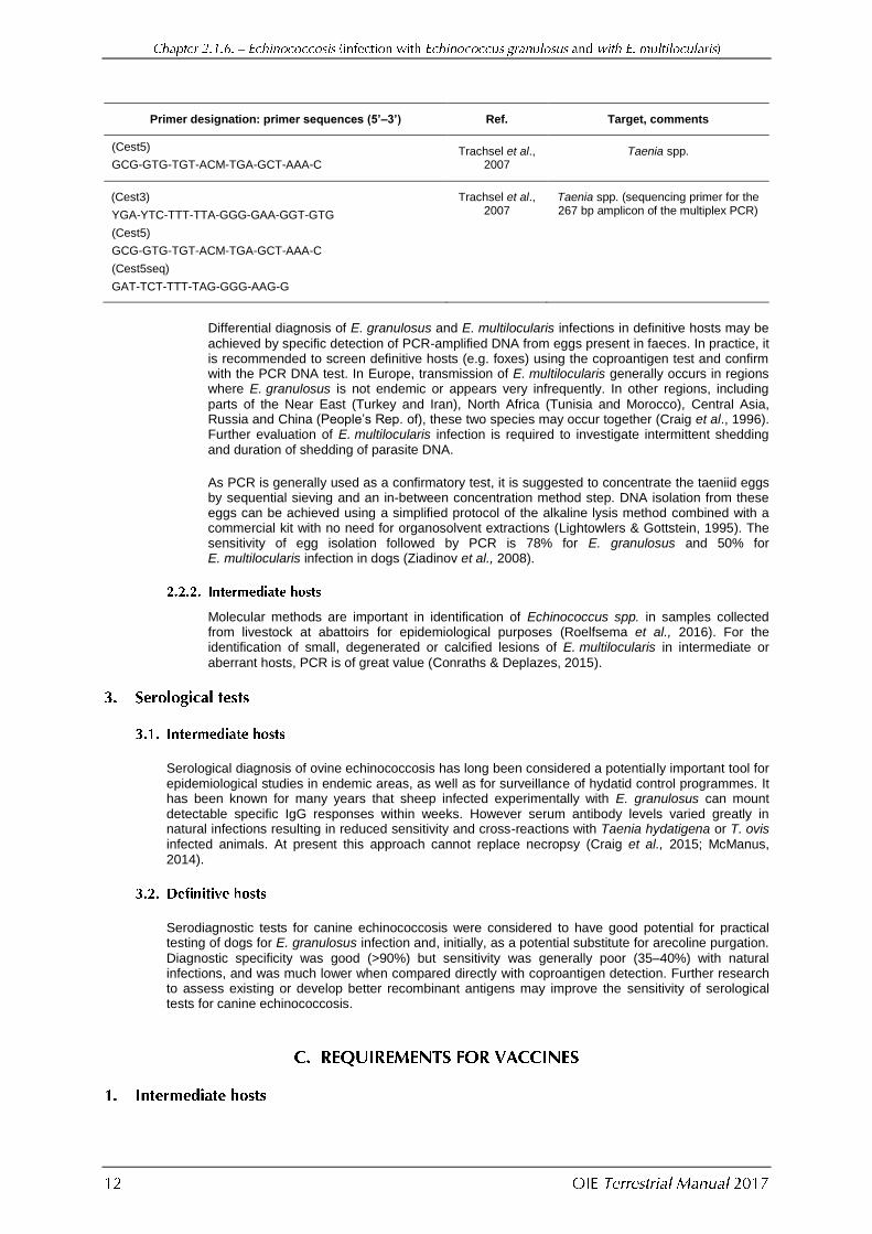
Primer designation: primer sequences (5’–3’) Ref. Target, comments
(Cest5)
GCG-GTG-TGT-ACM-TGA-GCT-AAA-C Trachsel et al.,
2007 Taenia spp.
(Cest3)
YGA-YTC-TTT-TTA-GGG-GAA-GGT-GTG
(Cest5)
GCG-GTG-TGT-ACM-TGA-GCT-AAA-C
(Cest5seq)
GAT-TCT-TTT-TAG-GGG-AAG-G
Trachsel et al., 2007
Taenia spp. (sequencing primer for the 267 bp amplicon of the multiplex PCR)
Differential diagnosis of E. granulosus and E. multilocularis infections in definitive hosts may be
achieved by specific detection of PCR-amplified DNA from eggs present in faeces. In practice, it is recommended to screen definitive hosts (e.g. foxes) using the coproantigen test and confirm with the PCR DNA test. In Europe, transmission of E. multilocularis generally occurs in regions where E. granulosus is not endemic or appears very infrequently. In other regions, including
parts of the Near East (Turkey and Iran), North Africa (Tunisia and Morocco), Central Asia, Russia and China (People’s Rep. of), these two species may occur together (Craig et al., 1996). Further evaluation of E. multilocularis infection is required to investigate intermittent shedding and duration of shedding of parasite DNA.
As PCR is generally used as a confirmatory test, it is suggested to concentrate the taeniid eggs by sequential sieving and an in-between concentration method step. DNA isolation from these eggs can be achieved using a simplified protocol of the alkaline lysis method combined with a commercial kit with no need for organosolvent extractions (Lightowlers & Gottstein, 1995). The sensitivity of egg isolation followed by PCR is 78% for E. granulosus and 50% for E. multilocularis infection in dogs (Ziadinov et al., 2008).
Molecular methods are important in identification of Echinococcus spp. in samples collected from livestock at abattoirs for epidemiological purposes (Roelfsema et al., 2016). For the identification of small, degenerated or calcified lesions of E. multilocularis in intermediate or
aberrant hosts, PCR is of great value (Conraths & Deplazes, 2015).
Serological diagnosis of ovine echinococcosis has long been considered a potentially important tool for epidemiological studies in endemic areas, as well as for surveillance of hydatid control programmes. It has been known for many years that sheep infected experimentally with E. granulosus can mount detectable specific IgG responses within weeks. However serum antibody levels varied greatly in natural infections resulting in reduced sensitivity and cross-reactions with Taenia hydatigena or T. ovis infected animals. At present this approach cannot replace necropsy (Craig et al., 2015; McManus, 2014).
Serodiagnostic tests for canine echinococcosis were considered to have good potential for practical testing of dogs for E. granulosus infection and, initially, as a potential substitute for arecoline purgation.
Diagnostic specificity was good (>90%) but sensitivity was generally poor (35–40%) with natural infections, and was much lower when compared directly with coproantigen detection. Further research to assess existing or develop better recombinant antigens may improve the sensitivity of serological tests for canine echinococcosis.
Application of an effective vaccine to reduce hydatid infection in livestock is likely to have a substantial impact on the rate of transmission of the disease to humans (Lightowlers, 2006). As E. granulosus belongs to the Taeniid family, many aspects of its immunological relationship with its intermediate host are similar to that occurring in Taenia species. Moreover, it was considered that the vaccine development approach used in Taenia species, such as the native host-protective antigens of T. ovis, would also be successful for E. granulosus. A recombinant antigen vaccine, EG95, was therefore developed in 1996 using an E. granulosus oncosphere protein expressed in Escherichia coli. The vaccine has been shown to produce high levels of protection (96–100%) against an experimental challenge infection with E. granulosus in sheep. Thereafter, the vaccine has been successfully applied in experimental trials in different countries in sheep and in other intermediate hosts. The EG95 vaccine has been licensed in some countries (Lightowlers, 2006).
Development of E. granulosus vaccines for dogs would ideally reduce worm fecundity and populations, and would be a crucial step towards the reduction (prevention) of the infection pressure on intermediate hosts, and thus reduce (prevent) infection in dogs. However no clear candidate molecules have yet been identified.
ABBASI I., BRANZBURG A., CAMPOS-PONCE M., ABDEL HAFEZ S.K., RAOUL F., CRAIG P.S. & HAMBURGER J. (2003). Copro-diagnosis of Echinococcus granulosus infection in dogs by amplification of a newly identified repeated DNA sequence. Am. J. Trop. Med. Hyg., 69, 324–330.
ALLAN J.C. & CRAIG P.S. (2006). Coproantigens in taeniasis and echinococcosis. Parasitol. Int., 55, S75–S80.
ALLAN J.C., CRAIG P.S., GARCIA NOVAL J., MENCOS F., LIU D., WANG Y., WEN H., ZHOU P., STRINGER R., ROGAN M.T. &
ZEYHLE E. (1992). Coproantigen detection for the immunodiagnosis of echinococcosis and taeniasis in dogs and humans. Parasitology, 104, 347–355.
BRETAGNE S., GUILLOUN J.P., MORAND M. & HOUIN R. (1993). Detection of Echinococcus multilocularis DNA in fox faeces using DNA hybridization. Parasitology, 106, 193–199.
CONRATHS F.J. & DEPLAZES P. (2015). Echinococcus multilocularis: Epidemiology, surveillance and state-of-the-art diagnostics from a veterinary public health perspective. Vet. Parasitol., 213, 149-161.
CRAIG P.S. (1997). Immunodiagnosis of Echinococcus granulosus and a comparison of techniques for diagnosis of canine echinococcosis. In: Compendium on Cystic Echinoccosis in Africa and in Middle Eastern Countries with
Special Reference to Morocco, Andersen F.L., Ouhelli H. & Kachani M., eds. Brigham Young University Print Services, Provo, Utah, USA.
CRAIG P.S., GASSER R.B., PARADA L., CABRERA P., PARIETTI S., BORGUES C., ACUTTIS A., AGULLA J., SNOWDEN R. &
PAOLILLO E. (1995). Diagnosis of canine echinococcosis: comparison of coproantigen and serum antibody tests with arecoline purgation in Uruguay. Vet. Parasitol., 56, 293–301.
CRAIG P.S., ROGAN M.T. & ALLAN J.C. (1996). Detection, screening and community epidemiology of taeniid cestode zoonoses: cystic echinococcosis, alveolar echinococcosis and neurocysticercosis. Adv. Parasitol. 38,
169–250.
CRAIG P.S., MASTIN A., VAN KESTERIN F. & BOUFANA, B (2015). Echinococcus granulosus: Epidemiology and state-of-the-art of diagnostics in animals. Vet. Parasitol., 213, 132–148.
DEPLAZES P. & ECKERT J. (1996). Diagnosis of Echinococcus multilocularis infection in final hosts. Appl. Parasitol., 37, 245–252.
DEPLAZES P., GOTTSTEIN B., ECKERT J., JENKINS D.J., WALD D. & JIMENEZ-PALACIOS S. (1992). Detection of Echinococcus coproantigens by enzyme-linked immunosorbent assay in dogs, dingoes and foxes. Parasitol. Res., 78, 303–308.
DINKEL A., VON NICKISCH-ROSENEGK M., BILGER B., MERLI M., LUCIUS R. & ROMIG T. (1998). Detection of Echinococcus multilocularis in the definitive host: coprodiagnosis by PCR as an alternative to necropsy. J. Clin. Microbiol., 36,1871–1876.
DUSCHER G., PROSL H. & JOACHIM A. (2005). Scraping or shaking – a comparison of methods for the quantitative determination of Echinococcus multilocularis in fox intestines. Parasitol. Res., 95, 40–42.
ECKERT J. (2003). Predictive values and quality control of techniques for the diagnosis of Echinococcus multilocularis in definitive hosts. Acta Trop., 85, 157–163.
ITO S. (1980). Modified Wisconsin sugar centrifugal-floatation technique for nematode eggs in bovine faeces. J. Jpn Vet. Med. Assoc., 33, 424–429.
KAMIYA M. (2007). Collaborative control initiatives targeting zoonotic agents of alveolar echinococcosis in the Northern Hemisphere. J. Vet. Sci., 8, 313–321.
LIGHTOWLERS M.W. (2006). Cestode vaccines: origins, current status and future prospects. Parasitology, 133,
S27–42.
LIGHTOWLERS M.W. & GOTTSTEIN B. (1995). Echinococcosis/hydatidosis: Antigens, immunological and molecular diagnosis. In: Echinococcus and Hydatid Disease, Thompson R.C.A. & Lymbery A.J., eds. CAB International, Wallingford, UK, 355–410.
LIGHTOWLERS M.W, LAWRENCE S.B., GAUCI C.G., YOUNG J., RALSTON M.J., MAAS D. & HEATH D.D. (1996). Vaccination against hydatidosis using a defined recombinant antigen. Int. J. Parasitol., 18, 457–462.
MATHIS A. & DEPLAZES P. (2006). Copro-DNA tests for diagnosis of animal taeniid cestodes. Parasitol. Int., 55,
S87–90.
MATSUO K. & KAMIYA H. (2005). Modified sugar centrifugal flotation technique for recovering Echinococcus multilocularis eggs from soil. J. Parasitol., 91, 208–209.
MCMANUS D.P. (2014). Immunodiagnosis of sheep infections with Echinococcus granulosus: in 35 years where have we come? Parasite Immunology, 36, 125–130.
MOKS E., SAARMA U. & VALDMANN H. (2005). Echinococcus multilocularis in Estonia. Emerg. Infect. Dis., 11, 1973–
1974.
MORISHIMA Y., TSUKADA H., NONAKA N., OKU Y. & KAMIYA M. (1999). Evaluation of coproantigen diagnosis for natural Echinococcus multilocularis infection in red foxes. Jpn J. Vet. Res., 46, 185–189.
NAIDICH A., MCMANUS D.P., CANOVA S.G., GUTIERREZ A.M., ZHANG W., GUARNERA E.A. & ROSENZVIT M.C. (2006). Patent and pre-patent detection of Echinococcus granulosus genotypes in the definitive host. Mol. Cell. Probes, 20, 5–10.
NAKAO M., LAVIKAINEN A., YANAGIDA T. & AKIRA I. (2013). Phylogenetic systematics of the genus Echinococcus (Cestoda: Taeniidae). Int. J. Parasitol., 43, 1017–1029.
NONAKA N., TSUKADA H., ABE N., OKU Y. & KAMIYA M. (1998). Monitoring of Echinococcus multilocularis infection in red foxes in Shiretoko, Japan, by coproantigen detection. Parasitology, 117 (Pt 2), 193–200.
ROELFSEMA J.H., NOZARI N. PINELLI E. & KORTBEEK L.M. (2016). Novel PCRs for differential diagnosis of cestodes. Exp. Parasitol., 161, 20–26.
ROMIG T., EBI D. & WASSERMANN M. (2015). Taxonomy and molecular epidemiology of Echinococcus granulosus sensu lato. Vet. Parasitol. 213, 76–84.
STEFANIC S., SHAIKENOV B.S., DEPLAZES P., DINKEL A., TORGERSON P.R. & MATHIS A. (2004). Polymerase chain reaction for detection of patent infections of Echinococcus granulosus (‘sheep strain’) in naturally infected dogs. Parasitol. Res., 92, 347–351.
STIEGER C., HEGGLIN D., SCHWARZENBACH G., MATHIS A. & DEPLAZES P. (2002). Spatial and temporal aspects of urban transmission of Echinococcus multilocularis. Parasitology, 124 (Pt 6), 631–640.
THIENPONT D., ROCHETTE F. & VANPARIJS O.F.J. (1979). Diagnosing helminthiasis by coprological examination. Janssen Research Foundation, Beerse, Belgium.
TRACHSEL D., DEPLAZES P. & MATHIS A. (2007). Identification of taeniid eggs in the faeces from carnivores based on multiplex PCR using targets in mitochondrial DNA. Parasitology, 134, 911–920.
VAN DER GIESSEN J.W., ROMBOUT Y.B., FRANCHIMONT J.H., LIMPER L.P. & HOMAN W.L. (1999). Detection of Echinococcus multilocularis in foxes in The Netherlands. Vet. Parasitol., 82, 49–57.
WORLD HEALTH ORGANIZATION (WHO)/OFFICE INTERNATIONAL DES EPIZOOTIES (OIE) (2001). WHO/OIE Manual on Echinococcosis in Humans and Animals: a Public Health Problem of Global Concern, Eckert J., Gemmell, M.A., Meslin F.-X., Pawlowski Z.S., eds. OIE (World Organisation for Animal Health), Paris, France, 1–265.
XIAO N., QIU J., NAKAO M., LI T., YANG W., CHEN X., SCHANTZ P.M., CRAIG P.S. & ITO A. (2006). Echinococcus shiquicus, a new species from the Qinghai-Tibet plateau region of China: Discovery and epidemiological implications. Parasitol. Int., 55, S233–236.
ZIADINOV I., MATHIS A., TRACHSEL D., RYSMUKHAMBETOVA A., ABDYJAPAROV T.A., KUTTUBAEV O.T., DEPLAZES P., TORGERSON P.R. (2008). Canine echinococcosis in Kyrgyzstan: Using prevalence data adjusted for measurement error to develop transmission dynamics models. Int. J. Parasitol., 38, 1179–1190.
*
* *
NB: There are OIE Reference Laboratories for Echinococcosis (see Table in Part 4 of this Terrestrial Manual or consult the OIE Web site for the most up-to-date list:
http://www.oie.int/en/our-scientific-expertise/reference-laboratories/list-of-laboratories/ ). Please contact the OIE Reference Laboratories for any further information on
diagnostic tests, reagents and vaccines for Echinococcosis/Hydatidosis
NB: FIRST ADOPTED IN 1989 AS ECHINOCOCCOSIS/HYDATIDOSIS. MOST RECENT UPDATES ADOPTED IN 2017


![Research Article Echinococcus granulosus Prevalence in ...downloads.hindawi.com/journals/jpr/2014/124358.pdf · Echinococcosis has been termed an emerging/reemerging disease [ , ]](https://img.pdfslide.net/doc/110x75/6023631a34bcce5f6c38aaba/research-article-echinococcus-granulosus-prevalence-in-echinococcosis-has-been.jpg)