Embed Size (px)
Citation preview
Wayne State University
Wayne State University Dissertations
1-1-2013
Human Cytomegalovirus Us17 Locus Fine-TunesInnate And Intrinsic Immune ResponsesStephen James GurczynskiWayne State University,
Follow this and additional works at: http://digitalcommons.wayne.edu/oa_dissertations
Part of the Virology Commons
This Open Access Dissertation is brought to you for free and open access by DigitalCommons@WayneState. It has been accepted for inclusion inWayne State University Dissertations by an authorized administrator of DigitalCommons@WayneState.
Recommended CitationGurczynski, Stephen James, "Human Cytomegalovirus Us17 Locus Fine-Tunes Innate And Intrinsic Immune Responses" (2013).Wayne State University Dissertations. Paper 840.
HUMAN CYTOMEGALOVIRUS US17 LOCUS FINE-TUNES INNATE AND INTRINSIC IMMUNE RESPONSES
by
STEPHEN JAMES GURCZYNSKI
DISSERTATION
Submitted to the Graduate School
of Wayne State University,
Detroit, Michigan
in partial fulfillment of the requirements
for the degree of
DOCTOR OF PHILOSOPHY
2013
MAJOR: IMMUNOLOGY AND MICROBIOLOGY
Approved by:
________________________________________ Advisor Date
________________________________________
________________________________________
________________________________________

ii
DEDICATION
This work is dedicated to my loving wife Laura and my son Raymond. Their love and
support has been instrumental in life.

iii
ACKNOWLEDGEMENTS
I would like to thank Drs. Thomas Shenk (Princeton University), Dong Yu (Washington
University), Wade Gibson (John Hopkins University), and Kezhong Zhang (Wayne State
University School of Medicine) for generously sharing recombinant viruses, antibodies, and
qRT-PCR facilities. The members of the Pellett lab, including, my advisor Dr. Phil Pellett, Dr.
Subhendu Das, Daniel Ortiz, and William Close for help and support over the years. I also thank
the Applied Genomics Technology Center (AGTC) at Wayne State University for RNA analysis
and hybridization onto the HT-12 beadarrays. This work was supported by NIH grants 1 R56
AI099390-01, R03 AI076568-01, and R21 AI076591-01.

iv
TABLE OF CONTENTS
Dedication ................................................................................................................................. ii
Acknowledgements ................................................................................................................. iii
List of Figures ........................................................................................................................... v
Chapter One: General introduction ......................................................................................... 1
Chapter Two: Transcriptional profiling of ΔUS17 infected fibroblasts ............................... 22
Introduction ............................................................................................................................ 22
Materials and methods .......................................................................................................... 24
Results .................................................................................................................................. 30
Discussion ............................................................................................................................. 40
Chapter Three: Role of US12 family proteins in regulating virion composition ................. 54
Introduction ............................................................................................................................ 54
Materials and methods .......................................................................................................... 56
Results .................................................................................................................................. 58
Discussion ............................................................................................................................. 64
Chapter Four: Conclusions and discussion ......................................................................... 73
References .............................................................................................................................. 76
Abstract ................................................................................................................................... 99
Autobiographical Statement ................................................................................................ 101
v
LIST OF FIGURES
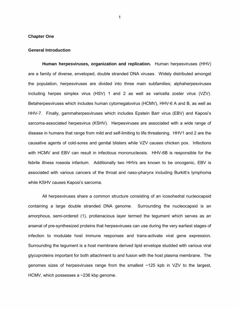
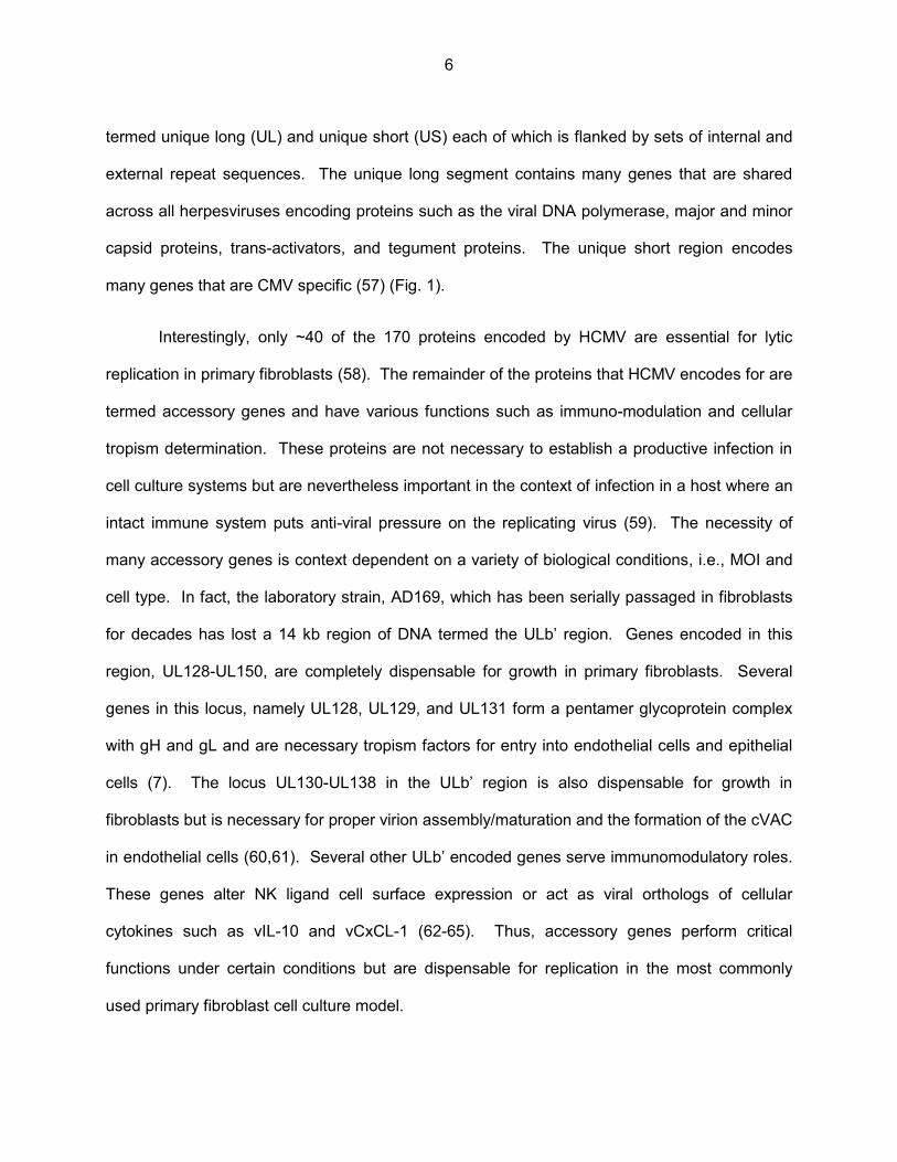
Figure 1: Genome map of HCMV strain Merlin .......................................................................... 18
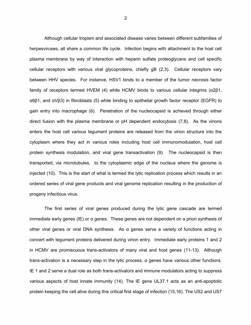
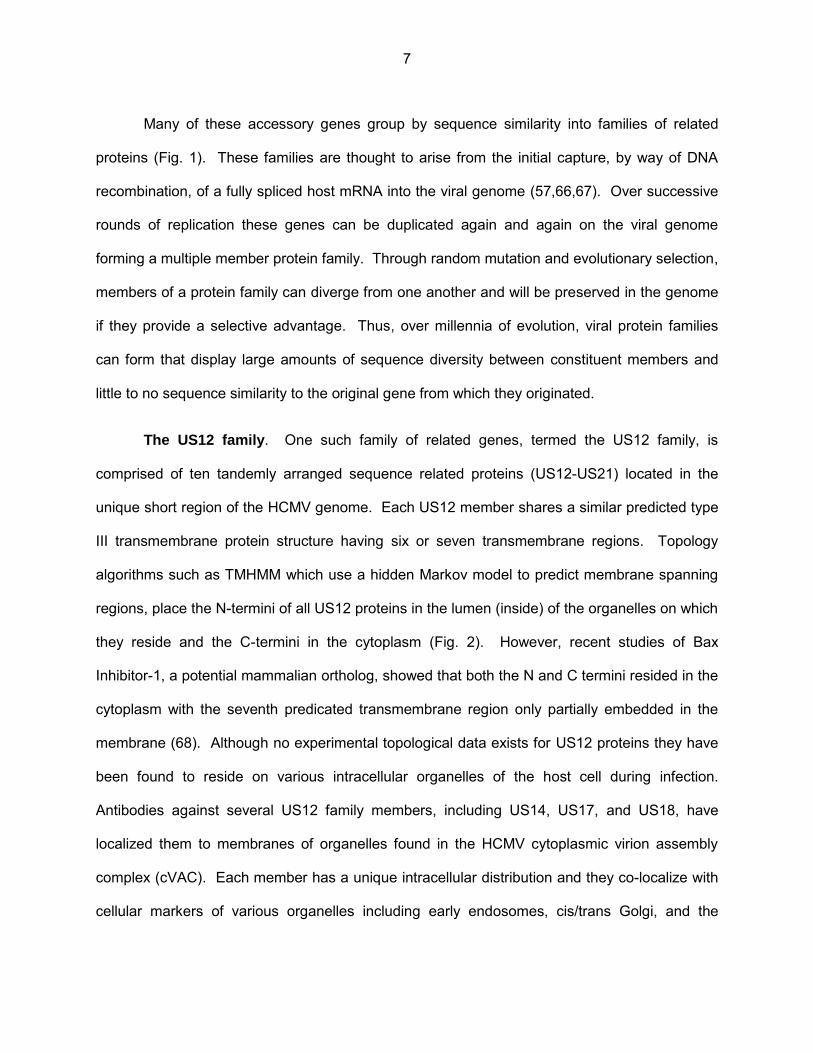
Figure 2: Predicted structure of US12 family proteins................................................................ 19
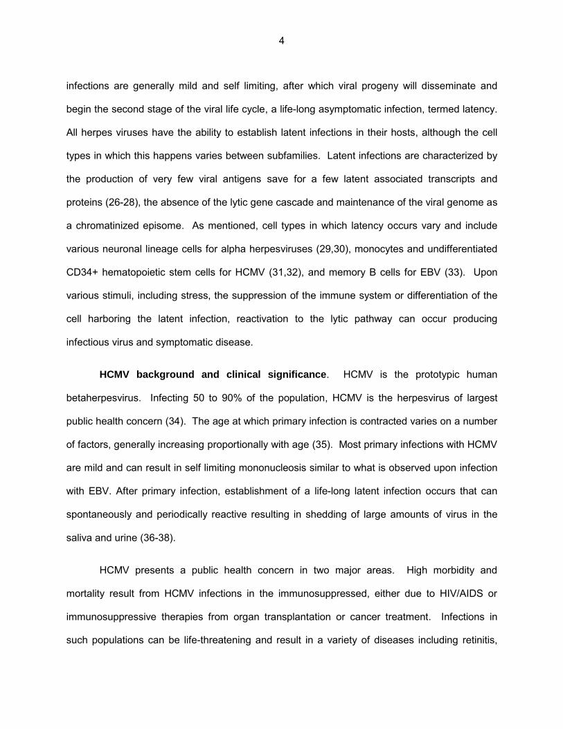
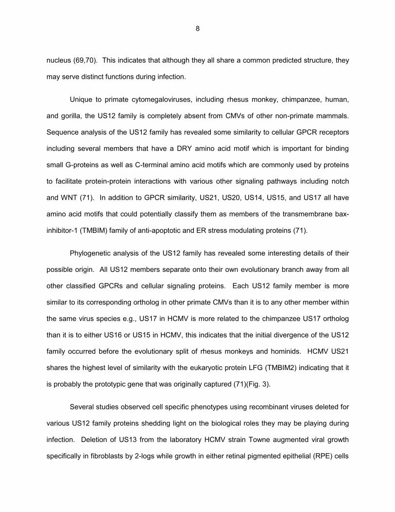
Figure 3: Duplication and divergence of the US12 gene family ................................................ 20
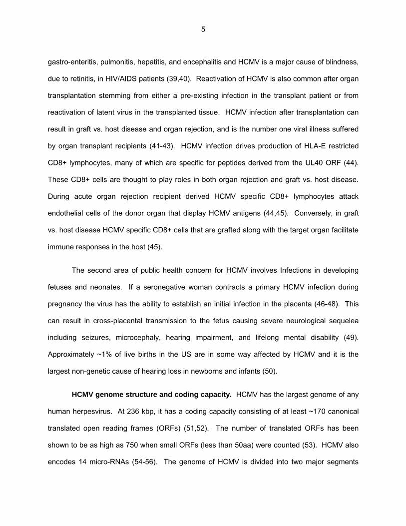
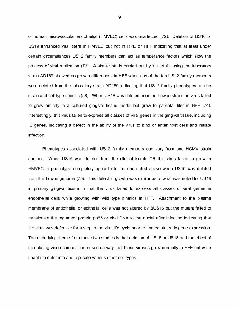
Figure 4: The HCMV cVAC and proposed path of virion egress ............................................... 21
Figure 5: Schematic of recombinant viruses used in this study ................................................ 42
Figure 6: Deletion of US17 does not significantly affect production of infectious virus in fibroblasts ................................................................................................................... 43
Figure 7: Deletion of US17 does not alter production of viral proteins ...................................... 44
Figure 8: Beadarray experiment overview and bio-informatics workflow ................................... 45
Figure 9: ΔUS17 alters small sets of host transcripts at both 12 and 96 hpi ............................. 46
Figure 10: SAM plots of genes differentially regulated by ΔUS17 compared to AD169 ....... 47-48
Figure 11: Transcripts modulated by ΔUS17 at 12 and 96 hpi found significant by SAM .......... 49
Figure 12: Cytoscape gene ontology maps of genes significantly regulated by ΔUS17 compared to parental at 12 hpi ................................................................................................. 50
Figure 13: Gene ontology analysis of SAM significant transcripts ............................................. 51
Figure 14: Transcript expression profiles for several GO categories highly significantly enriched at either 12 or 96 hpi ................................................................................................. 52
Figure 15: qRT-PCR verification of beadarray identified transcripts ......................................... 53
Figure 16: Linear Nycodenz gradients of infected cell culture supernatants ............................. 66
vi
Figure 17: HCMV genetic content of Nycodenz gradient fractions ............................................ 67
Figure 18: ΔUS17 changes virion composition ......................................................................... 68
Figure 19: ΔUS17 alters the genome-to-pfu ratio of gradient purified virions ............................ 69
Figure 20: ΔUS17 alters the genome to pfu ratio of infected cell culture supernatants ............. 70
Figure 21: Higher levels of tegument proteins are delivered per pfu to ΔUS17 infected fibroblasts ................................................................................................................ 71
Figure 22: Proposed model for US17 direct and indirect functions ........................................... 72
Chapter One
General Introduction
Human herpesviruses, organization and replication. Human herpesviruses (HHV)
are a family of diverse, enveloped, double stranded DNA viruses. Widely distributed amongst
the population, herpesviruses are divided into three main subfamilies; alphaherpesviruses
including herpes simplex virus (HSV) 1 and 2 as well as varicella zoster virus (VZV).
Betaherpesviruses which includes human cytomegalovirus (HCMV), HHV-6 A and B, as well as
HHV-7. Finally, gammaherpesviruses which includes Epstein Barr virus (EBV) and Kaposi’s
sarcoma-associated herpesvirus (KSHV). Herpesviruses are associated with a wide range of
disease in humans that range from mild and self-limiting to life threatening. HHV1 and 2 are the
causative agents of cold-sores and genital blisters while VZV causes chicken pox. Infections
with HCMV and EBV can result in infectious mononucleosis. HHV-6B is responsible for the
febrile illness roseola infantum. Additionally two HHVs are known to be oncogenic, EBV is
associated with various cancers of the throat and naso-pharynx including Burkitt’s lymphoma
while KSHV causes Kaposi’s sarcoma.
All herpesviruses share a common structure consisting of an icosohedral nucleocapsid
containing a large double stranded DNA genome. Surrounding the nucleocapsid is an
amorphous, semi-ordered (1), protienacious layer termed the tegument which serves as an
arsenal of pre-synthesized proteins that herpesviruses can use during the very earliest stages of
infection to modulate host immune responses and trans-activate viral gene expression.
Surrounding the tegument is a host membrane derived lipid envelope studded with various viral
glycoproteins important for both attachment to and fusion with the host plasma membrane. The
genomes sizes of herpesviruses range from the smallest ~125 kpb in VZV to the largest,
HCMV, which possesses a ~236 kbp genome.
1
Although cellular tropism and associated disease varies between different subfamilies of
herpesviruses, all share a common life cycle. Infection begins with attachment to the host cell
plasma membrane by way of interaction with heparin sulfate proteoglycans and cell specific
cellular receptors with various viral glycoproteins, chiefly gB (2,3). Cellular receptors vary
between HHV species. For instance, HSV1 binds to a member of the tumor necrosis factor
family of receptors termed HVEM (4) while HCMV binds to various cellular integrins (α2β1,
α6β1, and αVβ3) in fibroblasts (5) while binding to epithelial growth factor receptor (EGFR) to
gain entry into macrophage (6). Penetration of the nucleocapsid is achieved through either
direct fusion with the plasma membrane or pH dependent endocytosis (7,8). As the virions
enters the host cell various tegument proteins are released from the virion structure into the
cytoplasm where they act in various roles including host cell immunomodulation, host cell
protein synthesis modulation, and viral gene transactivation (9). The nucleocapsid is then
transported, via microtubules, to the cytoplasmic edge of the nucleus where the genome is
injected (10). This is the start of what is termed the lytic replication process which results in an
ordered series of viral gene products and viral genome replication resulting in the production of
progeny infectious virus.
The first series of viral genes produced during the lytic gene cascade are termed
immediate early genes (IE) or α genes. These genes are not dependent on a priori synthesis of
other viral genes or viral DNA synthesis. As α genes serve a variety of functions acting in
concert with tegument proteins delivered during virion entry. Immediate early proteins 1 and 2
in HCMV are promiscuous trans-activators of many viral and host genes (11-13). Although
trans-activation is a necessary step in the lytic process, α genes have various other functions.
IE 1 and 2 serve a dual role as both trans-activators and immune modulators acting to suppress
various aspects of host innate immunity (14). The IE gene UL37.1 acts as an anti-apoptotic
protein keeping the cell alive during this critical first stage of infection (15,16). The US2 and US7
2

proteins in HCMV act to block expression of MHC class I bound peptides at the cell surface
immediately after infection, stemming the activity of CD8+ cytotoxic T-cells (17). Thus, α genes
play important roles in setting the stage for a productive lytic infection.
The second class of genes produced termed early genes, or β genes, have expression
that is dependent on de novo protein production (α genes) but is not dependent on viral DNA
synthesis. These genes are associated with viral DNA replication and include the viral DNA
polymerase, and DNA processivity factor (HCMV UL54 and UL44) as well as nucleocapsid
assembly factors such as the major and minor capsid proteins (HCMV UL104, and UL105).
Capsid assembly takes place in the nucleus for all herpesviruses, after which, replicated
genomes are packaged and the assembled nucleocapsid exits the nucleus by way of budding
through the inner and outer nuclear membranes into the cytoplasm (18-21). This process of
capsid nuclear egress coincides with production of a third class of late genes, or γ genes.
Generally, γ genes are structural proteins, including tegument proteins, which aid in the final
assembly of the complete virion.
The final phase of the herpesvirus lifecycle, termed secondary envelopment, takes place
in the cytoplasm. The nucleocapsid aquires its full complement of tegument proteins as it
transits through the cytoplasm to the site of envelopment and eventual egress from the cell. This
process involves various organelles which differ between species of herpesvirus.
Alphaherpesviruses bud into vesicles derived from the Golgi apparatus (22,23). While HCMV
has been shown to use vesicles that have markers for both the trans-Golgi, and early
endosomes (24,25). After budding into the exit vesicle the viral particle is then trafficked to the
plasma membrane where fusion occurs releasing the virion into the extracellular space.
Primary infections with human herpesviruses generally begin with a lytic infection at the
mucosal epithelium of the mouth, nose, or genitals which serve as first sites of contact. Primary
3
infections are generally mild and self limiting, after which viral progeny will disseminate and
begin the second stage of the viral life cycle, a life-long asymptomatic infection, termed latency.
All herpes viruses have the ability to establish latent infections in their hosts, although the cell
types in which this happens varies between subfamilies. Latent infections are characterized by
the production of very few viral antigens save for a few latent associated transcripts and
proteins (26-28), the absence of the lytic gene cascade and maintenance of the viral genome as
a chromatinized episome. As mentioned, cell types in which latency occurs vary and include
various neuronal lineage cells for alpha herpesviruses (29,30), monocytes and undifferentiated
CD34+ hematopoietic stem cells for HCMV (31,32), and memory B cells for EBV (33). Upon
various stimuli, including stress, the suppression of the immune system or differentiation of the
cell harboring the latent infection, reactivation to the lytic pathway can occur producing
infectious virus and symptomatic disease.
HCMV background and clinical significance. HCMV is the prototypic human
betaherpesvirus. Infecting 50 to 90% of the population, HCMV is the herpesvirus of largest
public health concern (34). The age at which primary infection is contracted varies on a number
of factors, generally increasing proportionally with age (35). Most primary infections with HCMV
are mild and can result in self limiting mononucleosis similar to what is observed upon infection
with EBV. After primary infection, establishment of a life-long latent infection occurs that can
spontaneously and periodically reactive resulting in shedding of large amounts of virus in the
saliva and urine (36-38).
HCMV presents a public health concern in two major areas. High morbidity and
mortality result from HCMV infections in the immunosuppressed, either due to HIV/AIDS or
immunosuppressive therapies from organ transplantation or cancer treatment. Infections in
such populations can be life-threatening and result in a variety of diseases including retinitis,
4
gastro-enteritis, pulmonitis, hepatitis, and encephalitis and HCMV is a major cause of blindness,
due to retinitis, in HIV/AIDS patients (39,40). Reactivation of HCMV is also common after organ
transplantation stemming from either a pre-existing infection in the transplant patient or from
reactivation of latent virus in the transplanted tissue. HCMV infection after transplantation can
result in graft vs. host disease and organ rejection, and is the number one viral illness suffered
by organ transplant recipients (41-43). HCMV infection drives production of HLA-E restricted
CD8+ lymphocytes, many of which are specific for peptides derived from the UL40 ORF (44).
These CD8+ cells are thought to play roles in both organ rejection and graft vs. host disease.
During acute organ rejection recipient derived HCMV specific CD8+ lymphocytes attack
endothelial cells of the donor organ that display HCMV antigens (44,45). Conversely, in graft
vs. host disease HCMV specific CD8+ cells that are grafted along with the target organ facilitate
immune responses in the host (45).
The second area of public health concern for HCMV involves Infections in developing
fetuses and neonates. If a seronegative woman contracts a primary HCMV infection during
pregnancy the virus has the ability to establish an initial infection in the placenta (46-48). This
can result in cross-placental transmission to the fetus causing severe neurological sequelea
including seizures, microcephaly, hearing impairment, and lifelong mental disability (49).
Approximately ~1% of live births in the US are in some way affected by HCMV and it is the
largest non-genetic cause of hearing loss in newborns and infants (50).
HCMV genome structure and coding capacity. HCMV has the largest genome of any
human herpesvirus. At 236 kbp, it has a coding capacity consisting of at least ~170 canonical
translated open reading frames (ORFs) (51,52). The number of translated ORFs has been
shown to be as high as 750 when small ORFs (less than 50aa) were counted (53). HCMV also
encodes 14 micro-RNAs (54-56). The genome of HCMV is divided into two major segments
5
termed unique long (UL) and unique short (US) each of which is flanked by sets of internal and
external repeat sequences. The unique long segment contains many genes that are shared
across all herpesviruses encoding proteins such as the viral DNA polymerase, major and minor
capsid proteins, trans-activators, and tegument proteins. The unique short region encodes
many genes that are CMV specific (57) (Fig. 1).
Interestingly, only ~40 of the 170 proteins encoded by HCMV are essential for lytic
replication in primary fibroblasts (58). The remainder of the proteins that HCMV encodes for are
termed accessory genes and have various functions such as immuno-modulation and cellular
tropism determination. These proteins are not necessary to establish a productive infection in
cell culture systems but are nevertheless important in the context of infection in a host where an
intact immune system puts anti-viral pressure on the replicating virus (59). The necessity of
many accessory genes is context dependent on a variety of biological conditions, i.e., MOI and
cell type. In fact, the laboratory strain, AD169, which has been serially passaged in fibroblasts
for decades has lost a 14 kb region of DNA termed the ULb’ region. Genes encoded in this
region, UL128-UL150, are completely dispensable for growth in primary fibroblasts. Several
genes in this locus, namely UL128, UL129, and UL131 form a pentamer glycoprotein complex
with gH and gL and are necessary tropism factors for entry into endothelial cells and epithelial
cells (7). The locus UL130-UL138 in the ULb’ region is also dispensable for growth in
fibroblasts but is necessary for proper virion assembly/maturation and the formation of the cVAC
in endothelial cells (60,61). Several other ULb’ encoded genes serve immunomodulatory roles.
These genes alter NK ligand cell surface expression or act as viral orthologs of cellular
cytokines such as vIL-10 and vCxCL-1 (62-65). Thus, accessory genes perform critical
functions under certain conditions but are dispensable for replication in the most commonly
used primary fibroblast cell culture model.
6
Many of these accessory genes group by sequence similarity into families of related
proteins (Fig. 1). These families are thought to arise from the initial capture, by way of DNA
recombination, of a fully spliced host mRNA into the viral genome (57,66,67). Over successive
rounds of replication these genes can be duplicated again and again on the viral genome
forming a multiple member protein family. Through random mutation and evolutionary selection,
members of a protein family can diverge from one another and will be preserved in the genome
if they provide a selective advantage. Thus, over millennia of evolution, viral protein families
can form that display large amounts of sequence diversity between constituent members and
little to no sequence similarity to the original gene from which they originated.
The US12 family. One such family of related genes, termed the US12 family, is
comprised of ten tandemly arranged sequence related proteins (US12-US21) located in the
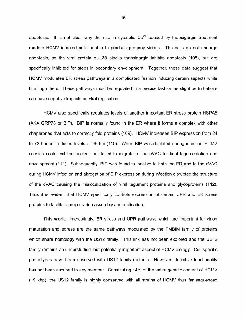
unique short region of the HCMV genome. Each US12 member shares a similar predicted type
III transmembrane protein structure having six or seven transmembrane regions. Topology
algorithms such as TMHMM which use a hidden Markov model to predict membrane spanning
regions, place the N-termini of all US12 proteins in the lumen (inside) of the organelles on which
they reside and the C-termini in the cytoplasm (Fig. 2). However, recent studies of Bax
Inhibitor-1, a potential mammalian ortholog, showed that both the N and C termini resided in the
cytoplasm with the seventh predicated transmembrane region only partially embedded in the
membrane (68). Although no experimental topological data exists for US12 proteins they have
been found to reside on various intracellular organelles of the host cell during infection.
Antibodies against several US12 family members, including US14, US17, and US18, have
localized them to membranes of organelles found in the HCMV cytoplasmic virion assembly
complex (cVAC). Each member has a unique intracellular distribution and they co-localize with
cellular markers of various organelles including early endosomes, cis/trans Golgi, and the
7
nucleus (69,70). This indicates that although they all share a common predicted structure, they
may serve distinct functions during infection.
Unique to primate cytomegaloviruses, including rhesus monkey, chimpanzee, human,
and gorilla, the US12 family is completely absent from CMVs of other non-primate mammals.
Sequence analysis of the US12 family has revealed some similarity to cellular GPCR receptors
including several members that have a DRY amino acid motif which is important for binding
small G-proteins as well as C-terminal amino acid motifs which are commonly used by proteins
to facilitate protein-protein interactions with various other signaling pathways including notch
and WNT (71). In addition to GPCR similarity, US21, US20, US14, US15, and US17 all have
amino acid motifs that could potentially classify them as members of the transmembrane bax-
inhibitor-1 (TMBIM) family of anti-apoptotic and ER stress modulating proteins (71).
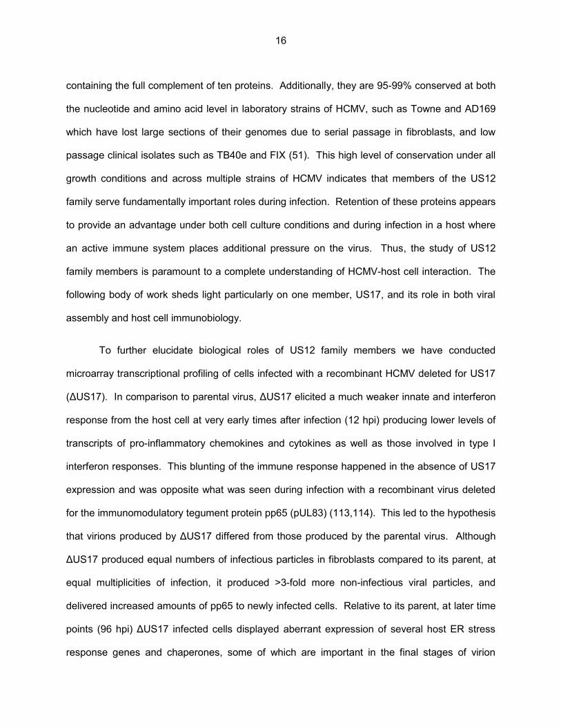
Phylogenetic analysis of the US12 family has revealed some interesting details of their
possible origin. All US12 members separate onto their own evolutionary branch away from all
other classified GPCRs and cellular signaling proteins. Each US12 family member is more
similar to its corresponding ortholog in other primate CMVs than it is to any other member within
the same virus species e.g., US17 in HCMV is more related to the chimpanzee US17 ortholog
than it is to either US16 or US15 in HCMV, this indicates that the initial divergence of the US12
family occurred before the evolutionary split of rhesus monkeys and hominids. HCMV US21
shares the highest level of similarity with the eukaryotic protein LFG (TMBIM2) indicating that it
is probably the prototypic gene that was originally captured (71)(Fig. 3).
Several studies observed cell specific phenotypes using recombinant viruses deleted for
various US12 family proteins shedding light on the biological roles they may be playing during
infection. Deletion of US13 from the laboratory HCMV strain Towne augmented viral growth
specifically in fibroblasts by 2-logs while growth in either retinal pigmented epithelial (RPE) cells
8
or human microvascular endothelial (HMVEC) cells was unaffected (72). Deletion of US16 or
US19 enhanced viral titers in HMVEC but not in RPE or HFF indicating that at least under
certain circumstances US12 family members can act as temperance factors which slow the
process of viral replication (73). A similar study carried out by Yu. et Al. using the laboratory
strain AD169 showed no growth differences in HFF when any of the ten US12 family members
were deleted from the laboratory strain AD169 indicating that US12 family phenotypes can be
strain and cell type specific (58). When US18 was deleted from the Towne strain the virus failed
to grow entirely in a cultured gingival tissue model but grew to parental titer in HFF (74).
Interestingly, this virus failed to express all classes of viral genes in the gingival tissue, including
IE genes, indicating a defect in the ability of the virus to bind or enter host cells and initiate
infection.
Phenotypes associated with US12 family members can vary from one HCMV strain
another. When US16 was deleted from the clinical isolate TR this virus failed to grow in
HMVEC, a phenotype completely opposite to the one noted above when US16 was deleted
from the Towne genome (75). This defect in growth was similar as to what was noted for US18
in primary gingival tissue in that the virus failed to express all classes of viral genes in
endothelial cells while growing with wild type kinetics in HFF. Attachment to the plasma
membrane of endothelial or epithelial cells was not altered by ΔUS16 but the mutant failed to
translocate the tegument protein pp65 or viral DNA to the nuclei after infection indicating that
the virus was defective for a step in the viral life cycle prior to immediate early gene expression.
The underlying theme from these two studies is that deletion of US16 or US18 had the effect of
modulating virion composition in such a way that these viruses grew normally in HFF but were
unable to enter into and replicate various other cell types.
9
Consistent with this, HCMV displays broad cellular tropism (fibroblasts, endothelial cells,
epithelial cells, monocytes/macrophage) and has adapted numerous strategies to enter into and
replicate in various cell types. For instance, HCMV entry into endothelial and epithelial cells is
mediated by the glycoprotein complex UL128-131/gH/gL and occurs through low pH
endocytosis (7,76); on the other hand entry into fibroblasts is mediated by another distinct
glycoprotein complex gH/gL/gO and occurs by direct, pH independent, fusion at the plasma
membrane (8,77). Changes in levels of one set of glycoproteins can shift virus tropism from
fibroblasts to endothelial/epithelial cells or vice versa while leaving tropism for the opposite cell
type unaffected or even enhanced.
Condition specific phenotypes have also been observed following modulation of levels of
tegument proteins in mature virions. A virus deleted for the tegument protein pp65 (pUL83) was
highly attenuated for growth in human fibroblasts at low multiplicities of infection (MOI), but grew
with wild type kinetics at high MOI (78). Further, pp65 is essential at all MOIs for growth in
human macrophages and its deletion altered virion levels of the tegument proteins pUL25,
pUL69, and pUL97 (79). Thus, changes in virion composition brought about by mutations to
US12 family members could have marked and varied effects that are dependent on a
combination of cell type and infection conditions.
The US12 family, relation to the TMBIM family of ER stress proteins. As mentioned
above, US12 family members share sequence similarity with the trans-membrane bax inhibitor-
1 (TMBIM) family of conserved eukaryotic anti-apoptotic seven transmembrane proteins (71).
These proteins localize to membranes of various cellular organelles and are thought to act as
rheostats that modulate apoptotic signaling, ER stress, and the unfolded protein response (80).
ER stress is a process that is initiated by any number of cellular stressors, including viral
infections, which cause a disruption to cellular homeostasis. The unfolded protein response is a
10

type of ER stress which results from an accumulation of unfolded proteins in the endoplasmic
reticulum, in the case of HCMV, UPR results from the large amounts of viral glycoproteins
produced during infection that must be correctly folded and transported through the secretory
apparatus of the cell. Several members of the TMBIM family have been shown to influence
intracellular calcium levels acting as pH dependent calcium channels allowing calcium to leak
from the endoplasmic reticulum to the cytoplasm (81-85). Bax inhibitor-1 (BI-1 / TMBIM6)
function as a negative regulator of the IRE1α branch of the UPR and over expression of BI-1
inhibits splicing of the ER stress associated transcription factor, XBP-1 that is directly processed
and activated by IRE1α (86,87). BI-1 has also been shown to interact with both the anti-
apoptotic protein BCL-2, and IP3 receptors in the ER (88,89). This interaction with IP3Rs links
BI-1 with autophagic pathways and BI-1 deficient cells showed a lower resting rate of
autophagic processes, however, BI-1s role in autophagy was shown to be independent of its
cytoprotective functions indicating that it has multiple biological roles (90).
Another TMBIM member, human Golgi associated anti-apoptotic protein (hGAAP /
TMBIM4), also influences ER Ca2+ levels and functions as a regulator of cell adhesion and
motility (91). Interestingly, poxviruses, specifically camelpox virus and vaccinia virus, have been
shown to encode orthologs of TMBIM like proteins. Termed viral Golgi-associated anti-
apoptotic proteins (vGAAP) these proteins localize to the Golgi of infected cells, and are non-
essential for virus growth in cell culture. Knockdown of vGAAP proteins caused an increase in
lethality in a mouse model indicating that they serve as temperance factors limiting the
replication or dissemination of virus in the host (92). Similar to hGAAP, vGAAP has been
shown to modulate levels of calcium in the ER (93).
Clearly, TMBIM proteins have a diverse array of important functions in cell biology.
HCMV is known to extensively modulate many of the pathways discussed above and it is
11
possible that the capture and divergence of the US12 family helps to facilitate this signaling by
serving as a virally controlled TMBIM like family of proteins similar to what was observed in
Poxviruses. Further, the pathways affected by TMBIM family members, i.e. ER stress and
apoptosis, have been implicated as important in the processes of HCMV virion maturation and
assembly. Therefore, it is highly possible that US12 family members serve as TMBIM orthologs
influencing virion assembly by modulating signaling pathways important in the construction of
mature virions.
Virion assembly and the cytoplasmic viral assembly complex (cVAC). At least 70
proteins have been detected in purified HCMV virion preparations (94,95) and the process of
HCMV virion maturation is presently not well understood. Relying on both viral and cellular
factors, cytoplasmic virion maturation is thought to occur in the cVAC, a virally induced
rearrangement of the host cell secretory apparatus consisting of ordered, concentric, rings of
various secretory organelles. Specifically, it consists of a region of endoplasmic reticulum at the
outer edge. Just inside the ER is an interlaced ring or basket like structure consisting of
membranes derived from both Golgi and trans-Golgi compartments, and a central region which
contains a collection of condensed EEA1 positive early endosomes as well as a microtubule
organizing center (MTOC) (96,97) (Fig. 4). The cVAC is formed from 72 to 96 hpi and
corresponds with production of high levels of infectious virus and is thought to be the site of final
tegumentation and secondary envelopment of maturating virions.
Many viral proteins, chiefly tegument and glycoproteins, converge in the cVAC during
the late stages of HCMV infection. In fact, the cVAC was first described as a confluence of the
viral tegument proteins pp28 (pUL99), pp150 (pUL32), pp65 (pUL83), the viral glycoproteins
gB(pUL55), gH(pUL75), and gp65 (97,98). Since then, many other viral proteins have been
shown to localize to this region including glycoproteins gO, gM, and gN, as well as the tegument
12
protein UL48 (99-101). While the exact function of the cVAC has not been elucidated it is clear
that this structure serves as the major congregation point for many HCMV structural proteins.
Particle types produced during HCMV infection. HCMV has been shown to produce
several different types of particles during infection that are able to be separated by various
density gradient centrifugation techniques. The most well characterized of these is the virion
with a diameter of ~250 nm consisting of the above mentioned conserved herpesvirus structure
of encapsidated genome, tegument layer, and lipid envelope. Interestingly, HCMV seems to
produce a very high particle to pfu ratio (i.e., the ratio of infectious units measured by plaque
assay to the number of particles counted by electron microscopy) producing several hundred to
several thousand non-infectious virions for every one infectious (102). These non-infectious
particles are difficult to isolate and study and thus the definition of exactly which structural
components are necessary to initiate a productive infection remains unclear.
HCMV also produces a particle, termed dense bodies, which are both 2 – 4 times larger
and more dense than infectious virons. Dense bodies are composed mainly of viral tegument
proteins with ~60% of their mass being made up of the tegument protein pp65. They possess a
lipid envelope similar to infectious virions but which differs in both the abundances and types of
incorporated glycoproteins (95). Dense bodies are able to enter into host cells independently of
infectious virions and can deliver immunomodulatory proteins, possibly serving as “bait”
particles that can elicit an immune response and thus draw host defenses away from the site of
the actual infection (103). A third type of particle termed the non-infectious enveloped particles
(NIEP) has also been isolated from HCMV infected cells (104). NIEP’s are essentially a
genome-less capsid (B-capsid) that has undergone the process of secondary envelopment.
Little is known about the possible functions of NIEPs although their lack of intact genomes most
certainly renders them unable to start a productive infection.
13
Even though virions, dense bodies, and NIEPs are composed of essentially the same
viral proteins the pathways involved in maturation of these particles differ in key areas. When
viral DNA encapsidation was inhibited virions accumulated in the nucleus of infected cells but
did not transit to the cytoplasm, nor were they secreted from the cell. Dense body assembly
remained unaltered even in the absence of DNA encapsidation and appears to rely solely on
cytoplasmic factors such as tegument protein levels (105). The viral protein UL103 was
subsequently shown to be important for egress of virions, dense bodies and NIEPs indicating
that while maturation pathways differ between the particle types, a common pathway is shared
for particle egress (105).
Cellular pathways involved in virion assembly. Cellular proteins and pathways play
a major role in both the formation of the cVAC and production of mature virions. Specifically,
those involved in ER stress and the unfolded protein response (UPR) are particularly important.
HCMV both induces and controls the three major branches of the unfolded protein response,
PKR like ER kinase (PERK), activating transcription factor 6 (ATF6), and inositol requiring
enzyme (IRE1α), activating certain aspects while inhibiting others. Various groups have shown
that control of UPR pathways by HCMV is essential for both proper virion assembly and
maturation. For instance, activation of PERK generally results in protein translation attenuation
by way of phosphorylation of eIF2a. Although HCMV infection results in an increase in PERK
phosphorylation and phospho- eIF2a, translation is not attenuated (106). Additionally, HCMV
infection induces IRE1α phosphorylation and splicing of the down-stream transcription factor
XBP1, but inhibits the production of XBP1 associated UPR related gene products important for
ER associated protein degradation (106). Consistent with this, chemicals that specifically cause
ER stress, such as thapsigargin, reduce the levels of infectious virus produced by inhibiting the
final steps of virion maturation and secondary envelopment (107). Thapsigargin inhibits the ER
Ca2+ ATPase, SERCA, thus increasing cytosolic levels of Ca2+ causing ER stress and eventually
14
apoptosis. It is not clear why the rise in cytosolic Ca2+ caused by thapsigargin treatment
renders HCMV infected cells unable to produce progeny virions. The cells do not undergo
apoptosis, as the viral protein pUL38 blocks thapsigargin inhibits apoptosis (108), but are
specifically inhibited for steps in secondary envelopment. Together, these data suggest that
HCMV modulates ER stress pathways in a complicated fashion inducing certain aspects while
blunting others. These pathways must be regulated in a precise fashion as slight perturbations
can have negative impacts on viral replication.
HCMV also specifically regulates levels of another important ER stress protein HSPA5
(AKA GRP78 or BIP). BIP is normally found in the ER where it forms a complex with other
chaperones that acts to correctly fold proteins (109). HCMV increases BIP expression from 24
to 72 hpi but reduces levels at 96 hpi (110). When BIP was depleted during infection HCMV
capsids could exit the nucleus but failed to migrate to the cVAC for final tegumentation and
envelopment (111). Subsequently, BIP was found to localize to both the ER and to the cVAC
during HCMV infection and abrogation of BIP expression during infection disrupted the structure
of the cVAC causing the mislocalization of viral tegument proteins and glycoproteins (112).
Thus it is evident that HCMV specifically controls expression of certain UPR and ER stress
proteins to facilitate proper virion assembly and replication.
This work. Interestingly, ER stress and UPR pathways which are important for virion
maturation and egress are the same pathways modulated by the TMBIM family of proteins
which share homology with the US12 family. This link has not been explored and the US12
family remains an understudied, but potentially important aspect of HCMV biology. Cell specific
phenotypes have been observed with US12 family mutants. However, definitive functionality
has not been ascribed to any member. Constituting ~4% of the entire genetic content of HCMV
(~9 kbp), the US12 family is highly conserved with all strains of HCMV thus far sequenced
15
containing the full complement of ten proteins. Additionally, they are 95-99% conserved at both
the nucleotide and amino acid level in laboratory strains of HCMV, such as Towne and AD169
which have lost large sections of their genomes due to serial passage in fibroblasts, and low
passage clinical isolates such as TB40e and FIX (51). This high level of conservation under all
growth conditions and across multiple strains of HCMV indicates that members of the US12
family serve fundamentally important roles during infection. Retention of these proteins appears
to provide an advantage under both cell culture conditions and during infection in a host where
an active immune system places additional pressure on the virus. Thus, the study of US12
family members is paramount to a complete understanding of HCMV-host cell interaction. The
following body of work sheds light particularly on one member, US17, and its role in both viral
assembly and host cell immunobiology.
To further elucidate biological roles of US12 family members we have conducted
microarray transcriptional profiling of cells infected with a recombinant HCMV deleted for US17
(ΔUS17). In comparison to parental virus, ΔUS17 elicited a much weaker innate and interferon
response from the host cell at very early times after infection (12 hpi) producing lower levels of
transcripts of pro-inflammatory chemokines and cytokines as well as those involved in type I
interferon responses. This blunting of the immune response happened in the absence of US17
expression and was opposite what was seen during infection with a recombinant virus deleted
for the immunomodulatory tegument protein pp65 (pUL83) (113,114). This led to the hypothesis
that virions produced by ΔUS17 differed from those produced by the parental virus. Although
ΔUS17 produced equal numbers of infectious particles in fibroblasts compared to its parent, at
equal multiplicities of infection, it produced >3-fold more non-infectious viral particles, and
delivered increased amounts of pp65 to newly infected cells. Relative to its parent, at later time
points (96 hpi) ΔUS17 infected cells displayed aberrant expression of several host ER stress
response genes and chaperones, some of which are important in the final stages of virion
16

assembly and egress. Our results suggest that US17 modulates host pathways to enable
production of virions that elicit an appropriately balanced host immune response.
17

Figure 1. Genome map of HCMV strain Merlin. Open reading frames are depicted as colored boxes. Similarity colored ORFs are gene families.
Used with permission, from Dolan et al. Journal of General Virology (2004), 85, 1301–1312
18
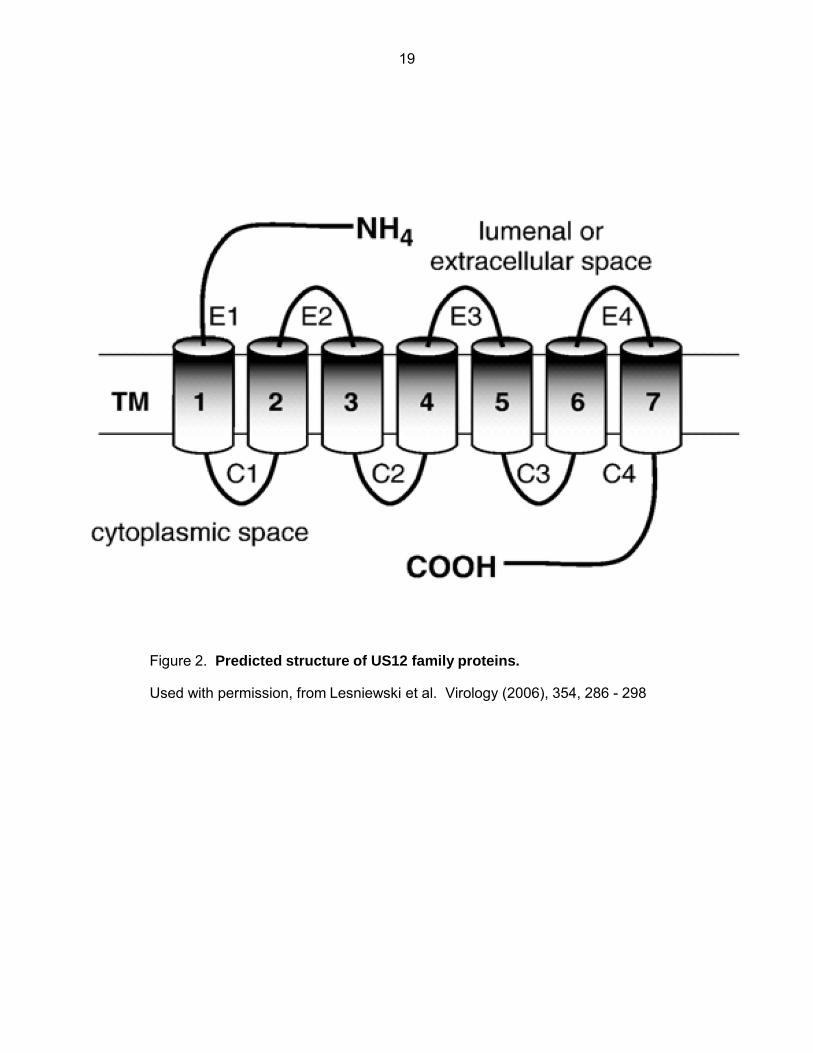
Figure 2. Predicted structure of US12 family proteins.
Used with permission, from Lesniewski et al. Virology (2006), 354, 286 - 298
19
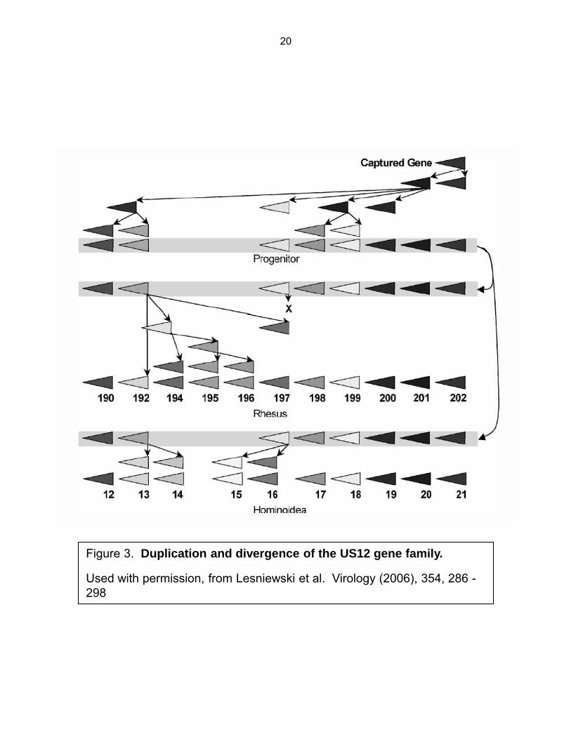
Figure 3. Duplication and divergence of the US12 gene family.
Used with permission, from Lesniewski et al. Virology (2006), 354, 286 - 298
20
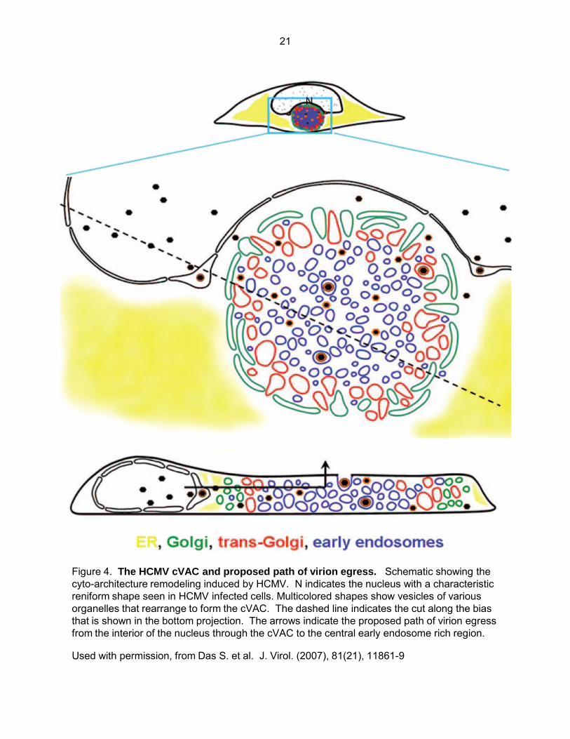
Figure 4. The HCMV cVAC and proposed path of virion egress. Schematic showing the cyto-architecture remodeling induced by HCMV. N indicates the nucleus with a characteristic reniform shape seen in HCMV infected cells. Multicolored shapes show vesicles of various organelles that rearrange to form the cVAC. The dashed line indicates the cut along the bias that is shown in the bottom projection. The arrows indicate the proposed path of virion egress from the interior of the nucleus through the cVAC to the central early endosome rich region.
Used with permission, from Das S. et al. J. Virol. (2007), 81(21), 11861-9
N
21

Chapter Two Cellular transcriptional profiling of ΔUS17 infected fibroblasts
Introduction.
The multi-protein nature of the US12 family has made study of individual members
difficult. It is unknown whether members can functionally complement each other thereby
mitigating discernible phenotypes when any particular member is deleted or mutated. Potential
complementation, coupled with the dispensability of US12 family members for growth in the
most commonly used cell culture models of HCMV has greatly hampered study of the US12
family. Currently, there are no functions ascribed to any of the US12 family members and these
proteins remain an untapped and potentially important area of study.
To further address the biological importance of US12 family members we have
performed cellular transcriptional analysis of cells infected with a recombinant HCMV deleted for
one member, US17. We previously found that US17 is expressed with late gene kinetics, and
localizes to the nucleus of infected fibroblasts beginning at 72 hpi. When imaged by
immunofluorescence microscopy using a polyclonal antibody directed against a C-terminal
epitope, interestingly, there was little co-localization between the N and C termini of US17 when
both were imaged simultaneously. Immunoblotting with the polyclonal antibody revealed that
two distinct species of US17 were present in infected cells, an ~80 kDa species and a smaller
~10 kDa species indicating that US17 is expressed in a segmented fashion (70). The biological
implications of this segmentation have not been elucidated and no biological function has been
ascribed to US17.
Analyzing cellular pathways perturbed by deletion of US17 is an effective way to glean
information about what roles it may be playing during infection. However, the limited information
22
available regarding the cellular roles of US12 family members makes generation of any specific
hypothesis, as to which genes they may be affecting, difficult. Therefore we have opted to take
an unbiased approach to transcriptional profiling.
Here we present data from an Illumina HT-12 v4 bead array from cells infected with
ΔUS17 and the laboratory strain of HCMV from which the deletion mutant was derived, AD169.
The HT-12 v4 is similar to a traditional microarray in that it measures expression levels of
mRNA transcripts from a sample of isolated whole cellular RNA. However, several benefits are
conferred by the platform over other methods of whole transcriptome analysis. The HT-12 v4
beadarray consists of 47,232 individual probes that give specific expression information for
39,809 coding sequences with well-defined or provisional annotations in the NCBI database as
well as 3,961 non-coding sequences. This high number of probes provides excellent coverage
and gives gene expression information for essentially every known annotated human transcript
allowing for robust downstream analysis of biological pathways affected by ΔUS17. Thus we
generated comprehensive cellular transcription profiles that have allowed us to elucidate a
potential role for US17 even in the absence of a differential growth phenotype.
23
Materials and methods
Cell culture, preparation of virus stocks, and virus purification. All experiments in
this study used normal human foreskin fibroblasts (HFF). All cells were used between
passages 10 and 15. Cells were cultured in DMEM (Hyclone/Thermo-Fischer, Waltham, MA )
supplemented with 10% fetal bovine serum, 1% penicillin/streptomycin, 1% Glutamax (Life
Technologies Grand, Island, NY), and 1% minimal non-essential amino acids. HCMV strain
AD169 (ATCC), the AD169 BAC pAD/Cre parental (provided courtesy of Dr. Dong Yu), and the
US17 deletion virus (ΔUS17) were cultured by inoculating confluent HFF monolayers at an MOI
of 0.001. Infected cells and supernatants were harvested 14 dpi and virus titers were
determined by plaque assay on confluent HFF monolayers. For all experiments, low passage
HFF were seeded onto 35 mm dishes 72 hr pre-infection at 3x10⁴ cells/cm2. Where applicable,
virions were concentrated by centrifugation of clarified supernatants through a 20% sorbitol
cushion at 60,000 x g for 1 h in a Beckman SW 41 Ti rotor. Subsequent purification of virions
was carried out by centrifuging concentrated virus through a 10-50% linear Nycodenz gradient
for 2 h at 110,000 x g in a Beckman SW 41 Ti rotor (95).
Recombinant viruses. A virus deleted for the entire US17 open reading frame was
constructed using a bacterial artificial chromosome (BAC) system (115). In short, E. coli (strain
EL250) which harbors a temperature inducible lambda pro-phage RED recombinase for
homologous linear recombination and an additional arabinose inducible FRT recombinase was
transformed with a BAC of HCMV strain AD169 pAD/Cre DH18 (provided courtesy of Dr.
Thomas Shenk), which is a full length BAC clone of HCMV strain AD169 that harbors a
transposon insertion cassette that disrupts the US17 open reading frame (58).
An insertion cassette was created by PCR amplification using a set of primers(forward:
ATCGCCACCGCCGTCgaagttcctattctctagaaagtataggaacttcAGACGTCAGGTGGCACTTTT;
24

reverse:AACGACGAGTTTTTCCGgaagttcctatactttctagagaataggaacttcAGCTCTTGATCCGGCA
AAC) consisting of a 5’ portion encoding for 15-17 bp of DNA directly flanking the US17 open
reading frame upstream of the start codon or downstream of the stop codon (capital letters), an
inner portion encoding a FLP recombinase site (lower case letters), and 20 bp at their 3’ ends
complementary to the ampicillin resistance gene of plasmid pPur (underlined) (Clontech,
Mountain View, CA). A second round of amplification used the product of the first reaction as
template and a set of primers consisting of 50 bases flanking the US17 open reading frame
(upstream primer:
ACACTCTATAAACGGTTTCTCATACGCGCCTTTTGATCGCCACCGCCGTC; downstream
primer: TTGGTGGAGACGGCCGGCGCGGCGGGTGGGGGAAACGACGAGTTTTTCCG). The
resulting cassette thus consisted of an inner core ampicillin resistance gene flanked by two FLP
recombinase sites with 50 bp of US17 ORF flanking DNA to facilitate linear recombination.
BAC-containing E. coli was shifted to 42°C for 15 min to activate the RED recombinase,
and then transformed with 300 ng of gel purified PCR product. Recombinants were selected on
LB agar plates containing 25 μg/ml ampicillin, and insertion of the cassette was confirmed by
HCMV genome restriction digestion with Hind III, and PCR from both within and outside the
US17 ORF. To remove the cassette and generate the final in frame deletion mutant, an
overnight culture of the previously described E. coli carrying the HCMV BAC and ampicillin
resistance gene cassette in place of the US17 ORF was subcultured 1/50 in fresh LB media and
incubated at 32°C until the culture reached OD600 = 0.5. Sterile arabinose was added to a final
concentration of 0.1% and the culture was incubated at 32°C for an additional hour to activate
the FLP recombinase. Serial dilutions were plated on non-selective media, colonies were
picked and screened for sensitivity to ampicillin, and the deletion was verified by Hind III
digestion. US17 mutants were further verified by PCR from both within and across the US17
25
open frame to confirm the absence of the ORF. The resulting mutant was deleted for the US17
ORF leaving only a 34 bp FLP scar.
To construct the US17 repair virus with a C-terminal V5 epitope tag, a scar less GalK
recombineering system was used (116). The US17 sequence from the AD169 genome was
PCR amplified using a set of primers that added the V5 epitope tag to the C-terminal end
(Forward primer: TGTGGATCCATGTCTCCGAACTCA, Reverse primer
TTCTCGAGTTACGTAGAATCGAGACCGAGGAGAGGGTTAGGGATAGGCTTACCCGCCATG
GTTCGCGTGAG Bold text represents start or stop codons, underlined text represents the V5
epitope sequence.) The amplimer was cloned as a BamHI / XhoI fragment into the PCDNA3.1
vector (Life Technologies, Grand Island, NY). Next, a GalK Kanamycin resistance cassette
containing 50 bp of up-stream and down-stream homologous US17 sequence was PCR
amplified from the plasmid C255 (117) (provided courtesy of Dr. Dong Yu) (Forward primer:
TTGGTGGAGACGGCCGGCGCGGCGGGTGGGGGAAACGACGAGTTTTTCCGCCTGTTGA
CAATTAATCATCG, reverse primer:
ACACTCTATAAACGGTTTCTCATACGCGCCTTTTGATCGCCACCGCCGTCCTCAGCAAAA
GTTCGATTTA. Bold text represents homologous AD169 sequence; non-bold text represents
the GalK/Kanr binding sequence.) This sequence was recombined into the ΔUS17 locus using
RED mediated recombination as described for the deletion mutant. Finally, the GalK/Kanr was
removed and replaced with the US17 C-terminal V5 sequence by constructing an amplimer
consisting of the US17-cV5 sequence with 50 bp of homologous US17sequence at either end
(Forward primer:
TTGGTGGAGACGGCCGGCGCGGCGGGTGGGGGAAACGACGAGTTTTTCCGTTACGTAG
AATCGAGACCGAGGA, reverse primer:
ACACTCTATAAACGGTTTCTCATACGCGCCTTTTGATCGCCACCGCCGTCATGTCTCCGA
ACTCAGAGGCCAC).
26
Protein analysis: immunoblotting and silver staining. At the indicated time points,
infected cell monolayers (MOI = 3.0) were washed once with ice cold PBS and lysed in RIPA
buffer (150 mM NaCl, 10 mM HEPES [pH 7.4], 1% Noniodet P40, 1% sodium deoxycholate,
0.1% SDS, 1x protease inhibitors (Roche, Indianapolis, IN)). After incubation for 5 min on ice,
lysates were clarified by centrifugation at 10,000 x g for 10 min at 4°C. Protein concentrations
were measured by BCA assay (Thermo Fisher, Waltham, MA); equal amounts of total protein
from each lysate, or protein from gradient purified virus fractions were run on 10%
polyacrylamide gels. Silver staining of polyacrylamide gels was carried out using a Pierce silver
stain kit (Thermo Scientific, Rockford, IL). Immunoblots were transferred to 0.1 µm
nitrocellulose membrane and then probed with antibodies against the following proteins: IE1 &
IE2, pp28, gB (Virusys, Taneytown, MD), pp65 (Fitzgerald Industries, Acton, MA), gH (Santa
Cruz Dallas, TX), and a polyclonal rabbit antibody against the C-terminus of UL48 (a gift from
Dr. Wade Gibson). Chemiluminescence was performed with Super Signal Pico West substrate
(Thermo, Waltham, MA) following the manufacturer’s directions.
Isolation of viral DNA or total cellular RNA for genome quantitation, microarrays,
and qRT-PCR analysis. To ensure accurate multiplicities of infection, one dish of cells was
trypsinized and counted immediately prior to infection to gauge final cell density. HFF (p12)
were then infected with either AD169 or pAD/CRE(ΔUS17) (MOI = 6.0) for 12 or 96 hr. After
washing once with PBS, 1 ml of Trizol (Life Technologies, Grand Island, NY) reagent was added
to each 35 mm dish. RNA was separated by addition of 200 μl chloroform to each 1 ml Trizol
sample followed by centrifugation at 12,000 x g for 15 min at 4°C. The aqueous phase was
transferred to a clean tube and RNA was precipitated by addition of 500 μl of 100% isopropyl
alcohol followed by centrifugation at 12,000 x g for 15 min at 4°C. The RNA pellet was washed
twice in 70% ethanol and then resuspended in 50 μl of nuclease free deionized water. DNase I
treatment was conducted with 2 U of RNase free DNase (New England Biolabs, Ipswich, MA)
27
following the manufacturer’s directions. RNA concentration was measured by UV spectroscopy
(260 nm : 280 nm) using a NanoDrop 1000 spectrophotometer (Thermo-Fisher, Waltham, MA).
RNA quality was assessed on an Agilent Biolyzer (Agilent Technologies Santa Clara, CA); all
samples had RIN values of 8 to 10. RNA was then hybridized on Illumina HT-12 v4 human
bead array chips. RNA quality assessment, chip hybridization, and array reading were
performed at the Wayne State University Advanced Genomics Technology Center.
cDNA for qRT-PCR analysis was generated from 1 µg of total isolated RNA using an
iScript first strand cDNA synthesis kit (Bio-Rad, Hercules, CA). Equal volumes of cDNA were
analyzed for all viruses using a custom Taq-man array (Applied Biosystems/Life Technologies,
Grand Island, NY) and the following pre-designed primer/probes sets: GAPDH
(Hs02758991_g1), IFNB1 (Hs01077958-s1), ISG15 (Hs00192713_m1), CCL5
(Hs00174575_m1), CXCL10 (Hs00171042_m1), IL6 (Hs00985639_m1), and TNFSF10
(Hs00921974_m1). An ABI 7500 fast thermocycler (Applied Biosystems/Life Technologies,
Grand Island, NY) with the cycling protocol supplied with the custom array.
Isolation of viral genomic DNA from cell culture supernatants was carried out using a
QIAamp MinElute Virus Spin Kit following the manufacture’s direction. Supernatants were first
clarified at 1000 rpm for 10 m and DNase treated with 2 U / 200 μl supernatant of DNase I(New
England Biolabs, Ipswich, MA) for 10 m. Quantitation of viral genomes was done using a
Syber-green based assay with primers specific for the HCMV UL83 ORF (forward primer:
GCAGCCACGGGATCGTACT, reverse primer: GGCTTTTACCTCACACGAGCATT). Data was
collected on a Bio-Rad MyIQ real-time thermocycler (40 cycles, 95 ºC for 15 s and 60 ºC for 1
min).
Bioinformatic analysis. Microarray analyses were performed using BRB-ArrayTools
(v. 4.2.0 beta 2) developed by Dr. Richard Simon and the BRB-ArrayTools Development Team.
28
Differential gene expression analysis was conducted using the significance analysis of
microarray (SAM) (118) option in BRB array using a false discovery rate of 0.001. Lists of
significantly differently expressed genes were generated for each mutant compared to AD169 at
each respective time point and expression values for these genes were then analyzed for other
pairwise comparisons (mutant vs. mock, AD169 vs. mock, etc.). Gene ontology categorization
of differentially expressed genes was carried out using Cytoscape v. 2.8.1 and the biological
gene ontology plugin, BiNGO (119). Gene ontology definitions and annotation files were
downloaded from http://www.geneontology.org/ and dated 01/19/2012. Genes were grouped
based on gene ontology biological function and only over-represented categories where
P<0.0001 were considered relevant for this study.
29
Results
Deletion of the US17 ORF does not significantly alter viral replication in primary
fibroblasts. A BAC mutant (ΔUS17) was constructed in which the entire US17 ORF was
replaced with a 32 bp frt scar (Fig. 5). This mutation did not alter the mapped polyA signal
shared by US18, US19, and US20 (120), and we verified US18 expression by
immunofluorescence with a previously described antibody (70). The deletion is likely upstream
of transcriptional signals for US16. This mutation deletes the C-terminal 68 aminio acids of
open reading frame cORF29 (RASCAL) (121). Expression of this protein was verified in strains
Towne and TB40e, but not AD169. No evidence of RASCAL expression was found in a detailed
translational analysis of cells infected with HCMV strain Merlin (53). In this same analysis, one
expressed ORF was identified that is expressed from an alternative translation initiation codon
within the US17 ORF, and another that is internal to the US17 ORF but in the opposite
orientation. We cannot discount the possibility that the ΔUS17 phenotypes trace at least in part
to effects on US16 or other proteins expressed from the US17 locus.
The construct was verified by viral genome restriction digestion and sequencing of PCR
amplimers that span a region from 100 bp upstream to 100 bp downstream of the US17 ORF.
One-step and multi-step growth analysis of ΔUS17 was performed by infecting low passage
HFF at MOI of 3.0 or 0.01. Cell culture supernatants were sampled every 24 to 48 h and
analyzed in triplicate by limiting dilution plaque assays. From 24 to 96 hpi (high MOI, one-step)
or 2 to 11 dpi (low MOI, multi-step) ΔUS17 grew to approximately the same titer as WT AD169,
and the parental BAC pAD/Cre (Fig. 6, panels A and B). A repair virus was also created in
which the US17 deletion was repaired by inserting the US17 sequence back into the mutant
HCMV genome fused in frame with a c-terminal V5 epitope tag (Rev17v5). This mutant had
growth characteristics similar to the deletion mutant and the parental virus. Immunoblotting for
30

various kinetic classes of viral proteins (Immediate early 1 and 2, and late proteins pp65 and
pp28) over a 96 h time course (MOI = 3.0) revealed no difference in the expression of viral
proteins between ΔUS17 and parental virus (Fig. 7).
Microarray: Experiment rationale. To further examine the biological role of US17 we
performed cellular transcriptional profiling of cells infected with ΔUS17 or HCMV strain AD169.
We designed the beadarray experiment to encompass both early (12 hpi) and late events (96
hpi) in the HCMV replication cycle. Analyzing cellular transcripts perturbed by ΔUS17 at these
time points gave detailed information on how deletion of US17 affects biological pathways at
different stages during HCMV replication. The 12 hpi time point analyzed cellular events that
happen before much de novo viral protein synthesis had taken place. Importantly, this time
point was before US17 production during infection. Since others have noted that US12 family
members influence virion assembly, we hypothesized that US17 may also be affecting virion
structure. Thus, the 12 hpi time point allowed us to discriminate events that are affected
primarily by changes to virion composition/structure. The late time point, 96 hpi, allowed for the
identification of any pathways or genes that are directly influenced by US17 expression. US17
is produced maximally from 72 hpi to at least 120 hpi, thus, 96 hpi coincides with both US17
expression and the development of the cytoplasmic viral assembly complex (cVAC) with the
goal being to study genes directly influenced by US17 that might be important for virion
assembly. Genes identified as differentially regulated in the absence of US17 (ΔUS17) at 96 hpi
served as hypothesis generators for down-stream experiments to identify a molecular
mechanism for US17 in human cytomegalovirus biology.
Biological conditions for infection were chosen so as to maximize detection of even
subtle changes to transcription caused by deletion of US17. Cells in culture are generally
asynchronous, i.e., they are all at different stages of the cell cycle. This makes detection of
31

phenotypes difficult as cells at different points in the cell cycle have different basal states, i.e.,
energetic, metabolic, homeostatic, and therefore respond differently to infection. To address
this, cells for the microarray experiment were seeded 72 h pre-infection and allowed to grow to
confluency. This gave the cells enough time to come to metabolic equilibrium. HCMV facilitates
a G1/S block in cells that is necessary for initiation of infection (122). Cells that are infected in S
phase or M phase of the cell cycle must finish and return back to G0/G1 before infection can
proceed (123). Synchronizing the cell population in a quiescent state (G0/G1) by growing to
confluency ensured that almost every cell that is infected in our assay began the infection cycle
at the same time. The high MOI of 6.0 chosen for this experiment further ensured that every
cell would become infected and minimized any “bystander” effect that could happen due to
uninfected cells responding to chemokines or cytokines produced by neighboring infected cells.
The beadarray is designed so that every probe is represented an average of 30 times
per well. These individual probes then get averaged together to give the final detection value
i.e. expression level of the transcript. Triplicate biological replicates were used, further
amplifying the statistical power of the assay. Together, the high number of technical replicates
per well coupled with the biological replication meant that each transcript expression level was
sampled an average of ~90 times per infection condition allowing for discrimination of even
subtle changes in gene expression (~ 1.35-fold from control).
Downstream analysis / statistical modeling of the bead array data was done using a
software package, BRB-ArrayTools, developed by the National Cancer Institute (NCI). We have
employed a variety of software features within BRB array tools that are specifically designed for
differential expression analysis of microarray data. Illumina beadarray data is directly
importable into BRB-ArrayTools and can be analyzed using beadarray specific plugins e.g. lumi
(124) meant to take full advantage of the unique features of the beadarray platform i.e., high
32
number of technical replicates per probe. Once data was imported into BRB-ArrayTools,
identification of differentially expressed genes was done using Significance Analysis of
Microarrays (SAM), a powerful statistical algorithm specifically designed for use in differential
gene expression analysis of microarray data (118). An overview of the beadarray experiment
and bioinformatics workflow is illustrated in Figure 8.
Microarray experiment: Quality control. Raw expression data for the 47,213 probes
from the arrays was imported and collated into BRB-ArrayTools using a beadarray-optimized
robust spline normalization (124). A pre-filter was applied to eliminate probes that showed no
signal differences (P < 0.001) across the full set of viruses, time points, and technical replicates;
signal intensities of probes for 10,672 unique transcripts were then passed along for
downstream analysis.
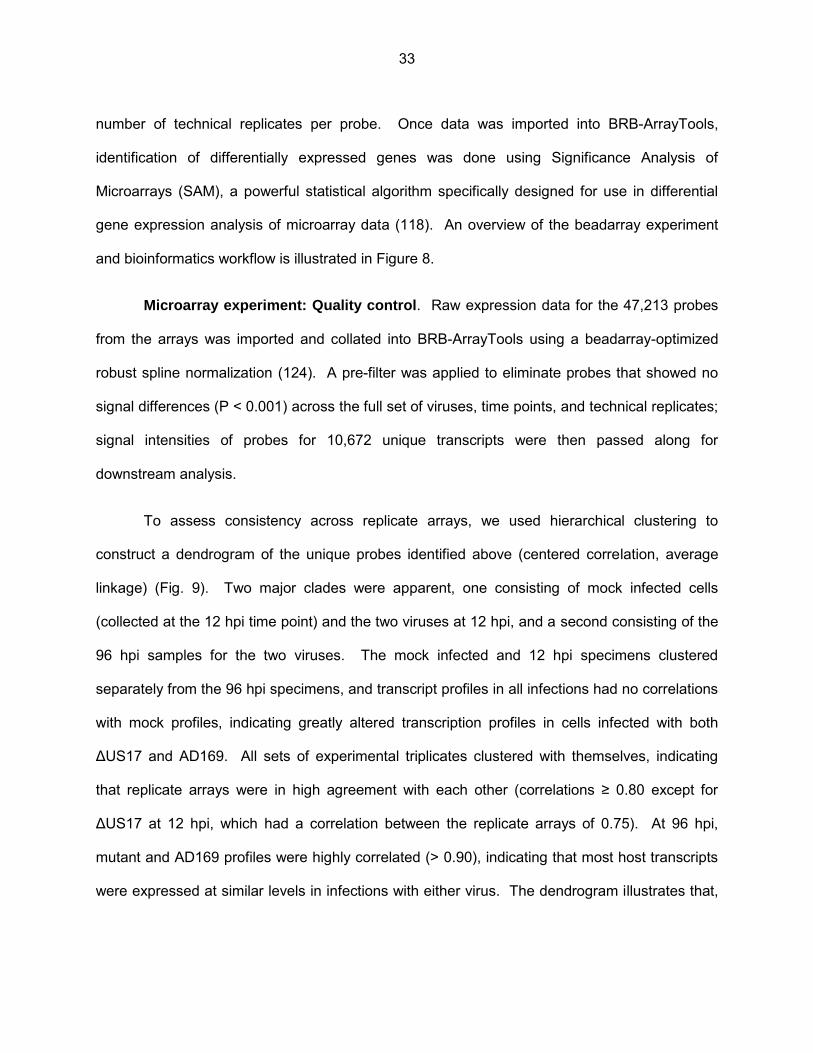
To assess consistency across replicate arrays, we used hierarchical clustering to
construct a dendrogram of the unique probes identified above (centered correlation, average
linkage) (Fig. 9). Two major clades were apparent, one consisting of mock infected cells
(collected at the 12 hpi time point) and the two viruses at 12 hpi, and a second consisting of the
96 hpi samples for the two viruses. The mock infected and 12 hpi specimens clustered
separately from the 96 hpi specimens, and transcript profiles in all infections had no correlations
with mock profiles, indicating greatly altered transcription profiles in cells infected with both
ΔUS17 and AD169. All sets of experimental triplicates clustered with themselves, indicating
that replicate arrays were in high agreement with each other (correlations ≥ 0.80 except for
ΔUS17 at 12 hpi, which had a correlation between the replicate arrays of 0.75). At 96 hpi,
mutant and AD169 profiles were highly correlated (> 0.90), indicating that most host transcripts
were expressed at similar levels in infections with either virus. The dendrogram illustrates that,
33

while, the transcript profile of ΔUS17 was highly similar to that of AD169 there were sufficient
differences to classify them as distinct entities.
To analyze overall gene transcript profiles, scatterplots were constructed for both time
points for all pairwise comparisons using the 10,672 probes that passed the filtering criteria (Fig.
10, panels A and B); note that SAM-significant differentially expressed probes are indicated as
either black or colored dots while non-SAM significant probes are indicated as light gray dots
(described in detail below). In comparison to mock infected cells, both viruses induced
numerous changes to the transcription profiles at either time point. At 12 hpi, both viruses
modulated over 1,800 probes infected by ≥ 1.5-fold vs. mock (Fig. 10A). At 96 hpi, changes to
the transcriptional profiles were even more pronounced. Both AD169 and ΔUS17 induced over
3,000 probes by ≥ 1.5-fold vs. mock infected cells (Fig. 10B). In contrast, there were relatively
few transcriptional differences between ΔUS17 and AD169, correlating with the similarity noted
on the Fig. 9 dendrogram and confirming that ΔUS17 modulated only a small percentage of
transcripts relative to its parent.
Differential gene expression analysis in cells infected with AD169 vs. ΔUS17. To
analyze differentially expressed transcripts more robustly, we examined the specific differences
in cellular transcript profiles between AD169 and ΔUS17 infections using significance analysis
of microarrays (SAM) with a stringent false discovery rate (FDR) of 0.001. SAM uses gene
specific t-tests to compute an expected fold change for each transcript, dε(i). It then uses
permutations of the data and compares the observed fold change, d(i) to the expected
foldchange. A threshold is set by using a false discovery rate (FDR) which is the number of
false positives that are statically likely in the data set. If the observed fold change is larger than
the threshold set for the expected fold change the gene is considered significantly differentially
regulated. Output plots of the SAM analysis are shown in Figure 11. At 12 hpi, of the 10,672
34
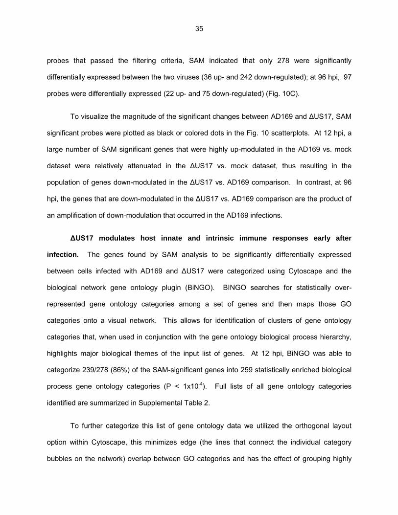
probes that passed the filtering criteria, SAM indicated that only 278 were significantly
differentially expressed between the two viruses (36 up- and 242 down-regulated); at 96 hpi, 97
probes were differentially expressed (22 up- and 75 down-regulated) (Fig. 10C).
To visualize the magnitude of the significant changes between AD169 and ΔUS17, SAM
significant probes were plotted as black or colored dots in the Fig. 10 scatterplots. At 12 hpi, a
large number of SAM significant genes that were highly up-modulated in the AD169 vs. mock
dataset were relatively attenuated in the ΔUS17 vs. mock dataset, thus resulting in the
population of genes down-modulated in the ΔUS17 vs. AD169 comparison. In contrast, at 96
hpi, the genes that are down-modulated in the ΔUS17 vs. AD169 comparison are the product of
an amplification of down-modulation that occurred in the AD169 infections.
ΔUS17 modulates host innate and intrinsic immune responses early after
infection. The genes found by SAM analysis to be significantly differentially expressed
between cells infected with AD169 and ΔUS17 were categorized using Cytoscape and the
biological network gene ontology plugin (BiNGO). BINGO searches for statistically over-
represented gene ontology categories among a set of genes and then maps those GO
categories onto a visual network. This allows for identification of clusters of gene ontology
categories that, when used in conjunction with the gene ontology biological process hierarchy,
highlights major biological themes of the input list of genes. At 12 hpi, BiNGO was able to
categorize 239/278 (86%) of the SAM-significant genes into 259 statistically enriched biological
process gene ontology categories (P < 1x10⁻⁴). Full lists of all gene ontology categories
identified are summarized in Supplemental Table 2.
To further categorize this list of gene ontology data we utilized the orthogonal layout
option within Cytoscape, this minimizes edge (the lines that connect the individual category
bubbles on the network) overlap between GO categories and has the effect of grouping highly
35
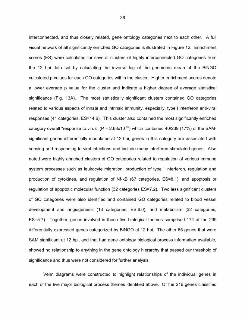
interconnected, and thus closely related, gene ontology categories next to each other. A full
visual network of all significantly enriched GO categories is illustrated in Figure 12. Enrichment
scores (ES) were calculated for several clusters of highly interconnected GO categories from
the 12 hpi data set by calculating the inverse log of the geometric mean of the BiNGO
calculated p-values for each GO categories within the cluster. Higher enrichment scores denote
a lower average p value for the cluster and indicate a higher degree of average statistical
significance (Fig. 13A). The most statistically significant clusters contained GO categories
related to various aspects of innate and intrinsic immunity, especially, type I interferon anti-viral
responses (41 categories, ES=14.6). This cluster also contained the most significantly enriched
category overall “response to virus” (P = 2.63x10-40) which contained 40/239 (17%) of the SAM-
significant genes differentially modulated at 12 hpi, genes in this category are associated with
sensing and responding to viral infections and include many interferon stimulated genes. Also
noted were highly enriched clusters of GO categories related to regulation of various immune
system processes such as leukocyte migration, production of type I interferon, regulation and
production of cytokines, and regulation of Nf-κB (67 categories, ES=8.1), and apoptosis or
regulation of apoptotic molecular function (32 categories ES=7.2). Two less significant clusters
of GO categories were also identified and contained GO categories related to blood vessel
development and angiogenesis (13 categories, ES:6.0), and metabolism (32 categories,
ES=5.7). Together, genes involved in these five biological themes comprised 174 of the 239
differentially expressed genes categorized by BiNGO at 12 hpi. The other 65 genes that were
SAM significant at 12 hpi, and that had gene ontology biological process information available,
showed no relationship to anything in the gene ontology hierarchy that passed our threshold of
significance and thus were not considered for further analysis.
Venn diagrams were constructed to highlight relationships of the individual genes in
each of the five major biological process themes identified above. Of the 216 genes classified
36
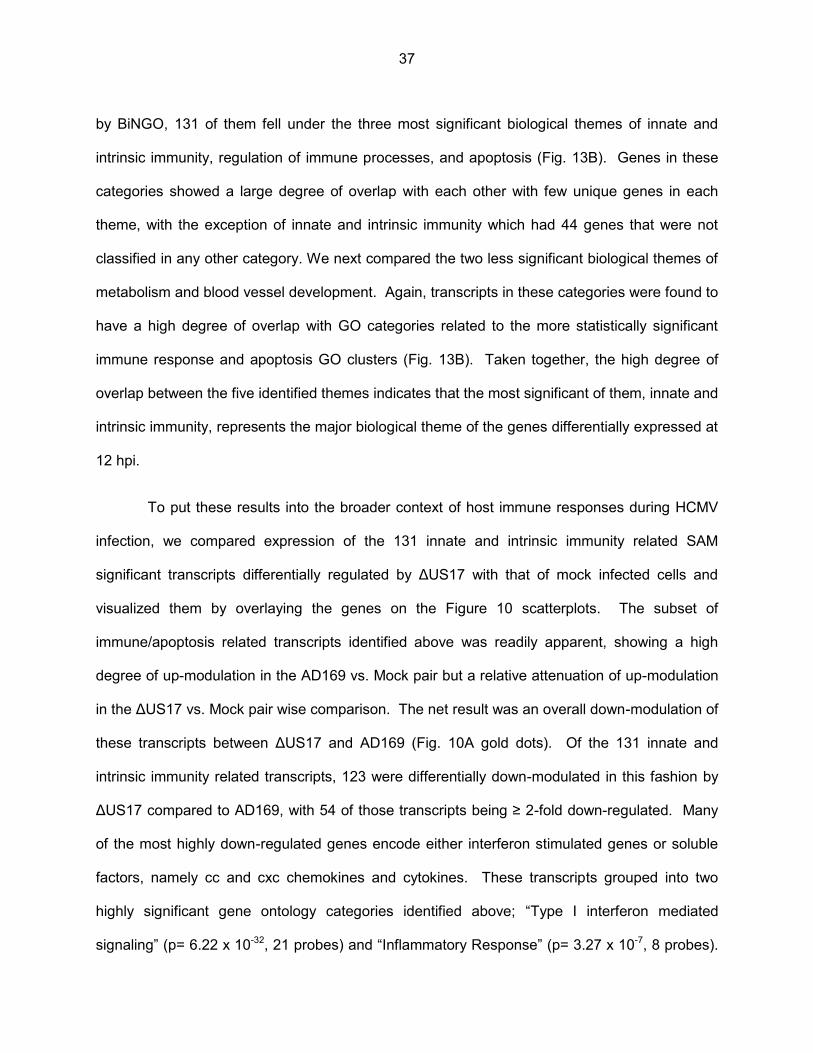
by BiNGO, 131 of them fell under the three most significant biological themes of innate and
intrinsic immunity, regulation of immune processes, and apoptosis (Fig. 13B). Genes in these
categories showed a large degree of overlap with each other with few unique genes in each
theme, with the exception of innate and intrinsic immunity which had 44 genes that were not
classified in any other category. We next compared the two less significant biological themes of
metabolism and blood vessel development. Again, transcripts in these categories were found to
have a high degree of overlap with GO categories related to the more statistically significant
immune response and apoptosis GO clusters (Fig. 13B). Taken together, the high degree of
overlap between the five identified themes indicates that the most significant of them, innate and
intrinsic immunity, represents the major biological theme of the genes differentially expressed at
12 hpi.
To put these results into the broader context of host immune responses during HCMV
infection, we compared expression of the 131 innate and intrinsic immunity related SAM
significant transcripts differentially regulated by ΔUS17 with that of mock infected cells and
visualized them by overlaying the genes on the Figure 10 scatterplots. The subset of
immune/apoptosis related transcripts identified above was readily apparent, showing a high
degree of up-modulation in the AD169 vs. Mock pair but a relative attenuation of up-modulation
in the ΔUS17 vs. Mock pair wise comparison. The net result was an overall down-modulation of
these transcripts between ΔUS17 and AD169 (Fig. 10A gold dots). Of the 131 innate and
intrinsic immunity related transcripts, 123 were differentially down-modulated in this fashion by
ΔUS17 compared to AD169, with 54 of those transcripts being ≥ 2-fold down-regulated. Many
of the most highly down-regulated genes encode either interferon stimulated genes or soluble
factors, namely cc and cxc chemokines and cytokines. These transcripts grouped into two
highly significant gene ontology categories identified above; “Type I interferon mediated
signaling” (p= 6.22 x 10⁻³², 21 probes) and “Inflammatory Response” (p= 3.27 x 10⁻⁷, 8 probes).
37

Expression levels of genes in these two categories are illustrated in Figure 14. Complete lists of
all SAM-significant genes at 12 hpi and 96 hpi, and transcripts common to both time points are
provided in Supplementary Table 1.
To further confirm the phenotype observed using the microarray, we employed a custom
qRT-PCR array that targets six innate and intrinsic immune response transcripts that showed a
high degree of down-modulation compared to AD169 at 12 hpi. We compared the parental virus
pAD/cre, a BAC version of AD169 which contains no deletion of primary sequence but does
contain a small 32 bp lox scar between the US28 and US29 ORFs, two independently derived
ΔUS17 mutant viruses (ΔUS17 and 2ΔUS17), and a US17 deletion repair virus (US17cV5).
The two independent ΔUS17 viruses showed the same down-modulation as was observed on
the microarray (Fig.15). Additionally, the repair virus restored transcript expression to parental
levels, indicating that deletion of the US17 locus had a specific effect on expression of immune
genes. Overall, these changes in gene expression at 12 hpi, a time that precedes de novo
expression of most HCMV genes, suggest changes in the way ΔUS17 virions are sensed by the
host cell.
Gene ontology analysis at 96 hpi. At 96 hpi, BiNGO categorized 86 of the 98 SAM
significant genes into 76 significantly enriched GO biological process categories (P < 1x10⁻⁴).
As evident in the scatterplots (Fig. 10 A and B), differences between ΔUS17 and AD169 were
generally smaller than at 12 hpi. Fourteen of the over-represented biological process categories
related to type I interferon, cytokine, or various other aspects of immune responses and another
five categories related to apoptosis (Supplementary Table 2). Together, these categories
contained 43 differentially regulated transcripts, 22 of which were identified by SAM analysis as
being differentially expressed between AD169 and ΔUS17 at both 12 hpi and 96 hpi (Fig. 10C).
Interestingly, for most of these genes, although the magnitude change relative to mock
38
decreased from 12 to 96 hpi, the directions of the transcriptional changes were consistent at
both times, with ΔUS17 blunting induction of certain immune response transcripts
(predominantly interferon responsive genes) while amplifying the suppression of other
transcripts (predominantly genes involved in tissue development). In sum, ΔUS17 produces
pronounced changes to host immune transcripts at 12 hpi that persist to at least 96 hpi.
Several ER stress and unfolded protein response transcripts were differentially regulated
at 96 hpi but not at 12 hpi. This included a number of heat-shock 60 kDa (HSP60) proteins, as
well as transcripts for proteins involved in ER stress and the ER associated degradation (ERAD)
pathways. Positive and negative effects were seen among transcripts associated with these
genes (Fig. 14). At least one of these genes (HSPA5 or Bip) is important for the biogenesis and
function of the HCMV cytoplasmic virion assembly complex (cVAC) (110,125). US17 shows its
highest levels of expression from 96 to 120 hpi, thus, transcripts modulated by ΔUS17 at late
times are probably a direct effect of the mutation rather than the indirect effects seen at 12 hpi.
39
Discussion US17 indirectly modulates host innate and intrinsic immune responses. As for
other infectious agents, modulation of immune responses is a fundamentally important aspect of
HCMV biology. The process of HCMV host cell immunomodulation begins at the very earliest
stages of viral attachment to the cell and is highly dependent on the structural components of
the virion, i.e., envelope glycoproteins such as gB and gH, and tegument proteins such as pp65
(114,126-128) reviewed in (9). We found that deletion of US17 had little impact on production of
infectious viral particles in fibroblasts, but nevertheless exerted a tangible influence over viral
replication. ΔUS17 markedly blunted the host cell anti-viral response at a very early time point
(12 h) after infection. Many interferon stimulated transcripts and transcripts encoding for pro-
inflammatory chemokines and cytokines were down-modulated by 3- to 5-fold when compared
to the parental virus. These changes to immune sensing of the host cell at 12 hpi happened in
the absence of US17 expression, thus we conclude that virions produced by ΔUS17 infected
cells differ in their composition from those produced by parental virus.
A role for US17 mediated manipulation of ER stress responses late in infection. In
contrast to the modulation of immune responses at 12 hpi, other effects were seen at 96 hpi, a
time that corresponds with both high expression of US17 and the formation of the cVAC (96).
Specifically, expression of some ER stress response and chaperone transcripts were
significantly altered by ΔUS17. By unknown mechanisms, HCMV modulates various pathways
and genes involved in ER stress and the unfolded protein response (106,108). One such gene,
HSPA5 (GRP78/BIP), which was up-regulated 1.6-fold by ΔUS17, is important for formation of
the cVAC and production of infectious virions (110,112,125). Several other important regulators
of ER stress were also modulated, including DDIT3 (also known as CHOP) (2.11-fold increase)
and CHAC1 (1.6-fold increase), both of which are known to promote apoptosis during the ER
stress response (129-131). Relative to mock-infected cells, ΔUS17 and its parent altered
40
regulation of most of these transcripts in opposite directions. This is in sharp contrast to the
sets of immune transcripts that at 12 hpi were modulated by ΔUS17 and its parent in the same
direction relative to mock-infected cells.
The modulation of gene expression by US17 at later stages of infection provides insights
into a potential mechanism of action for US17 and offers cellular targets that could be studied in
this context. We note that US12 family members share sequence similarity with the trans-
membrane bax inhibitor-1 (TMBIM) family of conserved eukaryotic anti-apoptotic seven
transmembrane proteins (71). These proteins localize to membranes of various cellular
organelles where they act as rheostats that modulate apoptotic and ER stress signaling by
influencing cellular calcium levels (80-84). Interestingly, over expression of Bax inhibitor-1,
showed lower levels of expression of both BIP and CHOP in response to the ER stress inducers
thapsigargin and tunicamycin (132). This is directly opposite to the phenotypes observed here
where deletion of US17 exhibited higher expression of both BIP and CHOP in response to
HCMV infection. Taken together, it is possible that US17 is a bax inhibitor-1 ortholog that
shapes virion composition by modulating levels of ER stress response and apoptosis genes
involved in regulation of protein folding, trafficking, and cVAC function.
41
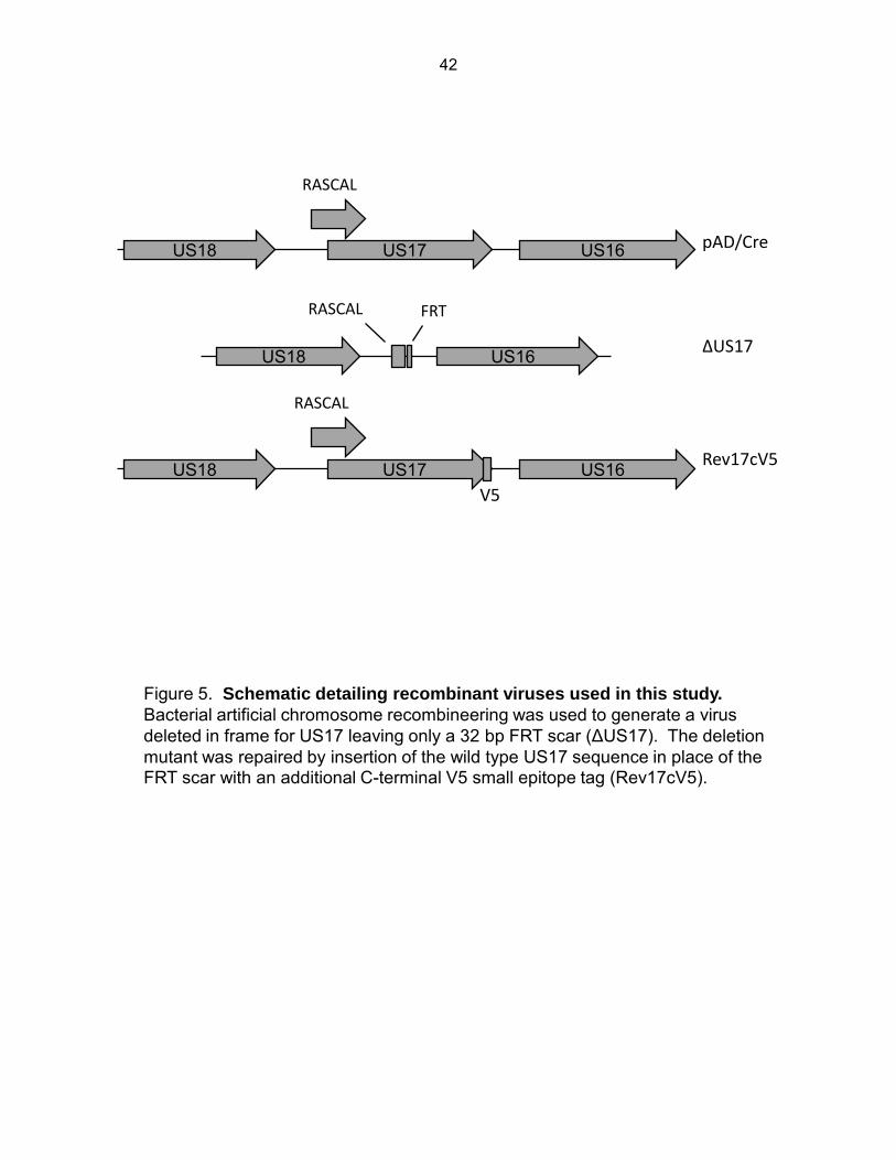
US18 US17 US16
RASCAL
US18 US16
FRT RASCAL
US18 US17 US16
RASCAL
V5
pAD/Cre
ΔUS17
Rev17cV5
Figure 5. Schematic detailing recombinant viruses used in this study. Bacterial artificial chromosome recombineering was used to generate a virus deleted in frame for US17 leaving only a 32 bp FRT scar (ΔUS17). The deletion mutant was repaired by insertion of the wild type US17 sequence in place of the FRT scar with an additional C-terminal V5 small epitope tag (Rev17cV5).
42
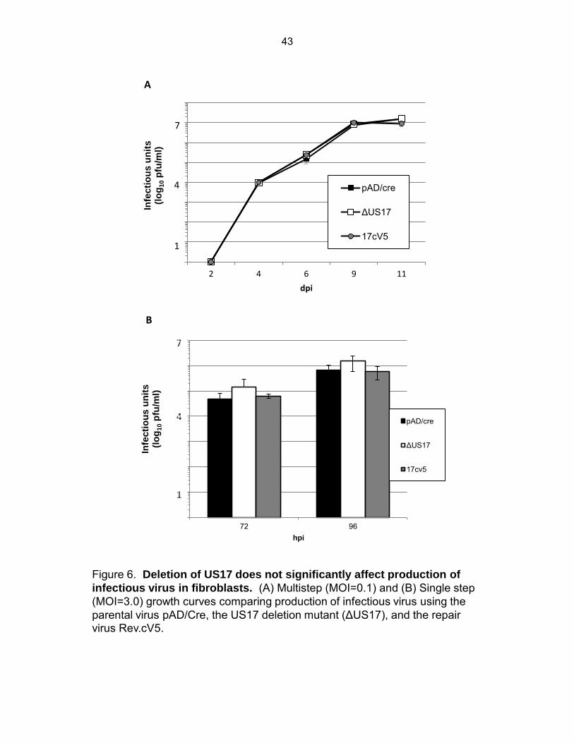
A
1E+00
1E+01
1E+02
1E+03
1E+04
1E+05
1E+06
1E+07
1E+08
2 4 6 9 11
Infe
cti
ou
s u
nit
s
(lo
g1
0 p
fu/m
l)
dpi
pAD/cre
ΔUS17
17cV5 1
4
7
1
4
7
1E+00
1E+01
1E+02
1E+03
1E+04
1E+05
1E+06
1E+07
72 96
Infe
cti
ou
s u
nit
s
(lo
g10 p
fu/m
l)
hpi
pAD/cre
ΔUS17
17cv5
B
Figure 6. Deletion of US17 does not significantly affect production of
infectious virus in fibroblasts. (A) Multistep (MOI=0.1) and (B) Single step (MOI=3.0) growth curves comparing production of infectious virus using the parental virus pAD/Cre, the US17 deletion mutant (ΔUS17), and the repair virus Rev.cV5.
43
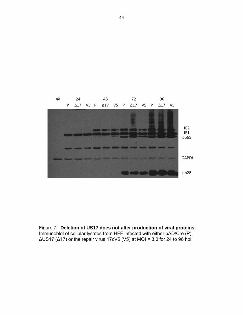
24
P Δ17 V5
48
P Δ17 V5
72
P Δ17 V5
96
P Δ17 V5
hpi
IE2 IE1
pp65
GAPDH
pp28
Figure 7. Deletion of US17 does not alter production of viral proteins. Immunoblot of cellular lysates from HFF infected with either pAD/Cre (P), ΔUS17 (Δ17) or the repair virus 17cV5 (V5) at MOI = 3.0 for 24 to 96 hpi.
44
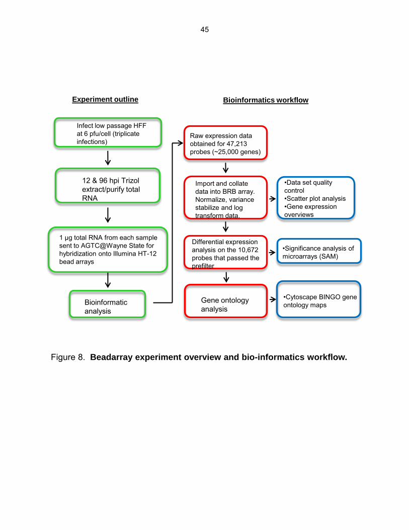
Infect low passage HFF at 6 pfu/cell (triplicate infections)
12 & 96 hpi Trizol extract/purify total RNA
1 μg total RNA from each sample sent to AGTC@Wayne State for hybridization onto Illumina HT-12 bead arrays
Bioinformatic analysis
Experiment outline
Raw expression data obtained for 47,213 probes (~25,000 genes)
Import and collate data into BRB array. Normalize, variance stabilize and log transform data.
Differential expression analysis on the 10,672 probes that passed the prefilter
•Data set quality control •Scatter plot analysis •Gene expression overviews
•Significance analysis of microarrays (SAM)
Bioinformatics workflow
Gene ontology analysis
•Cytoscape BINGO gene ontology maps
Figure 8. Beadarray experiment overview and bio-informatics workflow.
45
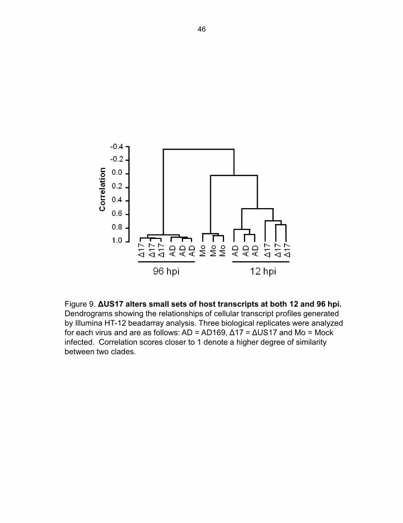
Figure 9. ΔUS17 alters small sets of host transcripts at both 12 and 96 hpi. Dendrograms showing the relationships of cellular transcript profiles generated by Illumina HT-12 beadarray analysis. Three biological replicates were analyzed for each virus and are as follows: AD = AD169, Δ17 = ΔUS17 and Mo = Mock infected. Correlation scores closer to 1 denote a higher degree of similarity between two clades.
46
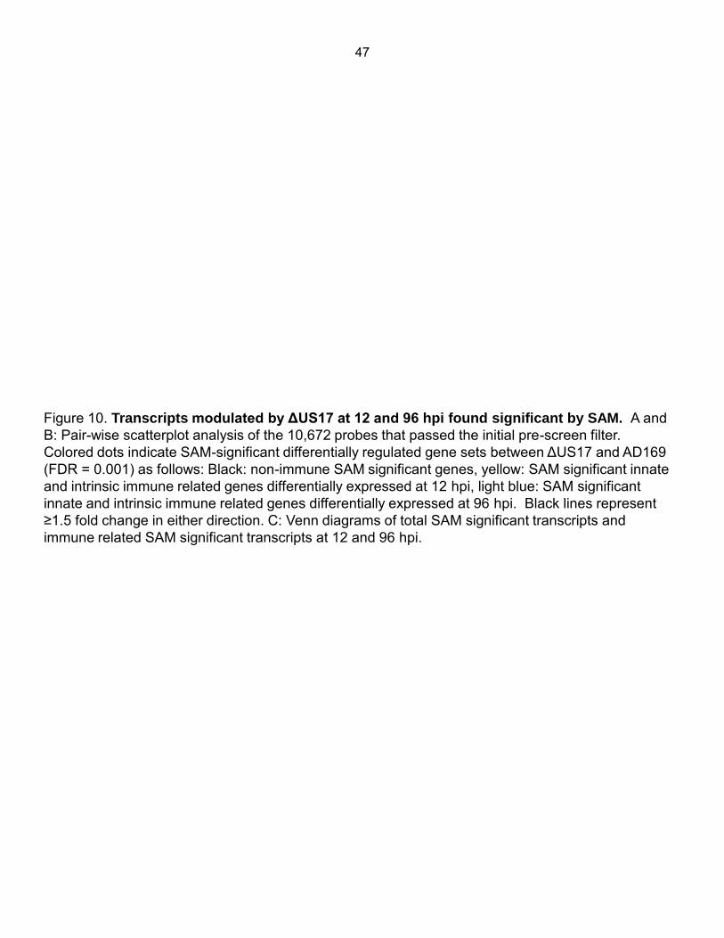
Figure 10. Transcripts modulated by ΔUS17 at 12 and 96 hpi found significant by SAM. A and B: Pair-wise scatterplot analysis of the 10,672 probes that passed the initial pre-screen filter. Colored dots indicate SAM-significant differentially regulated gene sets between ΔUS17 and AD169 (FDR = 0.001) as follows: Black: non-immune SAM significant genes, yellow: SAM significant innate and intrinsic immune related genes differentially expressed at 12 hpi, light blue: SAM significant innate and intrinsic immune related genes differentially expressed at 96 hpi. Black lines represent ≥1.5 fold change in either direction. C: Venn diagrams of total SAM significant transcripts and immune related SAM significant transcripts at 12 and 96 hpi.
47
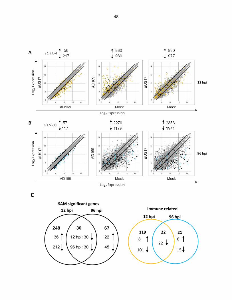
C
248
36
212
67
22
45
30
12 hpi: 30
96 hpi: 30
12 hpi 96 hpi
SAM significant genes Immune related
22
119
21
12 hpi 96 hpi
8
101
22 6
15
48
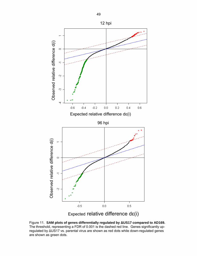
12 hpi
Obs
erve
d re
lativ
e di
ffere
nce
d(i)
Expected relative difference dε(i)
96 hpi
Obs
erve
d re
lativ
e di
ffere
nce
d(i)
Expected relative difference dε(i) Figure 11. SAM plots of genes differentially regulated by ΔUS17 compared to AD169. The threshold, representing a FDR of 0.001 is the dashed red line. Genes significantly up-regulated by ΔUS17 vs. parental virus are shown as red dots while down-regulated genes are shown as green dots.
49
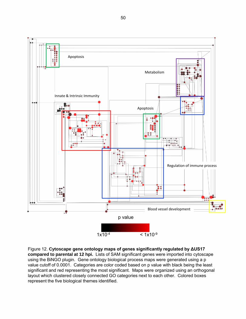
Innate & Intrinsic Immunity
Regulation of immune process
Apoptosis
Blood vessel development
Metabolism
p value
1x10⁻⁴ < 1x10⁻⁹
Apoptosis
Figure 12. Cytoscape gene ontology maps of genes significantly regulated by ΔUS17
compared to parental at 12 hpi. Lists of SAM significant genes were imported into cytoscape using the BiNGO plugin. Gene ontology biological process maps were generated using a p value cutoff of 0.0001. Categories are color coded based on p value with black being the least significant and red representing the most significant. Maps were organized using an orthogonal layout which clustered closely connected GO categories next to each other. Colored boxes represent the five biological themes identified.
50

44 38
26
23 11
14
10
A
B
C
49
36
7
1
7
0 28
ABC D
E
Group GO biological
theme
GO categories
(n=259)
Genes
(n=239)
Enrichment
score
A Innate immunity & Interferon production
41 131 14.6
B Regulation of immune response
67 93 8.1
C Apoptosis 32 86 7.2
D Blood vessel development/ angiogenesis
13 44 6.0
E Metabolism 32 71 5.7
Figure 13. Gene ontology analysis of SAM significant transcripts. (B) Table outlining the five biological themes identified. Listed are the numbers of individual gene ontology categories for each theme out of the 259 total GO categories identified as statistically significant (P < 0.0001). Also listed are numbers of SAM significant transcripts in each theme out of the 239 that could be categorized into a gene ontology biological process category. The enrichment score denotes the average level of significance of all of the GO categories in a particular theme. Higher enrichment scores indicate a more significant, mean log transformed p value for that theme. (A) Venn diagrams detailing numbers of differentially modulated SAM significant transcripts from the ΔUS17 vs. AD169 12 hpi comparison that grouped into the five most highly significant biological categories identified by BiNGO gene ontology biological process clustering. The left Venn diagram is a comparison of transcripts that grouped into either of the three most significant gene ontology themes of innate immunity and interferon production, regulation of immune response, or apoptosis. The right Venn diagram is a comparison of transcripts that grouped into either of the first three themes against the two minor identified gene ontology themes of blood vessel development and metabolism.
A
B
51
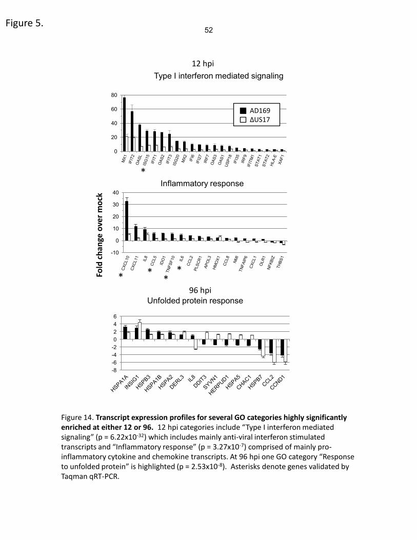
Figure 5.
0
20
40
60
80
Type I interferon mediated signaling
-10
0
10
20
30
40 Inflammatory response
AD169 ΔUS17
Fold
ch
ange
ove
r m
ock
* * *
*
12 hpi
96 hpi
-8 -6 -4 -2 0 2 4 6
Unfolded protein response
Figure 14. Transcript expression profiles for several GO categories highly significantly enriched at either 12 or 96. 12 hpi categories include “Type I interferon mediated signaling” (p = 6.22x10⁻³²) which includes mainly anti-viral interferon stimulated transcripts and “Inflammatory response” (p = 3.27x10⁻⁷) comprised of mainly pro-inflammatory cytokine and chemokine transcripts. At 96 hpi one GO category “Response to unfolded protein” is highlighted (p = 2.53x10⁻⁸). Asterisks denote genes validated by Taqman qRT-PCR.
*
52
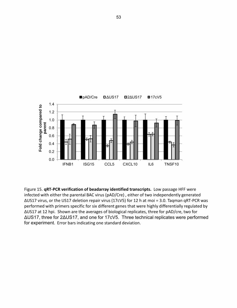
Figure 15. qRT-PCR verification of beadarray identified transcripts. Low passage HFF were infected with either the parental BAC virus (pAD/Cre) , either of two independently generated ΔUS17 virus, or the US17 deletion repair virus (17cV5) for 12 h at moi = 3.0. Taqman qRT-PCR was performed with primers specific for six different genes that were highly differentially regulated by ΔUS17 at 12 hpi. Shown are the averages of biological replicates, three for pAD/cre, two for ΔUS17, three for 2ΔUS17, and one for 17cV5. Three technical replicates were performed for experiment. Error bars indicating one standard deviation.
0.0
0.2
0.4
0.6
0.8
1.0
1.2
1.4
IFNB1 ISG15 CCL5 CXCL10 IL6 TNSF10
Fo
ld c
han
ge c
om
pare
d t
o
pare
nt
pAD/Cre ΔUS17 2ΔUS17 17cV5
53
Chapter Three
Role of US12 family proteins in regulating virion assembly.
Introduction
The HCMV virion has a highly complex structure. Analysis of purified virion preparations
has indicated that there may be as many as 70 different viral proteins incorporated to form a
mature infectious virion (94,95). We have previously shown, in chapter one of this work, that a
recombinant HCMV deleted for one member of the US12 protein family, ΔUS17, elicited a
blunted immune response from host cells at early times after infection (12 hpi). This time was
before US17 expression during infection, leading us to hypothesize that US17 may be
influencing virion composition.
The viral mediators of HCMV virion assembly are presently not well understood. Various
proteins in the US12 family, e.g. US16 and US18 (described previously), have been implicated
in modulating virion protein structure (74,75). These hypotheses have been driven largely by
inferring changes to virion composition from observations of growth phenotypes using deletion
mutants in various cell types. To date, no groups have elucidated any particular functions
associated with US12 family members nor have any specific changes to virion structure been
shown in the absence of US12 family member expression.
Thus, to directly study the effects of US17 on virion structure, we gradient purified virions
from ΔUS17 infected cell culture supernatants to compare protein composition, infectivity, and
levels of incorporated viral genomes to that of parental virions. We discovered that ΔUS17
virions differ from parental in a number of key ways. First, ΔUS17 virions had reduced amounts
of the viral glycoprotein, gH, incorporated into their structure. Second, fibroblasts infected with
ΔUS17 produced 3-fold more non-infectious genome-containing virions than parental virus.
54
Third, when inoculated at equal MOIs ΔUS17 virions delivered a 3-fold larger amount of the
tegument protein pp65 compared to parental virions.
US12 family members share some homology to the TMBIM family of eukaryotic ER
stress and anti-apoptotic proteins (133). The importance of ER stress pathways in virion
maturation, assembly complex formation and function (described in the general introduction of
this work), coupled with the sequence similarity of US12 family members to the TMBIM family of
ER stress and UPR related proteins and indicates that US12 proteins may indeed be playing a
role in modulating virion composition possibly by affecting pathways involved in protein folding
or protein trafficking of viral tegument and glycoproteins from the ER to the cVAC.
55
Materials and methods
Gradient purification of viral particles. Supernatants were harvested 10 dpi from low
passage (p 10) HFF infected at an MOI of 0.01 with the parental strain pAD/cre, ΔUS17, or the
US17 deletion repair virus US17cV5. Supernatants were clarified by centrifugation at 3,000
RPM for 15 m. Viral particles were subsequently concentrated by centrifugation through a
cushion of 20% sorbitol in TN buffer (50 mMol Tris pH 7.4, 100 mMol NaCl) at 60,000 xg for 1 h
using a Beckman SW 41 Ti rotor. Virion pellets were re-suspended in a small volume of TNE
(50 mMol Tris pH 7.4, 100 mMol NaCl, 10 mMol EDTA) and layered over a 10 – 50% linear
Nycodenz gradient prepared in TNE (Axis-shield, Oslo, Norway). Gradients were centrifuged at
110,000 xg for 2 h in a Beckman SW 41 Ti rotor and subsequently top fractionated into 1 ml
aliquots. To dialyze each fraction into PBS, Ultrafiltration of concentrated virus was carried out
with an Ambicon 10 kDa cutoff filter (EMD-millipore). Samples were first diluted with an
additional 1 ml PBS and centrifuged at 3,000 rpm for 30 – 45 m to concentrate to 100 µl. An
additional 2 ml of PBS was added and samples were centrifuged at 3,000 rpm for an additional
30 – 45 minutes until a final volume of 100 µl was obtained.
Immunofluorescent microscopy. HFF were seeded 72 hours pre-infection onto
gelatin coated 8 well chamber slides (Thermo Scientific, Rochester, NY) and infected at an MOI
of 3.0 with the indicated virus. 2 hpi cells were fixed in 4% para-formaldehyde for 10 m at room
temperature and quenched in 50 mm NH4Cl for 10 min. Cells were permeabilized in a blocking
buffer consisting of 10% normal goat serum, 5% glycine in PBS and 0.2% triton x-100. Antibody
staining was conducted using appropriately diluted primary anti-body against pp65 (Fitzgerald
Industries Acton, MA) and Alexa-fluor 488 secondary antibody (Molecular Probes/Life
Technologies Grand Island, NY). Images were captured using a Nikon Eclipse e800
microscope and total fluorescent signal was measured using Image J software. Three random
56
images were taken for each of two biological experiments. Images were background corrected
by measuring the average fluorescent intensity of an area that contained no pp65 signal.
Thresholds were set to measure only pp65 signal in the nucleus and the punctuate staining in
the cytoplasm of each cell. Total fluorescent intensity of the thresholded image was divided by
the number of pp65 positive nuclei to obtain the final pp65 signal intensity value.
.
57
Results
ΔUS17 induces changes in virion protein composition. Changes in cellular gene
expression at 12 hpi were unexpected (presented in Chapter 1 of this work), given that US17 is
a late gene whose expression is dependent on viral DNA synthesis, and is not detectably
expressed in infected cells until at least 48 hpi. We hypothesized that changes in transcription
of cellular interferon and innate immune genes at 12 hpi were the product of changes in virion
protein composition caused by the absence of US17. In order to directly analyze the protein
composition of ΔUS17, parental, or the US17 deletion repair virus US17cV5 virions, we
subjected infected cell-culture supernatants (MOI=0.01, 10 dpi) to density gradient
centrifugation through a 10-50% linear Nycodenz gradient. This separated viral particles based
on buoyant density and allowed for discrimination of any physical differences in the particles
produced by ΔUS17 compared to those produced by the parental virus. Nycodenz offers a non-
ionic, non-osmotic alternative for gradient purification of viral particles. Traditional linear
gradients of sucrose or potassium tartrate rely on high concentrations of salt which inactivate
many of the infectious particles thus making down-stream analysis difficult. Further, Nycodenz
gradients have been previously used to purify the various particle types from HCMV cell culture
supernatants, e.g., NIEPs, dense bodies (95).
Initial observations of the gradients revealed a thick milky-white band that concentrated
between fractions 4 and 7 which presumably contained the majority of the viral particles (Fig.
16). The band was located at approximately the same location in gradients of the parental
virus, ΔUS17, and the repair virus indicating that the particles produced by ΔUS17 were of a
similar density to parental virions. The gradients were top fractionated into 10-1 ml aliquots
(indicated as red lines in Fig. 16) dialyzed into PBS using an Ambicon ultra-filtration unit with a
10 kDa cutoff, and DNase treated to remove any non-encapsidated viral DNA. To determine
58
the relative amount of viral DNA present in each fraction, viral genomes were extracted and
subjected to qPCR using a set of primers specific for the HCMV UL83 ORF. As expected,
fractions 4 – 7 contained 92 – 98 % of the viral DNA in the entire gradient for each virus
coinciding with the observed milky band. Fraction 5 was especially enriched for viral genomes
and contained approximately 70% of the genetic content of the entire gradient (Fig. 17).
Fraction 5 showed similar levels of viral DNA for all three viruses in. However, differences in
amounts of viral genomes were detected in fractions 4, and 6. ΔUS17 displayed a lower level of
viral genomes present in fraction 4 while we observed lower levels of parental virus genomes in
fraction 7. Interpretation of these results is complicated and it is unclear if these differences are
due to particles of different densities being produced by ΔUS17 or do to loss of material during
the concentration, manual fractionation of the gradients, or ultrafiltration / dialysis. Given this
limitation we have preformed down-stream analysis using a variety of techniques, i.e., plaque
assay and immunoblotting, which allowed us to normalize based on the numbers of particles
present in each sample.
To analyze levels of specific viral proteins we compared the protein profiles of gradient-
purified virions present in fraction 5 by silver staining and immunoblotting for three tegument
proteins (pp65/pUL83, pp28/pUL99, and the large tegument protein/pUL48) as well as two virion
surface glycoproteins (gB/pUL55, and gH/pUL75). Differences in relative abundance were
noted for several protein bands on the silver stain, indicating that virions produced by ΔUS17
differ from those produced by the parental virus (Fig. 18A). These changes were not
attributable to an overall difference in the amount of protein loaded as the majority of the bands
observed were relatively equal in abundance. This indicates that virions produced by ΔUS17
differ from the parental virus with respect to their protein composition. Densitometric analysis of
the immunblot revealed no gross changes for any of the tegument proteins, pp65, pp28, or
pUL48. However, there was a ~2.8 fold decrease in ΔUS17 virion levels of the envelope
59
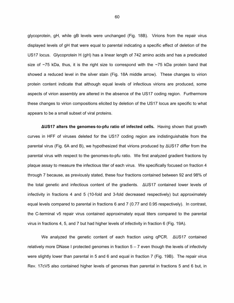
glycoprotein, gH, while gB levels were unchanged (Fig. 18B). Virions from the repair virus
displayed levels of gH that were equal to parental indicating a specific effect of deletion of the
US17 locus. Glycoprotein H (gH) has a linear length of 742 amino acids and has a predicated
size of ~75 kDa, thus, it is the right size to correspond with the ~75 kDa protein band that
showed a reduced level in the silver stain (Fig. 18A middle arrow). These changes to virion
protein content indicate that although equal levels of infectious virions are produced, some
aspects of virion assembly are altered in the absence of the US17 coding region. Furthermore
these changes to virion compositions elicited by deletion of the US17 locus are specific to what
appears to be a small subset of viral proteins.
ΔUS17 alters the genomes-to-pfu ratio of infected cells. Having shown that growth
curves in HFF of viruses deleted for the US17 coding region are indistinguishable from the
parental virus (Fig. 6A and B), we hypothesized that virions produced by ΔUS17 differ from the
parental virus with respect to the genomes-to-pfu ratio. We first analyzed gradient fractions by
plaque assay to measure the infectious titer of each virus. We specifically focused on fraction 4
through 7 because, as previously stated, these four fractions contained between 92 and 98% of
the total genetic and infectious content of the gradients. ΔUS17 contained lower levels of
infectivity in fractions 4 and 5 (10-fold and 3-fold decreased respectively) but approximately
equal levels compared to parental in fractions 6 and 7 (0.77 and 0.95 respectively). In contrast,
the C-terminal v5 repair virus contained approximately equal titers compared to the parental
virus in fractions 4, 5, and 7 but had higher levels of infectivity in fraction 6 (Fig. 19A).
We analyzed the genetic content of each fraction using qPCR. ΔUS17 contained
relatively more DNase I protected genomes in fraction 5 – 7 even though the levels of infectivity
were slightly lower than parental in 5 and 6 and equal in fraction 7 (Fig. 19B). The repair virus
Rev. 17cV5 also contained higher levels of genomes than parental in fractions 5 and 6 but, in
60

contrast to ΔUS17, this increase in genetic content corresponded to higher levels of infectious
virus compared to the parental virus. Taken together, when genome-to-pfu ratios were
calculated, ΔUS17 displayed over a 3 : 1 ratio of non-infectious viral particles compared to
parental in fractions 5, 6, and 7 (3.65, 3.55, and 4.12 to 1 respectively) while the repair virus had
ratios that were almost equal to the parent virus (1.65, 1.49, and 2.37 to 1 respectively) (Fig.
19C). This indicates that ΔUS17 is producing more non-infectious genome-containing viral
particles than either parental virus or the US17cV5 repair virus. These virions cannot be
accounted for by limiting dilution plaque assay but they are of a density that is similar to that of
the bulk of the infectious particles produced as they localize to the same fraction on the
gradient.
The previous experiment used a low MOI and relatively long time course of infection.
Thus to elucidate the time frame after infection when these non-infectious particles are
produced we repeated the experiment at a high multiplicity of infection (MOI=3.0) and shorter
time course of 24 to 96 h. At 72 and 96 hpi, times when equal numbers of infectious virions
were produced by both viruses (Fig. 20A), ΔUS17 produced 3.7- and 3.8-fold more genome-
containing viral particles (Fig. 20B). To obtain the genomes-to-pfu ratio, fold changes in levels
of DNase protected genomes were divided by fold changes in infectious titer between ΔUS17
and pAD/Cre. ΔUS17 produced 3.4 genome-containing non-infectious particles for every such
particle produced by the parental virus at 72 hpi (p=0.001) and a similar increase at 96 hpi
(3.3:1, p=0.0003) (Fig. 20C). The repair virus had ratios similar to those of the parental virus
(pAD/Cre).
The elevated genomes-to-pfu ratio of ΔUS17 was consistent over multiple experiments
using a variety of biological conditions, i.e., MOIs ranging from 0.001 to 6.0 with time courses
ranging from 3 dpi to 14 dpi. Although we did see changes in the numbers of infectious units
61
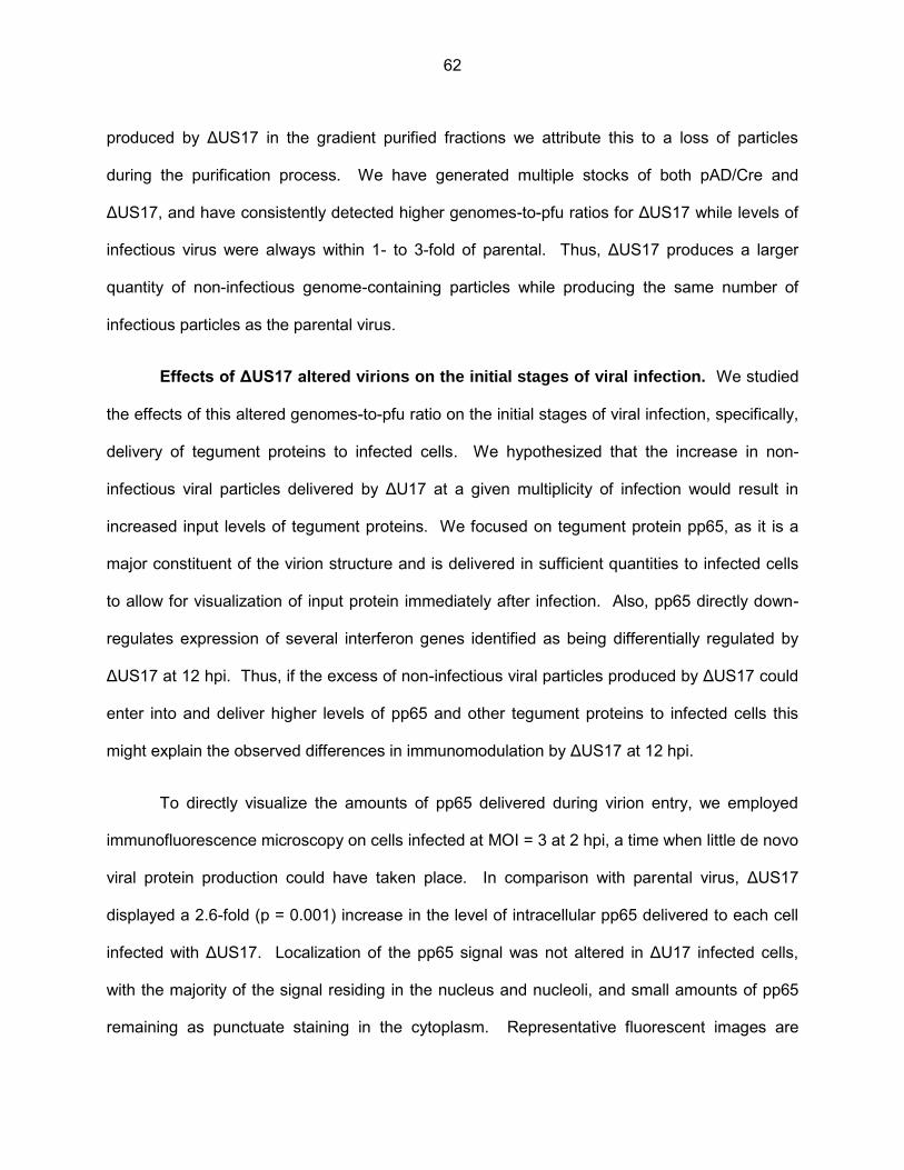
produced by ΔUS17 in the gradient purified fractions we attribute this to a loss of particles
during the purification process. We have generated multiple stocks of both pAD/Cre and
ΔUS17, and have consistently detected higher genomes-to-pfu ratios for ΔUS17 while levels of
infectious virus were always within 1- to 3-fold of parental. Thus, ΔUS17 produces a larger
quantity of non-infectious genome-containing particles while producing the same number of
infectious particles as the parental virus.
Effects of ΔUS17 altered virions on the initial stages of viral infection. We studied
the effects of this altered genomes-to-pfu ratio on the initial stages of viral infection, specifically,
delivery of tegument proteins to infected cells. We hypothesized that the increase in non-
infectious viral particles delivered by ΔU17 at a given multiplicity of infection would result in
increased input levels of tegument proteins. We focused on tegument protein pp65, as it is a
major constituent of the virion structure and is delivered in sufficient quantities to infected cells
to allow for visualization of input protein immediately after infection. Also, pp65 directly down-
regulates expression of several interferon genes identified as being differentially regulated by
ΔUS17 at 12 hpi. Thus, if the excess of non-infectious viral particles produced by ΔUS17 could
enter into and deliver higher levels of pp65 and other tegument proteins to infected cells this
might explain the observed differences in immunomodulation by ΔUS17 at 12 hpi.
To directly visualize the amounts of pp65 delivered during virion entry, we employed
immunofluorescence microscopy on cells infected at MOI = 3 at 2 hpi, a time when little de novo
viral protein production could have taken place. In comparison with parental virus, ΔUS17
displayed a 2.6-fold (p = 0.001) increase in the level of intracellular pp65 delivered to each cell
infected with ΔUS17. Localization of the pp65 signal was not altered in ΔU17 infected cells,
with the majority of the signal residing in the nucleus and nucleoli, and small amounts of pp65
remaining as punctuate staining in the cytoplasm. Representative fluorescent images are
62
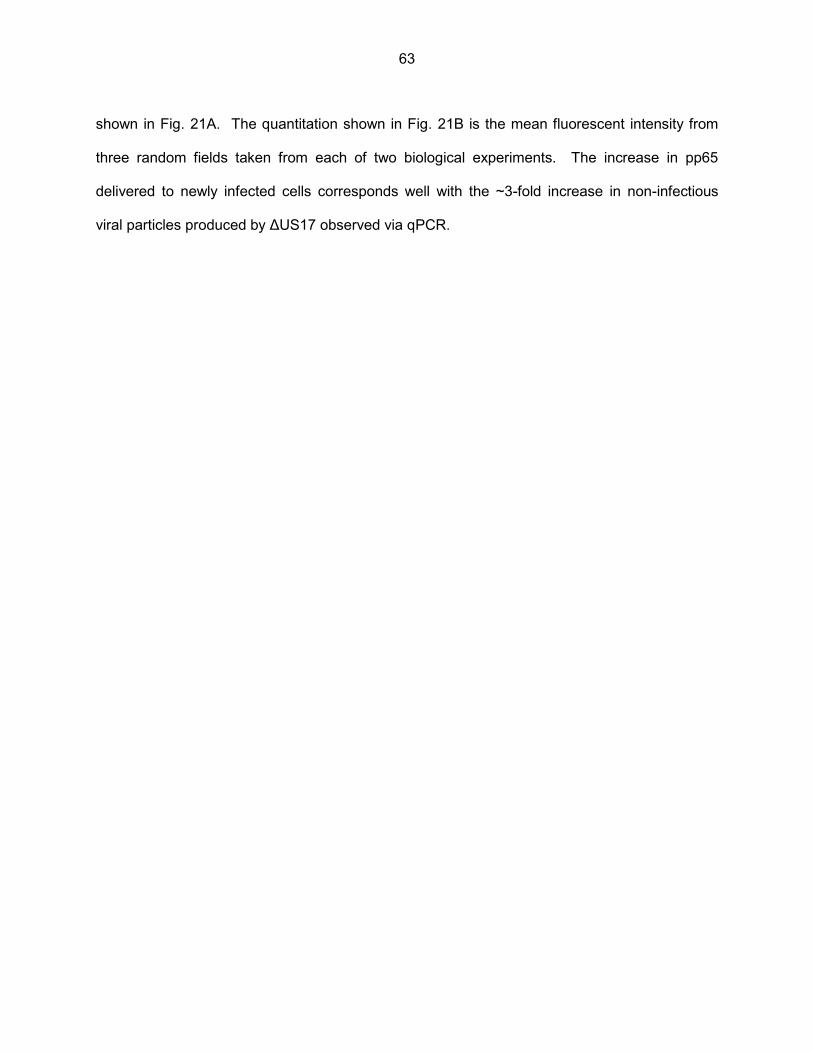
shown in Fig. 21A. The quantitation shown in Fig. 21B is the mean fluorescent intensity from
three random fields taken from each of two biological experiments. The increase in pp65
delivered to newly infected cells corresponds well with the ~3-fold increase in non-infectious
viral particles produced by ΔUS17 observed via qPCR.
63

Discussion. We previously demonstrated that fibroblasts infected with ΔUS17
produced virions elicited a blunted immune response from host cells at early times after
infection (12 hpi) in comparison to virions of the parental virus. This phenotype was in direct
contrast to other studies using recombinant HCMV deleted for the immunomodulatory tegument
protein pp65, which elicited a vastly increased interferon response from host cells (113,114).
We further observed that cells infected with ΔUS17 produced 3-fold more genome-containing
non-infectious viral particles compared to the parental virus. When cells were infected at equal
multiplicities of infection the larger number of non-infectious particles entered host cells and
delivered larger amounts of the immunomodulatory tegument protein pp65. The additional pp65
and possibly other tegument proteins delivered by the excess of non-infectious viral particles
produced by ΔUS17 likely explain the observed blunting of the host interferon response.
HCMV virion composition and genomes-to-pfu ratios. HCMV has a high particle-to-
pfu ratio, producing several hundred to several thousand non-infectious particles for every
infectious particle (102). A recombinant of strain AD169 deleted for US24 produced an equal
number of genome containing particles, but showed a 20- to 30-fold reduction in infectivity and a
~10-fold higher genome-to-pfu ratio (134). Although no differences were seen in the limited
amount of viral proteins assayed, the authors reasonably speculated that virion composition
may have been altered in ΔUS24. Here we showed that ΔUS17 modulates the genome-to-pfu
ratio in a different manner. This virus did not affect levels of infectious virus produced but
produced larger numbers of genome-containing non-infectious particles (a net increase in total
genome-containing particles). In comparisons of ΔUS17 virions with its parent or the US17v5
repair virus, levels of the virion glycoprotein gH were reduced and other differences in virion
composition were noted by silver staining. The exact molecular composition required for
infectivity of HCMV virions is not known; the ideal is likely to differ depending on the target cell
or tissue and other physiological conditions. We are using mass spectroscopy to more precisely
64
define the molecular properties of ΔUS17 virions to further elucidate the relationship between
virion composition and infectivity.
Glycoprotein H is part of the complex, gH/gL/gO, that is responsible for binding and entry
into fibroblasts (135-137). Although we did observed reduced levels of gH incorporated into
ΔUS17 virions, adsorption of individual virions was not altered as evidenced by the delivery of
pp65 to host nuclei shortly after infection. Aside from their roles in virion entry, HCMV
glycoproteins, especially gB and gH, elicit immune responses through induction of both nf-κB
and Sp1 pathways shortly after binding to the host plasma membrane (128,138). This induction
of the immune response has been linked to creation of a favorable environment inside the cell,
facilitating the early stages of HCMV replication by up-regulating expression of key cellular
transactivators. Therefore it is possible that the reduced levels of gH incorporated into ΔUS17
virions was not drastic enough to hinder virion entry but did contribute to an overall lack of
fitness for any particular virion thus increasing the numbers of seemingly non-infectious virions
during infection at high dilutions, i.e., limiting dilution plaque assay.
65
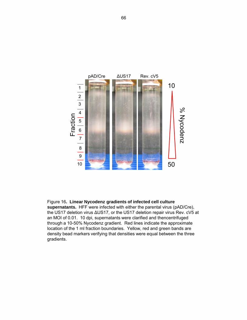
pAD/Cre ΔUS17 Rev. cV5
1
2
3
4
5
6
7
8
9
10
10
50
% N
ycodenz Fr
actio
n
Figure 16. Linear Nycodenz gradients of infected cell culture
supernatants. HFF were infected with either the parental virus (pAD/Cre), the US17 deletion virus ΔUS17, or the US17 deletion repair virus Rev. cV5 at an MOI of 0.01. 10 dpi, supernatants were clarified and thencentrifuged through a 10-50% Nycodenz gradient. Red lines indicate the approximate location of the 1 ml fraction boundaries. Yellow, red and green bands are density bead markers verifying that densities were equal between the three gradients.
66

0
500
1,000
1,500
2,000
2,500
3,000
3,500
2 3 4 5 6 7 8 9 10
Ge
no
me
s
(re
lati
ve f
old
ch
an
ges)
Fraction
pad DUS17 17cv5
Figure 17. HCMV genetic content of Nycodenz gradient fractions. Samples of each fraction were Dnase I treated to remove non-encapsidated viral DNA. Viral genomes were extracted with a Qiagen Virus Mini-Elute kit and relative levels of genomes were measured via qPCR for HCMV UL83.
67
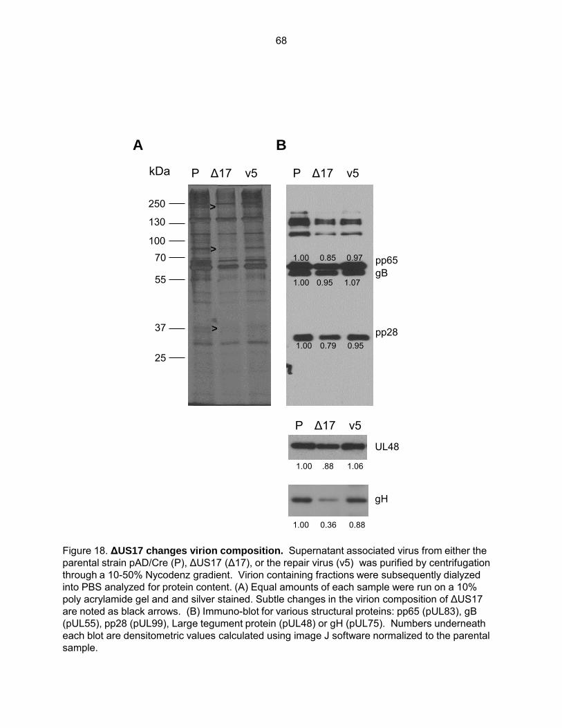
Figure 18. ΔUS17 changes virion composition. Supernatant associated virus from either the parental strain pAD/Cre (P), ΔUS17 (Δ17), or the repair virus (v5) was purified by centrifugation through a 10-50% Nycodenz gradient. Virion containing fractions were subsequently dialyzed into PBS analyzed for protein content. (A) Equal amounts of each sample were run on a 10% poly acrylamide gel and and silver stained. Subtle changes in the virion composition of ΔUS17 are noted as black arrows. (B) Immuno-blot for various structural proteins: pp65 (pUL83), gB (pUL55), pp28 (pUL99), Large tegument protein (pUL48) or gH (pUL75). Numbers underneath each blot are densitometric values calculated using image J software normalized to the parental sample.
P v5 Δ17 P v5 Δ17
25
37
55
70 100
130
250
kDa
pp65 gB
pp28
>
>
>
B A
P v5 Δ17
UL48
1.00 .88 1.06
0.36 0.88 1.00
1.00
1.00
1.00
0.85 0.97
0.79 0.95
0.95 1.07
gH
68

1.E+05
1.E+06
1.E+07
1.E+08
4 5 6 7
Infe
cti
ou
s u
nit
s
lo
g10p
fu/m
l
0.0
0.5
1.0
1.5
2.0
4 5 6 7
DN
ase p
rote
cte
d g
en
om
es
(
fold
ch
an
ge v
s.
fracti
on
5
pa
ren
t)
0
1
2
3
4
5
4 5 6 7
Gen
om
es-t
o-p
fu r
ati
o
(fo
ld c
ha
ng
e v
s. p
are
nt)
Fraction
pAD/Cre ΔUS17 Rev. cV5
5
6
7
8
C
B
A
Figure 19. ΔUS17 alters the genomes-to-pfu ratio of gradient purified virions Gradient fractions (from Fig. 16) were plaque assayed for infectivity (A), or total DNAse I protected viral genomic content by qPCR (B). (C) Genomes-to-pfu ratios were calculated by dividing the ratios of the fold changes (vs. parent) of relative genomes and infectivity.
69
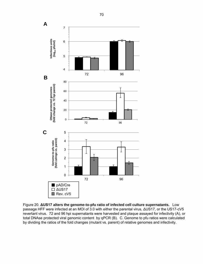
Figure 20. ΔUS17 alters the genome-to-pfu ratio of infected cell culture supernatants. Low passage HFF were infected at an MOI of 3.0 with either the parental virus, ΔUS17, or the US17-cV5 revertant virus. 72 and 96 hpi supernatants were harvested and plaque assayed for infectivity (A), or total DNAse protected viral genomic content by qPCR (B). C. Genome to pfu ratios were calculated by dividing the ratios of the fold changes (mutant vs. parent) of relative genomes and infectivity.
1E+04
1E+05
1E+06
1E+07
72 96
Infe
cti
ou
s u
nit
s
(lo
g10 p
fu/m
l)
0
20
40
60
80
72 96
DN
ase p
rote
cte
d g
en
om
es
(
fold
ch
an
ge v
s.
72 h
pi p
are
nt)
pAD/Cre ΔUS17 Rev. cV5
4
5
6
7
0
1
2
3
4
5
72 96
Gen
om
e-t
o-p
fu r
ati
o
(fo
ld c
ah
ng
e v
s. p
are
nt)
A
B
C
70
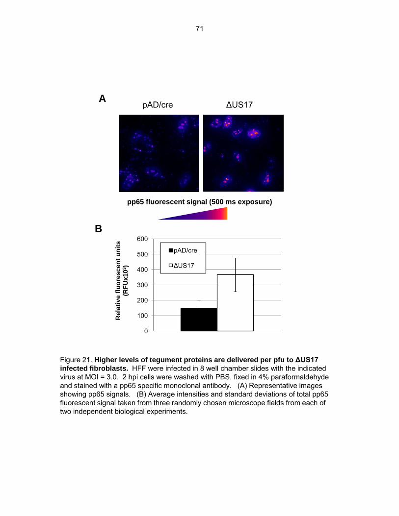
Figure 21. Higher levels of tegument proteins are delivered per pfu to ΔUS17
infected fibroblasts. HFF were infected in 8 well chamber slides with the indicated virus at MOI = 3.0. 2 hpi cells were washed with PBS, fixed in 4% paraformaldehyde and stained with a pp65 specific monoclonal antibody. (A) Representative images showing pp65 signals. (B) Average intensities and standard deviations of total pp65 fluorescent signal taken from three randomly chosen microscope fields from each of two independent biological experiments.
pAD/cre ΔUS17
pp65 fluorescent signal (500 ms exposure)
0
100
200
300
400
500
600
Rela
tiv
e f
luo
rescen
t u
nit
s
(RF
Ux10
3)
pAD/cre
ΔUS17
A
B
71
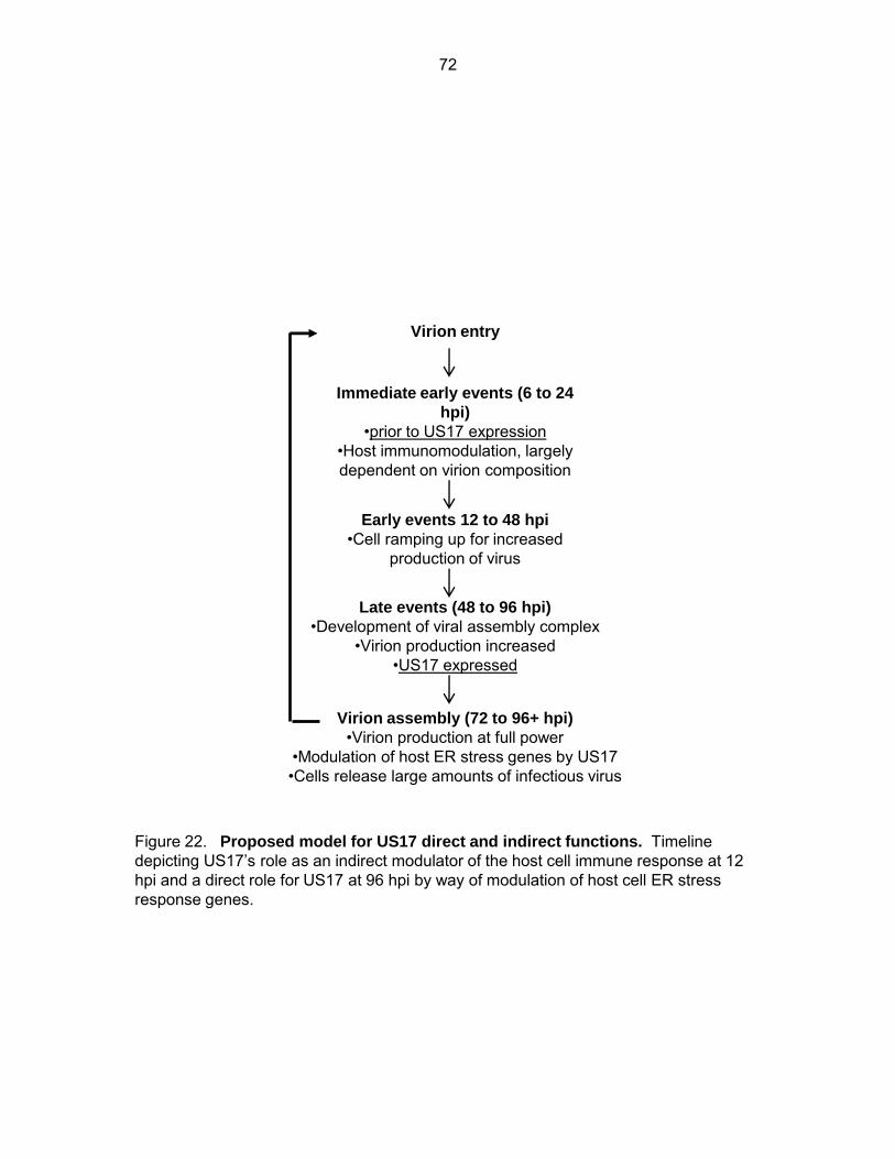
Virion entry
Immediate early events (6 to 24
hpi)
•prior to US17 expression •Host immunomodulation, largely dependent on virion composition
Early events 12 to 48 hpi
•Cell ramping up for increased production of virus
Late events (48 to 96 hpi)
•Development of viral assembly complex •Virion production increased
•US17 expressed
Virion assembly (72 to 96+ hpi)
•Virion production at full power •Modulation of host ER stress genes by US17 •Cells release large amounts of infectious virus
Figure 22. Proposed model for US17 direct and indirect functions. Timeline depicting US17’s role as an indirect modulator of the host cell immune response at 12 hpi and a direct role for US17 at 96 hpi by way of modulation of host cell ER stress response genes.
72
Chapter Four
Conclusions and Discussion
This work highlights the complex and incompletely understood process that is HCMV
immunomodulation and how factors such as virion composition can influence this process. In
Chapter Two we demonstrated that ΔUS17 induced altered host transcriptional profiles from
those of parental virions. At a very early time after infection (12 hpi) ΔUS17 elicited a reduced
immune response from the host cell as evidenced by decreased production of transcripts
associated with both interferon and innate immune responses. At a later time after infection (96
hpi) ΔUS17 differentially modulated transcripts encoding for genes involved in the ER stress
response. In Chapter Three we further demonstrated that virions produced by ΔUS17 differed
from parental virions, incorporating reduced amounts of the glycoprotein gH into their structure.
ΔUS17 produced a >3-fold larger genome-to-pfu ratio while producing an equal number of
infectious virus. When inoculated at an equal MOI this larger number of non-infectious particles
produced by ΔUS17 delivered a ~3-fold larger amount of the tegument protein pp65 compared
to parental. Additionally, UV inactivation of virions did not completely abrogate the
transcriptional phenotype of ΔUS17 indicating that the structural differences of ΔUS17 virions
contribute to the blunting of the host immune response (Data no shown).
At first, it seems counterintuitive that a virus would produce factors, such as US17, that
increase the immunogenic properties of the virus thus making it more easily recognizable to the
host’s immune system. However, there is an accumulating body of work demonstrating that
HCMV needs to strike a delicate balance of interferon and innate immune responses in order to
ensure proper reproduction and survival (139-141).
HCMV has evolved the ability to interact with, and in some instances necessitates use
of, host interferon response and anti-viral proteins. Two interferon inducible proteins, Viperin
73
(RSAD2) and IFI-16, enhance HCMV virion production and are specifically up-regulated by
during infection. These same proteins inhibit replication of HIV, HCV, RSV and several other
RNA viruses (142-146). HCMV utilizes IFI-16 in conjunction with pp65 as a trans-activator to
up-regulate transcription of immediate early proteins thus aiding in the establishment of a lytic
infection (113). RSAD2 interacts with another viral protein (UL37.1 vMIA) at the mitochondrial
membrane to facilitate production of ATP (140,147,148). Upon knock-down of IFI-16 or Viperin,
HCMV replicates less efficiently. In this way it appears that HCMV must counter-intuitively
stimulate, and more importantly balance, the host immune response to ensure proper
replication. If the immune response is activated too aggressively the cell will limit the spread of
infection by initiating apoptosis of infected cells before production of progeny virions. In
contrast, if there is too little immune response elicited by the virus, HCMV cannot initiate lytic
replication efficiently.
This balance must be struck immediately upon infection before the host anti-viral
response of the host cell overwhelms the virus and is therefore highly dependent on both virion
structure and the particle–to-pfu ratio both of which are important mediators of host immune
sensing. Therefore, it appears that HCMV has evolved US17 to modulate particle-to-pfu ratios
and virion composition in order to produce virions that elicit the correct immune response from
the host cell. Additionally the control of virion composition by US17 appears to be separate
from viral pathways that are important for making the most basic unit of an infectious virion, i.e.,
ΔUS17 is replication competent in cell culture but the virions produced by ΔUS17 elicit an
abnormal cellular response.
In addition to establishment of lytic replication, activation of the anti-viral response may
be important for HCMV replication in another way. It has been reported that undifferentiated
monocytes are an important reservoir of latently infected cells in the host (149-151). Monocytes
74

are a good viral reservoir because of their ability to travel throughout the body and deliver virus
to distant sites from that of the primary infection. Upon differentiation of these monocytes into
macrophage HCMV begins lytic replication and can then infect nearby cells of other lineages
(fibroblasts, endo/epithelial cells etc)(152,153). Thus it is possible that HCMV uses US17 to
control its immunomodulatory abilities through manipulation of particle-to-pfu ratios and virion
composition in an effort to “bait” monocytes and other immune effecter cells to the site of a
primary infection in order to establish latency. In fact, many of the most highly differentially
expressed chemokines and cytokines that were seen to be down-modulated by ΔUS17 virions
at 12 hpi, including cxcl10, ccl2, and ccl5, are known to be directly chemoattractive to
monocytes (154-157) further strengthening this hypothesis .
In sum, the previously detailed experiments have elucidated a role for US17 in HCMV
biology. We propose a model wherein US17 plays a role in the assembly of mature HCMV
virions by modulating levels of genes involved in ER stress and the unfolded protein response at
late times after infection thus affecting viral protein folding, trafficking, or incorporation into the
virion structure in the cVAC(Fig. 23). Further, the involvement of US17 and possibly other US12
family members is important in the formation of particles that elicit an appropriate immune
response from host cells.
75
REFERENCES
1. Yu, X., S. Shah, M. Lee, W. Dai, P. Lo, W. Britt, H. Zhu, F. Liu, and Z. H. Zhou. 2011.
Biochemical and structural characterization of the capsid-bound tegument proteins of
human cytomegalovirus. J.Struct.Biol. 174:451-460
2. Spear, P. G., M. T. Shieh, B. C. Herold, D. WuDunn, and T. I. Koshy. 1992. Heparan
sulfate glycosaminoglycans as primary cell surface receptors for herpes simplex virus.
Adv.Exp.Med.Biol. 313:341-353
3. Sawitzky, D., A. Voigt, H. Zeichhardt, and K. O. Habermehl. 1996. Glycoprotein B
(gB) of pseudorabies virus interacts specifically with the glycosaminoglycan heparin.
Virus Res. 41:101-108
4. Montgomery, R. I., M. S. Warner, B. J. Lum, and P. G. Spear. 1996. Herpes simplex
virus-1 entry into cells mediated by a novel member of the TNF/NGF receptor family.
Cell 87:427-436
5. Feire, A. L., H. Koss, and T. Compton. 2004. Cellular integrins function as entry
receptors for human cytomegalovirus via a highly conserved disintegrin-like domain.
Proc.Natl.Acad.Sci.U.S.A 101:15470-15475
6. Chan, G., M. T. Nogalski, and A. D. Yurochko. 2009. Activation of EGFR on
monocytes is required for human cytomegalovirus entry and mediates cellular motility.
Proc.Natl.Acad.Sci.U.S.A 106:22369-22374
7. Ryckman, B. J., M. A. Jarvis, D. D. Drummond, J. A. Nelson, and D. C. Johnson.
2006. Human cytomegalovirus entry into epithelial and endothelial cells depends on
76
genes UL128 to UL150 and occurs by endocytosis and low-pH fusion. J.Virol. 80:710-
722
8. Compton, T., R. R. Nepomuceno, and D. M. Nowlin. 1992. Human cytomegalovirus
penetrates host cells by pH-independent fusion at the cell surface. Virology 191:387-395
9. Kalejta, R. F. 2008. Tegument proteins of human cytomegalovirus.
Microbiol.Mol.Biol.Rev. 72:249-65, table
10. Bauer, D. W., J. B. Huffman, F. L. Homa, and A. Evilevitch. 2013. Herpes virus
genome, the pressure is on. J.Am.Chem.Soc. 135:11216-11221
11. Harris, S. M., B. Bullock, E. Westgard, H. Zhu, R. M. Stenberg, and J. A. Kerry.
2010. Functional properties of the human cytomegalovirus IE86 protein required for
transcriptional regulation and virus replication. J.Virol. 84:8839-8848
12. Hagemeier, C., S. M. Walker, P. J. Sissons, and J. H. Sinclair. 1992. The 72K IE1
and 80K IE2 proteins of human cytomegalovirus independently trans-activate the c-fos,
c-myc and hsp70 promoters via basal promoter elements. J.Gen.Virol. 73 ( Pt 9):2385-
2393
13. Yoo, Y. D., C. J. Chiou, K. S. Choi, Y. Yi, S. Michelson, S. Kim, G. S. Hayward, and
S. J. Kim. 1996. The IE2 regulatory protein of human cytomegalovirus induces
expression of the human transforming growth factor beta1 gene through an Egr-1
binding site. J.Virol. 70:7062-7070
14. Paulus, C. and M. Nevels. 2009. The human cytomegalovirus major immediate-early
proteins as antagonists of intrinsic and innate antiviral host responses. Viruses. 1:760-
779
77
15. Norris, K. L. and R. J. Youle. 2008. Cytomegalovirus proteins vMIA and m38.5 link
mitochondrial morphogenesis to Bcl-2 family proteins. J.Virol. 82:6232-6243
16. Sharon-Friling, R., J. Goodhouse, A. M. Colberg-Poley, and T. Shenk. 2006. Human
cytomegalovirus pUL37x1 induces the release of endoplasmic reticulum calcium stores.
Proc.Natl.Acad.Sci.U.S.A 103:19117-19122
17. Hesse, J., S. Ameres, K. Besold, S. Krauter, A. Moosmann, and B. Plachter. 2013.
Suppression of CD8+ T-cell recognition in the immediate-early phase of human
cytomegalovirus infection. J.Gen.Virol. 94:376-386
18. Sampaio, K. L., Y. Cavignac, Y. D. Stierhof, and C. Sinzger. 2005. Human
cytomegalovirus labeled with green fluorescent protein for live analysis of intracellular
particle movements. J.Virol. 79:2754-2767
19. Sanchez, V. and D. H. Spector. 2002. Virology. CMV makes a timely exit. Science
297:778-779
20. Klupp, B. G., H. Granzow, and T. C. Mettenleiter. 2011. Nuclear envelope breakdown
can substitute for primary envelopment-mediated nuclear egress of herpesviruses.
J.Virol. 85:8285-8292
21. Mettenleiter, T. C. 2004. Budding events in herpesvirus morphogenesis. Virus Res.
106:167-180
22. McMillan, T. N. and D. C. Johnson. 2001. Cytoplasmic domain of herpes simplex virus
gE causes accumulation in the trans-Golgi network, a site of virus envelopment and
sorting of virions to cell junctions. J.Virol. 75:1928-1940
78
23. Remillard-Labrosse, G., C. Mihai, J. Duron, G. Guay, and R. Lippe. 2009. Protein
kinase D-dependent trafficking of the large Herpes simplex virus type 1 capsids from the
TGN to plasma membrane. Traffic. 10:1074-1083
24. Tooze, J., M. Hollinshead, B. Reis, K. Radsak, and H. Kern. 1993. Progeny vaccinia
and human cytomegalovirus particles utilize early endosomal cisternae for their
envelopes. Eur.J Cell Biol. 60:163-178
25. Cepeda, V., M. Esteban, and A. Fraile-Ramos. 2010. Human cytomegalovirus final
envelopment on membranes containing both trans-Golgi network and endosomal
markers. Cell Microbiol. 12:386-404
26. Poole, E., A. Walther, K. Raven, C. A. Benedict, G. M. Mason, and J. Sinclair. 2013.
The myeloid transcription factor GATA-2 regulates the viral UL144 gene during human
cytomegalovirus latency in an isolate-specific manner. J.Virol. 87:4261-4271
27. Reeves, M. B. and J. H. Sinclair. 2010. Analysis of latent viral gene expression in
natural and experimental latency models of human cytomegalovirus and its correlation
with histone modifications at a latent promoter. J.Gen.Virol. 91:599-604
28. Farrell, M. J., A. T. Dobson, and L. T. Feldman. 1991. Herpes simplex virus latency-
associated transcript is a stable intron. Proc.Natl.Acad.Sci.U.S.A 88:790-794
29. Wigdahl, B. L., R. J. Ziegler, M. Sneve, and F. Rapp. 1983. Herpes simplex virus
latency and reactivation in isolated rat sensory neurons. Virology 127:159-167
30. Levine, M., A. L. Goldin, and J. C. Glorioso. 1980. Persistence of herpes simplex virus
genes in cells of neuronal origin. J.Virol. 35:203-210
79

31. Hargett, D. and T. E. Shenk. 2010. Experimental human cytomegalovirus latency in
CD14+ monocytes. Proc.Natl.Acad.Sci.U.S.A 107:20039-20044
32. Sindre, H., G. E. Tjoonnfjord, H. Rollag, T. Ranneberg-Nilsen, O. P. Veiby, S. Beck,
M. Degre, and K. Hestdal. 1996. Human cytomegalovirus suppression of and latency in
early hematopoietic progenitor cells. Blood 88:4526-4533
33. Babcock, G. J., L. L. Decker, M. Volk, and D. A. Thorley-Lawson. 1998. EBV
persistence in memory B cells in vivo. Immunity. 9:395-404
34. Boppana, S. B. and K. B. Fowler. 2007. Persistence in the population: epidemiology
and transmisson, In.
35. Staras, S. A., S. C. Dollard, K. W. Radford, W. D. Flanders, R. F. Pass, and M. J.
Cannon. 2006. Seroprevalence of cytomegalovirus infection in the United States, 1988-
1994. Clin.Infect.Dis. 43:1143-1151
36. Janssen, H. P., A. M. van Loon, M. J. Meddens, E. C. Eickmans-Josten, A. J.
Hoitsma, T. J. de Witte, and W. G. Quint. 1988. Immunological detection of
cytomegalovirus early antigen on monolayers inoculated with urine specimens by
centrifugation and cultured for 6 days as alternative to conventional virus isolation.
J.Clin.Microbiol. 26:1313-1315
37. Chou, S. and T. C. Merigan. 1983. Rapid detection and quantitation of human
cytomegalovirus in urine through DNA hybridization. N.Engl.J.Med. 308:921-925
38. Tamura, T., S. Chiba, Y. Chiba, and T. Nakao. 1980. Virus excretion and neutralizing
antibody response in saliva in human cytomegalovirus infection. Infect.Immun. 29:842-
845
80
39. Sugar, E. A., D. A. Jabs, A. Ahuja, J. E. Thorne, R. P. Danis, and C. L. Meinert.
2012. Incidence of cytomegalovirus retinitis in the era of highly active antiretroviral
therapy. Am.J.Ophthalmol. 153:1016-1024
40. Tyms, A. S., D. L. Taylor, and J. M. Parkin. 1989. Cytomegalovirus and the acquired
immunodeficiency syndrome. J.Antimicrob.Chemother. 23 Suppl A:89-105
41. Larsson, K., J. Aschan, M. Remberger, O. Ringden, J. Winiarski, and P. Ljungman.
2004. Reduced risk for extensive chronic graft-versus-host disease in patients receiving
transplants with human leukocyte antigen-identical sibling donors given polymerase
chain reaction-based preemptive therapy against cytomegalovirus. Transplantation
77:526-531
42. Onor, I. O., S. B. Todd, E. Meredith, S. D. Perez, A. K. Mehta, L. G. Marshall, S. J.
Knechtle, and S. I. Hanish. 2013. Evaluation of clinical outcomes of prophylactic versus
preemptive cytomegalovirus strategy in liver transplant recipients. Transpl.Int. 26:592-
600
43. Roux, A., G. Mourin, S. Fastenackels, J. R. Almeida, M. C. Iglesias, A. Boyd, E.
Gostick, M. Larsen, D. A. Price, K. Sacre, D. C. Douek, B. Autran, C. Picard, S.
Miranda, D. Sauce, M. Stern, and V. Appay. 2013. CMV driven CD8(+) T-cell
activation is associated with acute rejection in lung transplantation. Clin.Immunol.
148:16-26
44. Allard, M., P. Tonnerre, S. Nedellec, R. Oger, A. Morice, Y. Guilloux, E. Houssaint,
B. Charreau, and N. Gervois. 2012. HLA-E-restricted cross-recognition of allogeneic
endothelial cells by CMV-associated CD8 T cells: a potential risk factor following
transplantation. PLoS.One. 7:e50951
81
45. Iwaszko, M. and K. Bogunia-Kubik. 2011. Clinical significance of the HLA-E and
CD94/NKG2 interaction. Arch.Immunol.Ther.Exp.(Warsz.) 59:353-367
46. Cannon, M. J. 2009. Congenital cytomegalovirus (CMV) epidemiology and awareness.
J.Clin.Virol. 46 Suppl 4:S6-10
47. Demmler, G. J. 1996. Congenital cytomegalovirus infection and disease.
Adv.Pediatr.Infect.Dis. 11:135-162
48. Stagno, S., R. F. Pass, G. Cloud, W. J. Britt, R. E. Henderson, P. D. Walton, D. A.
Veren, F. Page, and C. A. Alford. 1986. Primary cytomegalovirus infection in
pregnancy. Incidence, transmission to fetus, and clinical outcome. Journal of the
American Medical Association 256:1904-1908
49. Mocarski, E. S., T. Shenk, R. F. Pass, and D. G. Paul. 2013. Cytomegaloviruses, p.
1960-2014. In: D. M. Knipe, P. M. Howley, D. E. Griffin, R. A. Lamb, M. A. Martin, B.
Roizman, I. C. Jeffrey, and R. R. Vincent (eds.), Fields Virology. 6 ed. Lippincott
Williams & Wilkins, Philadelphia.
50. Manicklal, S., V. C. Emery, T. Lazzarotto, S. B. Boppana, and R. K. Gupta. 2013.
The "silent" global burden of congenital cytomegalovirus. Clin.Microbiol.Rev. 26:86-102
51. Murphy, E., D. Yu, J. Grimwood, J. Schmutz, M. Dickson, M. A. Jarvis, G. Hahn, J.
A. Nelson, R. M. Myers, and T. E. Shenk. 2003. Coding potential of laboratory and
clinical strains of human cytomegalovirus. Proc.Natl.Acad.Sci.U.S.A 100:14976-14981
52. Davison, A. J., A. Dolan, P. Akter, C. Addison, D. J. Dargan, D. J. Alcendor, D. J.
McGeoch, and G. S. Hayward. 2003. The human cytomegalovirus genome revisited:
comparison with the chimpanzee cytomegalovirus genome. J.Gen.Virol. 84:17-28
82
53. Stern-Ginossar, N., B. Weisburd, A. Michalski, V. T. Le, M. Y. Hein, S. X. Huang, M.
Ma, B. Shen, S. B. Qian, H. Hengel, M. Mann, N. T. Ingolia, and J. S. Weissman.
2012. Decoding human cytomegalovirus. Science 338:1088-1093
54. Dhuruvasan, K., G. Sivasubramanian, and P. E. Pellett. 2010. Roles of host and viral
microRNAs in human cytomegalovirus biology. Virus Res.
55. Grey, F. and J. Nelson. 2008. Identification and function of human cytomegalovirus
microRNAs. J.Clin.Virol. 41:186-191
56. Grey, F., H. Meyers, E. A. White, D. H. Spector, and J. Nelson. 2007. A human
cytomegalovirus-encoded microRNA regulates expression of multiple viral genes
involved in replication. PLoS.Pathog. 3:e163
57. Dolan, A., C. Cunningham, R. D. Hector, A. F. Hassan-Walker, L. Lee, C. Addison,
D. J. Dargan, D. J. McGeoch, D. Gatherer, V. C. Emery, P. D. Griffiths, C. Sinzger,
B. P. McSharry, G. W. Wilkinson, and A. J. Davison. 2004. Genetic content of wild-
type human cytomegalovirus. J.Gen.Virol. 85:1301-1312
58. Yu, D., M. C. Silva, and T. Shenk. 2003. Functional map of human cytomegalovirus
AD169 defined by global mutational analysis. Proc.Natl.Acad.Sci.U.S.A 100:12396-
12401
59. Mocarski, E. S., Jr. 2002. Immunomodulation by cytomegaloviruses: manipulative
strategies beyond evasion. Trends Microbiol. 10:332-339
60. Umashankar, M., A. Petrucelli, L. Cicchini, P. Caposio, C. N. Kreklywich, M. Rak, F.
Bughio, D. C. Goldman, K. L. Hamlin, J. A. Nelson, W. H. Fleming, D. N. Streblow,
83
and F. Goodrum. 2011. A novel human cytomegalovirus locus modulates cell type-
specific outcomes of infection. PLoS.Pathog. 7:e1002444
61. Bughio, F., D. A. Elliott, and F. Goodrum. 2013. An endothelial cell-specific
requirement for the UL133-UL138 locus of human cytomegalovirus for efficient virus
maturation. J.Virol. 87:3062-3075
62. Lin, Y. L., P. C. Chang, Y. Wang, and M. Li. 2008. Identification of novel viral
interleukin-10 isoforms of human cytomegalovirus AD169. Virus Res. 131:213-223
63. Lockridge, K. M., S. S. Zhou, R. H. Kravitz, J. L. Johnson, E. T. Sawai, E. L. Blewett,
and P. A. Barry. 2000. Primate cytomegaloviruses encode and express an IL-10-like
protein. Virology 268:272-280
64. Tomasec, P., E. C. Wang, A. J. Davison, B. Vojtesek, M. Armstrong, C. Griffin, B. P.
McSharry, R. J. Morris, S. Llewellyn-Lacey, C. Rickards, A. Nomoto, C. Sinzger,
and G. W. Wilkinson. 2005. Downregulation of natural killer cell-activating ligand
CD155 by human cytomegalovirus UL141. Nat.Immunol. 6:181-188
65. Luttichau, H. R. 2010. The cytomegalovirus UL146 gene product vCXCL1 targets both
CXCR1 and CXCR2 as an agonist. J.Biol.Chem. 285:9137-9146
66. Weston, K. and B. G. Barrell. 1986. Sequence of the short unique region, short
repeats, and part of the long repeats of human cytomegalovirus. J Mol.Biol. 192:177-208
67. Chee, M. S., A. T. Bankier, S. Beck, R. Bohni, C. M. Brown, R. Cerny, T. Horsnell, C.
A. Hutchison, T. Kouzarides, J. A. Martignetti, E. Preddie, S. C. Satchwell, P.
Tomlinson, K. M. Weston, and B. G. Barrell. 1990. Analysis of the protein-coding
84
content of the sequence of human cytomegalovirus strain AD169.
Curr.Top.Microbiol.Immunol. 154:125-169
68. Carrara, G., N. Saraiva, C. Gubser, B. F. Johnson, and G. L. Smith. 2012. Six-
transmembrane topology for Golgi anti-apoptotic protein (GAAP) and Bax inhibitor 1 (BI-
1) provides model for the transmembrane Bax inhibitor-containing motif (TMBIM) family.
J.Biol.Chem. 287:15896-15905
69. Das, S. and P. E. Pellett. 2007. Members of the HCMV US12 family of predicted
heptaspanning membrane proteins have unique intracellular distributions, including
association with the cytoplasmic virion assembly complex. Virology 361:263-273
70. Das, S., Y. Skomorovska-Prokvolit, F. Z. Wang, and P. E. Pellett. 2006. Infection-
dependent nuclear localization of US17, a member of the US12 family of human
cytomegalovirus-encoded seven-transmembrane proteins. J.Virol. 80:1191-1203
71. Lesniewski, M., S. Das, Y. Skomorovska-Prokvolit, F. Z. Wang, and P. E. Pellett.
2006. Primate cytomegalovirus US12 gene family: A distinct and diverse clade of seven-
transmembrane proteins. Virology 354:286-298
72. Chee, M. S., S. C. Satchwell, E. Preddie, K. M. Weston, and B. G. Barrell. 1990.
Human cytomegalovirus encodes three G protein-coupled receptor homologues. Nature
344:774-777
73. Dunn, W., C. Chou, H. Li, R. Hai, D. Patterson, V. Stolc, H. Zhu, and F. Liu. 2003.
Functional profiling of a human cytomegalovirus genome. Proc.Natl.Acad.Sci.U.S.A
100:14223-14228
85
74. Hai, R., A. Chu, H. Li, S. Umamoto, P. Rider, and F. Liu. 2006. Infection of human
cytomegalovirus in cultured human gingival tissue. Virol.J. 3:84
75. Bronzini, M., A. Luganini, V. Dell'oste, A. M. De, S. Landolfo, and G. Gribaudo.
2012. The US16 gene of Human Cytomegalovirus is Required for Efficient Viral Infection
of Endothelial and Epithelial Cells. J.Virol.
76. Wang, D., Q. C. Yu, J. Schroer, E. Murphy, and T. Shenk. 2007. Human
cytomegalovirus uses two distinct pathways to enter retinal pigmented epithelial cells.
Proc.Natl.Acad.Sci.U.S.A 104:20037-20042
77. Vanarsdall, A. L., M. C. Chase, and D. C. Johnson. 2011. Human cytomegalovirus
glycoprotein gO complexes with gH/gL, promoting interference with viral entry into
human fibroblasts but not entry into epithelial cells. J.Virol. 85:11638-11645
78. Schmolke, S., H. F. Kern, P. Drescher, G. Jahn, and B. Plachter. 1995. The dominant
phosphoprotein pp65 (UL83) of human cytomegalovirus is dispensable for growth in cell
culture. J.Virol. 69:5959-5968
79. Chevillotte, M., S. Landwehr, L. Linta, G. Frascaroli, A. Luske, C. Buser, T.
Mertens, and E. J. von. 2009. Major tegument protein pp65 of human cytomegalovirus
is required for the incorporation of pUL69 and pUL97 into the virus particle and for viral
growth in macrophages. J.Virol. 83:2480-2490
80. Watanabe, N. and E. Lam. 2008. Arabidopsis Bax inhibitor-1: A rheostat for ER stress-
induced programmed cell death. Plant Signal.Behav. 3:564-566
81. Rojas-Rivera, D., R. Armisen, A. Colombo, G. Martinez, A. L. Eguiguren, A. Diaz, S.
Kiviluoto, D. Rodriguez, M. Patron, R. Rizzuto, G. Bultynck, M. L. Concha, J.
86

Sierralta, A. Stutzin, and C. Hetz. 2012. TMBIM3/GRINA is a novel unfolded protein
response (UPR) target gene that controls apoptosis through the modulation of ER
calcium homeostasis. Cell Death.Differ.
82. de, M. F., C. Gubser, M. M. van Dommelen, H. J. Visch, F. Distelmaier, A. Postigo,
T. Luyten, J. B. Parys, S. H. de, G. L. Smith, P. H. Willems, and F. J. van
Kuppeveld. 2009. Human Golgi antiapoptotic protein modulates intracellular calcium
fluxes. Mol.Biol Cell 20:3638-3645
83. Kim, H. R., G. H. Lee, K. C. Ha, T. Ahn, J. Y. Moon, B. J. Lee, S. G. Cho, S. Kim, Y.
R. Seo, Y. J. Shin, S. W. Chae, J. C. Reed, and H. J. Chae. 2008. Bax Inhibitor-1 Is a
pH-dependent regulator of Ca2+ channel activity in the endoplasmic reticulum. J Biol
Chem. 283:15946-15955
84. Xu, C., W. Xu, A. E. Palmer, and J. C. Reed. 2008. BI-1 regulates endoplasmic
reticulum Ca2+ homeostasis downstream of Bcl-2 family proteins. J Biol Chem.
283:11477-11484
85. Bultynck, G., S. Kiviluoto, N. Henke, H. Ivanova, L. Schneider, V. Rybalchenko, T.
Luyten, K. Nuyts, B. W. De, I. Bezprozvanny, J. B. Parys, S. H. de, L. Missiaen, and
A. Methner. 2012. The C terminus of Bax inhibitor-1 forms a Ca2+-permeable channel
pore. J.Biol.Chem. 287:2544-2557
86. Lisbona, F., D. Rojas-Rivera, P. Thielen, S. Zamorano, D. Todd, F. Martinon, A.
Glavic, C. Kress, J. H. Lin, P. Walter, J. C. Reed, L. H. Glimcher, and C. Hetz. 2009.
BAX inhibitor-1 is a negative regulator of the ER stress sensor IRE1alpha. Mol.Cell
33:679-691
87
87. Bailly-Maitre, B., B. F. Belgardt, S. D. Jordan, B. Coornaert, M. J. von Freyend, A.
Kleinridders, J. Mauer, M. Cuddy, C. L. Kress, D. Willmes, M. Essig, B. Hampel, U.
Protzer, J. C. Reed, and J. C. Bruning. 2010. Hepatic Bax inhibitor-1 inhibits
IRE1alpha and protects from obesity-associated insulin resistance and glucose
intolerance. J.Biol.Chem. 285:6198-6207
88. Xu, C., W. Xu, A. E. Palmer, and J. C. Reed. 2008. BI-1 regulates endoplasmic
reticulum Ca2+ homeostasis downstream of Bcl-2 family proteins. J.Biol.Chem.
283:11477-11484
89. Kiviluoto, S., L. Schneider, T. Luyten, T. Vervliet, L. Missiaen, S. H. de, J. B. Parys,
A. Methner, and G. Bultynck. 2012. Bax inhibitor-1 is a novel IP(3) receptor-interacting
and -sensitizing protein. Cell Death.Dis. 3:e367
90. Sano, R., Y. C. Hou, M. Hedvat, R. G. Correa, C. W. Shu, M. Krajewska, P. W. Diaz,
C. M. Tamble, G. Quarato, R. A. Gottlieb, M. Yamaguchi, V. Nizet, R. Dahl, D. D.
Thomas, S. W. Tait, D. R. Green, P. B. Fisher, S. Matsuzawa, and J. C. Reed. 2012.
Endoplasmic reticulum protein BI-1 regulates Ca(2)(+)-mediated bioenergetics to
promote autophagy. Genes Dev. 26:1041-1054
91. Saraiva, N., D. L. Prole, G. Carrara, B. F. Johnson, C. W. Taylor, M. Parsons, and G.
L. Smith. 2013. hGAAP promotes cell adhesion and migration via the stimulation of
store-operated Ca2+ entry and calpain 2. J.Cell Biol. 202:699-713
92. Gubser, C., D. Bergamaschi, M. Hollinshead, X. Lu, F. J. van Kuppeveld, and G. L.
Smith. 2007. A new inhibitor of apoptosis from vaccinia virus and eukaryotes.
PLoS.Pathog. 3:e17
88
93. Saraiva, N., D. L. Prole, G. Carrara, M. C. Maluquer de, B. F. Johnson, B. Byrne, C.
W. Taylor, and G. L. Smith. 2013. Human and viral Golgi anti-apoptotic proteins
(GAAPs) oligomerize via different mechanisms and monomeric GAAP inhibits apoptosis
and modulates calcium. J.Biol.Chem. 288:13057-13067
94. Gibson, W. 2008. Structure and formation of the cytomegalovirus virion.
Curr.Top.Microbiol.Immunol. 325:187-204
95. Varnum, S. M., D. N. Streblow, M. E. Monroe, P. Smith, K. J. Auberry, L. Pasa-Tolic,
D. Wang, D. G. Camp, K. Rodland, S. Wiley, W. Britt, T. Shenk, R. D. Smith, and J.
A. Nelson. 2004. Identification of proteins in human cytomegalovirus (HCMV) particles:
the HCMV proteome. J.Virol. 78:10960-10966
96. Das, S., A. Vasanji, and P. E. Pellett. 2007. Three-dimensional structure of the human
cytomegalovirus cytoplasmic virion assembly complex includes a reoriented secretory
apparatus. J.Virol. 81:11861-11869
97. Sanchez, V., K. D. Greis, E. Sztul, and W. J. Britt. 2000. Accumulation of virion
tegument and envelope proteins in a stable cytoplasmic compartment during human
cytomegalovirus replication: characterization of a potential site of virus assembly. J.Virol.
74:975-986
98. Sanchez, V., E. Sztul, and W. J. Britt. 2000. Human cytomegalovirus pp28 (UL99)
localizes to a cytoplasmic compartment which overlaps the endoplasmic reticulum-golgi-
intermediate compartment. J.Virol. 74:3842-3851
99. Theiler, R. N. and T. Compton. 2002. Distinct glycoprotein O complexes arise in a post-
Golgi compartment of cytomegalovirus-infected cells. J.Virol. 76:2890-2898
89

100. AuCoin, D. P., G. B. Smith, C. D. Meiering, and E. S. Mocarski. 2006.
Betaherpesvirus-conserved cytomegalovirus tegument protein ppUL32 (pp150) controls
cytoplasmic events during virion maturation. J.Virol. 80:8199-8210
101. Tandon, R. and E. S. Mocarski. 2008. Control of cytoplasmic maturation events by
cytomegalovirus tegument protein pp150. J.Virol. 82:9433-9444
102. Benyesh-Melnick, M., F. Probstmeyer, R. McCombs, J. P. Brunschwig, and V.
Vonka. 1966. Correlation between infectivity and physical virus particles in human
cytomegalovirus. J.Bacteriol. 92:1555-1561
103. Pepperl, S., J. Munster, M. Mach, J. R. Harris, and B. Plachter. 2000. Dense bodies
of human cytomegalovirus induce both humoral and cellular immune responses in the
absence of viral gene expression. J.Virol. 74:6132-6146
104. Irmiere, A. and W. Gibson. 1983. Isolation and characterization of a noninfectious
virion-like particle released from cells infected with human strains of cytomegalovirus.
Virology 130:118-133
105. Ahlqvist, J. and E. Mocarski. 2011. Cytomegalovirus UL103 controls virion and dense
body egress. J.Virol.
106. Isler, J. A., A. H. Skalet, and J. C. Alwine. 2005. Human cytomegalovirus infection
activates and regulates the unfolded protein response. J.Virol. 79:6890-6899
107. Isler, J. A., T. G. Maguire, and J. C. Alwine. 2005. Production of infectious human
cytomegalovirus virions is inhibited by drugs that disrupt calcium homeostasis in the
endoplasmic reticulum. J.Virol. 79:15388-15397
90
108. Xuan, B., Z. Qian, E. Torigoi, and D. Yu. 2009. Human cytomegalovirus protein pUL38
induces ATF4 expression, inhibits persistent JNK phosphorylation, and suppresses
endoplasmic reticulum stress-induced cell death. J.Virol. 83:3463-3474
109. Meunier, L., Y. K. Usherwood, K. T. Chung, and L. M. Hendershot. 2002. A subset of
chaperones and folding enzymes form multiprotein complexes in endoplasmic reticulum
to bind nascent proteins. Mol.Biol.Cell 13:4456-4469
110. Buchkovich, N. J., T. G. Maguire, Y. Yu, A. W. Paton, J. C. Paton, and J. C. Alwine.
2008. Human cytomegalovirus specifically controls the levels of the endoplasmic
reticulum chaperone BiP/GRP78, which is required for virion assembly. J.Virol. 82:31-39
111. Munger, J., B. D. Bennett, A. Parikh, X. J. Feng, J. McArdle, H. A. Rabitz, T. Shenk,
and J. D. Rabinowitz. 2008. Systems-level metabolic flux profiling identifies fatty acid
synthesis as a target for antiviral therapy. Nat.Biotechnol. 26:1179-1186
112. Buchkovich, N. J., T. G. Maguire, and J. C. Alwine. 2010. Role of the endoplasmic
reticulum chaperone BiP, SUN domain proteins, and dynein in altering nuclear
morphology during human cytomegalovirus infection. J.Virol. 84:7005-7017
113. Cristea, I. M., N. J. Moorman, S. S. Terhune, C. D. Cuevas, E. S. O'Keefe, M. P.
Rout, B. T. Chait, and T. Shenk. 2010. Human cytomegalovirus pUL83 stimulates
activity of the viral immediate-early promoter through its interaction with the cellular IFI16
protein. J.Virol. 84:7803-7814
114. Abate, D. A., S. Watanabe, and E. S. Mocarski. 2004. Major human cytomegalovirus
structural protein pp65 (ppUL83) prevents interferon response factor 3 activation in the
interferon response. J.Virol. 78:10995-11006
91
115. Lee, E. C., D. Yu, d. Martinez, V, L. Tessarollo, D. A. Swing, Court DL, N. A.
Jenkins, and N. G. Copeland. 2001. A highly efficient Escherichia coli-based
chromosome engineering system adapted for recombinogenic targeting and subcloning
of BAC DNA. Genomics 73:56-65
116. Warming, S., N. Costantino, Court DL, N. A. Jenkins, and N. G. Copeland. 2005.
Simple and highly efficient BAC recombineering using galK selection. Nucleic Acids Res.
33:e36
117. Qian, Z., B. Xuan, T. T. Hong, and D. Yu. 2008. The full-length protein encoded by
human cytomegalovirus gene UL117 is required for the proper maturation of viral
replication compartments. J.Virol. 82:3452-3465
118. Tusher, V. G., R. Tibshirani, and G. Chu. 2001. Significance analysis of microarrays
applied to the ionizing radiation response. Proc.Natl.Acad.Sci.U.S.A 98:5116-5121
119. Maere, S., K. Heymans, and M. Kuiper. 2005. BiNGO: a Cytoscape plugin to assess
overrepresentation of gene ontology categories in biological networks. Bioinformatics.
21:3448-3449
120. Guo, Y. W. and E. S. Huang. 1993. Characterization of a structurally tricistronic gene of
human cytomegalovirus composed of U(s)18, U(s)19, and U(s)20. J.Virol. 67:2043-2054
121. Miller, M. S., W. E. Furlong, L. Pennell, M. Geadah, and L. Hertel. 2010. RASCAL is a
new human cytomegalovirus-encoded protein that localizes to the nuclear lamina and in
cytoplasmic vesicles at late times postinfection. J.Virol. 84:6483-6496
122. Dittmer, D. and E. S. Mocarski. 1997. Human cytomegalovirus infection inhibits G1/S
transition. J.Virol. 71:1629-1634
92
123. Fortunato, E. A., V. Sanchez, J. Y. Yen, and D. H. Spector. 2002. Infection of cells
with human cytomegalovirus during S phase results in a blockade to immediate-early
gene expression that can be overcome by inhibition of the proteasome. J.Virol. 76:5369-
5379
124. Du, P., W. A. Kibbe, and S. M. Lin. 2008. lumi: a pipeline for processing Illumina
microarray. Bioinformatics. 24:1547-1548
125. Buchkovich, N. J., T. G. Maguire, A. W. Paton, J. C. Paton, and J. C. Alwine. 2009.
The endoplasmic reticulum chaperone BiP/GRP78 is important in the structure and
function of the HCMV assembly compartment. J.Virol.
126. Simmen, K. A., J. Singh, B. G. Luukkonen, M. Lopper, A. Bittner, N. E. Miller, M. R.
Jackson, T. Compton, and K. Fruh. 2001. Global modulation of cellular transcription by
human cytomegalovirus is initiated by viral glycoprotein B. Proc.Natl.Acad.Sci.U.S.A
98:7140-7145
127. Boehme, K. W., M. Guerrero, and T. Compton. 2006. Human cytomegalovirus
envelope glycoproteins B and H are necessary for TLR2 activation in permissive cells.
J.Immunol. 177:7094-7102
128. Yurochko, A. D., E. S. Hwang, L. Rasmussen, S. Keay, L. Pereira, and E. S. Huang.
1997. The human cytomegalovirus UL55 (gB) and UL75 (gH) glycoprotein ligands
initiate the rapid activation of Sp1 and NF-kappaB during infection. J.Virol. 71:5051-5059
129. Guo, F. J., Y. Liu, J. Zhou, S. Luo, W. Zhao, X. Li, and C. Liu. 2012. XBP1S protects
cells from ER stress-induced apoptosis through Erk1/2 signaling pathway involving
CHOP. Histochem.Cell Biol. 138:447-460
93
130. Mungrue, I. N., J. Pagnon, O. Kohannim, P. S. Gargalovic, and A. J. Lusis. 2009.
CHAC1/MGC4504 is a novel proapoptotic component of the unfolded protein response,
downstream of the ATF4-ATF3-CHOP cascade. J.Immunol. 182:466-476
131. Teske, B. F., M. E. Fusakio, D. Zhou, J. Shan, J. N. McClintick, M. S. Kilberg, and R.
C. Wek. 2013. CHOP induces activating transcription factor 5 (ATF5) to trigger
apoptosis in response to perturbations in protein homeostasis. Mol.Biol.Cell 24:2477-
2490
132. Lee, G. H., H. K. Kim, S. W. Chae, D. S. Kim, K. C. Ha, M. Cuddy, C. Kress, J. C.
Reed, H. R. Kim, and H. J. Chae. 2007. Bax inhibitor-1 regulates endoplasmic reticulum
stress-associated reactive oxygen species and heme oxygenase-1 expression.
J.Biol.Chem. 282:21618-21628
133. Hayajneh, W. A., A. M. Colberg-Poley, A. Skaletskaya, L. M. Bartle, M. M.
Lesperance, D. G. Contopoulos-Ioannidis, N. L. Kedersha, and V. S. Goldmacher.
2001. The sequence and antiapoptotic functional domains of the human cytomegalovirus
UL37 exon 1 immediate early protein are conserved in multiple primary strains. Virology
279:233-240
134. Feng, X., J. Schroer, D. Yu, and T. Shenk. 2006. Human cytomegalovirus pUS24 is a
virion protein that functions very early in the replication cycle. J.Virol. 80:8371-8378
135. Wang, D. and T. Shenk. 2005. Human cytomegalovirus virion protein complex required
for epithelial and endothelial cell tropism. Proc.Natl.Acad.Sci.U.S.A 102:18153-18158
136. Baldwin, B. R., C. O. Zhang, and S. Keay. 2000. Cloning and epitope mapping of a
functional partial fusion receptor for human cytomegalovirus gH. J.Gen.Virol. 81:27-35
94
137. Ryckman, B. J., M. C. Chase, and D. C. Johnson. 2008. HCMV gH/gL/UL128-131
interferes with virus entry into epithelial cells: evidence for cell type-specific receptors.
Proc.Natl.Acad.Sci.U.S.A 105:14118-14123
138. Yurochko, A. D. and E. S. Huang. 1999. Human cytomegalovirus binding to human
monocytes induces immunoregulatory gene expression. J.Immunol. 162:4806-4816
139. Viswanathan, K., M. S. Smith, D. Malouli, M. Mansouri, J. A. Nelson, and K. Fruh.
2011. BST2/Tetherin enhances entry of human cytomegalovirus. PLoS.Pathog.
7:e1002332
140. Seo, J. Y., R. Yaneva, E. R. Hinson, and P. Cresswell. 2011. Human cytomegalovirus
directly induces the antiviral protein viperin to enhance infectivity. Science 332:1093-
1097
141. Hansen, S. G., J. B. Sacha, C. M. Hughes, J. C. Ford, B. J. Burwitz, I. Scholz, R. M.
Gilbride, M. S. Lewis, A. N. Gilliam, A. B. Ventura, D. Malouli, G. Xu, R. Richards, N.
Whizin, J. S. Reed, K. B. Hammond, M. Fischer, J. M. Turner, A. W. Legasse, M. K.
Axthelm, P. T. Edlefsen, J. A. Nelson, J. D. Lifson, K. Fruh, and L. J. Picker. 2013.
Cytomegalovirus vectors violate CD8+ T cell epitope recognition paradigms. Science
340:1237874
142. Teng, T. S., S. S. Foo, D. Simamarta, F. M. Lum, T. H. Teo, A. Lulla, N. K. Yeo, E. G.
Koh, A. Chow, Y. S. Leo, A. Merits, K. C. Chin, and L. F. Ng. 2012. Viperin restricts
chikungunya virus replication and pathology. J Clin.Invest 122:4447-4460
95

143. McGillivary, G., Z. B. Jordan, M. E. Peeples, and L. O. Bakaletz. 2013. Replication of
respiratory syncytial virus is inhibited by the host defense molecule viperin. J
Innate.Immun. 5:60-71
144. Nasr, N., S. Maddocks, S. G. Turville, A. N. Harman, N. Woolger, K. J. Helbig, J.
Wilkinson, C. R. Bye, T. K. Wright, D. Rambukwelle, H. Donaghy, M. R. Beard, and
A. L. Cunningham. 2012. HIV-1 infection of human macrophages directly induces
viperin which inhibits viral production. Blood 120:778-788
145. Helbig, K. J., N. S. Eyre, E. Yip, S. Narayana, K. Li, G. Fiches, E. M. McCartney, R.
K. Jangra, S. M. Lemon, and M. R. Beard. 2011. The antiviral protein viperin inhibits
hepatitis C virus replication via interaction with nonstructural protein 5A. Hepatology
54:1506-1517
146. Wang, S., X. Wu, T. Pan, W. Song, Y. Wang, F. Zhang, and Z. Yuan. 2012. Viperin
inhibits hepatitis C virus replication by interfering with binding of NS5A to host protein
hVAP-33. J Gen.Virol 93:83-92
147. Rolle, S., A. M. De, D. Gioia, D. Lembo, L. Hertel, S. Landolfo, and M. Gariglio.
2001. The interferon-inducible 204 gene is transcriptionally activated by mouse
cytomegalovirus and is required for its replication. Virology 286:249-255
148. Hertel, L., A. M. De, B. Azzimonti, A. Rolle, M. Gariglio, and S. Landolfo. 1999. The
interferon-inducible 204 gene, a member of the Ifi 200 family, is not involved in the
antiviral state induction by IFN-alpha, but is required by the mouse cytomegalovirus for
its replication. Virology 262:1-8
96
149. Schrier, R. D., J. A. Nelson, and M. B. Oldstone. 1985. Detection of human
cytomegalovirus in peripheral blood lymphocytes in a natural infection. Science
230:1048-1051
150. Stanier, P., D. L. Taylor, A. D. Kitchen, N. Wales, Y. Tryhorn, and A. S. Tyms. 1989.
Persistence of cytomegalovirus in mononuclear cells in peripheral blood from blood
donors. BMJ 299:897-898
151. Taylor-Wiedeman, J., P. Sissons, and J. Sinclair. 1994. Induction of endogenous
human cytomegalovirus gene expression after differentiation of monocytes from healthy
carriers. J.Virol. 68:1597-1604
152. Soderberg-Naucler, C., K. N. Fish, and J. A. Nelson. 1997. Reactivation of latent
human cytomegalovirus by allogeneic stimulation of blood cells from healthy donors. Cell
91:119-126
153. Reeves, M. B., P. A. MacAry, P. J. Lehner, J. G. Sissons, and J. H. Sinclair. 2005.
Latency, chromatin remodeling, and reactivation of human cytomegalovirus in the
dendritic cells of healthy carriers. Proc Natl Acad Sci U S A 102:4140-4145
154. Nogueira, L. G., R. H. Santos, B. M. Ianni, A. I. Fiorelli, E. C. Mairena, L. A.
Benvenuti, A. Frade, E. Donadi, F. Dias, B. Saba, H. T. Wang, A. Fragata, M.
Sampaio, M. H. Hirata, P. Buck, C. Mady, E. A. Bocchi, N. A. Stolf, J. Kalil, and E.
Cunha-Neto. 2012. Myocardial chemokine expression and intensity of myocarditis in
Chagas cardiomyopathy are controlled by polymorphisms in CXCL9 and CXCL10.
PLoS.Negl.Trop.Dis. 6:e1867
97
155. Gouwy, M., S. Struyf, H. Verbeke, W. Put, P. Proost, G. Opdenakker, and D. J. Van.
2009. CC chemokine ligand-2 synergizes with the nonchemokine G protein-coupled
receptor ligand fMLP in monocyte chemotaxis, and it cooperates with the TLR ligand
LPS via induction of CXCL8. J.Leukoc.Biol. 86:671-680
156. de, N. P., C. Chenivesse, J. Gilet, H. Porte, H. Vorng, Y. Chang, A. F. Walls, B.
Wallaert, A. B. Tonnel, A. Tsicopoulos, and H. G. Zerwes. 2006. CCR5 usage by
CCL5 induces a selective leukocyte recruitment in human skin xenografts in vivo.
J.Invest Dermatol. 126:2057-2064
157. Schall, T. J., K. Bacon, K. J. Toy, and D. V. Goeddel. 1990. Selective attraction of
monocytes and T lymphocytes of the memory phenotype by cytokine RANTES. Nature
347:669-671
98
ABSTRACT
HUMAN CYTOMEGALOVIRUS US17 LOCUS FINE-TUNES INNATE AND INTRINSIC IMMUNE RESPONSES
by
STEPHEN J. GURCZYNSKI
December 2013
Advisor: Dr. Philip E. Pellett
Major: Immunology and Microbiology
Degree: Doctor of Philosophy
HCMV employs numerous strategies to combat, subvert, or co-opt host immunity. One
evolutionary strategy for this involves “capture” of a host gene and then its successive
duplication and divergence, forming a gene family, many of which have immunomodulatory
activities. The HCMV US12 family consists of ten tandemly arranged sequence-related genes in
the unique short region of the HCMV genome (US12-US21). Each gene encodes a protein
possessing seven predicted transmembrane domains, and patches of sequence similarity with
cellular GPCRs and the bax inhibitor-1 family of anti-apoptotic proteins.
We show that one member, US17, plays an important role during virion maturation.
Microarray analysis of cells infected with a recombinant HCMV deleted for US17 (ΔUS17)
revealed blunted host innate and interferon responses at early times after infection (12 hpi), a
pattern opposite that previously seen in the absence of the immunomodulatory tegument protein
pp65 (pUL83). Although ΔUS17 produced equal numbers of infectious particles in fibroblasts
compared to parental virus, at equal multiplicities of infection, it produced >3-fold more genome-
99

containing non-infectious viral particles, and delivered increased amounts of pp65 to newly
infected cells.
At later time points (96 hpi) ΔUS17 infected cells displayed aberrant expression of
several host ER stress response genes and chaperones, some of which are important for the
final stages of virion assembly and egress. Our results suggest that US17 modulates host
pathways which control virion composition enabling production of virions that elicit an
appropriately balanced host immune response.
100
AUTOBIOGRAPHICAL STATEMENT
I was born in Dearborn, Michigan in 1981 and have been a native of Michigan all of my
life. I attended Divine Child High School and graduated with honors in 1999. After high school I
attended the University of Michigan Dearborn where I majored in microbiology. College was my
first real introduction to biology and cemented my love for working in a laboratory. After
graduating with a B.S. in 2005 I went to work as a research assistant developing recognition
substrates for microbial biosensors in the laboratory of Dr. Matthew Jackson at the Wayne State
University School of Medicine. This project was multidisciplinary and exposed me to a wide
variety of scientific topics from microbiology to chemical engineering. During this time period I
also met my wife Laura who has been a great inspiration in my life. When the project ended in
2007 I was left with a choice of what I wanted to do for the rest of my life. Dr. Jackson urged me
to apply for the Ph.D. program at the School of Medicine. I joined the laboratory of Dr. Philip
Pellett as a Ph. D. candidate in 2008 and have spent the last several years studying human
cytomegalovirus cellular interactions, specifically, how HCMV virion protein composition
influences the innate immune system. The years building up to the completion of my Ph.D.
have been eventful, I’ve gotten married, had a son, Raymond, bought a house, and just recently
successfully defended my Ph. D. thesis. This truly has been the best time of my life and I feel
extremely fortunate that my life has taken the path that it has.
101