Embed Size (px)
Citation preview

Human DiseasesA Systemic ApproachSixth Edition
Chapter 5
Heredity and Disease
Mary Lou MulvihillMark Zelman
Paul HoldawayElaine Tompary
Jill Raymond
Chapter 5 Heredity and Disease
• Slide 10Sickle Cells• Slide 24Down Syndrome
Mulvihill, Zelman, Holdaway, Tompary, and RaymondHuman Diseases: A Systemic Approach, 6e
Copyright ©2006 by Prentice-Hall, Inc.Upper Saddle River, New Jersey 07458All rights reserved.
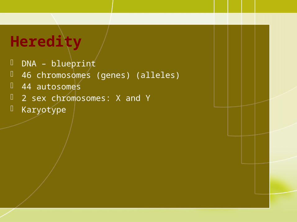
Heredity DNA – blueprint 46 chromosomes (genes) (alleles) 44 autosomes 2 sex chromosomes: X and Y Karyotype

Genetic Inheritance Alleles
– Homozygous– Heterozygous– Dominant– Recessive
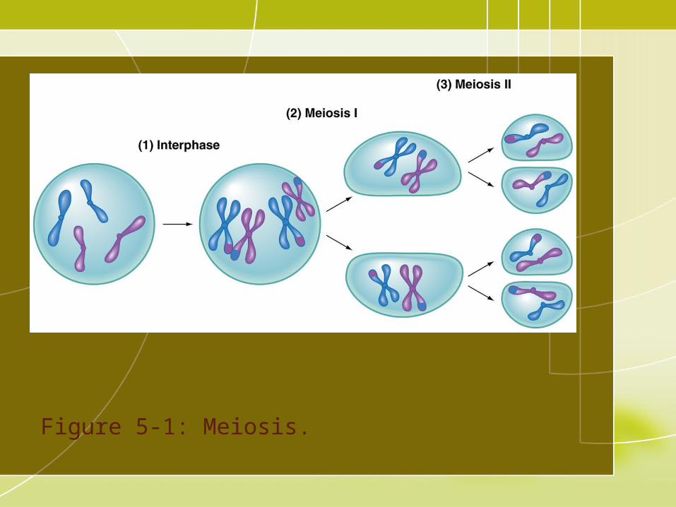
Figure 5-1: Meiosis.
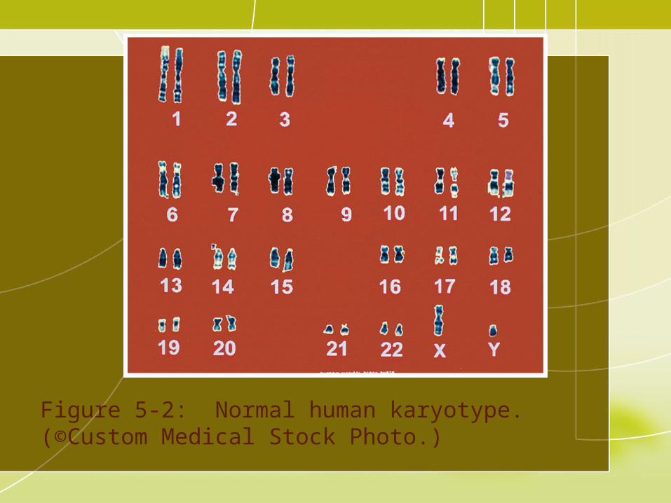
Figure 5-2: Normal human karyotype.(©Custom Medical Stock Photo.)

Table 5-1: Hereditary Disease Locations

Autosomal Dominant Transmission of a dominant allele 50% chance of being affected Disease appears in every generation Males and females equally being affected

Figure 5-3: Transmission of autosomal dominant disorders. (50% chance for an affected child).
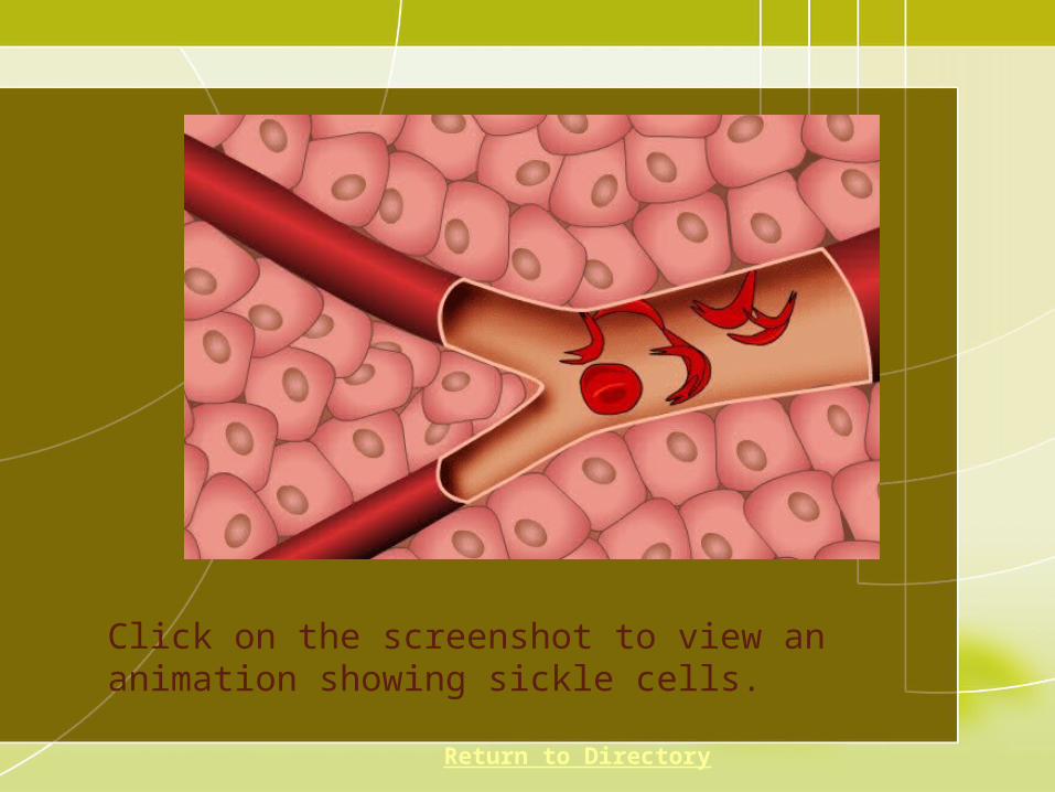
Click on the screenshot to view an animation showing sickle cells.
Return to Directory
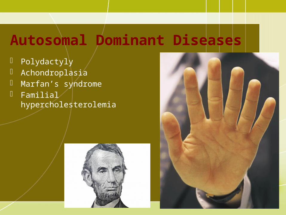
Autosomal Dominant Diseases Polydactyly Achondroplasia Marfan’s syndrome Familial hypercholesterolemia

Figure 5-4: A 12-year old Achondroplastic dwarf. Note the disproportion of the limbs to the trunk, the curvature of the spine, and the prominent buttocks.

Autosomal Recessive Disease manifests when individual is homozygous for
the defective allele Parents are carriers; they do not have the disease Child has a 25% chance of being affected Recessive allele appears more frequently in close
intermarriages

Figure 5-5: Transmission of recessive disorders (25% chance for an affected child).

Autosomal Recessive Diseases Phenylketonuria Galactosemia Sickle cell anemia Tay-Sachs disease Albinism
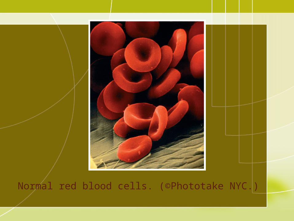
Normal red blood cells. (©Phototake NYC.)

Sickle blood cells. (©Photo Researchers, Inc.)

Figure 5-6: Enzyme block in phenylketonuria (PKU)

Autosomal Recessive Diseases Color blindness: inability to distinguish
colors Hemophilia Fragile X syndrome – a break or
weakness on long arm of X chromosome

Sex-Linked Inheritance Defective gene on X chromosome Defective X on male is unmasked and the trait is
expressed. Female is carrier for the disease; heterozygous Male transmits the defective allele to his daughters.

Figure 5-7: Transmission of sex-linked disorders.
Abnormal Chromosome Diseases Altered number or structure Failure of chromosome to separate during cell division Loss of autosome is usually incompatible with life

Patau Syndrome +13


Down Syndrome Caused by the presence of an extra autosome,
nondisjunction Results in mental retardation and shorter life
expectancy Characteristic appearance: slanted eyes, extra fold of
skin at upper medial corner of the eye, protrusion of the tongue, short nose
Short stature, underdeveloped sex organs

Click on the screenshot to view a video on the topic of Down syndrome.
Return to Directory
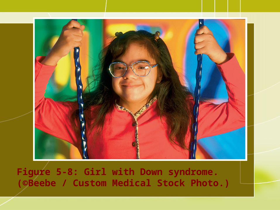
Figure 5-8: Girl with Down syndrome.(©Beebe / Custom Medical Stock Photo.)

Familial Disease Diseases run in families but means of inheritance are
not understood Most likely the effects of several genes working
together Examples: diabetes, allergies, familial polyposis

Cri Du Chat Syndrome Cat-like cry Caused by deletion of part of the short arm of
chromosome 5 Results in an abnormally small head with a deficiency
in cerebral brain tissue Widely spaced eyes and mental retardation

Sex Anomalies Turner’s syndrome: missing sex chromosome Klinefelter’s syndrome: extra sex chromosome Hermaphrodite: has both testes and ovaries Pseudohermaphrodite: has either

Figure 5-9: A 21-year-old patient with Turner’s syndrome. The chest is broad and the nipples are small and pale. Pubic hair is totally lacking.
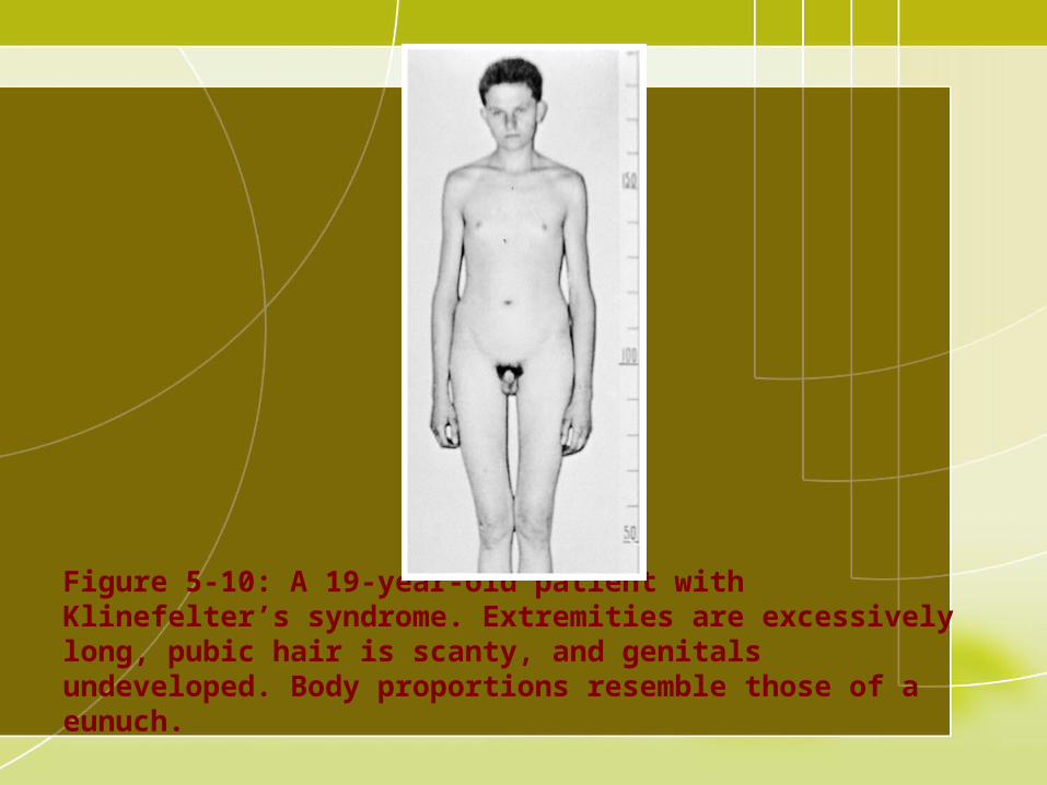
Figure 5-10: A 19-year-old patient with Klinefelter’s syndrome. Extremities are excessively long, pubic hair is scanty, and genitals undeveloped. Body proportions resemble those of a eunuch.
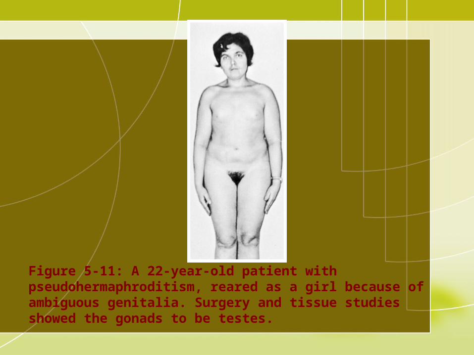
Figure 5-11: A 22-year-old patient with pseudohermaphroditism, reared as a girl because of ambiguous genitalia. Surgery and tissue studies showed the gonads to be testes.