Embed Size (px)
Citation preview

of January 13, 2019.This information is current as
MarkersMonocyte Cytokine Profile and SurfaceInvolves Soluble Factor(s) That Alters Human Glioma-Induced Immunosuppression
Gene M. ShearerE. Coligan, William H. Brooks, Thomas L. Roszman and
JohnDix, Andrew G. Brooks, Naomi Torres, Jon D. Shuman, Jian-Ping Zou, Lorri A. Morford, Claire Chougnet, Amy R.
http://www.jimmunol.org/content/162/8/48821999; 162:4882-4892; ;J Immunol
Referenceshttp://www.jimmunol.org/content/162/8/4882.full#ref-list-1
, 17 of which you can access for free at: cites 57 articlesThis article
average*
4 weeks from acceptance to publicationFast Publication! •
Every submission reviewed by practicing scientistsNo Triage! •
from submission to initial decisionRapid Reviews! 30 days* •
Submit online. ?The JIWhy
Subscriptionhttp://jimmunol.org/subscription
is online at: The Journal of ImmunologyInformation about subscribing to
Permissionshttp://www.aai.org/About/Publications/JI/copyright.htmlSubmit copyright permission requests at:
Email Alertshttp://jimmunol.org/alertsReceive free email-alerts when new articles cite this article. Sign up at:
Print ISSN: 0022-1767 Online ISSN: 1550-6606. Immunologists All rights reserved.Copyright © 1999 by The American Association of1451 Rockville Pike, Suite 650, Rockville, MD 20852The American Association of Immunologists, Inc.,
is published twice each month byThe Journal of Immunology
by guest on January 13, 2019http://w
ww
.jimm
unol.org/D
ownloaded from
by guest on January 13, 2019
http://ww
w.jim
munol.org/
Dow
nloaded from

Human Glioma-Induced Immunosuppression InvolvesSoluble Factor(s) That Alters Monocyte Cytokine Profile andSurface Markers1
Jian-Ping Zou,* Lorri A. Morford, † Claire Chougnet,* Amy R. Dix,† Andrew G. Brooks,‡
Naomi Torres,* Jon D. Shuman,‡ John E. Coligan,‡ William H. Brooks, † Thomas L. Roszman,†
and Gene M. Shearer2*
Patients with gliomas exhibit deficient in vitro and in vivo T cell immune activity, and human glioblastoma culture supernatants(GCS) inhibit in vitro T lymphocyte responses. Because APC are essential for initiating and regulating T cell responses, weinvestigated whether GCS would affect cytokines produced by monocytes and T cells from healthy donors of PBMC. Incubationof PBMC with GCS decreased production of IL-12, IFN-g, and TNF-a, and increased production of IL-6 and IL-10. The GCS-induced changes in IL-12 and IL-10 occurred in monocytes, and involved changes in IL-12 p40 and IL-10 mRNA expression.Incubation with GCS also resulted in reduced expression of MHC class II and of CD80/86 costimulatory molecules on monocytes.The immunosuppressive effects were not the result of IL-6 or TGF-b1 that was detected in GCS. However, it was due to a factor(s)that is resistant to pH extremes, differentially susceptible to temperature, susceptible to trypsin, and has a minimum molecularmass of 40 kDa. Our findings show that glioblastoma-generated factors that are known to suppress T cell responses alter thecytokine profiles of monocytic APC that, in turn, inhibit T cell function. This model indicates that monocytes can serve as anintermediate between tumor-generated immune-suppressive factors and the T cell responses that are suppressed in gliomas.TheJournal of Immunology,1999, 162: 4882–4892.
I t has long been recognized that an impaired cellular immuneresponse is a characteristic of many tumors in both animalmodels and human patients (1, 2). This diminished cellular
immunity is not necessarily limited to reactivity against tumor-specific Ags, but can include unresponsiveness to nontumor Agsand T cell mitogens (3, 4). Cytokine dysfunction appears to con-tribute to tumor-associated immune dysregulation, with decreasesof in vitro IL-2 and/or IFN-g production and increases in IL-4,IL-5, IL-6, and/or IL-10 production. Human tumors in which oneor more of these cytokine changes have been reported includeHodgkin’s lymphomas (4), cervical (5, 6) and ovarian carcinomas(7), melanomas (8), basal and squamous cell carcinomas (9), renalcell carcinomas (10), non-small cell lung cancer (11), and gliomas(12). Tumor-associated immune dysregulation can also be re-flected in T cells at the level of signal transduction, as defects inSTAT5 have been reported in a murine breast tumor model (13),and Janus kinase 3 (Jak3) expression is down-regulated by a sol-uble factor from a human renal cell carcinoma (14).
Human gliomas provide an interesting example of tumor-asso-ciated immune dysfunction. The in vitro responses of T cells from
patients who present with primary gliomas are impaired in theirability to respond in vitro to Ags and T cell mitogens by prolifer-ation and IL-2 production (3, 15, 16). Surgical removal of theprimary tumor can result in restoration of systemic in vitro re-sponses to T cell mitogens, which again declines with recurrenceof the tumor (17). Glioma patients also frequently fail to elicitdelayed skin reactions (15), and patients’ T cells express reducednumbers of high affinity IL-2R (18, 19). One of our laboratoriesrecently reported that T cells from glioma patients exhibit defectsin tyrosine phosphorylation of several proteins, reduced levels ofphospholipase Cg1 and p56lck, as well as reduced mobilization ofcalcium (20). Other studies demonstrated that cultures of glioblas-toma cell lines produce a factor(s) that inhibits Ag- and mitogen-stimulated proliferation and IL-2 production by T cells fromhealthy individuals (21, 22). These findings suggest that one ormore factors contained in glioma culture supernatant (GCS)3 ex-erts immunoregulatory effects on systemic cellular immunity, aswell as at the site of the primary tumor.
Although Th cell proliferation and IL-2 production have beendemonstrated to be defective in glioma patients and in cultures ofPBMC exposed to GCS (21), the possibility that these T cell de-fects have their origin in APC has not been addressed. The pro-duction of IL-12 and/or IL-10 and the stimulatory and costimula-tory molecules that are important for T cell activation are alteredin certain infectious diseases such as leprosy (23) and AIDS (24,25). Increased IL-10 production and mRNA expression have alsobeen reported in several tumors including gliomas (5, 8–11,26–29), and IL-12 has been used to inhibit the growth of murine
*Experimental Immunology Branch, National Cancer Institute, National Institutes ofHealth, Bethesda, MD 20892;†Department of Microbiology and Immunology, Uni-versity of Kentucky Medical Center, University of Kentucky, Lexington, KY 40536;and‡Laboratory of Immunogenetics, National Institute of Allergy and Infectious Dis-ease, National Institutes of Health, Rockville, MD 20852
Received for publication July 30, 1998. Accepted for publication Janaury 19, 1999.
The costs of publication of this article were defrayed in part by the payment of pagecharges. This article must therefore be hereby markedadvertisementin accordancewith 18 U.S.C. Section 1734 solely to indicate this fact.1 This work was supported by the Intramural Research Program, Division of BasicSciences, National Cancer Institute.2 Address correspondence and reprint requests to Dr. Gene M. Shearer, ExperimentalImmunology Branch, Building 10, Room 4B-36, NCI, National Institutes of Health,Bethesda, MD 20892-1360. E-mail address: [email protected]
3 Abbreviations used in this paper: GCS, glioblastoma culture supernatant; CASTA,Candida albicansAg; CGRP, calcitonin gene-related peptide; CNS, central nervoussystem; EIA, enzyme immunoassay; FLU, influenza A virus; SAC,StaphylococcusaureusCowan strain 1; TT, tetanus toxoid.
Copyright © 1999 by The American Association of Immunologists 0022-1767/99/$02.00
by guest on January 13, 2019http://w
ww
.jimm
unol.org/D
ownloaded from

tumors (30, 31). These findings raise the possibility that the de-pressed cellular immune condition associated with certain tumorscontributes to their neoplastic disease and is linked to cytokinedysregulation (2).
In the present study, we investigated whether exposure ofhealthy human blood donors’ PBMC and monocytes to GCSwould result in decreased IL-12 and increased IL-10 productionand mRNA expression. Because MHC class II and CD80/86 ex-pression have been shown to be down-regulated by IL-10 in hu-man and murine in vitro models (32, 33), we also tested for re-duced expression of MHC class II and CD80/86. Finally, we testedwhether in vitro Th response would be generated by a mixture ofautologous T cells and GCS-exposed monocytes. Our results in-dicate that GCS induces down-regulation of IL-12, MHC class II,and CD80/86, and concomitant up-regulation of IL-10 in mono-cytes. We also observed that proliferative responses to recall Agswere abrogated when monocytes were exposed to GCS beforemixing with autologous T cells. These findings suggest that thedefects seen in the Th of patients with gliomas originated as de-fective APC function that, in turn, resulted in aberrant signaling ofT cells and subsequent down-regulation of IL-2 and IFN-g pro-duction and gene expression. No single cytokine that we detectedin GCS has been demonstrated to induce all of these changes inmonocytes. The GCS activity was attributed to a factor(s) that isresistant to pH extremes, differentially susceptible to temperature,susceptible to trypsin, with a minimum molecular mass of approx-imately 40 kDa.
Materials and MethodsIsolation of PBMC
Samples of whole blood were provided for in vitro laboratory studies bythe Transfusion Medicine Department (National Institutes of Health, Be-thesda, MD), under a National Institute of Health Institutional ReviewBoard-approved protocol. The PBMC were separated on Lymphocyte Sep-aration Media (Organon Teknika, Rockville, MD), and resuspended at1.5 3 106 cells/ml in RPMI 1640 (Life Technologies, Rockville, MD),supplemented with 100 U/ml penicillin, 100mg/ml streptomycin, 5mMHEPES buffer, and 2mM glutamine (National Institutes of Health MediaUnit, Bethesda, MD), and 5% human AB1 serum (Sigma, St. Louis, MO).
Production of GCS from different glioblastoma cell lines
The SNB-19 and U251 glioma cell lines were used for production of GCS.Two independently carried SNB-19 lines were studied: Both originatedfrom Dr. Paul Kornblith (University of Pittsburgh, Pittsburgh, PA). Onehas been carried for several years by the University of Kentucky laboratory(Lexington, KY); the other was recently obtained as a cryopreserved sam-ple from American Type Culture Collection (ATCC, Manassas, VA). TheU251 line (34) is carried in National Cancer Institute laboratory. Thesecells were maintained in RPMI 1640 culture media containing 5% FCS,100 U/ml penicillin, 100mg/ml streptomycin, and 10mM HEPES buffer ina humidified, 37°C, 5% CO2 incubator. Cells were passaged at 4- to 7-dayintervals using 0.25% trypsin (Life Technologies, Grand Island, NY) inPBS (pH 7.3–7.4). Supernatants were harvested from all of the glioblas-toma lines after 4 to 7 days of culture in 5% FCS/RPMI 1640 medium.GCS was also generated by culturing the U251 cell line for 3 days inconditioned Cellgro Complete Serum Free Media (Mediatech, Herndon,VA) or FCS-free RPMI 1640 to obtain GCS samples for factor purificationstudies. Culture supernatants of these glioblastoma lines were tested di-rectly for factor activity by production of IL-12, IL-10, IFN-g, and T cellproliferation (see below). Supernatants from one of the SNB-19 cell linesgrown to confluency were concentrated (38–89-fold) on a Minitan tan-gential flow concentrator using 100-kDa molecular mass cutoff, low pro-tein binding, and regenerated cellulose filters to collect GCS. Followingconcentration, the GCS was filtered through 0.22-mm filters (Costar, Cam-bridge, MA) and stored at280°C until needed. In most experiments, GCSpreparations were used at a final 1/20 dilution. Supernatants from the otherSNB-19 and the U251 cell lines were either concentrated 20–40-fold usinga differential molecular mass cutoff Centricon Plus-80 Centrifugal FilterDevice (Millipore, Bedford, MA), or were tested without concentration.Culture supernatants exhibited activity, irrespective of whether they had
been concentrated. We verified that different preparations of GCS do notexert a toxic effect on PBMC cultured for 7 days.
As controls for the glioblastoma lines, we tested supernatants of ovariancarcinoma A2780, A2780/CP (35), and National Institute of Health-ovcar-3; the T2, U937, and K562 lymphoma lines; the prostate carcinomaPC-3 (National Institutes of Health-ovcar-3, T2, U973, K562, and PC-3were obtained from ATCC); and two EBV-transformed lymphoma celllines generated in our laboratory.
T cell function assay
Different preparations of GCS were tested for inhibition of T cell functionby culturing 1.53 106 PBMC/ml, or 13 106 T cells/ml with 0.53 106
autologous monocytes/ml in 200ml of culture media in 96-well flat-bottomculture plates (Costar) in a humidified, 37°C, 7% CO2 incubator. The cul-tures were either unstimulated, or were stimulated with PHA-M (1/80 di-lution) (Life Technologies) or a pool of recall Ags consisting of: influenzaA virus (FLU) (A/Bangkok/RX173, H3N2) (final dilution of 1/800); teta-nus toxoid (TT) (Connaught Laboratories, Swiftwater, PA) (final dilutionof 1/800); andCandida albicansAg (CASTA) (Greer Laboratories, Lenoir,NC) (10mg/ml). The cultures were pulsed with [3H]thymidine on day 2 forPHA and day 6 for recall Ags, harvested 20 h later using a Basic 96Harvester (Skatron Instruments, Sterling, VA), and counted in ab-spec-trometer (Wallac, Gaithersburg, MD).
Enrichment of monocytes and T cells
Enriched monocytes and T cells were obtained from elutriated lymphocyte-depleted and monocyte-depleted populations isolated from PBMC ofhealthy blood donors. Remixing experiments were performed using autol-ogous depleted and enriched cell populations.
To obtain enriched monocytes, lymphocyte-depleted PBMC were incu-bated on ice for 30 min with an Ab mixture consisting of mouse anti-human CD3, CD16, and CD19 mAb (IgG) (PharMingen, San Diego, CA),at 5mg of each mAb per 103 106 cells in 100ml PBS containing 10% FBS(PBS/FBS). The cells were washed three times in PBS/FBS, the cell pelletwas resuspended in PBS/FBS in the presence of Dynabeads M280 sheepanti-mouse IgG (Dynal, Oslo, Norway) (10 beads/cell), and the mixturewas incubated on ice for 30 min. The cell-bead mixture was exposed to amagnet through three cycles of magnetic separation and washing of theunattached cells. This procedure resulted in enrichment of monocytes togreater than 90% CD141 cells, determined by flow cytometry. The en-riched monocytes were tested by flow cytometry for the presence ofCD831 cells, a marker of mature dendritic cells (36), and none weredetected.
To obtain enriched T cells, monocyte-depleted cells were incubatedwith the Lympho-Quik-T Isolation Reagent (One Lambda, Canoga Park,CA), which depletes of all cell types except T cells by Ab-mediated, com-plement-dependent lysis (37).
Cytokine production and detection
The production of cytokines by PBMC, enriched monocytes, or monocytesplus autologous T cells was assessed by culturing 33 106 PBMC, 13 106
monocytes, or 13 106 monocytes plus 23 106 T cells in 2 ml of culturemedia in 24-well plates (Costar), respectively, in a humidified, 37°C, 7%CO2 incubator. Cells were either unstimulated or were stimulated withStaphylococcus aureusCowan strain 1 (SAC) (0.01%) (Pansorbin, Calbio-chem-Behring, La Jolla, CA). Culture supernatants were harvested after24 h and stored at280°C.
The IL-12 p70 heterodimer production was assessed by ELISA fromR&D (Minneapolis, MN). Total IL-12 p40, IL-2, IL-4, and IL-6 produc-tions were detected by ELISA from Genzyme (Cambridge, MA). IL-10,IL-5, IFN-g, and TNF-a productions were assessed in the supernatants of24-h SAC-stimulated cultures, using PharMingen capture and detectionAbs, as previously described (37). The limit for detection of these cyto-kines was in the range of 5–20 pg/ml.
Detection of IL-12 and IL-10 mRNA
Expression of hypoxanthine phosphoribosyltransferase (HPRT), IL-10, andIL-12 p40 mRNA was assessed on PBMC stimulated with SAC for 6 h,using a semiquantitative RT-PCR protocol, as previously described (37).
Detection of intracellular cytokines
PBMC were cultured for 6 h with or without stimulation in Teflon vials(Pierce Chemicals, Rockford, IL) in a 37°C, 7% CO2 incubator; thenBrefeldin A (Sigma) (5mg/ml) was added for an additional 18 h of incu-bation. In some experiments, PBMC were cultured without or with 10mg/ml Brefeldin A in 5% human AB1 serum, RPMI 1640 medium for 1
4883The Journal of Immunology
by guest on January 13, 2019http://w
ww
.jimm
unol.org/D
ownloaded from

day. Cell viability was tested by trypan blue exclusion or propidium iodidestaining. The cells also were analyzed by FACS with anti-CD3 and CD14staining. No differences were obtained in cell viability (.90%) in either theCD31 or CD141 populations in the presence or absence of Brefeldin A.Cells were harvested and washed in a staining buffer (PBS containing 1%FBS and 0.1% w/v sodium azide), preincubated with human IgG at 4°C for30 min to block FcR. The cells were then stained at 4°C for 30 min withfluorochrome-conjugated mAb specific for a cell surface Ag such as CD14and CD3 (PharMingen, San Diego, CA). The cells were subsequentlywashed with staining buffer, pelleted by centrifugation, and fixed in 500mlof fixing buffer (4% w/v paraformaldehyde in PBS) at 4°C for 30 min orovernight. The cells were washed in the staining buffer, pelleted by cen-trifugation, and resuspended in 100ml of permeabilization buffer (PBScontaining 1% FBS, 0.1% w/v sodium azide, 0.1% w/v saponin). The cellswere incubated for 30 min at 4°C with 0.5mg fluorochrome-conjugatedanti-cytokine Abs (anti-IL-12 p40 and p70, IL-10, IL-6, IFN-g, and TNF-afrom PharMingen). The cells were then washed twice in permeabilizationbuffer, resuspended in staining buffer, and analyzed by flow cytometryusing a FACScan (Becton Dickinson, San Jose, CA). The cells were gatedon monocytes or lymphocytes based on forward and side light scatter. Insome experiments, the binding of fluorochrome-conjugated anti-cytokinemAb was blocked by preincubation of the conjugated mAb with excessrecombinant cytokine (IL-12 p40 and IL-10; PharMingen).
Immunoprecipitation
To remove IL-6, TGF-b, and CGRP, GCS was diluted sevenfold in PBS.Anti-IL-6 (clone MQ2-13A5 rat IgG1, final concentration of 5mg/ml),anti-TGF-b mAb (mouse IgG1, final concentration of 10mg/ml), and rab-bit anti-human CGRP serum (final 1/120 dilution) were added singly or incombination. The GCS and Ab mixture was incubated overnight at 4°Cunder rotating conditions. An excess of GammaBind G Sepharose (Phar-macia Biotech, Piscataway, NJ) was added for 10 h, and the mixture wascentrifuged for 10 min at 20003 g. An excess of protein A-Sepharose(Pharmacia) was added to the supernatant, which was incubated overnightunder rotating conditions at 4°C. The mixture was centrifuged again, andthe supernatant was sterilized by passing through a 0.22-mm filter, thentested for ability to suppress the Th function and induce the cytokinechanges seen with the original GCS. We verified by specific ELISA orenzyme immunoassay (EIA) that the immunoprecipitation removed all de-tectable IL-6, TGF-b, and CGRP.
Ion exchange and gel filtration columns
The supernatants were harvested from the U251 glioblastoma line after 3days of culture in conditioned FCS-free RPMI 1640 media. The GCS sam-ples for factor purification studies were tested for binding to CM, Q, SP,and DEAE Sepharose Fast Flow Columns (Pharmacia Biotech). The un-bound fraction and eluted fractions were tested for GCS activity. Thebound fractions that contained GCS activity were fractionated on the Su-perdex 75 and Superdex 200 columns (Pharmacia Biotech). To determinethe molecular mass of the active factor(s), 25–50 fractions were each testedfor GCS activity by analysis of IL-12, IL-10, IFN-g production, and PHA-stimulation response of PBMC, and compared with the unfractionatedGCS, as described above.
Reagents
The additional following reagents were used in this study: anti-humanIL-10 neutralizing mAb (clone JES 319 F11; DNAX, Palo Alto, CA);anti-human IL-10R mAb (clone 37607.11; R&D); anti-human IL-6 neu-tralizing mAb (clone MQ2-13A5; PharMingen); paraformaldehyde and sa-ponin (Sigma); TGF-b1 human ELISA kit and PGE2 EIA kit (Biotrak,Amersham, Arlington Heights, IL); Ultrapure natural TGF-b1, humanrTGF-b2, and mouse monoclonal anti-human TGF-b1, TGF-b2, TGF-b3neutralizing Ab (Genzyme); human CGRP, rabbit anti-human CGRP se-rum, and high sensitivity EIA kit (Pennisula Laboratories Europe, Belmont,CA); and insoluble trypsin (Sigma).
ResultsInhibition of T cell proliferative responses by GCS
To test whether the preparations of GCS generated by the SNB-19glioblastoma cell lines inhibited in vitro T cell responses to a mi-togen and recall Ags, PBMC from healthy individuals were stim-ulated with PHA (Fig. 1A) or with a mixture of FLU, TT, andCASTA (Fig. 1B) in the absence or presence of GCS. The resultsindicate that GCS inhibited proliferative responses to both stimuli
in a dose-dependent manner at dilution ranging from 1/20 to1/20,000. Therefore, the GCS produced by the tumor cell linestrongly inhibited T lymphocyte responses to a T cell mitogen andto Th-dependent recall Ags that require intact APC function. Asnegative controls, we found that culture supernatants from three ofseven tumor lines and the two laboratory-generated EBV-trans-formed cell lines did not inhibit T cell proliferation or inducechanges in IL-12 and IL-10 production when added to PBMC (datanot shown).
Effect of GCS on cytokine production
Because SAC is a strong stimulator of IL-12 and IL-10 productionby monocytes, we tested different dilutions of GCS on SAC-stim-ulated IL-12 and IL-10 production in 24-h cultures of PBMC. Thedata in Fig. 2 demonstrate that GCS decreased IL-12 and increasedIL-10 in a dose-dependent way.
To determine the kinetics of cytokine production, GCS wasadded to PBMC at the time of SAC stimulation, and the cultureswere carried for 3, 6, 12, and 24 h. In addition to IL-12 and IL-10,we tested for other SAC-stimulated monokines and cytokines, in-cluding IFN-g, IL-6, and TNF-a. PBMC were also preincubatedwith GCS for 1 h, the GCS was washed out, and the treated PBMCwere stimulated with SAC for 3, 6, 12, and 24 h (Fig. 3). Thekinetics of the response of control cultures, either unstimulated orstimulated but not incubated with GCS, was also followed. Theproduction of IL-12 p70 and p40, as well as IFN-g was greatlyreduced by addition of GCS to SAC-stimulated PBMC (Fig. 3,A,B, andD). In contrast, GCS increased SAC-stimulated IL-10 pro-duction (Fig. 3C). IL-6 production was appreciably increased byGCS or SAC alone, and the combination of GCS and SAC inducedan additional increase (Fig. 3E). SAC-stimulated TNF-a produc-tion was reduced approximately twofold by GCS (Fig. 3F). Datasimilar to those shown in Fig. 3 were obtained in six independent
FIGURE 1. GCS inhibits PHA and recall Ag-induced proliferation ofPBMC in a dose-dependent manner. Effect of different dilutions of GCS onT lymphocyte proliferative responses to PHA (A) and a pool of recall Ags(B) consisting of FLU, TT, and CASTA. The PHA-stimulated cultureswere pulsed with [3H]thymidine after 2 days, and the recall Ag-stimulatedcultures were pulsed after 6 days of culture. Four experiments were per-formed with PBMC from four donors each (n 5 3, cpm mean per minute6SD), and the results shown are from one representative experiment.
4884 MONOCYTE DYSFUNCTION INDUCED BY GLIOMA
by guest on January 13, 2019http://w
ww
.jimm
unol.org/D
ownloaded from

experiments. We also observed that 48-h cultures of GCS and SACyielded results indistinguishable for the 24-h cultures (data notshown). These results indicate that GCS can rapidly induce a de-crease in the production of IL-12 and IFN-g and a concomitantincrease in IL-6 and IL-10 production. The 1-h preincubation ofPBMC with GCS before SAC stimulation induced changes in cy-tokine profiles that were similar to those observed when PBMCwere exposed to GCS and SAC simultaneously.
Culture supernatants generated by the two SNB-19 glioblastomalines as well as by the U251 glioblastoma line all induced de-
creased IL-12 and IFN-g, and increased IL-10 production, and alsoabolished PHA-stimulated T cell proliferation (Table I). Culturesupernatants from four of seven control tumor cell lines exhibitedweak GCS-like activity (A2780, National Institute of Health-ovcar-3, T2, PC-3); as noted above, supernatants from the threeother tumor lines did not show any GCS-like activity (A2780/CP,U937, K562).
The results of additional kinetic experiments in which GCS waspreincubated with PBMC for different time intervals before SACstimulation are summarized in Table II. Incubation of GCS withPBMC for as little as 3 min resulted in SAC-stimulated decreasedIL-12 p40 and IFN-g and increased IL-10 production. Preincuba-tion with GCS for 1 h was as effective as maintaining GCS in thecultures with SAC for 24 h.
Immunoregulatory factors contained in GCS
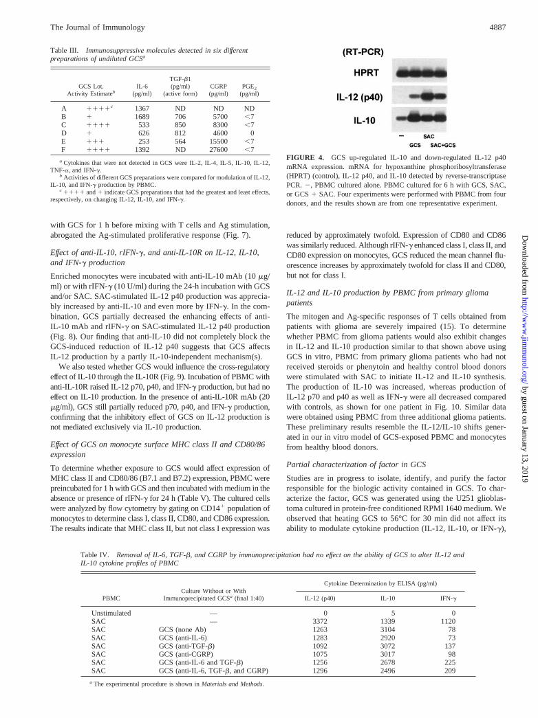
To identify immunoregulatory factors that might be contained inGCS, we tested six different lots of supernatants collected fromone of the SNB-19 glioblastoma cell lines that had been shown toinhibit in vitro T cell proliferation. The supernatants were found tocontain IL-6, TGF-b1, CGRP (38), and very low levels (below 7pg/ml) of PGE2 (Table III), but not detectable levels of IL-4, IL-10, IL-12, TNF-a, or IFN-g (data not shown). The levels of PGE2
detected were below those reported to reduce IL-12 production byhuman PBMC or dendritic cells (39, 40). Based on these data,three types of experiments were performed to determine whetherthe changes in cytokine production induced by GCS could be at-tributed to any of these immunosuppressive factors.
First, we tested whether the addition of exogenous IL-6, TGF-b1, TGF-b2, or CGRP to PBMC, either singly or in combination,would decrease IL-12 and/or increase IL-10 production. We addedIL-6 (10 ng/ml to 10 pg/ml), TGF-b1 or TGF-b2 (10 ng/ml to 10pg/ml), or CGRP (1mg to 10 pg) to SAC-stimulated PBMC, aswell as the combination of IL-6 (5 ng/ml), TGF-b1 (1 ng/ml),TGF-b2 (1 ng/ml), and CGRP (50 ng/ml). Under none of theabove conditions were changes detected in IL-12 or IL-10 produc-tion (data not shown). Second, treatment of GCS with neutralizingAbs against IL-6, CGRP, TGF-b1, TGF-b2, and TGF-b3 had noeffect on its ability to decrease IL-12 and IFN-g production and toincrease IL-10 production by PBMC. Third, we immunoprecipi-tated IL-6, TGF-b, and CGRP, either singly or in combination,with the same Abs used for inhibition. The GCS treated in this waydid not lose any of its ability to decrease IL-12 production. Onlyslight reductions were observed in ability to increase IL-10 anddecrease IFN-g by combination immunoprecipitation of IL-6 and
FIGURE 2. GCS affects SAC-stimulated IL-12and IL-10 production by PBMC in a dose-depen-dent manner. Effect of 10-fold changes in concen-tration of GCS on IL-12 (A) and IL-10 (B) pro-duction by SAC-stimulated PBMC for 24 h.Production of cytokines was measured by ELISA.Two experiments were performed with PBMCfrom two donors each (n5 2), and the resultsshown are from one representative experiment. #,Below detectable level.
FIGURE 3. GCS can rapidly induce cytokine production changes inSAC-stimulated PBMC. Effect of SAC and/or GCS on the kinetics of pro-duction of the cytokines indicated in the panels. See key for details ofmaterials contained in the cultures.21 h (hour) indicates the group thatwas preincubated with GCS for 1 h before washout and stimulation withSAC. GCS were used as final 1/20 dilution. Two experiments were per-formed with PBMC from two donors each (n 5 2), and the results shownare from one representative experiment.
4885The Journal of Immunology
by guest on January 13, 2019http://w
ww
.jimm
unol.org/D
ownloaded from

TGF-b (Table IV). Taken together, these results do not support theconclusion that the cytokine dysregulation induced by GCS is theresult of IL-6, TGF-b, or CGRP, acting singly or in combination.
Effect of GCS on IL-12 p40 and IL-10 mRNA expression
To determine whether GCS also affected IL-12 and IL-10 mRNA,expression of IL-12 p40 and IL-10 mRNA was analyzed in PBMCthat were incubated with SAC, GCS, and SAC1 GCS. The resultsof one representative of four independent experiments are shownin Fig. 4 for IL-12 p40 and IL-10. Incubation with GCS resulted ina modest increase in IL-12 p40 mRNA, and in a large increase inIL-10 mRNA. As previously reported (25), SAC stimulation aloneresulted in expression of IL-12 p40 and of IL-10 mRNA. Incuba-tion with GCS 1 SAC increased expression of IL-10 messageabove SAC alone, and decreased IL-12 p40 expression comparedwith SAC alone. These data are in agreement with the regulationof IL-12 and IL-10 production by GCS.
GCS induced changes in monocyte production of IL-12 andIL-10
To determine whether monocytes contained in the PBMC wereresponsible for the production of IL-12 and IL-10, 24-h SAC-stim-ulated PBMC were gated for CD141 and CD31 cells and stainedwith anti-IL-12 and anti-IL-10 Abs. IL-12 and IL-10 were detectedonly in the CD141 population. PBMC incubated alone or withGCS, SAC, or SAC1 GCS were used for isotype control staining(data not shown). The data presented in Fig. 5 illustrate the IL-12and IL-10 intracellular staining patterns using the same conditions.Incubation of PBMC with GCS indicated that 3.4% of the mono-cytes stained for intracellular IL-10, but only 0.3% stained forIL-12. Stimulation of PBMC with SAC alone resulted in the stain-ing of 8.1% for IL-10, 5.7% for IL-12, and 1.5% for both. ThePBMC incubated with SAC1 GCS resulted in a skewing towardIL-10-producing cells, as 13% stained for IL-10, 1.9% stained forIL-12, and 1.6% stained for both cytokines. Similar results wereobtained in four repetitive experiments. In contrast to the cells
gated for lymphocytes, no T cells were found that produced IL-10or IL-12 after a 24-h incubation with GCS, SAC, or SAC1 GCS(data not shown).
Effect of GCS on enriched SAC-stimulated monocytes
To determine whether incubation with GCS also affected IL-12p70, p40, and IL-10 produced by enriched monocytes, the follow-ing experiments were performed. The monocytes were enrichedfrom PBMC by elutriation, followed by negative selection of T, B,and NK cells. The enriched monocytes (shown.90% CD141, butundetectable,1% CD31, CD191, CD1a1, and CD161) werestimulated with SAC for 24 h in the absence or presence of GCS.The results indicate that GCS abrogated IL-12 production (Fig. 6A)and increased IL-10 production (Fig. 6B). GCS also decreasedSAC-stimulated IL-12 p40 production by twofold (data notshown). These results indicate that GCS can also affect IL-12 andIL-10 production by enriched monocytes in the absence of othercell types.
To determine whether T cells would affect SAC- and GCS-reg-ulated monokine production, we added purified autologous T cells(.95% CD31) to SAC-stimulated monocytes. The T cells en-hanced IL-12 fourfold, but addition of GCS greatly reduced IL-12production (Fig. 6A). Addition of autologous T cells increasedIL-10 production by twofold above monocytes alone, and additionof GCS further increased IL-10 production (Fig. 6B). We alsoassessed SAC-stimulated IFN-g production in the same cultures(Fig. 6C), and demonstrated that without T cells, this cytokine wasnot produced. Addition of T cells resulted in IFN-g production,which was reduced approximately fourfold by GCS.
To determine whether the observed GCS-induced changes inmonokine production would be reflected in Th cell function, amixture of autologous monocytes and T cells was stimulated withthe recall Ag mixture of FLU, TT, and CASTA, and thymidineincorporation measured 6 days later. Similar to the data obtainedfor IL-12 and IL-10 production, addition of GCS to cultures ofmonocytes during Ag stimulation, or preincubation of monocytes
Table I. Comparison of effects of culture supernatants from different glioblastoma lines on cytokine production and T cellproliferation
Culture Conditions
Cytokine Production of SAC-Stimulated PBMC (pg/ml)PHA-Stimulated PBMC[3H]TdR uptake (cpm)IL-12 (p70) IL-10 IFN-g
PBMC No GCS 64.9 540.4 9649.0 63917.0PBMC SNB-19b 1.4 1337.2 483.4 2683.0PBMC SNB-19c 14.3 1724.8 3514.8 16321.2PBMC U251d 6.2 1497.5 2292.6 6549.8
a A representative experiment from 10 or more experiments performed.b Concentrated SNB-19 supernatant from the University of Kentucky laboratory; final dilution of 1/20.c Unconcentrated SNB-19 supernatant from the NCI laboratory; final dilution of 1/2.d Unconcentrated U251 supernatant; final dilution of 1/2.
Table II. Effect of incubation of PBMC with GCS for different time intervals on SAC-stimulated IL-12, IL-10, and IFN-g production
PBMC Were Preincubatedin 37°C for 1 h
Content of24 h Cultured
Cytokine Determination by ELISA (pg/ml)
IL-12 (p70) IL-12 (p40) IL-10 IFN-g
PBMC SAC 34.36 8.6 48706 476 9326 189 33276 719PBMC SAC1GCS 2a . 10c 23.46 0.3 1b2.26 0.2 24.26 0.5Preincubation with GCS for 1 h SAC 2 . 10c 23.66 0.2 13.56 0.6 25.96 1.1Preincubation with GCS for 15–30 min SAC 2 . 10c 23.46 0.3 13.06 0.4 23.36 0.5Preincubation with GCS for 3–10 min SAC 26.36 1.1 21.86 0.1 12.56 0.2 22.86 0.4
a,b The fold increase (1) or fold decrease (2) change from SAC-stimulated PBMC based on the mean of four independent experiments (SEM).c Below detectable level. GCS was used at final dilution of 1/20 in the culture and preincubation system.
4886 MONOCYTE DYSFUNCTION INDUCED BY GLIOMA
by guest on January 13, 2019http://w
ww
.jimm
unol.org/D
ownloaded from
with GCS for 1 h before mixing with T cells and Ag stimulation,abrogated the Ag-stimulated proliferative response (Fig. 7).
Effect of anti-IL-10, rIFN-g, and anti-IL-10R on IL-12, IL-10,and IFN-g production
Enriched monocytes were incubated with anti-IL-10 mAb (10mg/ml) or with rIFN-g (10 U/ml) during the 24-h incubation with GCSand/or SAC. SAC-stimulated IL-12 p40 production was apprecia-bly increased by anti-IL-10 and even more by IFN-g. In the com-bination, GCS partially decreased the enhancing effects of anti-IL-10 mAb and rIFN-g on SAC-stimulated IL-12 p40 production(Fig. 8). Our finding that anti-IL-10 did not completely block theGCS-induced reduction of IL-12 p40 suggests that GCS affectsIL-12 production by a partly IL-10-independent mechanism(s).
We also tested whether GCS would influence the cross-regulatoryeffect of IL-10 through the IL-10R (Fig. 9). Incubation of PBMC withanti-IL-10R raised IL-12 p70, p40, and IFN-g production, but had noeffect on IL-10 production. In the presence of anti-IL-10R mAb (20mg/ml), GCS still partially reduced p70, p40, and IFN-g production,confirming that the inhibitory effect of GCS on IL-12 production isnot mediated exclusively via IL-10 production.
Effect of GCS on monocyte surface MHC class II and CD80/86expression
To determine whether exposure to GCS would affect expression ofMHC class II and CD80/86 (B7.1 and B7.2) expression, PBMC werepreincubated for 1 h with GCS and then incubated with medium in theabsence or presence of rIFN-g for 24 h (Table V). The cultured cellswere analyzed by flow cytometry by gating on CD141 population ofmonocytes to determine class I, class II, CD80, and CD86 expression.The results indicate that MHC class II, but not class I expression was
reduced by approximately twofold. Expression of CD80 and CD86was similarly reduced. Although rIFN-g enhanced class I, class II, andCD80 expression on monocytes, GCS reduced the mean channel flu-orescence increases by approximately twofold for class II and CD80,but not for class I.
IL-12 and IL-10 production by PBMC from primary gliomapatients
The mitogen and Ag-specific responses of T cells obtained frompatients with glioma are severely impaired (15). To determinewhether PBMC from glioma patients would also exhibit changesin IL-12 and IL-10 production similar to that shown above usingGCS in vitro, PBMC from primary glioma patients who had notreceived steroids or phenytoin and healthy control blood donorswere stimulated with SAC to initiate IL-12 and IL-10 synthesis.The production of IL-10 was increased, whereas production ofIL-12 p70 and p40 as well as IFN-g were all decreased comparedwith controls, as shown for one patient in Fig. 10. Similar datawere obtained using PBMC from three additional glioma patients.These preliminary results resemble the IL-12/IL-10 shifts gener-ated in our in vitro model of GCS-exposed PBMC and monocytesfrom healthy blood donors.
Partial characterization of factor in GCS
Studies are in progress to isolate, identify, and purify the factorresponsible for the biologic activity contained in GCS. To char-acterize the factor, GCS was generated using the U251 glioblas-toma cultured in protein-free conditioned RPMI 1640 medium. Weobserved that heating GCS to 56°C for 30 min did not affect itsability to modulate cytokine production (IL-12, IL-10, or IFN-g),
Table III. Immunosuppressive molecules detected in six differentpreparations of undiluted GCSa
GCS Lot.Activity Estimateb
IL-6(pg/ml)
TGF-b1(pg/ml)
(active form)CGRP(pg/ml)
PGE2
(pg/ml)
A 1111c 1367 ND ND NDB 1 1689 706 5700 ,7C 1111 533 850 8300 ,7D 1 626 812 4600 0E 111 253 564 15500 ,7F 1111 1392 ND 27600 ,7
a Cytokines that were not detected in GCS were IL-2, IL-4, IL-5, IL-10, IL-12,TNF-a, and IFN-g.
b Activities of different GCS preparations were compared for modulation of IL-12,IL-10, and IFN-g production by PBMC.
c 1111 and1 indicate GCS preparations that had the greatest and least effects,respectively, on changing IL-12, IL-10, and IFN-g.
Table IV. Removal of IL-6, TGF-b, and CGRP by immunoprecipitation had no effect on the ability of GCS to alter IL-12 andIL-10 cytokine profiles of PBMC
PBMCCulture Without or With
Immunoprecipitated GCSa (final 1:40)
Cytokine Determination by ELISA (pg/ml)
IL-12 (p40) IL-10 IFN-g
Unstimulated — 0 5 0SAC — 3372 1339 1120SAC GCS (none Ab) 1263 3104 78SAC GCS (anti-IL-6) 1283 2920 73SAC GCS (anti-TGF-b) 1092 3072 137SAC GCS (anti-CGRP) 1075 3017 98SAC GCS (anti-IL-6 and TGF-b) 1256 2678 225SAC GCS (anti-IL-6, TGF-b, and CGRP) 1296 2496 209
a The experimental procedure is shown inMaterials and Methods.
FIGURE 4. GCS up-regulated IL-10 and down-regulated IL-12 p40mRNA expression. mRNA for hypoxanthine phosphoribosyltransferase(HPRT) (control), IL-12 p40, and IL-10 detected by reverse-transcriptasePCR.2, PBMC cultured alone. PBMC cultured for 6 h with GCS, SAC,or GCS1 SAC. Four experiments were performed with PBMC from fourdonors, and the results shown are from one representative experiment.
4887The Journal of Immunology
by guest on January 13, 2019http://w
ww
.jimm
unol.org/D
ownloaded from

but did reduce its ability to abrogate PHA-stimulated T cell pro-liferation (Table VI). Heating GCS to 100°C for 30 min abrogatedthe activity responsible for modulating cytokine production. Ex-posure to pH extremes of 2 and 11 for 15 min resulted in retentionof 40–60% of the GCS activity for altering cytokine production,but had no affect on its ability to abolish PHA-stimulated T cellproliferation. All functional activity of GCS was lost by exposureto insoluble trypsin for 30 min at room temperature. The activityof GCS for all of the above parameters was retained by anion-exchange columns (Q and DEAE Sepharose), but not by cation-exchange columns (SP and CM Sepharose). Preliminary charac-terization by Superdex 200 gel filtration column experiments of theanion-exchange-retained fractions indicates a minimum molecularmass of approximately 40 kDa, although GCS functional activityfor changes in all three cytokines and T cell proliferation wasobserved over a wide molecular mass range up to approximately150 kDa (Table VI). This range of activity suggests various statesof aggregate formation, and is consistent with molecular sizing filterexperiments in which functional activity was present, but reduced bya 100-kDa filter (data not shown). Each condition that decreasedIL-12 also decreased IFN-g and increased IL-10 (Table VI).
DiscussionThe results of the present study suggest that the T cell defect(s)reported in glioma patients, and in PBMC cultured with GCS is theresult of negative response signals passed to the T cells by mono-cytes that have been modified upon exposure to this factor(s). Our
FIGURE 5. GCS increased IL-10-producing monocytes and decreasedIL-12-producing monocytes. Two-color cytometric analysis of intracellularstaining of monocytes for IL-12 and IL-10. PBMC were gated for mono-cytes and stained with anti-IL-12 FITC and anti-IL-10 phycoerythrin.PBMC were incubated alone (control) or with GCS, SAC, or SAC1 GCS.Four experiments were performed with PBMC from different donors, andthe results shown are from one representative experiment. Statistical anal-ysis of the four experiments indicates IL-12-producing cells (mean6 STD,%) in unstimulated control (0.496 0.33) and with GCS (0.376 0.43),p 50.0848; with SAC (8.536 3.06) and with SAC1 GCS (2.496 1.36),p 50.0072; IL-10-producing cells in unstimulated control (0.616 0.60) andwith GCS (3.606 0.38),p 5 0.0002; with SAC (8.036 2.53) and withSAC 1 GCS (12.326 3.87),p 5 0.0188.
FIGURE 6. The presence of T cells affects IL-12 and IL-10 productionby monocytes. Production of IL-12 p70 (A), IL-10 (B), and IFN-g (C) bySAC-stimulated monocytes with/without T cells: monocytes (M); mono-cytes1 GCS (M1 GCS); monocytes1 T cells (M 1 T); and monocytes1 T cells1 GCS (M1 T 1 GCS). Four experiments were performed withPBMC from four donors each (n 5 2), and the results shown are from onerepresentative experiment. #, Below detectable level.
FIGURE 7. GCS-pretreated monocytes lose APC function in recall Ag-induced T cell proliferative response. Recall Ag pool-stimulated T cell prolif-eration with autologous monocytes in the absence or presence of GCS (A); Tcells with GCS-pretreated monocytes (after preincubation with GCS for 1 h)in the absence or presence of GCS (B). Three experiments were performedwith purified T cells and autologous purified monocytes from four donors each(n 5 3), and the results shown are from one representative experiment.
4888 MONOCYTE DYSFUNCTION INDUCED BY GLIOMA
by guest on January 13, 2019http://w
ww
.jimm
unol.org/D
ownloaded from

findings also suggest that these monocytes with increased IL-10expression and decreased IL-12, class II, and CD80/86 expressionrepresent a primary cellular defect in glioma-associated immunedysfunction. Thus, these monocytes may serve as the intermediarybetween the immunosuppressive factor(s) produced by the tumorcells and the T cells that are affected. The GCS-induced changesthat we observed in the monocytes are those that initiate signalsessential for T cell activation. Using flow cytometry and intracel-lular staining, we demonstrated that the IL-12 and IL-10 detectedwere produced by monocytes and not by T cells, and we verifiedthese GCS-induced changes in IL-12 and IL-10 using purifiedmonocytes. Other cytokines that were affected by GCS were IL-6(increased), IFN-g (decreased), and TNF-a (decreased). The dem-onstration that exposure of monocytes to GCS before mixing withautologous T cells and Ag abolished proliferative responses indi-cated that the changes in monocyte IL-12, IL-10, MHC class II,and/or CD80/86 expression affected Th function. Importantly, the
data also show that PBMC from glioma patients exhibit a similarshift in IL-12 and IL-10 production.
It has been suggested that the immunosuppressive effect of gli-oma culture supernatant is the result from the synthesis of TGF-b(41, 42). However, our mAb inhibition and immunoprecipitationdata do not support the hypothesis that the factor responsible forrapidly inducing shifts in IL-12 and IL-10 expression/productionand down-regulation of MHC class II and CD80/86 was TGF-b,IL-6, CGRP, or a combination of these immunoregulatory mole-cules. It remains to be determined whether this glioma-generatedfactor, which appears to be a protein that has a minimum molecularmass of approximately 40 kDa, is a known immunoregulatory mol-ecule or represents a new immune modulator that affects monokineexpression. Other tumor-generated molecules have been reportedto inhibit in vitro T cell function. For example, adenocarcinoma-associated MUC1 mucin inhibits T cell proliferation that is revers-ible by IL-2 (43). In contrast, addition of rIL-2 to our GCS-treatedculture did not restore T cell function (data not shown). As controlsfor the GCS, we tested the supernatants of seven other tumor linesof different origins, and found that three had no effect when cul-tured with PBMC. In contrast, supernatants from four other tumorlines moderately decreased IL-12 and IFN-g production, increasedIL-10 production, and reduced PHA-stimulated T cell prolifera-tion. However, these effects were not as dramatic as those inducedby GCS. Therefore, factors that exert immunologic effects onmonocytes and T lymphocytes similar to those we report in thiswork for GCS may be produced by some but not all cell lines fromother types of tumor.
Our observations that anti-IL-10 and anti-IL-10R Abs did notcompletely neutralize the GCS-induced reduction in IL-12 indicatethat part of the GCS effect on IL-12 was independent of IL-10.Although glioma cell lines have been reported to produce IL-10(44), the SNB-19 glioblastomas that we studied did not produceIL-10. Instead, our experiments demonstrate the induction of IL-10gene expression and production in monocytes exposed to factor(s)contained in the SNB-19 culture supernatants.
Particularly noteworthy is our finding that GCS decreased bothMHC class II and CD80/86 costimulatory molecule expression,but did not affect MHC class I. Earlier reports indicated that IL-10reduced Ag-stimulated human T cell proliferation by decreasingMHC class II expression on monocytes and induced T cell anergy(32). In a murine model, IL-10 inhibited Con A-induced T cellproliferation when APC function was provided by macrophages,but not when provided by B cells (33). This inhibitory effect wasassociated with reduced costimulatory activity and B7 expression(33). Thus, in the human Ag-stimulated model, down-regulation ofclass II was adequate for loss of APC function leading to T cellanergy; in the murine mitogen-stimulated model, down-regulationof costimulatory molecules was sufficient for loss of APC functionrequired for T cell activation. It is also known that IL-10 candown-regulate IL-12 (25, 45), and therefore is likely to contributeto the decreased IL-12 production observed in the GCS-exposedmonocytes.
Astrocytes and microglial cells can be induced to become mac-rophage-like cells in the CNS. MHC molecules detected on mi-croglial cells are down-regulated under normal conditions, but canbe induced to express MHC class II Ags under pathologic condi-tions or upon exposure to IFN-g (46). Up-regulation of MHC mol-ecules can transform astrocytes into APC (47). Furthermore, in-creased expression of IL-12 and CD80/86 has been reported inmultiple sclerosis lesions (48). Although external stimuli can in-crease MHC expression in potential APC in the CNS, reduced orabsent expression of MHC is often considered to be the normal
FIGURE 8. Effect of anti-IL-10 mAb and rIFN-g on IL-12 productionby monocytes. Monocytes were incubated alone (control), with GCS, orSAC, or SAC1 GCS, and with medium, or anti-IL-10 mAb (10mg/ml),or rIFN-g (10 U/ml). Using anti-IL-10 mAb (JES 319 F11), 10mg/ml inabove culture condition shown completely neutralized IL-10 in the culturesample (data not shown). Two experiments were performed with PBMCfrom two donors each (n5 2), and the results shown are from one repre-sentative experiment. #, Below detectable level.
FIGURE 9. Anti-IL-10R mAb did not block GCS-induced changes incytokine production of PBMC. SAC-stimulated PBMC without/with anti-IL-10R mAb (20mg/ml) on the production of IL-12 (p70) (A), IL-12 (p40)(B), IL-10 (C), and IFN-g (D) in the absence or presence of GCS. Twoexperiments were performed with PBMC from two donors each (n 5 2),and the results shown are from one representative experiment. #, Belowdetectable level.
4889The Journal of Immunology
by guest on January 13, 2019http://w
ww
.jimm
unol.org/D
ownloaded from

situation. Our finding that GCS contains a factor(s) that down-regulates class II expression and counters the up-regulatory effectsof IFN-g on class II expression raises the possibility that reducedor absent MHC expression by astrocytes and glial cells is an activeprocess. This process could involve localized autocrine and/or
paracrine production of a GCS-like factor that would prevent dam-aging CNS inflammation and neuroimmunologic reactions. Thepotential for functional APC activity in the CNS would be furtherinhibited by factor-initiated reduced expression of the CD80/86costimulatory molecule. Thus, there could be two mechanisticallydistinct but related sites at which a glioma-generated factor pre-vents APC in the CNS from stimulating T cells: one interferingwith T cell activation via APC class II; TCR interaction, andthe other reducing costimulatory function via CD80/86; CD28interaction.
Glioma patients exhibit a number of immunologic abnormalitiesthat resemble those seen in AIDS patients (49), including de-creases in the absolute number and percentage of CD31 andCD41, but not of CD81 T lymphocytes (50). Furthermore, themonocyte cytokine and surface molecule expression pattern seenin AIDS and in our GCS-exposed monocytes are similar (24, 25,51–53). It is possible that HIV-infected and/or exposure to HIV-1protein (for example, Tat,nef, gp120, or gp41) or to tumor-derivedfactors such as GCS induce mechanistically similar changes inIL-12, IL-10, MHC class II, and CD80/86 expression by mono-cytes (54). These modified APC could then initiate anergic ratherthan stimulatory signals to Th cells that would contribute to ap-optotic-mediated depletion of the T cell repertoire reported inAIDS patients (55, 56), as well as to the loss of tumor-specific Tcells in cancer patients. Such factor-induced aberrant APC mayactivate common immunologic dysregulatory signals in the Th ofpatients with AIDS or immunosuppressive tumors. It is noteworthythat escape of malignant melanoma from T cell surveillance hasbeen suggested to be due to absence of expression of CD80/86 on
Table V. GCS down-regulates MHC class II and B7, but does not affect MHC class I expression on monocytesa
Cells Culture
Mean Channel Fluorescence (MCF)
HLA-DR(MHC class II)
CD80(B7.1)
CD86(B7.2)
HLA-A, B, C(MHC class I)
PBMC Media 572.6 (4.5)b 5.2 (3.4) 818.0 (4.8) 1557.1 (5.5)GCS-treated PBMC Media 283.9 (4.8) 5.2 (4.2) 414.3 (4.4) 1544.7 (5.5)
PBMC Media1 IFN-g 2011.8 (18.9) 30.2 (5.4) 876.8 (5.1) 2212.7 (7.1)GCS-treated PBMC Media 1 IFN-g 895.9 (12.8) 13.1 (6.1) 351.6 (6.5) 2445.3 (7.8)
a PBMC were preincubated with or without GCS (final 1:20) at 37°C for 1 h and washed twice with medium. PBMC (1.53 106/ml) were then cultured with or without humanIFN-g (100 U/ml) for 24 h. Cells were stained with anti-CD14-phycoerythrin/FITC and gated on CD141 monocytes to show the difference in the MCF of anti-HLA-DR, CD80,CD86, HLA-A, -B, -C-FITC/phycoerythrin staining, respectively. The data shown are representative of one of four experiments performed on PBMC from six donors.
b Numbers in parentheses indicate the MCF of the isotype control.
FIGURE 10. The primary glioma patient shows lower IL-12 and higherIL-10 production than the healthy donor. SAC-stimulated production ofIL-12 p70 (A), IL-12 p40 (B), IL-10 (C), and IFN-g (D) by PBMC from ahealthy control donor and a patient with a primary glioma. Three experi-ments were performed with PBMC from five healthy donors and four pri-mary glioma patients each (n5 2), and the results shown are from onerepresentative experiment. #, Below detectable level.
Table VI. Summary of partial characterization of activity contained inGCSa
Treatment of GCS
Test for GCS Functional Activity
Changes inIL-12, IL-10,and IFN-g
Inhibitionof PHA
proliferation
56°C for 30 min 111 2100°C for 30 min 2 1pH 2 for 15 min 11 111pH 11 for 15 min 1 111Trypsin for 30 min 2 2Bind to anion exchange column Yes YesBind to cation exchange column No NoGel filtration fraction;45 kDa 1111 11Gel filtration fraction;68 kDa 1111 11Gel filtration fraction;150 kDa 1111 11
a GCS generated by U251 glioblastoma line grown in protein-free media. Percentof effect compared with untreated GCS control:1111, 80–100%;111, 60–79%;11, 40–59%;1, 10–39%;2; activity completely lost.
4890 MONOCYTE DYSFUNCTION INDUCED BY GLIOMA
by guest on January 13, 2019http://w
ww
.jimm
unol.org/D
ownloaded from

the tumor cells (57). Tumor cell lines that are inoculated into can-cer patients for increasing tumor-specific immunity (58) should betested for expression of IL-10, IL-12, class II, and CD80/86. Ifthese cells function to present tumor Ags, but exhibit an APCprofile similar to that described in this work after GCS exposure,negative rather than positive signals could be transmitted to thetumor-specific T cells, resulting in anergy and clonal depletion.Thus, an appreciation and understanding of the negative immuno-regulatory signals initiated by suppressive factor-altered mono-cytic APC could be important in designing optimal therapeuticstrategies aimed at enhancing tumor-specific immunity by den-dritic cell immunization (59, 60). It will be important to determinewhether dendritic cells are more resistant than monocytes to tu-mor-derived immune dysregulatory-inducing factors such as GCS,and whether Ag presentation via dendritic cells can effectivelycounteract and/or circumvent the potential negative signals in-duced by monocytes that express increased IL-10 and decreasedIL-12, MHC class II, and CD80/86.
References1. Fujiwara, H., and T. Hamaoka. 1995. Regulatory mechanisms of antitumor T cell
responses in the tumor-bearing state.Immunol. Res. 14:271.2. Clerici, M., G. M. Shearer, and E. Clerici. 1998. Cytokine dysregulation in in-
vasive cervical carcinoma and other human neoplasias: time to consider the TH1/TH2 paradigm.J. Natl. Cancer Inst. 90:261.
3. Mahaley, M. S., W. H. Brooks, T. L. Roszman, D. D. Binger, L. Duka, andS. Richardson. 1977. Immunobiology of primary intracranial tumors.J. Neuro-surg. 46:463.
4. Gruss, H.-J., A. Pinto, J. Duyster, S. Poppema, and F. Herrmann. 1997.Hodgkin’s disease: a tumor with disturbed immunological pathways.Immunol.Today 18:156.
5. Clerici, M., M. Merola, E. Ferrario, D. Trabattoni, M. L. Villa, B. Stefanon,D. J. Venzon, G. M. Shearer, G. De Palo, and E. Clerici. 1996. Cytokine pro-duction patterns in cervical intraepithelial neoplasia: association with human pap-illomavirus infection.J. Natl. Cancer Inst. 89:245.
6. Tartour, E., A. Gey, X. Sastre-Garau, I. L. Surin, V. Mosseri, and W. H. Fridman.1998. Prognostic value of intratumoral interferong messenger RNA expressionin invasive cervical carcinomas.J. Natl. Cancer Inst. 90:287.
7. Merogi, A. J., A. J. Marrogi, R. Ramesh, W. R. Robinson, C. D. Fermin, andS. M. Freeman. 1997. Tumor-host interaction: analysis of cytokines, growth fac-tors, and tumor-infiltrating lymphocytes in ovarian carcinomas.Hum. Pathol.28:321.
8. Chen, Q., V. Daniel, D. W. Maher, and P. Hersey. 1994. Production of IL-10 bymelanoma cells: examination of its role in immunosuppression mediated by mel-anoma.Int. J. Cancer 56:755.
9. Kim, J., R. L. Modlin, R. L. Moy, S. M. Dubinett, T. McHugh, B. J. Nickoloff,and K. Uyemura. 1995. IL-10 production in cutaneous basal and squamous cellcarcinomas: a mechanism for evading the local T cell immune response.J. Im-munol. 155:2240.
10. Nakagomi, H., P. Pisa, I. K. Pisa, Y. Yamamoto, E. Halapi, K. Backlin, C. Juhlin,and R. Kiessling. 1995. Lack of interleukin-2 (IL-2) expression and selectiveexpression of IL-10 mRNA in human renal cell carcinoma.Int. J. Cancer 63:366.
11. Huang, M., J. Wang, P. Lee, S. Sharma, J. T. Mao, H. Meissner, K. Uyemura, andModlin. 1995. Human non-small cell lung cancer expresses a type 2 cytokinepattern.Cancer Res. 55:3847.
12. Urbani, F., A. Maleci, A. LaSala, R. Lande, and C. M. Ausiello. 1995. Defectiveexpression of interferon-g, granulocyte-macrophage colony-stimulating factor,tumor necrosis factora, and interleukin-6 in activated peripheral blood lympho-cytes from glioma patients.J. Interferon Cytokine Res. 15:421.
13. Pericle, F., R. A. Kirken, V. Bronte, G. Sconocchia, L. DaSilva, and D. M. Segal.1997. Immunocompromised tumor-bearing mice show a selective loss ofSTAT5a/b expression in T and B lymphocytes.J. Immunol. 159:2580.
14. Kolenko, V., Q. Wang, M. C. Riedy, J. O’Shea, J. Ritz, M. K. Cathcart,P. Rayman, R. Tubbs, M. Edinger, A. Novick, R. Bukowski, and J. Finke. 1997.Tumor-induced suppression of T lymphocyte proliferation coincides with inhi-bition of Jak3 expression and IL-2 receptor signaling.J. Immunol. 159:3057.
15. Brooks, W. H., M. G. Netsky, D. E. Normansell, and E. A. Horwitz. 1972.Depressed cell-mediated immunity in patients with primary intracranial tumors.J. Exp. Med. 136:1631.
16. Elliott, L. H., W. H. Brooks, and T. L. Roszman. 1984. Cytokinetic basis for theimpaired activation of lymphocytes from patients with primary intracranial tu-mors.J. Immunol. 132:1208.
17. Brooks, W. H., R. B. Latta, M. S. Mahaley, T. L. Roszman, L. Dudka, andC. Skaggs. 1981. Immunology of primary intracranial tumors. Part V. Correlationof a lymphocyte index and clinical status.J. Neurosurg. 54:331.
18. Elliott. L. H., W. Brooks, and T. L. Roszman. 1990. Role of interleukin-2 andIL-2 receptor expression in the proliferative defect observed in mitogen stimu-lated lymphocytes from patients with gliomas.J. Natl. Cancer Inst. 75:919.
19. Elliott, L. H., W. H. Brooks, and T. L. Roszman. 1990. Inability of mitogen-activated lymphocytes obtained from patients with malignant primary intracranialtumors to express high affinity interleukin 2 receptors.J. Clin. Invest. 86:80.
20. Morford, L. A., L. H. Elliott, S. L. Carlson, W. H. Brooks, and T. L. Roszman.1997. T cell receptor-mediated signaling is defective in T cells obtained frompatients with primary intracranial tumors.J. Immunol. 159:4415.
21. Roszman, T. L., W. H. Brooks, and L. H. Elliott. 1987. Inhibition of lymphocyteresponsiveness by a glial tumor cell-derived suppressive factor.J. Neurosurg.67:874.
22. Elliott, L. H., W. H. Brooks, and T. L. Roszman. 1992. Suppression of highaffinity IL-2 receptors on mitogen activated lymphocytes by glioma-derived sup-pressor factor.J. Neurooncol. 14:1.
23. Libraty, D. H., L. E. Airan, K. Uyemura, D. Jullien, B. Spellberg, T. H. Rea, andR. L. Modlin. 1997. Interferon-g differentially regulates interleukin-12 and in-terleukin-10 production in leprosy.J. Clin. Invest. 99:336.
24. Denis, M., and E. Ghadirian. 1994. Dysregulation of interleukin-8, interleukin-10, and interleukin-12 release by alveolar macrophages from HIV type 1-infectedsubjects.AIDS Res. Hum. Retroviruses 10:1619.
25. Chougnet, C., T. A. Wynn, M. Clerici, A. L. Landay, H. A. Kessler, J. Rusnak,G. P. Melcher, A. Sher, and G. M. Shearer. 1996. Molecular analysis of decreasedIL-12 production in HIV-infected individuals and in vitro reciprocal regulation ofIL-10 and IL-12.J. Infect. Dis. 174:46.
26. Bost, K. L., S. C. Bieligk, and B. M. Jaffe. 1995. Lymphokine mRNA expressionby transplantable murine B lymphocytic malignancies: tumor-derived IL-10 as apossible mechanism for modulating the anti-tumor response.J. Immunol. 154:718.
27. Huettner, C., W. Paulus, and W. Roggendorf. 1995. Messenger RNA expressionof the immunosuppressive cytokine IL-10 in human gliomas.Am. J. Pathol.146:317.
28. Kruger-Krasagakes, S., K. Krasagakis, C. Garbe, E. Schmitt, C. Huls,T. Blankenstein, and T. Diamantstein. 1994. Expression of interleukin-10 in hu-man melanoma.Br. J. Cancer 70:1182.
29. Smith, D. R., S. L. Kunkel, M. D. Burdick, C. A. Wilke, M. B. Orringer,R. I. Whyte, and R. M. Strieter. 1994. Production of interleukin-10 by humanbronchogenic carcinoma.Am. J. Pathol. 145:18.
30. Zou, J.-P., N. Yamamoto, T. Fujii, H. Takenaka, M. Kobayashi, S. H. Herrmann,S. F. Wolf, H. Fujiwara, and T. Hamaoka. 1995. Systemic administration ofrIL-12 induces complete tumor regression and protective immunity: response iscorrelated with a striking reversal of suppressed IFN-g production by anti-tumorT cells. Int. Immunol. 7:1135.
31. Fujiwara, H., J.-P. Zou, S. Herrmann, and T. Hamaoka. 1995. A sequence ofcellular and molecular events involved in IL 12-induced tumor regression.Res.Immunol. 146:638.
32. De Waal Malefyt, R., J. Haanen, H. Spits, M.-G. Roncarolo, A. te Velde,C. Figdor, K. Johnson, R. Kastelein, H. Yssel, and J. E. de Vries. 1991. Inter-leukin 10 (IL-10) and viral IL-10 strongly reduce antigen-specific human T cellproliferation by diminishing the antigen-presenting capacity of monocytes viadown-regulation of class II major histocompatibility complex expression.J. Exp.Med. 174:915.
33. Ding, L., P. S. Linsley, L.-Y. Huang, R. N. Germain, and E. M. Shevach. 1993.IL-10 inhibits macrophage costimulatory activity by selectively inhibiting theup-regulation of B7 expression.J. Immunol. 151:1224.
34. De Ridder, L. I., O. D. Laerum, S. J. Mork, and D. D. Bigner. 1987. Invasivenessof human glioma cell lines in vitro: relation to tumorigenicity in athymic mice.Acta Neuropathol. (Berl.) 72:207.
35. Behrens, B. C., T. C. Hamilton, H. Masuda, K. R. Grotzinger, J. Whang-Peng,K. G. Louie, T. Knutsen, W. M. McKoy, R. C. Young, and R. F. Ozols. 1987.Characterization of acis-diaminedichloroplatinum(II)-resistant human ovariancancer cell line and its use in evaluation of platinum analogues.Cancer Res.47:414.
36. Banchereau, J., and R. M. Steinman. 1998. Dendritic cells and the control ofimmunity. Nature 392:245.
37. Clerici, M., T. A. Wynn, J. A. Berzofsky, S. P. Blatt, C. W. Hendrix, A. Sher,R. L. Coffman, and G. M. Shearer. 1994. Role of interleukin-10 in T cell helpercell dysfunction in asymptomatic individuals infected with the human immuno-deficiency virus.J. Clin. Invest. 93:768.
38. Fox, F. E., M. Kubin, M. Cassin, Z. Niu, J. Hosoi, H. Torii, R. D. Granstein,G. Trinchieri, and A. H. Rook. 1997. Calcitonin gene-related peptide inhibitsproliferation and antigen presentation by human peripheral blood mononuclearcells: effects on B7, interleukin 10, and interleukin 12.J. Invest. Dermatol. 108:43.
39. Van der Pouw Kraan, T. C. T. M., L. C. M. Boeije, R. J. T. Smeenk, J. Wijdenes,and L. A. Aarden. 1995. Prostaglandin-E2 is a potent inhibitor of human inter-leukin 12 production.J. Exp. Med. 181:775.
40. Kalinski, P., C. M. U. Hilkens, A. Snijders, F. G. M. Snijdewint, andM. L. Kapsenberg. 1997. IL-12-deficient dendritic cells, generated in the pres-ence of prostaglandin E2, promote type 2 cytokine production in maturing humannaive T helper cells.J. Immunol. 159:28.
41. Wrann, M., W. Bodmer, R. de Martin, C. Siepl, R. Hofer-Warbinek, K. Frei,E. Hofer, and A. Fontana. 1987. T-cell suppressor factor from human glioblas-toma cells is a 12.5kd protein closely related to transforming growth factor-b.EMBO J. 6:1633.
42. Bodmer, S., K. Strommer, K. Frei, C. Siepl, N. DeTribolet, I. Heid, andA. Fontana. 1989. Immunosuppression and transforming growth factor-b in gli-oblastoma: preferential production of transforming growth factor-b2. J. Immunol.143:3222.
4891The Journal of Immunology
by guest on January 13, 2019http://w
ww
.jimm
unol.org/D
ownloaded from

43. Agrawal, B., M. J. Karntz, M. A. Reddish, and B. M. Longenecker. 1998. Cancer-associated MUC1 mucin inhibits human T-cell proliferation, which is reversibleby IL-2. Nat. Med. 4:43.
44. Hishii, M., T. Nitta, H. Ishida, M. Ebato, A. Kurosu, H. Yagita, K. Sato, andK. Okumura. 1995. Human glioma-derived interleukin-10 inhibits antitumor im-mune responses in vitro.Neurosurgery 37:1160.
45. Chougnet, C., M. Clerici, and G. M. Shearer. 1996. Immunoregulation by inter-leukin-12: role of IL12 in HIV disease/AIDS.Res. Immunol. 147:615.
46. Frei, K., C. Siepl, P. Groscurth, S. Bodmer, C. Schwerdel, and A. Fontana. 1987.Antigen presentation and tumor cytotoxicity by interferon-g-treated microglialcells.Eur. J. Immunol. 17:1271.
47. Fontana, A., W. Fierz, and H. Wekerle. 1984. Astrocytes present myelin basicprotein to encephalitogenic T-cell lines.Nature 307:273.
48. Windhagen, A., J. Newcombe, F. Dangond, C. Strand, M. N. Woodroofe,M. L. Cuzner, and D. A. Hafler. 1995. Expression of costimulatory moleculesB7-1 (CD80), B7-2 (CD86), and interleukin 12 cytokine in multiple sclerosislesions.J. Exp. Med. 182:1985.
49. Dix, A. R., J.-P. Zou, C. Chougnet, G. M. Shearer, L. A. Morford, W. H. Brooks,and T. L. Roszman. 1998. T cell-monocyte interactions in patients with glioblas-tomas [abstract]. InKeystone Symposium on Molecular and Cellular Biology: TLymphocyte Activation, Differentiation, and Death.(Abstr. 3026) Keystone, CO,p. 95.
50. Bhondeley, M. K., R. D. Mehra, N. K. Mehra, A. K. Mohapatra, P. N. Tandon,S. Roy, and V. Bijlani. 1988. Imbalances in T cell subpopulations in humangliomas.J. Neurosurg. 68:589.
51. Clerici, M., A. L. Landay, H. A. Kessler, R. A. Zajac, R. N. Boswell,S. C. Muluk, and G. M. Shearer. 1991. Multiple patterns of alloantigen pre-senting/stimulating cell dysfunction in patients with AIDS.J. Immunol. 146:2207.
52. Zwilling, B. S., J. Salkowitz, H. Laufman, and D. Pearl. 1991. Differences in theexpression of histocompatibility antigen-DR and in anti-mycobacterial activity ofmonocytes from HIV-infected individuals.AIDS 5:1327.
53. Dudhane, A., B. Conti, T. Orlikowsky, Z. Q. Wang, N. Mangla, A. Gupta,G. P. Wormser, and M. K. Hoffmann. 1996. Monocytes in HIV type 1-infectedindividuals lose expression of costimulatory B7 molecules and acquire cytotoxicactivity. AIDS Res. Hum. Retroviruses 12:885.
54. Shearer, G. M. 1998. HIV-induced immunopathogenesis.Immunity 9:587.55. Ameisen, J. C. 1992. Programmed cell death and AIDS: from hypothesis to
experiment.Immunol. Today 13:388.56. Imberti, L., A. Sottini, A. Bettinardi, M. Puoti, and D. Primi. 1991. Selective
depletion in HIV infection of T cells that bear specific T cell receptor Vb se-quences.Science 254:860.
57. Denfeld, R. W., A. Dietrich, C. Wuttig, E. Tanczos, J. M. Weiss, W. Vanscheidt,E. Schopf, and J. C. Simon. 1995. In situ expression of B7 and CD28 receptorfamilies in human malignant melanoma: relevance for T-cell-mediated anti-tumorimmunity. Int. J. Cancer 62:259.
58. Morton, D. L., L. J. Foshag, D. S. B. Hoon, J. A. Nizze, L. A. Wanek, C. Chang,D. G. Davtyan, R. K. Gupta, R. Elashoff, and R. F. Irie. 1992. Prolongation ofsurvival in metastatic melanoma after active specific immunotherapy with a newpolyvalent melanoma vaccine.Am. Surg. 216:463.
59. Rosenberg, S. A., J. C. Yang, D. J. Schwarzentruber, P. Hwu, F. M. Marincola,S. L. Topalian, N. P. Restifo, M. E. Dudley, S. L. Schwarz, P. J. Spiess et al.1998. Immunologic and therapeutic evaluation of a synthetic peptide vaccine forthe treatment of patients with metastatic melanoma.Nat. Med. 4:321.
60. Nestle, F. O., S. Alijagic, M. Gilliet, Y. Sun, S. Grabbe, R. Dummer, G. Burg, andD. Schandendorf. 1998. Vaccination of melanoma patients with peptide- or tumorlysate-pulsed dendritic cells.Nat. Med. 4:328.
4892 MONOCYTE DYSFUNCTION INDUCED BY GLIOMA
by guest on January 13, 2019http://w
ww
.jimm
unol.org/D
ownloaded from